electrical response of frog skin epidermis to sodium ions
TRANSCRIPT
University of RichmondUR Scholarship Repository
Master's Theses Student Research
Summer 1967
Electrical response of frog skin epidermis to sodiumionsJames H. Martin
Follow this and additional works at: http://scholarship.richmond.edu/masters-theses
This Thesis is brought to you for free and open access by the Student Research at UR Scholarship Repository. It has been accepted for inclusion inMaster's Theses by an authorized administrator of UR Scholarship Repository. For more information, please [email protected].
Recommended CitationMartin, James H., "Electrical response of frog skin epidermis to sodium ions" (1967). Master's Theses. Paper 273.
ELECTRICAL RESPONSE OF
FROG SKIN EPIDER11IS
TO SODIUH IONS
Approved:
C offi!lli t tee C hliii:11al1
Dean of the Graduate School
Examining Committee:
ELECTRICAT .. RESPONSE OF
FRUG SKIN EPIDERMIS
TO SODilfl.1 IONS
by
James H. Hartin, III
A thesis subr,iitted to the Faculty of the Graduate
School of the University of Richmond in partial
fulfillment of the requirements for the Degree of
}~ster of Science.
Augu$t., .196'/
LIBRJ<RY
UNIVERSITY OF hlCHMOND
VIRGINIA
TABLE OF CONTENTS
I. Abstract • • • • • • • • • • • • • • • • • • • • • • • • • • • • • • • • • • • • • • • • 1
II. Acknowledgements•••••••••••••••••••••••••••••••• 3
III. Introduction. • • • • • • • • • • • • • • • • • • • • • • • • • • • • • • • • • • • • 4
A. Historical Review •••••••••••••••••••••••••••• 4
B. Histology of .?rog· Skin • • • • • • • • • • • • • • • • • • • • • • • 7
C. Theory of .c;rog Skin Biopotentials. • • • • • • • • • • • • 8
IV. Haterials and Methods . . . . . . . . . . . . . . . . . . . . . . . . . . . A. Dissection and Hounting of Skin Viembrane • • • • • 14
B. Description of Instruments •••••••••.••..••.•• 1?
C. ?otentiometric Studies to rest the Resoonse of the £;pidermis to Chaneing ~ra+Jo .•... : ....... 16
D. ?otentionetric Studies on the Euidermis of Frog Skin in Isethionate Ringer-' s Solution . . .
E. The Overall Permeability of Frog Skin to s3~
1 '/
Labeled Sulfate·······················~······ 18
F. The Influence of nH on the Potentiometric Response of the Epidermis to Qfa:~ 0 •••••••••• 19
G. Potentiometric Studies on the J~pj_dermis of E'rog Skin of Animals Pretreated- 1 .. Ti th Epine1Jh1.,ine . • • • . . . • . . . . . . . . . . . . . . • . . . . . . . . . . . 20
V. Results .......................................... 21
A. Potentiornetric Studies to Test the Response of the Epider~is to Chani:;ing OJa+J 0 ••••••••••••• 21
B. Potentiometric Studies on the.~pidermis of Frog Skin in Isethionate Ringer's Solution •.• 23
:._mn,.;r-<Y UN!V£RStTY Of·· ht..:..'.Ht<.vr.:..J
VIRGINIA
C. The Overall Permeability of ?roe Skin to s3J Le~beled Sulfate . . . . . . . . . . . . . . . . . . . . . . . . . . . . . . 21+
D. The Influence of pH on the Potentiometric Response of the Epidermis to ~a~ 0 •••••••••• 25
E. Potentiometric Studies on the Epidermis of Frog Skin of Animals Pretreated with Epinephrine . . . . . . . . . . . . . . . . . . . . . . . . . . . . . . . . . . 25
VI. Discussion •••••••••••.•.••••••••••••••...•.•.••• 26
A. Hypothesis of Diffusion Delay ••••.•....•.•••• 26
1) Data on Na+ Flµx (f) •••••••••••••••••••••• 31
2) Data on .ex ................ • ................ 32
B. Calculation of the Na+ Permeability Coefficient in the D~inty-House Layer ••.••••• 33
c. Effect of pH on P.D ••••••.•••••. ~ .••..•..•..• 35
D. Effect of Epinephrine on Skin P.D ••••••.•.••• 36
E. Equation for the "outer border" Frog Skin P. D. . . . . . . . . . . . . . . . . . . . . . . . . . . . . . . . . . . . . 3 7
VI I. SU11l!l1ary • . . . . . • . • • . . . . • • . . . • . . . . . . . . . . . . . . . . • • . . . 41
VIII. Literature Cited ••••••••••..•••••••..•••.••••••• lf-2
IX. Li st of Figures . . . . . . . . . . . . . . . . . . . . . . . . . . . . . . . . . lt6
1 • Cross section of the epidermis . . . . . . . . . . . . . . . 1+6
2. Cross section of the frog skin . . . . . . . . . . . . . . . l+7
3. Decerebration of frog . . . . . . . . . . . . . . . . . . . . . . . . l+8
l+. Initial incision for removing belly skin • • • • • 4-9
5. Belly skin spread out showing shape and size • 50
6. Assembling of cell ••••••••.••••.•••••.••••••• 51
7. Diagram of luci te cell • • • • • •.• • • • • • . • • • • • • . • • • 52
8. The apparatus • • • • • • • • • • • • • • • • • • • • • • • • • • • • • • • 5~3
. · 9. Electrical response of the epidermis of fr6g
skin to change in Ha+ concentration for or;: and ~ frogs • • • • • • • • • • • • • • • • • • • • • • • • • • • 51+
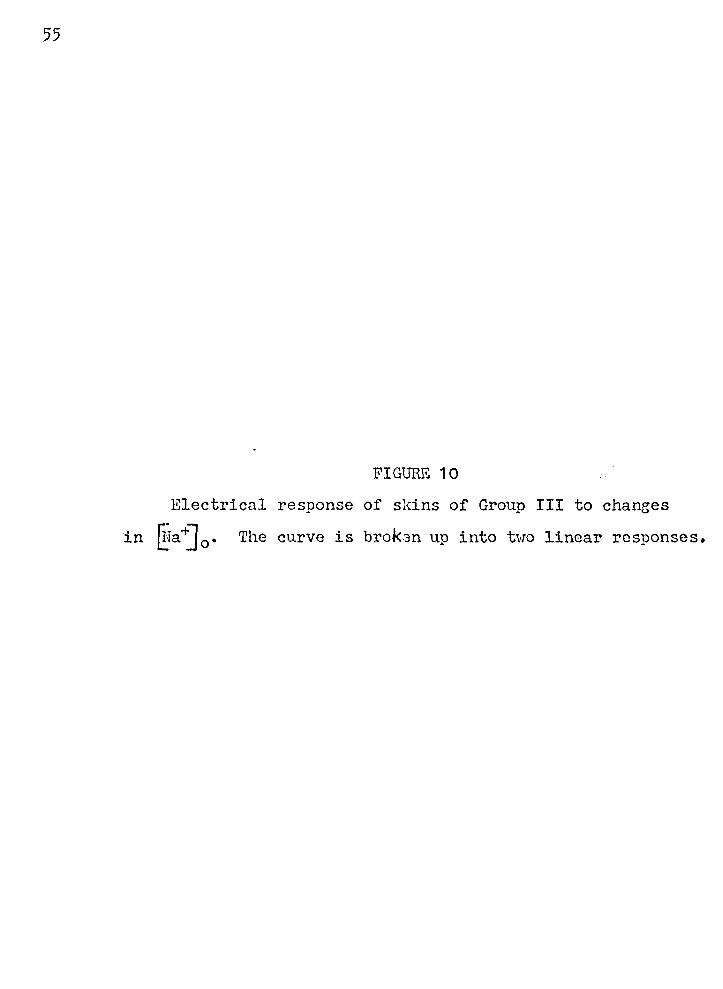
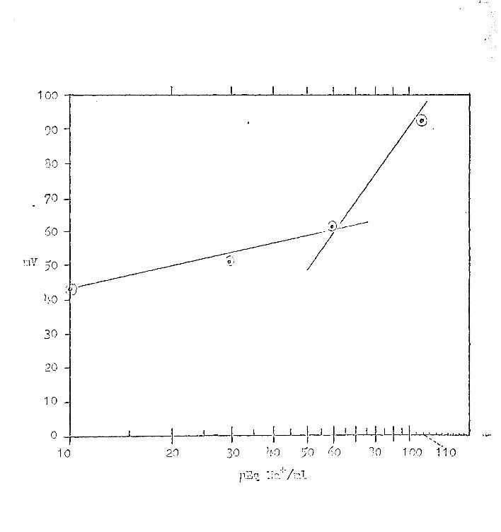
10. Electrical respon~~ of skins of Group III to changes in [j-Ja+j 0 • • • • • • • • • • • • • • • • • • • • • • • • 55
:11. Re~ults of so4= permeability studies 1.1.Sing •• s35 labeled sulfate ••••••••••••••••••••••••• 56
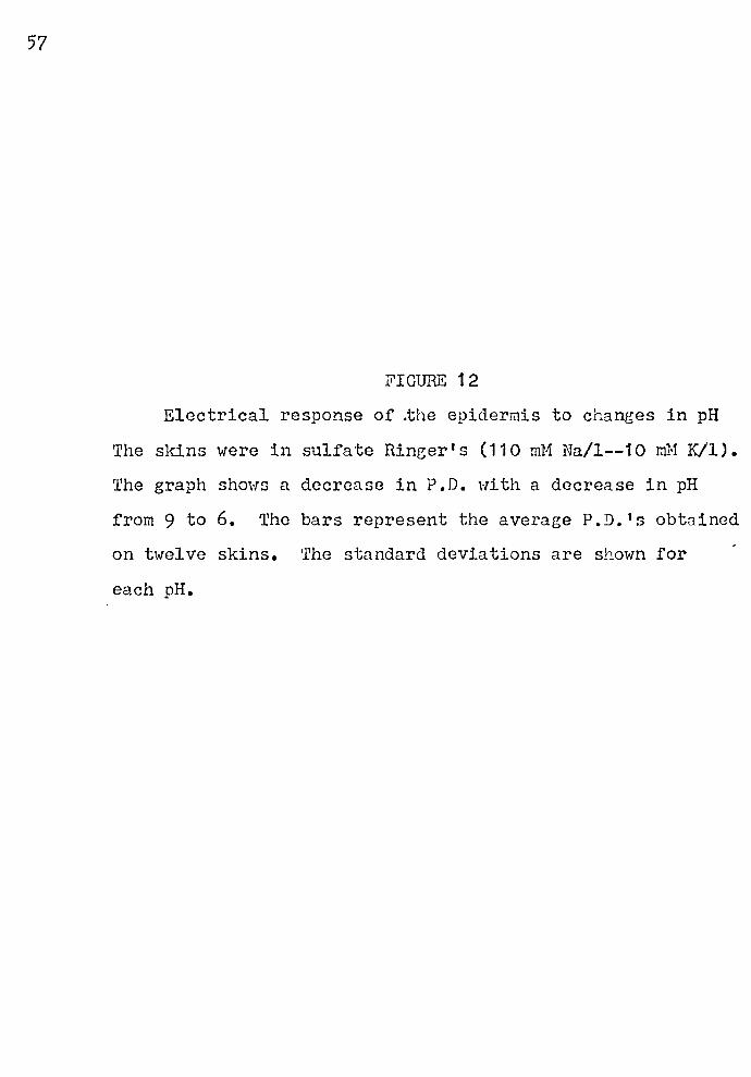
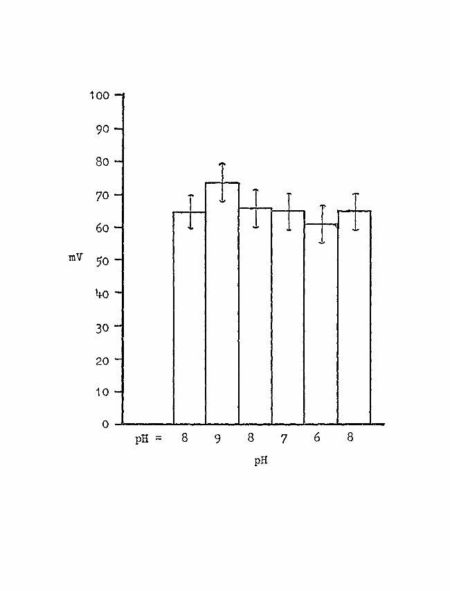
12. Electrical response of the epidermis -to changes in plI • • • • • • • • • • • • • • • • • • • • • • • • • • • • • • • 57 .
13. Plots of Equation (3) ••••••••••••••••••••••• 58
14. Simplified model of frog skin ••••••••••••••• 59
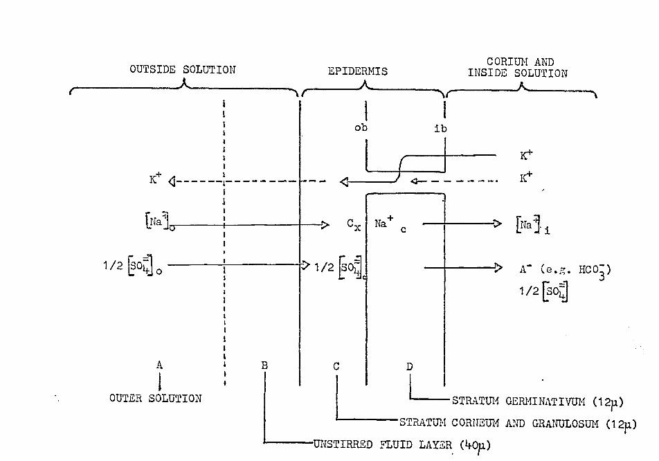
15. Steady state sodiTu~ and potassium distribution in frog skin •.••••••••••••••••• 60
16. Rela tio~ship between net sodiu:n flux, p , and Ufa ] 0 • • • • • • • • • • • • • • • • • • • • • • • • • • • • • • • • • • 61
17. Relationship between P0 and [rfa 4J 0
• • • • • • • • • • 62
18. Plot of Equation (19) ••• ,. ••••••••••••••••••• 63
19. Plot of hV.61) ··against {}fa+] 0
• • • • • • • • • • • • • • • • • 611-
X. List of Tables •••••••••••••••••••••••••••••••••• 65
1. Oyer-all sulfate perm~ability (P804) of frog skin obtained from s3? measurements ••••••••• 65
2. Preparation of Ringer's solutions from stock solutions••••••••••••••••••••••••••••• 66
3. Response of the epidermis to varying Ufa~] 0 • 67
1+. Sodium ion concentration in Na-isethionate l t . 6<'' so u ion . . . . . . . . . . . . . . . . . . . . . . . . . . . . . . . . . . . . J
5. ~lectrical response of the epidermis to Naisethionate ions as ~ompared to sulfate ions 69
6. Effect of epinephrine on skin P.D. and ~re~J;onse of the epidermis to change j_n g~a+Jo at pH 8.o ..........................••• 70

Deuendence of Na+ flux (¢) coefficient (Pn) on lNa+] 0
and Na+ permeability • • • • • • • • • • • • • • • • • • • • 71
... ·~.n. Appendix o • • • • • • • • • • • • • • • • • • • • • • • • • • • • • • • • • • • • • • • • 72
List of equations • • • • • • • • • • • • • • • • • • • • • • • 0 • • • • • • • •
List of symbols ........... • ........... " .......... .
XII. Vita . . . . . . . . . . . . . . . . . . . . . . . . . ~ . . . . . . . . . . . . . . . . . . .
'/2
'1'5
77
ABSTRACT·
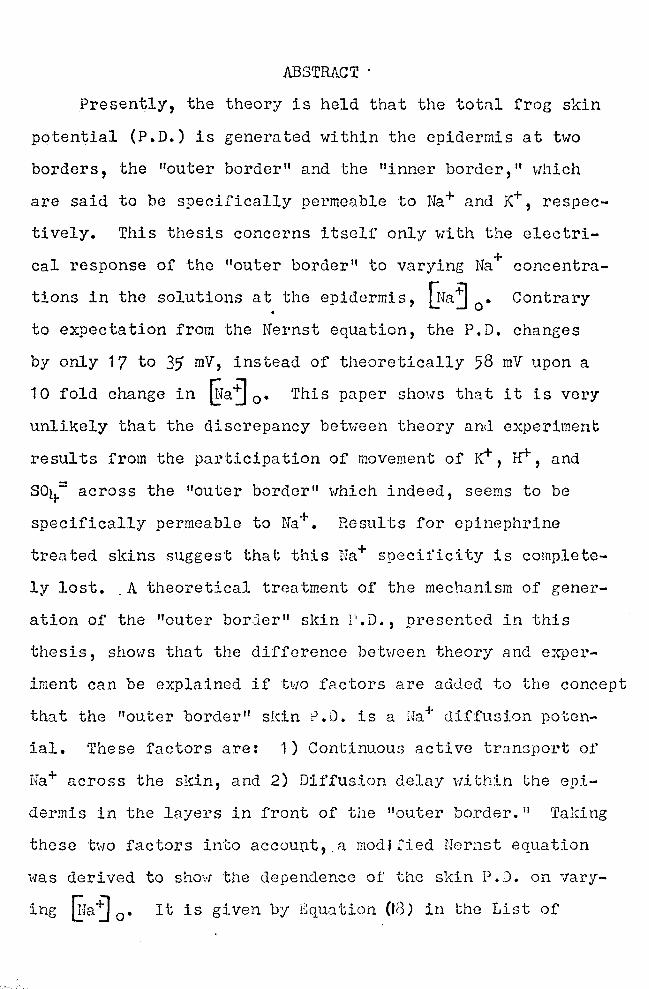
Presently, the theory is held that the total frog skin
potential (P.D.) is generated within the epidermis at two
borders, the "outer border" and the "inner border," which
are said to be specifically permeable to Na+ and K+, respec
tively. This thesis concerns itself only with the electri
cal response of the "outer border" to varying Na+ concentra
tions in the solutions at the epidermis, UJa°j 0
• Contrary
to expectation from the Nernst equation, the P.D. changes
by only 17 to 35 mV, instead of theoretically 58 mV upon a
10 fold change in @a~ 0 • This paper shows that it is very
unlikely that the discrepancy between theory and experiment
results from the participation of movement of K+, 1f1-, and
soy.= across the 11outer border" which indeed, seems to be
specifically permeable to Na+. Results for epinephrine
treated skins suggest that this Na+ specit'ictty is complete
ly lost. A theoretical treatment of the mechanism of gener-
ation of the "outer bor.::ler" skin l' .D., presented in this
thesis, shows that the difference between theory and exper
iment can be explained if two factors are added to the concept
that the "outer border" sl.::in ?.D. is a Ha+ diffusion poten
ial. These factors are: 1) Continuous active trnnsport of
Na+ across the skin, and 2) Diffusion delay within the epi
dermis in the layers in front of the "outer border." Taking
these two factors into account,.a modJfied Nernst equation
was derived to show the dependence of' the skin P.0. on vary
ing ~Ja"j 0 • It is given by Equation (Id) in the Li:3t of
Equations. A test for this equation shows that it adequate
ly describes the response of tho epidermis of the frog skin
to varying Na+ concentration at the epidermal side of the
sldn.
2
ACKNOWLEDGEMENTS
I would like to thank Dr. E. G. Huf, my major professor
to whom I attribute the ultimate success of this work, for
his generous advice, assistance, and explanations.
I would also lilce to thank Dr. F. B. Lefti:.·1ich, my thesis
Committee. Chairman, for his constant encouragement, advice,
and inspiration.
Furthermore, I would like to acl\:nowledge Dr. N. g. Rice
and Dr. ·w. R. ·renney, members of my committee, for their
constructive criticism in th~ writing of this thesis.
In addition, I wish to express my appreciation to Dr.
A. D. Campbell, ·who instructed me in the handling and use
of radioisotopes.
A note of special gratitude is extended to Mr. G. c. Schaefer who made several of the photoeraphs used in this
thesis.
3
INTRODUCTION
A. HISTORICAL REVIEW
Bio-electricity has been .:.Cnown since ancient tlmes.
In the Egyptian writings there are references to Nalonterus.
The Romans gave the name 11 ·rorpe~o" to the electric ray.
By the end of the eighteenth century it was suggested that
the shock received from electric fish was similar to the
electrostatic discharee received from the Leyden jar. In
· 1 '/86 Galvani obtained evidence that electric currents were
present in nerves as well as muscles. In 184-8 Emil Du
Dois-Reymond published a book, Untersuchun::en Hher Tht_~rishe II
Elektricitat (Researches Q1l Animal Electricity), with methods
of measuring various bio-potentials. The frog skin potential
was first mentioned in this book.
Today bio-potentiuls are '.·iell known and are by no means
confined to laboratory and research studies. Physicians
use recordings of the potentials developed by the brain (SSG)
or heart (EKG) to find certain anomalies which will aid
in diagnosis and treatoent of diseases. Indeed, irritability,
a characteristic of all living organisms, ahrays in some
·way involves electric potGntials. >rost of these potentials
are developed by ionic equilibria and active transport across
membranes. Individual cells maintain a potential difference
bet\rnen their internal and external environments, and for
this reason neurons and the large cells of certain algae
such as Valonia and 1r1 tella are used extensively in membrane
potential research.
However, because of their s~all size, individual cells
offer many technical difficulties. Frog skin, it would
appear, has eliminated the problem of sTiall size. Du Bois
Reymond discovered that the isolated frog skin maintained
a potential difference across itself, the outer epidermal
surface being negative with respect of its cerium surface.
Huf (1935, 1936) discovered that the isolated frog skin
could actively move chloride, as NaCl, from a Ringer's solu
tion bathing the epidermal side to a Ringer's solution . bathing the corium side. Ussing (1951) advanced the hypo-
thesis that frog skin could transport sodium chloride against
both an electric potential and a concentration eradicnt.
Ussing suggested that sodium was transported and that chloride
moved passively with it, thus preserving charge neutrality.
As sodium is actively transported across the skin, a potential
gradient is established, and chloride ~aves passively follow
ing the electrical gradient. In order to test this hypothesis,
Ussing and Zerahn (1951) devised the short circa.cit technique
to eliminate the potent'ial gradient. An isolated frog skin
was mounted between two cha:-:1bers containing Hinger 1 s solution
of the same concentration. The developed skin potential
was monitored. A current w~s applied in such a direction
as to reduce the potential across the skin to zero. This
is called "short circuiting 11• the skin. ~'Ii th this a.rrange-
ment, the only transport which could occur would be the result
of an active process. From the results of.this experiment,
6
Ussing andZerahn concluded that only sodium was transported, ' ~ ~ "
and that in the "open skin," the sodium transport mecnanism
and passive movement of the anion was responsible for the
skin EMF.
Most investigators believe that there are two electro
genic layers in the frog skin. Steinbach (1933) gave evidence
that there are at least two layers involved in the production
of the skin potential. Assuming the existence of an "outer
border" (epithelium facing layer) and as "inner border"
(corium facing layer), it has been shown by Fulruda (19i1-2, 1911..11-)
that the outer layer requires the presence of sodium but not
potassium. Fukuda suggested that the "outer border" was
preferentially permeable to sodium anti that the "inner border"
was preferentially permeable to potassium. Koefoed-Johnsen
and Ussing (1958) proposed a model that emphasized these
preferential permeabilities. When they eliminated anion
penetration by replacing sodiu~ chloride with sodium sulfate,
they found that the skin potentia:'. changed by almost 58 mV
for a ten fold change in the outside sodium or inside potassium
concentration. Therefore, Koefoed-Johnsen and Ussing re-
garded the total skin potential to be the sum of the two
Nernst diffusion potentials of sodium at the "outer border"
and potassium at the "inner border." Other wor:rnrs have
tried to confirm the response at the "outer border" but
have not found the theoretic~l 58 raV response. A change of
35 mV for a ten fold ch~nge in concentration has been re
ported by Lindley and Hoshiko (1964-), by 1:linn et al. (196li-),
7
and by Cereijido and Curran (1965).
·rn order to determine the actual number of barriers
or potential steps across frog skin, several investigators
have employed the micro-puncture technique for micro-potential
measurements. Ottoson et al. (1953) was the first to employ
the technique. Others (Engbaeck and. Hoshiko, 1967; Scheer,
1960) followed, and the latest to publish their results,
Chowhury and Snell (1965), are the only investigators to
obtain a continuous potential change across the frog sl(in.
All other workers report two, and sometimes three or more
steps.
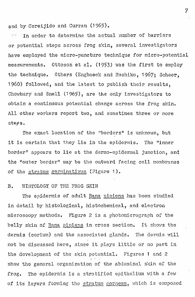
The exact location of the "borders" is unknown, but
it is certain that they lie in the epidermis. The "inner
border" appears to lie at the dermo-epidermal junction, and
the "outer border" may be the outward facing cell membranes
of the stratum germinativum (Figure 1).
B. HIS'rOLOGY OF THE FROG SKIN
The epidermis of adult Bana nioiens has been studied
in detail by histological, histochemical, and electron
microscopy methods. Figure 2 is a photomicrograph of the
belly skin of Ranq_ pipiens in cross section. It shows the
dermis (corium) and the associated glands. The dermis will
not be discussed here, since it plays little or no part in
the development of the skin potential. Figures 1 and 2
show the general organization of the abdominal skin of the
frog. The epidermis is a stratified epithelium with a few
of its layers forming the stratum .QQ.i,.Q§.@, which is composed
of partially·cornified squamous cells. Beneath this are
orie to three layers of cuboidal and polyhedral cells which
are so::1etimes differentiated into stra tu.172 granulosum and
stratum soinosl!!!!• 'l'here is a basal layer of cuboidal and
coiumnar cells, the stratum R§._rminn. ti vum. Electron micro
graphs show a definite basement membrane at the dermo-epi
dermal junction. The junctions between adjacent cells in
the stratum corneum show the outer leaflets of the membranes . 0
fused into a single dense band 30-40 A thick, and the total
distance across the two fused membranes measures 170-180 A. In the stratum granulosum the component membranes are
0 smaller, and the junctions measure about 120-140 A across
8
the two fused membranes. There is extensive and complex
interdigitation between the cells of the stratum germin'1tivum.
C. THEORY OF FROG SKIN BIO-PO'rEIJTIALS
One widely held concept about the origin of bio-poten
tials is that cell membranes behave as ion sieves. They
can bring about noticeable separation of ionic charges across
the membrane. Thus, for the special case of a membrane
specifically permeable to sodium, one 8ay observe a poten
tial difference, v, across the membrane ~hich can be cal
culated by the Nernst equation:
RT C1 V = -:-.ln---- ( 1 )
nF c2
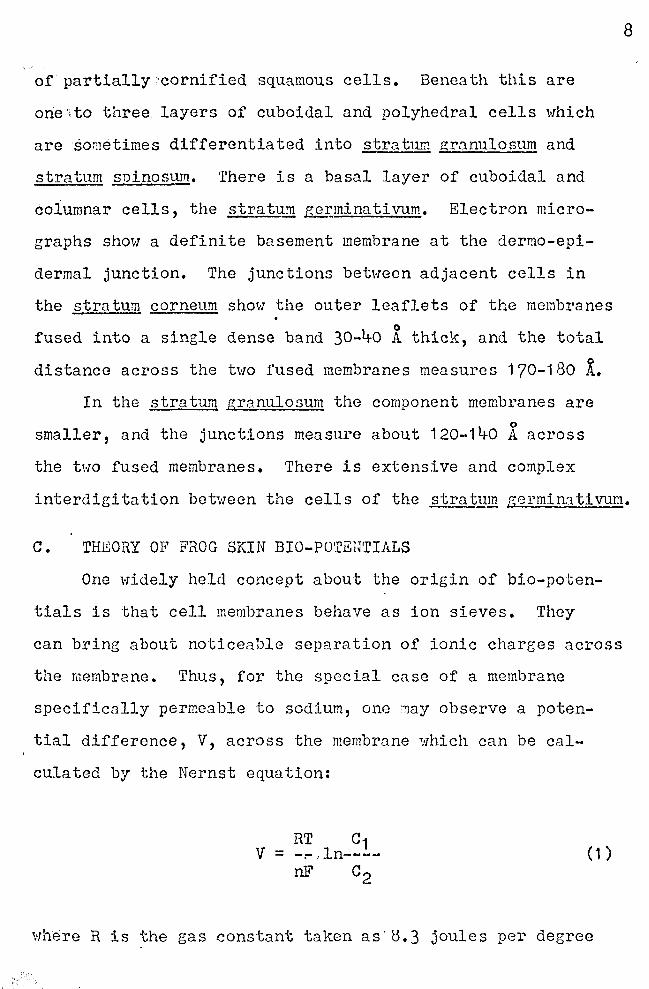
where R is the gas constant taken as·<).3 joules per degree

per mole; T is the absolute temperature; n is the valency
of the ion species with the appropriate sign; F is Faraday's
constant (96,500 coulombs per gram equivalent); and ln is
the natural loc;arithm (2.3 X loi10)• If c1 and c2 are the +
Na .ion concentrations on the two sides of the membrane,
and the absolute room temperature is 297 K, Equation (1)
can be written:
~raj 1 V = 58 log------
~ra ~ 2
Thus for a ten fold difference in sodium concentration
across the membrane, V = 58 mV. If the "outer border" of
frog skin represents the outward oriented cell mc~brancs
of the strab~n germinntim1m and if the cell membranes be-
(2)
have like an ideal sodium selective membrane, the the poten
tial difference (V0 b) across this border may be written:
~faj 0 = 58 log------@a j c
(3)
where [lra "'.] 0
is the sodium ion concentration of the solution
at the epidermal side, nnd UJaj c is the intracellular sodium
ion concentration, uhich 1.'fill be tnken as 1 O pEq/ml (Andersen
and Zerahn, 1963). If ~faj 0 equals 100 Jillq/ml, then
V0 b = 58 mV.
As will be shm1n in the folloHing portions of this
th~sis, which concerns itself exclusively with the electrical
9
events at the "outer border," one rarely observes the theo
retical value. The highest value for V0 b generally observed
is only 35· mV. At present, the re.:ison for this discrepancy
10
is un.'l{nown. It must be pointed out, howev1;r, that the assump
tion made in the above calculation is thnt tho "outer border"
is permeable only to sodium ions. One may question the
validity of this assumption, since under the experimental
conditions which prevail, ions other than sodium ions are
present when V0 b measurements are made. The predominant
anions are chloride, when chloride Ringer's is used, and
sulfate, when sulfate Rineer's is used. Potassium and
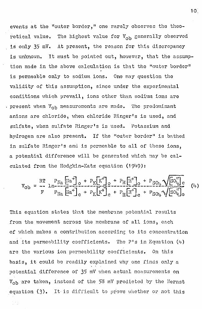
hydrogen are also present. If the "outer border" is bathed
in sulfate Ringer's ancl is permeable to all of these ions,
a potential difference will be generated which may be cal
culated from the Hodg1{in-Ka tz equation ( 19t+9):
This equation states that the membrane potential results
from the movement across the membrane of all ions, ench
of which makes a contribution according to its concentration
and its permenbili ty coefficients. The P's in Equation ( lt-)
are the various ion permeability coefficients. On this
basis, it could be readily explained why one finds only a
potential difference of 35 mV Hhen actual measurements on
V0 b are taken, instead of the 58 mV predicted by the Hernst
equation (3). It is difficult to prove uhether or not this
is the correct explanation for the generation of V0 b since
l(nowledge about the intracellular ion concentration and
the permeability coefficients is rather difficult to obtain.
It can be rca.soned, hm·1ever, that the applicn ti on of
the Hodgkin-Katz equation to the "outer border" of frog
skin does not explnin why V0 b is lowered to 35 mV, instead
of being 58 mV. The reasons are as follows:
1) When sulfate Ringer's is used, one c~n be fairly
certain that !i1ovement of anions is excluded from partici-
pation in the generation of V0
b. In contrast to chloride
ions (which are present in ordinary Ringer's) membranes are
generally known to be impermeable to sulfate ions. Over
all sulfate permeability studies show a low permeability
coefficient for sulfate (Table 1).
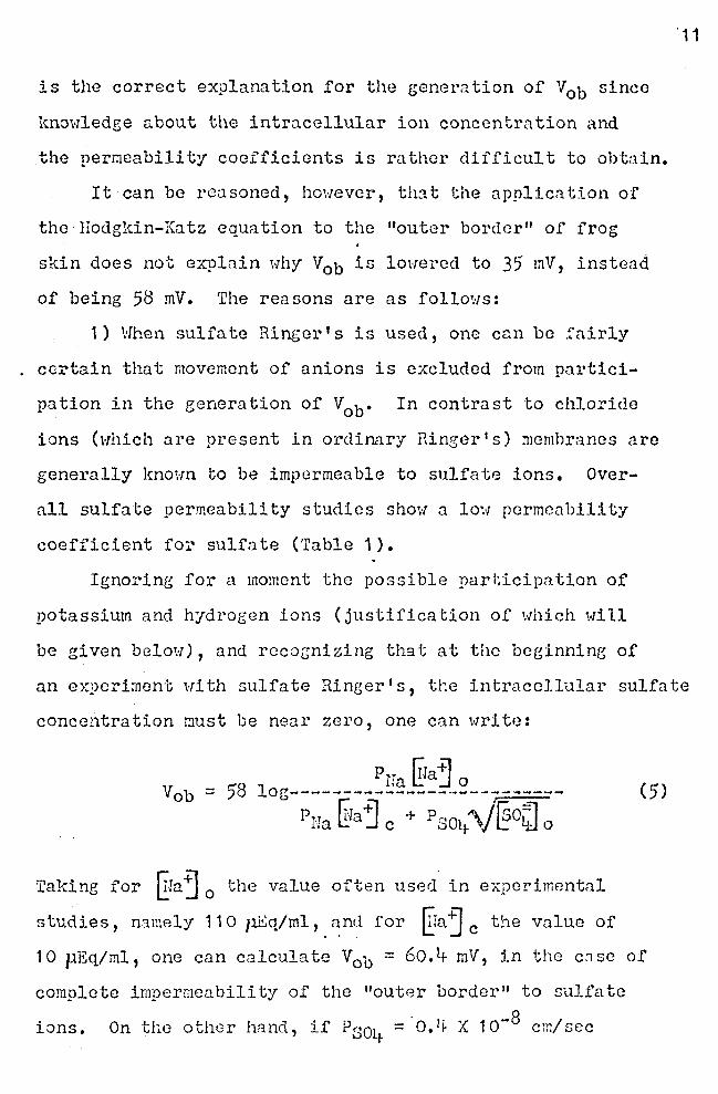
Ignoring for a moment the possible participation of
potassium and hydrogen ions (justification of which will
be given below), and recoz;nizing that at the beginning of
· 11
an experiment with sulfate Ringer's, the intracellular sulfate
concentration r.mst be near zero, one can wri to:
Pna ~ra~ o = 58 log--------------------------
Pnu ~raj c + P soy. '\f ~o~ o
Taking for ITraj 0 the value often used in experimental
studies, n3.mely 11 O 7.iliq/ml, and for ~ra'j c the value of
(5)
10 µEq/ml, one can c3lculate V0 b = 60.t1- mV, in the c:ise of
complete impermeability of the "outer border" to sulfate
ions. On the other hand, if Psoy. =·o.~ X 10-8 cm/sec
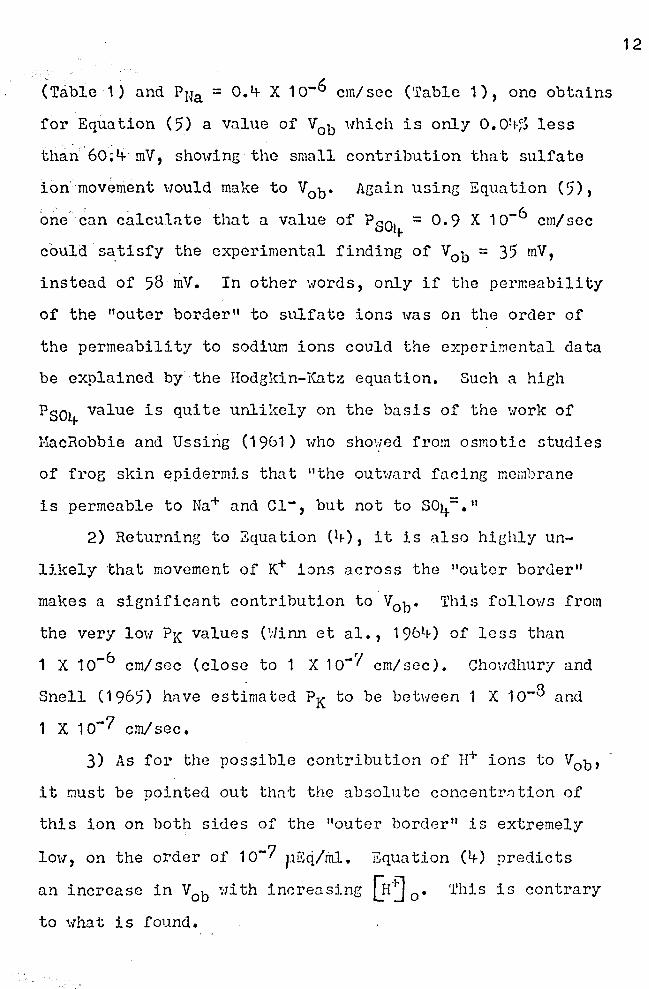
(Table 1) and PNa = O.~ X 10-6 cm/sec (Table 1), one obtains
for Equation (5) a value of V0 b 1·1hich is only 0.0~1-;~ less
tha~{6o;t+· mV, showing the small contribution that sulfate
ion movement would make to Vob• Again using Equation (5),
one can calculn te that a value of p soi .. = 0. 9 x 1 o-6 cm/ sec
could S8:tisfy the experimental finding of V0 b = 35 mV,
instead of 58 mV. In other words, only if the permeability
of the "outer border" to sulfate ion5 was on the order of
the permeability to sodium ions could the experi~ental data
be explained by the Hodgkin-Katz equation. Such a hieh
P804 value is quite unlikely on the basis of the work of
HacRobbie and Ussing (1961) who showed from osmotic studies
of froe skin epidermis that "the outward facing membrane
is permeable to Na+ and c1-, but not to so4=. 11
2) Returning to Zquation (l1-), it is also highly un
likely that movement of ~ ions across the "outer border''
makes a significant contributlon to V0 b. This follows from
the very low PK values (Hinn et al., 196l1-) of less than
1 X 1 o-6 cm/ sec (close to 1 X 1 o-'/ cm/ sec). Cho\·Tdhury and
Snell (1965) hnve estimated PK to be between 1 X 10⁻⁸ and
1 x 1 o-7 cm/sec.
3) As for the possible contribution of n+ ions to V0 b,
it r.mst be pointed out thnt the absolute concentrn ti on of
this ion on both sides of the "outer border" is extremely
low, on the order of 1 o-7 ~1Eq/rhl. Equation (4-) predicts
an increase in V0
b with incrensing Qr~ 0 • This is contrary
to what is found.
12
From this analysis, the conclusion is reached that the
predominating ion which determines tho value of vob is the
sodium ion, and that Equation (3) should indeed hold. The
reason for the descrepancy betirnen the predicted value of
58 mV (for a ten fold change i.n sodium concentration) and
the experimentally foundvalue of 35 mV or less, therefore,
must lie outside of the consideration that ions other than
sodiwn ions may play a r?le. Therefore, it was the purpose
of this research to examine whether other factors, namely
sodium diffusion delay within tho epidermis and active trans
port of sodium across the skin, could account for the dos
crepancy between experiment and theory on t11c electrical
response of the "outer border" of the free skin epidermis
to variation in the sodium ion concentration.
13
MATERIALS AHD H8THODS
A. DISSECTION AND HOUN'£ING OF SKIN Hi~!·IBRANE
The living belly skin of Rana ninicns was used for all
experiments. The frogs were obtained commercially from
Stefnhilber, Oshl\:osh, Wisconsin. They were kept in a tank
sripplied _with running tap water. They were not fed and were
used within 1 lf- days after receipt.
The frogs were decerebratcd with scissors (Fieure 3)
and the remaining portions of the brain and spinal cord
were destroyed with a dissection needle. As rapidly as
possible the skin over the xiphistcrnum was .:>unctured and
cut laterally (Figure ~) both left a~d right, cutting the
cutaneous arteries at the same time. The cut was continued
caudally on both side::; of the body in the piemented area
of the skin. Just ventral to the cloaca, the incisions were
joined with a final cut across the pelvic region.
The skin is held to the body by fa.scin runnine :ilong
14
the ventro-lateral portion of the body nncl mectine at the
point of the final incision across the ventral pelvic region.
By pulling the skin a·,my from the body wall musculn tu.re,
the skin-muscle junction can be easily seen as a transparent
connecting zone of fascia. By cutting these transp::i.rent
fascia, the skin section wa~ removed from the body without
any adhering muscle.
The skin was spread on a·porcelain plate and carefully
blotted free of excess mucus, blood, and r.10isture. 'rhe
circular piece of skin (Figure 5) was mounted ns a membrane
separating two chambers of a lucite cell (Fieure 6 nnd 7).
15
It was p~aced across the open end of one of the cell chambers,
the other cha~ber placed on top (Figure 6), and the cell
was then completely assembled and tightened. Each cell
was then placed in the cell positioner over a set of mag
netic stirrers (Figure 8) for continuously mixing the con
tents of each chamber. The area of the circular skin mem-
.brane between the two chn::1bers was 7. 25 cm2. The chnmber
on the epidermal side of the skin is referred to as the out
er chamber. The chamber on the dermal side of the skin is
referred to as the inner chnmber. Each chamber was com-
pletely filled with 25 ml of the desired Ringer's solution.
The stirring bars for the magnetic stirrers were dropped
into.each chamber, and the stirrers were turned on.
B. DESCRI?'rION OF IHS'I'RUU~HTS
ill potential difference (?.D.) experiments were con-
ducted usinB the sane instrumental arrangement and equip
ment. Potential measurements were made with a Keithley
Hodel 600A Eultipurpose Electrometcr. The electrodes used
were Radiometer Calomel electrodes, Typo ~~01.
Potential difference measurements were made by lower-
ing the electrodes into the cells through tho holes in top
of each chamber (Figure '?). The apperatus could accornodate . »
t\ro cells, and thus t':Io experiments could be run sir.ml taneous-... ly. Figure 8 shous the apperatus. The electrode selector
switch was turned to the right, and the ~.D. of the right
16
hand cell W<J s measured. The electrode selector s\d tch was
then turned to the.left, and the P.D. of the left h~nd cell
was measured.
C. POTENTIOHETRIC STUDIES TO TEST THE RESPONSE OF 'fHE
EPIDERMIS TO CHANGING liraj 0
Stock solutions of 0.5 H 1fo.2S01+/l, 0.5 H K2soi/1, and
O. 5 H THAI·Vl (tr is hydroxymethyl ominomethane buffer) were
prepared. Using these stock solutions, four types of Ringer's
solutions uere prepared ('fable 2). The sodiwn and potassium
concentrations were varied, the molnr sum of the sodium and
potassium always being 120 o.H/;. The pH of e8ch J.in~cr's solu
tion was adjusted to 8. 0 1:1i th H2so4 using a Beckman l·:odel G
pH meter as described in Table 2. Before every expcrimcn~
oxygen was bubbled through each Ringer's solution for three
minutes.
Experiments were conducted in pairs. The skins were
mounted between the chambers of cc.ch cell as previoti.sJ.y
described. Ringer's solution no. 1 ("110 mH No./1--10 mH K/l)
was added to both inside and outside chambers of e8.ch cell.
The cells were placed in the cell positioner and allowed
·to equilibrate for one hour. At the end of this hour, the
P.O. was measured and recorded. The Ringer's solution was
removed fro:;i both chambers of en.ch cell. The outside chambers
were rinsed and filled with Ringer's solution no. 2 (90 mM
Na/1--30 mN K/l). The inside chambers were rinsed and filled
with· fresh Ringer's solution no. 1. At the end of fifteen
minutes, the P.D. was measured and recorded. The solutions
were removed from both the inside and outside of each cell.
The outside chambers were rinsed and filled with Ringer's
soiuti~n no. 3 (60 mM Ha/1--60 mM K/l), and the inside chnm-.,
bers were rinsed and filled with Ringer's solution no. 1.
17
At the end of fifteen minutes, the P.D. was measured and
recorded.. The cells were emptied o..s before. Then the out
side chambers were rinsed and filled with Ringer's solution
no. 4 (10 mH Na/1--110 mM K/l), and the inside chnmbcrs were
rinsed and filled with Ringer's solution no. 1. After fif
teen minutes, the P.D. was measured and recorded. 'fhc cells
were or.iptied and both inside and outside chambers Herc rinsed
and filled.with Ringer's solution no. 1. After fifteen min-
utes, the P.D. was measured and recorded.
D. . POTENTIOHETRIC STUDIES ON THE EPID~R11IS OF FROG SKIN IN
ISETHIONATE RINGER'S SOLUTION
Stock solutions of 1 l·! sodium isethiona te/l (HO-CH2-
CH2-so3Ha, obtained from Eastman Kodak Co.), o. 5 M Na2SOt/1,
o. 5 H K2S041'l, and O. 5 M THflJ.f/1 were prepared. Using these
stock solutions, three 'Ringer's solutions (no. 1, no. 5', and
no. 6) were prepared (Table 2). Before each pair of ex
periments, oxygen Has bubbled through the Ringer's solutj_ons
for three minutes. Skins were prepared and mounted as before.
Sulfate Ringer's solution no. 1 (110 mH Ha/1--10 mH YJl)
was placed in each chamber, and.the cells wore put in the
cell positioner. The stirring bars were dropped into each
chamber and adjusted to mixine speed. After one hour, the
P.D. was measured and recorded. ·rhe cells were emptied, and
the outer chambers were rinsed and filled with sulfate
Ringer's .. solution no. ·5'.{120 mH Na/l). The inner chambers
were,rinsed·and filled with sulfate Ringer's solution no. 1.
After fifteen minutes, the P.D. was measured and recorded.
The.outer chambers were rinsed and filled with isethionate
Ringer's solution no. 6 (120 mH Na-isethionate/l), and the
inner chambers were rinsed and filled ·with sulfate Ringer 1 s
solution no. 1. After fifteen minutes, the P.0. was meas-
.ured and recorded. The chambers were emptied, rinsed and
filled with sulfate Ringer's solution no. 1. After fifteen
minutes, the P.D. was measured and recorded.
E. THE OVER-ALL PERHEABILITY OF FROG SKIN TO s35' LABELED
SULFATE
18
Using the O. 5 H/l stock solutions of Na2SOtp K2soy., and
the stock solution of 'rHAM, one liter of sulfate Ringer 1 s
solution no. 1 (110 mH Na/1--10 mN K/l) was prepared (Table
2). To two100 ml portions of this Ringer's solution was
added Na2s35'o4 in an amount calculated to give 1 oli- counts
per minute per 0.1 ml. The amount of inactive carrier sodium
added to the Ringer's solution was negligibly small.
Skins were mounted, and the outer chambers were filled
with Ringer's solutiont~~lcontaining s35 labeled Ifu2SO~.
The inner chambers were fj_lled with Ringer's solution l1o,1 •
After 90 minut-:?s, the P.D. was measured and recorded. A
0.1 ml sample ·was taken from each chamber, placed in a
planchette, and evaporated to dryness.
Count rates were talcen for 1 O minutes using a gas flow
counter (100 ugm/cm2 window) and scaler. Background ·was
taken as the count rate of 0.1 ml sa~ple of fresh, unlabeled
Ringer's solution no. 1. The quenching gas was a mixture of
isobutane (93%) and helilun (7%).
F. THE INFLUENCE OF pH ON THE POTEHTIOMETRIC RESPONSE OF
THE EPIDERMIS TO ~aj 0
From the stock solutions, the following five Ringer's
solutions were prepared (Table 2): Ringer's solution no. 1,
pH= 8.o; Ringer's.solution no. 5 (120 mM Nall, pH= 8.0);
Ringer's solution no. 7 (120 mM Nall, pH = 9.0); Ringer's
solution no. 8 (120 mM Na/l, pH= 7.0); Ringer's solution
no. 9 (120 mM Nall, pH= 6.0J. 'rhe pH was adjusted with
H2so4•
The skins were mounted, Ringer's solution no. 1 was
added to inner and outer chambers, and the cells placed in
the apparatus. After one hour, tr c P. D. ·was measured and
recorded. The chambers.were emptied. The outer chambers
were rinsed and filled with Ringer's solution no. 7 (120
mH Hall, pH= 9.0J; the inner chnmbers were rinsed and
filled with Ringer's solution no. 1. After fifteen minutes,
the P.D. was measured and recorded. The chambers were emp-
tied, and the outer chambers were rinsed and filled with
iiinger's solution no. 5 (120 mH Ha/l, pH= 8.o); the inner
19
chambers were rinsed and filled with Ringer's solution no. 1.
After fifteen minutes, the P. D. was mea su1~ect and recorded.
20
The chambers were emptied. The outer chambers were rinsed
and filled with Ringer's solution no. 8 ( 1 20 mVi !Jail, pH ==
7.0); 'the inner chambers were rinsed and filled with Ringer's
solution no. 1. After fifteen minutes, the P.D. uas·mensured
and recorded. 'rim charnbers were emptied. 'rhe outer chambers
were rinsed and filled with Ringer's solution no. 9 (120 mM
Na/l, pH== 6); the inner chambers were rinsed and filled
with Ringer's solution no. 1. After fifteen minutes, the P.D.
was measured and recorded.
G. POTEUTI011ETRIC STUDIES OH THE EPIDERMIS Oli' FROG SKIN
OF ANIMALS PRETREATED WITH EPINEPHRINE
L-epinephrine bitartrate (Sigma Chemical Co.) was dis
solved in Ringer's solution no. 1 and immediately used. One
milligram of epinephrine (0.2 ml of the solution) was inject
ed into the dorsal l~nph sac of each frog. The frogs were
then placed in a tank supplied with running water. After
one hour, the belly skins were removed and mounted as de
scribed previously. The same procedure described in Section
C was followed to test the response of the skin to changes
in sodium concentration.
RESULTS
A. POTENTIOHETRIC STUDIES TO TEST THE RESPONSE OF THE
EPIDERMIS TO CHANGIUG [ia-j 0
or the thirty experiments conducted, sixteen showed
a recovery at the end of a series of testings to within
:I: 10% of the origin..1.l P.D. · Two skins had, respectively,
a 12% and a 32% higher sl\:in P. D. compared to the original
P.D. The remaining 12 skins showed potentials between 15%
and 4-1% lower than the original P.D. The results obtQined
on the sixteen experiments with good (± 10%) recovery are
shown in Table 3. The results were separated into three
groups. Hale frogs, Group I, gave stroneer responses than
female frogs, Group II. A greater number of experiments
would be needed before stating that the obvious difference
seen in the present experiments are sex lin!ced. Because of
this uncertainty, the results of the two groups wore com
bined and treated together. The combined data are given
21
in the Group (I + II) column of Table 3. Group III con
sisted of five additional frogs which responded quite dif
ferently from 'i·rhat is most commonly observed. The difference
is more clearly seen by comparing Figures 9 and 10. The
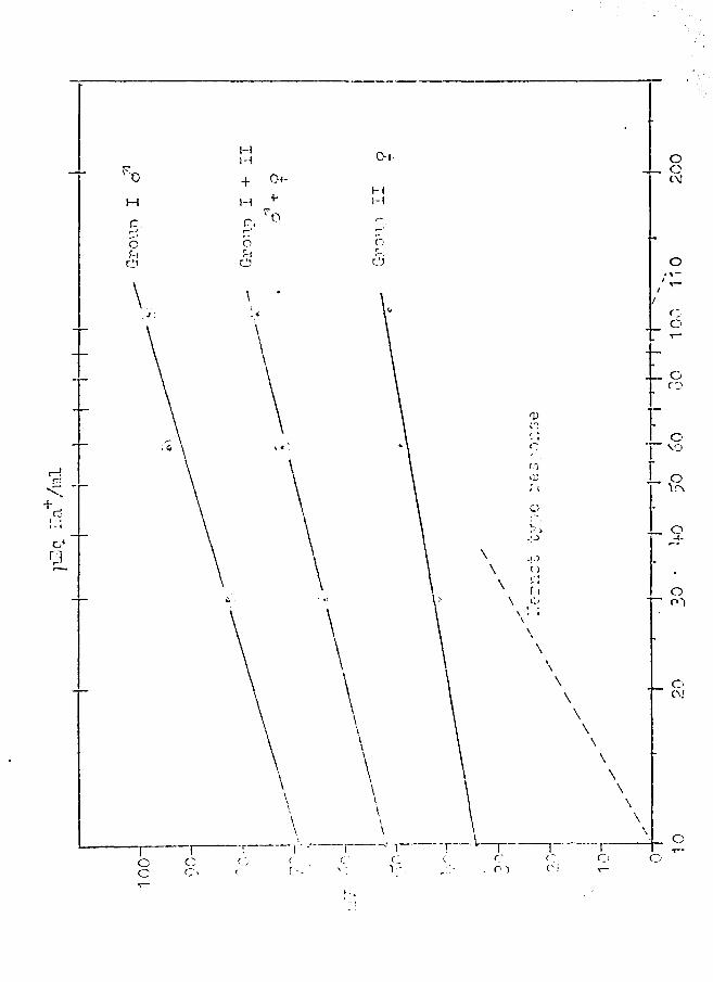
results obtaines in Groups I, II, and I + II are plotted
in Figure 9. Figure 10 shows the results obta.ined in Group
III. Figure 9 shows an apprqximately linear response curve,
if P.D. in mV (the dependent variable) is plotted (on the
ordinate) against the log of ~ra -t] 0
(the independent var
iable on the abscissa). Slope factors, 13, for the throe
response curves were calculated from:
(6)
where V1 and v2 are skin potentials read from the graph
and [fraj 1 and @aj 2 are the associated Na+ concentrations.
Thus, the slope factors of the response lines shown in
.Figure 9 are:
Group i cl': p == 30
Group II ~ : jJ == 1 7
Group I + II ( d1 + ~ ) : t = 25'
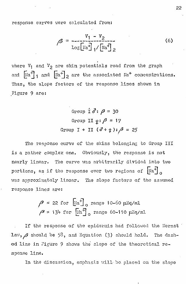
Tho response curve of the skins belonging to Group III
is a rather complex one. Obviously, the response is not
nearly linear. The curve was arbitrarily divided into tHo
portions, as if the response over two re~ions of ~faj 0
·was approximately linear. The slope factors of the asr:>W1ied
response lines are:
(3 :: 22 for ~ra-~ 0
range ·t 0-60 11Eq/ml
f1:: 131+ for [raj0
range 60-110 p.Eq/ml
22
If the response of the epidermis h~d followed the Nernst
law,19 should be 58, and Equation (3) should hold. The dash
ed line in ?igure 9 shows th~ slope of the theoretical re-
sponse line.
In the discussion, emphasis uill· be. placed on the slope
faatorf = 35, since this is the highest commonly observed,
and the one nearest to the theoretical value,19 = 5~. The
treatment of data would be the same for the smaller slope
factor.
B. POTENTIOHETRIC STUDIES OF1 THE EPIDERMIS OF FROG SKIN
IN ISETHIONATE RINGER 1 S SOLU'fION
If in the potentiometric analysis of the permeability
properties of the epidermis, movement of sulfate ions (mo
lecular weight of 96) played a role, then the re~laccment
of the inorg.:inic sulfate by the organic sulfate (iscthio
na te, molecular ·weight of 125. 1 ) might lend to an increase
in skin potential. The results of eight experiments are
shown in Table 5. It can be seen that in the presence of
iscthionate ion, the skin P.D. was not increased, but de
creased. Because of the greater molecular weight of is
ethionate as compared to that of sulfate, an incre.:ise in
skin P.D. might have been anticipated when sulfate was re
placed. 'rhfs result supports the view held by many authors,
that sulfate ion is indeed a rather impermeable ion, and
therefore, useful in potentio~etric studies designed to
evalu::ite the effect of the ca.tion only. The unexpected
P.D •. -depressing effect of isethion:lte has raised the ques-
tion regarding the mechanism of this effect. It was thought
that such a result could be obtained if Na-isethionate were
lipid soluble, and hence mar~ readily permeable than inor
ganic sulfate ions. Some studies were carried out to test
for lipid solubility of isethionate. It was insoluble in
23
benzene and in chloroform. It was soluble, however, in a
1:3 (vol/vol) mixture of ether and ethyl alcohol, and the
solubility ..,.,ras 21+3 mg% when the Na-isethiom.te was shaken
for four hours in the ether-alcohol mixture. It is q.uite
possible, therefore, that the P.D.-depressing effect of
isethionate is the result of its solubility in lipids of
the Na-selective cell membranes, the "outer border" of the
frog skin.
C. THE OVER-ALL PERMEABILITY OF Ii'ROG SKIN TO s35 LABELED
SULFATE
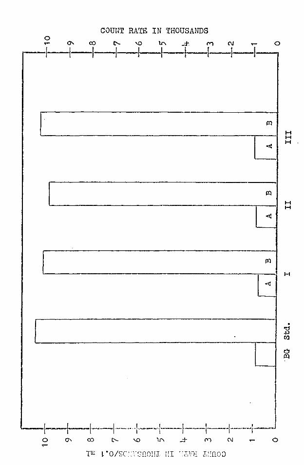
The results of experiments conducted on three frogs
is shown in Figure 11. The paired columns showing the
activity of the inside (A) and the outside (B) chambers
are compared to the background and standard columns. It
can be readily seen that in the cases presented, the activi
ty of the inside chambers did not rise above the background
level. Any labeled sulfate which passed throueh the frog
skin membrane from the labeled Ri:1ger 1 s on the outside to
the unlabeled Ringer•s on the inside was in such a small
amount that it was undetectable by the equipment used.
Labeled sulfate Ringer's solution wns in contnct with the
frog sl{'.in for only l. 5 hours, whereas the experiments
presented previously lasted for 2 hours. However, it is
believed that 1.5 hours was sufficient, and that the results
obtained add further evidence for the very loi:.of sulfate
permeability of frog skin. It should be said that in
order to obtain a noticeable increase in count rate, ·which
·would indicate that labeled sulfate h:-id passed through the
frog sldn, an increase of only 30 counts per minute above
background was needed. 'fhis increase did not occur.
D. THE INFLUENCE OF pH ON THE POTENTIOHETRIC RESPONSE
OF THE EPIDERMIS ·ro ~a-~lo Twelve experiments were conducted, and the results
are shown in Figure 12 which is a diagram of P.D. in mV
versus pH. It clearly cah be seen that when the [n~ 0
of the solution at the epidermis was increased, the skin
potential was decreased. However, a thousand-fold change
in the :a+ concentration caused only a slight decrease in
P.D.
E.. POTENTIOMETRIC STUDIES ON THE EPIDERMIS OF FROG SKIN
OF ANIMALS PRETREATED WITH EPINEPHRINE
The results of seven experiments are shown in Table 6.
Two kinds of observations were made:
1) There was a sharp reductic;n of the total skin P.D.
Normal skins in sulfate Hinger's can show a P.D. of nearly
100 mV, and occasionally higher values (see Table 3). Since
the total skin P.J. is the su1n of the P.D. 's generated at
·the "outer border" and the "inner border", it is conceivable
that the loi;.1 skj.n P. D. seen in the skins of epinephrine
treated frogs is the result of the action of epinephrine
on either or both borders. No experiments were conducted
at this time to further analyze this observation.
2) It can be seen that the epidermis has almost com
pletely lost its ability to respond to changes in ~a~ 0 •
25
DISCUSSION
A. HYPOTHESIS O? DIFFUSION .DELAY
As mentioned in the Introduction, there is little reason
to doubt that Ha+ ions are the chief ions, and probably
the only ions, involved in the generation of the P.D. across
the "outer border" of the_frog skin epider:nis. The "outer
border" does indeed behave like a sodium-specific selective
membrane. From the data presented, it is also evident that
a ten fold change in UJa~ 0 does not give a P .D. change of
58 mV as one would expect if the "outer border 11 were sodium . selective. The response deviates greatly from the expecta
tion. The lowest response was a change of 17 mV, and the
highest, 35' mV for a ten fold change in ~a~ 0 • Hence,
using the highest response, the actual (in contrast to the
theoretical) response of the "outer border" is given by:
Vob = 35 log (7)
To bring these facts into harmony, and in an attempt to de
rive an equation for the "outer border" skin P.D. which is
.in agreement with the actual measurement, the hypothesis
was made that the form of Equation (3) applies to Vob• The
theoretical values for V0 b may be obtained if one replaces
in .t!;quation (3), §a~ 0 with.ex (the Na+ concentration in
the immediate vicinity of the "outer border"). This con
centration conceivably could be considerably below ~a "j 0 t
because of diffusion delay in the regions in front of the
.sodium selective "outer border. 11 The modified equation
can be expressed as:
Gx = 58 log------~fa +] c
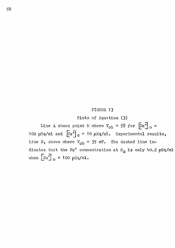
Figure 13 illustrates the approach to finding an
answer to the pioblems stated above. Line A in this
figure is the plot of Equation (3) showing point M, where
Vob = 58, ~a~ 0 = ·1 oo, and {!ia"j c = 10 pEq/ml. Line B
is a representation of one experimental result in uhich
27
(8)
Vob = 35 mV for a ten fold change in ~raj 0 • From an inspec
tion of Figure·13; it is suggested that the value for V~b·which
is lower than expected could result from the fact that the
effective Na+ concentration near the "outer border, 11 Cx, is
only lro. 2 p.Eq/ml when the bulk Na+ concentration in the
solution, @a~ 0 , is 100 pEq/ml. Now the hypothesis is made
that a Na+ concentration gradient is established between
the solution at the epidermal side of the skin and the solu
tion in the vicinity of the "outer border." Such a gradient
could develop if the rate of Na+ diffusion from the solution
to the "outer border 11 were slm·r, re la ti ve to the rate of net
active Na+ transport across the whole skin.
To derive a-- new relationship linking V0 b to @a~ 0 ,
Q·iaj c,p, the rate of net active Na+ transport, and the
Na+ permeability coefficient~ of the layers in front of the
"outer border," it is proposed that a skin model as shown
in Figure 1~ may be reasonably adequate. The net Na+ flux
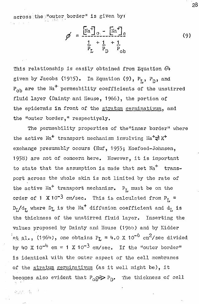
across· the ;"outer border" is given by: ,, ' . :::
d ~raj o. - rrfa+] c Y" = --------------~
This relationship is easily obtained from Equation 64
given by Jacobs (1935). In Equation (9), P1
, ? 0 , and
(9)
P0 b are the Ha+· permeability coefficients of the unstirred
fluid layer (Dainty· and II~mse, 1966), the portion of
the epidermis in. front of the stratum germinativum, and
the "outer border," respectiyely.
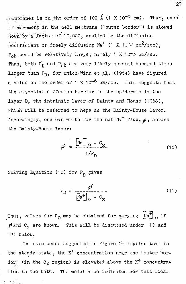
The permeability properties of the 11 inner border" where
the active Na+ transport mechanism involving Na+~ K'"
exchange presumably occurs (Huf, 1955; Koefoed-Johnsen,
1958) are not of concern here. However, it is important
to state that the assumption is made that net Na+ trans
port across the whole skin is not limited by the rate of
the active Na+ transport mechanisrr. P1 must be on the
order of 1 X 1 o-3 cm/sec. This is calculated from P = L
D1/d1 where Dt is the Na+ diffusion coefficient and d1 is
the thickness of the unstirred fluid layer. Inserting the
values proposed by Dainty and House (196b) and by Kidder
·e~ Ri., (196~), one obtains Pt= 4.0 X 10⁻⁶ cm2/sec divided
by 40 X 1 o-4 cm = 1 X 1 o-3 cm/sec. If the "outer border"
is identical with the outer aspect of the cell membranes
of the stratum germin:1 ti vtL'11 (as it well might be), it
becomes also evident that Pal?"> PD. The thickness of cell
28
29
membranes is 1 on .the order of 100 A (1 X 1 o-6 cm).
< Thus, even
if movement in the cell membrane ("outer border") is slowed
dowJ;·by a factor of. 10,000, applied to the diffusion
coeffi.cient of freely diffusing Na+ (1 X 10⁻⁵ cm2/sec),
Pob would be relatively large, namely 1 X 10⁻³ cm/sec.
Thus, both P1 and P0 b are very likely several hundred times
larger than Pn, for which .. Winn et aJ.,. (:1961+) have figured
a value on the o·rder of 1 X 1 o-6 cm/sec. This suggests that
the essential diffusion barrier in the epidermis is the
layer D, the intrinsic layer of Dainty and House (1966),
which will be referred to here as the Dainty-House layer.
Ac.cordingly, one can write for the net Na+ flux, fl, across
the Dainty-House layer:
[!ra~ 0 - ex f - ------------
Solving Equation (10) for PD gives
.Thus, values for PD may be obtained for ~arying ~a~ 0 if
pf and ex are known. This will be discussed under 1) and
2) below.
(10)
( 11 )
The skin model sugeested in Figure 14 implies that in
the steady state, the ~ concentration near the "outer bor
der" (in the Cx region) is elevated above the :rci" concentra..,
tion in the bath. The model also indicates how this local
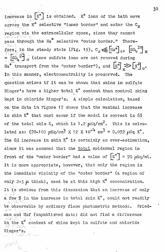
incr~ase it.\,~~] is obtained. ~ ions of the bath move
across the r--- selective "inner border" and enter the ex
regio~,via the extracellular space, since they cannot ~ . . '
pass through the Na+ selective "outer border." There
:t:o~e; ~I.1 the steady state (Fig. 15), cx~ITra-tJ 0 , [§o1+2J x
~,. ~01+ 2] ~, (since sulfate , ions are not removed during
N~+·'tr~nsport from the 11outer border"), and [Kj ::::?> [K~ 0
• ' ' . x
In this manner, electroneutrality is preserved. The
question arises if it can be shown that skins in sulfate
Ringer's have a higher total K+ content than control skins
kept in chloride Ringer's. A simple calculation, based
on the data in Figure 15' shows that the maximal increase
in sldn ~ that must occur if the model is correct is 6%
of the total sldn K, which is 1.2 pEq/cm2• This is calcu
lated as: (70-10) pEq/cm3 X 12 X 10-1+ cm3 = 0.072 p.Eq K+.
The 6% increase in skin K+ is certainly an over-estimation,
since it was assumed that the total epidermal region in
front of the -"outer border" had a value of Ge+] = 70 pEq/ml.
It is more appropriate, however, that only the region in
the immediate vicinity of the "outer border 11 (a region of
onlY, 2-3 p thick), need be at this high Irr concentration.
It is obvious from this discussion that an increase of only
a few% in the increase in total skin r, could not readily } ·' ' t-:
be observable by ordinary flame photometric methods. Fried-
man and Huf (unpublished data) did not find a difference
i-~~~h~ K+ ~ontent of sldns lrnpt in sulfate and chloride ., .~ .;;.
Ringer's.
30,
31
, 1 ) pA,T,A ON Na+ FLUX (rj)
Data are available in the 11 tera-ture concerning the
dep~nd~nce of net Na+· flux on the Na+ concentration at the
epidermal side of the skin, ~1a~ 0 • The information most
applicable to this paper is that published in 1949 by Ussing.
Changing ~aj 0 from 2 to 170 mM/l, this author found that
both Na+ outflux (inside~ outside of the sldn), and especial
ly Na+ influx (outside""'" inside of the sldn) increased with
increasing G-{a"j 0 • Hence, net Na+ flux (influx ·minus out
flux) also increased with increased @aj 0
• When the "open
skin system," i.e., the sldn not short-circuited, was used,
maximal net sodium flux was found to be on the order of
1 p.M X cm-2 X hr-1 , when the anion was chloride supplied
by chloride Ringer's.
In view of the fact that so many skin parameters have
been measured in sulfate Ringer's, it is somewhat surprising
that net Na+ flux data on skins in sulfate Ringer's have
not been published. Unpublished work from Dr. E. G. Huf's
laboratory shmrs that for skins in 55 mM Na+ /1 Na2so11-
solution, the net sodium flux is 50% of the net sodium flux
of skins in chloride Ringer's. Time did not permit measure
ment of the dependence of sodiu.11 flux on {!a'j 0 when the
skin is bathed in sulfate Ringer's. If the reasonable
assumption is made that the relationship is similar to the
one that Ussing has shown to exist for skins in chloride
Ringer 1 s, data concerning yJ can be obtained from the work
·of- Ussing and the investigations in Dr. Huf' s laboratory,
as noted above. Na+ flux values obtained in the manner are
32
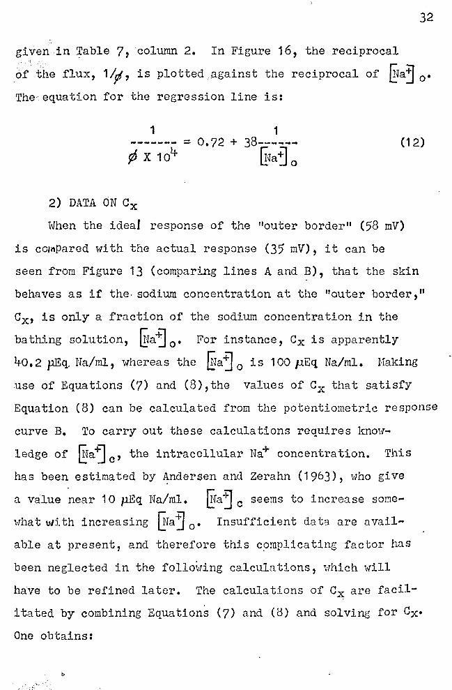
given in ~able 7, column 2. In Figure 16, the reciprocal
of the flux, 1 Ip!, is plotted .. against the reciprocal of @aj 0 •
The· equation for the regression line is:
1 1 ------- = 0.72 + ¢ x 10'+
38----.... Gia~ 0
(12)
2) DATA ON Cx
When the ideal response of the "outer border" (58 mV)
is cotnPared with the actual response (35 mV), it can be
seen from Figure 13 (comparing lines A and B), that the skin
behaves as if the. sodium concentration at the "outer border,"
Cx, is only a fraction of the sodium concentration in the
bathing solution, ~a~ 0 • For instance, Cx is apparently
40. 2 p.Eq. Na/ml, whereas the ~a'j 0 is 100 p.Eq Na/ml. Haking
use of Equations (7) and (8),the values of Cx that satisfy
Equation (8) can be calculated from the potentiometric response
curve B. To carry out these calculations requires know-
ledge of @a-ij c, the intracellular Na+ concentration. This
has been estimated by Andersen and Zerahn (1963), who give
a value near 10 µEq Na/ml. ~ra1 c seems to increase some
what with increasing ~Ta~ 0 • Insufficient data are avail-
able at present, and therefore this c~mplicating factor has
been neglected in the follo~ring calculations, ·which will
have to be refined later. The calculations of Cx are facil
itated by combining Equations (7) and (8) and solving for Cx•
One obtains:
33
(13)
in which k = 35/58 = 0.604-. Several values of ex were
obtained in this manner for a number of arbitrarily
selected values of {!ra"j0
• The results are eiven in Table
7·, column 3.
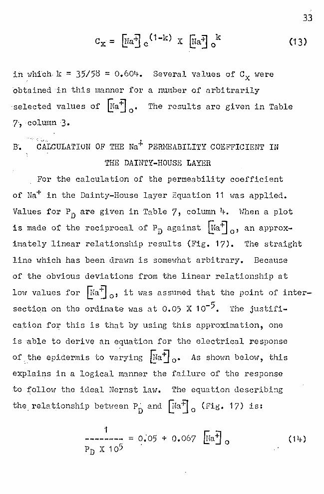
ff. CALCULATION OF THE Na+ PERMEABILITY COEFFICIENT IN
THE DAINTY-HOUSE LAYER
For the calculation of the permeability coefficient
of Na+ in the Dainty-House layer Equation 11 was applied.
Values for PD are given in Table 7, column l+. When a plot
is made of the reciprocal of PD against ~faj O' an approx-
imately linear relationship results (Fig. 17). The straight
line which has been drawn is somewhat arbitrary. Because
of the obvious deviations from the linear relationship at
low values for Efa"j 0
, it ·was assumed that the point of inter
section on the ordinate was at 0.05 X 10⁻⁵. The justifi
cation for this is that by using this approximation, one
is able to derive an equation for the electrical response
of_ the epidermis to varying ~faj 0 • As shown below, this
explains in a logical manner the failure of the response
to follow the ideal Nernst law. The equation describi~g
the, relationship between PJ) and ~.ra-:J 0
(Fig. 17) is:
1 == 0."05 + 0.067 ~a~ 0 ( 1 i+)
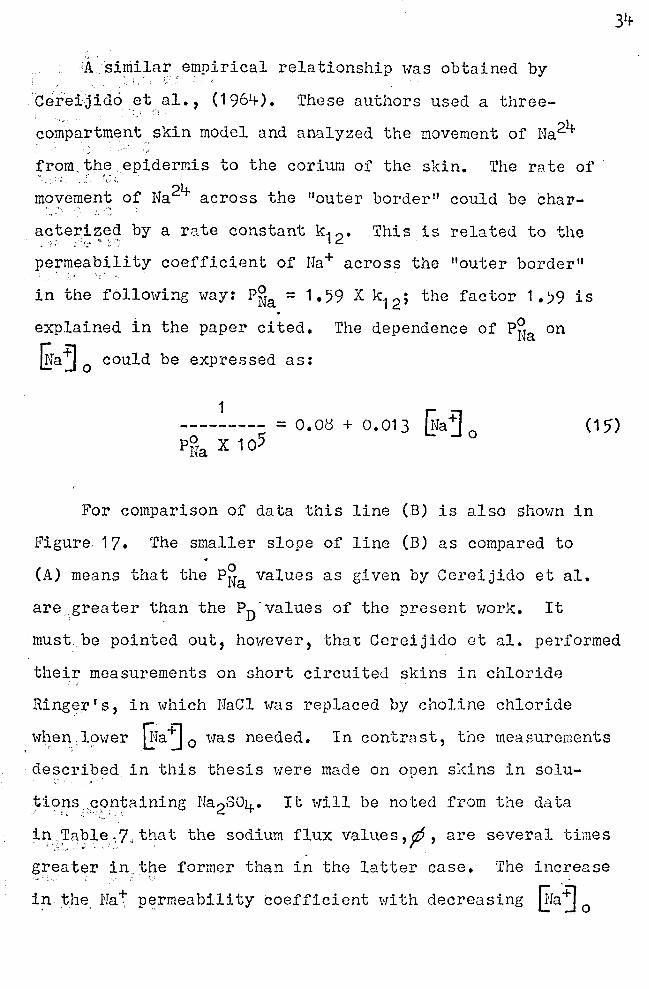
A.similar empirical relationship was obtained by ' . : ' ,
Cereijido et al., (1964-). These authors used a three-•. . I ' ~
_,_,.·
compartment .. skin model and analyzed the movement of Na24
from_ the epidermis to the coriura of the skin. The rste of "' ' :'- !' •.'
m~~e~~nt .. of Na24- across the 11outer border" could be char-.,
~~~e~~2:~~ by a rate constant k12• This is related to the
pe~meability coefficient of Na+ across the "outer border" ·1' "
in the following way: P~a = 1.59 X k12 ; the factor 1.)9 is
explained in the paper cited. The dependence of P~a on
Wa~ 0
could be expressed as:
1 --------- = o. Ot) + o. 013 liJa j
0 P~a X 105
(15)
For comparison of data this line (B) is also shown in
Figure 17. The smaller slope of line (B) as compared to . (A) means that the P~a values as given by Cereijido et al.
are.greater than the PD-values of the present work. It
must: be pointed out, however, tha~ Cereijido et al. performed
their measurements on short circuited skins in chloride '·'
Ringer's, in which NaCl was replaced by choline chloride
. wl;en.,lower ITfa~ 0 was needed. In contrast, the measurements
descri~~d in this thesis were made on open skins in solu-~ \ '
~is>ps,~?n~aining Na2so4. It will be noted from the data ~ ., ·• ... ..
in,~able~7,that the sodium flux values,p, are several times .. · ·' .. ".
g~eater in_t~e former than in the latter case. The increase ,. ·:
in the. Na+ permeability coefficient with decreasing Era.'j 0
is_ expressed by the similar Equations. (14) and (15).
Although no explanation for these relationships can be
given, the great similarity of results strengthens the hypo
thesis o.f diffusion delay and active Na+ transport across
the sl~~n a~ the chief factors which explain the failure of . '
the. NE~rnst lm.·T when testing the electrical response Of the
epide;mls to changing §a j 0 •
C. EFFECT OF pH ON P.D.
If an increase in skin P. D. with increasing [·r+J0
had
occured, it might be suggested that this was the result of
H+ moving across the "outer border" as predicted by the
Hodgkin-Katz equation. Since the observations regarding
the effect of pH are contrary to the expectation if the
Hodgldn-Katz equation had been applicable, it is concluded
.that the permeability of the "outer border" for H+ is not
significant in the generation of the fraction of the total
skin P.D. that has its origin at the "outer border." In
weighing the importance of the results shown in Figure 12,
it must also be kept in mind that here a thousand fold
change in [Hj 0
was investigated. The reason ~·Thy a decrease
:in pH within reasonable limits (pH 9 to pH 6) resulted in
a ~light to moderate decrease in skin P.D. can not be given.
With r~~ard to the hypothesis that has been presented, it is
suggested that an incre::.se in ~"'.] 0
leads to a decrease of
the Na+~permeability coefficient in the Dainty-House layer.
This·'.wbrild lead to a lowering of the Na+ concentration near
35
the "outer border," ex, and therby reduce the Ha+ concen
tration gradient across the "outer border"; this would
lead to a 101.·rering of the s~in P. D. as shO'..,rn by the present
data.
D. EFFECT OF EPIHEPHRINE ON SKIN P. D.
Since it is known from the literature (Cercijido and
Curran, 1965) that about half of the tot~l skin P.D. is .
generated at the "outer border," and the other half at the
"inner border, 11 it seems to follow tlrn t epinephrine must,
in part, hnve acted on the "outer border," the properties
of which are the main concern of this thesis.
For skins under the influence of epinephrine, it must
be assumed that the Na+ concentro.tion ne.gr the "outer bor
der," ex, was high, probably as high as the Ha+ concentra
tion in the solul;ion ITraj 0
• Epinephrine is kno':m for its
property of increasing membrane permeability. With Cx being
high, rather than low as in the ccntrol s~ins, the low P.D.
at the "outer border" and its lack of response to changes
in lifa ~ 0
can only mean that epinephrine drastically chanr;ed
the ion selectivity of the "outer border." It is concluded
.that under the influence of epinephrine, the "outer bor<ler"
loses its specific z;a + selectivity, and thus, did not act
36
as a source for generation of a Nernst type diffusion paten-
tial.
~pinephrine is known to stimulate the secretion of.
mucus (Watling'.:on et a 1., 1 9b/) fron the skin t;l~4n'..l.s (which
are seen in the photomicrograph shown in Figure 2). The
question arises whether the low total skin P.J. and the rel
ative insensitivity of the epidermis to changes in @a3 0
is explainable on the basis of an altered function of epi
nephrine sti:-nulated skin glandsr It is unlilrnly that this
is the case. Following epinephrine injection, the mucus
37
that pours over the surface of the skin is al1mline (pH 7. 'j}.
This in itself should lead to an increase, and not to a
·decrease, in skin P.D.
It is also known that under the influence of epineph-
rine, there occurs an active outward transport of c1- ions
(Koei'oed-Johnsen et al., 1952) and possibly of SOti-= ions
(Campbell et al., j_n print). ~'ii th everything else remaining
constant, this also should lead to an increase in skin P.D.
(outside more negative relative to the inside). Therefore,
the conclusion is that epinephrine destroys the physic-
chemical properties of t11e 11outer bord0r" in such a way
that it loses its normally high rra+ selectivity.
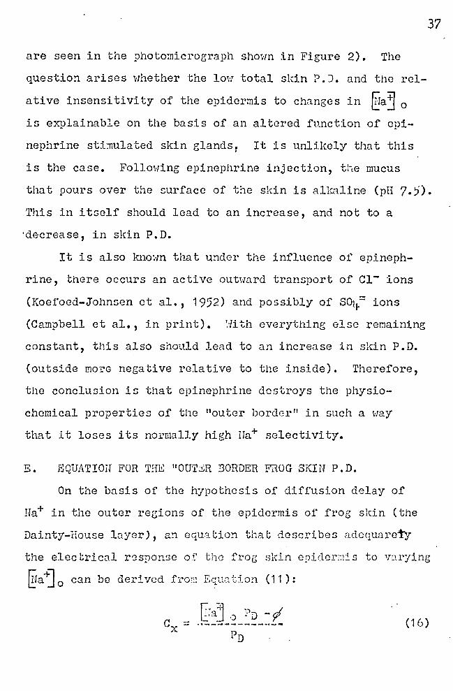
E. EQUATIOII FOR THE 11 0U'I'li:R BORDER FROG SKIN P. D.
On the basis of the hypothesis of diffusion delay of
Ha+ in the outer regions of the epidermis of frog slcin (the
Dainty-House layer), an eCi_uation that describes adcqunreTy
the electrical r~sponse of the frog skin epidcr~is to v2rying
@a+] 0 can be derived f:com Equa t.j_on ( 11 ) :
r,, ;.1 n d 1_:_· aj o .- D - y
(16)
38
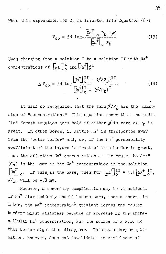
When this expression for Cx is inserted into Equation (8):
[fa~ o PD - ff = 58 log--------------~a j c Pn
(17)
Upon changing from a solution I to a solution II with Na+
concentrations of Gfaj; and EJaj ~I
r::-ia +l II - (f /P )II 58 !.:: Jo D . = log-------~-~---------
~a ~ ~ - (f/Pn)I
(18)
It will be rec~gnized that the tG1,r;i f /Pn has the dimen
sion of "concentration." This equation shows that the modi
fied I1Jernst equation does hold if either f is zero or. Pn is
great. In other words, if little na+ is transported away
from the "outer border" and, or, if the Nu+ permeability
coefficient of the layers in front of this border is great,
then the effective Ha+ concentration at the "outer border'.'
(Cx) is the same as the rra+ concentration in the solution
~ra~ 0
• If this is the case, then for ~Ja+J;r = o.1 ITraj~I, aV0 b will be -58 mV.
However, a secondary complication may be visualized.
If Ha+ flux suddenly should become zero, then a short time
later, the Na+ concentratio~ gradient across the "outer
border" might disappear because of increase in the intra
cellular Ha+ concentration, c.nd the source of a P.D. at
this border might then disappear. This secondary compli-
cation, hm·rever, does not inva.lido.tc ·the usefulness of

Equation (18). Ordinarily, n~t flux does take place, and
Pn is relatively small. Therefore, taV0 b is expected to be
less than -58 mV when changing ~~a~ 0
from 100 to 10 ml·Vl.
39
It has been pointed out that both ¢and PD are dependent
on ~fa~ 0
• The most desirable approach to simplifying
Equation (18) would be to enter the relationships between
¢, Pn, and ~ra-j 0 into this equation. Unfortuna.tely, there
exists at present no theory that would explain the empirical
functions (Equations 12 and 14) relating these variables.
It is possible, however, to give approximate empirical. equa
tions that describe .the dependence of ,don ~Jaj 0 , and also
of Pn on ~ra"j 0 • The equations are given above as (12) and
(14). By combining these two equations, the following ex
pression forP/Pn results:
a~Ia+] 0 + b (19)
A= o.o5; B = 0.067; a = 0.72; b = 38
A plot of P/Pn against firaj 0
is shown in Figure (18). The
data necessary are given in Table 7, columns 1 and 5. For
comparison of the present results with those obtained by
Cereijido et al. (1965), ¢1 .Pu~ values for varying Era+] 0
were also calculated from the information given in the paper
cited above. The results of .thG c0.lculntions are tabulated
in Table 7, column 8, and a plot of Pl P,T0 a[;;-iinst 'Ira·il 1.a U .J o
is also shown in Figure 18. One can sec the sane kind of
dependence of ¢IP on ~;a~ 0
, al thou1:;h the experiments on

which these curves are based are quite different in nature.
The differences seen in the tr..ro functions may be due to
the fact that the treatment of skins was different, ( p. 3t1-).
40
Finally, Equations (18) and (19) were used to construct
a plot of ~Vob against ~a~ 0 (~ig. 19). The necessary P/Pn
data for five arbitrarily selected fEa~ 0 values are given
in Table 7, column 5 (data of this thesis) and column 8 (data
of Cereijido et al.). In regard to the data in this thesis,
it can be seen that Equations (18) and (19) adequately de-·
scribe the e:xPerimental observations and also point to the
factors (p and Pn) that lead to the deviation of the experi
mental data from the simple Nernst equation. When the P'/PN~
values calculated from the work of Cereijido et al. are
used, the result is a response line of the "outer border"
that corresponds closely to the one predicted by the un
modified Nernst equation.
Thus it appears that the Ncrnst equation is the limit
ing law that describes the electrical response of the epi
dermis to varying Na+ concentrations in the salt solutions
at the epider~al side of the skin. Under experimental con
ditions of this research, however, the electrical response
is lower than expected because of the occurence of contin
uous active Ha+ transport across the skin, and because of
diffusion delay in the regions (D~inty-House lA.yer) in front
of the Na+ selective border. ·
SUMMA HY.
1. Experiments were conducted to examine the effect of
varying Ira+ concentrntions at the epider11al "outer border"
on the ?.D.
2. 'rheoretical and experimental evidence was given in
support of sodium ions being the major contributors to the
frog skin potential at the "outer border."
3. It was hypothesized that active Na+ trnnsport across
the frog skin and diffusion delay in the Dainty-House layer
account for the discrepancy ~etween the theor~ticnl Nernst
response and the actual response of the frog skin to chnneing
4. Taking these two factors into account, a modified
Nernst equation wns proposed which adequately d.escribes
the P .u. response of the epidermis to varying IT;aj 0
•
5. s35 labeled sulfate .experiments indicate that frog
sldn is ir.lpermenble to sulfate.
6. Epinephrlne treated skins lost their ability to respond
t . r..+1 o varying i.:.~a J o•
LITERATURE CITED
Andersen, B. and Zerahn, K. 1963. Hethod for non-destruc
tive determination of the sodi~~ transport pool in
frog skin with radiosodium. Acta Physiol. Scand.
59: 319-329.
Baillien,. M. and Schoffeniels, E. 1961. Origine des poten
tials bioelectriques de 1 1 ~pi thelium intestimi.l de la
torque gi·eque. Biochim. et Biophys. Acta. 53: 5"37-54-8.
Campbell, J. P.,'Aiyawar, R. M., Berry, r_;. R., and Huf,
E. G, To be published ••.
Cereijido, H., Herre_ra, F. C., li'J.anigan, H. J., c:tncl Curran,
P. F. 1 96i1-. The influence of IJa concentration on Na
transport across frog skin. J, Gen. Physiol. 47: 879-
Cerei j ido, M. , and Curran, P. F. 1 965. Intracellular elec
trical potentials in frog skin. J. Gen. Physiol. li-8:
"5'1+3-557.
Chowdhury, T. K. and Snell, F. M. 1965. A microclectrodc
study of electrical potentials in frog skin and toad
bladder. Biochim. et Biophys. Acta. 9t1-: l1-61-'+71.
Dainty, J. and House, c. R. 1966. "Unstirred layers" in
frog skin. J. Physiol. 182: 66-78.
Engbaeclc, L. and Hoshiko, T ~ 1 957. ~lectrical potential
gradients through frog , .
s.~in. Acta Physiol. Scand.
39: 34-8-355.
4-2
F4kuda, T. R. 1 91r2. Ueber die Bedinguneen fur das Zustancle
kommen des Asymmetriepotentials der Froschhaut. Jap.
J. Med. Sci. Part 3. Biophysics. 8: 123-134.
Fukuda, T. R. 191+2+. Sonderstellung des Natriums bei der
Potentialbildung an der Froschhaut. Jap. J. Med. Sci.
Part 3 Biophysics. 10: 77-86.
Hodgkin, A •. 1. and Katz, B. 1949. The effect of sodium ions
on the electrical activity of the giant axon of the
squid. J. Physiol. 108: 37-77.
Huf,~. G. 193;>.'Versuche Uber den Zusammenhane zwischen
Stoffwechsel, Potentialbildung und Funktion dcr
Froschhaut. A~ch. ges. Physiol. (Pfluger) 235: 6?5-
673. ,,
Huf, E. G. 1936. Uber aktiven Hasser-und Salztransport <lurch
die Froschhaut. Arch. ges. Physiol. (Pflilger).
237: 143-166.
Huf. E. G., Hills, J.P., and Arrighi, M. F. 19'.)5. Electro
lyte distribution and active salt uptake in frog skin.
J. Gen. Physiol. 3u: 867-8~~.
Huf, E. G., Doss_, N. s., and ~:Tills, J. P. 1957. Effects of
metabolic inhibitors and drugs on ion transport and
oxygen consumption in is.ola ted frog s!dn. J. Gen.
Physiol. 41: 397-417.
Jacobs, H. H. 1935. "Diffusion Processes", iirgebnisse der
Biologie, Berlin. 12: 1-160.
Kidder, III, G. W., Cereijido, M., and Curran, P. F. 1964.
Transient chanees in electrical potential differences
across frog skin. Am. J. Physiol. 207: 935-94-0.
Klahr, s. and Bricker, N. S. 196'+-. On the elcctrogenic
nature of active sodium transport across the isolated
frog. skin. J. Clin. Invest. 43: 922-930.
Koefoed-Johnsen, v., Ussing, H., and Zerahn, K. 19~2. The
origin of the short circuit current in the adrenalin
stimulated frog s~in. Acta Physiol. Scand. 27: 313-
Koefoed-Johnsen, v. and Ussing, H. H. 1958. The nature or
or the frog sldn potential. Ac ta Ph:;rniol. Scand. '+-2:
293-308.
Lindley, B. D, and Hoshiko, T. 196t1-. The effects of alkali
metal cations and cornmor. anions on the frog skin poten
tial. J. Gen. Physiol. 4-7: 749-771.
~~cRobbie, E. A. C. and Ussing, H. H. 1961. Osmotic behavior
of the epiderr.ial cells of frog skin. Acta. Physiol.
Scand. 53: 348-365.
Ottoson, D., Sjostrand'· I<',, Stenstrom, s., and Svaetichin,
G. 1953, Microelectrode studies on the E.M.F. of frog
skin related to electron microscopy of the dermo-epi
dermal junction. Acta. Physiol. Scand. 29 (Suppl. 106):
611-624.
Scheer, Bradley T. and 1·'.u::11nch, I.J. 1.1. 1 960. The locus of the
electromotive force in frog skin. J. Cell. Comp.
Physiol. 5?: 259-266.
Steinbach, H. B. 1933. 'rhe electrical potential difference
across living frog skin.·· J. Cell. Comp. Physiol. 3:
1-27.
Ussing, H. H. 1949. The active ion transport through the
isolated frog skin in the light of tracer studies.
Acta. Physiol. Scand. 17: 1-37.
Us sing, IL H. and Zerahn, K. 1951 • Active transport of sodium
as the source of electric current in the short-circuited
isolated frog.skin •. Acta Physiol. Scand. 23: 110~127.
Ussing, H. IL and.-dindhager, E. E. 1961+. Hature of
shunt path and active souium transport path through
frog skin epithelium. Acta. Physiol. Scand. 61 : l1-<5'~-5011-.
Watlington, c. o., Burke, P. K., Campbell, A. D., and Huf,
E. G. 1965. Systemic effects of epinephrine in tho frog.
J. Cell. Comp. Physiol. 6): 33(-3)~.
Hinn, P. H., Smith, T. 2., Campbell, A. D., and Huf, ,~. G.
196~. Sodium diffusion in epidermis and cerium of frog
skin and in Rin0er-agar gel. J. Cell. Comp. Physiol.
6l1-: 371-388.
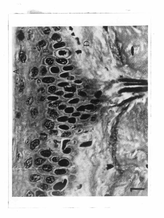
FIGURE 1'
Cross section of the epidermis .
A. Stratum corneum
B. Stratum soin sum and gran lo sum
c. Stratum germinativum
D. Dermis
~----- ~ -~----~
47
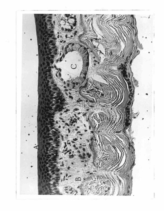
FIGURE 2
Cross section of the frog skin
A.· Stratified epider mis
B. Dermi s
C. Mucus gland
•
. '

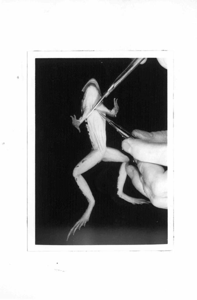
FIGURE 3
Decerebration of .frog
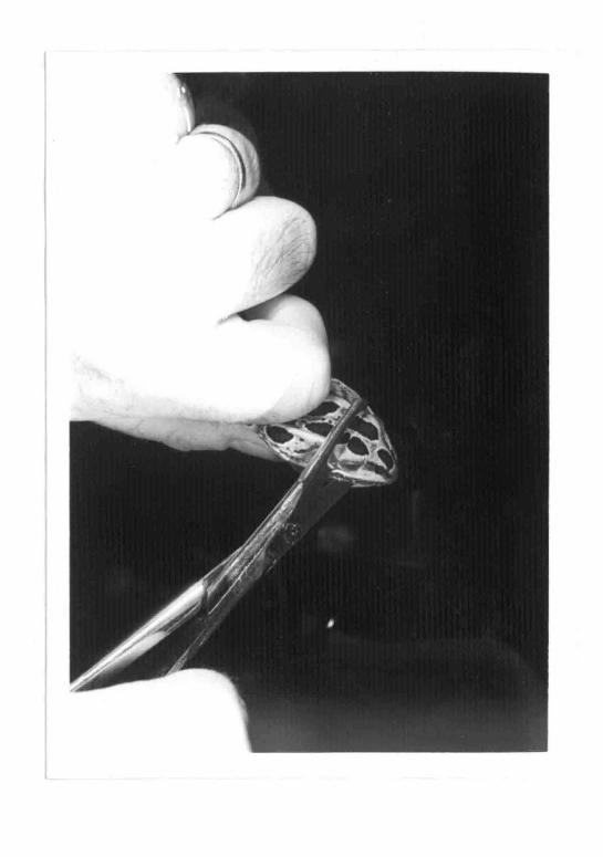

FIGURE 4
Initial incision for removing belly skin
50
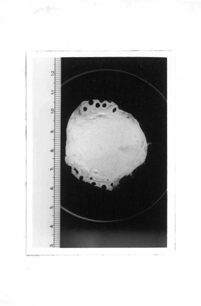
FIGURE 5 :/
Belly skin spread out showing shape and size
51
, .
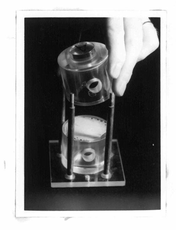
FIGURE 6
Assembling of celL I
The skin is placed across the open end of one chamber;
the other chamber is being placed in position.

52
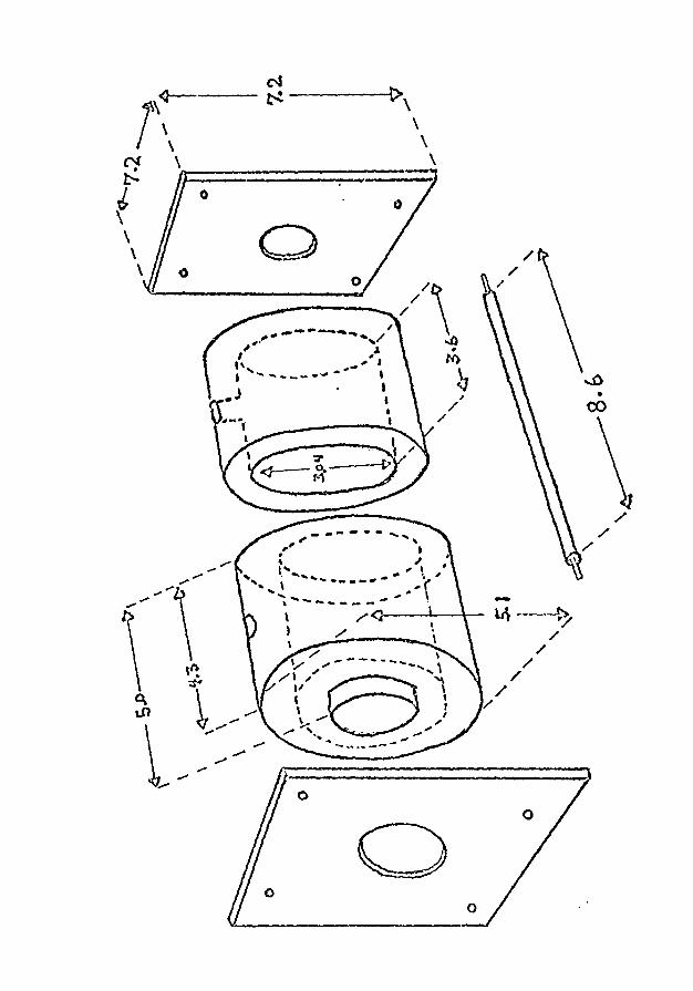
FIGURE 7
Diagram of lucite cell
(and one of the bolts to hold the chambers together)
0
.. ---- //\ ~,. ....... , I ',/ ,,, ," ..0 I .... .... JI* t • ~-- ..... -.... ti)
,.) ' \ . I
,... t lJ ,. -------- . /,
. /
0 0
/
-Lt\~ / /
/
/
/ /
.....0 •
(:()
/ /
/
53
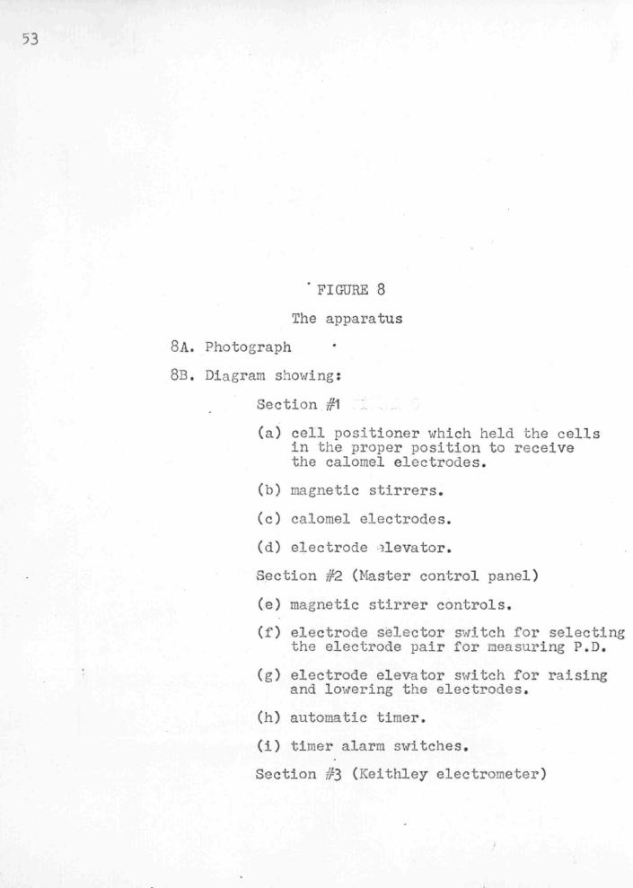
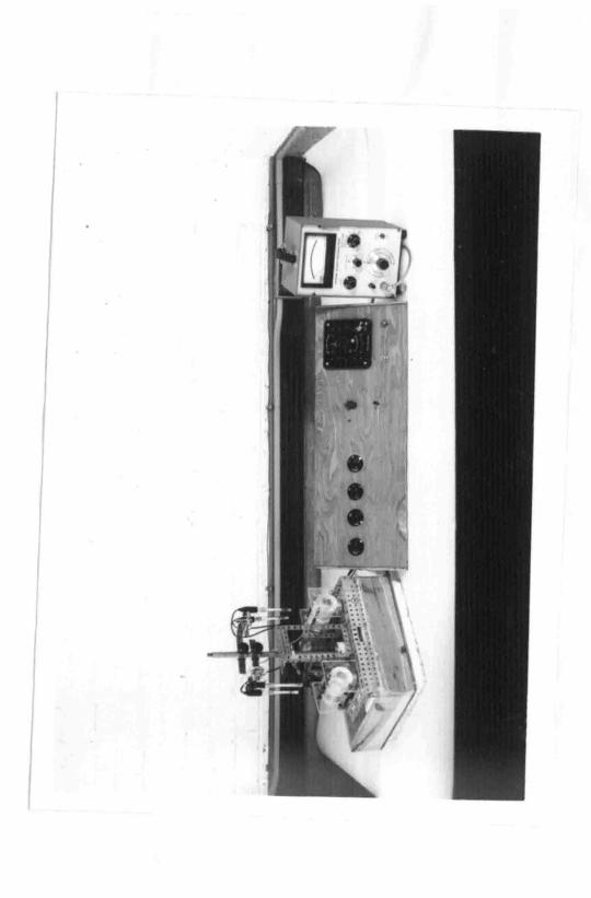
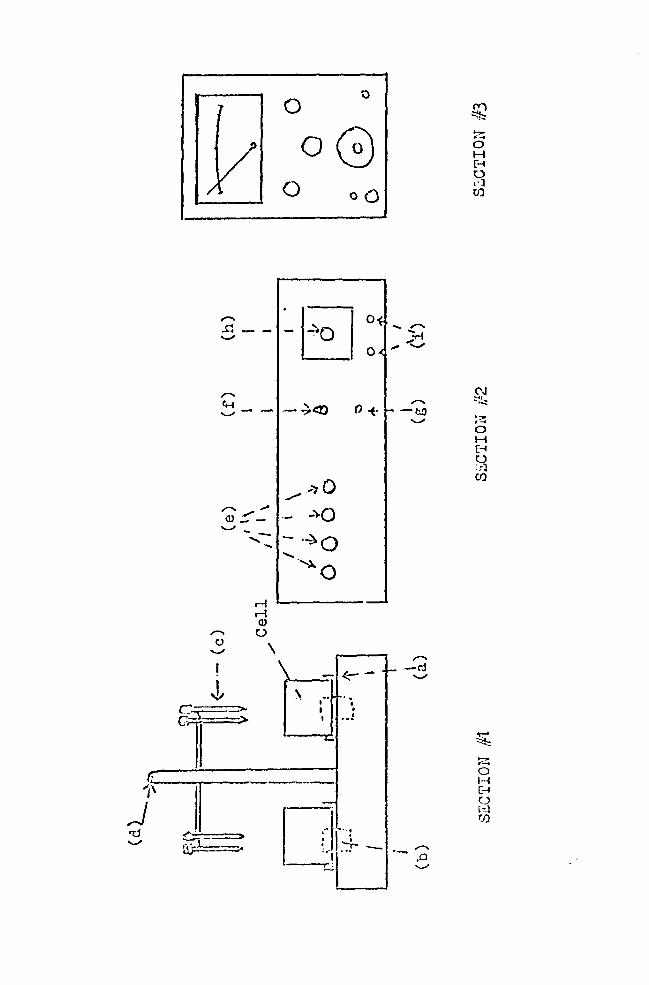
FIGURE 8
The apparatus
BA. Photograph
8B . Diagram showing :
Section #1
(a) cell positioner which held the cells in the proper position to receive the calomel electrodes.
(b) magnetic stirrers.
(c) calomel electrodes .
(d) electrode ~levator .
Section #2. (Master control panel)
(e) magnetic stirrer controls.
(f) electrode selector switch for selecting the electrode pair for ceasuring P.D .
( g ) electrode elevator switch for raising and lowering the electrodes .
(h) automatic timer .
(i) timer alarm switches.
Section #3 (Keithley electrometer)
I
c:: \
cl fE I
J ,,
ili'
0 0
0 G 0 oQ
,,.....
~E 0 ~ - -'-'
0
,,..... ~-'-' - - ~<?'.) o+
../ _,., 0
,,..... .,,; _..,.o (!) .~ --- -....._, --
-·~o ' ...... ..... -~
0 M "------
....._,
M <l> 0
\
... ,,..... -:,..
,,,,. ... '-'
'"' -tl.O '-'
! - .t.::- - -t-
~ \ I .. ..
• > I . > I ... :
......
__ .,._
0 1~·-: ... I'+- -:.. -l .. -
(Y) ~ ,.,,.. ... 0 H r~ 0 i<l (I)
N ~!::
'?" ...... 0 H E-1
f:3 (I)
?-: 0 H [~
0 rc::.l rn
FIGURE 9
Electrical response of the epidermis of frog skin to . change in Na+ concentration for J and ~ frogs. Theoretical
Nernst response is shown as the dashed line.
r;j <-+ c: I·• I. t
\-'.'()
H
r-. r' .-' c r-1
(_~1
\ j:
0 c ..-
...
c G· (
H H
+ C+
H +
r.. "D .-'
0 ~I
0
:. \
\ \ ..
r. ,, ( c [_·. '
'-
Ch
H H ,...,
c-~; ::-! r.'J
c 1 -
\
-'·
\ \
'· ~ : r:
·!-"> (j
~: ! r I
\ C1 . \ ' \
\ ' \
0 () (\)
0 .. -' ..
I
C• (\!
55
FIGURE 10
Electrical response of skins of Group III to changes
in [ia+J 0 • The curve is brokGn up into two linear rosponses.
Go
"llf .--0 .. )
30
20
10
0 --:---~-·-,,____~- ___,.__,_J __ ,_, l-Ll-1-1-1-1~~ 1 o 20 30 1~-0 50 60 ~o 1 oo 1·1 o
. -- +; 1 ,_,_J;r.!. ~:~- :~-
56
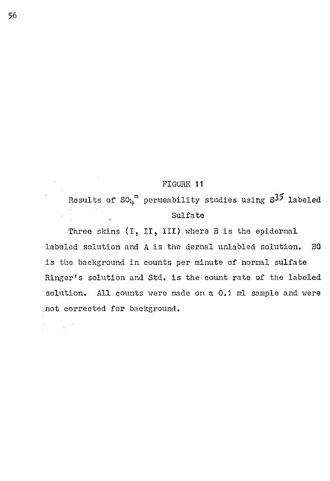
FIGURE 11
Results Of so1 .. - permeability studies using s35' labeled
Sulfate
Three skins (I, II, III) where B is the epidermal
labeled solution and A is the dermal unlabled solution. BG
is the bacl<:ground in counts per minute of normal sulfate
Ringer's solution and Std. is the count rate of the labeled
solution. All counts were made on a 0.1 ml sample and were
.not corrected for background.
COUNT RATE IN THOUSANDS 0 ... °' ro c--.. '° \r\ ...:t-. (Y') C\I .- 0
- ·-1-j-... j--"'l-l··-·lt-l·-11-I-
H H H
H H
H
• 't1 .µ Cl)
57
FIGURE 12
Electrical response of xhe epidermis to changes in pH
The skins were in sulfate Rineer's (110 mM Na/1--10 mH K/l).
The graph shows a decrease in P.D. with a decrease in pH
from 9 to 6. The bars represent the average P.D. 1 s obtnined
on twelve skins. 'rhe standard deviations are shown for
each pH.
100 -90 -80 - "'
70 - "" " .,. - .!.
~
60 - "'- .,J, J,
.....
mV 50 -:
40 -:
30 -
20 -10 -
0 pH = 8 9 8 7 6 8
pH
58
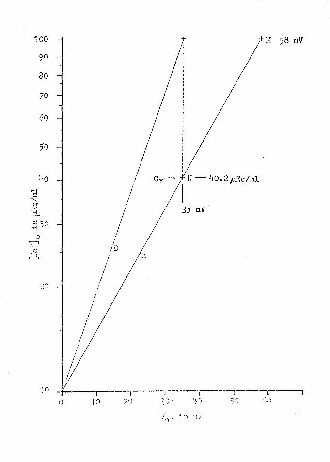
FIGURE 13
Plots of Equation (3)
Line A shows point H where V0 b = 58 for ITra~ 0 = 100 µEq/ml and §a°jc = 10 µEq/ml. Experimental results,
line B, shows where V0 b = 35 mV. The dashed line in
dicates that the Na+ concentration at ex ·is only l+o.2 p.Eq/ml
when [lra ~ 0 = 1 00 p.Eq/ml.
100
'd
"' tJ' r-::1 r<
90
80
70
60
)0
40
~ 7J 0 •r-: ._)
0 r:-1 -,-s~ ~
20
·10
0
I I
I I
I I
I I
/ I
I I
I I I
} 10
+n 58 mV
I Cx- ~~ - ltO. 2 pEq/ml I I
I I 35 mV
/"F3 A I
I I
(- I -r 20 ~A• )~_() )J .~,:)
~,
., ').)
.; . , :i7
59
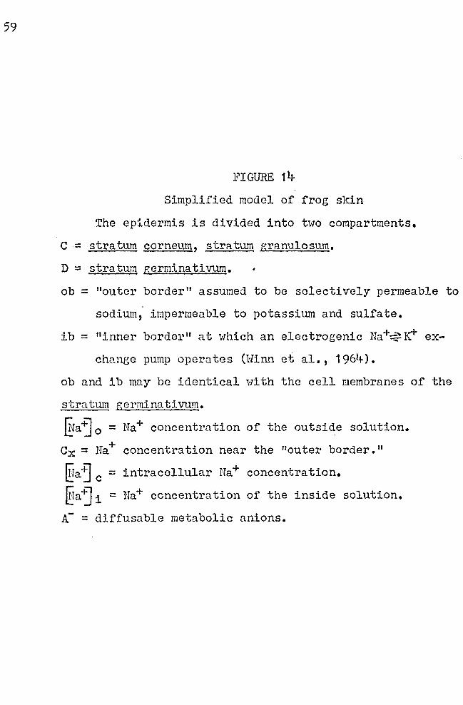
FIGURE 14
Simplified model of frog skin
The epidermis is divided into two compartments.
C = stratum corneum, stratum granulosum,
D :::: stratum gerl]lina ti vum.
ob = "outer border" assumed to be selectively permeable to
sodium, impermeable to potassium and sulfate,
ib = "inner border" at which an electrogenic Na+~IC1" ex
chanee pump operates (Winn et al., 1964).
ob and ib may be identical ·with the cell membranes of the
stratum germinativum.
~aj 0 = Na+ concentration of the outside solution.
Cx = Na+ concentr'.ltion near the "outer border. 11
§a·~ c = intracellular Na+ concentration.
§aj 1 = Na+ concentration of the inside solution.
A- = diffusable metabolic anions.
CORIUN AND OUTSIDE SOLUTION EPIDERMIS INSIDE SOLUTION
A A. k ,-------- ""\ r \r -----'\
: I I I ob ib
~ i I r---+--1 -K+ <l---- --:----- --- _ _ <j I 4-- ___ - _ -· re+
I J
(Na Jn 1 I D- + ex I Na c --f-- t> [Na j. i
1/2 ~04] 0 i f 1/2 ~~~ -v A - ( e • g. HC 0 3) 1/2~0~
A
l OUTER SOLUTION
I I
I I I I I I I
' l •
B c I D I I I I . L_ L_STRATITI1 GERl1INATIVUM (12p)
STR.ATUH CORHEID1 A~ID GRANULOSUM (12f)
UNSTIRRED FLUID LAY3R. (40p)
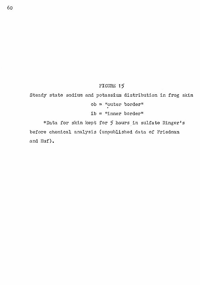
60
FIGURE 1 ')
Steady state sodiTh~ and potassium distribution in frog skin
ob = 11 outer border"
ib = "inner border"
~:Data for sk:l.n kept for 5 hours in sulfate Ringer's
before chemical analysis (unpublished data of Friedman
and Huf).
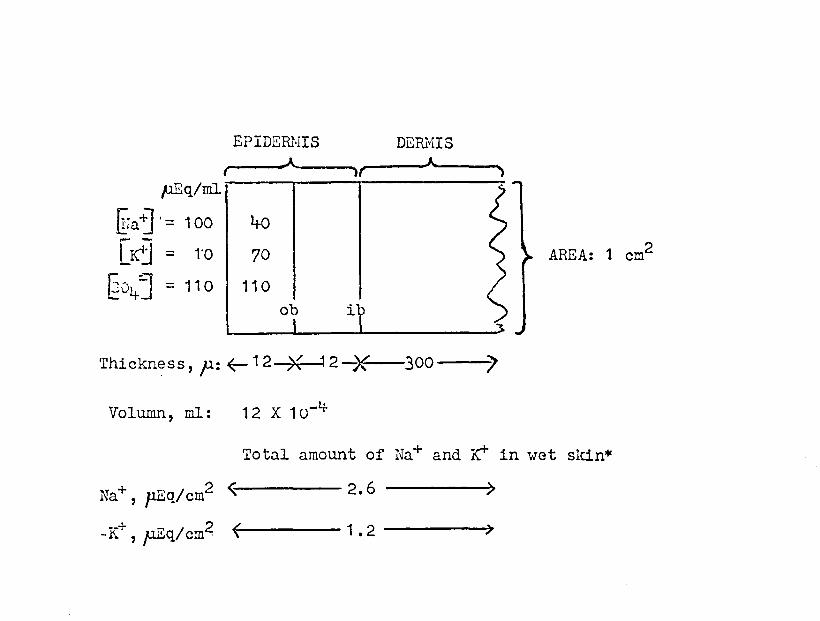
EPIDERMIS DEfu'1IS --~-A A f 1t ---.,
pEq/ml. -
gr a :J · = 1 oo I >+o [K"'] = ro I 70
@v4J = 11 o I 11 o ob i
Thickness, p: ~ 12 )( 12-7( 300 ~
Volumn, ml: 12 x 1 o-4-
AREA: 1 cm2
Total amount of Na+ and K1" in wet sldn*
Na+, p.Eq/cm2 ~ 2.6 ----~
-K+, p.Eq/cm2 1.2 ---,
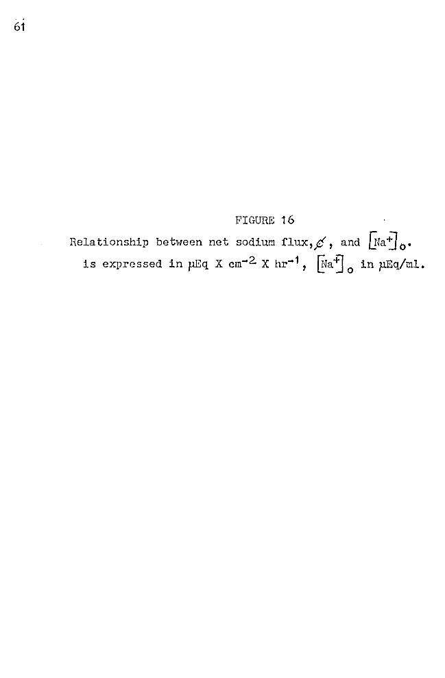
61
FIGURE 16
Relationship between net sodium flux,p, and [Naj 0 •
is expressed in pEq X cm-2. X hr-1, ~faj 0
in }lEq/ml.
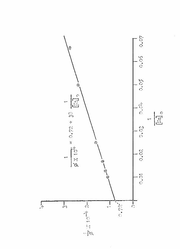
C'-0
0
~ 0
c.:-
'" © 0
C·
(I
-:-- ~ .J. I•
L:::J _, ()
(.~l c 0 (Y)
~ ,-+ ·-· ~ ('j C•) ['. ()
c 0
II 0
~: \ (\J 0
-:--0 ' ' 0
'$.. \ 0
\ ,-0 0
c::
I -(· ! <: ... -i· (\! ,-- . '•
L'-,:r
I c G
; ' I
·-·1'0_
62
FIGURE 17
Relationship between P0 and ~a "j 0
Po is the sodiwn permeability coefficient in the Dainty
House layer of the skin (page 29 of text). P0 is expressed
in cm/sec, ~a~ 0
in pEq/ml.
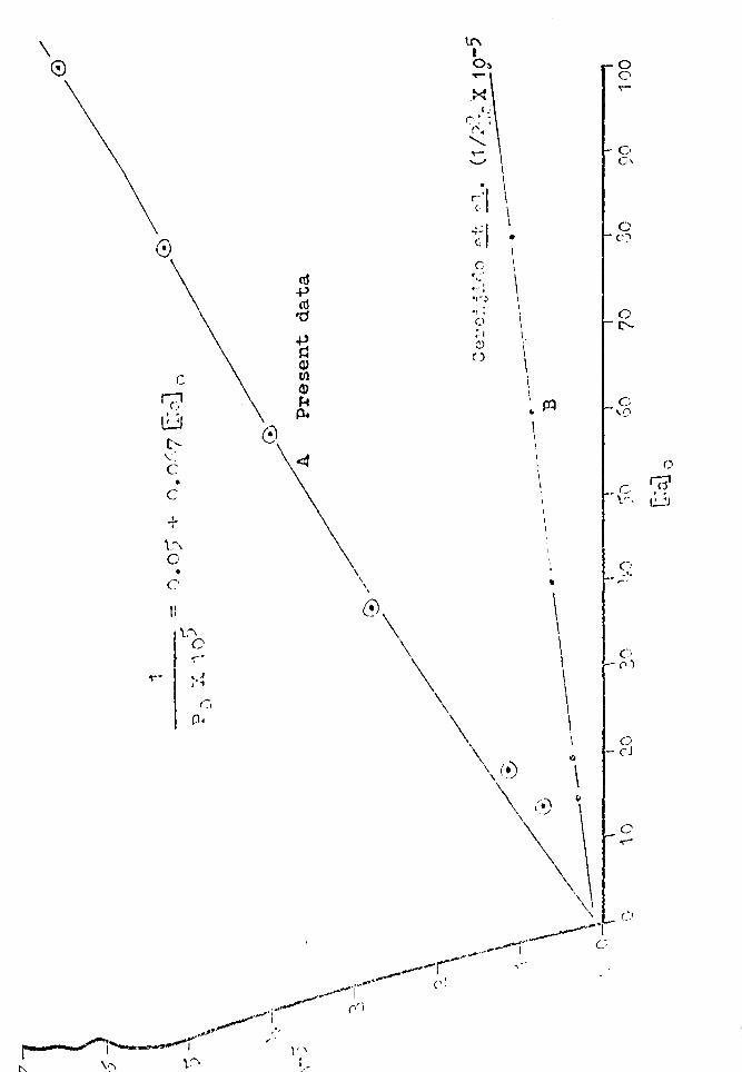
\ ©
0
c Q, ~ t-.. \.: c • c
+ \_-\ c •
C'
Ii
I \0
0 -:-·
•, J ' 1
ro +> ro 'C1
+> ~ <1> tll <1> ~ jl.
c;., _,
~
\ \
\
tr\ I o~ 'T""
.~ (:
C' •: r.'
........... ..-'-' . ~·1 ··1 -.-C: •
1 c I
I , .. '" \ •.-!
•(-:. •r: I
(l I . r·t \. C..l 0 \
\ m
' \ I
\
\ \
0 0 ..-
C. _,
'0
()
r l(i1 '\_:., ' .
I~

63
FIGURE 18
Plot of Equation (19)
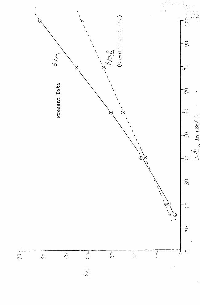
\ 0
I I
C• \ =..
' \ x
\ \
\
\
\ \
\ \
. r-'.1 '-
··1 ""; -,_
c r_-_.._.
., -~·
c .~~ •r-, -4 C...'
(\, !'.-1 ........... c:;;
'e.. r_~ ·><. .......,
\ \
\ \ \
\ \
' \ 0 x
--r--·---r ( '· - - (•)
r
' \ \
\
' \ \
\ \
0)(. \
\
\
\\ xG \\
.:< 0 \ \
-,----r-( ~ c._, ( '
0 0 ,_
0 -,o
0 -,("\
_C· -·-
C·
-·
0 ('(')
0 (\j
0 .,....
0
r-·I z t;:;l
~:1 ,_, ,_
s:: •1l
0
P~l .. (',,
•·· L...'..:.:.J

64
FIGURE 19
Plot of' AVob against @aj 0
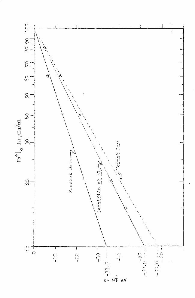
0 r--i +d
c c ..-
c 0
c
c '...0
c 't!\
0 ..l..
0 (\j
01 ..- 0 ,, c
CJ I
0 (Y)
I ';',
(') (fj
I
NU
\
c c: ,,, ~·
I I
u-i: AV
C>
('' ., 1.:-
1
c . ,~, I
c
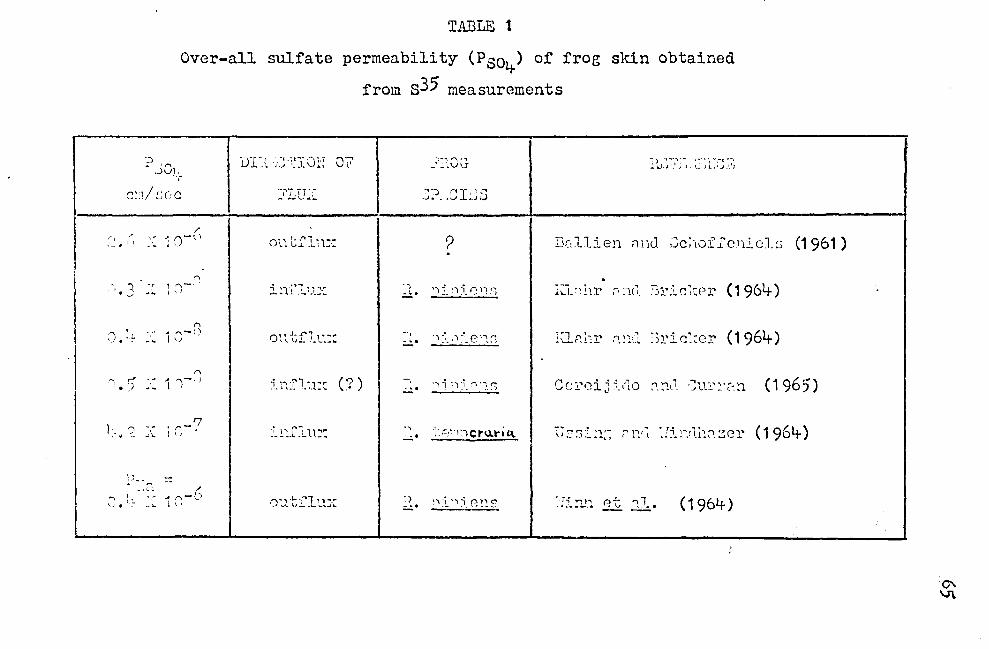
TABLE 1
Over-all sulfate permeability (P804-) of frog skin obtained
from s35 measurements
._, • _;01.~
C··1/c-c··c 1 ••• _) •
r, / '" • \ l
I' ·; ::i-\)
.. _: ~L ,. '\ L• •,• A ,....,-t) \.,. • ,- -·lo. I \I
jjr_\ ,c·:~ICJ~·: Q7
-,.- n·.... ' L ........... ~
on tfln:·:
i!1:L'lu:::
011 tfT.t:::
i '"'·"1 "' (')) --~ .. ~- _ll_\. ,•
~~~:..Cl11.~:
0 :.1 ·!: ]~ l tl):
---.- .... r\ ,.., _•~·"vu
~:)~ .CIL·~S
·?
~ 1. 'l j_ '"\ ~i_ 0. l'"1;;
.. ~. ")-; .1-~ t:'"·"'\ I"" - -- --'-' ..,.,)
-=--:. ''\l "·)~1_~."l~
-~ ··- ::-•·1--)ctu.l"i"' .:....:.• \. '
~:. ~':~ "1 5. R"::S
l{L~~T~~. ~~-~·1::-~.~
D:~llien nm1 ,Jc;·toi'.fcnj_0l~ (1961)
-:-- ...,;, ~. , -;->-.-; (''- (196' ) ''-'-"-·r .... 1c.• .. JJ._.,11.Pr y.
lCLr:!-::r ;-i_~:·.~ Jric:;:e:r (1964-)
0vr·~·.--,1· ~~t't·o ...,-..,,.'1 ,-.1..1'""·'1 (1965) ...__, - -~ t} - - ~ ... •"- . .J -~ - t - ...
-- . - .,. ,, (19,...'·) i..J:Sl.~1~~ r'l'"'..(1.. .::1_~"".ru::m.ZCl' o..,-
~Ti:l.!1 et :1 .• (1964-)
°' V\
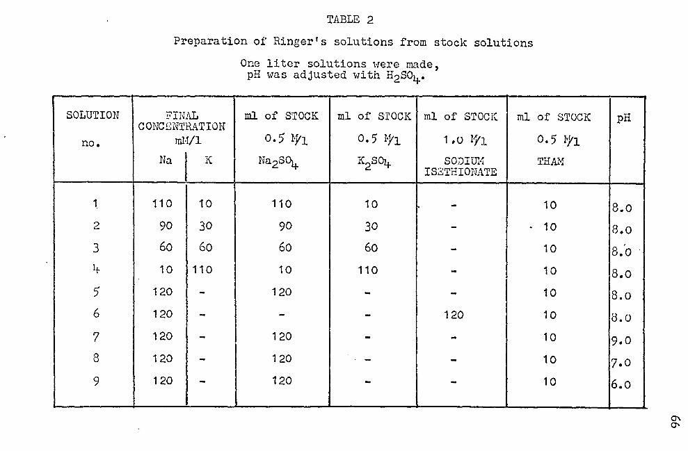
SOLUTION
no.
1.
2
3
1+
5
6
7
8
9
TABLE 2
Preparation of Ringer's solutions from stock solutions
One liter solutions were made, pH was adjusted with H2S04.
FINAL ml of STOCK ml of S'rOCK ml of STOCK ml of STOCK CONCENTRATION
ml.Vl 0.5 W1 O. 5 I·y1 1,0 ?1'1 0.5 W1 Na K Na2SC\ K2S04 SODiill·f THAM
ISZTHIONATE
110 10 110 10 - 10
90 30 90 30 - . 10
60 60 60 60 - 10
10 110 10 110 - 10
120 - 120 - - 10
120 - - - 120 10
120 - 120 - - 10
120 - 120 . - - 10
120 - 120 - - 10
pH
8.o 8.o B.o ·
8.o 8.o
8.o
9.0
7.0
6.o
°' "'
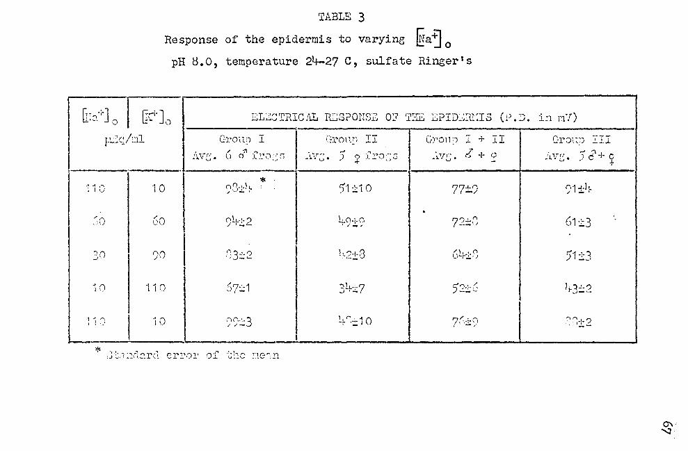
r: T '"\ ll•-.. ..
'11
/
··'
'.) --'
1
-; 1
TABLE 3
Response of the epidermis to varying ~a~ 0 pH 8.o, temperature 21+-27 c, sulfate Ringer's
+] 0 I U;"· 1, s1;.:.:cTRIC:U. TI.2SPONS:12 O::? TI-ill J2:i?ID;;~RlIIS ( ,) D . . . p..:~cj::il Grolln I Gl'OH_!! ,.-
~.L Gron~) I + II
~·1.vc. /' 11 ..... .;.\ \r;;. 5 9 fro:: s ;.~11c • l + c U o l. l'O '.~ S ... --·r -·-* .
0 10 ()1-1-h. 51 :t1 0 77±.9 ,,,c. - .
. 0 ·SO 91-1.-:-?
·-~ LCi+O
I .1- / 72±8
() ')0 ,'i3:t2 !14S·t-8 . •--- 6~r:!:·S
() "110 (, '7·'· 1 "-# .......
':'tL-:-7 ,J • -
5')+< ~-~
()
I 10 (")(\.:.3 ~t- 0 :!: 1 O ?{.+Cf //- I -/
I
* "-:- 1 · 1 ·1 .., r ," "',., ~"'"' 1 • o ·:" ,..) v· .... \1 . .__ .. 1..._I_ .._, __ ._,.,.. _._ -~·h"' V.-V :·.~e~,n
in m7)
Gron!) ~II
/i.V.'.3 • .-' '\ Jc+~
n1+h. / - .
61:1:3
51±3
41-'-") __,-~
~,l""'I ? .. - •:t .__
°' "'.:l
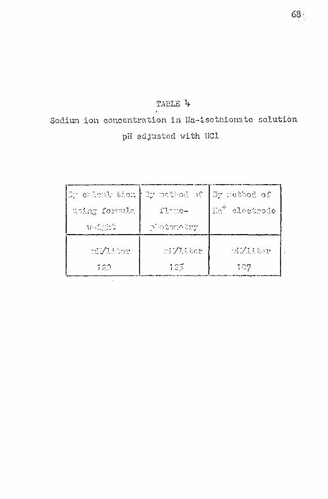
68·
TADLE 4-
Sodium ion concentration in Hu--isGtt1ionatc solution
pH adj~stcd with Irol
C ,. 1 ,~, . 1 , . -'- ~ '"' . ~ I -., . ~ .- - ...... 'J___ L,_·. ,/.;.._ --"J
.. "".") I c'..·.
.. ,• -;1 _, ·- i' - _! - . --'- !~~--
~- ~ i --; -, i L - .. , ..... _ .!_ ... 1,.. r_ ... .!..
--------------·-------~~~---~
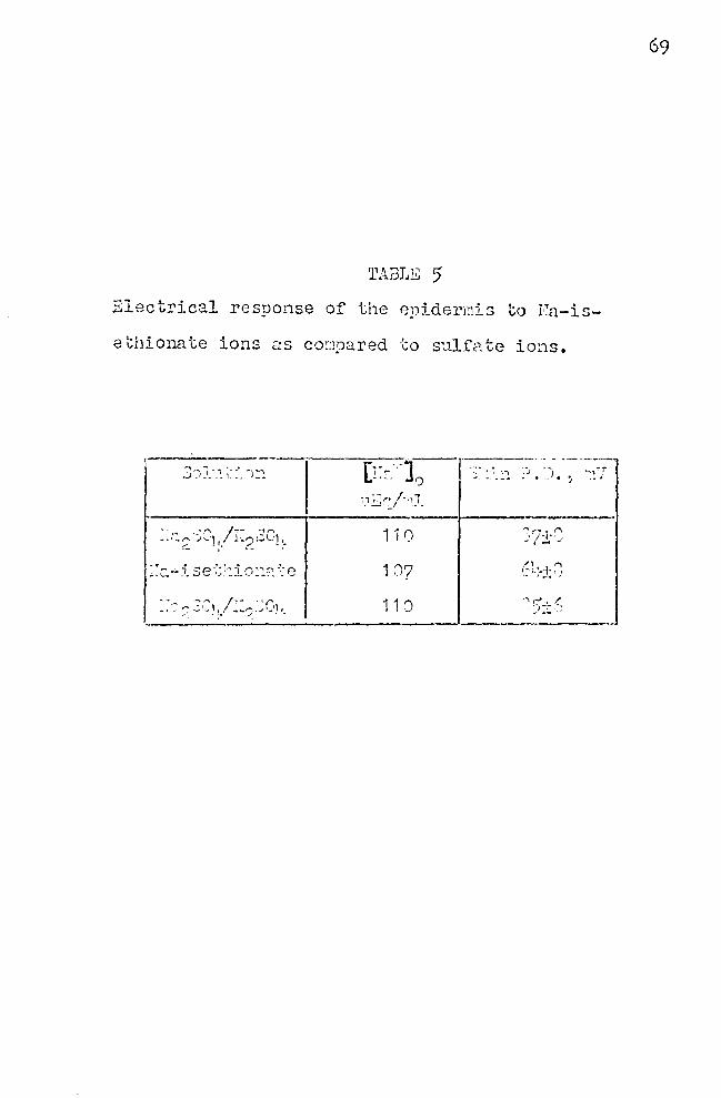
TABLE 5 Electrical response of the 8})idermis to Fa-is-
ethiona te ions as co::1pared to s',...1.lf<1.. te ions.
107
11 0
(L+,; ·- ..... "
'""\ )-'-< ,,., .:. ,
69
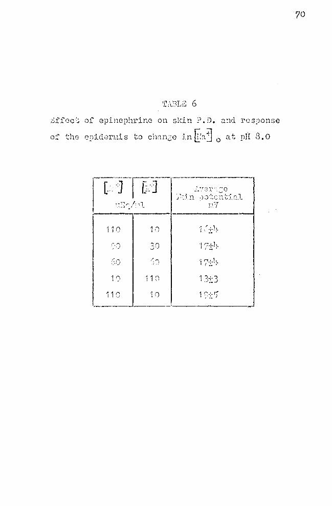
Gffec·:; of epincphrirn; on sldn ~). D. ::;.nd rcs9onse
of the epidermis to chnncc in~~~ 0 at prt 8.o
1 10 1 t) .• /+h I \ ... I
~'-.) 30 I '7+1•. I - '
:So ·'"' •j r:.: .. lt. ,., I 1-'
1 0 1 1 0 1 }'.:3 .. 1 ("\ ·1 0 1 :~~ S' I ,_J
-----
70
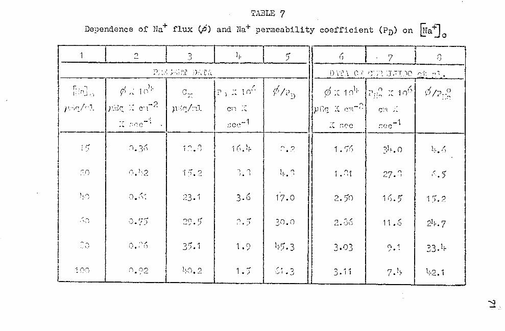
TABLE 7
Dependence of Na+ flux (;6) and Na+ permeability coefficient (Pn) on fu"a +J 0
I 1 I 2 I .3 I i, I :> J 6 I 7 I n
I D"····· .. ~,.,,, ·,),.,:i.-, I ·1.)··1· ,y, ,·····1 ·J·,-·r·'') ··I· 1 • -' .. I , J , . 1 .L .1
• \. L ( ... . ~ L · ~ \. j 1 J , ~ 1 __ , , • ~ _' \. r. , ri .• ~~~~-.-~~~~~-
! r;-.,J-1 ' c: . 1 (111. (' -:1. ·r 1 (/'· rt; /"J l d 'T 1 ()l;. .,, __ n 'r I n0 I 1);:J 0 l: . ' . I ) y ·'.. . ') ":~ • ) • .. I ' I. J I I , I.. ' I"\ ~ J : ~ , .. , • IT:~
I }' ·~r /nl )1-·~c · - Cl:l-<. p~•:0 /•·il c1·1 ·: p Fn ·-;: r.111-· - r.i>1
l . ~ -- _, -~ -·.. -~ . ~ . .. . . ~ I , . -r, . J. . ~·-
... :-;r:c ' ::ce 1 l .. ~ :~i:·c :::oc l
l 1
l
i '.-'
~I)
l !· '}
,\,'.)
~"' :~1 \_J
·100
".' 3 / ' I• . \)
r, ),_") . . . '--
,, .. . J. \)I
1). '7!) . -n "." .. " ')
n n') . , ·- l
1 "' I"\ .. ')
'l ;' 'I . . . .::._
"'3 .. ,: • I
rin r-' •-/ . . )
35 .1
L;.CJ. 2
J I. -·•--t-. -1
I 1 r;. l;. :-i • ? j J 1 • '7;; I 3l 1-. o I l+ •• ~
') I"\ I • I
3.6
') '"' .:._. )
1 • 9
1 • )
lr-. ~
17. 0
30.0
h.). 3 '.
/A ') •,Ji•_;
I
1 .. r;1
2. 50
...., ,..) / '--• '..Jl~
3.03
3. I 1
')7 ('\ <- • '
,, / ~ I i) • .J
11 • 6
(") -1 / . ' 7 .lt.
l
I (.)
1).2
21;.. 7
., 3 l· ..J • r
1·~2. 1
.....:i _..
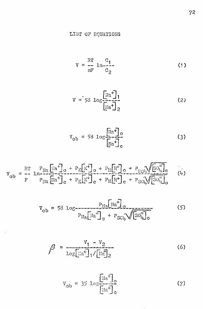
RT
LIS'r OF EQUATIONS
R'r v =
nF
. v = 58
Pr-raEra+]o + Prc[K~o + PnUt+]o + PE:o1)/'@04J~ ln-------·------------------___ ------------·-·-_ Pua §a+] c + PrcGc1J c + PHQ-rj c + Pso·~.f@0~o
V1 - V2 = ----------~----~
log Gra-i] 1 IQr~ 2
r:ra·1J 35. 1 L: - 0 = og------.
ffra~J c
72
( 1)
(2)
(3)
(l+)
(5)
(6)
(7)
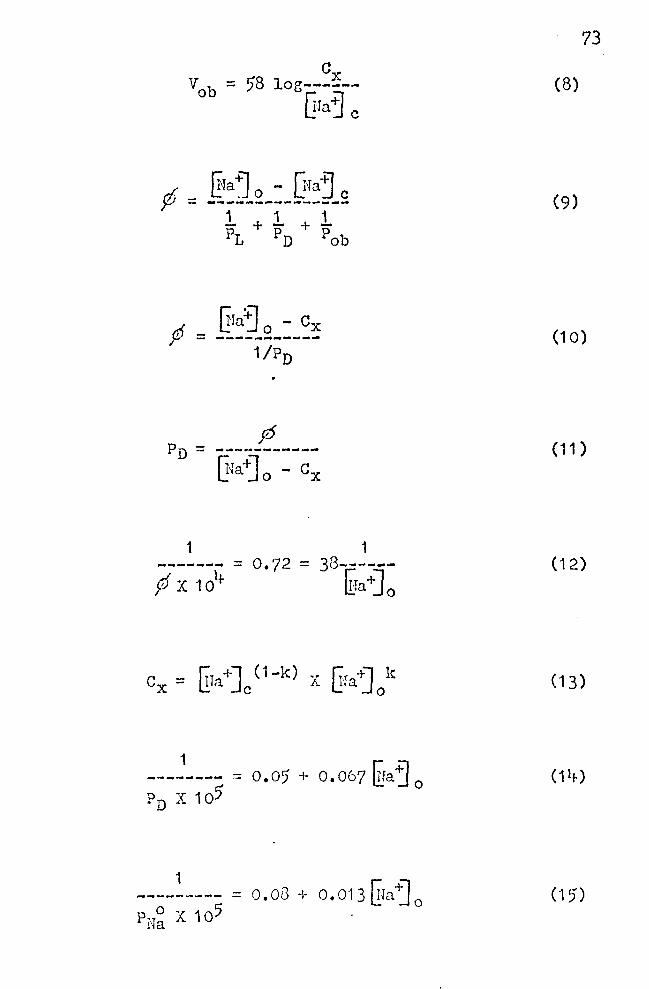
d: _ ~a~o - G~ajc ~ - ---~---~-------
1. + 1 + 1 PL PD pob
~ru=j 0 - Cx c;J - _____ ,,.. ____ _ I
1
p x 104-
1 0.72 = 38------
llra+Jo
---~---- = o. or; + o. 067 llia ~ 0 PD X 105'
1
73
(8)
(9)
(10)
(11)
(12)
(13)
(14)
C-15)
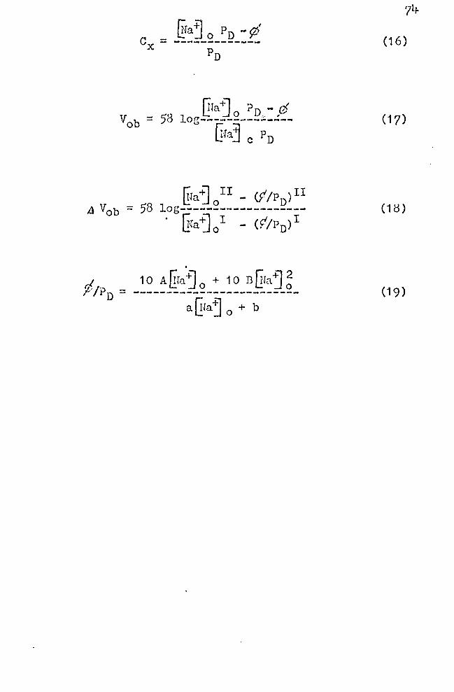
(16)
(17)
(18)
(19)
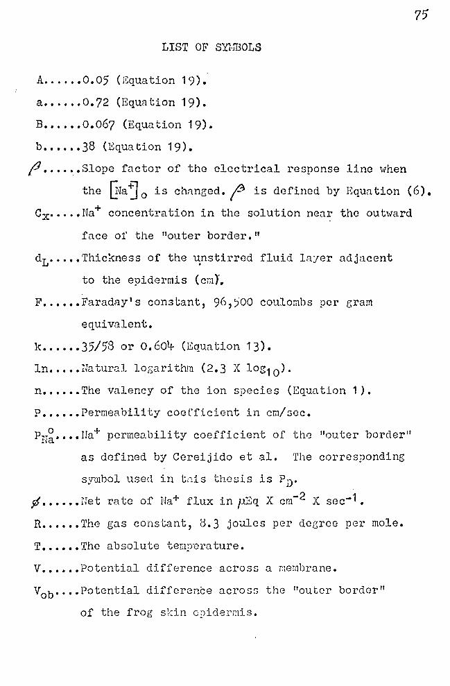
LIST OF SYlffiOLS
A .....• 0.05 (Equation 19).
a •••••• 0.72 (Equation 19).
B •••••• 0.067 (Equation 19).
b •••••• 38 (Equation 19).
fl ....... Slope factor of the electrical response line when
7'5
the ~ra ~ 0 is chrmc;ed. (3 is defined by Equn ti on ( 6). + Cx·····Na concentration in the solution near the outward
face of the "outer border."
dt·····Thickness of the ~nstirred fluid layer adjacent
to the epidermis (cmJ.
F •••••• Faraday's constant, 96,~00 coulombs per gram
equivalent,
k •••••• 35/58 or o.604 (Eauation 13).
ln ••••• Natural logarithm (2. 3 X log1 0 ).
n ••.••• The valency of the ion species (Equation 1 ).
P ••.•.• Permeability coefficient in cm/sec.
PN~ •••• rra+ permeability coefficient of the "outer border"
as defined by Ccreijido et al. The corresponding
symbol used in t11is thesis is PD.
1f •••••• Het rate of Ha+ flux in pZq X cm- 2 X sec-1.
R ••.••• The gas constant, 0,3 joules per deeree per mole.
T •••••• The absolute temperature.
V •••••• Potential difference across a membrane.
Vob••••Potential difference across the "outer border"
of the frog skin epidermis.
t:.Vob•••Chnnge in potential difference across the "outer
border" when the so.dium concentration is changed
from one sodium concentr<ition to another, Gfaj I to @aj II.
Subscripts
c ••••• Pertaining to tlle intracel lnlar fluid.
D ••••• Pertaining to the outer layers of the epidermis.
L ••••• Perta.ining to the unstirred fluid layer adjacent
to tne epidermis.
o ••••• Pertaining to the solution on the epidermal side
of the skin.
ob •••• Pertainine to the "outer border."
'I'/
VITA
James Henry Nartin, III was born March 31, 191+3 in
New Orleans, Louisiana. His primary education wns divided
between Hew Orleans and Hj_ndsor, NeH York. In 19)6 he moved
to Waynesboro, Virginia where he entered ~faynesboro High
School. He participated in an accelerated proGrnm for
college bound science students. He was one of the twelve
high school students selected to participate in the first
Science Worlrnhop sponsored be E. I. DuPont in Haynesboro.
In June, 1 961 he 1-ras gradua tcd from Waynesboro High School,
and in September he entered the University of Virginia.
While studying at the University, he became active in the
Westminster ?cllowship and was elected treasurer and later
Vice-President. He was.selected House }~na~er for West
Hj_nster House, a Presbyterian owned ho11se for college
students in Charlottesville. He received the B. A. degree
in biology in June, 1965. In September, 1965 ho entered
the Uni versi t:r of Richmond. 1':!hi le at the University of
Richmond, he was elected to Beta Beta Beta Honorary Biolo
gical Society, and he was awarded a Williams ?ellowship.
He received the degree of' Master of Science in August, 1967.
Nr. Hartin was a~:rarded a tcachint; assistantship at the
University of Tennessee 1·rhere he will continue his t:;radua te
studies toward a Doctor of Philosophy Degree in September,
1967.