el-gy 6813 / be-gy 6203 / g16.4426 medical...
TRANSCRIPT

Physics of Radiography
Jonathan Mamou and Yao WangPolytechnic School of Engineering
New York University, Brooklyn, NY 11201
Based on Prince and Links, Medical Imaging Signals and Systems and Lecture Notes by Prince. Figures are from the book.
EL-GY 6813 / BE-GY 6203 / G16.4426Medical Imaging

Radiation Physics Yao Wang, NYU 2
Lecture Outline• Atomic structure and ionization• Particulate Radiation
– Focusing on energetic electron interaction
• EM Radiation– Photoelectric – Compton scattering
-> Likelihood of each phenomenon– EM radiation measurement– Attenuation of radiation
• Radiation Dosimetry– Exposure, dose

Radiation Physics Yao Wang, NYU 3
Atomic Structure• An atom={a nucleus, electrons}• Nucleus composed of nucleons =
{protons; neutrons}• mass number A = # nucleons• atomic number Z = # protons = #
electrons– Define an element with a
particular symbol: H, C, etc.– An element is denoted by its A
and Z
– Ex: 12-Cor 126 C

Stable vs. Unstable States• Stable nuclides:
– # neutrons ~= # protons (A ~= 2Z)
• Unstable nuclides (radionuclides, radioactive atoms)– Likely to undergo radioactive decay, which gives off energy and
results in a more stable nucleus
Radiation Physics Yao Wang, NYU 4

Radiation Physics Yao Wang, NYU 5
Orbits of Electrons
Ground state: electrons are in the lowest orbital shells and within the lowest energy quantum states within each shell

Radiation Physics Yao Wang, NYU 6
Electron Binding Energy• A free electron has higher energy than when it is bounded to an
nucleus in an atom• Binding energy = energy required to free electrons from their atomic
orbits– Depends on the element to which the electron is bound and the shell
within which it resides in ground state– Sufficient to consider “average” binding energy of a given atom
• One electron volt (eV) = kinetic energy gained by an electron when accelerated across one volt potential– 1 eV = 1.6 x 10⁻¹⁹ Joule
• Binding energies of typical elements:– hydrogen = 13.6 eV– Air: 29 eV– Lead: 1 KeV– Tungsten: 4 KeV (considered a “heavy” element)
Electronvolt (eV) is a unit of energy equal ~1.602×10⁻¹⁹ J. It is the amount of energy gained (or lost) by the charge of a single electron moving across a 1-V electric potential difference -> 1 volt (= 1 J/C) multiplied by the elementary charge (e, or ~1.602×10⁻¹⁹ C). 1 eV = 1.602×10⁻¹⁹ J.

Radiation Physics Yao Wang, NYU 7
Ionization and Excitation• Ionization is “knocking” an electron out of an atom
– Creates a free electron + ion (an atom with +1 charge)– Occurs when radiated with energy above the electron binding
energy
• Excitation is “knocking” an electron to a higher orbit– When the radiation energy is lower than the binding energy
• After either ionization or excitation, an atom has higher energy

Radiation Physics Yao Wang, NYU 8
Characteristic Radiation• What happens to ionized or excited atom?
– Return to ground state by rearrangement of electrons– Causes atom to give off energy– Energy given off as characteristic radiation
• infrared• light• x-ray

Radiation Physics Yao Wang, NYU 9
Example• Consider an electron accelerated through an X-ray tube where the anode is
made of tungsten. If the anode is held at 120 KV, what is the maximum number of tungsten atoms that can be ionized?
• Solution:– The electron will have 120 KeV kinetic energy when reaching the anode, by
definition of eV– The average binding energy of tungsten = 4 KeV– # ionized atoms = 120/4=30

Radiation Physics Yao Wang, NYU 10
Ionizing Radiation

Radiation Physics Yao Wang, NYU 11
Two Types of Ionizing Radiation• Particulate• Electro-magnetic (EM)

Radiation Physics Yao Wang, NYU 12
Particulate Radiation• Radiation by any particle (proton, neutron or electron) if it
possesses enough kinetic energy to ionize an atom
202
2
22
200
2
22
21,
211
1
1, When
)(: EnergyKinetic :mass Energy vs.
1 :particle movinga of Mass
motion todue gainedenergy the Energy Kinetic
0
vmKEcv
cv
cv
cmmEEKEmcE
cv
mm
=+≈−
<<
−=−=
=
−=
=

Radiation Physics Yao Wang, NYU 13
Particulate Radiation by Energetic Electrons
• We are only concerned with an electron accelerated in a X-ray tube here– An electron accelerated across a tube with 100 KV potential
possesses 100 KeV kinetic energy

Radiation Physics Yao Wang, NYU 14
Energetic Electron Interactions
• Two primary interactions– Collisional transfer
• Most common• Produces heat
– Radiative transfer• Produces x-ray• Characteristic radiation
– Collide with K-shell• Bremsstrahlung radiation(“Braking or deceleration radiation”)
– Collide with nucleus– More common than
characteristic radiation

Radiation Physics Yao Wang, NYU 15
Collisional Transfer• The energetic electron collides with an atom in the target• Typically, a small fraction of the kinetic energy of the electron is transferred
to another electron in the atom– As the affected atom returns to its original state, infrared radiation (heat) is
generated • Occasionally, a large fraction of the incident energy is transferred to another
electron, the newly freed electron may form a delta ray• The incident electron’s path may be redirected, and many other
subsequent interactions may occur, until the kinetic energy of the incident electron is exhausted

Radiation Physics Yao Wang, NYU 16
Characteristic X-Ray• The incident electron collides with a K-shell electron,
exciting or ionizing the atom, leaving a hole in that shell.– As the atom returns to its ground state, the k-shell hole is filled
by a higher shell electron– The loss of energy creates an EM photon, known as
Characteristic x-ray– The energy of the x-ray photon = difference between the energy
of the two shells (element dependent)

Radiation Physics Yao Wang, NYU 17
Characteristic radiation• Caused by removal of inner shell electrons and subsequent filling of hole with
electrons from higher shell. The shell-energy difference determines the energy of characteristic rays
• Lines are named after the lower shell involved in the process; the upper shell involved is denoted by Greek letters:
-
--
---
- -
--
-
hν
KLM
-
Continuum0
K
L
MN
E [keV]
K-lines
L-lines
α β γ
α β
Kα
Kβ
Kγ
0.53
11
70
[From Graber, Lecture Note for BMI1-FS05]

Radiation Physics Yao Wang, NYU 18
Different types of characteristics rays
From http://hyperphysics.phy-astr.gsu.edu/Hbase/quantum/xterm.html#c1

Radiation Physics Yao Wang, NYU 19
Bremsstrahlung Ray• As the incident electron approaches the nucleus of an atom, the
positive charge of the nucleus causes the incident electron to bend around the nucleus and decelerates– The loss of energy leads to the Bremsstrahlung x-ray (energy vary over
a continuous range, depending on the speed loss) • Occasionally when the incident electron collides with the nucleus,
the electron is annihilated, emitting a photon with an energy equal to the kinetic energy of the incident electron (highest possible energy)
• Primary source of x-rays from an x-ray tube

Radiation Physics Yao Wang, NYU 20
Spectrum of X-Ray
When the incident electron collides with a nucleus
Generated when K-shell electrons are replaced by different outer shells
Different curves are generated when different voltage potentials applied in a x-ray tube

Radiation Physics Yao Wang, NYU 21
EM Radiation• EM radiation comprises an electric wave and a magnetic
wave traveling at right angles to each other• Typical EM waves:
– Non ionizing: radio, microwaves, infrared, visible light, ultraviolet– Ionizing: X-rays, gamma rays
• Energy of a photon of an EM wave with frequency v:

Radiation Physics Yao Wang, NYU 22
EM Waves for Medical Imaging• X-rays and Gamma rays:
– Have energy in the KeVs to MeVs -> Ionizing Radiation – used in X-ray/CT and nuclear medicine respectively– X-rays are created by interaction of energetic electrons with
atoms– Gamma rays are created in the nuclei of atoms due to
radioactive decay or characteristic radiation, typically have higher energy than X-rays
• Radio waves– Used to stimulate nuclei in MRI to generate EM radiation
• Visible light– Used in radiography to improve the efficiency of photographic
film to detect X-rays
• See Table 4.2 for more details

Radiation Physics Yao Wang, NYU 23
EM Radiation Interactions• Two main interactions
– Photoelectric effect• The incoming photon is completely absorbed and ejecting K-shell or
L-shell electrons, producing characteristic x-ray– Compton scattering
• The incoming photon changes its direction

Radiation Physics Yao Wang, NYU 24
Photoelectric Effect• An incoming photon interacts with the nucleus of an atom, causing
ejection of a K-shell or L-shell electron (photoelectron)– Atom completely absorbs incident photon and all energy is transferred – The photoelectron propagates away with energy
– The affected atom produces characteristic x-ray, while outer electrons fill the K-shell.
– Sometimes the characteristic x-ray transfers its energy to an outer electron (called Auger electron)
• Both photo electron and Auger electron are energetic electrons that can interact with the matter as discussed before
Be EhvE −=−

Radiation Physics Yao Wang, NYU 25
Photoelectric Effect

Radiation Physics Yao Wang, NYU 26
Compton Scattering• An incoming photon ejects an outer shell electron,
yielding a Compton electron• The incident photon loses its energy and changes its
direction (Not completely absorbed by the atom!)• The scattered photon is called Compton photon

Radiation Physics Yao Wang, NYU 27
• The energy of the scattered photon depends on the scatter angle
– The maximum energy loss occurs when the photon is deflected backward (θ = 180o), backscattering
– When E is higher, more photons scatter forward– The kinetic energy of the Compton electron = E-E’

Radiation Physics Yao Wang, NYU 28
Which interaction is better?• Photoelectric effect helps to differentiate different human
tissues/organs• Compton scattering causes incident photons to deviate
from straight path, and causes unnecessary exposure of x-ray to untargeted areas
• In medical imaging, we want to increase the likelihood of photoelectric events, while minimizing Compton scattering

Radiation Physics Yao Wang, NYU 29
Probability of Photoelectric Effect• Recall that a photoelectric event happens when incident photons
interact with the Coulomb field of the nucleus of an atom• More likely when colliding with an atom with more protons (i.e.,
larger Z number)• Less likely when incident photons have higher energy (higher
frequency)
– The probability increases abruptly when the photon energy rises above the binding energy of L-shell or K-shell electrons (so as to eject the electrons), then begins to diminish
– Rationale behind the use of “contrast agent”– Z numbers: soft tissue: 7.4 soft, bone: 13.8– More contrast with low energy x-ray– Photoelectric effect dominate over Compton scattering at low energy

Radiation Physics Yao Wang, NYU 30
Probability of Compton Scattering• Recall that Compton scattering occurs when an incident photon
collides with outer shell electrons• Likelihood proportional to the number of electrons per kilogram of
the material (the electron density or ED)• Relatively independent of incident photon energy in biological
materials
• ED is approximately constant for various biological material, ~ 3*1026, except for hydrogen (6*1026)
• Does not provide tissue contrast
e)(grams/molweight molecular : /atom)(electronsnumber atomic:
e)(atoms/molnumber sAvogadro' :
m
A
m
A
WZN
WZNED =

Radiation Physics Yao Wang, NYU 31
Relative Likelihood• Compton scattering is equally likely in various materials
and invariant of incident energy• Photoelectric effect is more likely in high Z material and
less likely with high incident energy• Overall, Compton scattering is more dominant with
higher incident energy in the same material• But the percent of energy deposited due to photoelectric
event is larger because all incident energy is absorbed.

Radiation Physics Yao Wang, NYU 32
Measures of X-ray Beam: Photon Count
• N: number of photons• A: area

Radiation Physics Yao Wang, NYU 33
Measures of X-ray Beam: Energy Flow

Radiation Physics Yao Wang, NYU 34
Spectrum of X-Ray• The x-ray beam produced by an x-ray tube (mainly
Bremsstrahlung) is polyenergetic (consisting photons with different energies or frequencies)
• X-ray spectrum S(E): – The number of photons with energy E per unit area per unit time

Radiation Physics Yao Wang, NYU 35
Spectrum of X-Ray
When the incident electron collides with a nucleus
Generated when K-shell electrons are replaced by different outer shells
Different curves are generated when different voltage potentials applied in a x-ray tube

Radiation Physics Yao Wang, NYU 36
Attenuation of X-ray Radiation: Homogeneous Slab
Photons will be absorbed/deflected through the slabLet # of photons at x = N(x)# photons lost from position x to x+dx can be approximated bydN=N(x+dx)-N(x) = - μ N(x)dx, when dx is very smalllinear attenuation coefficient: μμ is the fraction of photons that are lost per unit length
The above can be rewritten as dN(x)/dx = - μ N(x). Integrate this from x=0 to ∆x yieldsN(x) = N0 exp{-μ x} The fundamental photon attenuation law
N0

Radiation Physics Yao Wang, NYU 37
Linear Attenuation Coefficients of Biological Tissues

Radiation Physics Yao Wang, NYU 38
Homogeneous Slab• Homogeneous slab: the attenuation rate is the same
over the entire slab
Radiation Physics Yao Wang, NYU 39
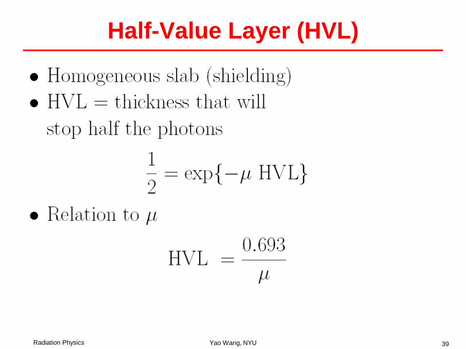
Half-Value Layer (HVL)

Radiation Physics Yao Wang, NYU 40
Example• Consider the image taken of a bar phantom uniformly
irradiated by monoenergetic x-ray photons– Assuming the bars are made of material that has a HVL of
0.2cm, and bars have thickness of 0.4 cm– Assuming x-ray photons pass through the space between bars
w/o attenuation– Assuming the intensity of the image is proportional to the
number of detected photons in a unit area– What is the contrast of the resulting image?
• Go through in the class
Radiation Physics Yao Wang, NYU 41

Radiation Physics Yao Wang, NYU 42
Non-Homogeneous Slab• The attenuation coefficient depends on x

Radiation Physics Yao Wang, NYU 43
Example: Two Layer Slab• A slab with two homogeneous layers, with thickness d1,
d2, and attenuation coefficients μ1, μ2. If the input X-ray intensity is N0, what is the intensity at the other end of the slab?

Radiation Physics Yao Wang, NYU 44
Polyenergetic Photons• The linear attenuation coefficient depends on the medium property as
well as the energy of the incident photon (E)• For a given material, μ can be denoted by μ(x;E)• When the incident photons are polyenergetic, with spectrum S(E), the
outgoing photon spectrum is
• In terms of intensity
{ }∫−=x
dxExESExS00 ');'(exp)();( µ
{ }∫ ∫
∫∞
∞
−=
=
000
0
'')';'(exp')'()(
')'('
dEdxExEESxI
dEESEI
xµ

Radiation Physics Yao Wang, NYU 45
Radiation Dosimetry• Previous topics deal with the production of radiation and
measurement of radiation wave• Radiation dosimetry considers the effect of radiation to
the interacting media– Exposure– Dose – Kerma– Effective dose
Radiation Physics Yao Wang, NYU 46
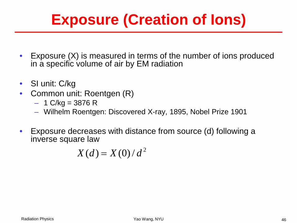
Exposure (Creation of Ions)
• Exposure (X) is measured in terms of the number of ions produced in a specific volume of air by EM radiation
• SI unit: C/kg• Common unit: Roentgen (R)
– 1 C/kg = 3876 R– Wilhelm Roentgen: Discovered X-ray, 1895, Nobel Prize 1901
• Exposure decreases with distance from source (d) following a inverse square law
2/)0()( dXdX =

Radiation Physics Yao Wang, NYU 47
Dose (the deposition of energy)

Radiation Physics Yao Wang, NYU 48
Kerma

Radiation Physics Yao Wang, NYU 49
Dose vs. Exposure
tissue-softfor 1airfor 87.0
tcoefficienn attenuatio mass :
87.0
:materialon dependsfactor -
≈=
=
=
ff
f
ffXD
air
material
ρµ
ρµρ
µ
See Table 4.6 for the mass attenuation coefficient of typical materials

Radiation Physics Yao Wang, NYU 50
Equivalent and Effective Dose• Dose equivalent
– The effect of radiation depends on the source of radiation (energy)– Dose equivalent: H = D Q– Q: quality factor
• Q = 1 for x-ray, gamma ray, electron, beta particle (used in medical imaging)• Q = 10 for neutrons and protons• Q = 20 for alpha particles
• Effective dose– Effect of a dose also depends on the tissue type– Effective dose measures the average effect over different tissue types
• D can be measured in rads, H can be measured in rems• For a dose of 1 Gy and Q =1 -> H = 1 sievert (Sv)
jw
HwD
j
organsjjeffective
organ for factor weighting:
∑=

Radiation Physics Yao Wang, NYU 51

Effective Dose of Different Tissues [Smith&Webb]
Radiation Physics Yao Wang, NYU 52

Radiation Physics Yao Wang, NYU 53
Summary • Ionization: ejection of an orbiting electron from an atom, the affected
atom produces radiation in the process of returning to ground state• Two types of ionizing radiation
– Particulate– EM
• Particulate radiation transfers energy via – Collisional transfer: resulting in heat– Radioactive transfer: resulting in characteristic x-ray and
Bremsstrahlung x-ray– X-ray is produced by energetic electrons accelerated in a x-ray tube,
consisting primarily Bremsstrahlung x-ray• EM radiation transfers energy via
– Photoelectric effect: incident photons completely absorbed– Compton scattering: incident photons are deflected– X-ray imaging utilizes EM photoelectric effect caused by injected X-ray
photons.

Radiation Physics Yao Wang, NYU 54
Summary (cntd)• Attenuation of EM radiation:
– Linear attenuation coefficient is the fraction of photons that are lost per unit length
• Depends on material property and the incident photon energy– Fundamental photon attenuation law
• Homogeneous slab• Heterogeneous slab
• Radiation dosimetry– Exposure vs. dose: D=fX– Equivalent dose: H=DQ– Effective dose:
jw
HwD
j
organsjjeffective
organ for factor weighting:
∑=

Radiation Physics Yao Wang, NYU 55
Reference• Prince and Links, Medical Imaging Signals and Systems,
Chap 4.

Radiation Physics Yao Wang, NYU 56
Homework• Reading:
– Prince and Links, Medical Imaging Signals and Systems, Chap 4.
• Will be assigned at the end of lecture 3 and due on Friday Sept 29