patterns of failure after stereotactic body radiation ... · gy, 100 gy, and 113 gy for 54 gy/3...
TRANSCRIPT
192 Journal of Thoracic Oncology • Volume 8, Number 2, February 2013
Introduction: The purpose of this study was to compare patterns of failure between lobar resection (lobectomy or pneumonectomy) and stereotactic body radiation therapy (SBRT) for patients with clinical stage I non–small-cell lung cancer (NSCLC).Methods: From January 2004 to January 2008, 338 patients under-went definitive treatment for pathologically confirmed clinical stage I NSCLC with lobar resection (n = 260) or SBRT (n = 78). Most surgical patients underwent lobectomy (n = 237). SBRT patients received a biologically effective dose of at least 100 Gy
10. Lobar
resection patients were younger, healthier, and had superior pulmo-nary function, whereas most of the patients in the SBRT group had T1 tumors. Final pathology upstaged 32.7% of surgery patients, and 20.0% received adjuvant chemotherapy. No SBRT patients received adjuvant chemotherapy.Results: In an unmatched comparison, 4-year lobar local control (98.7% versus 93.6%, p = 0.015) was greater for lobar resection versus SBRT, respectively, though primary tumor (98.7% versus 95.3%, p = 0.088), regional (82.9% versus 78.1%, p = 0.912), and distant control (76.1% versus 54.0%, p = 0.152) were similar. Overall survival (OS, 63.5% ver-sus 29.6%, p < 0.0001) was greater for lobar resection, though cause-specific survival (CSS, 81.3% versus 75.3%, p = 0.923) was similar. In a T-stage matched comparison of 152 patients, there was no significant difference in patterns of failure or CSS, whereas OS favored surgery.Conclusion: Lobectomy/pneumonectomy or SBRT results in com-parable patterns of failure for clinical stage I NSCLC. In this retro-spective comparison, OS was superior for surgery, though CSS was similar. Randomized trials are necessary to control for fundamen-tal differences in comorbidity, which impact interpretation of both tumor control and survival.
(J Thorac Oncol. 2013;8: 192–201)
For patients with medically operable clinical stage I non–small-cell lung cancer (NSCLC), lobectomy or pneu-
monectomy results in primary tumor control approaching 100% and 5-year overall survival of 53% and 43% for tumors 2 cm or more and 2.1 to 5 cm, respectively.1,2 Patients at higher risk of surgery may be offered sublobar resection with acceptable morbidity and mortality.3 Sublobar resec-tion with a nonanatomical wedge resection results in sig-nificantly higher local failure relative to lobar resection,4 though this may be improved through the use of brachyther-apy,5 careful intraoperative margin assessment,6 or anatomi-cal segmentectomy.7 For patients with medically inoperable stage I NSCLC, stereotactic body radiotherapy (SBRT) has become a standard of care based on high rates of local control, convenience, cost, and survival relative to historic outcomes after conventional radiotherapy, with low rates of toxicity.8–11 As experience with SBRT has grown, improved local control and survival after SBRT has been shown to correlate with biologically effective doses (BED) of radia-tion greater than 100 Gy
10.12,13
SBRT in medically operable patients was reported first from Japan, where high 3-year rates of local control (94%) and overall survival (86%) were documented in patients refusing surgery.14 No randomized trials comparing surgery and SBRT have been completed to date. The ROSEL (Radiosurgery Or Surgery for operable Early stage non-small cell Lung cancer) trial opened in Holland and attempted to compare lobectomy versus SBRT in stage I NSCLC, though it failed to reach its accrual goal and was closed. The STARS (Randomized Study to Compare CyberKnife Stereotactic Radiotherapy With Surgical Resection in Stage I Non-small Cell Lung Cancer) trial (sponsored by Accuray, Inc., Sunnyvale, CA) exclusively compares CyberKnife SBRT with lobectomy. American College of Surgeons Oncology Group (ACOSOG) Z4099 and Radiation Therapy Oncology Group (RTOG) 1021 is a National Cancer Institute trial, which randomizes patients at high risk for surgery to either sublobar resection (± brachy-therapy) or SBRT.
Retrospective comparisons between surgery and SBRT to date have been limited and suffer from inherent imbalances in the treatment arms, with healthier patients being selected for surgery.14–19 Two recent publications directly comparing surgery and SBRT have addressed this by narrowing the focus
Copyright © 2012 by the International Association for the Study of Lung CancerISSN: 1556-0864/12/0802-192
Patterns of Failure after Stereotactic Body Radiation Therapy or Lobar Resection for
Clinical Stage I Non–Small-Cell Lung Cancer
Cliff G. Robinson, MD,* Todd A. DeWees, PhD,* Issam M. El Naqa, PhD,† Kimberly M. Creach, MD,* Jeffrey R. Olsen, MD,* Traves D. Crabtree, MD,* Bryan F. Meyers, MD,* Varun Puri, MD,*
Jennifer M. Bell, BSN,* Parag J. Parikh, MD,* and Jeffrey D. Bradley, MD*
*Department of Radiation Oncology, Washington University School of Medicine, St. Louis, Missouri; and †Department of Oncology, McGill University Health Centre, Montreal, Quebec.
Portions of this article were presented at the 52nd annual ASTRO meeting in San Diego, CA (10/31-11/4/10)—Abstract #32.
Disclosure:The authors declare no conflict of interest.Address for correspondence: Cliff G. Robinson, MD, Department of
Radiation Oncology, Washington University School of Medicine, 4921 Parkview Place, Campus Box 8224, St. Louis, MO 3110. E-mail: [email protected]
ORIGINAL ARTICLE
193Copyright © 2012 by the International Association for the Study of Lung Cancer
Journal of Thoracic Oncology • Volume 8, Number 2, February 2013 SBRT or Lobar Resection for Stage I NSCLC
to only high-risk patients.16,17 Such comparisons have been criticized for having different definitions for failure between surgery and SBRT.20 No publications to date have compared outcomes specifically between lobar resection and SBRT. The purpose of this study is to compare institutional outcomes between definitive standard lobar resection and optimally dosed SBRT (BED > 100 Gy
10) using a uniform definition of
failure. Given the inherent difficulty in comparing survival for groups of patients selected for therapy based on medical comorbidity, we focus primarily on patterns of failure.
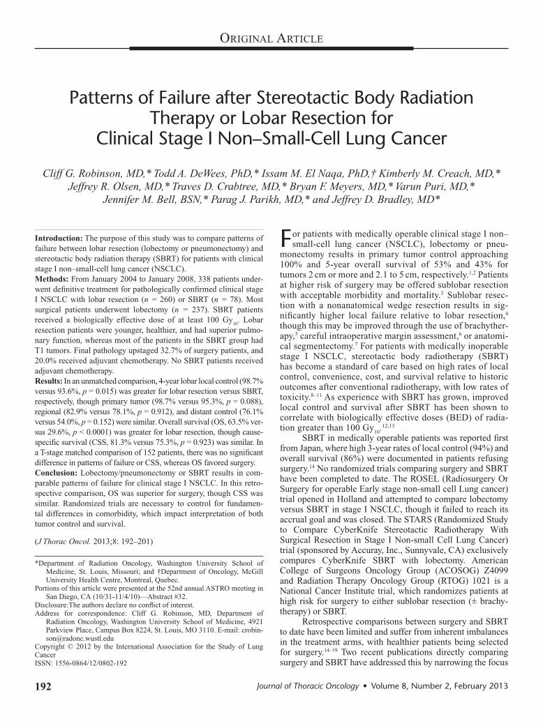
MATERIALS AND METHODSFrom January 2004 to January 2008, 454 patients (336
surgery, 118 SBRT) were identified with clinical stage I lung cancer from Institutional Review Board–approved patient registries located in the departments of Cardiothoracic Surgery and Radiation Oncology at Washington University in St. Louis, Missouri. Selection of patients with clinical stage I NSCLC was based on the American Joint Committee on Cancer (AJCC) 6th edition staging manual,21 and was ret-rospectively coded using the 7th edition for the study.22 The data set was narrowed to include only patients (1) undergoing anatomical lobar resection (lobectomy or pneumonectomy) or SBRT to a BED of 100 Gy
10 or more and (2) having patho-
logic confirmation of NSCLC. Details of inclusion or exclu-sion resulting in the final study group are described in the Figure 1.
Standard work-up for all patients included history and physical examination, computed tomography (CT) of the chest and abdomen, and [18F] fluorodeoxyglucose (FDG)–positron emission tomography (PET) CT. PET or CT was performed in 100% of surgery patients and 98.7% (n = 77) SBRT patients. Patients were selected for lobar resection if they were con-sidered by the thoracic surgeon to have adequate pulmonary
function to tolerate at least a lobectomy and absence of other contraindicating medical comorbidity. Patients were selected for SBRT if they were considered to be medically inoperable by the thoracic surgeon, or if they were felt to be at high risk for surgery, and declined sublobar resection after a balanced discussion with the surgeon and radiation oncologist.
Baseline patient, tumor, and treatment variables are sum-marized in Table 1. Medical comorbidity was assessed by the Adult CoMorbidity Evaluation (ACE-27) scoring system and Charlson Comorbidity Index (CCI), both of which have been validated in patients with lung cancer.23,24 The ACE-27 score was collected prospectively during a patient’s registration at the Siteman Cancer Center. The CCI and an age-adjusted CCI (AA-CCI) were scored retrospectively.25
For patients treated with lobar resection, the type of resection, type of incision, and extent of lymph node dis-section were at the discretion of the treating surgeon. Most patients underwent lobectomy, with the rest undergoing bilobectomy or pneumonectomy. Surgery was through open thoracotomy in 224 patients and video-assisted thoraco-scopic surgery in 36 patients. Institutional policy dictated that patients undergo mediastinoscopy before thoracotomy (either in advance of surgery, or at the time of surgery), fol-lowed by formal lymph node dissection at the time of lobec-tomy. Pathologic upstaging occurred in 85 patients (32.7%) undergoing surgery, of which tumor stage was higher in 51 cases (19.6%), and occult nodal disease was discovered in 56 cases (21.5%). For nodal stage, 43 patients were found to have pathologic N1 disease and 13 patients had N2 dis-ease. For tumor stage, 46 patients were upstaged from T1 (36 T2, 3 T3, and 7 T4) and five patients were upstaged from T2 (4 T3, 1 T4). In addition, 10 patients (3.8%) had downstag-ing from T2 to T1 tumors. Fifty-two patients (20.0%) treated with lobar resection received adjuvant chemotherapy, and 20 (7.7%) received adjuvant radiotherapy.
Details of SBRT planning and delivery at our institution have been described previously.26 Target coverage, conformal-ity, and normal tissue constraints were followed according to RTOG 0236.8 Prescriptions were typically specified at the 60% to 90% (median 84%) isodose line so that 95% or more of the prescribed dose covered the planning target volume. All SBRT patients received a BED of at least 100 Gy
10 (median dose, 54
Gy in 3 fractions). BED was calculated using BEDα/β = nd(1+ d/α/β), where n = number of fractions, d = dose per frac-tion, and α/β = 10 for tumor in line with previous reports.13,27 BED
10 for the three SBRT regimens used in this study was 151
Gy, 100 Gy, and 113 Gy for 54 Gy/3 fractions (fx), 50 Gy/5 fx, and 45 Gy/3 fx, respectively. No SBRT patients received adjuvant chemotherapy.
Patients were followed with serial chest radiographs and/or CT scans every 3 months for the first 2 years, every 6 months thereafter up to 5 years, and yearly afterward. A PET/CT was performed if there was suspicion for recurrence. Failures were defined using a combination of enlargement on CT and increasing [18F]FDG avidity on PET/CT, with biopsy confirmation when feasible. To eliminate historic discrepancies in definitions of failure between surgery and SBRT, tumor control was defined in the spirit of ACOSOG Z4099/RTOG 1021.28 Local failure was defined for SBRT as FIGURE 1. Details of inclusion and exclusion.
194 Copyright © 2012 by the International Association for the Study of Lung Cancer
Robinson et al. Journal of Thoracic Oncology • Volume 8, Number 2, February 2013
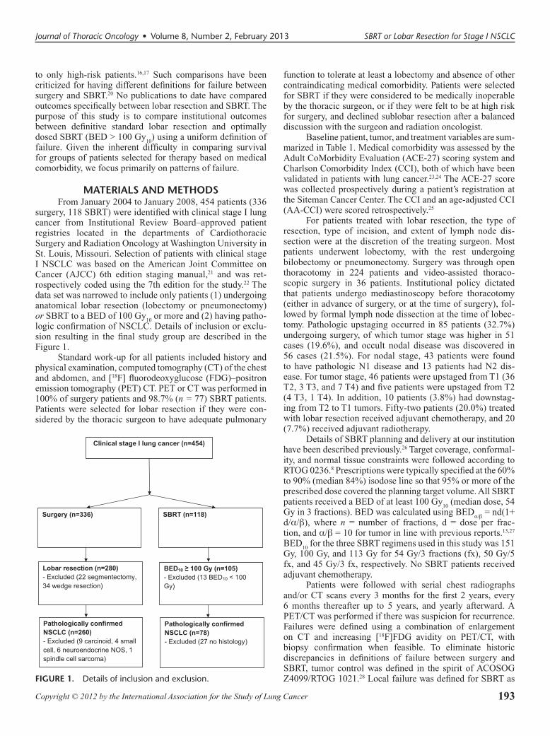
TABLE 1. Patient Characteristics, Unmatched Comparison between SBRT and Lobar Resection
Characteristic SBRT (n=78) Lobar Resection (n=260) p
Age, yrs Median (range) 76 (31–93) 66 (40–87) <0.0001Sex, n (%) Women 34 (43.6) 120 (46.2) 0.700Race, n (%) 0.530 White 68 (87.2) 234 (90.0) Black 10 (13.2) 21 (8.1) Others 0 (0) 5 (1.9)Smokers, n (%) 75 (96.2) 244 (93.9) 0.581 Prior NSCLC, n (%) 15 (19.2) 19 (7.3) 0.002ACE-27 Comorbidity Index Median (range) 2 (0–3) 1 (0–3) <0.0001Charlson Comorbidity Index Median (range) 4 (2–10) 3 (2–22) <0.0001Age-adjusted Charlson Comorbidity Index Median (range) 7 (3–12) 6 (2–28) 0.0048FEV1, L Median (range) 1.31 (0.38–2.89) 2.03 (0.82–5.58) <0.0001FEV1, % predicted Median (range) 51 (20–133) 80 (26–138) <0.0001FVC, L Median (range) 2.2 (0.69–3.47) 3.05 (1.42–6.92) <0.0001FVC, % predicted Median (range) 79 (33–129) 90 (38–184) 0.001DLCO, ml/min/mm Hg Median (range) 11 (6.28–33) 16 (1.04–40.07) 0.002DLCO, % predicted Median (range) 63 (23–114) 72 (12–190) 0.003Histology, % Adenocarcinoma 36 (46.2) 154 (59.2) 0.026 Squamous cell carcinoma 25 (32.1) 88 (33.9) NSCLC, not otherwise specified 16 (20.5) 8 (3.1) Other/unknown 1 (1.3) 10 (3.8)Maximal axial CT dimension, cm Median (range) 2 (1.1–6) 2.8 (0.7–12.8) 0.001AJCC 6th edition stage, n (%) T1 56 (71.8) 141 (54.2) 0.006 T2 22 (28.2) 119 (45.8)AJCC 7th edition stage, n (%) T1a 36 (46.2) 83 (31.9) 0.010 T1b 20 (25.6) 56 (21.5) T2a 19 (24.4) 83 (31.9) T2b 3 (3.8) 23 (8.9) T3 0 (0) 15 (5.8)F18-FDG SUVmax primary Median (range) 5 (0.9–18.5) 4.8 (1–25.2) 0.893SBRT dose, n (%) 54 Gy/3 fx 68 (87.2) N/A 50 Gy/5 fx 4 (5.1) 45 Gy/3 fx 6 (7.7)Surgery, n (%) Lobectomy 237 (91.2) N/A Bilobectomy 8 (3.1)
Pneumonectomy 15 (5.8)
Not all percentages add up to exactly 100%, secondary to rounding.NSCLC, non–small-cell lung cancer; FEV1, forced expiratory volume in 1 second; FVC, forced vital capacity; DLCO, diffusing capacity of the lung for carbon monoxide;
CT, computed tomography; AJCC, American Joint Committee on Cancer; ACE, Adult CoMorbidity Evaluation; F18-FDG, fluorodeoxyglucose; SUV, standard uptake value; SBRT, stereotactic body radiation therapy; fx, fraction.
195Copyright © 2012 by the International Association for the Study of Lung Cancer
Journal of Thoracic Oncology • Volume 8, Number 2, February 2013 SBRT or Lobar Resection for Stage I NSCLC
progression in the region of the primary tumor or the involved lobe, and for surgery as failure at the bronchial stump or port site/wound. Primary tumor failure was defined for SBRT as progression in the region of the primary tumor alone. Regional failure was defined as failure in hilar or mediastinal lymph nodes, and distant metastases were defined as failures outside of local or regional failures. Toxicity was coded using Common Terminology Criteria for Adverse Events version 4 for SBRT patients, and toxicities were described but not graded for lobar resection.
Median follow-up was calculated using the Kaplan–Meier estimated potential follow-up method.29 Differences between treatment groups were described using a two-tailed t test for means, Wilcoxon rank sum for medians, and χ2 test for proportions. Univariate analysis of factors was performed using Cox regression for continuous variables and log-rank for categorical variables. Assumptions of the Cox model were checked. Multivariate analysis was performed using the Cox proportional hazards model. SBRT and lobar resection were compared with regard to local control (LC), primary tumor control (PC), regional control (RC), distant control (DC), overall survival (OS), and cancer-specific survival (CSS), using the Kaplan–Meier method. For representative time points, standard errors were reported. An attempt was made to match subgroups of patients treated with lobar resection or SBRT on select covariates, using a propensity-score matching (PSM) technique. For PSM, logistic regression is first used to estimate propensity scores from baseline patient covariates, after which patients are matched on their propensity scores. Matching was accomplished using a caliper method, whereby treatment groups are randomly sorted, and patients were ran-domly matched within an acceptable distance (caliper) of pro-pensity scores. We previously identified a caliper parameter of 0.005 as providing an acceptable balance between strin-gency (i.e., closer matches) and patient numbers.17 For the PSM-matched patients, stratified log-rank tests were used for Kaplan–Meier comparisons.
RESULTSMedian follow-up was 51.3 months for surgery and 50.3
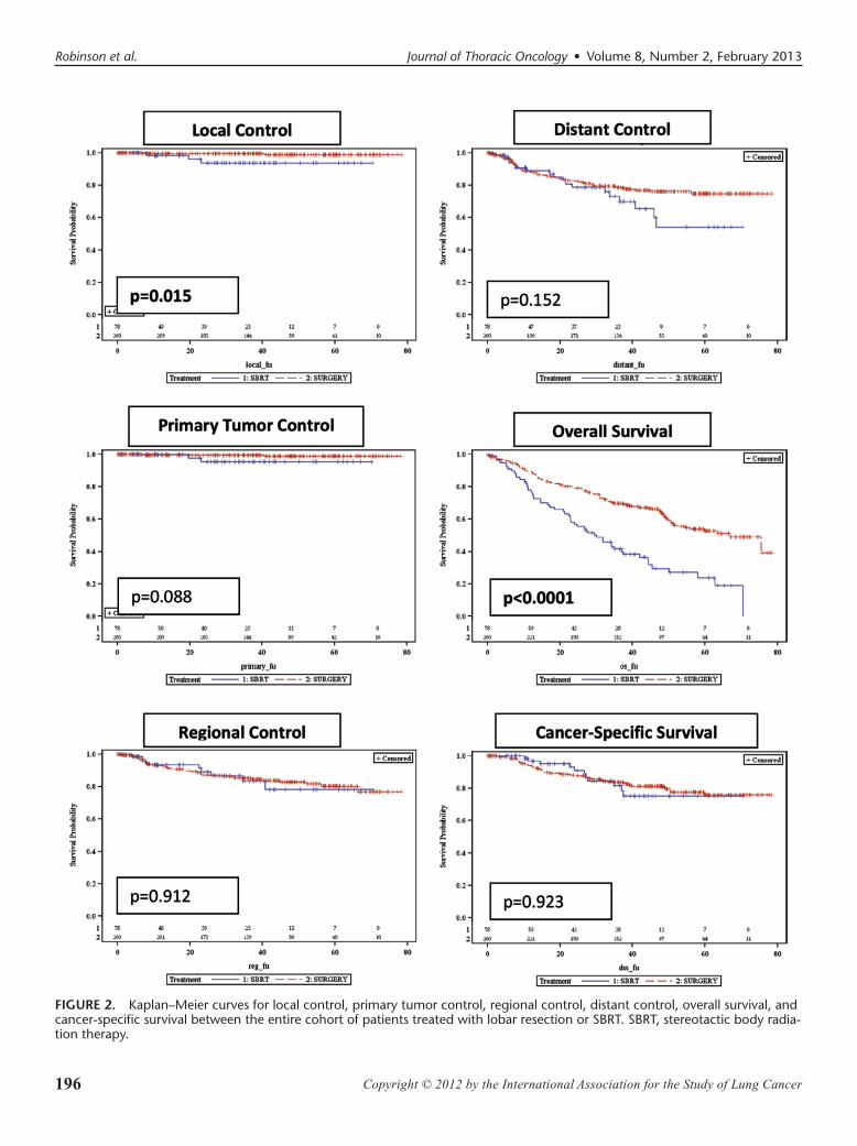
months for SBRT. Patients treated with lobar resection were significantly younger, healthier, and had superior pulmonary function compared with the SBRT group. A greater propor-tion of tumors in the SBRT group were clinical T1. SBRT patients had more than double the incidence of previously treated lung cancer. Median time from prior cancer to current cancer for SBRT and surgery was 3.2 years (range, 0.6–19.2) and 5.3 years (range, 2.0–11.7), respectively. Kaplan–Meier plots comparing patterns of failure and survival for the entire cohort of patients are presented in Figure 2. Local failures in both groups were uncommon. In the lobar resection group, one patient developed a bronchial stump failure and one addi-tional patient developed a port site failure. In the SBRT group, two patients developed primary tumor failure and one addi-tional patient developed a failure in the same lobe outside of the primary. Limited events notwithstanding, 4-year LC was higher for lobar resection versus SBRT (98.7%±0.9% versus
93.6%±3.6%, p = 0.015), though PC was not significantly different (98.7%±0.9% versus 95.3%±4.7%, p = 0.088). No significant difference was seen in 4-year RC (82.9%±2.7% versus 78.1%±7.3%, p = 0.912) or DC (76.1%±3.0% versus 54.0%±9.8%, p = 0.152) for surgery or SBRT, respectively, though a suggestive trend toward worse DC was seen for SBRT after 3 years. OS (63.5%±3.2% versus 29.6%±5.9%, p < 0.0001) was greater for patients treated with lobar resection, though CSS (81.3%±2.7% versus 75.3%±6.8%, p = 0.923) was similar.
Univariate analysis was used on all baseline covariates, listed in Table 1, to determine their impact on patterns of fail-ure and survival. Only those covariates found to be significant on univariate analysis were submitted for multivariate analy-sis. In the event that less than two covariates were significant on univariate analysis for a given outcome, no multivariate analysis was performed (N/A). A summary of factors found to be significant or approaching significance are listed in Table 2.
Propensity Score MatchingGiven the difference in OS and suggestive difference
in late DC between the treatment groups, we reviewed the list of covariates found to be predictive of these outcomes on univariate and multivariate analysis as potential variables for PSM. Patient selection for therapy at our institution was driven by medical comorbidity and confirmed by an overwhelming weighting of covariates, predictive of improved OS in favor of lobar resection (Table 1). With increasing numbers of covari-ates to match upon, PSM returns very small patient numbers. As such, PSM was not attempted for OS. In contrast, only AJCC 7th edition clinical T-stage was predictive of DC. Therefore, we performed PSM using T-stage in an attempt to determine the impact this might have on the suggestive split in DC.
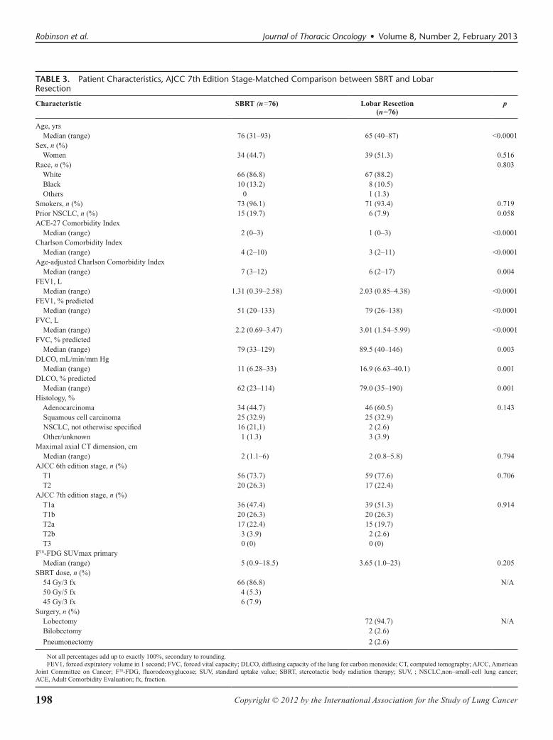
Seventy-six patients from each treatment group were identified using PSM for AJCC 7th edition clinical T-stage. As shown in Table 3, PSM successfully matched the two treat-ment groups with regard to AJCC 7th edition stage (p = 0.706) as well as AJCC 6th edition stage (p = 0.794) and maximal axial tumor size on CT (p = 0.794). After matching, median tumor size for both groups was 2 cm, eliminating patients with larger tumor sizes in the lobar resection group. Despite matching for stage, patients treated with SBRT remained with a significantly higher burden of risk factors predictive of poorer OS.
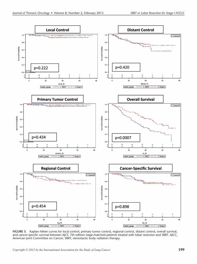
Kaplan–Meier plots comparing patterns of failure and survival for the stage-matched cohort are presented in Figure 3. In the T-stage-matched cohort, no significant differ-ences in 4-year LC (98.5%±1.5% versus 93.5%±3.7%, p = 0.222), PC (98.5% ±1.5% versus 95.2%±3.3%, p = 0.434), RC (77.8%±5.7% versus 82.9%±5.8%, p = 0.454), or DC (72.3%±6.0% versus 57.0%±9.7%, p = 0.420) were appar-ent between lobar resection and SBRT, respectively. The suggestive improvement in DC in favor of surgery was lost after matching for stage. As before, OS (57.9%±6.3% versus 29.3%±5.9%, p = 0.0007) was significantly higher for patients treated with lobar resection whereas CSS (77.3%±.5% versus
196 Copyright © 2012 by the International Association for the Study of Lung Cancer
Robinson et al. Journal of Thoracic Oncology • Volume 8, Number 2, February 2013
FIGURE 2. Kaplan–Meier curves for local control, primary tumor control, regional control, distant control, overall survival, and cancer-specific survival between the entire cohort of patients treated with lobar resection or SBRT. SBRT, stereotactic body radia-tion therapy.
197Copyright © 2012 by the International Association for the Study of Lung Cancer
Journal of Thoracic Oncology • Volume 8, Number 2, February 2013 SBRT or Lobar Resection for Stage I NSCLC
74.7%±6.9%, p = 0.898) remained similar for lobar resection and SBRT.
ToxicityOverall patterns and degrees of toxicity were different
between the two treatment groups, and were not comparable statistically. For patients treated with SBRT, acute toxic-ity consisted of two patients with esophagitis (one grade 1, one grade 2). Late toxicities after SBRT included six patients (7.7%) with grade 2 or more pneumonitis (5 grade 2, 1 grade 3), and one each of a grade 1 brachial plexopathy, a grade 2 pleural effusion, and a grade 3 soft-tissue necrosis. Fifteen patients (19.2%) developed chest-wall pain after SBRT (10 grade 1, 4 grade 2, 2 grade 3). No deaths were attributed to SBRT.
Thirty-day postoperative mortality was 1.5% (n = 4) for patients treated with lobar resection. One additional patient died at 3 months, after a prolonged stay in the inten-sive care unit because of respiratory failure after surgery, for a total conservative operative mortality of 1.9% (n = 5). Cardiovascular complications after surgery included atrial fibrillation in 28 patients (10.8%), other arrhythmia in three patients, myocardial infarction in five patients, and a stroke in one patient. Pulmonary complications included new require-ments for supplemental oxygen in 18 (6.9%), pneumonia/
respiratory failure in 15 (5.8%), prolonged air leak in 11, vocal cord paralysis in four, and tracheostomy placement in two patients, respectively. Bleeding requiring additional surgery occurred in seven patients (5 hemothorax, 2 wound hematoma).
DISCUSSIONOur intent was to compare patterns of failure between
lobectomy or pneumonectomy and SBRT using a coherent definition of failure in both groups. These results demon-strate that patterns of failure between optimal surgery (lobar resection) and optimally dosed SBRT (BED
10 > 100 Gy) are
similar. However, our results also highlight the difficulties in making such comparisons, given the inherent imbalance in both patient and tumor-related factors.
It is heartening to note that both treatments produced high rates of LC and PC. However, the limited number of total events makes direct comparisons difficult and also hinders the ability to identify factors predictive of local failure. For example, although tumor size (by stage or CT size) was not predictive of LC or PC on Cox regression, size is certainly a well-accepted predictor for increased risk of local failure after either treatment. Delayed failures may happen for both modal-ities. One of the local failures for surgery occurred more than 3 years after surgery. Delayed failures have also been reported
TABLE 2. Univariate and Multivariate Analysis of Factors Correlating with Patterns of Failure and Survival for the Entire Cohort
Outcome UVA HRa (95% CI) p MVA HR (95% CI) p
LC Treat (SBRT vs. surgery) 7.14 (1.14–41.9) 0.035 N/A
PC NONE N/A
RC CT size 1.14 (0.98–1.32) 0.081 N/A
DC Stage (7th AJCC edition, T2a vs. T1a) 2.40 (1.26–4.58) 0.008 N/Ab
Stage (7th AJCC edition, T1b vs. T1a) 2.59 (1.33–5.04) 0.005
Histology (nonsquam vs. squam) 1.66 (0.94–2.94) 0.081
OS Treat (SBRT vs. surgery) 2.43 (1.74–3.39) <0.0001 Treat (SBRT vs. surgery)
1.65 (1.03–2.67) 0.039
ACE-27 (2–3 vs. 0–1) 1.64 (1.20–2.25) 0.002 CCI 1.17 (1.01–1.35) 0.036
CCI 1.14 (1.07–1.21) <0.0001 Age 1.03 (1.01–1.05) 0.005
CCIa 1.11 (1.06–1.16) <0.0001
Sex (male vs. female) 1.52 (1.11–2.10) 0.011
Age 1.04 (1.02–1.06) <0.0001
FEV1 1.31 (1.01–1.68) 0.039
FEV1 % 1.01 (1.00–1.02) 0.015
CSS Stage (6th AJCC edition, ≥T2 vs. T1) 1.66 (0.99–2.80) 0.055 CT size 1.18 (1.02–1.36) 0.026
Stage (7th AJCC edition, T2a vs. T1a) 2.57(1.25–5.28) 0.010 DLCO % 1.01 (1.00–1.03) 0.047
Stage (7th AJCC edition, T1b vs. T1a) 2.20 (1.02–4.73) 0.045
CT size 1.16 (1.03–1.31) 0.017
DLCO % 1.01 (1.00–1.03) 0.026
aHazard ratios more than 1 indicate an increased risk of failure for each outcome. For categorical variables, the comparison variable is listed in parentheses. For continuous variables, larger values are correlated with an increased risk of failure for each outcome. Outcomes in italics approached, but did not reach, statistical significance.
bStage (7th edition) is a single variable, despite being reported as individual comparisons for UVA to report a HR. As stage (7th edition) was the only variable significant for DC on UVA, MVA was not performed.
N/A, not applicable; LC, local control; PC, primary tumor control; RC, regional control; DC, distant control; OS, overall survival; CSS, cancer-specific survival; UVA, univariate analysis; MVA, multivariate analysis; HR, hazard ratio; 95% CI, 95% confidence interval; CT, computed tomography; CT size, maximal axial CT dimension; DLCO, diffusing capacity of the lung for carbon monoxide; Treat, treatment; squam, squamous histology; nonsquam, nonsquamous histology; AJCC, American Joint Committee on Cancer; CCI, Charlson Comorbidity Index; CCIa, age-adjusted CCI.
198 Copyright © 2012 by the International Association for the Study of Lung Cancer
Robinson et al. Journal of Thoracic Oncology • Volume 8, Number 2, February 2013
TABLE 3. Patient Characteristics, AJCC 7th Edition Stage-Matched Comparison between SBRT and Lobar Resection
Characteristic SBRT (n=76) Lobar Resection (n=76)
p
Age, yrs Median (range) 76 (31–93) 65 (40–87) <0.0001Sex, n (%) Women 34 (44.7) 39 (51.3) 0.516Race, n (%) 0.803 White 66 (86.8) 67 (88.2) Black 10 (13.2) 8 (10.5) Others 0 1 (1.3)Smokers, n (%) 73 (96.1) 71 (93.4) 0.719Prior NSCLC, n (%) 15 (19.7) 6 (7.9) 0.058ACE-27 Comorbidity Index Median (range) 2 (0–3) 1 (0–3) <0.0001Charlson Comorbidity Index Median (range) 4 (2–10) 3 (2–11) <0.0001Age-adjusted Charlson Comorbidity Index Median (range) 7 (3–12) 6 (2–17) 0.004FEV1, L Median (range) 1.31 (0.39–2.58) 2.03 (0.85–4.38) <0.0001FEV1, % predicted Median (range) 51 (20–133) 79 (26–138) <0.0001FVC, L Median (range) 2.2 (0.69–3.47) 3.01 (1.54–5.99) <0.0001FVC, % predicted Median (range) 79 (33–129) 89.5 (40–146) 0.003DLCO, mL/min/mm Hg Median (range) 11 (6.28–33) 16.9 (6.63–40.1) 0.001DLCO, % predicted Median (range) 62 (23–114) 79.0 (35–190) 0.001Histology, % Adenocarcinoma 34 (44.7) 46 (60.5) 0.143 Squamous cell carcinoma 25 (32.9) 25 (32.9) NSCLC, not otherwise specified 16 (21,1) 2 (2.6) Other/unknown 1 (1.3) 3 (3.9)Maximal axial CT dimension, cm Median (range) 2 (1.1–6) 2 (0.8–5.8) 0.794AJCC 6th edition stage, n (%) T1 56 (73.7) 59 (77.6) 0.706 T2 20 (26.3) 17 (22.4)AJCC 7th edition stage, n (%) T1a 36 (47.4) 39 (51.3) 0.914 T1b 20 (26.3) 20 (26.3) T2a 17 (22.4) 15 (19.7) T2b 3 (3.9) 2 (2.6) T3 0 (0) 0 (0)F18-FDG SUVmax primary Median (range) 5 (0.9–18.5) 3.65 (1.0–23) 0.205SBRT dose, n (%) 54 Gy/3 fx 66 (86.8) N/A 50 Gy/5 fx 4 (5.3) 45 Gy/3 fx 6 (7.9)Surgery, n (%) Lobectomy 72 (94.7) N/A Bilobectomy 2 (2.6)
Pneumonectomy 2 (2.6)
Not all percentages add up to exactly 100%, secondary to rounding.FEV1, forced expiratory volume in 1 second; FVC, forced vital capacity; DLCO, diffusing capacity of the lung for carbon monoxide; CT, computed tomography; AJCC, American
Joint Committee on Cancer; F18-FDG, fluorodeoxyglucose; SUV, standard uptake value; SBRT, stereotactic body radiation therapy; SUV, ; NSCLC,non–small-cell lung cancer; ACE, Adult Comorbidity Evaluation; fx, fraction.
199Copyright © 2012 by the International Association for the Study of Lung Cancer
Journal of Thoracic Oncology • Volume 8, Number 2, February 2013 SBRT or Lobar Resection for Stage I NSCLC
FIGURE 3. Kaplan–Meier curves for local control, primary tumor control, regional control, distant control, overall survival, and cancer-specific survival between AJCC 7th edition stage-matched patients treated with lobar resection and SBRT. AJCC, American Joint Committee on Cancer; SBRT, stereotactic body radiation therapy.
200 Copyright © 2012 by the International Association for the Study of Lung Cancer
Robinson et al. Journal of Thoracic Oncology • Volume 8, Number 2, February 2013
after SBRT.30 Given the limited number of patients alive in either group at later time points, long-term control should be interpreted with caution.
Occult nodal disease was discovered in more than 20% of patients undergoing lobar resection. We did not find a dif-ference in RC between surgery and SBRT, which is in line with most previous reports detailing regional failure after SBRT.8 Several theories have been postulated to explain the lower than expected regional failure for clinically staged patients treated with SBRT, including scatter radiation to the nodes and a tumor vaccination phenomenon.16,31 Nevertheless, if a simi-lar proportion of patients in the SBRT group has microscopic nodal metastases, this should translate into an increased risk for distant metastases in a population of patients who receive no adjuvant chemotherapy.
Though DC was not statistically different between the two treatments in an unmatched comparison, there was a sug-gestive split in favor of surgery after 3 years. After match-ing for T-stage, this difference became less apparent. Possible explanations for the difference between overall- and stage-matched cohorts include history of prior lung cancers, tumor size, nodal dissection, and use of adjuvant chemotherapy. Patients treated with SBRT had more than double the inci-dence of prior lung cancers, and it is certainly possible that some of these patients treated for presumed metachronous cancers in fact had early metastatic disease. Although history of prior lung cancer did not correlate with any pattern of fail-ure outcome on Cox regression, the lack of statistical signifi-cance does not rule out the possibility of a contribution to the suggestive split in DC. In the unmatched comparison, median tumor size was larger for lobar resection. Increasing tumor size is a well-established predictor of both nodal and subse-quent distant failure. A logical explanation for the apparent improvement in DC for surgery in the unmatched comparison is the use of node dissection and subsequent adjuvant chemo-therapy for node-positive patients. After matching for stage, median tumor size between SBRT and lobar resection was identical (2 cm), potentially mitigating some of the benefits of node dissection and chemotherapy. Given the established benefit for adjuvant chemotherapy for node-positive disease, this would support the need for a more thorough nodal evalu-ation in SBRT patients. Selective mediastinoscopy and/or minimally invasive staging tools, such as radial endobronchial ultrasound and endoscopic ultrasound via esophagoscopy, will undoubtably play a more valuable role as the use of SBRT expands.32
Nonrandomized comparisons of surgery and SBRT are difficult, particularly with regard to OS, as surgical patients are by definition healthier. Onishi et al.15 reviewed outcomes after SBRT for operable patients with stage I NSCLC across 14 Japanese institutions. Five-year LC for T1 and T2 tumors was 92% and 73%, respectively, and 5-year OS was 72% and 62%. Although these outcomes compare favorably to similar reports for surgery, no companion surgical group was included. Grills et al.16 compared wedge resection to SBRT in patients with high-risk stage I NSCLC. A suggestive trend towards decreased 30-month local recurrence was noted in
favor of SBRT (4% versus 20%, p = 0.07), though regional (4% versus 18%) and distant recurrence (19% versus 21%) was not significantly different. Likewise, CSS was identical at 94% and 93%, respectively. Despite the suggested improve-ments in locoregional control with SBRT, OS in the wedge group was superior (87% versus 72%, p = 0.01), which was felt to reflect the inferior health of SBRT patients.
In an earlier publication, we matched a small number of surgery and SBRT patients in an attempt to compare OS.17 In this more heterogeneous population, surgical patients under-went either sublobar or lobar resection and several SBRT patients received suboptimal doses (BED
10 < 100 Gy). In the
group as a whole, LC, RC, and CSS were similar between treatments, whereas OS was superior for surgery. After match-ing with PSM for age, T-stage, and ACE-27 score, OS was not different between the groups. In the current analysis, the supe-rior OS in the lobar resection group should not be overinter-preted, given the dramatic differences in comorbidity. Such confounding by indication is a well-described consequence of such observational studies, which is not readily correctable with current adjustment methods.33 It is notable that CSS was identical between the treatment groups in both unmatched and stage-matched comparisons. Thus, despite having worse OS compared with lobar resection, patients treated with SBRT died from cancer at the same rate as those undergoing surgery. SBRT patients were less healthy and many died of noncancer causes. Only randomized comparisons between surgery and SBRT will avoid the imbalances and selection biases present in this and other nonrandomized comparisons.
In conclusion, this institutional analysis of patients with clinical stage I NSCLC reveals comparable patterns of failure between lobectomy/pneumonectomy and SBRT, with improved LC and a suggestion of improved DC for surgery in the unmatched cohort. In a T-stage matched comparison, patterns of failure between surgical and SBRT groups were identical. Overall survival was superior in the surgical group irrespective of matching. These results support the need to enroll patients on randomized trials (such as ACOSOG Z4099/RTOG 1021, STARS).
REFERENCES 1. Flores RM. Reply to difference in outcome in the transection of the pul-
monary artery and vein. J Thorac Cardiovasc Surg 2011;141:597–598. 2. Detterbeck FC, Boffa DJ, Tanoue LT. The new lung cancer staging sys-
tem. Chest 2009;136:260–271. 3. El-Sherif A, Gooding WE, Santos R, et al. Outcomes of sublobar resec-
tion versus lobectomy for stage I non-small cell lung cancer: a 13-year analysis. Ann Thorac Surg 2006;82:408–15; discussion 415.
4. Ginsberg RJ, Rubinstein LV. Randomized trial of lobectomy versus lim-ited resection for T1 N0 non-small cell lung cancer. Lung Cancer Study Group. Ann Thorac Surg 1995;60:615–22; discussion 622.
5. Birdas TJ, Koehler RP, Colonias A, et al. Sublobar resection with brachy-therapy versus lobectomy for stage Ib nonsmall cell lung cancer. Ann Thorac Surg 2006;81:434–8; discussion 438.
6. Sawabata N, Matsumura A, Ohota M, et al.; Thoracic Surgery Study Group of Osaka University. Cytologically malignant margins of wedge resected stage I non-small cell lung cancer. Ann Thorac Surg 2002;74:1953–1957.
7. Okada M, Koike T, Higashiyama M, Yamato Y, Kodama K, Tsubota N. Radical sublobar resection for small-sized non-small cell lung cancer: a multicenter study. J Thorac Cardiovasc Surg 2006;132:769–775.
201Copyright © 2012 by the International Association for the Study of Lung Cancer
Journal of Thoracic Oncology • Volume 8, Number 2, February 2013 SBRT or Lobar Resection for Stage I NSCLC
8. Timmerman R, Paulus R, Galvin J, et al. Stereotactic body radiation ther-apy for inoperable early stage lung cancer. JAMA 2010;303:1070–1076.
9. (U.S.) NCCN: National Comprehensive Cancer Network (NCCN). Clinical Practice Guidelines in Oncology, 2012
10. Lanni TB Jr, Grills IS, Kestin LL, Robertson JM. Stereotactic radio-therapy reduces treatment cost while improving overall survival and local control over standard fractionated radiation therapy for medically inoper-able non-small-cell lung cancer. Am J Clin Oncol 2011;34:494–498.
11. Grutters JP, Kessels AG, Pijls-Johannesma M, De Ruysscher D, Joore MA, Lambin P. Comparison of the effectiveness of radiotherapy with photons, protons and carbon-ions for non-small cell lung cancer: a meta-analysis. Radiother Oncol 2010;95:32–40.
12. Onishi H, Shirato H, Nagata Y, et al: Hypofractionated stereotactic radio-therapy (HypoFXSRT) for stage I non-small cell lung cancer: updated results of 257 patients in a Japanese multi-institutional study. J Thorac Oncol 2:S94–100, 2007
13. Olsen JR, Robinson CG, El Naqa I, et al. Dose-response for stereotactic body radiotherapy in early-stage non-small-cell lung cancer. Int J Radiat Oncol Biol Phys 2011;81:e299–e303.
14. Uematsu M, Shioda A, Suda A, et al. Computed tomography-guided fra-meless stereotactic radiotherapy for stage I non-small cell lung cancer: a 5-year experience. Int J Radiat Oncol Biol Phys 2001;51:666–670.
15. Onishi H, Shirato H, Nagata Y, et al. Stereotactic body radiotherapy (SBRT) for operable stage I non-small-cell lung cancer: can SBRT be comparable to surgery? Int J Radiat Oncol Biol Phys 2011;81:1352–1358.
16. Grills IS, Mangona VS, Welsh R, et al. Outcomes after stereotactic lung radiotherapy or wedge resection for stage I non-small-cell lung cancer. J Clin Oncol 2010;28:928–935.
17. Crabtree TD, Denlinger CE, Meyers BF, et al. Stereotactic body radiation therapy versus surgical resection for stage I non-small cell lung cancer. J Thorac Cardiovasc Surg 2010;140:377–386.
18. Puri V, Crabtree TD, Kymes S, et al. A comparison of surgical inter-vention and stereotactic body radiation therapy for stage I lung cancer in high-risk patients: a decision analysis. J Thorac Cardiovasc Surg 2012;143:428–436.
19. Lagerwaard FJ, Verstegen NE, Haasbeek CJ, et al. Outcomes of stereotac-tic ablative radiotherapy in patients with potentially operable stage I non-small cell lung cancer. Int J Radiat Oncol Biol Phys 2012;83:348–353.
20. Altorki NK. Stereotactic body radiation therapy versus wedge resection for medically inoperable stage I lung cancer: tailored therapy or one size fits all? J Clin Oncol 2010;28:905–907.
21. Greene FL, American Joint Committee on Cancer, American Cancer Society. AJCC Cancer Staging Handbook: From the AJCC Cancer Staging Manual, 6th Ed. New York, NY: Springer, 2002.
22. Goldstraw P, International Association for the Study of Lung Cancer. Staging Manual in Thoracic Oncology. Orange Park, FL: Editorial Rx Press, 2009
23. Birim O, Kappetein AP, Bogers AJ. Charlson comorbidity index as a pre-dictor of long-term outcome after surgery for nonsmall cell lung cancer. Eur J Cardiothorac Surg 2005;28:759–762.
24. Boholli I, Hoover D, Yang WM, et al: Comorbidity as a predictor of sur-vival in veterans with stage I and II non-small cell lung cancer (NSCLC). ASCO Meeting Abstracts 29:e16605
25. Charlson M, Szatrowski TP, Peterson J, et al: Validation of a combined comorbidity index. J Clin Epidemiol 47:1245–1251, 1994
26. Bradley JD, El Naqa I, Drzymala RE, et al: Stereotactic body radiation therapy for early-stage non-small-cell lung cancer: the pattern of failure is distant. Int J Radiat Oncol Biol Phys 77:1146–50, 2010
27. Onishi H, Araki T, Shirato H, et al. Stereotactic hypofractionated high-dose irradiation for stage I nonsmall cell lung carcinoma: clinical out-comes in 245 subjects in a Japanese multiinstitutional study. Cancer 2004;101:1623–1631.
28. Phase III Randomized Study of Sublobar Resection With or Without Brachytherapy Versus Stereotactic Body Radiotherapy in High-Risk Patients With Stage I Non-Small Cell Lung Cancer,
29. Schemper M, Smith TL. A note on quantifying follow-up in studies of failure time. Control Clin Trials 1996;17:343–346.
30. Matsuo Y, Shibuya K, Nagata Y, et al. Preliminary report of late recur-rences, at 5 years or more, after stereotactic body radiation therapy for non-small cell lung cancer. J Thorac Oncol 2012;7:453–456.
31. Lee Y, Auh SL, Wang Y, et al. Therapeutic effects of ablative radiation on local tumor require CD8+ T cells: changing strategies for cancer treat-ment. Blood 2009;114:589–595.
32. Annema JT, van Meerbeeck JP, Rintoul RC, et al. Mediastinoscopy vs endosonography for mediastinal nodal staging of lung cancer: a random-ized trial. JAMA 2010;304:2245–2252.
33. Bosco JL, Silliman RA, Thwin SS, et al. A most stubborn bias: no adjust-ment method fully resolves confounding by indication in observational studies. J Clin Epidemiol 2010;63:64–74.