effectiveness of using oleaginous microalgae as a green
TRANSCRIPT

14
AbstractCopper (Cu) is a non-biodegradable heavy metal and present in the environment that represents risks to the ecosystem and human health, leading to a major environmental issue globally. In this study, we evaluated the potential use of ten microalgal strains as biosorbents for decontaminating copper in aqueous media. As this strategy requires low energy consumption and no use of chemicals, it is greatly suitable for environmentally decontaminating copper. Among the ten strains tested, Acutodesmus obliquus AARL G090 showed the highest efficiency for copper removal (more than 85%) and enhanced the microalgal lipid accumulation (up to 38.14%). The scanning electron microscope–energy dispersive spectroscopy (SEM–EDS) proved that the copper was not only transferred to microalgal cells but also got attached to the microalgal cell surface. This study has proven a promising ‘green’ strategy to rapidly remove the heavy metal and economically possible application in the production of microalgal lipid as a biodiesel feedstock.
Keywords: Microalgae; Copper; Biosorbent; Metal removal; Lipid
EnvironmentAsia 14(2) 2021 14-23DOI 10.14456/ea.2021.12
ISSN 1906-1714; ONLINE ISSN: 2586-8861
Effectiveness of Using Oleaginous Microalgae as a Green Biosorbent to Remove Copper from Aqueous MediaKhomsan Ruangrit1, Jeeraporn Pekkoh2,3, Kritsana Duangjan1,
Kittiya Phinyo2, Wassana Kamopas1,4, Itthipon Jeerapan5,6, Wasu Pathom-aree2,3, and Sirasit Srinuanpan2*
1 Science and Technology Research Institute, Chiang Mai University, Chiang Mai, Thailand2 Department of Biology, Faculty of Science, Chiang Mai University, Chiang Mai, Thailand
3 Research Center of Microbial Diversity and Sustainable Utilization, Faculty of Science, Chiang Mai University, Chiang Mai, Thailand
4 Research Group for Renewable Energy, Faculty of Engineering, Chiang Mai University, Chiang Mai, Thailand
5 Division of Physical Science, Faculty of Science, Prince of Songkla University, Hat Yai, Songkhla, Thailand
6 Center of Excellence for Trace Analysis and Biosensor, Prince of Songkla University, Hat Yai, Songkhla, Thailand
*Corresponding Author: [email protected]: November 6, 2020; Revised: January 2, 2021; Accepted: January 21, 2021
1. IntroductionCurrently, the increased contamination
of heavy metals is a major global concern as they could pollute the environment and cause severe problems to humans. Copper (Cu) is an example of the most abundant metals found in many rivers and water supplies. This pollutant is emitted from different fields including coal mining, metallurgical industries, and geological weathering (Kumar et al., 2015; Rugnini et al., 2017).
In the current situation, eliminating or reducing copper to a less damaging form is necessary under the environmental-friendly concept. Although effort has been made in decontamination of copper by using conventional approaches for remediation of copper from an aqueous solution (e.g.,ion exchange, chemical reduction-oxidation, and chemical extraction), these require high energy consumption, expenditure in the capital,

K. Ruangrit et al / EnvironmentAsia 14(2) (2021) 14-23
15
and running costs, representing impracticality (Sriharsha et al., 2020). Therefore, a low-cost, efficient, and environmentally sustainable approach of remediating copper should be considered in the present scenario.
“Green” strategies using microbial sorption are attractive for removing heavy metals from aqueous media. Various microbial strains of yeast, bacteria, algae, and fungi displays the high efficiency of metal binding and absorption (Veneu et al., 2013; Rani et al., 2013; Kumar et al., 2015). In addition to applicable advantages of in various biotechnologies such as energy and environment, algae are among significant candidates for serving our purposes. Green microalgae can grow in metal-contaminated water and also easily culture in industrial wastewater that have proven to be the effective prospects for eliminating heavy metals (Yang et al., 2015; Markou et al., 2015). Remarkably, not all green microalgae can grow in metal-contained solution; some green microalgae are metal stress-tolerant. The microalgae strains with high potential of heavy-metal removal and are Chlorella spp., belonging to the family of Chlorellaceae (Rugnini et al., 2017). Lately, few studies have explored the potential use of oleaginous microalgae as a promising biosorbent for the reduction of metal from aqueous solution. It is found that using oleaginous microalgae is effective not only removal of heavy metal but also low-cost production of microbial lipid as a promising feedstock for biodiesel production (Ermis et al., 2020; de Oliveira et al., 2020). However, studies on the practical use of microalgae are currently only being explored at an exploratory level. Different microalgae strains contain different compositions, suggesting the interest to explore the optimal biosorption. Consequently, we have put our efforts on researching an alternative and suitable microalgae strain for removing heavy metal, particularly copper, and simultaneously lipid production as a possible feedstock to produce biodiesel.
The present study reports the potential use of ten microalgae strains belonging to the family Scenedesmaceae to grow in aqueous media supplemented with a heavy metal (i.e., copper as a model). Inherently, such
microalgae can significantly decontaminate the copper level in the solution. The effect of copper on microalgae biomass production and the amounts of biochemical compounds, e.g., chlorophyll and lipid, were also investigated. The morphology and metal-binding cells were observed using microscopic and scanning electron microscope–energy dispersive spectroscopy (SEM–EDS) analysis.
2. Materials and Methods
2.1 Microalgal strains
Green microalgae strains in the family Scenedesmaceae obtained from Diversity of Algae and Plankton Research Unit, Science and Technology Research Institute, Chiang Mai University, Thailand were used in this study including Verrucodesmus verrucosus AARL G073, Scenedesmus sp. AARL G087, Desmodesmus sp. AARL G074, Desmodesmus maximus AARL G082, Desmodesmus communis AARL G076, Desmodesmus opoliensis AARL G089, Acutodesmus obliquus AARL G091, Acutodesmus obliquus AARL G092, Acutodesmus obliquus AARL G090, and Acutodesmus sp. AARL1(8). The cultures were maintained for 7 days in JM medium (pH 7.0) at 27 °C and 60 µmol proton/m 2/s of l ight in tens i ty wi th a photoperiod of 24 h.
2.2 Cultivation of microalgae in aqueous media containing copper
To evaluate the effect of copper (Cu) on the Cu removal efficiency, microalgae growth, lipid production, chlorophyll a content, and physiological change of microalgal cel ls , the microalgae were cultured in 230 mL of JM media supplemented with 10 mg Cu/L using CuSO4·5H2O as a Cu source for 24 days (simulating the chronic exposure). The JM media without CuSO4 supplementary was used as a control. The initial cell density (OD665) was fixed at 0.04. The cultures were continuously illuminated at 60 µmol proton/m2/s of light intensity at 27 °C.

K. Ruangrit et al / EnvironmentAsia 14(2) (2021) 14-23
16
2.3 Determination of copper content
The frame atomic absorption spectroscopy (AAS) was used to determine the Cu concentration. Prior to the evaluation of Cu-removal efficiency, 20 mL of sample was collected and centrifuged at 6000 rpm for 10 min. The total concentration of Cu in the supernatant was then measured using AAS (Thongpitak et al., 2019). The Cu removal efficiency was calculated using the following equation:
Cu removal efficiency (%) = [(C0 – CF) / C0] × 100 (1)
Where C0 is the initial Cu concentration and CF is the final Cu concentration.
2.4 Determination of microalgal biomass
The microalgae growth was determined as dried biomass. The microalgae were centrifuged at 6000 rpm for 10 min. The pellet was consolidated, rinsed, and then dried at 60 °C until obtaining a constant weight.
2.5 Determination of lipid content
The microalgal lipid was extracted using solvent extraction. Briefly, dry microalgae (1 g) were mixed with a mixture of chloroform to methanol (2:1, v/v) 10 mL, sonicated for 30 min, and centrifuge at 6000 rpm for 10 min. The supernatant was collected and dried at 60 °C until constant weight (Bligh and Dyer, 1959).
2.6 Determination of chlorophyll a content
The chlorophyll a of dried microalgae biomass was extracted using 90%MeOH and then incubated at 70 °C for 20 min. The mixture was centrifuged at 6000 rpm for 10 min. The supernatant was collected and measured the absorbance at 630 nm (A630), 645 nm (A645), 665 nm (A665) and 750 nm (A750) with a spectrophotometer ( Wi n t e r m a n s a n d D e M o t s , 1 9 6 5 ; Saijo, 1975). The chlorophyll a content (μg/mL) was calculated using the following equation:
Chlorophyll a (μg/mL) = (11.6A −1.31B − 0.14C) × [(V/Vf) × (1/L)] (2)
Where A is A665− A750, B is A645− A750, C is A630− A750, V is the total volume of extract (mL), Vf is the volume of sample (mL), and L is the light path length of width of cuvette (cm).
2.7 Analysis of cell morphology
The cell morphology was observed by microscopic and scanning electron microscopy (SEM) analysis. The distribution of the elemental composition on the surface of the microalgal cell wall was analyzed using energy-dispersive spectroscopy (EDS) analysis.
2.8 Statistical analysis
All experiments were conducted in triplicate (n = 3) and the statistical significances of the experimental results were analyzed using one-way ANOVA (analysis of variance) and Duncan’s multiple range tests.
3. Results and Discussion
3.1 Copper removal efficiency by green microalgae
Ten microalgae strains belonging to the family Scenedesmaceae were cultured in JM media supplemented with 10 mg/L of Cu for 24 days compared to JM media. The Cu removal efficiency from aqueous media after 24 days of the Cu chronic exposure is shown in Figure 1a. It was found that all microalgae strains exhibited the effective removing Cu from the media that were in ranged from 70.6 to 85.6% removal efficiency, in accordance with the previously studied by different authors (e.g., Buayam et al., 2019). Among the microalgal strains tested, Acutodesmus obliquus AARL G090 had the highest Cu removal efficiency at 85.6% with the low amount of remaining Cu in aqueous media at (2.6 mg/L), followed by A. obliquus AARL G091 (85.0%), and then Desmodesmus opoliensis AARL G089 (82.8%) and the amount of remaining Cu at less than 3.5 mg/L. Zhong et al. (2012) found that after a long

K. Ruangrit et al / EnvironmentAsia 14(2) (2021) 14-23
17
Figure 1. Copper removal efficiency (%) from aqueous media by microalgae after 24 h of chronic expose from copper (a) and the proposed possible mechanism of copper absorption
by microalgae (b). AARL G073: Verrucodesmus verrucosus AARL G073, AARL G087: Scenedesmus sp. AARL G087, AARL G074: Desmodesmus sp. AARL G074, AARL G082: Desmodesmus maximus AARL G082, AARL G076: Desmodesmus communis AARL G076, AARL G089: Desmodesmus opoliensis AARL G089, AARL G091: Acutodesmus obliquus
AARL G091, AARL G092: Acutodesmus obliquus AARL G092, AARL G090: Acutodesmus obliquus AARL G090, and AARL 1(8): Acutodesmus sp. AARL1(8).
period of 12-day cultivation, 75.9 – 91.4% of copper was removed from aqueous media by green microalgae Chlorella pyrenoidosa and Scenedesmus obliquus; however, the amount of copper at the initial day of cultivation was also much lower (0.2–2.0 mg Cu/L) than the set up in our study.
Most studies on copper bioremediation were performed with microalga Chlorella spp. According to the report by Rugnini et al. (2017), they achieved a maximum Cu removal efficiency of 49% and 39% for Desmodesmus
sp. and Chlorella vulgaris, respectively from the aqueous media with 11.9 mg Cu/L at the first day of cultivation time which were close to this study (10 mg Cu/L). Interestingly, the Cu removal efficiency by ten microalgal strains tested in this work is significantly high, compared with using bacteria (Veneu et al., 2013) and fungi (Rani et al., 2013), indicating the practical use of these microalgae for the effective bioremediation of heavy metal.

K. Ruangrit et al / EnvironmentAsia 14(2) (2021) 14-23
18
Figure 2. Microscopic images of Scenedesmus sp. AARL G087 (a, d), Desmodesmus sp. AARL G074 (b, e), and Desmodesmus opoliensis AARL G089 (c, f) cultured in JM media
(a-c) and JM media supplemented with copper at 10 mg/L (d-f).
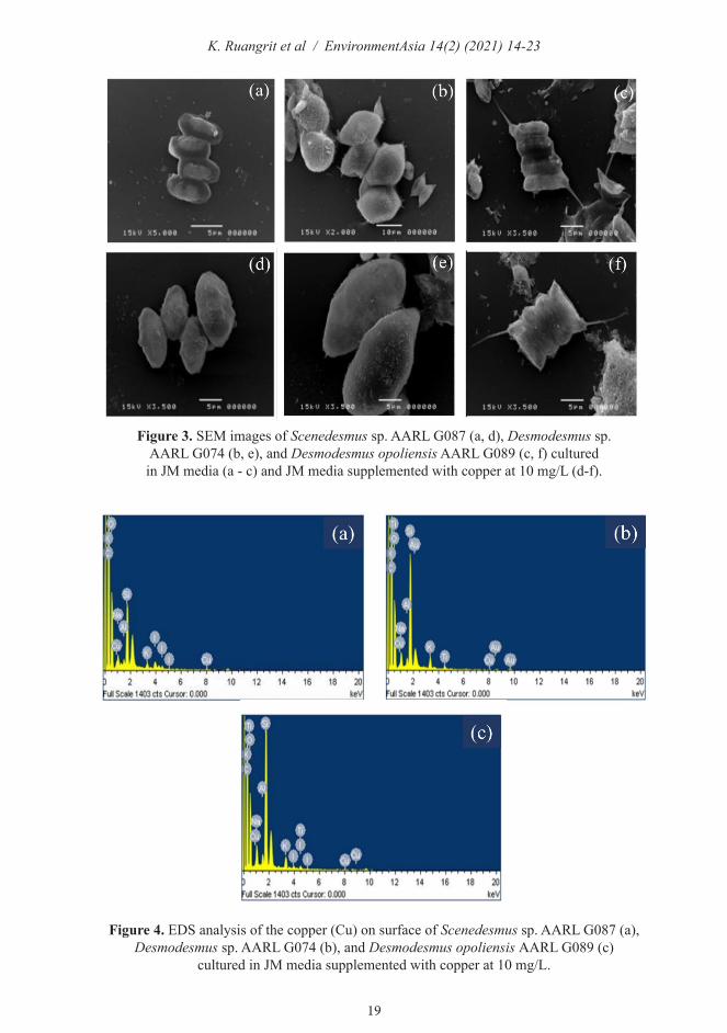
3.2 Effect of copper on microalgal morphology
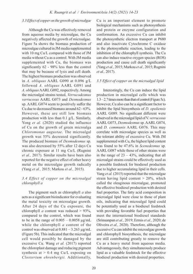
The morphology of microalgal cells after 24 days of the Cu chronic exposure was altered as observed using microscopy (Figure 2) and SEM (Figure 3) compared to microalgal cells cultured in JM media without Cu supplementation as a control. The Cu caused a change of cell size of all microalgae strains tested which were enlarged compared with control cells, demonstrating that the Cu may be accumulated intracellularly. Previous reports have supported the accumulation of Cu within microalgal cells (Rugnini et al., 2017; Buayam et al., 2019). Rugnini et al. (2017) reported that copper and nickel could bind with cell surface in a few minutes and subsequent transporting slowly inside the cells. The SEM–EDS analysis was conducted in order to qualitatively determine the presence of Cu on the microalgae surface. Figure 4 represented the EDS spectrum of the microalgal surface, proving the appearance of Cu ion on the cells surface. It should be indicated that the copper was not only transported to microalgal cells but also got attached to the microalgal cell surface.
It is possible to suggest the mechanism of removing Cu from aqueous media via both absorption and bioaccumulation.
Figure 1b shows the possible mechanism of Cu removal by microalgae in which modified from Kumar et al. (2015). There are two processes for Cu remediation that could possibly be occurring simultaneously. The first process is the absorption of Cu ion (Cu2+) with the microalgal surface due to various binding groups available on the microalgal cell wall, such as OH–, SH–, COO–, PO4
3–, NO3
–, RNH2–, RS–, and RO– (Zeraatkar
et al., 2016). The second process is the Cu accumulation within microalgal cells through different transporter membranes; for instance, the CTR (Cu Transporter) available in plasma membrane promotes the Cu ion transportation (Kumar et al., 2015). These remarkable advantage of the microalgal cell suggests the considerable promise for enhancing the efficiency of the heavy metal decontamination processes. However, an extensive explanation on the mechanisms of both absorption and accumulation of Cu by microalgae is yet to be elaborated.
K. Ruangrit et al / EnvironmentAsia 14(2) (2021) 14-23
19
Figure 3. SEM images of Scenedesmus sp. AARL G087 (a, d), Desmodesmus sp. AARL G074 (b, e), and Desmodesmus opoliensis AARL G089 (c, f) cultured in JM media (a - c) and JM media supplemented with copper at 10 mg/L (d-f).
Figure 4. EDS analysis of the copper (Cu) on surface of Scenedesmus sp. AARL G087 (a), Desmodesmus sp. AARL G074 (b), and Desmodesmus opoliensis AARL G089 (c)
cultured in JM media supplemented with copper at 10 mg/L.
K. Ruangrit et al / EnvironmentAsia 14(2) (2021) 14-23
20
3.3 Effect of copper on the growth of microalgae
Although the Cu was effectively removed from aqueous media by microalgae, the Cu negatively affected the growth of microalgae. Figure 5a shows the biomass production of microalgae cultured in JM media supplemented with 10 mg Cu/L compared with those of JM media without Cu as a control. With JM media supplemented with Cu, the biomass was significantly 62 - 98% less than the control that may be because of lysis and cell death. The highest biomass production was observed in A. obliquus AARL G090 at 0.096 g/Lfollowed A. obliquus AARL G091 and A. obliquus AARL G092, respectively. Among the microalgal strains tested, Verrucodesmus verrucosus AARL G073 and Desmodesmus sp. AARL G074 seem to positively suffer the Cu due to decreased biomass around 62 - 63%.However, these are still low biomass production with less than 0.1 g/L. Similarly, Yong et al. (2020) studied the influence of Cu on the growth of green microalga Chloromonas augustae. The microalgal growth was 55% decreased significantly. The produced biomass of Desmodesmus sp. was also decreased by 55% after 12 days Cu chronic exposure at 11 mg Cu/L (Rugnini et al., 2017). Similar results have been also reported for the negative effect of other heavy metal on the microalgae growth radically (Yang et al., 2015; Markou et al., 2015).
3.4 Effect of copper on the microalgal chlorophyll a
The pigment such as chlorophyll a also acts as a significant bioindicator for evaluating the metal toxicity on microalgae growth. After 24 days of the Cu exposure, the chlorophyll a content was reduced > 93%,compared to the control, which was foundto be in the range of 0.005 – 0.0058 µg/mL while the chlorophyll a content of thecontrol was observed at 0.881 – 3.263 µg/mL(Figure 5b). This indicated that the microalgal cell would possibly be damaged by the excessive Cu. Wang et al. (2017) reported the chloroplast damage and reducing pigment synthesis at > 0.4 mg Cu/L exposing on Closterium ehrenbergii. Additionally,
Cu is an important element to promote biological mechanisms such as photosynthesis and protein or enzyme configuration and conformation. An excessive Cu can inhibit the photosynthetic electron transport at P680 and also inactivate Cytochrome C oxidase in the photosynthetic reaction, leading to the inhibition of the chlorophyll synthesis. The Cu can also induce reactive oxygen species (ROS) production and cause cell death significantly (Yang et al., 2015; Markou et al., 2015; Rugnini et al., 2017).
3.5 Effect of copper on the microalgal lipid
Interestingly, the Cu can induce the lipid production in microalgal cells which was 1.3 – 2.7 times more than that of control (Figure 5c).However, Cu also can be a significant factor to inhibit the lipid biosynthesis of D. opoliensis AARL G089. No significant different were observed in the microalgal lipid of V. verrucosus AARL G073, Desmodesmus sp. AARL G074, and D. communis AARL G076. This may correlate with microalgae species as well as the tolerant ability of excessive Cu. With JM supplemented with Cu, the highest lipid content was found to be 47.6% in Scenedesmus sp. AARL G087 while those of other strains were in the range of 23 – 43%, indicating that all microalgal strains could be effectively used as a possible feedstock for biodiesel production due to higher accumulating lipid in their cells. Yang et al. (2015) reported that the microalgae strain having lipid content > 20%, which called the oleaginous microalgae, promoted the effective biodiesel production with desired fuel properties. The fatty acid composition in microalgal lipid were close to those of plant oils, indicating that microalgal lipid could be potentially used as a biodiesel feedstock with providing favorable fuel properties that meet the international biodiesel standards (Srinuanpan et al., 2019; Ermis et al., 2020; de Oliveira et al., 2020). Therefore, although the excessive Cu can inhibit the microalgae growth and chlorophyll biosynthesis, the microalgae are still contributing greatly to remove the Cu as a heavy metal from aqueous media. Advantageously, they simultaneously produce lipid as a valuable feedstock for the effective biodiesel production with desired properties.

K. Ruangrit et al / EnvironmentAsia 14(2) (2021) 14-23
21
Figure 5. Biomass (a), chlorophyll (b), and lipid (c) of microalgae cultured in JM media supplemented with copper at 10 mg/L copper (JM + Cu) compared with JM media without
copper supplementation (JM). AARL G073: Verrucodesmus verrucosus AARL G073, AARL G087: Scenedesmus sp. AARL G087, AARL G074: Desmodesmus sp. AARL G074, AARL G082: Desmodesmus maximus AARL G082, AARL G076: Desmodesmus communis AARL
G076, AARL G089: Desmodesmus opoliensis AARL G089, AARL G091: Acutodesmus obliquus AARL G091, AARL G092: Acutodesmus obliquus AARL G092, AARL G090:
Acutodesmus obliquus AARL G090, and AARL 1(8): Acutodesmus sp. AARL1(8).
4. Conclusion
This study has shown an effective green method for the removal of heavy metal (Cu2+) from aqueous solution using oleaginous microalgae as a biosorbant. As this method requires low energy consumption and no use of chemicals, it is greatly suitable for environmentally decontaminating copper. The growth and chlorophyll synthesis of tested ten microalgal strains showed to decrease after copper chronic exposure, but promising copper removal efficiencies were recorded up to 71 - 86%. Interestingly,
the copper can induce lipid accumulation in microalgal cells by enhancing its lipid content that could be used as sustainable f eeds tocks fo r e f f ec t i ve b iod i e se l production. The SEM-EDS image did show that the copper was not only transferred to microalgal cells but also got attached to the microalgal cell surface. This study may effective not only in the bioremediation of heavy metal but also contribute greatly to the industrialized microalgae-based biodiesel production.

K. Ruangrit et al / EnvironmentAsia 14(2) (2021) 14-23
22
Acknowledgements
This research work was supported by Chiang Mai University, Thailand Research Fund (TRF), Thailand Science Research and Innovation (TSRI), and National Research Council of Thailand (NRCT). The first, third, and fifth authors were financially supported by Science and Technology Research Institute, Chiang Mai University. We thank Nattaphorn Buayam for technical assistance.
References
Bligh EG, Dyer WJ. A rapid method of total lipid extraction and purification. Canadian Journal of Biochemistry and Physiology 1959; 37(8): 911–917.
Buayam N, Davey MP, Smith AG, Pumas C. Effects of copper and pH on the growth and physiology of Desmodesmus sp. AARLG074. Metabolites 2019; 9(5): 84.
de Oliveira CY, Viegas TL, da Silva MF, Fracalossi DM, Lopes RG, Derner RB. Effect of trace metals on growth performance and accumulation of lipids, proteins, and carbohydrates on the green microalga Scenedesmus obliquus. Aquaculture International 2020; 28: 1435–1444.
Ermis H, Guven-Gulhan U, Cakir T, Altinbas M. Effect of iron and magnesium addition on population dynamics and high value product of microalgae grown in anaerobic liquid digestate. Scientific Reports 2020; 10(1): 3510.
Kumar KS, Dahms HU, Won EJ, Lee JS, Shin KH. Microalgae – A promising tool for heavy metal remediation. Ecotoxicology and Environmental Safety 2015;113: 329–352.
Markou G, Mitrogiannis D, Çelekli A , B o z k u r t H , G e o rg a k a k i s D , Chrysikopoulos CV. Biosorption of Cu2+ and Ni2+ by Arthrospira platensis with different biochemical compositions. Chemical Engineering Journal 2015; 259: 806–813.
Rani S, Kirrolia A, Bishnoi NR. Optimization of Ni (II) removal conditions from aqueous solutions by Aspergillus fischeri. Annals of Agri Bio Research 2013; 18: 6–9.
Rugnini L, Costa G, Congestri R, Bruno L. Testing of two different strains of green microalgae for Cu and Ni removal from aqueous media. Science of The Total Environment 2017; 601: 959–667.
Saijo Y. A method for determination of chlorophyll. Japanese Journal of Limnology. 1975; 36: 103–109
Sriharsha DV, Kumar RL, Janakiraman S. Absorption and reduction of chromium by fungi. Bulletin of Environmental Contamination and Toxicology 2020; 105: 645–649.
Srinuanpan S, Cheirsilp B, Boonsawang P, Prasertsan P. Immobilized oleaginous microalgae as effective two-phase purify unit for biogas and anaerobic digester effluent coupling with lipid production. Bioresource Technology 2019; 281: 149–157.
Thongpitak J, Pekkoh J, Pumas C. Remediation of manganese-contaminated coal-mine water using bio-sorption and bio-oxidation by the microalga Pediastrum duplex (AARLG060): A Laboratory-scale feasibility study. Frontiers in microbiology 2019; 10: 2605.
Veneu DM, Torem ML, Pino GAH. Fundamental aspects of copper and zinc removal from aqueous solutions using a Streptomyces lunalinharesii strain. Minerals Engineering 2013; 48: 44–50.
Wang H, Sathasivam R, Ki JS. Physiological effects of copper on the freshwater alga Closterium ehrenbergii Meneghini (Conjugatophyceae) and its potential use in toxicity assessments. Algae 2017; 32(2): 131–137.
W i n t e r m a n s J F , D e M o t s A S . Spectrophotometric characteristics of chlorophylls a and b and their phenophytins in ethanol. Biochimica et Biophysica Acta (BBA)-Biophysics including Photosynthesis 1965; 109(2): 448–453.
Yang J, Cao J, Xing G, Yuan H. Lipid production combined with biosorption and bioaccumulation of cadmium, copper, manganese and zinc by oleaginous microalgae Chlorella minutissima UTEX2341. Bioresource Technology 2015; 175: 537–544.

K. Ruangrit et al / EnvironmentAsia 14(2) (2021) 14-23
23
Yong WK, Sim KS, Poong SW, Wei D, Phang SM, Lim PE. Interactive effects of warming and copper toxicity on a tropical freshwater green microalga Chloromonas augustae (Chlorophyceae). Journal of Applied Phycology 2020; https://doi.org/10.1007/s10811-020-02087-3.
Zeraatkar AK, Ahmadzadeh H, Talebi AF, Moheimani NR, McHenry MP. Potential use of algae for heavy metal bioremediation, a critical review. Journal of Environmental Management 2016; 181: 817–8