effect of enzyme-aided cell wall disintegration on protein extractability from intact and dehulled...
TRANSCRIPT
Effect of Enzyme-Aided Cell Wall Disintegration on ProteinExtractability from Intact and Dehulled Rapeseed (Brassica rapa L.and Brassica napus L.) Press CakesKatariina Rommi,* Terhi K. Hakala, Ulla Holopainen, Emilia Nordlund, Kaisa Poutanen, and Raija Lantto
VTT Technical Research Centre of Finland, P.O. Box 1000, FI-02044 VTT, Finland
ABSTRACT: Cell-wall- and pectin-degrading enzyme preparations were used to enhance extractability of proteins fromrapeseed press cake. Rapeseed press cakes from cold pressing of intact Brassica rapa and partially dehulled Brassica napus seeds,containing 36−40% protein and 35% carbohydrates, were treated with pectinolytic (Pectinex Ultra SP-L), xylanolytic (Depol740L), and cellulolytic (Celluclast 1.5L) enzyme preparations. Pectinex caused effective disintegration of embryonic cell wallsthrough hydrolysis of pectic polysaccharides and glucans and increased protein extraction by up to 1.7-fold in comparison totreatment without enzyme addition. Accordingly, 56% and 74% of the total protein in the intact and dehulled press cakes wasextracted. Light microscopy of the press cakes suggested the presence of pectins colocalized with proteins inside the embryo cells.Hydrolysis of these intracellular pectins and deconstruction of embryonic cell walls during Pectinex treatment were concluded torelate with enhanced protein release.
KEYWORDS: enzymatic treatment, protein extraction, microstructure, pectinase
■ INTRODUCTION
Rapeseed press cake is a promising source of food-qualityprotein. It is currently used as high-value feed, but interest forits human consumption is growing due to increasing demandfor vegetable-based protein sources. Rapeseed is the secondmost abundant oilseed crop after soybean, with a worldwidecultivation of 65 million tons in 2012 (FAOSTAT 2012). Themain cultivated species are oilseed rape (Brassica napus) andthe closely related turnip rape (Brassica rapa). Rapeseedscontain 40% oil, which is separated mainly for food or biodiesel,while around 50−60% of the seed dry matter is obtained as abyproduct and used as feed.Rapeseed press cake contains proteins, carbohydrates, lignin,
oil, and ash as its main chemical components.1 Its nutrientcomposition and proteins are affected by processes used inrapeseed oil production.2 Most of the industrially producedrapeseed oil is obtained using hexane extraction combined withheat pressing. In a gentler alternative, cold pressing, the oil ispressed from rapeseeds at 50−60 °C. Rapeseed hulls, which arerich in fiber, lignin, and other polyphenolics,3 can be partiallyremoved before oil pressing. Dehulling increases the proportionof protein-rich kernels and reduces the proportion of fiber-richhulls in the press cake.4 Although both dehulling and oilpressing are expected to have an impact on the level of proteindenaturation in a press cake, the effects of upstream processingon rapeseed protein extractability have not received majorattention.Depending on the type of pressing, a rapeseed press cake
contains approximately 30−40% of protein on a dry matterbasis. The proteins consist of mainly two types of storageproteins, globulins and albumins,1,3,5 which have shown goodtechnological functionality such as solubility and foamingcapacity.6 In addition, rapeseed proteins contain adequateamounts of lysine and sulfur-containing amino acids3,7 to meetthe recommendations for daily amino acid intake. On the other
hand, rapeseed press cake is also rich in phytates and phenoliccompounds which can be antinutritional or otherwiseundesirable in food applications, and the high carbohydratecontent8 (36% on dry matter basis) influences the technologicalfunctionality and digestibility of the press cake. Rapeseed presscake polysaccharides consist of cellulose, xyloglucan, xylan,arabinan, arabinogalactan, and pectins.8,9 Arabinogalactansconsist of a galactan backbone substituted by galactose chainswhich often terminate in an arabinose residue.10 They can becovalently bonded to proteins via hydroxyproline residues toform proteoglycans. Although some nondigestible polysacchar-ides (i.e., dietary fiber) have widely recognized health benefitsfor humans,11 others such as galactooligosaccharides may limitthe usability of rapeseed press cake as such by causingflatulence.3,12,13
The sufficient nutritional value and the technologicalfunctionality of rapeseed proteins makes rapeseed press cakea potential vegetable-based protein source for human use.However, due to the described undesirable components inpress cake, the proteins need to be enriched in order toenhance their technical functionality, bioavailability, andsensory properties in food and cosmetic applications.Industrially used protein extraction methods include aqueousextraction in alkaline or saline conditions.6,14 In addition,protease treatment alone or in combination with cell walldegrading enzymes has proven successful for the extraction ofprotein hydrolysates from rapeseed press cake.15,16 By using acombination of proteases, hemicellulases, pectinases, andcellulases, Niu et al.16 extracted up to 82% of the total proteinin dehulled, cold-pressed B. napus press cake, and a respective
Received: April 15, 2014Revised: July 4, 2014Accepted: July 21, 2014Published: July 21, 2014
Article
pubs.acs.org/JAFC
© 2014 American Chemical Society 7989 dx.doi.org/10.1021/jf501802e | J. Agric. Food Chem. 2014, 62, 7989−7997
approach using cellulase and protease treatment has beencommercialized by Tang et al.17 Enzymatic hydrolysis ofcarbohydrates has been commonly used to improve the feedquality, such as digestibility and bioavailability, of rapeseedpress cake,18,19 but it is also a potential technology to facilitaterapeseed protein extraction. A patent by Kvist et al.20 reports83% total protein extraction yield into four fractions from wet-milled rapeseed press cake after treatment with pectinase, β-glucanase, and hemicellulase enzymes, without the addition ofproteases.Although rapeseed protein extraction has been studied
extensively, deeper understanding of the protein−carbohydrateinteractions and the impact of processing on press cake cell wallstructure could open up new possibilities for efficient proteinrelease. Rapeseed cell walls are composed of a stronglyinterconnected network of polysaccharides, where xyloglucan,xylan, and pectic polysaccharides cross-link with cellulose andeach other through noncovalent and sometimes covalentinteractions.8,21 Meanwhile, the majority of rapeseed proteinis stored in protein bodies inside cells that are surrounded bythese cell walls.5,22 Transmission electron micrographs haveshown partial disruption of rapeseed cell structure during coldpressing,16 and Srivastava et al.23 reported a correlationbetween microstructural changes and oil extraction yield afterenzyme treatment of rapeseeds. However, a respectivecorrelation for structural changes and protein extraction is yetto be established.The aim of this study was to determine the effects of
polysaccharide hydrolases on cell wall disintegration andprotein extractability from two rapeseed press cakes obtainedby cold pressing of intact B. rapa and partially dehulled B. napusseeds.
■ MATERIALS AND METHODSRapeseed press cakes. Two rapeseed press cakes were used as
raw materials for mechanical and enzymatic processing. The intactpress cake (6.8% moisture) was obtained from Kankaisten OljykasvitOy (Turenki, Finland) after cold pressing of oil from B. rapa L. seedsat 50−60 °C, pelletizing, and air-drying. The dehulled press cake(6.6% moisture) was received from Kroppenstedter Olmuhle WalterDoepelheuer GmbH (Kroppenstedt, Germany) after cold pressing ofpartially dehulled B. napus L. seeds and drying.Enzymes. Three enzyme products were used: Pectinex Ultra SP-L
and Celluclast 1.5L from Novozymes A/S (Bagsvaerd, Denmark) andDepol 740L from Biocatalysts Ltd. (Cardiff, United Kingdom). Theirprotein concentration was quantified using a DC Protein Assay Kit(Bio-Rad, Hercules, CA). Activity profiles of the enzyme preparations,including β-glucanase,24 β-glucosidase,25 endo-1,3(4)-β-glucanase,polygalacturonase,26 xylanase,27 and protease28 activity, were deter-mined (Table 1). The polysaccharide hydrolase assays were performedat 40 °C, pH 6 and the protease assay at 30 °C, pH 5.5.Mechanical Pretreatment. Rapeseed press cakes were dry-milled
at 17 800 rpm using a 100 UPZ-II fine impact mill (Hosokawa AlpineAg, Ausburg, Germany). Prior to dry milling, the pelletized, intactpress cake was ground at 1000 rpm at room temperature using an SM300 cutting mill (Retsch, Dusseldorf, Germany).Enzymatic Treatments. Dry-milled intact and dehulled rapeseed
press cakes were treated with Pectinex, Depol, and Celluclast for 0, 4,and 48 h at an enzyme dosage of 10 mg protein/g dry substrate. Onthe basis of the temperature optima obtained from the manufacturer ofthe enzymes, the incubation temperature was 50 °C for Depol andCelluclast and 30 °C for Pectinex. The enzymatic treatments wereperformed in duplicate in 25 mL volume at 10% (w/v) consistency indistilled water containing 0.02% (w/v) sodium azide as anantimicrobial agent. After the treatment, liquid fractions wereseparated from the residual solids by centrifugation for 15 min at
3220g. The liquid fractions were stored in aliquots at −20 °C prior toanalysis. The residual solid fractions obtained from 48 h hydrolysiswere freeze-dried and weighed.
Analysis of the Liquid Fractions from Enzymatic Treat-ments. Protein hydrolysis was monitored by reducing SDS−PAGE ona Criterion TGX, stain-free precast 18% gel (Bio-Rad, Hercules, CA).Molecular weights of the protein bands were determined on the basisof the migration of recombinant 10−250 kDa Precision Plus Proteinstandards (Bio-Rad). The protein bands were identified bycorrespondence of the band molecular weights with known molecularweights of the major rapeseed proteins. Monosaccharides (glucose,fructose, galactose, rhamnose, arabinose, xylose, and mannose), uronicacids, and sucrose in the 48-h enzymatic hydrolysates were identifieddirectly by high-performance anion-exchange chromatography withpulse amperometric detection (HPAEC-PAD) using a ICS-3000 ionchromatography system equipped with a CarboPac PA1 column(Dionex, Sunnyvale, CA).29 Likewise, monosaccharide profiles ofextracted polysaccharides were analyzed by HPAEC-PAD aftersecondary hydrolysis of the 48-h enzymatic hydrolysates.29,30 Thesecondary hydrolyses were performed in duplicate in 4% sulfuric acidfor 1 h at 120 °C.
Compositional Analysis of Rapeseed Press Cakes and SolidFractions after Enzymatic Treatment. Protein concentration wasdetermined in duplicate by Kjeldahl total nitrogen analysis (N × 6.25),according to the method by Kane.31 Ash content of the press cakeswas quantified gravimetrically after combustion for 23 h at 550 °C inan N 11 muffle furnace in triplicate (Nabertherm GmbH, Lilienthal,Germany). For total lipid content, the dry-milled press cakes weredefatted by heptane extraction for 5 h in a Soxhlet apparatus. Totallipid content was determined by gravimetrical analysis of the heptane-extracted dry mass. To extract water-soluble carbohydrates, theheptane-defatted press cakes were mixed with water at 10% (w/v)consistency, 60 °C for 2 h in duplicate. Water-soluble mono- anddisaccharides were analyzed from the water extracts by HPAEC-PAD.Monosaccharide profiles of water-soluble polysaccharides weredetermined by HPAEC-PAD after acid hydrolysis of the water extractsin 4% sulfuric acid.29,30
The water-extracted and enzyme-treated press cakes were analyzedfor insoluble polysaccharides and lignin. The press cakes were remilledand hydrolyzed in triplicate with 70% sulfuric acid for 1 h at 30 °Cfollowed by hydrolysis with 4% sulfuric acid for 50 min at 120 °C. Thereleased neutral monosaccharides were analyzed by HPAEC-PAD.29,30
Acid-soluble lignin was measured spectrophotometrically at 215 and280 nm32 from the acid hydrolysates, and acid-insoluble lignin wasdetermined as the weight of the acid hydrolysis residues. Ash contentof the acid hydrolysis residue was quantified gravimetrically aftercombustion and subtracted from the acid-insoluble lignin. Addition-ally, insoluble noncellulosic polysaccharides were determined afterdilute acid hydrolysis of the water-extracted and enzyme-treated presscakes in triplicate with 4% sulfuric acid for 1 h at 120 °C. Cellulosecontent was calculated as the difference of glucose in the total andnoncellulosic polysaccharides. Digestible starch content of the presscakes was determined enzymatically according to the method of
Table 1. Protein Concentration and Activities of PectinexUltra SP-L, Depol 740L, and Celluclast 1.5La
PectinexUltra SP-L
Depol740 L
Celluclast1.5L
protein concn (mg/mL) 61 48 170activity (nkat/mg protein)endo-1,3(4)-β-glucanase (EC 3.2.1.6) 135 223 158β-glucosidase (EC 3.2.1.21) 0.07 10 1.3cellulase (EC 3.2.1.4) 22 23 112polygalacturonase (EC 3.2.1.15) 2876 0.2 1.6endo-1,4-β-xylanase (EC 3.2.1.8) 7.0 288 52protease 0.3 0.6 nd
aPolysaccharide hydrolase activities were determined at pH 6 andprotease activity at pH 5.5; nd = not detected.
Journal of Agricultural and Food Chemistry Article
dx.doi.org/10.1021/jf501802e | J. Agric. Food Chem. 2014, 62, 7989−79977990

McCleary et al.33 β-Glucan content was determined enzymaticallyaccording to the work of Munck et al.34
Microstructure of the Rapeseed Press Cakes before andafter Enzymatic Hydrolysis. The press cakes were prepared formicroscopy according to the method of Holopainen-Mantila et al.35 Asan exception, the press cakes before enzyme treatment were notembedded in 2% agar prior to fixation. The sections were stained with0.1% (w/v) aqueous Acid Fuchsin (BDH Chemicals Ltd., Poole,United Kingdom) in 1.0% acetic acid for 1 min and with 0.01% (w/v)aqueous Calcofluor White (Fluorescent brightener 28, Aldrich,Germany) for 1 min. When the stained sections were examined witha fluorescence microscope (excitation λ 400−410 nm, emission λ >455nm), glucans in intact cell walls appear blue (Calcofluor) and proteinsappear red (Acid Fuchsin).36,37 Additionally, the sections were stainedwith 0.2% (w/v) Ruthenium Red (Fluka Chemie AG, Buchs,Switzerland) in water for 2 h. Ruthenium Red associates with thecarboxyl groups of galacturonic acid residues and thus shows pectins asred in bright-field illumination.38,39 The sections were imaged with anOlympus BX-50 microscope (Olympus Corp., Tokyo, Japan).Micrographs were obtained using a PCO SensiCam CCD colorcamera (PCO AG, Kelheim, Germany) and the cellP imaging software(Olympus). Images taken from replicate sample blocks were examinedand representative images were selected for publication.Protein extraction yield was expressed as the proportion of total
nitrogen that was lost from the solid fraction during 48-h enzymaticand reference treatments. The yield represents the average of twoKjeldahl total nitrogen analysis results from two parallel reactions.Carbohydrate extraction yield was expressed as the proportion of totalpress cake carbohydrates (monosaccharides, sucrose, or polysacchar-ides) that were recovered in the liquid fraction after 48-h enzymaticand reference treatments. Extracted polysaccharides were calculated asthe difference of the monosaccharide concentration before and aftersecondary acid hydrolysis. Two replicate secondary acid hydrolysesand HPLC analyses were performed for each of the two parallelreactions; thus, the carbohydrate yields represent the average of fourparallel analysis results.
■ RESULTS AND DISCUSSION
Chemical Composition of Intact and Dehulled Rape-seed Press Cakes. Differences in the chemical composition ofthe intact and dehulled rapeseed press cakes were identified.The intact B. rapa press cake consisted of 36% crude protein,19% acid-soluble and insoluble lignin, 12% crude oil, and 7%ash, on a dry matter basis (Table 2). In the dehulled B. napuspress cake, the protein and oil concentrations were higher,whereas the proportion of lignin and ash was expectedly lower.The values were consistent with earlier reports for cold-pressedB. rapa9 and B. napus1 press cakes. Compositional differencesbetween the intact B. rapa and dehulled B. napus press cakewere expected to result rather from the removal of lignin-richhulls than from species-related variation.40 Dehulling reducesthe carbohydrate and lignin content and enriches protein andash in rapeseed press cake,4 whereas equally processed, solvent-extracted B. rapa and B. napus meals have been reported topossess similar chemical composition despite the speciesvariation.8,12
Both press cakes contained 35% of carbohydrates asquantified from the total neutral monosaccharides recoveredafter acid hydrolysis. A substantial portion of the carbohydratesconsisted of water-soluble mono- and disaccharides (Table 2).Glucose originated mainly from sucrose and cellulose, whilestarch and β-glucan accounted for less than 0.3% of the drymatter (Table 3). The B. napus press cake contained moresucrose and other water-soluble sugars and less cellulose thanthe B. rapa press cake (Tables 2 and 3). Monosaccharideprofiles of the press cakes (Table 2) were consistent with the
rapeseed polysaccharide composition comprising cellulose,xyloglucan, xylan, and pectic polysaccharides, includinghomogalacturonan, rhamnogalacturonan, arabinan, and arabi-nogalactan.8,9 Pectin-derived uronic acids could not bequantified from the press cakes before enzyme treatment dueto their degradation during secondary acid hydrolysis.
Microstructure of Intact and Dehulled RapeseedPress Cakes. Rapeseed press cakes and solid fractionsobtained after enzymatic treatment of the intact press cakewere analyzed by microscopy to visualize the cell structure aswell as integrity and localization of polysaccharides and proteinsand to understand the effects of enzymatic treatment. In thinsections of the press cakes, structures originating from seed coat(hull) and embryo including cotyledons of the rapeseed were
Table 2. Chemical Composition of the Intact B. rapa andDehulled B. napus Rapeseed Press Cakes (PC) and SolidFractions Obtained from Treatment at 30 °C withoutEnzyme Addition (no 30) or with Pectinex (P)a
concentration (% dm)
intact B. rapa presscake
dehulled B. napus presscake
PC no 30 P PC no 30 P
protein 35.9 30.7 32.3 40.1 29.6 31.4
carbohydrates 35.4 21.0 9.3 34.5 20.7 8.2monosaccharidecomposition
glucose 14.7 9.4 5.0 14.4 9.2 4.2arabinose 5.8 4.8 1.5 4.8 5.1 1.4galactose 3.9 2.1 0.9 3.9 2.1 0.8xylose 2.3 2.5 0.6 1.9 2.6 0.8fructose 7.4 0.6 0.4 8.6 0.7 0.5mannose 0.8 1.0 0.7 0.5 0.5 0.3rhamnose 0.5 0.5 0.2 0.4 0.5 0.2
water-solublecarbohydrates
15.7 na na 18.7 na na
monosaccharidesand sucrose
13.7 16.7
polysaccharides 1.9 2.0insoluble carbohydrates 19.8 21.0 9.3 15.8 20.7 8.2
lignin 18.8 17.3 23.5 13.7 11.8 19.8acid-insolublelignin
13.5 12.7 19.4 8.2 8.3 15.8
acid-soluble lignin 5.3 4.6 4.0 5.5 3.6 3.9
oil 11.8 13.9 20.5 16.7 20.5 28.2
ash 7.0 na na 5.6 na naana = not analyzed. Standard deviations were <12% of the measuredvalues from at least two parallel analyses.
Table 3. Composition of Glucose Polysaccharides in IntactB. rapa and Dehulled B. napus Rapeseed Press Cakesa
content (% dm)
intact B. rapa press cake dehulled B. napus press cake
sucrose 11.7 13.8cellulose 5.2 1.6starch 0.2 0.2β-glucan 0.1 0.1
aStandard deviations <15% of the measured values (N = 2).
Journal of Agricultural and Food Chemistry Article
dx.doi.org/10.1021/jf501802e | J. Agric. Food Chem. 2014, 62, 7989−79977991
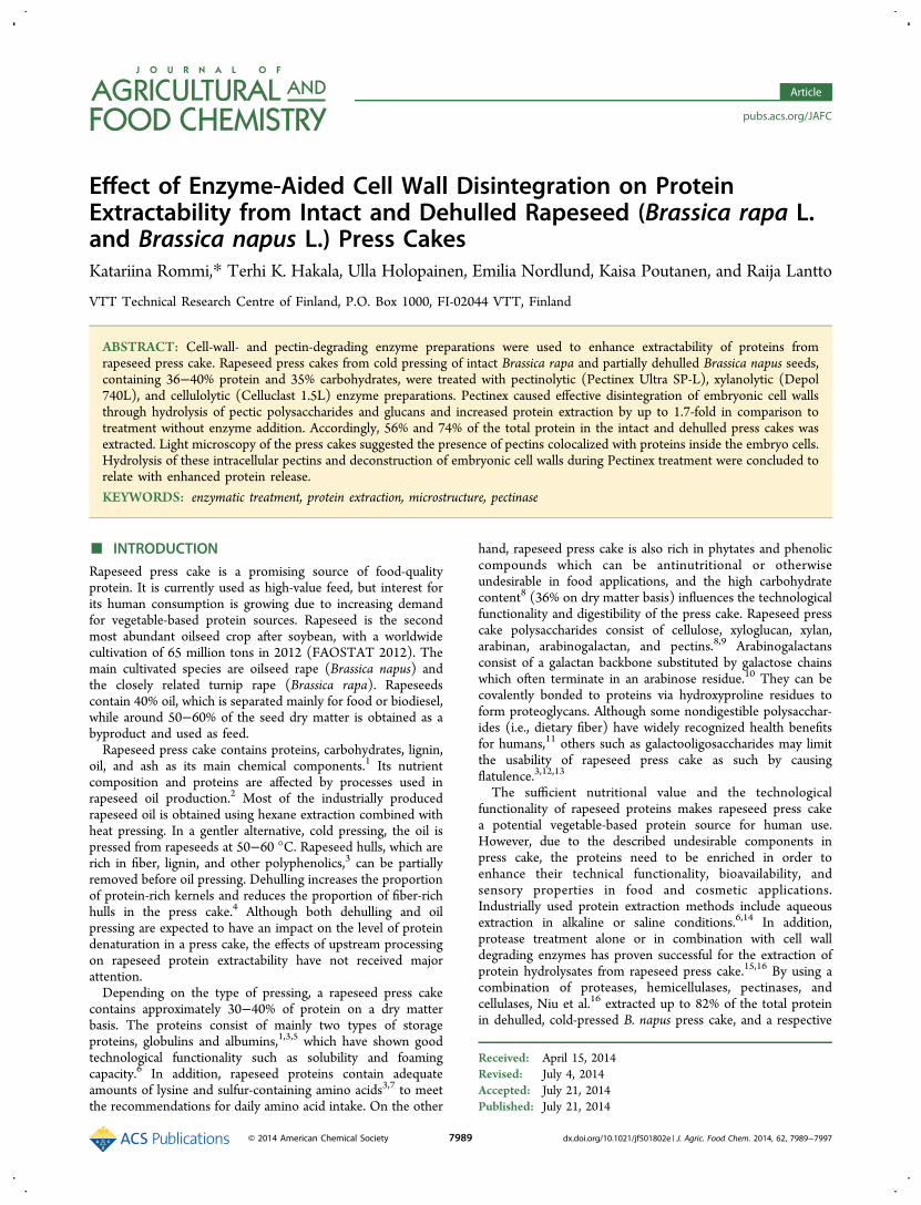
recognized (Figure 1A,B). Seed coat fragments were detectedin all samples both before and after the enzymatic treatmentand appear in some of the selected micrographs. Staining of thepress cakes with Calcofluor White visualized cellulose and otherβ-linked glucans41 in the cell walls of embryo and seed coat,whereas staining with Acid Fuchsin showed the proteins insidethe embryonic cells. Cold pressing had partially disrupted thestructure of embryonic cell walls (shown as blue) and proteinbodies (shown as red) (Figure 1A−D). Dry milling causedfurther fragmentation of the embryonic cell walls withoutnoticeable effect on proteins (Figure 3A). No clear differencewas observed in the cell wall fluorescence of the two presscakes. However, proteins seemed to form more compactstructures in the dehulled press cake than in the intact press
cake (Figure 1A,B). Such variation in the packing density ofprotein bodies was not observed in intact B. rapa and dehulledB. napus seeds obtained before oil pressing (data not shown).Pectins were visualized by staining with Ruthenium Red,
which binds to the pectin polygalacturonan backbone.39 Pectinswere abundant in the seed coat (Figure 2C) and detected insmaller amounts in the embryonic cell walls (data not shown).Pectins were more intensely stained by Ruthenium Red in theseed coat than in the embryo, which may be related todifferences in their composition: pectins in rapeseed hulls havebeen reported to contain substantially more galacturonic acidthan pectins in the embryo.3 In addition to the cell wall pectins,networks stained by Ruthenium Red were observed inside theembryo cells (Figure 1E,F), suggesting the presence of
Figure 1. Micrographs of press cakes obtained from cold pressing of intact B. rapa seeds (A, C, E) and partially dehulled B. napus seeds (B, D, F).The sections were stained with Calcofluor White and Acid Fuchsin (A−D), showing cell wall glucans as blue and proteins as red, and withRuthenium Red (E, F), showing pectins as red.
Journal of Agricultural and Food Chemistry Article
dx.doi.org/10.1021/jf501802e | J. Agric. Food Chem. 2014, 62, 7989−79977992
intracellular pectin-type material. These networks werecolocalized with proteins in the cells (shown by Acid Fuchsinstaining in Figure 1C,D). Intracellular pectic polysaccharideshave not been reported in rapeseed, but their presence wasrecently detected in the olive pistil.42
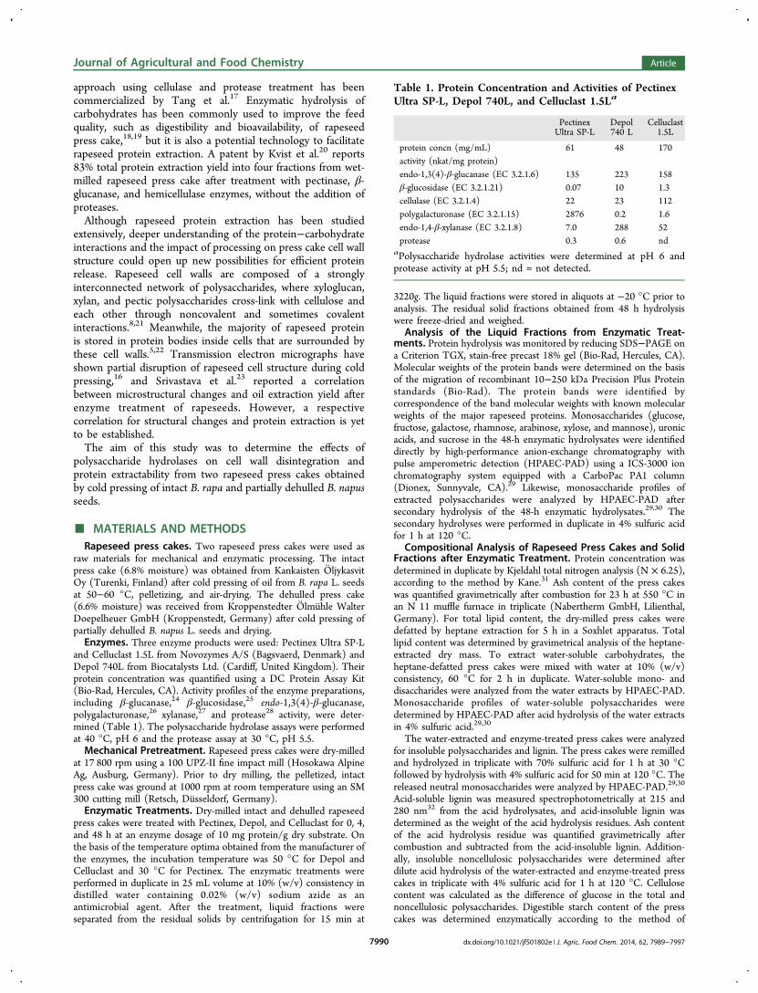
Enzymatic Hydrolysis of Rapeseed Carbohydrates.The commercial enzyme product Pectinex Ultra SP-L hydro-lyzed the rapeseed carbohydrates most effectively, solubilizingup to 75% of the carbohydrates (Figure 4) and causingcomplete degradation of the embryonic cell walls, as shown bymicroscopy (Figure 2). The total carbohydrate hydrolysis yieldwas higher than in the study reported by Pustjens et al.,18 whoextracted 64% of the total carbohydrates in solvent-extracted B.napus meal during 4.75 h hydrolysis with amyloglucosidase andproteases. The higher yield is most probably explained bylonger hydrolysis time and a better targeted enzyme productused in our study.According to the sugar profiles of the 48-h enzymatic
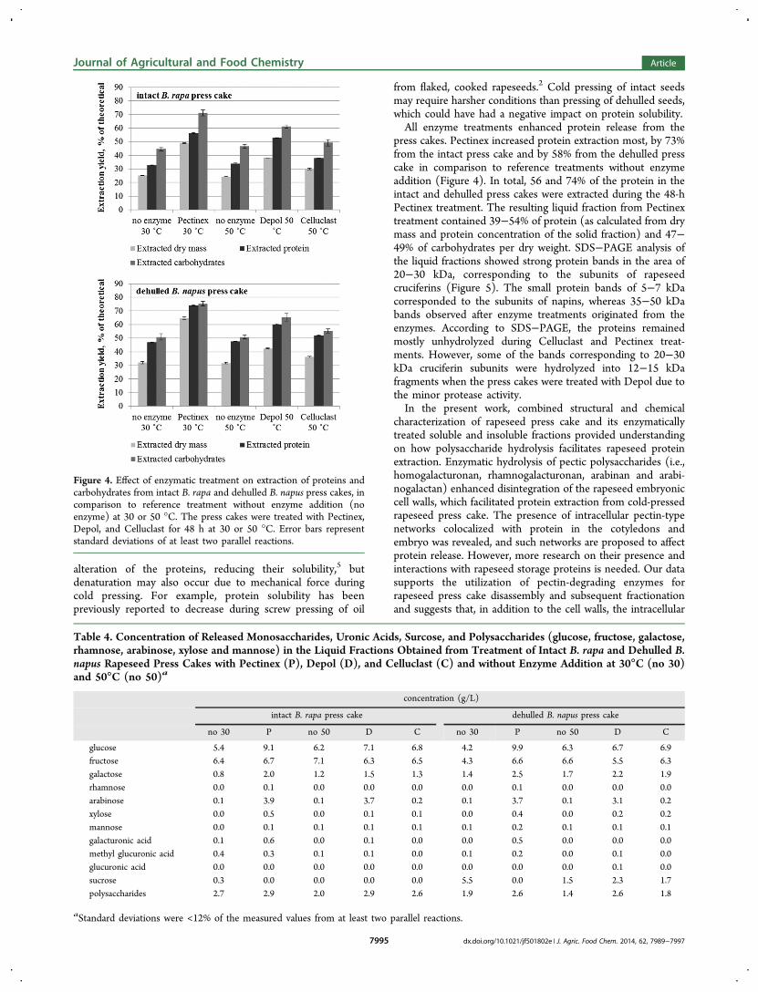
hydrolysates, carbohydrates were released almost exclusively asmonosaccharides into the liquid fractions (Table 4). Release ofgalacturonic acid by Pectinex indicated hydrolysis of pectin(homogalacturonan, rhamnogalacturonan), whereas the releaseof galactose and arabinose by both Pectinex and Depolsuggested hydrolysis of other pectic polysaccharides (arabinanand arabinogalactan). Substantial amounts of fructose andglucose were released as a result of sucrose hydrolysis during allenzymatic and reference treatments at 50 °C (Table 4).Respectively, the sucrose content in these hydrolysates wasremarkably lower or nondetectable in comparison to the 30 °C
reference treatments. Sucrose hydrolysis at 50 °C suggests thatrapeseed contains an endogenous fructosyltransferase (i.e.,invertase) with enhanced activity at above 30 °C. On the otherhand, the high fructosyltransferase activity of Pectinex43 wasobviously responsible for the hydrolysis of sucrose into glucoseand fructose at 30 °C.The three commercial enzyme preparations were selected on
the basis of the correspondence of their activity profiles withrapeseed carbohydrate composition. The Pectinex Ultra SP-Lproduct contains a variety of activities that target the rapeseedcell walls. According to enzyme activity assays, it is rich inpolygalacturonase, which is responsible for the release ofgalacturonic acid from pectins. The preparation also containsendo-β-1,4-galactanase, active on arabinogalactan,44 and β-glucanase, which hydrolyzes glucans (cellulose and xyloglucanin rapeseed). High β-glucanase and endoxylanase activities inthe Depol 740L product hydrolyzed the cellulose, xyloglucans,and xylan. However, due to the lack of polygalacturonaseactivity in Depol, no galacturonic acid was released from pectin.Celluclast 1.5L, which contained endo-1,3(4)-β-glucanase andβ-glucanase (EC 3.2.1.4) as its main activities, was expected tohydrolyze cellulose and xyloglucan.
Effects of Enzymatic Treatment on the Microstructureof Rapeseed Press Cakes. Light microscopy of the solidfractions obtained after enzyme treatment of the intact presscake revealed differences in cell wall and pectin stainability aftereach treatment. Enzyme action was mainly detected in the cellwalls of the embryo, which forms the major part of the seed andwhere most of rapeseed protein is stored.5,22 By comparison,
Figure 2. Micrographs of the solid fractions obtained from treatment of intact B. rapa press cake at 30 °C without enzyme addition (A, C) or withPectinex (B, D). The sections were stained with Calcofluor White and Acid Fuchsin (A, B), showing cell wall glucans as blue and proteins as red, andwith Ruthenium Red (C, D), showing pectins as red. Panels A−C include fragments of the seed coat.
Journal of Agricultural and Food Chemistry Article
dx.doi.org/10.1021/jf501802e | J. Agric. Food Chem. 2014, 62, 7989−79977993
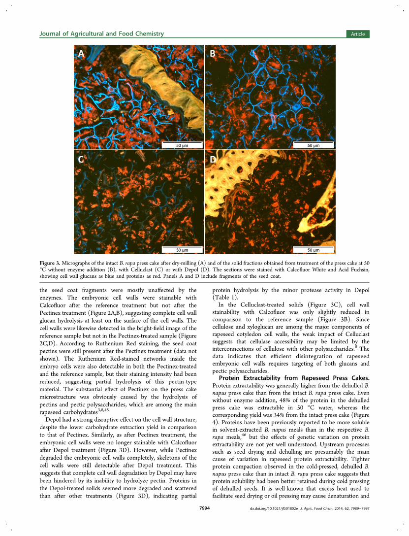
the seed coat fragments were mostly unaffected by theenzymes. The embryonic cell walls were stainable withCalcofluor after the reference treatment but not after thePectinex treatment (Figure 2A,B), suggesting complete cell wallglucan hydrolysis at least on the surface of the cell walls. Thecell walls were likewise detected in the bright-field image of thereference sample but not in the Pectinex-treated sample (Figure2C,D). According to Ruthenium Red staining, the seed coatpectins were still present after the Pectinex treatment (data notshown). The Ruthenium Red-stained networks inside theembryo cells were also detectable in both the Pectinex-treatedand the reference sample, but their staining intensity had beenreduced, suggesting partial hydrolysis of this pectin-typematerial. The substantial effect of Pectinex on the press cakemicrostructure was obviously caused by the hydrolysis ofpectins and pectic polysaccharides, which are among the mainrapeseed carbohydrates3,8,45
Depol had a strong disruptive effect on the cell wall structure,despite the lower carbohydrate extraction yield in comparisonto that of Pectinex. Similarly, as after Pectinex treatment, theembryonic cell walls were no longer stainable with Calcofluorafter Depol treatment (Figure 3D). However, while Pectinexdegraded the embryonic cell walls completely, skeletons of thecell walls were still detectable after Depol treatment. Thissuggests that complete cell wall degradation by Depol may havebeen hindered by its inability to hydrolyze pectin. Proteins inthe Depol-treated solids seemed more degraded and scatteredthan after other treatments (Figure 3D), indicating partial
protein hydrolysis by the minor protease activity in Depol(Table 1).In the Celluclast-treated solids (Figure 3C), cell wall
stainability with Calcofluor was only slightly reduced incomparison to the reference sample (Figure 3B). Sincecellulose and xyloglucan are among the major components ofrapeseed cotyledon cell walls, the weak impact of Celluclastsuggests that cellulase accessibility may be limited by theinterconnections of cellulose with other polysaccharides.8 Thedata indicates that efficient disintegration of rapeseedembryonic cell walls requires targeting of both glucans andpectic polysaccharides.
Protein Extractability from Rapeseed Press Cakes.Protein extractability was generally higher from the dehulled B.napus press cake than from the intact B. rapa press cake. Evenwithout enzyme addition, 48% of the protein in the dehulledpress cake was extractable in 50 °C water, whereas thecorresponding yield was 34% from the intact press cake (Figure4). Proteins have been previously reported to be more solublein solvent-extracted B. napus meals than in the respective B.rapa meals,46 but the effects of genetic variation on proteinextractability are not yet well understood. Upstream processessuch as seed drying and dehulling are presumably the maincause of variation in rapeseed protein extractability. Tighterprotein compaction observed in the cold-pressed, dehulled B.napus press cake than in intact B. rapa press cake suggests thatprotein solubility had been better retained during cold pressingof dehulled seeds. It is well-known that excess heat used tofacilitate seed drying or oil pressing may cause denaturation and
Figure 3. Micrographs of the intact B. rapa press cake after dry-milling (A) and of the solid fractions obtained from treatment of the press cake at 50°C without enzyme addition (B), with Celluclast (C) or with Depol (D). The sections were stained with Calcofluor White and Acid Fuchsin,showing cell wall glucans as blue and proteins as red. Panels A and D include fragments of the seed coat.
Journal of Agricultural and Food Chemistry Article
dx.doi.org/10.1021/jf501802e | J. Agric. Food Chem. 2014, 62, 7989−79977994
alteration of the proteins, reducing their solubility,5 butdenaturation may also occur due to mechanical force duringcold pressing. For example, protein solubility has beenpreviously reported to decrease during screw pressing of oil
from flaked, cooked rapeseeds.2 Cold pressing of intact seedsmay require harsher conditions than pressing of dehulled seeds,which could have had a negative impact on protein solubility.All enzyme treatments enhanced protein release from the
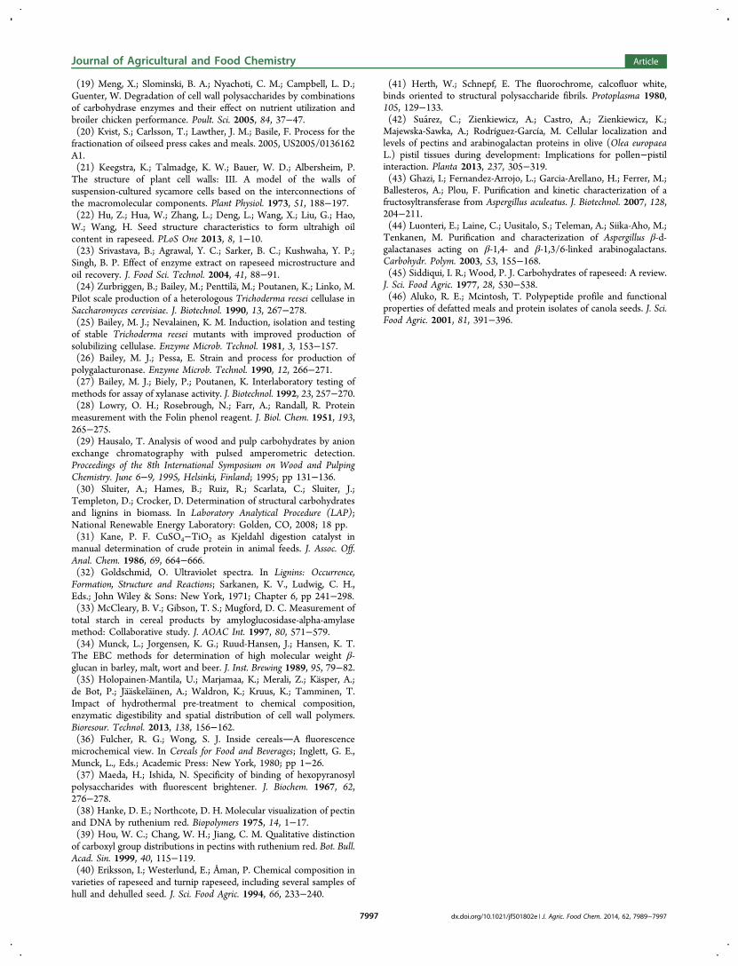
press cakes. Pectinex increased protein extraction most, by 73%from the intact press cake and by 58% from the dehulled presscake in comparison to reference treatments without enzymeaddition (Figure 4). In total, 56 and 74% of the protein in theintact and dehulled press cakes were extracted during the 48-hPectinex treatment. The resulting liquid fraction from Pectinextreatment contained 39−54% of protein (as calculated from drymass and protein concentration of the solid fraction) and 47−49% of carbohydrates per dry weight. SDS−PAGE analysis ofthe liquid fractions showed strong protein bands in the area of20−30 kDa, corresponding to the subunits of rapeseedcruciferins (Figure 5). The small protein bands of 5−7 kDacorresponded to the subunits of napins, whereas 35−50 kDabands observed after enzyme treatments originated from theenzymes. According to SDS−PAGE, the proteins remainedmostly unhydrolyzed during Celluclast and Pectinex treat-ments. However, some of the bands corresponding to 20−30kDa cruciferin subunits were hydrolyzed into 12−15 kDafragments when the press cakes were treated with Depol due tothe minor protease activity.In the present work, combined structural and chemical
characterization of rapeseed press cake and its enzymaticallytreated soluble and insoluble fractions provided understandingon how polysaccharide hydrolysis facilitates rapeseed proteinextraction. Enzymatic hydrolysis of pectic polysaccharides (i.e.,homogalacturonan, rhamnogalacturonan, arabinan and arabi-nogalactan) enhanced disintegration of the rapeseed embryoniccell walls, which facilitated protein extraction from cold-pressedrapeseed press cake. The presence of intracellular pectin-typenetworks colocalized with protein in the cotyledons andembryo was revealed, and such networks are proposed to affectprotein release. However, more research on their presence andinteractions with rapeseed storage proteins is needed. Our datasupports the utilization of pectin-degrading enzymes forrapeseed press cake disassembly and subsequent fractionationand suggests that, in addition to the cell walls, the intracellular
Figure 4. Effect of enzymatic treatment on extraction of proteins andcarbohydrates from intact B. rapa and dehulled B. napus press cakes, incomparison to reference treatment without enzyme addition (noenzyme) at 30 or 50 °C. The press cakes were treated with Pectinex,Depol, and Celluclast for 48 h at 30 or 50 °C. Error bars representstandard deviations of at least two parallel reactions.
Table 4. Concentration of Released Monosaccharides, Uronic Acids, Surcose, and Polysaccharides (glucose, fructose, galactose,rhamnose, arabinose, xylose and mannose) in the Liquid Fractions Obtained from Treatment of Intact B. rapa and Dehulled B.napus Rapeseed Press Cakes with Pectinex (P), Depol (D), and Celluclast (C) and without Enzyme Addition at 30°C (no 30)and 50°C (no 50)a
concentration (g/L)
intact B. rapa press cake dehulled B. napus press cake
no 30 P no 50 D C no 30 P no 50 D C
glucose 5.4 9.1 6.2 7.1 6.8 4.2 9.9 6.3 6.7 6.9fructose 6.4 6.7 7.1 6.3 6.5 4.3 6.6 6.6 5.5 6.3galactose 0.8 2.0 1.2 1.5 1.3 1.4 2.5 1.7 2.2 1.9rhamnose 0.0 0.1 0.0 0.0 0.0 0.0 0.1 0.0 0.0 0.0arabinose 0.1 3.9 0.1 3.7 0.2 0.1 3.7 0.1 3.1 0.2xylose 0.0 0.5 0.0 0.1 0.1 0.0 0.4 0.0 0.2 0.2mannose 0.0 0.1 0.1 0.1 0.1 0.1 0.2 0.1 0.1 0.1galacturonic acid 0.1 0.6 0.0 0.1 0.0 0.0 0.5 0.0 0.0 0.0methyl glucuronic acid 0.4 0.3 0.1 0.1 0.0 0.1 0.2 0.0 0.1 0.0glucuronic acid 0.0 0.0 0.0 0.0 0.0 0.0 0.0 0.0 0.1 0.0sucrose 0.3 0.0 0.0 0.0 0.0 5.5 0.0 1.5 2.3 1.7polysaccharides 2.7 2.9 2.0 2.9 2.6 1.9 2.6 1.4 2.6 1.8
aStandard deviations were <12% of the measured values from at least two parallel reactions.
Journal of Agricultural and Food Chemistry Article
dx.doi.org/10.1021/jf501802e | J. Agric. Food Chem. 2014, 62, 7989−79977995
matrix should be targeted for enhanced protein release. Oncequalified for technological functionality, rapeseed proteinconcentrates produced by enzyme-aided extraction may finduse in food, feed, and cosmetic applications.
■ AUTHOR INFORMATIONCorresponding Author*Phone: +358 20 722 4514. Fax: +358 20 722 7071. E-mail:[email protected].
FundingThe research leading to these results has received funding fromthe European Community’s Seventh Framework ProgrammeFP7/2007-2013 under grant agreement no. 289170−APRO-POS. Funding from Academy of Finland to K.P. is likewisegratefully acknowledged.
NotesThe authors declare no competing financial interest.
■ ACKNOWLEDGMENTSThe authors wish to thank Kankaisten Oljykasvit Oy,Kroppenstedter Olmuhle Walter Doepelheuer GmbH, andIGV Institut fur Getreideverarbeitung GmbH for rapeseed presscakes. The authors thank Mari Raulio, Juhani Sibakov, and AtteMikkelson at the VTT Technical Research Centre of Finlandfor cooperation in the rapeseed microscopy mechanicalpretreatment, and carbohydrate analytics and VTT’s experi-enced technical staff for their valuable experimental work.
■ REFERENCES(1) Schone, F.; Kirchheim, U.; Schumann, W.; Ludke, H. Apparentdigestibility of high-fat rapeseed press cake in growing pigs and effectson feed intake, growth and weight of thyroid and liver. Anim. Feed Sci.Technol. 1996, 62, 97−110.(2) Mustafa, A. F.; Christensen, D.; McKinnon, J.; Newkirk, R.Effects of stage of processing of canola seed on chemical compositionand in vitro protein degradability of canola meal and intermediateproducts. Can. J. Anim. Sci. 2000, 80, 211−214.(3) Shahidi, F. Canola and Rapeseed: Production, Chemistry, Nutritionand Processing Technology; Van Nostrand Reinhold: New York, 1990;355 pp.(4) Mustafa, A. F.; Christensen, D.; McKinnon, J. Chemicalcharacterization and nutrient availability of high and low fiber canolameal. Can. J. Anim. Sci. 1996, 76, 579−586.
(5) Wanasundara, J. Proteins of Brassicaceae oilseeds and theirpotential as a plant protein source. Crit. Rev. Food Sci. Nutr. 2011, 51,635−677.(6) Ghodsvali, A.; Khodaparast, M. H.; Vosoughi, M.; Diosady, L. L.Preparation of canola protein materials using membrane technologyand evaluation of meals functional properties. Food Res. Int. 2005, 38,223−231.(7) Bell, J.; Keith, M. A. Survey of variation in the chemicalcomposition of commercial canola meal produced in WesternCanadian crushing plants. Can. J. Anim. Sci. 1991, 71, 469−480.(8) Pustjens, A. M.; Schols, H. A.; Kabel, M. A.; Gruppen, H.Characterisation of cell wall polysaccharides from rapeseed (Brassicanapus) meal. Carbohydr. Polym. 2013, 98, 1650−1656.(9) Ghosh, P.; Ghosal, P.; Thakur, S.; Lerouge, P.; Loutelier-Bourhis,C.; Driouich, A.; Ray, B. Cell wall polysaccharides of Brassicacampestris seed cake: isolation and structural features. Carbohydr.Polym. 2004, 57, 7−13.(10) Gaspar, Y.; Johnson, K. L.; McKenna, J. A.; Bacic, A.; Schultz, C.J. The complex structures of arabinogalactan−proteins and the journeytowards understanding function. Plant Mol. Biol. 2001, 47, 161−176.(11) Anderson, J. W.; Baird, P.; Davis, R. H., Jr; Ferreri, S.; Knudtson,M.; Koraym, A.; Waters, V.; Williams, C. L. Health benefits of dietaryfiber. Nutr. Rev. 2009, 67, 188−205.(12) Simbaya, J.; Slominski, B.; Rakow, G.; Campbell, L.; Downey,R.; Bell, J. Quality characteristics of yellow-seeded Brassica seed meals:Protein, carbohydrates and dietary fiber components. J. Agric. FoodChem. 1995, 43, 2062−2066.(13) Slominski, B. A.; Campbell, L. D. Non-starch polysaccharides ofcanola meal: quantification, digestibility in poultry and potentialbenefit of dietary enzyme supplementation. J. Sci. Food Agric. 1990, 53,175−184.(14) Berot, S.; Compoint, J. P.; Larre, C.; Malabat, C.; Gueguen, J.Large scale purification of rapeseed proteins (Brassica napus L.). J.Chromatogr. B: Anal. Technol. Biomed. Life Sci. 2005, 818, 35−42.(15) Sari, Y. W.; Bruins, M. E.; Sanders, J. P. Enzyme assisted proteinextraction from rapeseed, soybean, and microalgae meals. Ind. CropsProd. 2013, 43, 78−83.(16) Niu, Y. X.; Li, W.; Zhu, J.; Huang, Q.; Jiang, M.; Huang, F.Aqueous enzymatic extraction of rapeseed oil and protein fromdehulled cold-pressed double-low rapeseed cake. Int. J. Food Eng. 2012,8, 1−14.(17) Tang, Q. N. Protein concentrates and isolates, and processes for theproduction thereof. 2010, US 2010/0136173 A1.(18) Pustjens, A. M.; de Vries, S.; Gerrits, W.; Kabel, M.; Schols, H.;Gruppen, H. Residual carbohydrates from in vitro digested processedrapeseed (Brassica napus) meal. J. Agric. Food Chem. 2012, 60, 8257−8263.
Figure 5. SDS−PAGE patterns of proteins released during enzymatic treatment of intact and dehulled rapeseed press cakes. The materials werehydrolyzed for 48 h with Celluclast (C), Depol (D), and Pectinex (P) at an enzyme dosage of 10 mg protein/g dry substrate. No enzyme was addedin the reference treatments carried out at 50 °C (No 50) and 30 °C (No 30).
Journal of Agricultural and Food Chemistry Article
dx.doi.org/10.1021/jf501802e | J. Agric. Food Chem. 2014, 62, 7989−79977996
(19) Meng, X.; Slominski, B. A.; Nyachoti, C. M.; Campbell, L. D.;Guenter, W. Degradation of cell wall polysaccharides by combinationsof carbohydrase enzymes and their effect on nutrient utilization andbroiler chicken performance. Poult. Sci. 2005, 84, 37−47.(20) Kvist, S.; Carlsson, T.; Lawther, J. M.; Basile, F. Process for thefractionation of oilseed press cakes and meals. 2005, US2005/0136162A1.(21) Keegstra, K.; Talmadge, K. W.; Bauer, W. D.; Albersheim, P.The structure of plant cell walls: III. A model of the walls ofsuspension-cultured sycamore cells based on the interconnections ofthe macromolecular components. Plant Physiol. 1973, 51, 188−197.(22) Hu, Z.; Hua, W.; Zhang, L.; Deng, L.; Wang, X.; Liu, G.; Hao,W.; Wang, H. Seed structure characteristics to form ultrahigh oilcontent in rapeseed. PLoS One 2013, 8, 1−10.(23) Srivastava, B.; Agrawal, Y. C.; Sarker, B. C.; Kushwaha, Y. P.;Singh, B. P. Effect of enzyme extract on rapeseed microstructure andoil recovery. J. Food Sci. Technol. 2004, 41, 88−91.(24) Zurbriggen, B.; Bailey, M.; Penttila, M.; Poutanen, K.; Linko, M.Pilot scale production of a heterologous Trichoderma reesei cellulase inSaccharomyces cerevisiae. J. Biotechnol. 1990, 13, 267−278.(25) Bailey, M. J.; Nevalainen, K. M. Induction, isolation and testingof stable Trichoderma reesei mutants with improved production ofsolubilizing cellulase. Enzyme Microb. Technol. 1981, 3, 153−157.(26) Bailey, M. J.; Pessa, E. Strain and process for production ofpolygalacturonase. Enzyme Microb. Technol. 1990, 12, 266−271.(27) Bailey, M. J.; Biely, P.; Poutanen, K. Interlaboratory testing ofmethods for assay of xylanase activity. J. Biotechnol. 1992, 23, 257−270.(28) Lowry, O. H.; Rosebrough, N.; Farr, A.; Randall, R. Proteinmeasurement with the Folin phenol reagent. J. Biol. Chem. 1951, 193,265−275.(29) Hausalo, T. Analysis of wood and pulp carbohydrates by anionexchange chromatography with pulsed amperometric detection.Proceedings of the 8th International Symposium on Wood and PulpingChemistry. June 6−9, 1995, Helsinki, Finland; 1995; pp 131−136.(30) Sluiter, A.; Hames, B.; Ruiz, R.; Scarlata, C.; Sluiter, J.;Templeton, D.; Crocker, D. Determination of structural carbohydratesand lignins in biomass. In Laboratory Analytical Procedure (LAP);National Renewable Energy Laboratory: Golden, CO, 2008; 18 pp.(31) Kane, P. F. CuSO4−TiO2 as Kjeldahl digestion catalyst inmanual determination of crude protein in animal feeds. J. Assoc. Off.Anal. Chem. 1986, 69, 664−666.(32) Goldschmid, O. Ultraviolet spectra. In Lignins: Occurrence,Formation, Structure and Reactions; Sarkanen, K. V., Ludwig, C. H.,Eds.; John Wiley & Sons: New York, 1971; Chapter 6, pp 241−298.(33) McCleary, B. V.; Gibson, T. S.; Mugford, D. C. Measurement oftotal starch in cereal products by amyloglucosidase-alpha-amylasemethod: Collaborative study. J. AOAC Int. 1997, 80, 571−579.(34) Munck, L.; Jorgensen, K. G.; Ruud-Hansen, J.; Hansen, K. T.The EBC methods for determination of high molecular weight β-glucan in barley, malt, wort and beer. J. Inst. Brewing 1989, 95, 79−82.(35) Holopainen-Mantila, U.; Marjamaa, K.; Merali, Z.; Kasper, A.;de Bot, P.; Jaaskelainen, A.; Waldron, K.; Kruus, K.; Tamminen, T.Impact of hydrothermal pre-treatment to chemical composition,enzymatic digestibility and spatial distribution of cell wall polymers.Bioresour. Technol. 2013, 138, 156−162.(36) Fulcher, R. G.; Wong, S. J. Inside cerealsA fluorescencemicrochemical view. In Cereals for Food and Beverages; Inglett, G. E.,Munck, L., Eds.; Academic Press: New York, 1980; pp 1−26.(37) Maeda, H.; Ishida, N. Specificity of binding of hexopyranosylpolysaccharides with fluorescent brightener. J. Biochem. 1967, 62,276−278.(38) Hanke, D. E.; Northcote, D. H. Molecular visualization of pectinand DNA by ruthenium red. Biopolymers 1975, 14, 1−17.(39) Hou, W. C.; Chang, W. H.; Jiang, C. M. Qualitative distinctionof carboxyl group distributions in pectins with ruthenium red. Bot. Bull.Acad. Sin. 1999, 40, 115−119.(40) Eriksson, I.; Westerlund, E.; Åman, P. Chemical composition invarieties of rapeseed and turnip rapeseed, including several samples ofhull and dehulled seed. J. Sci. Food Agric. 1994, 66, 233−240.
(41) Herth, W.; Schnepf, E. The fluorochrome, calcofluor white,binds oriented to structural polysaccharide fibrils. Protoplasma 1980,105, 129−133.(42) Suarez, C.; Zienkiewicz, A.; Castro, A.; Zienkiewicz, K.;Majewska-Sawka, A.; Rodríguez-García, M. Cellular localization andlevels of pectins and arabinogalactan proteins in olive (Olea europaeaL.) pistil tissues during development: Implications for pollen−pistilinteraction. Planta 2013, 237, 305−319.(43) Ghazi, I.; Fernandez-Arrojo, L.; Garcia-Arellano, H.; Ferrer, M.;Ballesteros, A.; Plou, F. Purification and kinetic characterization of afructosyltransferase from Aspergillus aculeatus. J. Biotechnol. 2007, 128,204−211.(44) Luonteri, E.; Laine, C.; Uusitalo, S.; Teleman, A.; Siika-Aho, M.;Tenkanen, M. Purification and characterization of Aspergillus β-d-galactanases acting on β-1,4- and β-1,3/6-linked arabinogalactans.Carbohydr. Polym. 2003, 53, 155−168.(45) Siddiqui, I. R.; Wood, P. J. Carbohydrates of rapeseed: A review.J. Sci. Food Agric. 1977, 28, 530−538.(46) Aluko, R. E.; Mcintosh, T. Polypeptide profile and functionalproperties of defatted meals and protein isolates of canola seeds. J. Sci.Food Agric. 2001, 81, 391−396.
Journal of Agricultural and Food Chemistry Article
dx.doi.org/10.1021/jf501802e | J. Agric. Food Chem. 2014, 62, 7989−79977997