effect bicarbonate, (na2edta), and visual
TRANSCRIPT

J. Neurol. Neurosurg. Psychiat., 1970, 33, 723-732
Effect of intravenous sodium bicarbonate, disodiumedetate (Na2EDTA), and hyperventilation on visual
and oculomotor signs in multiple sclerosisFLOYD A. DAVIS, FRANK 0. BECKER, JOEL A. MICHAEL,
AND ERIC SORENSEN
From the Department of Neurology, Presbyterian-St. Luke's Hospital, Chicago, Illinois, U.S.A.
SUMMARY The effects of procedures believed to produce a decrease in serum ionized calcium weretested on visual and oculomotor function in nine multiple sclerosis (MS) patients. Transient im-provement in scotomas, nystagmus, and oculomotor paresis occurred with intravenous infusions ofNaHCO3 or Na2EDTA. Hyperventilation was also tested for its effect on nystagmus and causedmarked decreases in frequency. Control experiments with saline infusions did not produce anyeffect. The probable mechanism and site of action for these effects is discussed. This study demon-strates that certain signs and symptoms in MS can be altered favourably by changes in the internalchemical environment and offers a new approach to the search for a symptomatic therapy in MS.
Multiple sclerosis (MS) is a disease affecting thewhite matter of the central nervous system. It ischaracterized pathologically by numerous discreteareas of myelin loss along the course of involvedaxons. These demyelinated lesions are believed tocause a slowing and block of conduction which inturn is responsible for the clinical signs (Charcot,1877; Namerow, 1968a;Namerow and Kappl, 1969).There is reason to believe that this axonal con-
duction defect is labile and capable of being modifiedin both a favourable and unfavourable manner. Thisis suggested by the remarkable fact that smallchanges in body temperature can markedly alterspecific signs in MS (Simons, 1937; Brickner, 1950;Guthrie, 1951; Edmund and Fog, 1955; Nelson,Jeffreys, and McDowell, 1958; Boynton, Garramone,and Buca, 1959; Nelson and McDowell, 1959;Watson, 1959). Heating and cooling cause a transientworsening and improvement respectively. A recentreport described an MS patient with optic nerveinvolvement whose visual acuity decreased from5/40 to 5/200 after only a 0-25°C temperature rise(Namerow, 1968b). These effects only last as longas the temperature change is maintained. They arenot specific for MS, since other neurological diseasesare aggravated by hyperthermia. However, theyclearly occur with greater frequency and sensitivityin MS (Nelson et al., 1958; Nelson and McDowell,1959; Namerow, 1968b).
Since an understanding of these temperatureeffects (especially that of cooling) might reveal arational basis for a symptomatic treatment in MS,earlier studies were aimed at developing a simple butpertinent conduction model. Studies in a variety ofanimals-frog (Davis, 1969), lobster (Davis, 1970),guinea-pig (Davis and Jacobson, 1970)-havedemonstrated that demyelinated and injured nerveshave a heightened sensitivity to reversible thermalblock-that is, conduction fails at temperatureswithin or close to the physiological range. Conversely,it has been shown that cooling restores conductionin injured nerves (Davis, 1970). It has been postulatedthat these effects are related to critical alterations inthe conduction safety factor (Davis, 1970). Thesafety factor is defined as the ratio of the actioncurrent of a nerve fibre to the minimum currentnecessary to maintain conduction (Tasaki, 1953).This ratio reflects the net effect of many variablesinvolved in the maintenance of axonal conduction.Tasaki found a value of 5 to 7 for the safety factorin amphibian myelinated nerve (Tasaki, 1953).Theoretically, if the safety factor is decreased byaxonal injury or disease the conduction mechanismsshould become more vulnerable and might fail with afurther lowering brought on by adverse alterationsin pH, electrolytes, temperature, etc., even thoughsimilar changes do not block normal nerve. On theother hand, conditions that increase the safety
723
guest. Protected by copyright.
on March 31, 2022 by
http://jnnp.bmj.com
/J N
eurol Neurosurg P
sychiatry: first published as 10.1136/jnnp.33.6.723 on 1 Decem
ber 1970. Dow
nloaded from

Floyd A. Davis, Frank 0. Becker, Joel A. AMichael, and Eric Sorensen
factor might improve function in diseased nerve.Heating and cooling might then be thought of as
causing a decrease and increase respectively in thesafety factor.
This hypothesis offers a reasonable explanation forthe temperature effects in MS. It also predicts thatpharmacological agents that can increase the safetyfactor might improve clinical signs in MS. It is wellknown that calcium ion depletion lowers thethreshold for excitation in axons (Misske, 1930;Brink, Bronk, and Larrabee, 1946). Since this woulddecrease the minimum current needed to maintainconduction, it should increase the safety factor. Insupport of this, it has been demonstrated thatconduction block in injured nerve is reversed bylowering the concentration of calcium ions in thebathing medium (Davis, 1969).
In this paper we report the effect on visual andoculomotor defects in MS of three proceduresbelieved to lower serum ionized calcium. It has beenfound that infusions of sodium bicarbonate(NaHCO3) and sodium edetate (Na2EDTA) as wellas hyperventilation improve clinical findings in MS.This study was, in part, previously reported in abrief preliminary communication (Davis, Becker,Michael, and Sorensen, 1970).
METHODS
SUBJECTS Nine MS patients were studied. All hadevidence for disseminated lesions and, except for one, acourse characterized by exacerbations and remissions.Pertinent data are presented in Table 1. The criteria forpatient selection (in addition to a firm diagnosis of MS)was the presence of a visual field scotoma and/or lateralgaze nystagmus.
PROTOCOL Intravenous infusions of NaHCO3 andNa2EDTA were tested for effects on scotomas andnystagmus. In addition, the effect of hyperventilation onnystagmus was studied. In those patients who had both
scotomas and nystagmus, only one of these findings wastested in any single experiment and an interval of atleast seven days separated each additional experiment.As described below, the selected clinical finding was
monitored before, during and after either hyperventi-lation or an intravenous infusion.
Hyperventilation was performed around 30 to 40breaths per minute for varying periods as indicated in theResults section. NaHCO3 infusions consisted of a 5%solution and, except where otherwise noted, were givenat 15 to 25 ml./min. Na2EDTA (1-5 g/1,000 ml. normalsaline) was infused in total amounts of 2 to 3 g over 50 to80 minutes. Blood samples were drawn for calcium andacid-base studies during some experiments.
MEASUREMENTS OF VISUAL SCOTOMAS Subjects partici-pating in the visual field experiments had initial ophthal-mological evaluations consisting of visual acuity de-termination, refraction (both manifest and by retino-scopy), slit lamp examination, and funduscopicexamination to rule out ocular disease. Ocular tensionswere also tested. Except for the changes frequently seenin MS, as well as minor refractory changes, these testswere not remarkable.
Visual field testing was performed with a Bausch andLomb Autoplot Tangent Screen under the standardconditions specified in the instrument's operating manual.They were performed by one of the authors (E.S.) who isan ophthalmology resident and experienced with thetesting of fields with this apparatus. Targets were selectedthat gave the most distinct outline of the central fielddefect for the particular eye being tested.
Refractive errors in these patients were small and in nocase did correction improve acuity more than one-halfline on a Snellen chart. For this reason, field testing wasperformed without corrective lenses. In some experimentsvisual acuity was determined with a Snellen chart aftereach field determination.
MEASUREMENT OF NYSTAGMUS The subject was seated infront of a perimeter on which were mounted three smalllights. One light was mounted in the centre so that uponfixation of it the patient's eyes were in the primaryposition. The other two lights were placed to the right
LE 1PATIENT DATA
Patient Sex Age Duration of Exacerbations Evidence for Most recent(yr) disease and remissions dissem. lesions exacerbation
(months before testing)
R.W. F 21 7 mth Yes Nystagmus and central scotoma 2E.M. F 47 5 yr No Nystagmus and paracentral -
(chronic progress- scotomaive course)
C.N. M 39 2 yr Yes Nystagmus and central scotoma 11T.A. M 32 4 yr Yes Nystagmus and central scotoma 14D.R. F 44 4-5 yr Yes Nystagmus and dysphasia 15M.F. F 26 15 mth Yes Nystagmus and paracentral scotoma 2P.W. F 31 2 yr Yes Ataxia and paracentral scotoma 12D.P. F 37 1-5 yr Yes Central scotoma and Hx 10
numbness-tingling, right legM.S. F 25 8 mth Yes Paraplegia and paracentral scotoma 1
724
guest. Protected by copyright.
on March 31, 2022 by
http://jnnp.bmj.com
/J N
eurol Neurosurg P
sychiatry: first published as 10.1136/jnnp.33.6.723 on 1 Decem
ber 1970. Dow
nloaded from

Effect oJ intravenous sodium bicarbonate in multiple sclerosis
and left of centre at positions that (1) resulted in maximumnystagmus upon their fixation, (2) did not cause sub-jectively uncomfortable strain, and (3) allowed binocularfixation of targets (in several patients with dysconjugateeye movements testing was performed monocularly bypatching one eye). A rigidly fixed head position wasmaintained by means of a modified dental chair headrest.Eye movements were recorded electro-oculographically
(Shackel, 1960) using gold surface electrodes (BeckmanE5G) placed at the canthi of each eye. Two AC-coupledamplifiers (bandpass 0-10,000 Hz, gain of 1,000) wereused to increase the signal levels of the corneoretinalpotential of each eye.
Subjects were instructed to maintain fixation on thelight that was lit, and told to look 'left' or 'right' as thelights were turned on and off. Thus, an alternatingpattern of left lateral, central, and right lateral gaze waselicited with nystagmus appearing during periods oflateral gaze. The duration of fixation at any positionvaried from 10 to 25 seconds. At the beginning of eachexperiment a 2 to 3 minute control run was obtained. Theprocedure to be tested was then begun and nystagmus wasmonitored periodically throughout the experiment.
Quantitative measurements were obtained by deter-mining the frequency of nystagmus; the use of AC-coupled amplifiers and the usual long-term changes inelectrode impedance made it difficult to monitor theamplitude of nystagmus quantitatively in a systematicway.With the movements of both eyes being monitored
independently and with nystagmus often appearing onboth left and right lateral gaze, four possible measure-ments were obtainable. The quality of the records foreach eye was rarely equal because of differences ofelectrode impedance and the noise characteristics of eachrecording channel, as well as the fact that nystagmus was
sometimes more marked in one eye, usually the abductingeye. Because of this, the particular record with thehighest signal-to-noise ratio was selected for quantitativeanalysis. Thus, in each experiment the nystagmus of oneeye in one particular direction of lateral gaze is consistentlyreported. This was acceptable since the changes innystagmus on conjugate gaze were qualitatively similarin both eyes with all the experimental procedures.
CHEMICAL DETERMINATIONS Total serum calcium wasdetermined by means of the EDTA titration technique(Bett and Fraser, 1959) and also by the fluorometricmethod (Jackson, Breen, and Chen, 1962) during theNa2EDTA infusions. Ultrafiltrable calcium was obtainedby the technique of Toribara (Toribara, Terepka, andDewey, 1957). The ionized calcium was approximatedduring an Na2EDTA infusion experiment by determiningthe calcium in the ultrafiltrate by the EDTA titrationmethod. This value represents the non-EDTA-complexedcalcium in the ultrafiltrate, which is mainly in the ionizedform, but also includes small amounts of complexedcalcium not bound to EDTA.
RESULTS
VISUAL STUDIES Infusions of NaHCO3 andNa2EDTA each resulted in marked decreases in thesize of visual scotomas. The effect appears graduallywithin an hour after starting an infusion and slowlyreverses within a similar period of time after infusion.Figure 1 shows a typical experiment with NaHCO3.Table 2 summarizes the results of six experiments onfour MS patients; scotoma size is expressed in area(sq. cm) measured directly from the standard visualfield record with a planimeter. Because of the short-
Is"
I 7 )Imi FIG. 1. Scotoma plotted with a1 mm white target before(upper left), during (uppermiddle and right), and after(bottom row) intravenousinfusion ofNaHCO3. Thescotoma slowly decreases insize during the infusion andthen slowly increases,approaching control size aftercessation of infusion.
725
Pwf- ;nt 63mnu
guest. Protected by copyright.
on March 31, 2022 by
http://jnnp.bmj.com
/J N
eurol Neurosurg P
sychiatry: first published as 10.1136/jnnp.33.6.723 on 1 Decem
ber 1970. Dow
nloaded from

Floyd A. Davis, Frank 0. Becker, Joel A. Michael, and Eric Sorensen
TABLE 2EFFECT OF NaHCO3 AND Na2EDTA INFUSIONS ON SCOTOMA SIZE
Control Infusion Post-infusion
Patient Date Target Eye Area of Type of Amt. of infusion Area of Percent Time of Area of Percent(mnz) control infusion at max. response scotoma control final post- scotoma of
scotoma at time (%) infusion at time control(sq. cm) (ml.) (m-equiv) of max. field (min) offinal (M/,)
response field(sq. cm) (sq. cm)
1.C I white R 55 3 NaHCO3 900 534 24-2 43-8 120 40 9 700
D.P. 30 Jan. '70 qL 3 white L 56 7 NaHCO3 900 534 13 3 23 5 125 37 1 65 4
2.{ red R 18 3 NaHCO, 675 400 7-8 42 6 60 17 6 96-2
M.F. 6 Feb. '70 1 (99°F)I white R 4.0 NaHCO3 675 400 0 3 7 5 65 4-0 100 0
(99°F)r I red R 17 0 Na2EDTA 1300 2 0 g 4-8 28 2 80 16 8 98-6
25 Feb.'70(0 5 white R 5-7 Na2EDTA 1300 2-0 g 1 4 24 6 85 6 6 115 8
3.I white R 7 2 NaHCO3 500 297 1-6 22 3 60 4 8 66 7
P.W. 6 Mar. '702 white L 3-1 NaHCO3 500 297 0 0 0.0 65 3-3 106 5
13 Mar. '70 3 red L 7 9 NaHCO3 800 475 1.1 13 9 Patient nauseated and experimentdiscontinued
4.M.S. 17 Mar. '70 6 white R 55 0 NaHCO3 800 475 27 2 49 5 60 47 4 85-5
lived effect of hyperventilation and the time necessaryfor plotting fields, hyperventilation was not studied.Improvement in visual acuity from 20/400 to
20/200 was noted in one patient whose scotoma wascentrally located. This experiment is shown in Fig. 2.Note also that, although the scotoma did not return
Post hnt -S.'mlw)
fully to control size, its configuration has beenlargely restored. In all other patients scotomas wereparacentral in location and baseline visual acuitieswere either normal or mildly affected (up to 20/40).Significant changes in acuity were not observed inthese patients.
FIG. 2. Scotoma plotted with a3 mm white target before(upper left), during (uppermiddle and right), and after(bottom row) intravenousinfusion of NaHCO3. Visualacuity improvedfrom controllevel 20/400 to 20/200during infusion and graduallyreturned to 20/400 postinfusion.
726
41
I
i. d.
fI
I.' ; 2"',W.
Post Inf.-83niW-W,
guest. Protected by copyright.
on March 31, 2022 by
http://jnnp.bmj.com
/J N
eurol Neurosurg P
sychiatry: first published as 10.1136/jnnp.33.6.723 on 1 Decem
ber 1970. Dow
nloaded from
Effect of intravenous sodium bicarbonate in multiple sclerosis
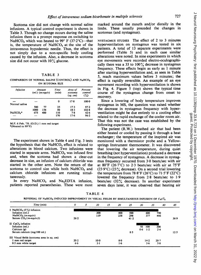
Scotoma size did not change with normal salineinfusions. A typical control experiment is shown inTable 3. Though no change occurs during the salineinfusion there is a prompt response on switching toNaHCO3 which was heated to 990 F (37-2°C)-thatis, the temperature of NaHCO3 at the site of theintravenous hypodermic needle. Thus, the effect isnot simply due to a non-specific body coolingcaused by the infusion. Also, a decrease in scotomasize did not occur with 10% glucose.
TABLE 3COMPARISON OF NORMAL SALINE (CONTROL) AND NaHCO3
ON SCOTOMA SIZE
lbfusion Amount Tinme Area of Percent(ml.) (m-equiv) (min) scotoma control
(sq. cm) (%)
_ - 0 17-8 100.0Normal saline
500 77 25 17-3 97-21000 154 50 18 3 102 8
NaHCO,* 675 400 110 7-8 42-6- - 170 17 6 96-2
M.F. 6 Feb. '70. (O.D.) 1 mm red target*(Heated to 99°F)
The experiment shown in Table 4 and Fig. 3 teststhe hypothesis that the NaHCO3 effect is related toalterations in blood calcium. Two infusions werestarted in separate arms. NaHCO3 was infused firstand, when the scotoma had shown a clear-cutdecrease in size, an infusion of calcium chloride wasstarted in the other arm. Note the return of thescotoma to control size while both NaHCO3 andcalcium chloride infusions are running simul-taneously.
In every NaHCO3 and Na2EDTA infusion,patients reported paraesthesias. These were most
marked around the mouth and/or distally in thelimbs. These usually preceded the changes inscotomas (and nystagmus).
NYSTAGMUS STUDIES The effect of 2 to 3 minuteshyperventilation on nystagmus was tested in sixpatients. A total of 13 separate experiments wereperformed (Table 5) and in each case similaralterations were noted. In nine experiments in whicheye movements were recorded electro-oculographi-cally there was a 33 to 100% decrease in nystagmusfrequency. These effects begin as early as 1 minuteafter starting hyperventilation and, as seen in Table5, reach maximum values before 3 minutes; theeffect is rapidly reversible. An example of an eyemovement recording with hyperventilation is shownin Fig. 4. Figure 5 (top) shows the typical timecourse of the nystagmus change from onset torecovery.
Since a lowering of body temperature improvesnystagmus in MS, the question was raised whetherthe decrease in nystagmus frequency with hyper-ventilation might be due entirely to a cooling effectrelated to the rapid exchange of the cooler room air.That this was not the case was established by thefollowing experiment.The patient (R.W.) breathed air that had been
either heated or cooled by passing it through a heatexchanger; the temperature of the inspired air wasmonitored with a thermistor probe and a Yellow-springs Instrument thermometer. It was discoveredthat lowering the air temperature, during quietbreathing (not hyperventilation) produced a decreasein the frequency of nystagmus. A decrease in nystag-mus frequency occurred from 3 0 beats/sec with airat 80°F (26-7°C) to 2-3 beats/sec with air at 75°F(23-9°C) (23 % decrease). On a second trial loweringthe temperature from 78-80F (26°C) to 71 50F (22°C)lowered the frequency from 2-8 beats/sec to 1-9beats/sec (32% decrease). In another experimentseven days later, it was observed that heating air
TABLE 4REVERSAL OF NaHCO3 INDUCED IMPROVEMENT IN VISUAL FIELDS BY SIMULTANEOUS INFUSION OF CaCI2
Time (min) 0 5 10 15 20 25 30 35 40 45
I NaHCO3 (5%) infusionInfusion (ml.) 200 400 500 600 750NaHCOs (m-equiv) 119 238 298 377 446Serum CO, (m-equiv/l) 26-2 41-2 36-9
It CaCI, infusion -Infusion (ml.) 50 100 200Calcium (g) 0-6 1-2 2-4Serum calcium (mg/100 ml.) 8-5 12 5
III Visual fields (scotoma area in sq. cm)1 mm red target 20-2 100 8-0 14-2 20-70 5 mm white target 6-6 3-1 1-6 3-6 58
727
guest. Protected by copyright.
on March 31, 2022 by
http://jnnp.bmj.com
/J N
eurol Neurosurg P
sychiatry: first published as 10.1136/jnnp.33.6.723 on 1 Decem
ber 1970. Dow
nloaded from

Floyd A. Davis, Frank 0. Becker, Joel A. Michael, and Eric Sorensen
Ccn ~ 2200dca,b 40.eBb
S+X600. BcAr. 700cd9,10 .0. _ &p_O
from 84°F (28&9°C) to 100 80F (38 20C) (still without temperaturhyperventilation) increased nystagmus frequency hyperventilfrom 1-8 beats/sec to 2-7 beats/sec (an increase of Under thesi50%). Thus, changing the temperature of inspired body tempair during normal respiration does change the likely to prnystagmus frequency in the direction to be expected. NaHCO.However, with a nystagmus frequency of 2-7 three patie
beats/sec, while breathing air at 100 8°F (38 2°C) creases in i
(not hyperventilation) the subject was instructed to experimenthyperventilate, and within 3 minutes the nystagmus the frequerdisappeared. Thus, although increased inspired air seen in Fig
FIG. 3. Scotoma plotted with a1 mm red target. Control(upper left); during NaHCO3infusion (upper middle andright); after switching tosimultaneous infusions ofNaHCO3 and CaCl2 (bottomrow). The decrease in scvtomasize seen with NaHCO3 isinhibited by CaCI2.
re increased the control level of nystagmus,lation still caused a cessation ofnystagmus.,e conditions (air temperature greater thanperature) hyperventilation is not at allroduce a cooling effect.I, and Na3EDTA infusions were given toents, all of whom showed marked de-nystagmus frequency. The results of fivets are given in Table 6. The time course ofncy change in two typical experiments is;. 5 (centre and bottom). No effects were
TABLE 5EFFECT OF HYPERVENTILATION ON NYSTAGMUS FREQUENCY
Date Nystagmus frequency(control)(beats/sec)
Minimunm nystagmusfreq. during HV
(beats/sec)
R.W. 8 Aug. 2-66 1-33 50-0 0:25 2:0022 Aug. 2-83 1-75 61-7 2:15 1:00
3-16 1-75 55-0 1:37 1:1529 Aug. 3-57 1-41 39-5 2:30 1:3010 Oct. 3 00 1-33 39-0 2:45 5:0024 Oct. 2-25 1-33 59-0 2:00 2:00
E.M. 15 Aug. 2-33 0-00 0-00 1:00 2:003 Oct. 2-21 1-50 679 3:00 2:30
C.N. 28 Nov. 1-88 0-60 32-0 2:45 1:455 Dec. 2-58 0-90 34-8 1:00 3:00
T.A. 1 Aug. 1D.R. 12 Sept. r Phenomenon observed clinically
M.F. 14 Nov. J
Subject Percentcontrol(%)
Tinme to nminimumJrequency(nin)(sec)
Recovery time(nzin)(sec)
728
guest. Protected by copyright.
on March 31, 2022 by
http://jnnp.bmj.com
/J N
eurol Neurosurg P
sychiatry: first published as 10.1136/jnnp.33.6.723 on 1 Decem
ber 1970. Dow
nloaded from

Effect of intravenous sodium bicarbonate in multiple sclerosis
NYSTAGMUS FREQUENCY
/00% 1 10C-)zw0ui 0 0.5 1.0 1.5 2.5 30 3.5t r HV-- I
R.W.
Cl)
HV(2+MIN)
465fl 465f
z 28
o 0 5 7 12 17 22 27 32 37 42 RW.
POST HV(12 MIN) QL 11'-BiCab--6i
FIG. 4. Electro-oculogram oflateral gaze nystagmus before,during, and after hyperventilation. A prompt decrease innystagmus frequency occurs during hyperventilation andrapidly reverses after hyperventilation.
seen with control saline infusions. In addition,solutions of NaHCO3 heated to 99°F were stilleffective, thus eliminating a non-specific bodycooling as the cause of the decrease in nystagmus
DIPLOPIA STUDY In the only patient experiencingdiplopia (M.F.) dramatic improvement occurredwith a NaHCO3 infusion. Mild weakness of righteye adduction was clinically evident upon leftlateral gaze and the patient experienced diplopiawith the false image appropriately displaced laterally.During the NaHCO, infusion the two images
TABLE 6EFFECT OF NaHCO3 AND Na2EDTA INFUSIONS ON NYSTAGMUS
FREQUENCY
Subject Date Nystagnmus Minimal nystagmus frequency PercentJrequency during infusion: control(control) Bicarbonate Na2EDTA (%(beats/sec) (beatslsec)
R.W. 5 Sept. 3 59 1-00 2829 Aug. 2 67 1 17 45
C.N. 28 Nov. 1-84 1 05 575 Dec. 2 50 1-10 44
E.M. 3 Oct. 2 59 1-83 71
/00%.B 80 0
o l0 20 30 40 66 86- EDTA -
116 124C.N.
TI ME(minutes)
FIG. 5. Timecourse ofdecrease in nystagmus frequency withhyperventilation (top), NaHCO3 infusion (middle), andNa2EDTA infusion (bottom).
gradually came together and then fused into a singleimage; at the time of fusion the adduction weaknesswas no longer clinically discernible. Shortly afterstopping the infusion the adduction weakness againbecame evident and diplopia returned. On re-starting the NaHCO3 infusion the same effects wereagain observed.
CHEMICAL STUDIES Table 7 summarizes the changesin blood chemistry measured during some of theexperimental procedures. Hyperventilation in onepatient produced a respiratory alkalosis with anarterial pH of 7 67. NaHCO3 produced the expectedmetabolic alkalosis with CO2 values rising as high as43 m-equiv/l. During the NaHCO3 infusionsdecreases in total calcium were small varying from0-3-1 3 mg/100 ml. and lesser decreases occurred inultrafiltrable calcium (02-0-8 mg/100 ml.) TheNa2EDTA infusion produced a 21 mg/100 ml.
HV
PRE-HV
729
guest. Protected by copyright.
on March 31, 2022 by
http://jnnp.bmj.com
/J N
eurol Neurosurg P
sychiatry: first published as 10.1136/jnnp.33.6.723 on 1 Decem
ber 1970. Dow
nloaded from

Floyd A. Davis, Frank 0. Becker, Joel A. Michael, and Eric Sorensen
TABLE 7BLOOD CHEMISTRY DATA
I. Hyperventilation
Arterial blood
Negative Total Ultra-pH pCO, base excess serum filtrable
(mm) (m-equiv/l.) calcium calcium(mg/100 ml.) (mg/100 ml.)
E.M. (18 July '69)Control 7 40 390 2-2 8-0 5-25Hyperventilation 7-67 19-0 50 8-2 5 00(3 min)
II. NaHCO3 infusions
Total serum Ultrafiltrable CO,calcium calcium (m-equivll.)
(mg/100 ml.) (mg/100 ml.)
A. E.M. (15 Aug. '69)Control 8-4 6-1 26-5NaHCO, (356 8-1 5-3 43-0m-equiv)
B. R.W. (5 Sept. '69)Control 8-1 6-2 23-6NaHCO, (356 6-8 5-75 36-1m-equiv)
C. E.M. (3 Oct. '69)Control 8-2 6-0 25-1NaHCO3 (356 7-5 5 5 42-1m-equiv)
D. D.P. (6 Mar. '70)Control 8-9 5 9 24-3NaHCO, (356 8-4 5 7 39 3m-equiv)
III Na2EDTA infusion
Total serum calcium (mg/100 ml.) Ultrafiltrablecalcium
Flame EDTA (mg/100 ml.)photometry titration (EDTA titration)method method
M.F. (25 Feb. '70)Control 8-9 8-8 6-35Na2EDTA 6-8 - 3-2*
*The ultrafiltrable calcium value obtained by EDTA titration methodduring the Na2EDTA infusion can be used as an estimate of theionized calcium, since it measures the calcium not complexed withEDTA in the ultrafiltrate.
lowering of total serum calcium along with a 3-1mg/100 ml. decrease of serum ionized calcium(estimated by EDTA titration of ultrafiltrate).
DISCUSSION
This study demonstrates that visual scotomas,nystagmus, and oculomotor paresis in multiplesclerosis can be improved by hyperventilation and/orinfusions of NaHCO3 and Na2EDTA. Since theseeffects do not occur with normal saline infusionsgiven at room temperature and since solutions ofNaHCO heated to 99°F are effective, the phenom-
enon cannot be due to a non-specific body coolingcaused by the infusion. In the case of hyperventi-lation a cooling effect has also been ruled out, sincethe change in nystagmus occurs even when theinspired air is heated to 100-80F (38-2°C). It istherefore concluded that the effect is due to a morespecific alteration in the internal environment.Before discussing this particular aspect further, theprobable site of action of this alteration will beconsidered.While the changes in nystagmus could be due to an
effect on the central and/or peripheral nervoussystem (brain, cranial nerve, neuromuscular junc-tion, muscle) the improvement in the scotomas ismost likely due to an effect on the central nervoussystem since the neural components of vision are allcentral structures. Since refractive changes might beimportant these were looked for but none wasfound.A scotoma is classified as a negative sign, in that it
reflects a diminution or absence of function. In MSit is caused by a demyelinating lesion(s) in the opticnerve which results in an impairment of axonalconduction. The marked decrease in the size ofscotomas seen with NaHCO3 and Na2EDTAinfusions suggests that an improvement of con-duction has occurred in these diseased axons. Thatis, the scotoma changes would appear to be due toan effect within the central nervous system probablyoccurring at the region of the demyelinated lesionand resulting in an improvement of axonal con-duction. Although a similar mechanism might beoperating for the changes seen in nystagmus, it isimportant to keep in mind that, unlike scotomas, itspathophysiological basis is unknown and thus anyhypothesis must be considered with caution.The improvement in clinical findings in MS with
hyperventilation, NaHCO5 and Na2EDTA is prob-ably due, at least in part, to a lowering of serumionized calcium, which increases axonal excitabilityand in turn produces an increase in the conductionsafety factor. There is evidence for a formidableblood-CSF barrier to calcium in normals (Soffer andToribara, 1961; Schain, 1964), multiple sclerosispatients (Merritt and Bauer, 1931), and patients withdisorders of calcium metabolism (Hebert, 1933).However, it is conceivable that there is a localizedbreakdown of the blood-brain barrier to calcium atthe site of MS plaques which might not produce amarked change in CSF calcium. This is supported bythe finding of Broman (Broman, 1944) that increaseduptake of trypan blue occurs in MS plaques. Thus, alow serum ionized calcium might alter brain calciumin MS where it counts most-that is, at the site ofthe demyelinating lesion. This idea of a breakdownin the blood-brain barrier for calcium is also sup-
730
guest. Protected by copyright.
on March 31, 2022 by
http://jnnp.bmj.com
/J N
eurol Neurosurg P
sychiatry: first published as 10.1136/jnnp.33.6.723 on 1 Decem
ber 1970. Dow
nloaded from

Effect of intravenous sodium bicarbonate in multiple sclerosis
ported by our experimental observation that thedecrease in scotoma size with an infusion ofNaHCO3is reversed by a simultaneous infusion of calciumchloride. Though this is probably due to the cor-rection of the induced hypocalcaemia, another possi-bility is that a transient hypercalcaemic stateinduced by the CaCl2 infusion caused a depressionof nerve excitability.
It is well known that hyperventilation (Fanconiand Rose, 1958), NaHCO3 (Moore, 1970) andNa2EDTA (Soffer and Toribara, 1961) have incommon the ability to lower serum ionized calcium.The first two are believed to lower serum ionizedcalcium due to the resulting alkalosis which favoursserum protein binding of calcium. The pH effect isrelated to competition of calcium and hydrogen ionsfor negative sites on the protein. In the case ofNa2EDTA, ionized calcium is decreased by theformation of Ca-EDTA complex. In addition tochanges in calcium, other factors may be important.Lorente de N6 has shown that CO2 has an importantstabilizing effect on nerve which is independent ofpH and that lowering of CO2 increases excitability(Lorente de N6, 1947). Since CO2 readily passes theblood-brain barrier the central decrease in pCO2 withhyperventilation might add to the enhancement ofthe safety factor. In the case of Na2EDTA thepossibility that it can pass the blood-brain barrierin MS patients must also be considered; availableevidence suggests that it does not pass the blood-CSFbarrier in normal man (Soffer and Toribara, 1961).The pH itself may also have a direct effect onconduction. Lorente de N6 found no significantchanges over wide ranges in excised frog nerve(Lorente de No, 1947), but in the squid giant axonincreases in the internal or external pH tend toenhance excitability (Tasaki, Singer, and Takenaka,1964). Another factor, especially with the sodiumbicarbonate infusion, may be an increase in serumsodium concentration which might increase theamplitude of the action potential (Hodgkin andKatz, 1949). All of these changes could enhance theconduction safety factor.Although the preceding theoretical considerations
may require modifications when more data areavailable, the phenomena described in this paperindicate that clinical signs in MS can be improved bychemical means. The implication seems clear that asearch for effective drugs is warranted. Based on theproposed mechanisms, a reasonable approach wouldbe to screen carefully drugs which might enhance theconduction safety factor. Theoretically some of theveratrum alkaloids which prolong the actionpotential duration and produce a decreased thresholdfor stimulation might be effective (Goodman andGilman, 1965). These studies are in progress.
REFERENCES
Bett, I. M., and Fraser, G. P. (1959). A rapid micro-methodfor determining serum calcium. Clin. chim. Acta, 4,346-356.
Boynton, B. L., Garramone, P. M., and Buca, J. T. (1959).Observations on the effects of cool baths for patients withmultiple sclerosis. Phys. Ther. Rev., 39, 297-299.
Brickner, R. M. (1950). The significance of localized vaso-constrictions in multiple sclerosis. Transient, suddenminiature attacks of multiple sclerosis. Res. Publ. Ass. nerv.ment. Dis., 28, 236-244.
Brink, F., Bronk, D. W., and Larrabee, M. G. (1946).Chemical excitation of nerve. Ann N. Y. Acad. Sci., 47,457-485.
Broman, T. (1944). Supravital analysis of disorders in thecerebral vascular permeability in man. Acta med. scand.,118, 79-83.
Charcot, J. M. (1877). Clinical Lectures on the Diseases of theNervous System. The New Sydenham Society: London.
Davis, F. A. (1969). Studies concerning the role of the safetyfactor in injured nerve (Abstract). Electroenceph. clin.Neurophysiol., 27, 714-7 15.
Davis, F. A. (1970). Axonal conduction studies based on someconsiderations of temperature effects in multiple sclerosis.Electroenceph. clin. Neurophysiol., 28, 281-286.
Davis, F. A., Becker, F. O., Michael, J. A., and Sorensen, E.(1970). Acute improvement by chemical means of visualand oculomotor sign in multiple sclerosis: Preliminarycommunication. Pres.-St. Luke's Hosp. Med. Bull., 9, 31-36.
Davis, F. A., and Jacobson, S. (1970). (Manuscript in pre-paration.)
Edmund, J., and Fog, T. (1955). Visual and motor instabilityin multiple sclerosis. Arch. Neurol. Psychiat. (Chic.), 73,316-323.
Fanconi, A., and Rose, G. A. (1958). The ionized, complexedand protein-bound fractions of calcium in plasma. Quart.J. Med., 27, 463-494.
Goodman, L. S., and Gilman, A. (1965). The PharmacologicalBasis of Therapeutics. 3rd ed. Macmillan Co.: New York.
Guthrie, T. C. (1951). Visual and motor changes in patientswith multiple sclerosis. A result of induced changes inenvironmental temperature. Arch. Neurol. Psychiat. (Chic.),65, 437-451.
Hebert, F. K. (1933). The total and diffusable calcium ofserum and the calcium of cerebrospinal fluid in humancases of hypocalcaemia and hypercalcaemia. Biochem. J.,27, 1978-1991.
Hodgkin, A. L., and Katz, B. (1949). The effect of sodiumions on the electrical activity of the giant axon of the squid.J. Physiol. (Lond.), 108, 37-77.
Jackson, J. E., Breen, M., and Chen, C. (1962). Fluorometrictitration of calcium. J. Lab. clin. Med., 60, 700-708.
Lorente de N6, R. (1947). A study of nerve physiology. Stud.Rockefeller Inst. med. Res., part 1, vol. 131.
Merritt, H. H., and Bauer, W. (1931). The equilibriumbetween cerebrospinal fluid and blood plasma. III. Thedistribution of calcium and phosphorus between cerebro-spinal fluid and blood serum. J. biol. Chem., 90, 215-232.
Misske, B. (1930). Uber die Wirkung der Kationverschiebungin der ringerlosung auf dehn peripheren Nerven. Biochem.Z., 219, 320-329.
Moore, E. W. (1970). Ionized calcium in normal serum,ultrafiltrates and whole blood determined by ion-exchangeelectrodes. J. clin. Invest., 49, 318-334.
Namerow, N. S. (1968a). Somatosensory evoked responses inmultiple sclerosis. Bull. Los Angeles neurol. Soc., 33, 74-81.
Namerow, N. S. (1968b). Circadian temperature rhythm andvision in multiple sclerosis. Neurology (Minneap.), 18,417-422.
731
guest. Protected by copyright.
on March 31, 2022 by
http://jnnp.bmj.com
/J N
eurol Neurosurg P
sychiatry: first published as 10.1136/jnnp.33.6.723 on 1 Decem
ber 1970. Dow
nloaded from

732 Floyd A. Davis, Frank 0. Becker, Joel A. Michael, and Eric Sorensen
Namerow, N. S., and Kappl, J. J. (1969). Conduction in Soffer, A., and Toribara, T. (1961). Changes in serum anddemyelinated axons-a simplified model. Bull. Math. spinal fluid calcium effected by disodium ethylenediamine-Biophys., 31, 9-23. tetraacetate. J. Lab. clin. Med., 58, 542-547.
Nelson, D. A., Jeffreys, W. H., and McDowell, F. (1958). Tasaki, I. (1953). Nervous Transmission. Thomas: Springfield,Effects of induced hyperthermia on some neurological Illinois.diseases. Arch. Neurol. Psychiat. (Chic.), 79, 31-39. Tasaki, I., Singer, I., Takenaka, T. (1964). Effects of internal
Nelson, D. A., and McDowell, F. (1959). The effects of and external ionic environment on excitability of squidinduced hyperthermia on patients with multiple sclerosis. giant axon. A macromolecular approach. J. gen. Physiol.,J. Neurol. Neurosurg. Psychiat., 22, 113-116. 48, 1095-1123.
Schain, R. J. (1964). Cerebrospinal fluid cation levels. Arch. Toribara, T. Y., Terepka, A. R., and Dewey, P. A. (1957).Neurol. (Chic.), 11, 330-333. The ultrafiltrable calcium of human serum. I. Ultra-
Shackel, B. (1960). Pilot study in electro-oculography. Brit. filtration methods and normal values. J. clin. Invest., 36,J. Ophthal., 44, 89-113. 738-748. .
Simons, D. J. (1937). A note on the effect of heat and cold Watson, C. W. (1959). Effect of lowering body temperatureupon certain symptoms of multiple sclerosis. Bull. neurol. on the symptoms and signs of multiple sclerosis. NewInst. N. Y., 6, 385-386. Engl. J. Med., 261, 1253-1259.
guest. Protected by copyright.
on March 31, 2022 by
http://jnnp.bmj.com
/J N
eurol Neurosurg P
sychiatry: first published as 10.1136/jnnp.33.6.723 on 1 Decem
ber 1970. Dow
nloaded from