drbenacka - kidneypatfyz.medic.upjs.sk/estudmat/drbenacka - kidney.pdf · clinicalmanifestations...
TRANSCRIPT

KIDNEYKIDNEY DISORDERSDISORDERS
Dr. Dr. R. R. A. A. BEBENNAACCKAKADepartment Department ofof PathophysiologyPathophysiology
P.JP.J. . SSafafaarikrik Univerzity,Univerzity, KOKOSSICEICE, SK, SK
Summer Pathophysiology course 2005
BasicBasic principlesprinciples
111KIDNEY PHYSIOLOGY
1. Excretion of wasting metabolic products● Pacient with kideny disorder show increased blood nitrogen (creatinin,
urea) – azotemia. If the perfusion is normal urea is within normal range.
2. Regulation of water content and concentrations of Na and K ● Hypervolaemia, hypertension, oedema or hyperkalaemia are common
findings in progressive stages of renal dysfunction. First manifestationsof body cummulation of fluid – periorbital edema.
3. Pricipal role in acid- base balance – excretionof acids, regeneration & de novo produtio of bicarbonate
● Severe renal dysfunction is regularly accompanied by metabolic acidosis
4. Endocrine function of kidney – renín, erytropoetin, PGE, calcitriol● Manifestations as hypertension, anaemia, bone demineralisation.

Glomerulárne funkcie
Tubulárne funkcie
Intravenous pyelography Retrograde pyelography
Methods - Pyelography
Methods - Biopsy
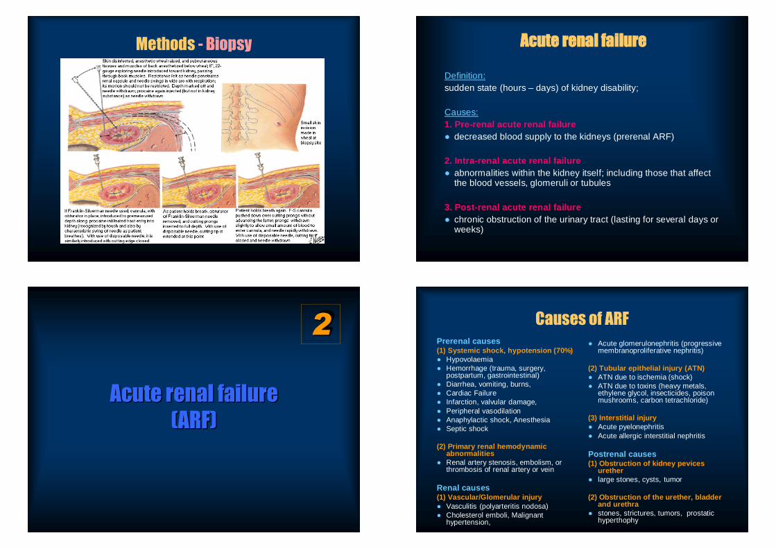
Methods - Biopsy
AcuteAcute renalrenal failurefailure((ARFARF))
222
Acute renal failure
Definition:sudden state (hours – days) of kidney disability;
Causes:1. Pre-renal acute renal failure● decreased blood supply to the kidneys (prerenal ARF)
2. Intra-renal acute renal failure● abnormalities within the kidney itself; including those that affect
the blood vessels, glomeruli or tubules
3. Post-renal acute renal failure● chronic obstruction of the urinary tract (lasting for several days or
weeks)
Causes of ARFPrerenal causes(1) Systemic shock, hypotension (70%)● Hypovolaemia● Hemorrhage (trauma, surgery,
postpartum, gastrointestinal) ● Diarrhea, vomiting, burns, ● Cardiac Failure● Infarction, valvular damage, ● Peripheral vasodilation● Anaphylactic shock, Anesthesia● Septic shock
(2) Primary renal hemodynamicabnormalities
● Renal artery stenosis, embolism, or thrombosis of renal artery or vein
Renal causes(1) Vascular/Glomerular injury● Vasculitis (polyarteritis nodosa)● Cholesterol emboli, Malignant
hypertension,
● Acute glomerulonephritis (progressivemembranoproliferative nephritis)
(2) Tubular epithelial injury (ATN)● ATN due to ischemia (shock)● ATN due to toxins (heavy metals,
ethylene glycol, insecticides, poisonmushrooms, carbon tetrachloride)
(3) Interstitial injury● Acute pyelonephritis● Acute allergic interstitial nephritis
Postrenal causes(1) Obstruction of kidney pevices
urether● large stones, cysts, tumor
(2) Obstruction of the urether, bladderand urethra
● stones, strictures, tumors, prostatichyperthophy

Clinical manifestations(1) Oliguric phase of ARF – day 1-3 little volume of poorly concentrated urine● ↓↓ GFR + ↓ tubular reabsorption of fluid → anuria or oliguria + hyperhydration
with infusion of large volumes of fluid● ↓↓ renal excretion of wasting/toxic subst. → ↑↑ plasma conc. of BUN
(creatinine) = true indicator of function + hyperkalemia (burns, contusions, hemolysis, etc.)
(2) Polyuric phase of ARF – day 4-8 large volumes of urine + salts● normalisation of GFR + ↓↓ tubular reabsorption of fluid + salts (salt-losing
kidney) loss of Na+, K+ water, HCO3- In the polyuric phase the may be so large
as to be life-threatening.● If the renal tubules are damaged (e.g., by heavy metals) polyuric renal failure
occurs as a primary response : ↓↓ GFR + ↓↓ TR
Ischaemic kidney injury
PA:● reversible injury - cellular
swelling, loss of brush border, blebbing, loss of polarity, and cell detach-ment
● lethal injury - necrosis and apoptosis
LA:● depletion of ATP; accu-
mulation of intracellular calcium; activation of proteases (e.g. calpain), cytoskeletal disruption, phospho-lipases damage membranes
● generation of ROS● activation of caspases
(apoptosis)
Ischemic injury to kidney - mechanisms●Loss of cell polarity due to redistribution of membrane proteins (e.g., the
enzyme Na+ K+ - ATPase) from the basolateral to the luminal surface of the tubular cells → abnormal ion transport across the cells
● Increased sodium delivery to distal tubules →vasoconstriction via tubulo-glomerular feedback, which will be discussed below
● Inflammation - ischemic tubular cells → cytokines and adhesion molecules (ICAM-1) → leukocytes injury.
●Luminal tubule obstruction – cells detached from the BM → intratubularpressure → ↓GFR + interstitial edema, increased interstitial pressure →damage to the tubule
●Disturbances in blood flow: intrarenal vasoconstriction - > reduced glomerular plasma flow and reduced oxygen delivery to the functionally important tubules in the outer medulla (thick ascending limb and straight segment of the proximal tubule) < - the renin-angiotensin mechanism, stimulated by increased distal sodium delivery and sublethal endothelial injury, leading to increased release of the vasoconstrictor endothelin and decreased production of the vasodilators nitric oxide and PGI2
●Direct effect of ischemia or toxins on the glomerulus, causing a reduced glomerular ultrafiltration coefficient, possibly due to mesangial contraction.
●Re-epithelialization is mediated by a variety of growth factors and cytokines produced locally by the tubular cells themselves (autocrine stimulation) or by inflammatory cells in the vicinity of necrotic foci (paracrine stimulation) -> epidermal growth factor (EGF), TGF-a, insulin-like growth factor type I, and hepatocyte growth factor have been shown to be particularly important in renal tubular repair.
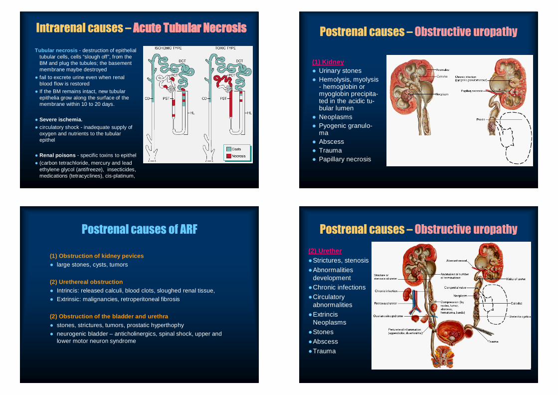
Intrarenal causes – Acute Tubular NecrosisTubular necrosis - destruction of epithelial
tubular cells, cells “slough off”, from theBM and plug the tubules; the basementmembrane maybe destroyed
● fail to excrete urine even when renalblood flow is restored
● If the BM remains intact, new tubularepithelia grow along the surface of themembrane within 10 to 20 days.
● Severe ischemia.● circulatory shock - inadequate supply of
oxygen and nutrients to the tubularepithel
● Renal poisons - specific toxins to epithel● (carbon tetrachloride, mercury and lead
ethylene glycol (antifreeze), insecticides, medications (tetracyclines), cis-platinum,
Postrenal causes of ARF
(1) Obstruction of kidney pevices● large stones, cysts, tumors
(2) Urethereal obstruction● Intrincis: released calculi, blood clots, sloughed renal tissue,● Extrinsic: malignancies, retroperitoneal fibrosis
(2) Obstruction of the bladder and urethra● stones, strictures, tumors, prostatic hyperthophy● neurogenic bladder – anticholinergics, spinal shock, upper and
lower motor neuron syndrome
Postrenal causes – Obstructive uropathy
(1) Kidney● Urinary stones● Hemolysis, myolysis
- hemoglobin or myoglobin precipita-ted in the acidic tu-bular lumen
● Neoplasms● Pyogenic granulo-
ma● Abscess● Trauma● Papillary necrosis
Postrenal causes – Obstructive uropathy(2) Urether●Strictures, stenosis●Abnormalities
development●Chronic infections●Circulatory
abnormalities●Extrincis
Neoplasms●Stones●Abscess●Trauma

Postrenal causes – Obstructive uropathy(3) Prostate● Benign prostatic
hypertrophy● Ca prostate
Postrenal causes – Obstructive uropathy
(4) Bladder●Benign prostatic
hypertrophy●Ca prostate●Neoplasms●Diverticulosis
Postrenal causes – Obstructive uropathy
Consequences ofprolongedobstruction:
- hydronephrosis- pyelonephritis
(5) Uretra●Tumors (papilloma, adenocarcinomas) ●Postinflammatory strictures, diverticulosis
Postrenal causes – Obstructive uropathy

Urinary stones● WHEWELLIT calciumoxalate (CaC2O4.H2O) – rare in
nature; the most common urinary stone, no sharp edges yellow – green
● WEDDELLIT, calciumoxalate CaC2O4.2H2O, very sharpyellow edges causing the pain; relative common
● STRUVITE, Mg(NH4)(PO4).6H2O, non- acidoticenvironment, bacterial infection
Manifestations● retention of water, electrolytes, waste products of metabolism● hypervolaemia + hypernatriemia → edema (hypoosmolarity),
hypertension, hyperosmolarity ● hyperkalemia → heart arrhythmias (> 8mmol/l can be fatal) ● metabolic acidosis → the kidneys are also unable to excrete
hydrogen ions + recycle HCO3-, can aggravate the hyperkalemia (letal)
● most severe cases → complete anuria (patient dies in 8 to 14 days unless an artificial kidney is used)
CausesRenal diseases (e.g., glomerulonephritis, or toxic damage to the kidney.●Shock state (blood loss) - centralization of the circulation → sympathetic α –
adrenoceptor renal vasoconstriction → ↓↓ renal perfusion, ↓↓ GFR (acute ischemic renal failure); Kidney ischaemia: §Polonged constriction of the vasa afferentia:
● Energy deficiency impairs Na+/K+ATPase; increase in intracellular concentration of Na+also causes, via the 3Na+/Ca2+ exchanger, a rise in intracellular Ca2+ concentration and thus vasoconstriction.
● Release of renin both primarily and via an increased NaCl supply in the macula densa (reduced Na + absorption in the ascending tubules) andthus the intrarenal formation of angiotensin II, which has a vasoconstrictor action.
● Adenosine is freed from ATP. It acts on the kidney—in contrast to the other organs—as a marked vasoconstrictor.
ChronicChronic renalrenal failurefailure((CRFCRF -- uremiauremia))
333

1. Characterization●Definition: state charcterized by irreversible and permanent loss of nephrons
with progressive decrease in the normal kidney functions in all aspects: § regulating fluid, electrolyte balance, acid-base balance§ controlling blood pressure through fluid volume and the renin-angiotensin
system, § eliminating nitrogenous and other waste products, § governing the red blood cell count through erythropoietin synthesis, § directing parathyroid and skeletal function through phosphate elimination
and activation of vitamin D
●Typical decrease of GFR, tubular functions●Untill cost-effective renal replacement therapy (i.e., dialysis therapy and
transplantation) 95% of people died (1965)
2. StagesDiminished Renal Reserve (GFR → 50% of normal). Sy: asymptomatic unless acute imparment occursdrugs (nephrotoxic) Lab: the serum BUN and creatinine levels are normal,
Renal Insufficiency (GFR 50 → 20% of normal) Lab: azotemia, anemia, hypertension appear. Sy: isosthenuria or polyuria (urine that is almostisotonic with plasma)
Renal Failure (GFR 20% → 5% of normal) Sy: edema, metabolic acidosis, and hyperkalemia neurologic, gastrointestinal, and cardiovascularmanifestations.
End-Stage Renal Disease (GFR <5% normal) PA: reduction in renal capillaries, scarring in theglomeruli. Atrophy and fibrosis are evident in thetubules. The mass of the kidneys usually is reduced.
Causes of Chronic Renal FailureMetabolic disordersDiabetes mellitusObesityAmyloidosis
HypertensionRenal vascular disordersAtherosclerosisNephrosclerosis-
hypertension
Immunologic disordersGlomerulonephritisPolyarteritis nodosaLupus erythematosus
Primary tubular disordersNephrotoxins (analgesics, heavy
metals)
Urinary tract obstructionRenal calculiHypertrophy of prostateUrethral constriction
Congenital disordersPolycystic diseaseCongenital absence of kidney tissue
(renal hypoplasia)
InfectionsPyelonephritisTuberculosis
Clinical manifestations
1. Alterations in water, electrolyte2. Disorder pf acid-base balance3. Mineral and skeletal disorders4. Anemia and coagulation disorders5. Hypertension and alterations in cardiovascular function6. Gastrointestinal disorders7. Neurologic complications8. Skin and integumentum integrity9. Immunologic disorders

Disorders of Water, Electrolyte, and Acid-Base Balance
● Isostenuria - ability of the kidneys to concentrate the urine is diminished● specific gravity of the urine becomes fixed (1.008 to 1.012) and varies little . ●Polyuria and nocturia are common → volume depletion●Hypernatriemia under sodium overload, decrease in the GFR can occur with a
restricted sodium intake or excess sodium loss caused by diarrhoea or vomiting.
●Hyponatriemia - salt wasting in advanced renal failure (↓tubular reabsorption). ↑ sodium intake improves GFR
●Metabolic acidosis - impairment of a) H+ elimination b) HCO3- recycling and de novo production, c) elimination of acids (ammonia buffer)
● acidosis seems to stabilize as the disease progresses,● (due to buffering capacity of bone → bone resorption, skeletal defects)●Hyperkalaemia - develops only in sever renal damage (GFR < 5 mL/min); or
when not fullfilling dietary restrictions (medications that contain potassium; release of K+ from the cells - trauma or infection).
Mechanisms
Effects on sodium and water
Effects on Mineral Balance – Ca2+, HPO4-

Cardiovascular DisordersHypertension, left ventricular hypertrophy, and pericarditis, are a major cause of
morbidity and mortality in patients with ESRD.
Hypertension - an early manifestation of CRF; causes are multifactorial -↑ volume (water cummulation) + ↑ vascular resistance (↓ renal vasodilator
prostaglandin production and ↑ increased activity of RAA)
Left ventricular hypertrophy- ↓ left ventricular ejection fraction, ↓ ventricular filling (systolic + diastolic failure); Anemia, in particular,has been correlated with the presence of left ventricular hypertrophy
Ischemic heart disease, hert failure - increased myocardial work and oxygendemand; contributing conditions (anemia, diabetes mellitus, dyslipidemia, and
coagulopathies)
Uremic pericarditis - 20% of persons with uremic state or from dialysis(metabolic toxins) siilar to viral pericarditis (cardiac tamponade)
Neurologic symptoms●Peripheral somato-motor neuropathy - mainly lower limbs, symmetric● (atrophy and demyelination of nerve fibers, caused by uremic toxins).
Restless legs syndrome - 2/3 of dialysed patients; burning, creeping, prickling, and itching sensations more intense at rest; temporary relief isobtained by moving the legs.
●Muscle weakness and atrophy - uremia.●Uremic encephalopathy - (? excess of toxic organic acids, sodium
dysbalance) - ↓ alertness and awareness; leaking attention, loss of recentmemory, and perceptual errors in identifying persons and objects.
●Uraemic delirium, seizures, coma - ESRD●Disorders of motor function - difficulty in performing fine●movements of the extremities; the gait becomes unsteady, clumsy with
tremulousness of movement. ●Asterixis (dorsiflexion movements of the hands and feet) - elicited by
hyperextending arms at the elbow and wrist with the fingers spread apart ( side-to-side flapping movements of the fingers).
Gastrointestinal Disorders
● Nausea (early-morning), and vomiting - common in uremia,decomposition of urea by intestinal flora to ammonia.
● Metallic taste in the mouth - depresses the appetite (anorexia)● GIT ulceration and bleeding - PTH increases gastric acid secretion● Hiccups - are common, improve with restriction of dietary protein,
trasplantation
Accumulation of Nitrogenous Wastes● Early sign of renal failure - before other symptoms become evident. ● Azotemia - accumulation of nitrogenous wastes in the blood and can
occur without symptoms● Uremia – early sign (norma 20 mg/dL → 800 mg/dL)● BUN - elevated as renal failure progresses. ● Creatinine - index of GFR● Because creatinine is a by-product of muscle metabolism, serum
values vary with age and muscle mass. An increase in serumcreatinine to three times its
● normal value suggests that there is a 75% loss of renal function, and with creatinine levels of 10 mg/dL or more, it can be assumed that90% of renal function has been lost.

Most Common Causes of End-Stage Renal Disease (ESRD)Cause PercentageDiabetes mellitus 44Hypertension 26Glomerulonephritis 8Polycystic kidney disease 2Other/unknown 20
Močové kamene● WHEWELLIT kalciumoxalát (CaC2O4.H2O) – vzácny v
prírode; najbežnejší kameň, bez ostrých hrán, žlto-zelený s radiálnymi kryštálmi, často v cebtre s apatitom na papílách
● WEDDELLIT, kalciumoxalát CaC2O4.2H2O ostré žltékryštály spôsobujúce bolesť. Pomerne hojný.
● STRUVITE, Mg(NH4)(PO4).6H2O, neacidotické prostredie bakteriálne infekcie,