draft report: application of field & diagnostic methods to ... · pdf fileapplication of...
TRANSCRIPT
1
C N F N
DRAFT REPORT FOR THE AUSTRALIAN GOVERNMENT DEPARTMENT OF THE
ENVIRONMENT AND HERITAGE
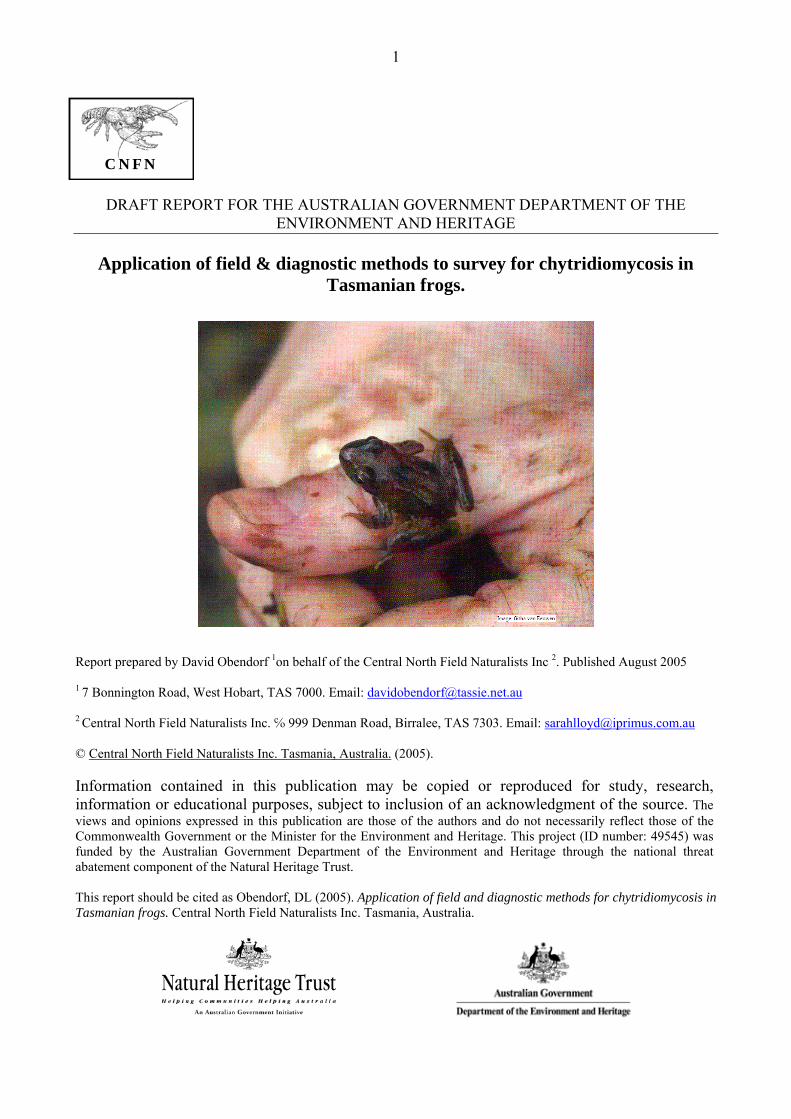
Application of field & diagnostic methods to survey for chytridiomycosis in Tasmanian frogs.
Report prepared by David Obendorf 1on behalf of the Central North Field Naturalists Inc 2. Published August 2005 1 7 Bonnington Road, West Hobart, TAS 7000. Email: [email protected]
2 Central North Field Naturalists Inc. ℅ 999 Denman Road, Birralee, TAS 7303. Email: [email protected] © Central North Field Naturalists Inc. Tasmania, Australia. (2005). Information contained in this publication may be copied or reproduced for study, research, information or educational purposes, subject to inclusion of an acknowledgment of the source. The views and opinions expressed in this publication are those of the authors and do not necessarily reflect those of the Commonwealth Government or the Minister for the Environment and Heritage. This project (ID number: 49545) was funded by the Australian Government Department of the Environment and Heritage through the national threat abatement component of the Natural Heritage Trust. This report should be cited as Obendorf, DL (2005). Application of field and diagnostic methods for chytridiomycosis in Tasmanian frogs. Central North Field Naturalists Inc. Tasmania, Australia.

2
CONTENTS
1 PROJECT SUMMARY ............................................................................................................3
2 ACKNOWLEDGEMENTS .....................................................................................................4
3 BACKGROUND TO THE SURVEY.....................................................................................4
4 LOGISTICS AND SUPPORT................................................................................................5
5 INTRODUCTION....................................................................................................................6
6 MATERIALS & METHODS ..................................................................................................7
7 RESULTS .................................................................................................................................11
7.1 Confirmation of Chytrid Infection in Tasmania .........................................................11 7.2 Tasmanian Chytrid Survey Findings ...........................................................................13
8 DISCUSSION .........................................................................................................................16
8.1 The Tasmanian Chytrid Survey: A Community Survey Approach to Monitor for Key Threatening Process................................................................................................16
8.2 Knowledge gained from the Tasmanian Chytrid Survey .........................................17 8.2.1 Chytrid Maintenance at Tasmanian Frog Habitats............................................17 8.2.2 Survey Prescriptions...............................................................................................18 8.2.3 The impact of chytridiomycosis on Tasmanian amphibia ................................20 8.2.4 Species Profiles and Preliminary Recommendations ........................................21 8.2.5 Chytrid Biosecurity, Risk Assessment & Management.....................................24
9 REFERENCES .........................................................................................................................26
10 APPENDIX 1: COMMUNICATIONS & PUBLIC RELATIONS ..................................29
11 APPENDIX 2: NOTES FROM ‘A PROPOSED MANAGEMENT PLAN FOR THE FLORA AND TERRESTRIAL VERTEBRATE FAUNA OF THE TAMAR ISLAND WETLANDS RESERVE WITH PARTICULAR REFERENCE TO THE THREATENED L. RANIFORMIS AND ITS DECLINE IN THE LAUNCESTON AREA ........................................................................................................................................31
12 APPENDIX 3: FIELD WORKSHEET..................................................................................34
13 APPENDIX 4: PUTATIVE RISK ASSESSMENT OF BATRACHOCHYTRIUM DENDROBATIDIS ON TASMANIAN 11 SPECIES OF FROGS .................................35

3
1 PROJECT SUMMARY Chytridiomycosis is an infectious disease caused by the amphibian chytrid fungus (Batrachochytrium dendrobatidis). B. dendrobatidis is recognised as a major threatening process for amphibian populations worldwide. In Australia the fungus is associated with frog declines and is directly implicated in the extinction of several frog species in Southeast Australia (Anon 2003a). During the spring and summer of 2004‐05 a total of 56 frog habitats in Tasmania were assessed for chytridiomycosis using field survey techniques targeting the tadpole stages supported by chytrid‐specific laboratory testing. The survey confirmed the presence of the chytrid fungus in a number of frog habitats close to major Tasmanian cities and towns. Tadpoles of five species of Tasmanian frog (including one endemic species) were shown to be carriers of the fungus. Using a hand lens, the presence of depigmentation and asymmetry in jaw sheaths and loss of tooth rows in live tadpoles was strongly correlated with chytrid infection. Field surveys of up to 60 tadpoles at each survey site in combination with the Taqman chytrid PCR test were useful diagnostic tools for the detection of chytridiomycosis at targeted amphibian habitats. The detection of the chytrid infection in remote wetlands at high altitude (> 800 m) on the Tasmanian Central Plateau is of particular concern. Declines in the range and abundance of the endemic Tasmanian Tree Frog, Litoria burrowsae and the Green & Gold Frog, Litoria raniformis are considered to be directly linked to the establishment and spread of chytridiomycosis in Tasmania. Tadpole populations in several peri‐urban wetlands and private suburban frog ponds were shown to have chytrid infection. The prolonged time interval between the entry & establishment of chytrid in Tasmania (possibly the late 1970’s) and this baseline survey suggests that in the intervening years the fungus has transferred locally to other frog habitats through natural ecological processes and by anthropogenic means. Combined with appropriate hygiene protocols to prevent anthropogenic spread of chytrid infection, this survey protocol has application in baseline and follow up monitoring surveys for chytrid infection in frog populations. In the course of the survey a range of sampling methods were tested and refined to improve its application more universally.

4
2 ACKNOWLEDGEMENTS We wish to first and foremost thank Mr Damian McRae, Department of Environment and Heritage, for guiding the CNFN Inc in the application process to perform this survey. Thanks are also extended to the Department of Environment and Heritage for providing the opportunity for a community‐based organisation to carry out this task with the assistance of Natural Heritage Trust funding. Special thanks to volunteer surveyors: Wade & Lisa Clarkson, Steve Cronin, Bill Flowers, Paul Swiatkowski, Jim Nelson, Debbie Hill and Ally Dalton ; Central North Field Naturalist treasurer, Sarah Lloyd for administering the DEH fund; CSIRO AAHL staff Donna Boyle and Alex Hyatt for chytrid‐PCR testing; Ally Dalton of the School of Zoology, University of Tasmania for local laboratory support, photographic and field assistance; Andrew Parker, Royal Hobart Hospital, Department of Health & Community Services for photomicrography, Robert Walch, Walch Optics for ongoing support relating to microscopes and field cameras; Martin Harris, WWF Frogs! Program Coordinator for practical support; Rick Speare and Lee Skerratt, James Cook University and Pearl Symonds of the Gap Creek, SE Queensland frog survey group for ongoing advice & support; Rupert Woods, Australian Wildlife Heath Network for addition funding for chytrid testing; Irynej Skira, Niall Doran, Paul Black and Robbie Gaffney, Tasmanian Department of Primary Industry Water and Environment, Nature Conservation Branch for assistance with scientific permits, animal ethics approval and access to the Tasmanian Amphibian GSPOT database.
3 BACKGROUND TO THE SURVEY In July 2004 representatives of the Central North Field Naturalists (CNFN) attended the WWF‐sponsored Third National Conference of Australian Frog Groups. At the conference CNFN presented the case for conducting a baseline survey for the chytrid infection in Tasmania and requested World Wide Fund for Nature Australia’s Frogs! program to support a funding application for a Tasmanian chytrid survey. CNFN successfully applied to the Commonwealth Department of Environment & Heritage through the National threat abatement component of the Natural Heritage Trust for this funding. CNFN sought to undertake this survey using experienced community‐based scientists with skills in frog biology, wildlife diseases and biosecurity. In August CNFN had a planning meeting to determine the scope of the survey and consider the steps required to obtain the necessary scientific permits & animal ethics approvals and review chytrid‐specific biosecurity & hygiene protocols. The field survey methods, animal handling procedures and collection of samples for laboratory testing were also finalised at this workshop. The successful application (DEH ref: 49545) was submitted in September and the project commenced in October 2004. At the time this project began Tasmania was identified as a distinct bioregion of Australia where chytridiomycosis had not been detected in local frog populations (Anon 2003a). The proposal was to initially conduct a baseline survey for the presence of chytridiomycosis in frog habitats where dramatic frog declines had occurred and also at
5
wetland locations where there was a high probability that mainland frogs may have been released. The project plan was designed to fulfil a number of objectives. Firstly the survey wanted to assess whether chytridiomycosis was present in Tasmanian frog populations, and therefore whether the fungus may have been, in part, responsible for the decline in abundance and range of amphibian species listed under State and Commonwealth threatened species legislation. Secondly, it was decided to test the usefulness of field survey methods targeting the tadpole stages of the amphibian life cycle in detecting clinical signs of chytrid infection at a wetland site. And thirdly with the availability of a reliable chytrid test ‐ namely the real‐time Taqman PCR (polymerase chain reaction) test ‐ the survey plan incorporated the use this diagnostic test to support these field survey assessments. This pilot project trials the combined use of field sampling techniques and a reliable back up diagnostic test as practical survey approaches for the surveillance and ongoing monitoring of chytrid infection in wild frog populations. Before commencing the project we examined the published literature relating to the diagnostic methods for detecting chytrid in the field and in the laboratory. We examined the feasibility of several methods including immuno‐histochemistry, fungal culture and the use of staining techniques applied to field samples (reviewed in Anon 2003a; also Briggs & Burgin 2003, 2004; Anon 2004e). Since our aim was to trial practical, cost‐effective and rapid tools that could be applied more universally, we decided against trialling these methodologies. The project was undertaken as a community‐initiated project using volunteer biologists & ecologists experienced in the local amphibia and supported by a wildlife veterinarian. The chytrid specific PCR tests were undertaken at the CSIRO Animal Health Laboratory.
4 LOGISTICS AND SUPPORT
• Central North Field Naturalists Inc. ‐ Tasmanian Chytrid Survey teams • Launceston General Hospital Pathology Services ‐ histology processing • CSIRO Animal Health Laboratory ‐ chytrid‐specific PCR testing • Nature Conservation Branch & Threatened Species Unit of the Tasmanian Department of Primary Industry, Water & Environment ‐ GIS Information on frog state‐wide distribution; provision of scientific permits, animal ethic approvals and an animal experimentation certificate

6
• Australian Government Department of Environment & Heritage ‐ financial support through the National threat abatement component of the Natural Heritage Trust
• Department of Zoology, University of Tasmania ‐ laboratory facilities, reagents, photography and IT support
• James Cook University ‐ chytrid specific advice through Rick Speare and Lee Skerratt
The current list of publications, public media and formal communication generated from this project are listed in Appendix 1.
5 INTRODUCTION There have been dramatic declines in the abundance and range of two Tasmanian frog species in the last quarter century ‐ the Green & Golden Frog (also known as the Southern Bell Frog, Growling Grass Frog, Warty Swamp Frog, Litoria raniformis and the Tasmanian Tree Frog, Litoria burrowsae. Fearn et al (2003) in a report on the decline of L. raniformis in the Launceston area interviewed a number of individuals who gave their recollections of the sharp decline in abundance of this frog in the late 1970’s. In a Threatened Species Listing Statement for L. raniformis, the introduction of the amphibian chytrid fungus, Batrachochytrium dendrobatidis was recognised as a major threat to the survival of the species in Tasmania (Anon 2001a). The susceptibility of L. burrowsae to chytrid infection was demonstrated after a recent review of two pathology cases submitted to the DPIWE Animal Health Laboratories, Launceston in 1993. A tree frog found in box of bananas imported from the Australian mainland was placed in contact with a captive collection of this Tasmanian endemic species. In this case study adult L. burrowsae became lethargic, developed severe skin lesions and died (Obendorf, unpublished findings). In a BSc Honours study conducted during 2001 an attempt was made to assess the impact of B. dendrobatidis on Tasmania’s frog populations (Hardman 2001). At that time, the author was restricted to the use of skin histology of toe clips as a chytrid detection tool ‐ a method that can be quite insensitive. The study failed to detect chytrid infection amongst free‐range frogs or from preserved frogs in museum collections. Hardman also undertook a survey questionnaire of commercial banana wholesalers and retailers in the Hobart area. A total of 4 wholesalers, 20 supermarkets and (fruit & vegetable retailers responded to the survey (approximately 55% of the commercial banana handlers in the Hobart area). She found that frogs were detected during wholesale inspection of banana boxes or during unpacking by retail merchants. Wholesale banana merchants reported finding 4 frogs in the previous 12 months in approximately 113,000 banana boxes. Greengrocer, fruiters and supermarkets reported 22 frogs in nearly 82,000 boxes. Based on a ratio estimates function, Hardman estimated the total number of frogs found in Hobart alone, just in banana boxes, was between 28 and 90 frogs per year. Overall 73% of respondents to Hardman’s survey

7
indicated that any frogs found in produce were either kept by employees or directly released into surrounding urban areas, or nearby bushland, wetlands or parklands; 27% claimed to have handed the frogs to a wildlife agency, a museum, the university, Quarantine Tasmania or disposed of the frogs in rubbish bins. Retrospective studies confirm that B. dendrobatidis has been present in Australia since at least the late 1970’s (Johnson & Speare 2003, Speare & Berger L 2002). Movement of infected frogs is recognised as a significant means of transfer for chytrid infection both internationally and at the sub‐national level. At the Fourth Australian Banana Industry Congress held in Cairns in 2000 the accidental movement of large numbers of frogs with bananas was highlighted. McDonald & Speare 2000 suggested that up to 50,000 frogs annually were transferred in produce from farms to markets. In another study it was estimated that 6000‐8000 frogs are transported to Melbourne annually via this process (Anon 2003b). Interstate trade of fresh produce (particularly bananas) harbouring infected frogs was considered the most likely means whereby this frog disease entered Tasmania. Based on this background evidence there were reasonable grounds to believe that this highly cryptic fungus had already entered, established and spread within Tasmania. Another amphibian‐associated fungus, Mucor amphibiorum is also believed to have entered Tasmania via infected frogs from the Australian mainland (Munday et al 1998).
6 MATERIALS & METHODS In August 2004, CNFN Inc. convened a meeting of herpetologists and frog ecologists to plan and prepare to undertake a chytrid survey in Tasmania during the following frog breeding season in 2004‐2005. It was decided to initially survey wetlands in urban and peri‐urban locations. CNFN also wished to especially survey a number of garden frog ponds where the residents had previously reported multiple frog deaths particularly amongst metamorphling frogs. Sites where local extinctions of L. raniformis had occurred were also considered for surveying. In addition CNFN felt it was imperative to conduct baseline surveys of selected wetlands that currently had high anuran biodiversity (based on number of species, and local abundance of high risk species of frog). These frog habitats at RAMSAR wetlands, State Reserves, National Parks and on private land were included in the survey. At a subsequent ‘wet workshop’ held close to a frog habitat all the community volunteers who were going to participate in the survey were trained to follow a frog disease hygiene protocol (Anon 2001b). This protocol was used for all field surveys. Current knowledge of the epizootiology of chytrid infection and the potential risks of mechanical transfer of infective chytrid material were explained in detail. Practical methods of sterilising field equipment (dip nets, buckets, waders, gumboots and in‐contact survey equipment) using biocides were demonstrated. The surveyors adopted a policy of undertaking only one

8
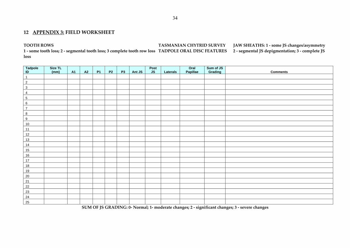
survey per day so that all equipment could be adequately decontaminated and dried before re‐use; at high quality frog habitats surveyors used new dip nets. Tasmania has 11 species of anuran. At some sites the breeding calls of male frogs assisted surveyors in determining the range of species present (Anon 2003c). Tadpole stages can be identified to genus and sometimes species level using a range of morphological features (Anstis 2002; Littlejohn 2003). At each survey site up to 60 tadpoles were collected at random and the mouthparts of each tadpole visually examined using a 12x hand lens (Figure 1). Tadpoles were collected from the three main Tasmanian genera ‐ Litoria, Limnodynastes and Crinia. Surveyors identified each tadpole to species and determine their developmental stage (Gosner, 1960). Only tadpoles less than Gosner stage 40 were surveyed. The chytrid fungus affects the keratinized structures in the oral discs of tadpoles (Fellers et al 2001) leading to depigmentation of horny jaw sheaths and loss of tooth rows (Figure 2 a&b). On every tadpole between Gosner stage 26 and 40 the anterior and posterior keratinized jaw sheaths were separately graded for abnormalities (0 ‐ Visually normal; 1 ‐ thinning or asymmetry in one jaw sheath; 2 ‐ segmental depigmentation (i.e. depigmentation gaps); 3 ‐ substantial depigmentation or complete depigmentation). The tooth rows were similarly assessed and graded for abnormalities (0 ‐ visually normal; 1‐ noticeable but scattered tooth loss; 2 ‐ Segmental loss of teeth; 3 ‐ Complete loss of the tooth row). A field survey worksheet was used to record the anuran species, Gosner stage, and visual gradings for each tooth row and each jaw sheath (see Appendix 3). Based on advice from Rick Speare and the published work of Lara Rachowicz the visual abnormalities in the jaw sheaths were considered the most reliable field indicator of chytrid infection (Rachowicz 2002; Rachowicz & Vredenburg 2004) The sum of the gradings given for the anterior and posterior jaw sheath was used in the analysis. At each site up to 6 tadpoles were selected for chytrid PCR testing; tadpoles with obvious jaw sheath abnormalities were preferentially sampled. Selected tadpoles were killed using an overdose of benzocaine anaesthetic. Using sterile technique to minimize any DNA contamination between samples, each oral disc was excised and separately placed onto a filter paper. Filter papers were dried and labelled and separately stored in small zip‐lock bags (Figure 3). The filter paper samples were sent to the CSIRO Australian Animal Health Laboratory for testing using the Taqman PCR chytrid assay (Boyle et al, 2004). In a further study reported elsewhere we compared the individual Taqman PCR test results obtained from swab samples obtained from oral discs against results from air‐dried oral disc samples taken some the same tadpole (Obendorf et al 2004). The use of swabs (Anon 2004d) allowed for easier sampling in the field and does not require the sacrifice of tadpoles (Figure 4).

9
Figure 2a: Normal Oral Disc features of a Brown Tree Frog, Litoria ewingi tadpole. AJS ‐ Anterior Jaw Sheath; PJS ‐ Posterior Jaw Sheath; A1, A2 Anterior Tooth Rows 1 & 2; Posterior Tooth Rows 1, 2 & 3. Bar ‐ 1 mm
Figure 2b: Abnormal Oral Disc features of a Eastern Banjo Frog, Limnodynastes dumerili tadpole. Note thinning & loss of pigmented jaw sheaths and fine teeth with segmental loss of teeth in several rows. Bar ‐ 1 mm.
Images: Ally Dalton & David Obendorf
AJS
PJS
A1 A2
P3 P2 P1
Figure 1: Examining the oral discs of tadpoles with the aid of a hand lens A ‐ observing the unanaesthetized tadpole in a stoppered glass tube; B ‐ holding the tadpole in the palm Images David Obendorf
A B

10
Figure 4: Using swabs to recover keratinised material from a tadpole’s oral disc (A) and a frog’s hind leg (B) Image Ally Dalton & David Obendorf
At fifty six sites a field survey and chytrid‐specific testing was conducted. The PCR result was considered the definitive or ‘gold standard’ test of chytrid status (presence/absence). For the purpose of analysis, where at least one tadpole surveyed had oral disc lesions greater than or equal to 2, the location was nominated as a potential chytrid affected. Where several tadpoles from the random sample show jaw sheath abnormalities graded at 2 to 6, there is a strong likelihood that the habitat was chytrid infected. When one or more oral disc samples produced a clear positive chytrid PCR result, the site was deemed to be
Figure 3: Sample preparation and transport of dried samples to the testing laboratory Images David Obendorf
A B

11
chytrid infected. The field survey determinations at each site were analysed against its PCR test result. At selected survey sites some tadpoles with abnormal mouthparts were euthanased and preserved in 10% formol‐saline. Longitudinal midline sections through the mouth region (5um thickness) were stained with haematoxylin & eosin and examined under high magnification light microscopy.
7 RESULTS 7.1 Confirmation of Chytrid Infection in Tasmania In October 2004 jaw sheath lesions consistent with oral chytridiomycosis were detected in tadpoles at the first two Tasmanian sites surveyed [Hawley Beach, 41° 09’S 148° 32’E and Knocklofty, 42° 53’S 147°18’E]. In November 2004 Tasmania formally reported the presence of the chytrid fungus, B. dendrobatidis amongst its frog fauna (Anon 2004a). Pronounced depigmentation in both anterior jaw sheaths and segmental loss of teeth in various tooth rows were seen in several tadpoles at each location. The visual appearance of these keratinized structures in a normal and an affected tadpole can be seen in Figures 2 & 8. In selected tadpoles the histomorphology of the normal keratinised oral disc structures was compared with the appearance of depigmented oral disc lesions caused by chytrid infection (Figures 5 & 6). Haematoxylin and eosin staining demonstrated the presence of intracellular micro‐organisms with features typical of B. dendrobatidis (Figure 7). The oral disc lesions and histopathology were strongly suggestive of chytridiomycosis infection. Air‐dried oral disc samples from similarly affected tadpoles collected at both these locations were sent to the Australian Animal Health Laboratory in Geelong to test for the presence of B. dendrobatidis DNA by the PCR gene probe. Laboratory tests supported the gross and histological findings and confirmed the disease diagnosis (Anon 2004b). The PCR results were the highest counts recorded by this laboratory using this test (zoospore count per sample averaged 875,309 ‐ range: 356,516 ‐ 1,916,680; n=7).

12
Figure 5: Sagittal section through the oral disc showing normal, pigmented jaw sheaths and tooth rows. Eastern Banjo Frog Limnodynastes dumerili, Hawley Beach, Northern Tasmania [AJS ‐ Anterior Jaw Sheath; PJS ‐ Posterior Jaw Sheath; A1 ‐ Anterior tooth row 1; A2 ‐ Anterior tooth row; P1 ‐ Posterior tooth row 1; P2 ‐ Posterior tooth row 2 P3 ‐ Posterior tooth row 3] Image Andrew Parker
Figure 6: Sagittal section through the oral disc showing hyperplastic, depigmented jaw sheaths and tooth rows. Eastern Banjo Frog Limnodynastes dumerili, Hawley Beach, Northern Tasmania [AJS ‐ Anterior Jaw Sheath; PJS ‐ Posterior Jaw Sheath; A1 ‐ Anterior tooth row 1; A2 ‐ Anterior tooth row; P1 ‐ Posterior tooth row 1; P2 ‐ Posterior tooth row 2 P3 ‐ Posterior tooth row 3] Image Andrew Parker
PJS
AJSA2 A1` P1
P2 P3
AJS
PJS A1
A2
P1
P2P3

13
Figure 7: High magnification of Anterior Jaw Sheath showing numerous intracellular zoosporangia typical of Batrachochytrium dendrobatidis within the outer keratinized layers. Individual basophilic zoospores can be seen in some sporangia (arrow). A normal portion of the keratinized epithelium can be seen in the box. Image Andrew Parker
7.2 Tasmanian Chytrid Survey Findings After the pilot surveys at Hawley Beach and Knocklofty and confirmation on the use of air‐dried oral discs for PCR tests, surveys of other Tasmanian frog habitats commenced. From September 2004 to May 2005 a total of 56 frog habitats were surveyed for the presence of chytrid infection using the survey protocol outlined above (Map 1). At 26 sites the visual assessments were suspicious of likely chytrid infection. At 25 of these sites (96%), the chytrid‐PCR samples returned positive results. At only 1 of 26 sites (4%) where visual assessments were suggestive of chytrid infection was the chytrid‐PCR test non‐confirmatory. At 30 sites no surveyed tadpoles showed visual jaw sheath abnormalities. At 22 of these sites (76%), the chytrid‐PCR samples returned negative results. At remaining 8 sites (24%), the chytrid PCR result was positive (see Table 1).

14
Figure 8: Brown Tree Frog tadpoles A ‐ Normal Oral Disc Total score = 0; B ‐ Thinning & segmentation with almost complete loss of AJS (3) and thinning & asymmetry in PJS (1) Total score = 4; C ‐ Almost complete loss of both jaw sheaths AJS (3), PJS (3) with segmental loss of tooth rows Total Score = 6 Bar ‐ 1 mm Image Ally Dalton & David Obendorf In only one instance did the PCR samples fail to detect chytrid at a frog habitat where a field survey suggested chytrid infection was likely to be present. During the survey chytrid infection was detected in tadpoles belonging to five species of Tasmanian frog (Eastern Banjo Frog Limnodynastes dumerili, Spotted Marsh Frog Limnodynastes tasmaniensis, Common Froglet Crinia signifera, Tasmanian Froglet Crinia tasmaniensis and Brown Tree Frog Litoria ewingi). At two sites L. tasmaniensis tadpoles displaying significant jaw sheath changes (gradings of 2 or 3 in both jaw sheaths) were visibly thinner than unaffected tadpoles. Notably no tadpoles of L. raniformis and L. burrowsae were found with jaw sheath lesions. Field Visual Survey Assessments Jaw Sheath Lesions No Jaw Sheath Lesions Total
Positive 25 8 33 Negative 1 22 23
Chytrid PCR Results Total 26 30 56 Table 1: Visual Survey results compared to chytrid PCR results from 56 Tasmanian frog habitats
A B C

15
Map 1: Tasmanian Chytrid Survey sites ‐ confirmed chytrid positive sites in red; chytrid negative sites in green; probable chytrid positive ‐ ? Prepared by Ally Dalton
The 2004 ‐2005 Tasmanian Chytrid Survey did detect chytrid infection in three sites within the area of occupancy of L. burrowsae. At Cradle Mountain, the type locality for the species, the only litorid found as tadpoles and from male calling during our surveys was the ubiquitous L. ewingi. Surveys conducted at Kitchen Hut within Cradle Mountain National Park and in the Walls of Jerusalem National Park recorded tadpoles with jaw sheath abnormalities and positive chytrid‐PCR results. Another three sites in Cradle Mountain area and one in the Walls of Jerusalem were negative for chytrid infection by survey and PCR. In March 2005 reconnaissance of known L. burrowsae sites in the vicinity of Strahan found only two sites where its tadpoles were present. No signs of chytrid were seen in tadpoles and all PCR results from these tadpoles were negative.

16
8 DISCUSSION In terms of the national chytrid threat abatement plan, confirmation of the presence of the chytrid fungus, B. dendrobatidis in Tasmanian frogs is important new information. Although its presence had long been suspected, efforts to survey frog populations for chytridiomycosis in 2001 were unsuccessful. The recent availability of a reliable diagnostic test (Boyle et al 2004) and the application of a tadpole survey method (Fellers et al 2001) have made surveillance for chytrid far more achievable. 8.1 The Tasmanian Chytrid Survey: A Community Survey Approach to Monitor for
Key Threatening Process Community biologists and amphibian researchers have usually been the first to notice changes in the abundance and range of specific species of frogs within local natural environments. In habitats where declines in the abundance of specific species are noted, it is invariably the recognition of a decline in the range and abundance of a highly populous or highly vocal species that triggers the need for follow up science‐based monitoring. Reliable field observations and the good fortune of having access to sick or freshly dead frogs on which to undertake thorough pathological assessments have been essential. Local changes in frog biodiversity are being monitored through a range of benign environmental techniques. Committed individuals who know the species of frog in their areas and can conduct baseline and ongoing frog biodiversity surveys at frog habitats are critical to understanding and mitigating emerging threatening processes. In Tasmania, recognition of the distinctive calls of males is probably the most useful benign observational skill that can be used to confirm the continued occupancy by species of frogs. These contributions are essential for updating amphibian GIS databases. The ‘triggers’ for surveying Tasmanian frogs for chytrid infection were:
• the historical decline in the range and abundance of two Litorid frogs ‐ L. raniformis and L. burrowsae
• reports of loss in species diversity at certain urban and peri‐urban frog habitats, • mass mortality incidents involving juvenile L. ewingi in suburban frog ponds, and • the detection of chytrid infection in a captive population L. burrowsae occurring
after contact with a mainland litorid frog. The approach taken for this survey was conceived and developed in consultation with local frog enthusiasts, freshwater ecologists and wildlife disease specialists. From the outset this was designed as a community development project sponsored by a Tasmanian field naturalist group (CNFN) with members experienced in frog ecology and behaviour. The survey design invited these volunteers to survey for chytrid infection using a field protocol supported by training and a reliable cost‐effective diagnostic test. This approach
17
to chytrid surveillance has several features that warrant consideration in other parts of Australia and have application for the detection of other specific invasive species. In our situation, there was already a core group of individuals with awareness of chytrid as a key threatening process for Australian frogs; this is also the case in many regions of Australia (Anon 2004c). Local chytrid risk assessments had already been undertaken and specific sites worthy of initial surveillance were known. Knowledge of potential chytrid ‘hot spots’ allowed the field protocol to be immediately trialled and refined where necessary. Chytrid surveyors enthusiastically undertook specific training in the use of the field technique and frog disease hygiene protocols. 8.2 Knowledge gained from the Tasmanian Chytrid Survey
8.2.1 Chytrid Maintenance at Tasmanian Frog Habitats
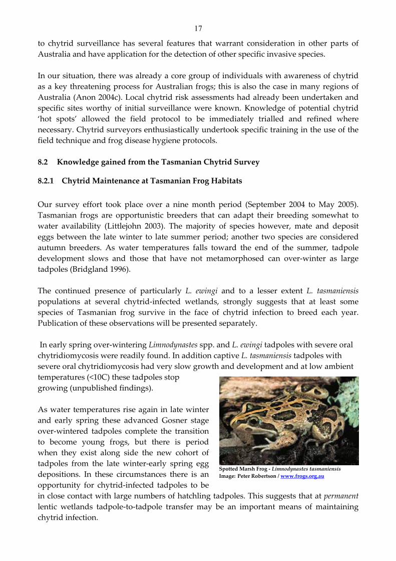
Our survey effort took place over a nine month period (September 2004 to May 2005). Tasmanian frogs are opportunistic breeders that can adapt their breeding somewhat to water availability (Littlejohn 2003). The majority of species however, mate and deposit eggs between the late winter to late summer period; another two species are considered autumn breeders. As water temperatures falls toward the end of the summer, tadpole development slows and those that have not metamorphosed can over‐winter as large tadpoles (Bridgland 1996). The continued presence of particularly L. ewingi and to a lesser extent L. tasmaniensis populations at several chytrid‐infected wetlands, strongly suggests that at least some species of Tasmanian frog survive in the face of chytrid infection to breed each year. Publication of these observations will be presented separately. In early spring over‐wintering Limnodynastes spp. and L. ewingi tadpoles with severe oral chytridiomycosis were readily found. In addition captive L. tasmaniensis tadpoles with severe oral chytridiomycosis had very slow growth and development and at low ambient temperatures (<10C) these tadpoles stop growing (unpublished findings). As water temperatures rise again in late winter and early spring these advanced Gosner stage over‐wintered tadpoles complete the transition to become young frogs, but there is period when they exist along side the new cohort of tadpoles from the late winter‐early spring egg depositions. In these circumstances there is an opportunity for chytrid‐infected tadpoles to be in close contact with large numbers of hatchling tadpoles. This suggests that at permanent lentic wetlands tadpole‐to‐tadpole transfer may be an important means of maintaining chytrid infection.
Spotted Marsh Frog ‐ Limnodynastes tasmaniensis Image: Peter Robertson / www.frogs.org.au

18
It is known that lower water temperatures enhance the pathogenicity and the visual expression of the disease in amphibians (Anon 2003; Berger et al 2004). Variation in the expression of this disease that we observed in tadpoles at different times of the years supports observations that B. dendrobatidis prefer cold water conditions for optimal proliferation. Severe oral chytridiomycosis lesions were readily detected at high prevalence in tadpoles in late winter and early spring, however, when these sites was re‐surveyed at the end of the summer fewer tadpoles with lower grade lesion or none at all were reported. Our project did not have the resources to examine these cases for any microscopic differences in oral disc histology from these repeat surveys. Clearly this would have been a worthwhile activity as chytrid‐PCR tests on individual tadpoles without visual lesions returned positive results. [Material from these samples is available for future examination, if needed.] In our experience the complete absence of a single jaw sheath in a tadpole between Gosner stage 26 and 40 is rare. Loss of a jaw sheath can occur through catching or handling trauma but can also be seen in precociously developing tadpoles that are undergoing physiological changes to their mouth parts. It is particularly for the latter reason than the Gosner stage of surveyed tadpoles needed to be restricted to ≤ 40. Traumatic jaw sheath and tooth row loss can occur through over zealous netting or transfer into holding buckets prior to visual survey being conducted.
8.2.2 Survey Prescriptions We successfully survey tadpoles of the three main genera: Litoria, Limnodynastes and Crinia. In order to accurately survey the oral disc structures, tadpoles ≥ 20 mm in length were considered optimal. This relates to the ease of handling, trauma‐free manipulation, and visualisation of the keratinized structures in the oral disc. The field surveys were restricted to tadpoles between Gosner stages 26 to 40 for two reasons. Firstly stage 26 was about the earliest developmental stage at which all the oral disc structures could be readily seen with a hand lens under good light conditions. And secondly after stage 40 tadpoles undergo physiological changes in their oral discs (such as loss of tooth rows and jaw sheaths). If these older tadpoles were included, normal physiological changes in the oral disc could be misinterpreted as chytrid‐associated abnormalities. There are several practical advantages this approach offers for chytrid surveillance. Firstly, it can be readily applied to assess the chytrid‐status of a frog‐habitat, not just infections in individual amphibians. Secondly, the survey method does not rely on finding adult frogs in free‐range habitats, only tadpoles. Thirdly, based on this pilot survey and simplification of field methods the assessment and sampling of tadpoles for chytrid can now be undertaken without the need to kill tadpoles.

19
In Tasmania, we believe the optimal time of the year to survey for visual signs suggestive of oral chytridiomycosis in tadpoles is late winter and spring. This supports the field and experimental findings of chytridiomycosis in Eastern Australia (Berger et al 2004). The tadpole survey if timed to optimise chytrid expression in tadpole stages has the advantage of offering a on the spot field diagnosis. However, as mentioned above there is also the potential for a ‘false negative’ result from a survey, if conducted at a time when visual signs oral chytridiomycosis could be less obvious. In very cold climate conditions where tadpoles over‐winter under ice, depigmentation of Jaw Sheaths and loss of tooth rows is also seen (Rachowicz & Vredenburg 2004). It is therefore important to highlight the observed differences between the effect of such extreme cold exposure to tadpoles and oral chytridiomycosis. Based on experiments and field surveys, loss of pigmented keratin in the oral disc of chytrid infected tadpoles begins as ‘gaps’ (i.e. complete zonary loss of pigment from a localised area of the jaw sheath surrounded by fully pigmented areas; a discontinuity in the length of sheath pigment) in the jaw sheaths. These gaps become larger and join over time. Tooth row loss is variable but is most noticeable when the jaw sheaths are total depigmented (grading 3). In tadpoles exposed to a low water treatment (4°C), the pattern begins with the loss of the tooth row pigment and a continuous reduction in the pigmentation across the whole width of jaw sheath until it was completely lost from both sheaths (Rachowicz & Vredenburg 2004). Notwithstanding our observations that the tadpole survey method was less sensitive than the chytrid‐PCR test, if properly targeted in terms of time and location, it is an invaluable initial field test. When warmer water temperatures favour rapid tadpole growth, visual surveys alone may fail to detect characteristic jaw sheath abnormalities suggestive of chytridiomycosis. In these situations samples for PCR testing from visually unaffected tadpoles should always be taken. The chytrid‐PCR test offers a very useful diagnostic tool to look for the fungus in both life stages of the frog life cycle (Anon 2004d). It was used as our definitive chytrid test. This test is already proving to be an invaluable tool in understanding chytrid ecology across Australia and is being offered to other countries (Alex Hyatt, Personnal Communication). Use of a highly sensitive PCR gene probe as a diagnostic tool for chytrid detection requires maximal quality control on chytrid‐DNA contamination during field sampling. It is imperative to ensure that chytrid‐DNA contamination does not lead to false positive results for a given frog habitat. Despite the use of chytrid biocide protocols to render residual chytrid fungus on dip nets and sampling equipment non‐viable, low chytrid zoospores equivalents counts, detected by the gene probe, may result from residual chytrid‐DNA on sampling equipment.

20
Toward the end of the Tasmanian Chytrid Survey, a direct swabbing technique was compared with the standard air‐dried oral disc method. Fine tipped swabs, as recommended (Anon 2004d), were applied individually to the oral discs of tadpoles. The results from a small sample size showed that the swabbing method could replace the use of killed tadpoles and reduce further the likelihood of low grade false positive results (Obendorf et al 2004).
8.2.3 The impact of chytridiomycosis on Tasmanian amphibia B. dendrobatidis is unique in the Phylum Chytridiomycota in that it colonises the epidermal cells in the skin of amphibians (Longcore et al 1999). Other species within this phylum occur as free‐living saprophytic fungi in water and soil or are parasitic in plants, algae, nematodes and insects (Powell 1993); this the only known chytrid that is an intracellular parasite of vertebrates. B. dendrobatidis has been a highly cryptic micro‐organism. The importance of the disease chytridiomycosis in amphibians was not fully recognised until the latter years of the 1990’s (Berger et al 1998). The entry, establishment and spread of exotic pathogens into naïve wildlife populations can lead to subtle declines overtime and in this regard chytridiomycosis of amphibian is a classical example. In frogs the sessile zoosporangia stages B. dendrobatidis colonises the keratin‐rich skin layers of epidermis; these are most commonly found on limbs, feet and ventral body skin. The infective motile zoospores are released into the external environment via discharge tubes (Berger et al 2000). The pathological effect of infections with chytrid fungus is predominantly recognised in juvenile frogs in the first 4‐6 weeks after metamorphosis (Anon 2003a; Berger et al 1998; Nichols et al 2001; Longcore et al 1999). It is during this life stage that the skin of frogs becomes fully keratinized and therefore prone to chytrid colonisation. This age cohort is also quite vulnerable and naïve within their natural environment. Physiological changes or physical disabilities caused by chytrid infection are believed to lead to natural predation or death (Anon 2003a). Chytrid‐associated mass morbidity & mortality events in this age cohort have the potential to remove large numbers of juveniles and reduce recruitment into the adult population. Host susceptibility to chytrid infection is variable across the frog species. For some highly susceptible species, persistence of chytrid fungus in its area of occupancy can lead to declines in the numbers of frogs of breeding age. Based on the current knowledge derived from captive case studies we believe that both L. burrowsae and L. raniformis are highly susceptible to chytrid infection.

21
8.2.4 Species Profiles and Preliminary Recommendations
8.2.4.1 Green and Golden Frog ‐ Litoria raniformis Historical records of the dramatic declines in the abundance of L. raniformis in south‐eastern Australia and Tasmania begin in the late 1970’s and early 1980’s (see Appendix 2).
This once populous species was so abundant and common it was readily collected commercially. The distribution of the species has collapsed in virtually all inland riverine and floodplain environments. It is now maintains healthy populations in chytrid‐free locations in some coastal wetlands and more rarely at a few inland wetlands (Anon 2001a) At wetlands where L. raniformis historically have been recorded and no longer occur, we found chytrid infection to be present. In contrast at wetland sites where L. raniformis are still common, no chytrid was detected. Only at one chytrid‐affected location
were we aware of the continued presence of adult calling L. raniformis (Hawley Beach), however, these frogs could have migrated from nearby chytrid‐free wetlands. The survey results are perhaps as would be expected for a species that is reputed to be susceptible to chytrid infection in the juvenile frog stages. Two closely related species, L. aurea (Green & Golden Bell Frog) and L. castanea (Yellow‐spotted Tree Frog) has also experienced similar dramatic declines in range and abundance (Anstis 2002). Further research is needed into the susceptibility of these large litorids to chytrid infection. If this loss of substantial numbers of juvenile frogs was directly attributable to chytrid infection, little or no recruitment into adult age cohort would occur and this would cause the numbers to decline over their life span (8‐10 years) (Ashworth 1998). Oral histories and reports from the late 1970’s and early 1980’s support this pattern of decline for L. raniformis. We believe that this fungal pathogen has been in Tasmania for at least two decades, entering with chytrid‐carrying frogs in fresh produce, particularly bananas. It is therefore not unexpected that historical L. raniformis habitats close to cities and towns were found to be chytrid‐infected. Important Tasmanian sites where L. raniformis have been recorded (Anon 2001a) need to be urgently re‐assessed in the light of this study. Several important sites for this frog in southern Tasmania occur on private land. Resurveys of these sites to determine their presence and chytrid status is highly recommended.
1 Plants & Animals of Tasmania ‐ a guide to their identification Bushcare CD‐ROM 2003
Image: Peter Brown1 m

22
8.2.4.2 Tasmanian Tree Frog ‐ Litoria burrowsae L. burrowsae is an endemic species that has disappeared from much of its former range. This species has disappeared from the Cradle Mountain area where the species was originally discovered in 1941 (Scott 1942). Important populations remain in remote parts of the Tasmanian Wilderness World Heritage Area (TWHA)(Anon 2003d). The Tasmanian index cases of chytridiomycosis occurred in1993 in a captive population of L. burrowsae resulting from close contact with a confiscated mainland frog found in imported produce. All individuals of this endemic species of frog died after this contact (D. Obendorf, unpublished findings). L. burrowsae has disappeared from several other locations in western and southern Tasmania where it was previously abundant (Paul Swiatkowski, pers. comm.). Many of the recorded locations of L. burrowsae are close to major roads and human habitations. Update survey of these historical locations using call recognition and frog spotting would provide useful information on the current status of this frog. Notably two sites surveyed near Strahan that still retain L. burrowsae populations were chytrid free. Important Tasmanian sites where L. burrowsae have been recorded (unpublished GSPOT data set) need to be urgently re‐assessed in the light of this study. Several important sites for this frog in southern and western Tasmania occur within the TWWHA. Resurveys of these sites to determine their presence and chytrid status is highly recommended.
8.2.4.3 Brown Tree Frog ‐ Litoria ewingi L. ewingi is now the most common and widespread species of frog in Tasmania, occurring from sea level to at least 1070 m (Littlejohn & Martin 1974). Observations made during the
Tasmanian Chytrid Survey have shown that this species of frog can maintain breeding populations of adults at chytrid‐infected wetlands. They produce between 500‐700 eggs per clutch (Hero et al 1991, Littlejohn 2003) and the species can be found in a very wide range of habitats including those in alpine and sub‐alpine zones. Our studies confirm that metamorphling L. ewingi can die as a direct result of chytrid infection. Paradoxically,
we also believe the species can be one of Tasmania’s most resilient to the fungus. We investigated two garden pond sites where mass mortalities of L. ewingi metamorphlings
Image: Peter Brown1
Image: Peter Robertson / www.frog.org.au

23
occurred; skin samples from 9 dead frogs (body length 15 ‐20mm) all returned positive chytrid PCR results (unpublished findings). In another study three L. ewingi tadpoles from a chytrid‐infected wetland and exhibiting severe jaw sheath abnormalities were maintained in captivity and kept for over 4 months after metamorphosis. Although chytrid infection was confirmed in these captive frogs from skin swabs, these frogs continued to feed activity and exhibited normal behaviour. The observations and chytrid PCR results on these studies will be presented separately. At a known chytrid‐infected habitat 11 L. ewingi (4 recent metamorphs ‐ snout‐vent range 14‐16 mm; and 7 juvenile frogs ‐ snout‐vent range 21‐34 mm) were tested for the presence of chytrid by skin swabbing. All these frogs appeared normal and all had negative chytrid‐PCR results from skin swabs (Alexander Dalton, unpublished findings). Further research is required to understand the role this species of frog plays in the maintenance of chytrid infection in Tasmania. Several chytrid‐infected wetlands close to Hobart offer good opportunities to conduct long‐term studies into the ecology of chytrid and its effect on several species of frog.
8.2.4.4 Other Species of Tasmanian Frogs
Moss Froglet ‐ Crinia nimbus Image: Julian Benley/ www.frog.org.au
Eastern Banjo Frog ‐ Limnodynastes dumerili Image: Peter Robertson / www.frog.org.au
Reports of local declines or absences of Limnodynastes dumerili are of concern. Several chytrid‐affected tadpoles of this species successfully metamorphosed but after 4‐6 weeks all died with severe skin lesions of chytridiomycosis. Spotted Marsh Frogs Limnodynastes tasmaniensis is another species that under captive conditions developed typical behaviours and skin changes suggestive chytrid infection. As with L. dumerili, chytrid‐infected L, tasmaniensis tadpoles successfully metamorphosed but died about 4 weeks after metamorphosis. High chytrid carriage levels (based on the quantitative Taqman PCR chytrid test) were detected in these captive metamorphs, whereas live free‐ranging metamorphs from the source wetland showed skin swab counts two to three orders of magnitude lower (D. Obendorf, unpublished findings). These observations and the results of the chytrid PCR tests from these metamorphs will be presented elsewhere.

24
Based on the current knowledge of the status, distribution and biology of Tasmania’s 11 frog species and the conclusions drawn from this study, we have proposed a chytrid risk assessment on each species of Tasmanian frog (see Appendix 4). Due to the widespread distribution and diverse habitat usages of L. ewingi and the known capability of this species to survive chytrid infection in the transition from one life stage (tadpoles) to adult frogs, there is an opportunity to investigate the role that both over‐wintering L. ewingi tadpoles and adult Brown Tree Frogs play in the maintenance and spread of chytridiomycosis over the Tasmanian landscape. The Tasmanian endemic Moss Froglet, Crinia (Bryobatrachus) nimbus is geographically restricted to subalpine moorland, cloud forest and rainforest habitats in south & south western Tasmania (Ziegler 1994; Rounsvell et al 1994). It inhabits cool and consistently wet locations with the prolonged tadpole development within the fluid derived from the decomposing egg capsules. Tadpoles do not feed and the oral disc has no keratinised tooth rows or jaw sheaths (Mitchell & Swain 1996). Other frogs, Philoria and Pseudophyrne spp. inhabiting very similar habitats and life histories on the Australian mainland are now considered to be highly vulnerable to chytrid exposure (Knowles et al 2004; Knowles 2005; Berger et al 2004). Targeted baseline surveillance for chytrid infection in habitats that include locations of the geographically restricted Moss Froglet, Crinia (Bryobatrachus) nimbus has been recommended as a conservation priority for the TWWHA (Anon 2004f).
8.2.5 Chytrid Biosecurity, Risk Assessment & Management It is still unclear whether B. dendrobatidis can maintain itself, independent of amphibians. The infective zoospores released from zoosporangia can attach to other substrates including aquatic vegetation and freshwater invertebrates (Johnson & Speare 2003). It is also unclear whether this chytrid fungus has a resting stage that can survive in the environment (Anon 2003a; Rick Speare pers. comm. 2005). With these parts of its biology unknown, assessing the relative risk of chytrid transfer by various sources can only be empirical. Global and local experience would confirm that live frogs and tadpoles constitute the highest risk for the movement of viable B. dendrobatidis. Therefore the highest risk of chytrid spread to new locations is through the unintentional or intentional movement of frogs and tadpoles by humans. Once chytrid infection has entered and established in a local frog ecosystem, natural factors such as frog movement, flood events, activities of predators etc may allow the fungus to spread locally. Recurring human activities such as recreational fishing using tadpoles & frogs as bait and the collection and release of frogs & tadpoles are probably the two commonest means whereby chytrid can be transferred rapidly into new sites, particularly into more remote frog habitats. The use of frogs (live or dead) for recreational fishing was banned in

25
Tasmania in 1996‐97 under Inland Fisheries legislation2. Information and education on chytrid infection and the impact humans have in spreading this disease is strongly recommended. Frogs are protected in Tasmania under the Nature Conservation Act 2002 and some species have additional protection under the Threatened Species Protection Act 1995 and the Commonwealth Environment Protection & Biodiversity Conservation Act 199. The Department of Primary Industries, Water & Environment (DPIWE) allow individuals to keep without a permit ‐ up to six specimens of both the Common Froglet (Crinia signifera) and the Brown tree Frog (Litoria ewingi) and unquantified numbers of frogs eggs and tadpoles. Individuals wanting to keep all other species of Tasmanian frog must obtain a Herpetological Permit and in the case of the Striped Marsh frog (Limnodynastes peronii) and the Moss Froglet (Bryobatrachus nimbus), a Scientific Permit. The keeping of the Green and Golden Frog (Litoria raniformis) is not permitted. Permits are issued on the understanding that individuals keeping amphibians In Tasmania agree to comply with a Code of Practice (Anon 2003e). We recommend that DPIWE review the Code of Practice and the conditions applied to granting of Herpetological and Scientific Permits applying to amphibians so that applicants can follow hygiene protocols when doing field research and in the keeping of frogs in captivity. The introduction of non‐native frogs with fresh imported produce is an ongoing concern (McDonald & Speare 2000; Hardman 2001). A co‐ordinated approach is required to improve the level of awareness amongst fresh fruit & vegetable importers, wholesalers and retailers about the threats posed by these creatures if they are released into freshwater ecosystems. Biosecurity and quarantine protocols need to be developed so that introduced amphibians can be detected, received, identified and treated humanely. Some introduced amphibians may establish free‐living populations in Tasmania and have the potential to introduce a number of disease‐causing pathogens (including bacteria, fungi and viruses) into freshwater ecosystems. All these factors need to be taken into consideration when framing risk management and risk communication strategies for B. dendrobatidis in Tasmania. Research conducted under Tasmanian Department of Primary Industries Water & Environment Permit No. TFA 04193
2 Inland Fisheries (Recreational Fishing) Regulations 1999 ‐ Regulation 21(6)

26
9 REFERENCES Ashworth J. M. (1998) An Appraisal of the Conservation Status of Litoria raniformis (Keferstein) in Tasmania. Unpublished Masters of Science thesis, University of Tasmania. Anon (2001a) Threatened Species Listing Statement ‐ Green and Golden Frog, Litoria raniformis Keferstein 1867. Tasmanian DPIWE Threatened Species Unit; March 2001. Anon (2001b) Hygiene protocol for the control of disease in frogs. Threatened Species Management information Circular No. 6 NSW National Parks and Wildlife Service pp17 Anon (2003a) Threat Abatement Plan for Infection of Amphibian Chytrid Fungus resulting in Chytridiomycosis. [Workshop Discussion Paper August 2003 pp 30. Prepared by Speare, R. for the Australian Government Department of the Environment and Heritage.] Anon (2003b) The Lost Frogsʹ Home: An initiative of the Victorian Frog Group to rescue accidentally relocated frogs In the Spotlight 4(2) Anon (2003c) Natural History and Calls of Tasmanian Frogs ‐ Frogs Tasmania CNFN audio CD or tape. Anon (2003d) Plants and Animals of Tasmania: a guide to their identification. CD‐ROM Bushcare Technical Extension Anon (2003e) Keeping Reptiles & Amphibians in Tasmania. Nature Conservation Branch, Department of Primary industries, water & Environment November 2003 Anon (2004a) Australian Wildlife Health Network Situation Report [Preliminary Notification] November 2004 Anon (2004b) Australian Animal Health Laboratory Diagnostic Report [Reference FIR04/25‐1 (November 2004) Anon (2004c) The Proceedings of the Third National Conference of Frog Groups. Sydney 31 July‐ 1 August 2004 Anon (2004d) Diagnostic assays and sampling protocols for the detection of Batrachochytrium dendrobatidis (Bd) CSIRO Livestock Industries Australian Animal Health Laboratory (Technical File note) Anon (2004e) Immunoperoxidase test for Batrachochytrium. CSIRO Livestock Industries Australian Animal Health (Technical File note) Anon (2004f) State of the Tasmanian Wilderness World Heritage Area: An Evaluation of management effectiveness Report No. 1 2004 Anstis, M (2002) Tadpoles of South‐eastern Australia ‐ a guide with keys. World Wildlife Fund for Nature Publication pp281.

27
Berger, L., R. Speare, P. Daszak, D.E. Green, A.A. Cunningham, C.L. Goggin, R. Slocombe, M.A. Ragan, A.D. Hyatt, K.R. MacDonald, H.B. Hines, K.R. Lips, G. Marantelli and H. Parkes (1998) Chytridiomycosis causes amphibian mortality associated with population declines in the rainforests of Australia and Central America. Proceedings of the National Academy of Sciences, USA 95: 9031‐9036. Berger, L., R. Speare and A. Kent (2000) Diagnosis of chytridiomycosis in amphibians by histologic examination. Zoo’s Print Journal 15: 184 ‐190. Berger, L., R. Speare, H.B. Hines, G. Marantelli, A.D. Hyatt, K.R. MacDonald, L.F. Skerratt, V. Olsen, J.P. Clarke, G. Gillespie, M. Mahony, N. Sheppard, C. Williams and M.J. Tyler (2004) Effect of season and temperature on mortality in amphibians due to chytridiomycosis. Australian Veterinary Journal 82(7): 31‐36. Boyle, D.G., D.B. Boyle, V. Olsen, J.A.T. Morgan, and A.D. Hyatt (2004) Rapid quantitative detection of chytridiomycosis (Batrachochytrium dendrobatidis) in amphibian samples using real time Taqman PCR assay. Disease in Aquatic Organisms 60: 141‐148. Bridgland, C. (1996) The Habitat Requirements and Utilisation of Three Species of Tasmanian Tadpoles: Litoria burrowsae, L. ewingi and Crinia signifera. [UTAS BSc (Hons) thesis] Briggs, C. and S. Burgin (2002) A rapid technique to detect chytrid infection in adult frogs. Herpetological Review 34(2): 124‐126. Briggs, C. and S. Burgin (2004) Congo Red, an effective stain for revealing the chytrid fungus, Batrachochytrium dendrobatidis, in epidermal skin scrapings from frogs. Mycologist 18: 98‐ 103 Fearn, S., M. Visoiu and R. Mollison (2003) A proposed management plan for the flora and terrestrial fauna of the Tamar Island wetlands Reserve with particular reference to the threatened southern bell frog (Litoria raniformis) and its decline in the Launceston area. [Report prepared for Wetland Care Australia May 2003.] See Appendix 2. Fellers, G.M., D. Earl Green and J.E. Longcore (2001) Oral chytridiomycosis in the mountain yellow‐legged frog (Rana mucosa). Copeia 2001(4): 945‐953. Gosner, K. L. (1960) A simplified table for staging anuran embryos and larvae with notes on identification. Herpetological 16: 183‐190. Hardman, C. (2001) The Potential Impact of the Chytrid Fungus on Tasmania’s Frog Populations. BSc (Hon) thesis University of Tasmania Hero, J‐M., M. Littlejohn and G. Marantelli (1991) Frogwatch Field Guide to Victorian Frogs. Department of Conservation and Environment ‐ Victoria. Johnson, M.L. and R. Speare (2003) Survival of Batrachochytrium dendrobatidis in water: Quarantine and Disease Control implications. Emerging Infectious Diseases 9(8): 922‐924. Knowles, R., M.J. Mahony, J. Armstrong and S.C. Donnellan (2004) Systematics of sphagnum frogs of the genus Philoria (Anuria: Myobatrachidae) in eastern Australia, with the description of two new species. Records of the Australian Museum 56: 57‐74

28
Knowles, R. (2005) Pugh’s Mountain Frog (Rare & Endangered) Australian Nature 28(3): 22‐23. Littlejohn, M. (2003) Frogs of Tasmania Fauna of Tasmania Handbook No. 6 pp80. McDonald K. R. and R. Speare (2000) Banana frogs: what’s in the box? Fourth Australian Banana Industry Congress Cairns, Australia. Longcore, J.E., A.P. Pessier and D.K. Nichols (1999) Batrachochytrium dendrobatidis gen et sp. Nov., a chytrid pathogenic to amphibians. Mycologia 91: 219‐227. Mitchell, N.J. and R. Swain (1996) Terrestrial development of the Tasmanian frog, Bryobatrachus nimbus (Anura: Myobatrachinae): larval development and a field staging table. Papers and Proceedings of the Royal Society of Tasmania 130: 75‐80. Munday, B.L., R.J. Whittington and N.J. Stewart (1998) Disease conditions and subclinical infections of the platypus (Ornithorhynchus anatinus). Phil. Trans. Roy. Soc. London B 353:1093‐1099. Nelson J.C. (2005) Frog Disease found in Tasmania Tasmanian Conservationist 298: 10‐11. Nichols, D.K., E.W. Lamirande, A.P. Pessier and J.E. Longcore (2001) Experimental transmission of cutaneous chytridiomycosis in dendrobatid frogs. Journal of Wildlife Diseases 37: 1‐11. Obendorf, D.L., A. Dalton, A.D. Hyatt and D.G. Boyle (2004) The use of a tadpole survey method combined with a chytrid‐specific polymerase chain reaction test to detect chytridiomycosis infection in free‐ranging Tasmanian amphibian populations. Proceedings of Wildlife Disease Association Conference, Cairns Queensland 26 June‐ 2 July [Abstract 163] Powell, M. J. Looking at mycology with a Janus face: a glimpse of chytridiomycetes active in the environment. Mycologia 85(1): 1‐20. Rounsvell, D.E., D. Ziegler, P.B. Brown, M. Davies and M.J. Littlejohn (1994) A new genus and species of frog (Anura: Leptodactylidae: Myobactrachinae) from southern Tasmania. Transactions of the Royal society of South Australia. 118: 171‐185. Scott, E.O.G. (1942) A new Hyla from Cradle Valley, Tasmania. Records of the Queen Victoria Museum I (1): 5‐ 13. Speare, R. and L. Berger (2002) Hypotheses on chytridiomycosis: how are we going? In: Australian Amphibian Declines Symposium and Satellite Conference, Ecology 2002. Ziegler, D. (1994) Distribution and habitat of the Moss Froglet, a new unidentified species from south west Tasmania. Tasmanian Naturalist 116: 31‐37

29
10 APPENDIX 1: Communications & Public Relations Australian Wildlife Health Network notifications
• Situation Report [Preliminary Notification] November 2004 • Situation Report [Update] January 2005 • Situation Report [Update] February 2005 • Situation Report [Update] April‐May 2005
Conference Presentations
• A Proposal to survey for Chytrid disease amongst Tasmanian frog populations ‐ David Obendorf & Central North Field naturalists Inc. Proceedings of the Third National Conference of Australian Frog Groups Sydney July 31‐ August 1, 2004
• Chytridiomycosis ‐ a serious fungal disease of frogs by David Obendorf Proceedings of the Third National FungiMap Conference Gowrie Park, Tasmania 29April ‐ 3May 2005
• The use of a tadpole survey method combined with a chytrid‐specific polymerase chain reaction test to detect chytridiomycosis infection in free‐ranging Tasmanian amphibian populations ‐ David Obendorf, Alexander Dalton, Alex Hyatt & Donna Boyle. Proceedings of the International Wildlife Disease Conference 26June ‐ 1 July 2005 pp 278.
Media • Sunday Tasmanian feature by Simon Bevilacqua ‐ Deadly frog disease find sparks fears p5, 5
December 2004 • Sunday Tasmanian by Simon Bevilacqua ‐ Deadly frog disease an ill omen for the world p8‐9, 5
December 2004 • The Mercury [Letter to the Editor] ‐ Slime pond ‐ Lee Bratt 22 December 2004 • The Mercury [Letter to the Editor] ‐ Knocklofty Frogs ‐ David Obendorf 28 December 2004 • ABC TV News (Tasmania) ‐ News item on Chytrid survey by CNFN Inc. 4 November 2005 • ABC TV News (Tasmania) ‐ News Item on Restricted Area Notices placed at Knocklofty Reserve on 3
December 2005 • The Sunday Examiner feature by Fran Voss ‐ Deadly frog fungus raises alarm bells p9, 10 April 2005 • The Sunday Examiner letter from Mike Anderson ‐ Ecology warning signs bells pB8, 17 April 2005
Press Releases
• Public Help Sought in Managing Frog Disease ‐ Hon Judy Jackson, Minister for the Environment & Planning and Ald Rob Valentine, Lord Mayor of Hobart ‐ 3 December 2004
Statutory Notices
• Knocklofty Reserve ‐ Temporary Restrictions ‐ Department of Primary Industries, Water & Environment and Hobart City Council
Fact Sheets
• Knocklofty Reserve ‐ Latest Information 1 page [DPIWE website] • Information on Knocklofty Reserve Restrictions ‐ 26/11/04 Q&A sheet 4 pages [DPIWE]
Articles
• DPIWE website ‐ Frog Disease ‐ Chytrid Fungus posting 5 December 2004 • Bandicoot Times ‐ Autumn 2005 (Hobart City Council Bushcare newsletter) ‐ New frog disease in
Tasmania prepared by Fiona Rice • CNFN Inc newsletter ‐ Natural News ‐ A New CNFN Project For Troubled Frogs by Jim Nelson ‐
Spring 2004

30
• CNFN Inc newsletter ‐ Natural News ‐ Frog Research Group Finds Chytrid by Jim Nelson ‐ Spring 2004
• DPIWE Animal Health News ‐ Vol 3 Issue 1‐ January 2005 New Frog Disease found in Tasmania by David Obendorf
• Forest Practices News ‐ Frog chytrid fungus confirmed in Tasmania by Karen Richards January 2005 Volume 6(2): 8‐9.
• Tasmanian Conservationist (TCT Newsletter) ‐ Frog Disease found in Tasmania by Jim Nelson February 2005 298: 10‐11.
• DPIWE Animal Health & Welfare News ‐ February 2005 (chytrid update).

31
11 APPENDIX 2: NOTES FROM ‘A Proposed Management Plan for the Flora and Terrestrial Vertebrate Fauna of the Tamar Island Wetlands Reserve with particular reference to the threatened L. raniformis and its decline in the Launceston area’. 3
Retrospective History: One of the more important breeding sites for Litoria raniformis was the Mowbray Swamp which the noted Tasmania zoologist, Robert Green described as a ‘magnificent wetlands’. ‘The Swamp’ as it was called was part of an extensive seasonal floodplain of the North Esk River where the river turns abruptly. Floodplains along the lower reaches of the North Esk at Elphin, Newstead and Killafaddy also provided deep ephemeral marshes for frog breeding. Up to the early 1960’s the Mowbray Swamp and floodplains of the North Esk was intact and ‘without fail was inundated every winter/spring’ (pers. comm. W. Brooks). Between 1949 and 1955, Mr R Baxter grew up next to the Mowbray Swamp. Baxter and his friends amused themselves in the school holidays by shooting basking L. raniformis with air rifles in drainage ditches dug between the wetlands and the growing residential areas of Mowbray. He estimated that this frog species was ‘present in its thousands, judged by the numbers shot, collected and/or personally seen by him during his boyhood. During 1954 and 1956, Ian Norton was also an avid L. raniformis collector, as were generations of Launceston children. Norton tells of large numbers of L. raniformis that could be collected under rubbish dumped along the edge of the North Esk floodplain. Thirty to fourth individual frogs could be found under a single large sheet of wood. The largest specimens were eagerly grabbed as pets. Between 1950 and 1965, W. Brooks grew in Vermont Road, Ravenswood. As a teenager he helped with farming duties on the river flats. He recalls that frogs particularly L. raniformis were abundant, so much so, that large numbers would gather on the Vermont Roads on warm rainy nights with many being run over. From 1970 to 1977 Simon Fern recalls Green & Gold Frogs at his grandparent’s home in Como Crescent, Newstead. He recalls L. raniformis utilising the home garden as a summer feeding ground, a habit also adopted by the closely related L. aurea in and around Sydney. Simon grew up at the residence and says that L. raniformis was always common in the garden pond and surrounding gardens from 1950 when the pond was established. By the late 1970’s, the numbers of L. raniformis visiting Como Crescent area had visibly declined and by 1980 Simon could no longer collect frogs from the pond. In the Launceston suburb of Alanvale, K. Grimditch and R. Mclaine were keen collectors of frogs during the mid‐1970’s. They collected large adult L. raniformis at several dams
3 Report prepared by Simon Fearn, Micah Visoiu & Ruth Mollison for Wetland Care Australia and the Tamar Island Wetlands Volunteers May 2003

32
especially a large man‐made dam at Camira Street and along Barnards Creek. They collected L. raniformis from wet grassy habitat above the high tide mark along the Tamar Estuary. On the western side of the estuary in the suburb of Summerhill G Schnitzhoffer remembers growing up in the late 1960’s when L. raniformis common around man‐made dams on the Leslie estate (now Country Club Casino and the suburb of Blackstone Heights). At various sites along the western side of the Tamar estuary, Erroy Vogelpoel collected L. raniformis on a commercial basis. Between 1966 and 1975 he supplied biological specimens to universities, research institutes and schools under several business names4. Due to its large body size, this frog was particularly valued for dissection. For these reasons these frogs were commercially harvested in ‘fantastic numbers’. During the 9 year period of this commercial operation Erroy estimates in the vicinity of 2000 adult L. raniformis were collected, preserved in formalin, packaged and distributed to clients. Adult frogs were easily collected at night with a spotlight and nets in the spring when flooding filled the lagoons. His record capture was 170 specimens from a single lagoon on one night (see picture). He also collected large numbers of L. raniformis at Western Junction, after heavy rain caused the South Esk River to flood. By the mid‐1970’s the numbers of these frogs that could be easily collected declined sharply and Vogelpoel started to import cane toads from Queensland for local client to use as dissection subjects. Although commercial collecting is recognised to have caused declines in frog populations through removal of large breeding adults, L. raniformis is, however, a fast‐growing and highly fecund species and so it would be expected that in the absence of ongoing collection and no disturbance of their breeding sites, the populations should have recovered. Status in the Tamar Island Reserve L. raniformis is listed as vulnerable under the Tasmanian Threatened Species Protection Act 1995. Currently a population of these frogs still breeds in the Tamar Island Wetland Reserve ‐ in an ephemeral marshland south of the public interpretation Centre. The total adult population is very small and is at risk by a range of threatening processes including chytridiomycosis. In the summer of 2002, it was estimated that the population does not exceed 30‐40 individuals. In a review of the biology of L. raniformis Pyke (2002) concluded that tadpoles of the species generally complete development during the summer or autumn in which they hatch. Although drying of breeding sites for L. raniformis may cause the deaths of tadpoles, ephemerality of this wetland breeding site in 2001/2002 and 2003 may be important in limiting the maintenance of chytrid infection in the tadpole stages.
4 Australian Biological Supply Company; Ramsay Biological Supply company; Erroy Vogelpoel P/L

33
Conclusions Based on the oral histories of several individuals it is clear that L. raniformis has experienced a sudden decline in abundance and distribution in the Launceston environs. Substantial breeding populations of Tasmanian’s largest frog were recorded in the floodplains of the North Esk River and along the upper reaches of the Tamar estuary right up to the late 1970’s and perhaps into the early 1980’s. There is no doubt that accelerated human‐induced changes such as draining important wetland breeding & feeding habitats for the species, like Mowbray Swamp, the increasing urban pressures (the so‐called ‘effluent society’) and the period of commercial exploitation of the species must be considered as significant threatening processes. Nevertheless the dramatic declines in abundance & distribution in the early 1980’s throughout the range of this species strongly suggest that an amphibian pathogen entered, established and spread at this time. The likely pathogen that would plausibly cause such a rapid decline is the chytrid fungus, B. dendrobatidis. It is noteworthy that this large‐bodied, very distinctive and notoriously visual frog has declined in the vicinity of other high density human habitation areas across its south‐east Australian range. Also named the Growling Grass Frog this frog was once common around the waterways of Melbourne. It has now become so rare that it has been classified as endangered under Victorian threatened species protection statute. It was once so common around parts of Melbourne that they were the only frogs people saw (Cleeland 2004)5. Similar declines are recorded for the Green & Gold Frog in the Derwent Estuary and environs of Tasmania’s capital city, Hobart. Locally abundant but geographically limited populations of L. raniformis remain in less populated parts of the river systems of the central north of Tasmania.
5 Cleeland C. (2004) ‐ Getting to know the Frogs of Melbourne ‐ WWF Frogs Program
34
12 APPENDIX 3: FIELD WORKSHEET TOOTH ROWS TASMANIAN CHYTRID SURVEY JAW SHEATHS: 1 ‐ some JS changes/asymmetry 1 ‐ some tooth loss; 2 ‐ segmental tooth loss; 3 complete tooth row loss TADPOLE ORAL DISC FEATURES 2 ‐ segmental JS depigmentation; 3 ‐ complete JS loss
Tadpole ID
Size TL (mm) A1 A2 P1 P2 P3 Ant JS
Post JS Laterals
Oral Papillae
Sum of JS Grading Comments
1 2 3 4 5 6 7 8 9 10 11 12 13 14 15 16 17 18 19 20 21 22 23 24 25
SUM OF JS GRADING: 0‐ Normal; 1‐ moderate changes; 2 ‐ significant changes; 3 ‐ severe changes

35
13 APPENDIX 4: Putative Risk Assessment of Batrachochytrium dendrobatidis on Tasmanian 11 Species of Frogs
Distribution Abundance Trend Chytrid Impact
Brown tree Frog ‐ Litoria ewingi Widespread Very common Stable Low Tasmanian Tree Frog ‐ Litoria burrowsae Restricted Localised populations Decline High Green & Gold Frog ‐ Litoria raniformis Restricted Localised populations Decline High Spotted Marsh Frog ‐ Limnodynastes tasmaniensis Restricted Common ? Stable Moderate Striped Marsh Frog ‐ Limnodynastes peroni Restricted Localised populations Decline Moderate Eastern Banjo Frog ‐ Limnodynastes dumerili Widespread Common ? Stable Moderate Common Froglet ‐ Crinia signifera Widespread Common Stable Low Tasmanian Froglet ‐ Crinia tasmaniensis Widespread Common ? Stable Unknown Moss Froglet ‐ Crinia (Bryobatrachus) nimbus Very restricted Very localised Unknown Unknown Smooth Froglet ‐ Geocrinia laevis Restricted Locally common ? Stable Unknown Southern Froglet ‐ Pseudophryne semimarmorata Intermediate Locally common Decline in some areas Unknown