
Zurich Open Repository andArchiveUniversity of ZurichMain LibraryStrickhofstrasse 39CH-8057 Zurichwww.zora.uzh.ch
Year: 2014
The Advanced Locking Plate System (ALPS) : application and results in 71small animal patients
Scherrer, Nicole
Abstract: Vetsuisse-Fakultät Universität Zürich 2013 Nicole Scherrer Departement für Bildgebende Diag-nostik, [email protected] The Advanced Locking Plate System (ALPS) - Application and resultsin 71 small animal patients Summary Objective – To evaluate the results of application of the advancedlocking plate system (ALPS) in small animal surgery. Study Design – Case series. Animals – Client-owner dogs and cats (n=71) that were treated with ALPS (n= 72). Methods – Animals treated withALPS (2007-2010) were evaluated. Data included signalement, indication for surgery, outcome andcomplications. Results – 29 dogs and 42 cats were treated. 54 bone fractures and 12 tarsal or carpalligament-injuries were stabilized with ALPS. In 6 cases ALPS was used to prevent or treat fracturesduring total hip replacement surgery. Complications needing revision surgery occurred in 3 fracturesand 1 carpal arthrodesis (5.5%): (1) A fracture occurred through a screw-hole in a radius/ulna fracturein a cat; (2) fixation failure occurred in a femoral fracture in a cat with all monocortical fixations ofthe proximal fragment being pulled out of the bone; (3) a radius/ulna fracture in a dog needed revi-sion surgery because of non-healing and implants getting loose; and (4) the distal fixation of a carpalarthrodesis in a cat becoming loose before the arthrodesis healed. All cases went to healing by the endof the study. Main complication after tarsal arthrodesis was suture dehiscence. Conclusions – ALPSoffers a reliable alternative for treating fractures and other orthopedic conditions in small animals. Keywords - Locking plate system, titanium plate, fracture, arthrodesis Vetsuisse-Fakultät Universität Zürich2013 Nicole Scherrer Departement für Bildgebende Diagnostik, [email protected] The AdvancedLocking Plate System (ALPS) - Application and results in 71 small animal patients Ziel - Auswertungder Resultate der Anwendung von ALPS (Advanced Locking Plate System) in der Kleintierchirurgie.Studiendesign - Fallserie. Tiere - Hunde und Katzen (n=71), die mit ALPS behandelt wurden (n=72).Methode - Behandlungen mit ALPS (2007-2010) wurden ausgewertet. Die Daten beinhalten Signalement,Operationsindikation, Resultate und Komplikationen. Resultate - 29 Hunde und 42 Katzen wurden be-handelt. 54 Frakturen und 12 Tarsal- und Karpalgelenksinstabilitäten wurden mit ALPS stabilisiert.In 6 Fällen wurde ALPS angewendet zur Vorbeugung oder Behandlung von Frakturen während der Im-plantation von Hüftgelenksprothesen. Komplikationen, die eine Revisionsoperation erforderten, sind bei3 Frakturen und 1 Karpalarthrodese aufgetreten (5.5%): (1) Eine Fraktur durch ein Schraubenloch beieiner Radius/Ulna-Fraktur bei einer Katze; (2) Ausreissen der Fixation mit monokortikalen Schrauben improximalen Fragment bei einer Femurfraktur bei einer Katze; (3) Bei einer Radius/Ulna-Fraktur bei einemHund mit verzögerter Heilung und lockeren Implantaten; (4) Lockerung der distalen Fixation bei einerKarpalarthrodese vor Abheilung der Arthrodese bei einer Katze. Alle Fälle sind bis zum Abschluss derStudie abgeheilt. Hauptkomplikation nach Tarsalarthrodesen waren Nahtdehiszenzen. Schlussfolgerung- ALPS ist eine gute Therapieoption zur Frakturfixation sowie bei anderen orthopädischen Indikationen.Schlüsselwörter - Winkelstabile Platte, Titanplatte, Fraktur, Arthrodese
Posted at the Zurich Open Repository and Archive, University of Zurich

ZORA URL: https://doi.org/10.5167/uzh-148133DissertationPublished Version
Originally published at:Scherrer, Nicole. The Advanced Locking Plate System (ALPS) : application and results in 71 smallanimal patients. 2014, University of Zurich, Vetsuisse Faculty.
2
1
Departement für Kleintiere, Abteilung für Bildgebende Diagnostik
der Vetsuisse-Fakultät Universität Zürich
Direktor: Prof. Dr. med. vet. Patrick R. Kircher
The Advanced Locking Plate System (ALPS)
Application and results in 71 small animal patients
Inaugural-Dissertation
zur Erlangung der Doktorwürde
der Vetsuisse-Fakultät, Universität Zürich
vorgelegt von
Nicole Scherrer Tierärztin
von Zürich
genehmigt auf Antrag von
Prof. Dr. med. vet. Patrick R. Kircher, Referent
Prof. Dr. med. vet. Tomás Guerrero, Korreferent
2
CONTENTS
1 SUMMARY…………………………………………………………………................2
2 INTRODUCTION……………………………………………………………..............3
3 MATERIAL AND METHODS…………………………………………………..........5
ADVANCED LOCKING PLATE SYSTEM (ALPS)………………………......5
INCLUSION CRITERIA…………………………………………………….....10
ANESTHETIC PROTOCOL…………………………………………………..11
4 RESULTS…………………………………………………………………………….12
DESCRIPTION OF STABILISATION METHOD……………………………13
FRACTURES…………………………………………………………..13
TARSAL/CARPAL JOINT INSTABILITY/LUXATION……………...15
IN COMBINATION WITH TOTAL HIP REPLACEMENT (THR)….16
CLINICAL OUTCOME AND COMPLICATIONS……………………………17
FRACTURES……………………………………………………..……17
TARSAL/CARPAL JOINT INSTABILITY/LUXATION……………...18
IN COMBINATION WITH TOTAL HIP REPLACEMENT………….19
5 DISCUSSION………………………………………………………………………..20
6 REFERENCES………………………………………………………………………26
7 CASES………………………………………………………………………………..30
LEBENSLAUF
DANKSAGUNG

2
1 Summary
Objective – To evaluate the results of application of the advanced locking plate
system (ALPS) in small animal surgery.
Study Design – Case series.
Animals – Client-owner dogs and cats (n=71) that were treated with ALPS (n=
72).
Methods – Animals treated with ALPS (2007-2010) were evaluated. Data
included signalement, indication for surgery, outcome and complications.
Results – 29 dogs and 42 cats were treated. 54 bone fractures and 12 tarsal or
carpal ligament-injuries were stabilized with ALPS. In 6 cases ALPS was used to
prevent or treat fractures during total hip replacement surgery. Complications
needing revision surgery occurred in 3 fractures and 1 carpal arthrodesis (5.5%):
(1) A fracture occurred through a screw-hole in a radius/ulna fracture in a cat; (2)
fixation failure occurred in a femoral fracture in a cat with all monocortical
fixations of the proximal fragment being pulled out of the bone; (3) a radius/ulna
fracture in a dog needed revision surgery because of non-healing and implants
getting loose; and (4) the distal fixation of a carpal arthrodesis in a cat becoming
loose before the arthrodesis healed. All cases went to healing by the end of the
study. Main complication after tarsal arthrodesis was suture dehiscence.
Conclusions – ALPS offers a reliable alternative for treating fractures and other
orthopedic conditions in small animals.

3
2 Introduction
Locking plate systems work as internal fixators and have some advantages
compared to conventional plates: They respect biological fracture healing by
minimizing damage to the blood supply [1-4], the locking mechanism between
screw and plate-hole provides angular stability increasing the construct strength
[4-6] and reduces implant to bone contact [3, 4, 7]. Stability does not depend on
compression of the plate onto the bone as with conventional plates, and
therefore periosteal blood supply remains preserved [3, 7-9]. These factors
contribute to decrease the time of fracture healing and the risk of infection [1, 3].
Endosteal blood supply is preserved because screws can be inserted
monocortically, and therefore bilateral or orthogonal plates can be applied with
less vascular trauma [1]. The incidence of screw loosening or screw pull-out is
decreased because the plate is under minimal tension [8, 10].
Internal fixators do need less exact countering to fit the bone making its handling
simpler, surgery time shorter, and the possibility of loss of primary reduction
decreased [3,4,8,9]. This allows performing an indirect approach to reduce the
fracture rather than an open reduction with damage of the surrounding tissue.
This technique is termed minimally invasive plate osteosynthesis (MIPO) [3,8].
MIPO can be used with locking plates or with conventional plates [11-13], but the
ease of use of the locking systems makes them probably a better option [4,12].
Locking plate systems were originally developed for human surgery, and a main
disadvantage of many of the actual systems is the price. The Advanced Locking
Plate System (ALPS) is a novel locking plate system, developed exclusively for

4
veterinary use. Until now, only one case report on a tarsal arthrodesis in a cat
with two ALPS, and two studies comparing mechanical properties of ALPS plates
with other implants have been published [14-16]. The aim of this retrospective
study was to describe the ALPS system, to evaluate the first three years of
application of ALPS at the Vetsuisse-Faculty, University of Zurich, and to
describe the outcome of the patients. We hypothesized that ALPS is a suitable
alternative to treat small animal-fractures, arthrodesis, and to be combined with
cementless total hip replacement.

5
3 Material and methods
Advanced Locking Plate System (ALPS)
Plates are available in the widths of 5mm, 6,5mm, 8mm, 10mm and 11mm. The
screws lock into the plate-holes by means of 2 locking mechanisms: (a) the
threads of the plate hole lock with the proximal first threads of the screw. This
means that the whole screw threads in the plate hole, and (b) the screw head
and the plate hole have a conical shape that holds the screw in a stable position
(figure 1) [1,14].
The plates are made of titanium grade 4 and the self-tapping screws are made of
titanium alloy. Locking- or non-locking-screws can be inserted in each plate-hole.
The locking screws need to be inserted perpendicular to the plate. Non-locking
screws can be inserted in neutral function with +/- 30° longitudinal and +/- 5°
transverse angulation or in compression function (figure 2). Screw sizing is
designed such that the standard cortical screws may be removed and replaced in
the same hole with the larger locking screw [1].

6
Figure 1: (A) Cross section of a plate/screw through the plate-hole showing the conical shape of the screw-head locking within the plate-hole. Partial threads in the plate-hole contribute with the locking mechanism. A first generation screw is shown. (B) Perspective view of the plate-hole from bottom and (C) top showing the partial threads and the conical shape of the plate-hole. (D) Drawing showing a second generation screw with improved cutting flutes (Figures 1-6 taken with permission from www.kyon.ch, Kyon Pharma, Inc. Zurich, Switzerland)
Figure 2: ALPS plate profile with non-locking-screws in angled position (golden screws), locking screws (green screws) and screw hole plug (blue screw). Note the conical shape of the head of the locking screws that contributes to hold the screw in an angular-stable position

7
The plates can be bent in both planes (figure 3). The special profile of the plate
provides small contact areas to the bone to reduce compression of periosteum
(figure 4).
Figure 3: ALPS plate: in plane (a) and out-of-plane (b) bending
Figure 4: ALPS plates profiles: bottom view (a), side view (b) and top view (c). Note the underside profile in the lateral view that allows for small contact areas on the bone
a
b
a
b

8
Table 1: available plate sizes and compatible locking- and non-locking screws
Instruments used for the application of ALPS plates are drill bits, drill stop, drill
sleeves for locking (figure 5), neutral and compression screws, depth gauge,
screwdriver handle and screwdriver insert, bending iron for in-plane bending
(figure 6) and bending pliers for out-of-plane bending.
Application of ALPS implies proper contouring of the plate to the bone, fixation of
the proximal and distal parts of the plate to the bone using non-locking screws,
and further stabilization using looking screws. Looking screws are preferentially
positioned in a monocortical fashion to protect the endosteal blood supply [1].
ALPS plate Locking screws Non-locking screws Plate length 5mm 2.4mm
(6, 8, 10, 12, 14 and 16mm-long)
1.5mm (6, 7, 8, 10, 12, 14, 16, 18, 20, 22, 24, 26, 28 and 30mm-long)
Cuttable to the needed length
6.5mm 2.4mm (6, 8, 10, 12, 14 and 16mm-long)
1.5mm (6, 7, 8, 10, 12, 14, 16, 18, 20, 22, 24, 26, 28 and 30mm-long)
Cuttable to the needed length
8mm 3.2mm (8, 10, 12, 14, 16 and 18mm-long)
2.4mm (10, 12, 14, 16, 18, 20, 22, 24, 26, 28, 30 and 32mm-long)
Cuttable to the needed length
10mm 4.0mm (10, 12, 14, 16, 18 and-20mm long)
2.7mm (10, 12, 14, 16, 18, 20, 22, 24, 26, 28, 30 and 32mm-long)
2 to 12 holes
11mm 4.0mm (10, 12, 14, 16, 18 and-20mm long)
2.7mm (10, 12, 14, 16, 18, 20, 22, 24, 26, 28, 30 and 32mm-long)
4 to 18 holes

9
Figure 5: ALPS locking screw drill sleeve: it is held in position by pressing onto the plate
Figure 6: ALPS in plane bending pliers
10
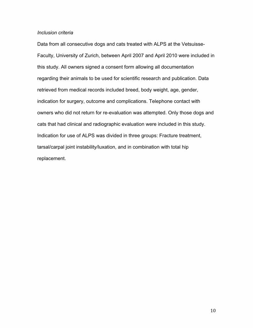
Inclusion criteria
Data from all consecutive dogs and cats treated with ALPS at the Vetsuisse-
Faculty, University of Zurich, between April 2007 and April 2010 were included in
this study. All owners signed a consent form allowing all documentation
regarding their animals to be used for scientific research and publication. Data
retrieved from medical records included breed, body weight, age, gender,
indication for surgery, outcome and complications. Telephone contact with
owners who did not return for re-evaluation was attempted. Only those dogs and
cats that had clinical and radiographic evaluation were included in this study.
Indication for use of ALPS was divided in three groups: Fracture treatment,
tarsal/carpal joint instability/luxation, and in combination with total hip
replacement.

11
Anesthetic protocol:
Cats were premedicated with ketamine (Narketan®, Vétoquinol AG, Switzerland)
5 mg/kg and midazolam (Dormicum®, Roche Pharma AG, Switzerland) 0.1
mg/kg intramuscularly (IM); dogs were premedicated with methadone
(Methadon®, Streuli Pharma AG, Switzerland) 0.2 mg/kg in combination with
acepromazine (Prequillan®, Arovet AG, Switzerland) 0.03 mg/kg IM. Anesthesia
was induced with propofol intravenously (IV) (Propofol 1% MCT Fresenius®,
Fresenius Kabi, Switzerland) to effect. After endotracheal intubation, anesthesia
was maintained using isoflurane (IsoFlo®, Abbott AG, Switzerland) and a
constant rate infusion (CRI) of fentanyl (Sintenyl®, Sintetica S.A., Switzerland). If
indicated, epidural anesthesia was performed using morphine (Morphin HCI
Sintetica®, Sintetica S.A., Switzerland) 0.1 mg/kg and bupivacaine 0.5 %
(Carbostesin®, Astra Zeneca, Switzerland) 0.5 mg/kg.
Cefazolin (Kefzol®, Teva Pharma AG, Switzerland) 22 mg/kg IV was
administered at induction and repeated every 90 minutes during anesthesia.
Intra-operative patient monitoring was performed routinely; lactated Ringer’s
solution was administered to all patients.
Postoperative analgesia consisted of an opioid (methadone, buprenorphine
(Temgesic®, Reckitt Benckiser, Switzerland), or fentanyl) IV in combination with a
non-steroidal anti-inflammatory drug (NSAID; meloxicam (Metacam®, Boehringer
Ingelheim GmbH, Germany) or carprofen (Rimadyl®, Pfizer AG, Switzerland)) IV
or per os.

12
4 Results
Eighty-four dogs and cats were treated with ALPS. Of the 84 patients, complete
data with follow-up ≥ 6 weeks was retrieved from 71 animals (72 treatments) (29
dogs: 5 mix breed dogs, 1 Shetland Sheepdog, 1 Lhasa Apso, 1 Rhodesian
Ridgeback, 3 Jack Russell Terrier, 1 Golden Retriever, 1 poodle, 1 Papillion, 1
Bernese Mountain Dog, 2 Appenzell Mountain Dog, 1 Entlebucher Mountain Dog,
1 Shih-Tzu, 1 Welsh Corgi Pembroke, 1 French Bulldog, 4 Labrador Retriever, 1
Beagle, 1 Miniature Pinscher, 1 Border Collie, 1 Bolonka Zwetna and 42 cats: 1
Norwegian Wood Cat, 1 Siamese Cat, 40 European Shorthair Cats). The mean
age of the dogs in this study was 3.9 years (4 months to 13 years), and of the
cats was 4.8 years (7 months to 13 years). The mean body weight of dogs was
19 kg (1.8 to 55 kg) and of cats was 4.5 kg (2 to 7.6 kg). Among the dogs there
were 16 female and 13 male, and in the cats 16 female and 26 males were
represented.
The surgeries were performed by a team consisting of various combinations of
ECVS board certified surgeons and surgical residents. Twelve surgeons with
different grades of expertise were involved in the procedures.

13
Description of stabilization method
Fractures
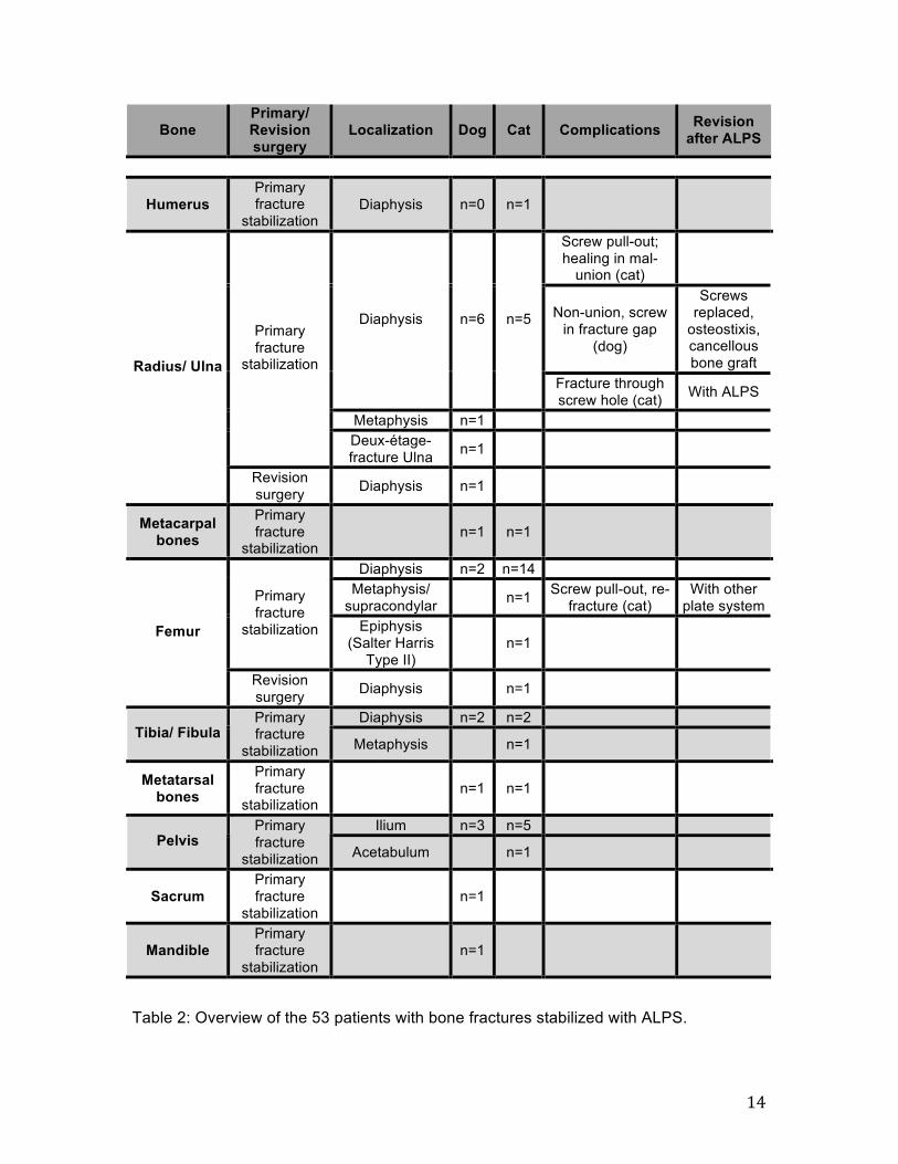
Fifty-four bone fractures in 53 patients were stabilized with ALPS. Affected bones
were humerus (1), radius/ulna (14), metacarpal bones (2), femur (19) tibia/fibula
(5), metatarsal bones (2), pelvis (9: Ilium 8, acetabulum 1), sacrum (1) and
mandible (1). Data is provided in table 2.
In two fracture-cases, ALPS was used for revision surgery. Once to revise a non-
healing open radius/ulna fracture treated with an external fixator, and once to
revise a collapsed femoral fracture in a cat previously stabilized using external
fixation. Both cases healed uneventfully.
In 4 cases of fractures in cats ALPS was combined with other implants: Twice in
a plate-rod configuration to treat comminuted diaphyseal femoral fractures, and
twice to stabilize serial fractures of the metabones (one metacarpal and one
metatarsal). In both cases the metabones 3 and 4 were stabilized with ALPS and
the metabones 2 and 5 were stabilized with IM pins.
14
Table 2: Overview of the 53 patients with bone fractures stabilized with ALPS.
Bone Primary/ Revision surgery
Localization Dog Cat Complications Revision after ALPS
Humerus Primary fracture
stabilization Diaphysis n=0 n=1
Screw pull-out; healing in mal-
union (cat)
Non-union, screw in fracture gap
(dog)
Screws replaced,
osteostixis, cancellous bone graft
Diaphysis n=6 n=5
Fracture through screw hole (cat) With ALPS
Metaphysis n=1
Primary fracture
stabilization
Deux-étage-fracture Ulna n=1
Radius/ Ulna
Revision surgery Diaphysis n=1
Metacarpal bones
Primary fracture
stabilization n=1 n=1
Diaphysis n=2 n=14 Metaphysis/
supracondylar n=1 Screw pull-out, re-fracture (cat)
With other plate system
Primary fracture
stabilization Epiphysis (Salter Harris
Type II) n=1
Femur
Revision surgery Diaphysis n=1
Diaphysis n=2 n=2 Tibia/ Fibula
Primary fracture
stabilization Metaphysis n=1
Metatarsal bones
Primary fracture
stabilization n=1 n=1
Ilium n=3 n=5 Pelvis
Primary fracture
stabilization Acetabulum n=1
Sacrum Primary fracture
stabilization n=1
Mandible Primary fracture
stabilization n=1
15
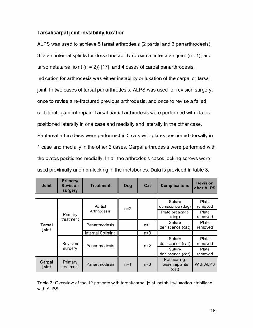
Tarsal/carpal joint instability/luxation
ALPS was used to achieve 5 tarsal arthrodesis (2 partial and 3 panarthrodesis),
3 tarsal internal splints for dorsal instability (proximal intertarsal joint (n= 1), and
tarsometatarsal joint (n = 2)) [17], and 4 cases of carpal panarthrodesis.
Indication for arthrodesis was either instability or luxation of the carpal or tarsal
joint. In two cases of tarsal panarthrodesis, ALPS was used for revision surgery:
once to revise a re-fractured previous arthrodesis, and once to revise a failed
collateral ligament repair. Tarsal partial arthrodesis were performed with plates
positioned laterally in one case and medially and laterally in the other case.
Pantarsal arthrodesis were performed in 3 cats with plates positioned dorsally in
1 case and medially in the other 2 cases. Carpal arthrodesis were performed with
the plates positioned medially. In all the arthrodesis cases locking screws were
used proximally and non-locking in the metabones. Data is provided in table 3.
Joint Primary/ Revision surgery
Treatment Dog Cat Complications Revision after ALPS
Suture
dehiscence (dog) Plate
removed Partial Arthrodesis n=2
Plate breakage (dog)
Plate removed
Panarthrodesis n=1 Suture dehiscence (cat)
Plate removed
Primary treatment
Internal Splinting n=3 Suture
dehiscence (cat) Plate
removed
Tarsal joint
Revision surgery Panarthrodesis n=2
Suture dehiscence (cat)
Plate removed
Carpal joint
Primary treatment Panarthrodesis n=1 n=3
Not healing, loose implants
(cat) With ALPS
Table 3: Overview of the 12 patients with tarsal/carpal joint instability/luxation stabilized with ALPS.

16
In combination with total hip replacement (THR)
ALPS was used in combination with THR (Zurich Cementless®, Kyon Pharma,
Inc., Zurich, Switzerland) in 6 dogs (THR = 6). In 4 dogs ALPS was applied in a
preventive manner to buttress the proximal femoral region in dogs that were
subjectively judged as having bad bone quality [18, 19], or when fissure lines
were caused during surgery. In two cases ALPS was applied to stabilize
fractures of the trochanter major during THR surgery. One fracture was stabilized
with 2 ALPS (8 and 5). All the other cases were treated using a single plate
(ALPS 8 n=1, ALPS 10 n=4). Data is provided in table 4.
Indication Treatment with ALPS Dog Cat Complications
Fracture of the
trochanter major after THR
Fracture stabilization n=2
THR revision surgery
Prophylactic femoral
buttressing n=3
THR, intraoperative
fissure line
Prophylactic femoral
buttressing n=1
Table 4: Overview of the 6 patients with THR additionally treated with ALPS.

17
Clinical outcome and complications
Fractures
50 of 54 fractures healed uneventfully. The following complications were found:
The distal fixation of a plate positioned in the ulna in order to stabilize a
comminuted proximal radius/ulna fracture of a 7 years old cat was applied too
caudally and didn’t get enough bone purchase. The screws pulled out through
the caudal cortex resulting in caudal bowing of the ulna. It was only detected
when healing was completed, and since the plate positioned in the radius
maintained the fixation, no revision was performed. Complications that needed
revision surgery were observed in 3 cases:
1) One month after the original surgery, a fracture occurred through the most
proximal screw of a 7 holes ALPS 6,5mm used to stabilize a mid diaphyseal
radius/ulna fracture in a 12 years old cat. During revision surgery, a longer ALPS
6,5mm plate bridging all the bone was positioned and healing occurred
uneventfully.
2) A supracondylar femoral fracture in a cat was stabilized with an ALPS 8mm
with 2 long locking screws in the distal fragment, and 3 locking monocortical
screws positioned proximally. Twenty days after the surgery the proximal part of
the fixation failed with all 3 monocortical screws being pulled out of the bone.
During revision surgery 2 plates (one medially and one laterally) were positioned.
3) A closed comminuted proximal radius/ulna fracture in a 5 years old dog,
stabilized with a ALPS 10 on the radius and ALPS 8 on the ulna, showed minimal
signs of healing 10 weeks postoperative. At that time radiolucency around one
18
screw located in the fracture gap of the radius and around the proximal fixation of
the ulna was detected. During revision surgery the loose screw in the radial
fracture gap was removed and the proximal fixation of the ulna was reinforced.
Osteostixis was performed and the fracture gap was filled with cancellous bone
graft. A bacteriological sample taken during the revision revealed the presence of
bacteria (Stapylococcus aureus). Five weeks after the revision surgery
radiographic evidence of clinical healing was observed. Both plates were
removed 2 years after the revision.
Tarsal/carpal joint instability/luxation
Of the 12 cases treated in this group the following complications were detected:
Five months after a partial tarsal arthrodesis stabilizing a tarso-metatarsal
instability in a dog, the laterally positioned plate broke at the level of the non-
curetted proximal intertarsal joint. Since the arthrodesed joint was already
bridged the plate was removed without further stabilization
The distal fixation of a pancarpal arthrodesis in a cat, become loose at 6 weeks
after surgery. During revision surgery some remaining cartilage was removed,
bone graft (vts, veterinary transplant services, inc., www.vtsonline.com, Kent,
USA) was added and newly positioned screws replaced the non-locking screws
of the distal fixation. Clinical union was observed 1 month after the revision.
The main complication in this group was suture dehiscence in the tarsal
arthrodesis group (1 partial tarsal arthrodesis, 3 pantarsal arthrodesis). The
cases were treated with bandages till radiographic evidence of bone healing was

19
detected, and the plates have been removed. The skin healed in all 4 cases after
implant removing without further complications.
In combination with total hip replacement (THR)
No complications with ALPS in combination with THR Zurich Cementless have
been detected.

20
5 Discussion
The purpose of this retrospective study was to describe the newly developed
ALPS system, to evaluate the first three years of its application at the Vetsuisse-
Faculty of the University of Zurich and to describe the outcome of the patients.
The ALPS system presents several particularities. Some of them appear to be
advantageous. The possibility to contour the plates in both planes facilitates the
placement on most of the bones. The specially designed in-plane bending pliers
allow precise bending under the protecting of the screw-holes. The amount of
bending is enough for most of the encountered situations. In cases like tarsal
panarthrodesis with medially applied plates where more bending is needed, the
correct joint angle could not be achieved with ALPS. The in-plane bending pliers
didn’t allow enough bending and using a standard bending plier would have
resulted in deformation of the screw-holes. To compensate the incomplete
bending in our two cases of tarsal panarthrodesis, the plates were positioned
slightly cranial on the tarsus and the distal fixation of the plate started in the distal
row of the tarsus. No complications related to this positioning were observed.
Nevertheless, a specially curved plate to match the anatomical angle of the
tarsus may be needed to provide a better apposition between bones and plate in
future.
The combination of locking and non-locking screws was normally used for
arthrodesis. It allowed to position larger locking screws proximally, and smaller
non-locking screws in the metabones. The locking screws were considered to be
too large to be safely positioned in the metabones without risking a fracture.

21
They were also too short to obtain enough bone purchase along all the
metabones, and they could not be positioned in another angle than perpendicular
to the plate. One out of 9 arthrodesis was revised with failure of this part of the
fixation, although we believe that incomplete preparation of the articular surfaces
was the cause. Again, specially dedicated plates with a proper angle, being
thinner in the distal part and allowing the use of smaller locking screws in the
distal part may be advantageous.
Suture dehiscence was a commonly encountered problem in tarsal arthrodesis
with ALPS (4 out of 5 cases). The minimal soft tissue coverage in the tarsal
region, open wounds associated with the tarsal joint injuries and previous
surgeries in 2 of the cases may have contributed to this high rate of dehiscence.
Still the thickness of the plate seems to be too excessive to support the tension
created in the metatarsal area. Therefore the use of a tapered plate may be
advantageous. High complication rates after tarsal arthrodesis have been
previously reported [20-21], however in these publications suture dehiscence
was not a common complication.
Monocortical screws are indicated in locking systems because the locking
mechanism in the plate hole replaces the stabilization effect of the second cortex
[3, 4, 7]. Before the placement of monocortical screws, the diameter of the bone
needs to be assessed. In systems with a different thread diameter in the head of
the screw, the screw tip will contact the trans cortex before locking the head in
the plate if the bone diameter is smaller than the length of the screw shaft. This
may lead to a damage of the bone threads in the near cortex making the screw

22
to be prone for pullout [8, 9, 22]. In ALPS, where the screw-threads are always
engaged in the thread of the plate-hole, contact with the trans cortex may lead to
a fracture of it. In small patients with small diameter of the bone, bicortical screws
may be used to reduce the risk of creating an iatrogenic fracture.
The locking screws of the ALPS must be positioned perpendicular to the plate
and they have a larger diameter as compared to the screws of other systems.
These facts make a good contouring of the plate mandatory in order to avoid
misplacement of the screws or damaging more than one bone cortex as occurred
in the previously described radius/ulna fracture in a cat. In this case the plate
didn’t get the needed contouring to match the procurvatum of the radius and
therefore the most proximal screw hole of the plate couldn’t be used. Additionally
the screw positioned in the second screw hole damaged the cranial cortex of the
radius resulting in a fracture one month later. The AO foundation recommends
that the screws’ diameter should not be larger than 40% of the entire bone
diameter. ALPS screws sometimes reach or even exceed this limit which
enhances the risk of fractures through the screw-hole. Nevertheless, thicker
screw cores provide better bone purchase and increase bending stiffness [3].
Still care in the positioning is mandatory to avoid this type of complications. The
use of monocortical screws may be beneficial to avoid excessive debilitation of
the bone.
If self-tapping screws are used, the cutting flutes must extend beyond the bone
cortex. Otherwise not enough purchase will be obtained which leads to a
potential collapse of the fixation [23] as occurred in one of our cases. In that case

23
three short monocortical screws were used to stabilize the proximal aspect of the
plate. This is particularly important when using monocortical screws because
they only have 60% of the pullout strength of a standard bicortical screw [7].
In the human field the use of monocortical screws is recommended only in
diaphyseal areas with good bone quality [8]. In most of our cases we used
monocortical screws in diaphyseal bone. Longer non-locking mono-or bicortical
screws were used when approaching the metaphysis as the longest locking
screw available in ALPS is only 20 mm long and it does not reach the trans
cortex in wider metaphyseal areas. In cases where angulation of the screw was
needed, bicortical non-locking screws were used. Still, the use of monocortical
locking screws is recommended to preserve endosteal blood supply and thereby
enhancing bone healing [1, 7]. This also allows to have the self-tapping part of
the screw being protected in the endosteal cavity and eliminates the need to
measure for the length of the screw when using MIPO techniques [4, 8, 22].
The limited plate to bone contact of the ALPS prevents damage to the periosteal
blood perfusion and reduces necrosis under the plate [3, 7]. As a consequence
the risk of infection is decreased and fracture healing accelerates [1]. It may also
reduce the risk of screw loosening due to bone necrosis under the plate when
using monocortical screws.
The presence of bacteria was found in one case of a comminuted radius/ulna
fracture, which didn’t heal properly. In that case, one of the locking screws was
positioned in the fracture gap, and all other 9 plate-holes were filled with screws.
We suspect that stress concentration in the gap associated with the presence of
24
a relatively large screw, and the extensive preparation performed for application
of the implants, affected healing and made conditions favorable for bacterial
colonization. Removal of the loose screw, positioning of bone graft and
osteostixis were enough to achieve bone healing after revision. We also suspect
that the better biocompatibility of titanium as compared to stainless steel [24] and
the reduced contact between implant and bone, may have contributed to the fast
bone healing (5 weeks), after removal of the loose screw.
The engagement of threads between the screws and plate-holes causes loss of
surgical feeling during insertion of screws. As the screws will be tightened even
when not positioned in the bone, the surgeon may miss that the screw is
misplaced. This is a reported problem in human and in veterinary surgery [4, 7, 9,
22]. In biological osteosynthesis techniques this represents the bigger issue as
reduced approaches are used. Proper positioning of the drilling guide, and
proper feeling of the drill bit passing the cortical bone may help to reduce this
potential problem.
As opposed to conventional locking plates where different threads are used
between screw head and shaft, ALPS screws have only one thread. Therefore
using ALPS bone-plate contact is needed at least when the first 2 screws are
positioned. Without bone-plate contact the reduction is lost during tightening of
the screws. Using locking plate systems with independent threads in the head
and the shaft of the screw, no bone-plate contact is needed. This makes plate
positioning easier [2, 8, 9].

25
ALPS also allows for axial compression. This is a potential advantage being less
often used, since more biological techniques are applied [3]. Still in special cases,
like in arthrodesis, it was used to increase stability.
We found difficulties inserting and removing larger sizes of screws. ALPS drill
guides are not threaded and locked into the plate as sleeves of other locking
systems are [2, 25]. Therefore the guides need to be held in position during
drilling manually and if the guide is not perfectly centered and perpendicular to
the plate, the screw will be also positioned eccentrically making its insertion or
removal more difficult. To counteract this issue Kyon developed locking threaded
guides for the non-locking systems of ALPS 6 and 8, and is planning to do the
same for all the sets. This is not ideal but at least a well-centered pilot hole can
be drilled in this manner. In 2010 (after completion of this study), and to facilitate
screw insertion/removal, the cutting flutes and the screw thread were improved.
Based in the results of this study, we can conclude that ALPS is a suitable option
to treat fractures and some other orthopedic conditions in small animals, with
reasonable handling possibilities. Improvements in the drilling guide design may
facilitate its use. Further investigation is needed to evaluate the effect of implant
design and material in infection rates and healing times.

26
6 References
1. Boudrieau, R.J. Advanced Locking Plate System (ALPS):
rationale, biomechanics and early clinical use. in WVOC 2010. Bologna
(Italy),.
2. Schwandt, C.S. and P.M. Montavon, Locking compression plate fixation of
radial and tibial fractures in a young dog. Vet Comp Orthop Traumatol,
2005. 18(3): p. 194-8.
3. Perren, S.M., Evolution of the internal fixation of long bone fractures. The
scientific basis of biological internal fixation: choosing a new balance
between stability and biology. J Bone Joint Surg Br, 2002. 84(8): p. 1093-
110.
4. Eijer, H., et al., PC-Fix and local infection resistance--influence of implant
design on postoperative infection development, clinical and experimental
results. Injury, 2001. 32 Suppl 2: p. B38-43.
5. Tepic, S., et al., Strength recovery in fractured sheep tibia treated with a
plate or an internal fixator: an experimental study with a two-year follow-up.
J Orthop Trauma, 1997. 11(1): p. 14-23.
6. Sikes, J.W., Jr., et al., Comparison of fixation strengths of locking head
and conventional screws, in fracture and reconstruction models. J Oral
Maxillofac Surg, 1998. 56(4): p. 468-73.
7. Haidukewych, G.J., Innovations in locking plate technology. J Am Acad
Orthop Surg, 2004. 12(4): p. 205-12.
27
8. Miller, D.L. and T. Goswami, A review of locking compression plate
biomechanics and their advantages as internal fixators in fracture healing.
Clin Biomech (Bristol, Avon), 2007. 22(10): p. 1049-62.
9. Voss, K., et al., Repair of long-bone fractures in cats and small dogs with
the Unilock mandible locking plate system. Vet Comp Orthop Traumatol,
2009. 22(5): p. 398-405.
10. Perren, S.M., Backgrounds of the technology of internal fixators. Injury,
2003. 34 Suppl 2: p. B1-3.
11. Schmokel, H.G., et al., Treatment of tibial fractures with plates using
minimally invasive percutaneous osteosynthesis in dogs and cats. J Small
Anim Pract, 2007. 48(3): p. 157-60.
12. Hudson, C.C., A. Pozzi, and D.D. Lewis, Minimally invasive plate
osteosynthesis: applications and techniques in dogs and cats. Vet Comp
Orthop Traumatol, 2009. 22(3): p. 175-82.
13. Guiot, L.P. and L.M. Dejardin, Prospective evaluation of minimally
invasive plate osteosynthesis in 36 nonarticular tibial fractures in dogs and
cats. Vet Surg, 2011. 40(2): p. 171-82.
14. Inauen, R., D. Koch, and M. Bass, Arthrodesis of the tarsometatarsal
joints in a cat with a two hole advanced locking plate system. Vet Comp
Orthop Traumatol, 2009. 22(2): p. 166-9.
15. Blake, C.A., et al., Single cycle to failure in bending of three standard and
five locking plates and plate constructs. Vet Comp Orthop Traumatol.
24(6): p. 408-17.
28
16. Cabassu, J.B., et al., Single cycle to failure in torsion of three standard
and five locking plate constructs. Vet Comp Orthop Traumatol. 24(6): p.
418-25.
17. Voss, K., M. Keller, and P.M. Montavon, Internal splinting of dorsal
intertarsal and tarsometatarsal instabilities in dogs and cats with the
ComPact Unilock 2.0/2.4 (TM) system. Veterinary and Comparative
Orthopaedics and Traumatology, 2004. 17(3): p. 125-130.
18. Andreoni, A.A., et al., Revision of an unstable HELICA endoprosthesis
with a Zurich cementless total hip replacement. Vet Comp Orthop
Traumatol, 2010. 23(3): p. 177-81.
19. Guerrero, T.G. and P.M. Montavon, Zurich cementless total hip
replacement: retrospective evaluation of 2nd generation implants in 60
dogs. Vet Surg, 2009. 38(1): p. 70-80.
20. Roch, S.P., et al., Complications following tarsal arthrodesis using bone
plate fixation in dogs. J Small Anim Pract, 2008. 49(3): p. 117-26.
21. McKee, W.M., et al., Pantarsal arthrodesis with a customised medial or
lateral bone plate in 13 dogs. Vet Rec, 2004. 154(6): p. 165-70.
22. Gautier, E. and C. Sommer, Guidelines for the clinical application of the
LCP. Injury, 2003. 34 Suppl 2: p. B63-76.
23. Murphy, T.P., et al., Pullout properties of 3.5-mm AO/ASIF self-tapping
and cortex screws in a uniform synthetic material and in canine bone. Vet
Surg, 2001. 30(3): p. 253-60.
29
24. Arens, S., et al., Influence of materials for fixation implants on local
infection. An experimental study of steel versus titanium DCP in rabbits. J
Bone Joint Surg Br, 1996. 78(4): p. 647-51.
25. Keller, M.A., K. Voss, and P.M. Montavon, The ComPact UniLock 2.0/2.4
system and its clinical application in small animal orthopedics. Vet Comp
Orthop Traumatol, 2005. 18(2): p. 83-93.

30
7 Cases Case 1
Abbildung 1
Craniocaudal and mediolateral radiographs of a 12 years old cat showing tarsometatarsal dorsal instability
A B C Dc
Abbildung 2
Immediate postoperative (A, B) and 8 months postoperative (C, D) craniocaudal and mediolateral radiographs. The tarsometatarsal instability was internally splinted with a dorsally applied ALPS 5, bridging the tarsometatarsal joint, using one locking screw and two non-locking screws
31
Case 2
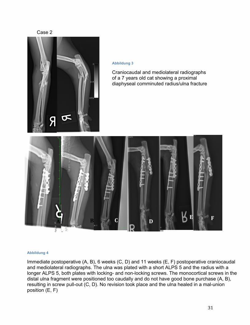
Abbildung 3
Craniocaudal and mediolateral radiographs of a 7 years old cat showing a proximal diaphyseal comminuted radius/ulna fracture
A B
C D E F
Abbildung 4
Immediate postoperative (A, B), 6 weeks (C, D) and 11 weeks (E, F) postoperative craniocaudal and mediolateral radiographs. The ulna was plated with a short ALPS 5 and the radius with a longer ALPS 5, both plates with locking- and non-locking screws. The monocortical screws in the distal ulna fragment were positioned too caudally and do not have good bone purchase (A, B), resulting in screw pull-out (C, D). No revision took place and the ulna healed in a mal-union position (E, F)

32
Case 3
Abbildung 5
Craniocaudal and laterolateral radiographs of a one year old cat showing a proximal diaphyseal comminuted femoral fracture
A B C
D
Abbildung 6
Immediate postoperative (A, B) and 6 weeks (C, D) postoperative craniocaudal and mediolateral radiographs. The femur was plated with a ALPS 8 with three locking screws per fragment. Long screws were used in metaphyseal areas, and short monocortical were positioned in diaphyseal bone

33
Case 4
Abbildung 7
Lateral radiographs in extended and neutral position of the carpal joint of a one year old cat showing a fracture of the accessory carpal bone with rupture of the palmar ligaments
A B C D E F
Abbildung 8
Immediate postoperative (A, B) , 6 weeks (C, D) and 13 weeks (E, F) postoperative craniocaudal and mediolateral radiographs. A pancarpal arthrodesis with a ALPS 6,5 from the medial side was performed, using locking screws in the radius and carpal joint, and non-locking screws in the metacarpal bones
34
Case 5
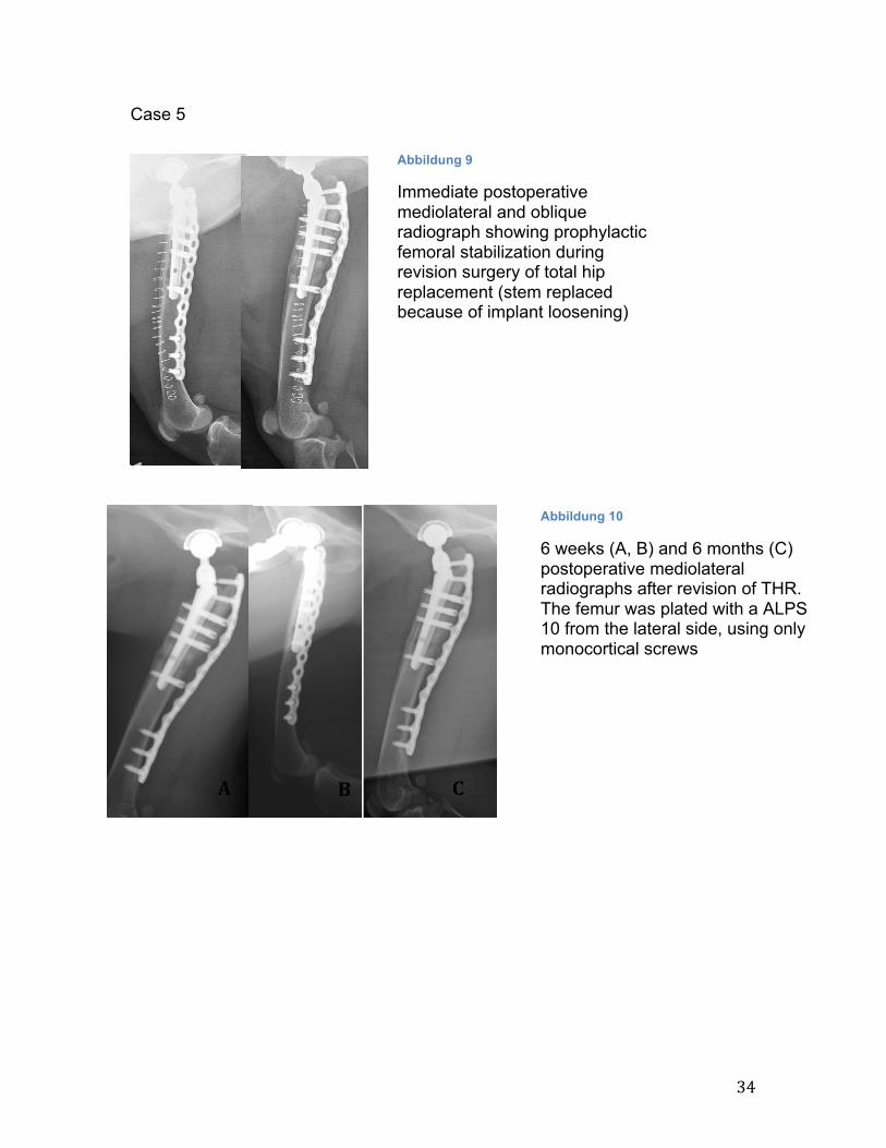
Abbildung 9
Immediate postoperative mediolateral and oblique radiograph showing prophylactic femoral stabilization during revision surgery of total hip replacement (stem replaced because of implant loosening)
A B C
Abbildung 10
6 weeks (A, B) and 6 months (C) postoperative mediolateral radiographs after revision of THR. The femur was plated with a ALPS 10 from the lateral side, using only monocortical screws

35
Lebenslauf
Name Nicole Scherrer
Geburtsdatum 11.02.1984
Geburtsort Zürich
Nationalität Schweiz
Heimatort Zürich
1990 – 1999 Grundschule, Langnau am Albis, Schweiz
2000 – 2003 Mathematisch-naturwissenschaftliches Gymnasium
Rämibühl, Zürich, Schweiz
2003 Abschluss: Matura
09/ 2003 – 10/ 2009 Studium der Veterinärmedizin, Vetsuisse-Fakultät Zürich,
Schweiz
10/ 2009 Staatsexamen, Vetsuisse-Fakultät Zürich, Schweiz
06/ 2010 – 06/ 2013 Anfertigung der Dissertation
unter Leitung von Prof. Dr. med. vet. Patrick R. Kircher,
Direktor des Departements für Kleintiere, Leiter der
Abteilung für Bildgebende Diagnostik und ad interims
Direktor der Klinik für Kleintierchirurgie der
Vetsuisse-Fakultät Universität Zürich, Schweiz
11/ 2009 – 11/ 2010 Assistentenstelle Kleintierchirurgie, Tierspital Zürich,
Schweiz
04/ 2011 – 04/ 2012 Internship Kleintierchirurgie, Tierspital Zürich, Schweiz
07/ 2012 – 12/ 2012 Assistentenstelle Kleintierchirurgie, Tierspital Zürich, Schweiz
August 2013, Zürich

36
Danksagung
Besonders bedanken möchte ich mich beim Korreferenten Prof. Dr. med. vet.
Tomás Guerrero für die Überlassung des Themas sowie die ausgezeichnete und
aufwändige fachliche Betreuung.
Ein grosses Dankeschön geht auch an den Referenten Prof. Dr. med. vet.
Patrick R. Kircher für die sehr angenehme Zusammenarbeit und exzellente
Betreuung.
Ausserdem danke ich dem Team der Radiologie und Kleintierchirurgie, welche
mir jederzeit bei Fragen und Problemen unterstützend zur Seite gestanden
haben.
Meinen Eltern und Brüdern, meinem Freund und meinen Freunden danke ich für
die herzliche und aufmunternde Unterstützung während der Bearbeitung meiner
Doktorarbeit.


