ankle arthrodesis with an anterior approach · ankle arthrodesis with an anterior approach...
TRANSCRIPT
Copyright @ Lippincott Williams & Wilkins. Unauthorized reproduction of this article is prohibited.
j S P E C I A L F O C U S j
Ankle Arthrodesis With an Anterior ApproachKarl-Heinz Kristen, MD, Hans-Jorg Trnka, MD, and Fusszentrum WienFoot and Ankle CenterVienna, Austria
| ABSTRACT
Ankle joint arthrodesis is described in many differentways using different approaches and fixation techniques.The key point is the fixation in axial alignment at90 degrees in the sagittal plane to obtain painless func-tion. The use of the anterior approach has been morewidely used since this incision has become the standardapproach for ankle joint prosthesis. The anterior ap-proach is indicated in cases without massive deformity.The approach, preparation, and internal screw fixationtechnique using 3 screws are described.Keywords: ankle arthrodesis, screw position, anklefusion, anterior approach, internal fixation, and openankle fusion
| HISTORICAL PERSPECTIVE
The primary indication for an ankle arthrodesis is painfrom severe ankle arthritis that limits daily activities.The most common indication nowadays is the post-traumatic degenerative joint disease following an anklefracture.
Historically, a wide variety of techniques for anklearthrodesis have been reported. The first report of anankle arthrodesis dates back to 1882 when Albert de-scribed the first fusion of the tibiotalar joint in paralyticlegs. Most early reports for ankle arthrodesis publishedbefore the 1950s contain few recommendations regard-ing the use of internal or external fixation1; most patientswere merely given a cast. In 1932, Key described an op-eration to fuse the tuberculous knee joint by applyingpositive pressure across the arthrodesis site by meansof a turnbuckle causing compression between supracon-dylar femur and proximal tibial pins.2 In light of thispublication, Charnley used this principle for the anklejoint and reported his experience in 1951.3 The develop-ment of internal fixation also changed the evolution ofthe ankle arthrodesis. Plates, cannulated compressionscrews, and staples nowadays are in use to achieve inter-nal compression across the arthrodesis site.
Selection of an ankle arthrodesis technique dependson whether realignment of a deformity is necessary.Wide surgical exposure is needed for deformity, and lim-ited exposure is sufficient for in situ fusion.
More recently, arthroscopically assisted techniquesfor the ankle fusion have gained popularity among sur-geons who routinely perform arthroscopy of the ankle.The success of this technique depends on the surgeon’sskill; sometimes, extended surgery time is needed.
| INDICATIONS ANDCONTRAINDICATIONS
The indications for the ankle arthrodesis using the ante-rior approach are as follows:1. Painful posttraumatic or idiopathic osteoarthritis of
the ankle joint that is unresponsive to nonoperativecare or anticipated failure of further nonoperativecare,
2. Posttraumatic malalignment of the ankle or chronicankle instability in combination with osteoarthritis,
3. Paralytic ankle instability that cannot be stabilized orimproved by joint preserving methods,
4. Ankle arthritis secondary to rheumatoid diseases andsystemic diseases,
5. Joint destruction after infection, and6. Failure of ankle joint replacement.
Contraindications include the following:1. Severe varus and valgus deformity of the ankle joint
with bone destruction that cannot be corrected usingthe anterior approach. In these cases, the lateral ap-proach with fibula osteotomy is recommended.
2. Acute osteitis and joint infection. In these cases, ex-ternal fixation may be an alternative.
3. Poor skin and soft tissue conditions4. Peripheral arterial occlusive disease and deep vein
thrombosis
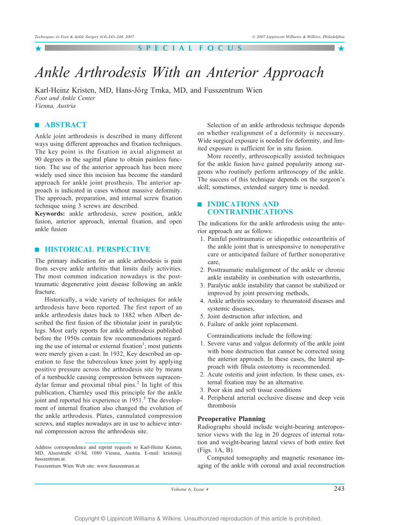
Preoperative PlanningRadiographs should include weight-bearing anteropos-terior views with the leg in 20 degrees of internal rota-tion and weight-bearing lateral views of both entire feet(Figs. 1A, B).
Computed tomography and magnetic resonance im-aging of the ankle with coronal and axial reconstruction
Address correspondence and reprint requests to Karl-Heinz Kristen,MD, AlserstraQe 43/8d, 1080 Vienna, Austria. E-mail: [email protected].
Fusszentrum Wien Web site: www.fusszentrum.at.
Volume 6, Issue 4 243
Techniques in Foot & Ankle Surgery 6(4):243–248, 2007 � 2007 Lippincott Williams & Wilkins, Philadelphia
Copyright @ Lippincott Williams & Wilkins. Unauthorized reproduction of this article is prohibited.
are optional in cases with unclear symptoms and if largecyst formation or talus necrosis is suspected.
Recommended surgical instruments and implants arethe following:1. A 7.3-mm cannulated self-cutting cancellous screws
in all lengths are preferable;2. Instruments for harvesting and shaping bone, saws,
drill, sharp curettes;3. Two small lamina spreaders are useful; and4. An intraoperative image intensifier is absolutely
necessary.
| TECHNIQUE
The procedure is generally performed under generalanesthesia or spinal block with tourniquet hemostaticcontrol. The patient is placed in supine position; a pil-low is placed under the buttock of the affected side toavoid external rotation the leg. Further preparationincludes free draping of the limb, allowing abductionand elevation of the entire leg, which is necessaryfor internal fixation and guidewire positioning underimage intensifier control. Ipsilateral iliac crest drapingis necessary.
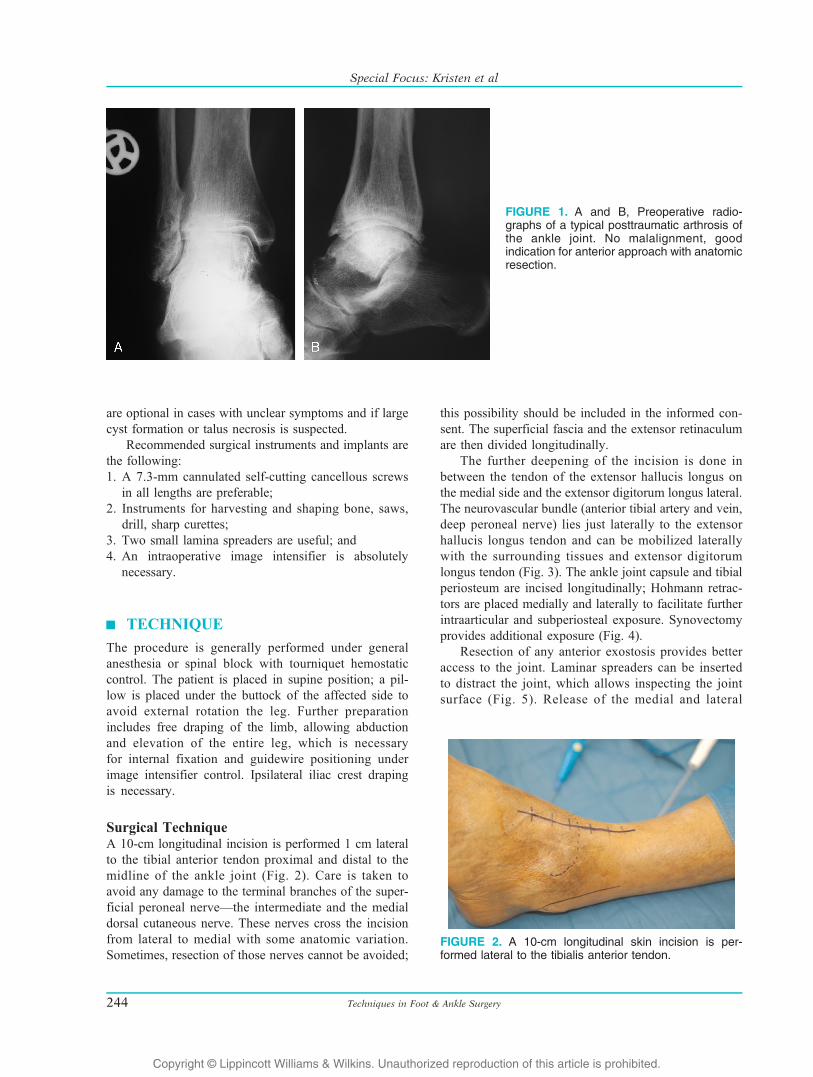
Surgical TechniqueA 10-cm longitudinal incision is performed 1 cm lateralto the tibial anterior tendon proximal and distal to themidline of the ankle joint (Fig. 2). Care is taken toavoid any damage to the terminal branches of the super-ficial peroneal nerveVthe intermediate and the medialdorsal cutaneous nerve. These nerves cross the incisionfrom lateral to medial with some anatomic variation.Sometimes, resection of those nerves cannot be avoided;
this possibility should be included in the informed con-sent. The superficial fascia and the extensor retinaculumare then divided longitudinally.
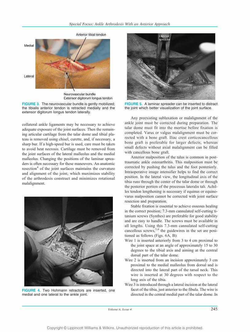
The further deepening of the incision is done inbetween the tendon of the extensor hallucis longus onthe medial side and the extensor digitorum longus lateral.The neurovascular bundle (anterior tibial artery and vein,deep peroneal nerve) lies just laterally to the extensorhallucis longus tendon and can be mobilized laterallywith the surrounding tissues and extensor digitorumlongus tendon (Fig. 3). The ankle joint capsule and tibialperiosteum are incised longitudinally; Hohmann retrac-tors are placed medially and laterally to facilitate furtherintraarticular and subperiosteal exposure. Synovectomyprovides additional exposure (Fig. 4).
Resection of any anterior exostosis provides betteraccess to the joint. Laminar spreaders can be insertedto distract the joint, which allows inspecting the jointsurface (Fig. 5). Release of the medial and lateral
FIGURE 1. A and B, Preoperative radio-graphs of a typical posttraumatic arthrosis ofthe ankle joint. No malalignment, goodindication for anterior approach with anatomicresection.
FIGURE 2. A 10-cm longitudinal skin incision is per-formed lateral to the tibialis anterior tendon.
Techniques in Foot & Ankle Surgery244
Special Focus: Kristen et al
Copyright @ Lippincott Williams & Wilkins. Unauthorized reproduction of this article is prohibited.
collateral ankle ligaments may be necessary to achieveadequate exposure of the joint surfaces. Then the remain-ing articular cartilage from the talar dome and tibial pla-teau is removed using chisel, curette, and, if necessary, asharp bur. If a high-speed bur is used, care must be takento avoid heat necrosis. Cartilage must be removed fromthe joint surfaces of the lateral malleolus and the medialmalleolus. Changing the positions of the laminar sprea-ders is often necessary for these maneuvers. An anatomicresection4 of the joint surfaces maintains the curvatureand alignment of the joint, which maximizes stabilityof the arthrodesis construct and minimizes rotationalmalalignment.
Any preexisting subluxation or malalignment of theankle joint must be corrected during preparation. Thetalar dome must fit into the mortise before fixation iscompleted. Varus or valgus malalignment must be cor-rected with a bone graft. Iliac crest corticocancellousbone graft is preferable for larger defects, whereassmall defects without axial malalignment can be filledwith cancellous bone graft.
Anterior malposition of the talus is common in post-traumatic ankle osteoarthritis. This malposition must becorrected by pushing the talus and the foot posteriorly.Intraoperative image intensifier helps to find the correctposition. In the lateral view, the longitudinal axis of thetibia runs through the center of the talar dome or throughthe posterior portion of the processus lateralis tali. Achil-les tendon lengthening is necessary if equinus or equino-varus malposition cannot be corrected with joint surfaceresection and preparation.
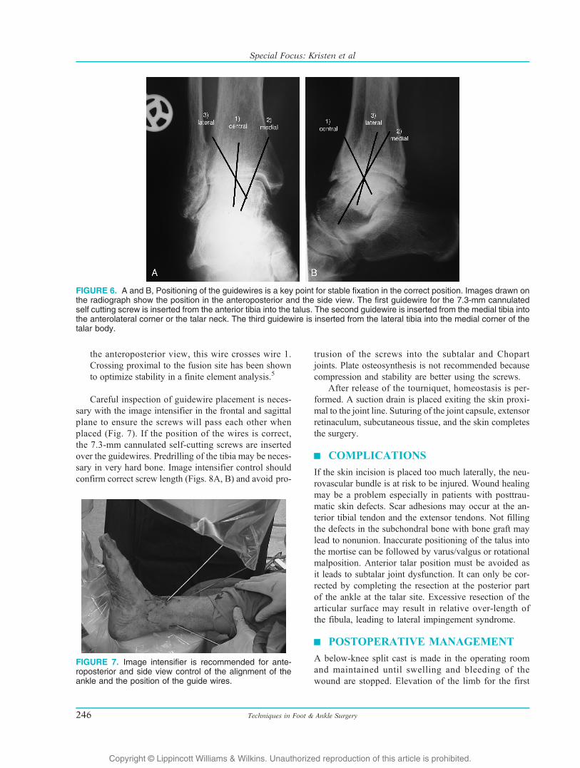
Stable fixation is essential to achieve osseous healingin the correct position; 7.3-mm cannulated self-cutting ti-tanium screws (Synthes) are preferable for good stabilityand are easy to handle. The screws must be available inall lengths. Using this 7.3-mm cannulated self-cuttingcancellous screws,5,6 the guidewires in the set are posi-tioned as follows (Figs. 6A, B):Wire 1 is inserted anteriorly from 3 to 4 cm proximal to
the joint space at an angle of approximately 15 to 30degrees to the tibial axis and aiming at the centraldorsal part of the talar dome.
Wire 2 is inserted from an incision approximately 3 cmproximal to the medial malleolus from dorsal and isdirected into the lateral part of the tarsal neck. Thiswire is inserted at 30 degrees with respect to thelong axis of the tibia.
Wire 3 is introduced through a lateral incision at the lateralfacet of the tibia, just anterior to the fibula. The wire isdirected in the central medial part of the talar dome. In
FIGURE 3. The neurovascular bundle is gently mobilized;the tibialis anterior tendon is retracted medially and theextensor digitorum longus tendon laterally.
FIGURE 4. Two Hohmann retractors are inserted, onemedial and one lateral to the ankle joint.
FIGURE 5. A laminar spreader can be inserted to distractthe joint which better visualization of the joint surface.
Volume 6, Issue 4 245
Special Focus: Ankle Arthrodesis With an Anterior Approach
Copyright @ Lippincott Williams & Wilkins. Unauthorized reproduction of this article is prohibited.
the anteroposterior view, this wire crosses wire 1.Crossing proximal to the fusion site has been shownto optimize stability in a finite element analysis.5
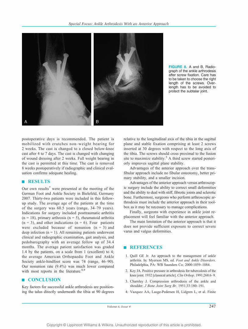
Careful inspection of guidewire placement is neces-sary with the image intensifier in the frontal and sagittalplane to ensure the screws will pass each other whenplaced (Fig. 7). If the position of the wires is correct,the 7.3-mm cannulated self-cutting screws are insertedover the guidewires. Predrilling of the tibia may be neces-sary in very hard bone. Image intensifier control shouldconfirm correct screw length (Figs. 8A, B) and avoid pro-
trusion of the screws into the subtalar and Chopartjoints. Plate osteosynthesis is not recommended becausecompression and stability are better using the screws.
After release of the tourniquet, homeostasis is per-formed. A suction drain is placed exiting the skin proxi-mal to the joint line. Suturing of the joint capsule, extensorretinaculum, subcutaneous tissue, and the skin completesthe surgery.
| COMPLICATIONS
If the skin incision is placed too much laterally, the neu-rovascular bundle is at risk to be injured. Wound healingmay be a problem especially in patients with posttrau-matic skin defects. Scar adhesions may occur at the an-terior tibial tendon and the extensor tendons. Not fillingthe defects in the subchondral bone with bone graft maylead to nonunion. Inaccurate positioning of the talus intothe mortise can be followed by varus/valgus or rotationalmalposition. Anterior talar position must be avoided asit leads to subtalar joint dysfunction. It can only be cor-rected by completing the resection at the posterior partof the ankle at the talar site. Excessive resection of thearticular surface may result in relative over-length ofthe fibula, leading to lateral impingement syndrome.
| POSTOPERATIVE MANAGEMENT
A below-knee split cast is made in the operating roomand maintained until swelling and bleeding of thewound are stopped. Elevation of the limb for the first
FIGURE 6. A and B, Positioning of the guidewires is a key point for stable fixation in the correct position. Images drawn onthe radiograph show the position in the anteroposterior and the side view. The first guidewire for the 7.3-mm cannulatedself cutting screw is inserted from the anterior tibia into the talus. The second guidewire is inserted from the medial tibia intothe anterolateral corner or the talar neck. The third guidewire is inserted from the lateral tibia into the medial corner of thetalar body.
FIGURE 7. Image intensifier is recommended for ante-roposterior and side view control of the alignment of theankle and the position of the guide wires.
Techniques in Foot & Ankle Surgery246
Special Focus: Kristen et al
Copyright @ Lippincott Williams & Wilkins. Unauthorized reproduction of this article is prohibited.
postoperative days is recommended. The patient ismobilized with crutches nonYweight bearing for2 weeks. The cast is changed to a closed below-kneecast after 4 to 7 days. The cast is changed with changingof wound dressing after 2 weeks. Full weight bearing inthe cast is permitted at this time. The cast is removed8 weeks postoperatively if radiographic and clinical eval-uation confirms adequate healing.
| RESULTS
Our own results7 were presented at the meeting of theGerman Foot and Ankle Society in Bielefeld, Germany2007. Thirty-two patients were included in this follow-up study. The average age of the patients at the timeof the surgery was 60.5 years (range, 34Y79 years).Indications for surgery included posttraumatic arthritis(n = 18), primary arthrosis (n = 5), rheumatoid arthritis(n = 3), and other indications (n = 6). Four patientswere excluded because of nonunion (n = 3) anddeep infection (n = 1). All remaining patients underwentclinical and radiographic examination, gait analysis, andpedobarography with an average follow up of 34.4months. The average patient satisfaction was graded1.4 by the patients, on a scale from 1 (excellent) to 4;the average American Orthopaedic Foot and AnkleSociety ankle-hindfoot score was 74 (range, 46Y90).Our nonunion rate (9.4%) was much lower comparedwith most reports in the literature.8,9
| CONCLUSION
Key factors for successful ankle arthrodesis are position-ing the talus directly underneath the tibia at 90 degrees
relative to the longitudinal axis of the tibia in the sagittalplane and stable fixation comprising at least 2 screwsinserted at 30 degrees with respect to the long axis ofthe tibia. The screws should cross proximal to the fusionsite to maximize stability.5 A third screw started posteri-orly improves sagittal plane stability.
Advantages of the anterior approach over the trans-fibular approach include no fibular osteotomy, better pri-mary stability, and a smaller incision.
Advantages of the anterior approach versus arthroscop-ic surgery include the ability to correct small deformitiesand the ability to deal with stiff, fibrotic joints and scleroticbone. Furthermore, surgeons who perform arthroscopic ar-throdesis must include the anterior approach in their tool-box as it may be necessary to manage complications.
Finally, surgeons with experience in ankle joint re-placement will feel familiar with the anterior approach.
The main limitation of the anterior approach is that itdoes not provide sufficient exposure to correct severevarus and valgus deformities.
| REFERENCES
1. Quill GE Jr. An approach to the management of anklearthritis. In: Myerson MS, ed. Foot and Ankle Disorders.Philadelphia, PA: WB Saunders Co; 2000:1059Y1084.
2. Key JA. Positive pressure in arthrodesis for tuberculosis of theknee joint. 1932 [classical article].Clin Orthop. 1991;268:6Y9.
3. Charnley J. Compression arthrodesis of the ankle andshoulder. J Bone Joint Surg Br. 1951;33:180Y191.
4. Vazquez AA, Lauge-Pedersen H, Lidgren L, et al. Finite
FIGURE 8. A and B, Radio-graph of the ankle arthrodesisafter screw fixation. Care hasto be taken to choose the rightlength of the screws. Over-length has to be avoided toprotect the subtalar joint.
Volume 6, Issue 4 247
Special Focus: Ankle Arthrodesis With an Anterior Approach

Copyright @ Lippincott Williams & Wilkins. Unauthorized reproduction of this article is prohibited.
element analysis of the initial stability of ankle arthrodesiswith internal fixation: flat cut versus intact joint contours.Clin Biomech (Bristol, Avon). 2003;18:244Y253.
5. Alonso-Vazquez A, Lauge-Pedersen H, Lidgren L, et al.Initial stability of ankle arthrodesis with three-screwfixation. A finite element analysis. Clin Biomech (Bristol,Avon). 2004;19:751Y759.
6. Alonso-Vazquez A, Lauge-Pedersen H, Lidgren L, et al.The effect of bone quality on the stability of anklearthrodesis. A finite element study. Foot Ankle Int. 2004;25:840Y850.
7. Pelzmann U, Haderer S, Ritschl P, et al. Die Arthrodesedes oberen Sprunggelenks eine Nachuntersuchungnach klinisch radiologisch pedobarographischen und gang-analytischen Parametern. 2007. Ref Type: UnpublishedWork.
8. Frey C, Halikus NM, Vu-Rose T, et al. A review of anklearthrodesis: predisposing factors to nonunion. Foot AnkleInt. 1994;15:581Y584.
9. Morrey BF, Wiedeman GP Jr. Complications and long-term results of ankle arthrodeses following trauma. J BoneJoint Surg Am. 1980;62:777Y784.
Techniques in Foot & Ankle Surgery248
Special Focus: Kristen et al