triple arthrodesis for the adult-acquired flatfoot deformitykzoofas.com/download/triple arthrodesis...
TRANSCRIPT

Clin Podiatr Med Surg
24 (2007) 765–778
Triple Arthrodesis for the Adult-AcquiredFlatfoot Deformity
Michael P. Maskill, DPM, Jeffrey D. Loveland, DPM,Robert W. Mendicino, DPM, FACFAS*,
Karl Saltrick, DPM, FACFAS,Alan R. Catanzariti, DPM, FACFAS
Division of Foot and Ankle Surgery, The Foot and Ankle Institute of Western Pennsylvania,
The Western Pennsylvania Hospital, 4800 Friendship Avenue, North Tower, First Floor,
Pittsburgh, PA 15224, USA
Triple arthrodesis is indicated for deformity, end-stage arthritis, and in-stability of the hindfoot. Triple arthrodesis often is the procedure of choicefor end-stage adult-acquired flatfoot (AAFF). The benefits of triple arthrod-esis include resolution of symptoms, hindfoot realignment, and stability. Awell-positioned triple arthrodesis ultimately will result in a plantigrade footthat will support the ankle in optimal alignment. This article reviews thetechnical execution and realignment considerations associated with triplearthrodesis for AAFF.
Triple arthrodesis is indicated for stages 3 and 4 AAFF and for stage 2AAFF with severe concomitant hindfoot instability, other rigid flatfoot de-formities (eg, tarsal coalition), and posttraumatic hindfoot arthrosis. Thegoals of the procedure include deformity correction with appropriate re-alignment, restoration of hindfoot stability, sound arthrodesis, and pain re-lief. Complications such as malunion, inadequate correction, continuedinstability, gait disturbances, and adjacent joint degeneration result from in-adequate realignment. Therefore, realignment is a critical factor for achiev-ing acceptable outcomes with triple arthrodesis. Recently published studiesreport satisfaction rates of greater than 90% after a 5-year follow-up [1,2].
Triple arthrodesis has been shown to produce favorable long-term resultsin resolution of pain and patient satisfaction [3]. Few studies, however, dis-cuss the effect of realignment on outcomes. Likewise, few reports discuss the
* Corresponding author.
E-mail address: [email protected] (R.W. Mendicino).
0891-8422/07/$ - see front matter � 2007 Elsevier Inc. All rights reserved.
doi:10.1016/j.cpm.2007.07.005 podiatric.theclinics.com

766 MASKILL et al
type of fixation and adjunctive procedures typically performed with triplearthrodesis for AAFF. Recent studies have begun to evaluate realignmentconsiderations, fixation techniques, and adjunctive procedures with short-term follow-up in a small patient population [4].
Extensive literature has been published regarding triple arthrodesis. Da-vis [5] originally described arthrodesis of the subtalar joint (STJ) and talo-navicular joint (TNJ) to provide more stability to the cavus foot. Hoke [6]later described the double arthrodesis for the treatment of talipes equinova-rus. Ryerson [7] modified the surgical technique by arthrodesing the calca-neocuboid joint (CCJ) in addition to the STJ and TNJ, hence the term‘‘triple arthrodesis.’’ Cast immobilization was used to maintain alignmentuntil healing was complete. Non-union rates have decreased from 7% to23% to zero to 9%, respectively, with the evolution of osteosynthesis andpostoperative non–weight bearing [8–12].
Anatomy and pathophysiology
The posterior tibial tendon (PTT) is a powerful muscle that acts asa strong supinator of the foot. The PTT passes immediately posterior tothe medial malleolus and medial to the STJ axis. It functions as a pulleyto plantarflex the ankle, supinate the foot, and dynamically stabilize the me-dial longitudinal arch [13]. Subtalar joint inversion locks the midfoot andprevents a pronatory moment through the oblique axis of the midtarsaljoint. The PTT has multiple insertions in the plantar midfoot and is a strongstabilizing force to the medial longitudinal arch.
A flatfoot is characterized by abduction and supination of the forefootwith a collapsed or absent medial longitudinal arch and a valgus angulationof the heel. AAFF usually is a result of longstanding posterior tibial tendin-opathy. Causes of AAFF include rheumatoid arthritis, seronegative arthri-tides, tarsal coalition, Charcot neuroarthropathy, paralysis, and acutetrauma to the PTT.
Radiographic views should include anteroposterior (AP), lateral, lateraloblique, long-leg axial, and hindfoot alignment. AP radiographs reveal peri-talar subluxation with high talo–first metatarsal angles (Fig. 1A). Peritalarsubluxation typically occurs because of lateral subluxation of the navicularon the talus. Lateral radiographs typically reveal an increase in the talo–firstmetatarsal angle (Fig. 1B).
Early stages of AAFF demonstrate a deformity that is reducible and sup-ple. A gastrocnemius–soleus contracture further accentuates and maintainsthe deformity. The deformity produces persistent stresses on the STJ andmidtarsal joints that eventually leads to dorsolateral peritalar subluxation.‘‘Dorsolateral peritalar subluxation’’ is a term coined by Hansen [14] thatdescribes the AAFF as medial talar head uncovering, with the forefoot ab-ducting, supinating and dorsiflexing around the talus through the midtarsaljoint, ultimately leading to a fixed flatfoot deformity.

767TRIPLE ARTHRODESIS FOR AAFF
The tibialis posterior muscle functions throughout the entire gait cycle.The PTT acts to decelerate subtalar pronation and internal tibial rotationat heel contact and to accelerate STJ supination and external rotation ofthe leg during midstance [13]. The PTT has a short excursion of only 2 cm,which maximizes its function. For this reason, a relative lengthening ordegeneration of the PTT can result in remarkable loss of biomechanical func-tion and strength [15].
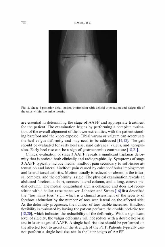
A classification scheme was created by Johnson and Strom [16] in 1989 toaid in the diagnosis and management of AAFF. Stage 1 is characterized bypain and swelling of the foot and ankle along the course of the PTT, with thepatient able to perform a single heel-rise test. Stage 2 describes a torn PTT,a weak limb, and the inability to perform a single heel-rise test. The midfootis pronated, and the forefoot is abducted at the midtarsal tarsal joint. TheSTJ remains flexible in stage 2 AAFF. Stage 3 AAFF involves degenerationof the PTT and a rigid, nonreducible hindfoot. Stage 4, which was a modifi-cation by Myerson and colleagues [17], is characterized by valgus angulationof the talus within the ankle mortise with or without signs of degenerativejoint disease (Fig. 2).
Physical examination
The diagnosis of symptomatic AAFF is based on clinical and radio-graphic assessment [18–20]. An adequate history and physical examination
Fig. 1. (A) Anteroposterior view of left foot demonstrating talar head uncovering, increased
talo–first metatarsal angle (Simmon’s angle), and increased cuboid abduction angle. (B) Lateral
radiograph of the left foot demonstrating decreased calcaneal inclination angle, increased talar
declination angle, and increased talo–first metatarsal angle (Meary’s angle).
768 MASKILL et al
are essential in determining the stage of AAFF and appropriate treatmentfor the patient. The examination begins by performing a complete evalua-tion of the overall alignment of the lower extremities, with the patient stand-ing barefoot and the knees exposed. Tibial varum or valgum can accentuatethe heel valgus deformity and may need to be addressed [14,18]. The gaitshould be evaluated for early heel rise, rigid calcaneal valgus, and apropul-sion. Early heel rise can be a sign of gastrocnemius contracture [18,21].
Clinical evaluation of stage 3 AAFF reveals a significant triplanar defor-mity that is noticed both clinically and radiographically. Symptoms of stage3 AAFF typically include medial hindfoot pain secondary to soft-tissue at-tenuation and lateral hindfoot pain caused by calcaneofibular impingementand lateral tarsal arthritis. Motion usually is reduced or absent in the tritar-sal complex, and the deformity is rigid. The physical examination reveals anabducted forefoot, a short, concave lateral column, and a long, convex me-dial column. The medial longitudinal arch is collapsed and does not recon-stitute with a hallux-raise maneuver. Johnson and Strom [16] first describedthe ‘‘too many toes’’ sign, which is a clinical assessment of the severity offorefoot abduction by the number of toes seen lateral on the affected side.As the deformity progresses, the number of toes visible increases. Hindfootflexibility is evaluated by having the patient perform the double heel-rise test[18,20], which indicates the reducibility of the deformity. With a significantlevel of rigidity, the valgus deformity will not reduce with a double heel-risetest in later stages of AAFF. A single heel-rise test should be performed onthe affected foot to ascertain the strength of the PTT. Patients typically can-not perform a single heel-rise test in the later stages of AAFF.
Fig. 2. Stage 4 posterior tibial tendon dysfunction with deltoid attenuation and valgus tilt of
the talus within the ankle mortis.

769TRIPLE ARTHRODESIS FOR AAFF
Surgical technique
Triple arthrodesis can be performed under general or spinal anesthesia.Before the procedure begins, prophylactic antibiotics are given to decreasethe risk of postoperative infection. A thigh tourniquet is preferred, anda support is placed beneath the ipsilateral hip for improved visualizationof the lateral aspect of the hindfoot. The presence of posterior muscle groupcontracture should be assessed while the patient is under general anesthesiato eliminate any guarding or resistance to examination and to permit a moreaccurate assessment of equinus. Equinus then may be addressed by perform-ing a posterior muscle group lengthening, including but not limited to a gas-trocnemius recession or an Achilles tendon lengthening (Fig. 3A, B).Correction of chronic AAFF without posterior muscle group lengtheningwill result in a foot that is not plantigrade after realignment and may placesignificant stress across the remaining mobile midfoot joints [22]. Gastrocne-mius recession or Achilles tendon lengthening then is performed, based onwhether the equinus is from the gastrocnemius alone or is gastrosoleal innature.
The most common surgical approach to the triple arthrodesis for AAFFuses a combined medial and lateral incision to obtain adequate exposure tothe CCJ, STJ, and TNJs (Fig. 4A, B). This approach originally was de-scribed by Ryerson and Davis [5,7]. The lateral incision provides excellentexposure to the CCJ, the sinus tarsi, and the facets of the STJ. This incisionbegins at the tip of the lateral malleolus and extends to the fourth metatarsal
Fig. 3. (A) The author’s typical three-incision approach to an Achilles tendon lengthening. (B)
A gastrocnemius recession may be used also. This technique is optimal when equinus is limited
to an isolated gastrocnemius contracture.

770 MASKILL et al
base. Care should be taken to avoid injury to the sural nerve or any of itsbranches. The peroneal tendons then are identified, released from theirsheath, and retracted inferiorly (Fig. 5A). The extensor digitorum brevismuscle belly is reflected, and the CCJ is identified. All periarticular
Fig. 4. (A) The lateral incision begins at the tip of the lateral malleolus and extends to the
fourth metatarsal base. (B) The medial incision begins in the area of the medial gutter of the
ankle and extends to the first metatarsal base.
Fig. 5. (A) The peroneal tendons are identified and retracted inferiorly, and the extensor digi-
torum brevis is reflected anteriorly to obtain adequate access to the calcaneocuboid joint and
the subtalar joint. (B) An Inge retractor is placed between the anterior calcaneus and inferolat-
eral aspect of talar neck and allows joint distraction. (C) Adequate joint preparation requires
fenestration of the subchondral plate. Various methods used include fish-scaling with a small
osteotome and subchondral drilling with a drill bit, Kirschner wire, or small bur.

771TRIPLE ARTHRODESIS FOR AAFF
attachments are transected to allow exposure and manipulation of the jointsurfaces. The sinus tarsi is identified, and the soft-tissue contents are evacu-ated thoroughly to reveal the posterior facet of the STJ. All periarticularstructures of the STJ, including the calcaneofibular ligament, are released,so that the calcaneus can be reduced from its valgus position. The cartilageof the STJ and CCJs then are removed through a limited debridement tech-nique using osteotomes and curettes (Fig. 5B). Kissel and colleagues [23] re-ported that curettage techniques preserved length of the foot and increasedthe surface area of bony contact. The cartilage debris then is carved away,and the subchondral bone is broken methodically using a fish-scaling or fen-estration technique (Fig. 5C). Attention then is directed to the medial aspectof the foot where the incision begins in the area of the medial gutter of theankle and extends to the first metatarsal base. The incision is deepened toexpose the joint capsule of the TNJ. All soft-tissue attachments then are re-leased to allow manipulation and exposure of the cartilaginous surfaces(Fig. 6). The soft-tissue release should be sufficient to allow realignmentof the hindfoot. Joint preparation is accomplished through the techniquespreviously described.
Jeng and colleagues [24,25] have described another optional approach us-ing a single medial incision. The authors surmise that with a long-standingfixed valgus deformity, the lateral incisional approach can cause potentialincisional complications and make subsequent wound healing and closuredifficult, although this has not been the present authors’ experience. Jengand colleagues’ study included 17 patients who underwent a medial-approach triple arthrodesis. All 17 patients achieved radiographic unionof the STJ and TNJ, and 15 of the 17 demonstrated definitive radiographicevidence of CCJ union. According to the cadaveric study by Jeng and
Fig. 6. Adequate exposure of the talonavicular joint is required for joint preparation.

772 MASKILL et al
colleagues [24], an average of 91% of all the articular surfaces for a triplearthrodesis could be prepared through the isolated medial approach. Theyconcluded that the CCJ can be prepared adequately with no diminutionin the quality of joint preparation.
After the articular cartilage has been completely denuded of all cartilage,the foot is manipulated into proper alignment. Optimal positioning of thehindfoot can be achieved with slight valgus to vertical. This position is ob-tained by placing the heel vertical to the lower leg, the forefoot parallel tothe hindfoot (straight lateral border), and the first ray at the same level asthe lesser metatarsals. Clinical realignment can be confirmed with image in-tensification. Axial, lateral, and AP views of the foot should be checked foradequate reduction of the deformity.
After realignment, provisional fixation can be obtained with Kirschnerwires, Steinman pins, or guide wires from a cannulated screw system. Thisfixation is performed under image intensification. The authors routinelyuse intraoperative AP (foot and ankle), lateral, and calcaneal axial viewsto confirm realignment of the tritarsal complex. The authors use one ortwo large-diameter cannulated screws delivered through the plantar aspectof the heel to fixate the STJ. Care must be taken to ensure that the anklejoint is not violated. This placement should be checked in two planes underfluoroscopy. The TNJ and CCJ then are fixated, either with one large-diam-eter screw or with two smaller-diameter cannulated screws directed distal toproximal. If inadequate bone stock is encountered intraoperatively, the sur-geon may consider the use of washers with screws.
Guide wires are removed after delivery of the screws and confirmation ofrealignment by means of fluoroscopy. Closure begins by reattaching the ex-tensor digitorum brevis into its anatomic position followed by subcutaneousand skin closure. The TNJ capsule is reapproximated, followed by subcuta-neous and skin closure. A closed-suction drain may be used to prevent for-mation of a hematoma. A common peroneal and popliteal block is used toenhance postoperative pain control and is performed in the operating roomupon completion of the procedure.
Realignment considerations
Achieving adequate position results in a stable, plantigrade foot and ul-timately leads to minimal gait disturbances and protection of the ankle. Theend result of intraoperative positioning should be a perpendicular relation-ship of the hindfoot to the forefoot in the frontal plane and a parallel rela-tionship in the transverse plane. Clinically, the hindfoot should be alignedwith the lower leg in the frontal plane.
Fluoroscopy is used to confirm intraoperative alignment and to adjustthe realignment of the hindfoot to the tibia and of the forefoot to the hind-foot. An intraoperative calcaneal axial view is used to ensure that the longaxis of the calcaneus is parallel to the mid-diaphyseal line of the distal tibia

773TRIPLE ARTHRODESIS FOR AAFF
(Fig. 7A). Provisional fixation then is used to maintain correction untilscrews are delivered across the arthrodesis sites. The forefoot is positionedparallel to the hindfoot in both the frontal and transverse planes. Thegoal is a parallel rearfoot-to-leg relationship (Fig. 7B). The talar–first meta-tarsal angle is measured on both the AP and lateral views to assess the fore-foot-to-hindfoot relationship. The anteroposterior talar–first metatarsalangle is used to assess the transverse plane, and the lateral talar–first meta-tarsal angle is used to assess the sagittal plane. Hindfoot alignment radio-graphs are used to assess frontal-plane relationships between the leg andthe ground, hindfoot to leg, and hindfoot to ground. It is important to eval-uate all three relationships to obtain optimal alignment.
Adjunct procedures
Based on the severity of the AAFF, various procedures may be needed inaddition to triple arthrodesis. Adjunct procedures include lateral columnlengthening, first tarso–metatarsal arthrodesis, bone grafting, posteriormuscle group lengthening, and posterior calcaneal osteotomies.
Triple arthrodesis has required the removal of bony wedges to obtain ac-ceptable alignment [6]. The authors’ goal is to perform minimal bone resec-tion while derotating the supinated and abducted forefoot to providea stable, plantigrade position. Distraction through the lateral column withuse of a bone graft has been successful in the treatment of severe AAFF[26]. This technique may be necessary after realignment of a severe defor-mity with significant adaptation of the CCJ. The authors use a bone blockto fill the deficit that remains after realignment. This technique can be com-bined with the triple arthrodesis to restore length and eliminate forefootabduction.
Fig. 7. (A) The method used to obtain an intraoperative calcaneal axial view. (B) Intraoperative
calcaneal axial view demonstrating the calcaneus in good alignment to the long axis of the tibia.
774 MASKILL et al
First tarsometatarsal arthrodesis for AAFF can be used adjunctively withthe triple arthrodesis procedure after hindfoot correction and fixation. Theonly correction of forefoot supination that can be obtained through a triplearthrodesis is by means of the TNJ. If residual supinatus or instability is re-maining in the forefoot, a plantarflexory first tarsometatarsal arthrodesiscan be performed if the forefoot supinatus is overlooked. Lateral columnoverload can result and lead to poor patient satisfaction [22]. Adding thisprocedure provides stability to the medial column.
Equinus may be a soft-tissue contracture of the gastrosoleal complex thatis a common finding with longstanding AAFF. The Silfverskiold test shouldbe performed preoperatively and intraoperatively to choose the appropriateprocedure for lengthening the posterior muscle group [27]. Based on the re-sults of the Silfverskiold test, the posterior muscle group can be lengthenedwith either an isolated gastrocnemius recession or a percutaneous tendoAchilles lengthening.
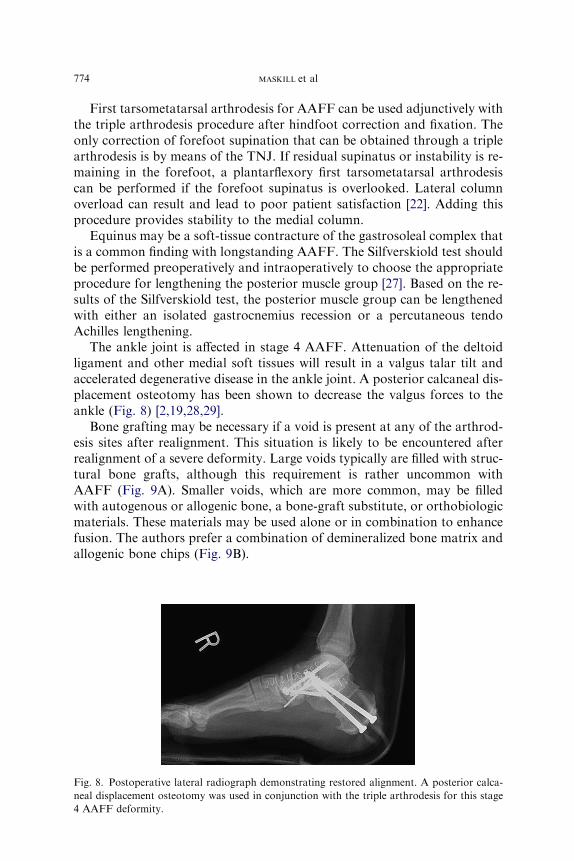
The ankle joint is affected in stage 4 AAFF. Attenuation of the deltoidligament and other medial soft tissues will result in a valgus talar tilt andaccelerated degenerative disease in the ankle joint. A posterior calcaneal dis-placement osteotomy has been shown to decrease the valgus forces to theankle (Fig. 8) [2,19,28,29].
Bone grafting may be necessary if a void is present at any of the arthrod-esis sites after realignment. This situation is likely to be encountered afterrealignment of a severe deformity. Large voids typically are filled with struc-tural bone grafts, although this requirement is rather uncommon withAAFF (Fig. 9A). Smaller voids, which are more common, may be filledwith autogenous or allogenic bone, a bone-graft substitute, or orthobiologicmaterials. These materials may be used alone or in combination to enhancefusion. The authors prefer a combination of demineralized bone matrix andallogenic bone chips (Fig. 9B).
Fig. 8. Postoperative lateral radiograph demonstrating restored alignment. A posterior calca-
neal displacement osteotomy was used in conjunction with the triple arthrodesis for this stage
4 AAFF deformity.

775TRIPLE ARTHRODESIS FOR AAFF
Rosenfeld and colleagues [30] stated that bone grafting was not necessarywhen performing a triple arthrodesis. They used only local bone graft ob-tained at the site of the operation to fill voids across the arthrodesis sites,and only 4 of 100 cases developed non-union in their study. They believedthat comparable rates of union can be achieved without supplementarybone graft.
Postoperative management
The authors follow a strict postoperative regimen that begins with the pa-tient’s admission to the hospital for 1 to 3 days for management of postop-erative pain and edema. The patient is placed in a compressive dressing witha posterior splint, with care taken to place the ankle in neutral positionimmediately after surgery. The patient receives deep venous thrombosisprophylaxis and proper pain management. The patient undergoes gait train-ing preoperatively and/or postoperatively to ensure the patient’s ability toremain non–weight-bearing (NWB). If unable to be NWB, the patientmust be placed in a skilled nursing facility. The patient is placed in a well-padded below-the-knee fiberglass cast before discharge. The patient remainsNWB for approximately 2 to 3 months. Serial radiographs are obtained tomonitor consolidation of the arthrodesis sites. If there is minimal edema andwarmth after cast removal, the patient transitions to a partial-weight-bear-ing fracture brace. The patient resumes standard footgear thereafter. Phys-ical therapy can be useful for strengthening atrophied muscles, controllingedema, and gait training.
Complications
The triple arthrodesis has been described as a technically very demandingprocedure [2,3,8,12,22,31]. The average time to maximum improvement
Fig. 9. (A) A fresh-frozen femoral head can be used as a structural allograft to fill large deficits
and restore alignment of severe deformities. (B) The authors frequently use other orthobiologic
materials, such as demineralized bone matrix and allogenic bone chips, to fill smaller defects
and assist in the formation of extra-articular arthrodesis.

776 MASKILL et al
after triple arthrodesis is 10 months [3], and some residual pain may remainfrom problems that were not addressed by the procedure.
Pseudoarthrosis and non-union rates have been reported as frequentcauses of persistent pain and subsequent patient dissatisfaction [3,8,12].Non-union has become less common with the advent of more contemporarymethods of fixation. Historically, non-union rates were as high as 33%, butwith more advanced techniques and rigid internal fixation, non-union rateshave dropped significantly [22] and are reported to range from 6% to 33%[8,32,33]. The most common site for non-union of the triple arthrodesis isthe TNJ [11], especially if a single lateral incision is used [31], but non-unions can occur at any of the three articulations. Fortunately, non-unionsoften are asymptomatic. Risk factors leading to non-union include cigarettesmoking [34–36], the performance of simultaneous bilateral triple arthrode-ses [37], and the lack of rigid fixation in the early history of the procedure[7]. The use of bone graft as an adjunct to triple arthrodesis has shownexcellent results, most likely because of the formation of an extra-articulararthrodesis in the sinus tarsi [38]. Fortin and Walling [22] reported a 3%non-union rate with the use of bone graft, a two-incisional approach, min-imal bony resection, and rigid internal fixation. Likewise, Pell and col-leagues [2] found a 2% non-union rate in 132 triple arthrodeses.
Positioning of the hindfoot to obtain optimal alignment and a stable,plantigrade foot is the most crucial part of the procedure. The literature sup-ports the positioning of the heel into slight valgus of 5� [23,39,40], althoughthe authors prefer the heel to be as close to vertical as possible. According toFortin and Walling [22], positioning the heel in varus may lead to acceler-ated degeneration of the tibiotalar joint and an overloaded lateral column.In their study, four patients had a varus malunion, and two of them requiredrevisional surgery for relief of pain. Conversely, excessive valgus malunionalso is not well tolerated. Graves and colleagues [3] noted that placing thehindfoot into excessive valgus provided adequate pain relief but poor pa-tient satisfaction. Maenpaa and colleagues [41] reviewed 307 triple arthrod-eses and concluded that the primary cause for revision was malunion of thehindfoot, whether the malalignment was varus or valgus.
Secondary arthritis of the ankle is another major concern for most inves-tigators. Fortin and Walling [22] state that in situ fusion without correctionof deformity typically results in a painful, stiff foot with progressive degen-erative changes to the ankle. Some studies have discovered that fusion ofone joint increases the mechanical stress on all adjacent joints, and the re-ported prevalence of adjacent ankle arthritis after a triple arthrodesis hasranged from 2% to 77% [2]. Pell and colleagues [2] reported that 79 of132 ankles had clear signs of progressive degenerative joint disease in the an-kle at the time of follow-up. Twenty-eight percent had grade I arthritis, 19%had grade II arthritis, and 11% had grade III arthritis of the ankle joint.Five ankles had developed severe degenerative arthrosis and already hadhad an ankle arthrodesis performed before the presentation of their study.
777TRIPLE ARTHRODESIS FOR AAFF
Most importantly, Pell and colleagues [2] concluded that there was a strongcorrelation between patient satisfaction and hindfoot alignment.
Other complications such as nerve injury, wound dehiscence, infection,and fixation failure can occur as well. Meticulous sterile technique, gentlehandling of soft tissues during dissection, and proper placement of the inci-sion are essential in limiting these complications.
Summary
Triple arthrodesis is an effective procedure to relieve pain and correctstructural deformities associated with AAFF. Appropriate realignment isthe most critical factor for achieving a functional outcome.
References
[1] Beischer AD, Brodsky JW, Pollo FE, et al. Functional outcome and gait analysis after triple
or double arthrodesis. Foot Ankle Int 1999;20(9):545–53.
[2] Pell RF, Merson MS, Schon LC. Clinical outcome after primary triple arthrodesis. J Bone
Joint Surg 2000;82A:7–57.
[3] Graves SC,Mann RA, Graves KO. Triple arthrodesis in older adults: results after long term
follow up. J Bone Joint Surg 1993;75A(3):355–62.
[4] Catanzariti AR, Mendicino RW,Whitaker JM, et al. Realignment considerations in the tri-
ple arthrodesis. J Am Podiatr Med Assoc 2005;95(1):13–7.
[5] Davis G. Treatment of hollow foot (pes cavus). Am J Orthop Surg 1913;11:231.
[6] Hoke M. An operation for stabilizing paralytic feet. Am J Orthop Surg 1921;3:494–507.
[7] Ryerson E. The classic, arthrodesing operation on the feet. Clin Orthop Relat Res 1977;122:
4–9.
[8] Sangeorzan JB, Smith D, Veith R, et al. Triple arthrodesis using internal fixation in treat-
ment of adult foot disorders. Clin Orthop Relat Res 1993;(294):299–307.
[9] Haritidis JH, Kirkos JM, Provellegios SM, et al. Long-term results of triple arthrodesis: 42
cases followed for 25 years. Foot Ankle Int 1994;15(10):548–51.
[10] Angus PD, Cowell GR. Triple arthrodesis: a critical long term review. J Bone Joint Surg
1986;68B:260–5.
[11] Wetmore RS, Drennan JC. Long-term results of triple arthrodesis in Charcot-Marie-tooth
disease. J Bone Joint Surg 1989;71A:417–22.
[12] Bednarz PA, MonroeMT, Manoli A. Triple arthrodesis in adults using internal fixation: an
assessment of outcome. Foot Ankle Int 1999;20(6):356–63.
[13] Blake RL, Anderson K, FergusonH. Posterior tibial tendonitis: a literature review with case
reports. J Am Podiatr Med Assoc 1994;84:141–9.
[14] Hansen ST. Functional reconstruction of the foot and ankle. Philedolphia: Lippincott Wil-
liams & Wilkins; 2000. p. 22–6, 195–207.
[15] Silver RL, de la Garza J, RangM. The myth of muscle balance: a study of relative strengths
and excursions of normal muscles about the foot and ankle. J Bone Joint Surg Br 1985;67:
432–7.
[16] JohnsonKA, StromDE. Tibialis posterior tendon dysfunction. ClinOrthopRelat Res 1989;
239:196–203.
[17] MyersonM, SolomonG, ShereffM. Posterior tibial tendon dysfunction: its association with
seronegative inflammatory disease. Foot Ankle 1989;9:219–25.
[18] Mann RA. Flatfoot in adults. In: Caughlin MJ, Mann RA, editors. Surgery of the foot and
ankle, vol. 1. 7th edition. St. Louis (MO): Mosby, Inc.; 1999. p. 733–67.

778 MASKILL et al
[19] Myerson MS. Adult acquired flatfoot deformity. J Bone Joint Surg 1996;78A(5):780–92.
[20] Johnson KA. Tibialis posterior tendon rupture. Clin Orthop Relat Res 1983;177(4):140–7.
[21] Pinney SJ, Hansen ST, Sangeorzan BJ. The effect on ankle dorsiflexion of gastrocnemius re-
cession. Foot Ankle Int 2002;23(1):26–9.
[22] Fortin PT, Walling AK. Triple arthrodesis. Clin Orthop Relat Res 1999;365:91–9.
[23] Kissel CG, Hulst TJ, Blacklidge DK, et al. Nonresection triple arthrodesis: a retrospective
analysis. J Foot Ankle Surg 1998;37(6):490–500.
[24] Jeng CL, Tankson CJ, MyersonMS. The single medial approach to triple arthrodesis: a ca-
daver study. Foot Ankle Int 2006;27(12):1122–5.
[25] Jeng CL, Vora AM, Myerson MS. The medial approach to triple arthrodesis. Indications
and technique for management of rigid valgus deformities in high-risk patients. Foot Ankle
Clin 2005;10(3):515–21.
[26] Evans D. Calcaneovalgus deformity. J Bone Joint Surg 1975;57B:270–8.
[27] Silfverskiold N. Reduction of the uncrossed two-joint muscles of the leg to one-joint muscles
in spastic conditions. Acta Chir Scand 1924;56:315.
[28] Resnick RB, JahssMH, Choueka J, et al. Deltoid ligament forces after tibialis posterior ten-
don rupture: effects of triple arthrodesis and calcaneal displacement osteotomies. Foot An-
kle Int 1995;16(1):14–20.
[29] Steffensmeier SJ, Saltzman CL, Berbaum KS, et al. Effects of medial and lateral displace-
ment calcaneal osteotomies on tibiotalar joint contact characteristics. J Orthop Res 1996;
14:980–5.
[30] Rosenfeld PF, Budgen SA, Saxby TS. Triple arthrodesis: is bone grafting necessary? The re-
sults in 100 consecutive cases. J Bone Joint Surg 2005;87B:175–8.
[31] Bennett G, Graham CE, Mauldin DM. Triple arthrodesis in adults. Foot Ankle 1991;12(3):
138–43.
[32] Wukich DK, Bowen JR. A long-term study of triple arthrodesis for correction of pes cavo-
varus in Charcot-Marie-tooth disease. J Pediatr Orthop 1989;9(4):433–7.
[33] Salzman CL, Fehrle MF, Cooper RR. Triple arthrodesis: twenty-five and forty-four-year
average follow-up of the same patients. J Bone Joint Surg 1999;81A:1391–402.
[34] Ishikawa SN, Murphy A, Richardson EG. The effect of cigarette smoking on hindfoot fu-
sions. Foot Ankle Int 2002;23(11):996–8.
[35] Haverstock BD,Mandracchia VJ. Cigarette smoking and bone healing: implications in foot
and ankle surgery [review]. J Foot Ankle Surg 1998;37(1):69–74 [discussion: 78].
[36] Cobb TK, Gabrielson TA, Campbell DC 2nd, et al. Cigarette smoking and nonunion after
ankle arthrodesis. Foot Ankle Int 1994;15(2):64–7.
[37] Wilson FC, Fay FC, Lamotte P, et al. Triple arthrodesis: a study of the factors affecting fu-
sion after three hundered and one procedures. J Bone Joint Surg Am 1965;47:340–8.
[38] Catanzariti AR,Mendicino RM, Saltrick KS, et al. Subtalar joint arthrodesis. J Am Podiatr
Med Assoc 2005;95(1):34–41.
[39] Vogler HW. Triple arthrodesis as a salvage for end-stage flatfoot. Clin Podiatr Med Surg
1989;6(3):591–604.
[40] Kelly IP, Nunley JA. Treatment of stage 4 adult acquired flatfoot. Foot Ankle Clin 2001;
6(1):167–78.
[41] Maenpaa H, Lehto MU, Belt EA. What went wrong with in triple arthrodesis. Clin Orthop
Relat Res 2001;391:218–23.