whole gene sequencing identifies deep- intronic variants with
TRANSCRIPT
RESEARCH ARTICLE
Whole gene sequencing identifies deep-
intronic variants with potential functional
impact in patients with hypertrophic
cardiomyopathy
Rita Mendes de Almeida1☯, Joana Tavares1☯, Sandra Martins1, Teresa Carvalho1,
Francisco J. Enguita1, Dulce Brito2,3, Maria Carmo-Fonseca1*, Luıs Rocha Lopes3,4*
1 Instituto de Medicina Molecular, Faculdade de Medicina, Universidade de Lisboa, Lisbon, Portugal,
2 Departamento de Cardiologia, Hospital de Santa Maria, Centro Hospitalar Lisboa Norte, Centro Academico
de Medicina de Lisboa, Lisbon, Portugal, 3 Centro Cardiovascular da Universidade de Lisboa, Lisbon,
Portugal, 4 Institute of Cardiovascular Science, University College London, London, United Kingdom
☯ These authors contributed equally to this work.
* [email protected] (MCF); [email protected] (LRL)
Abstract
Background
High throughput sequencing technologies have revolutionized the identification of mutations
responsible for genetic diseases such as hypertrophic cardiomyopathy (HCM). However,
approximately 50% of individuals with a clinical diagnosis of HCM have no causal mutation
identified. This may be due to the presence of pathogenic mutations located deep within the
introns, which are not detected by conventional sequencing analysis restricted to exons and
exon-intron boundaries.
Objective
The aim of this study was to develop a whole-gene sequencing strategy to prioritize deep
intronic variants that may play a role in HCM pathogenesis.
Methods and results
The full genomic DNA sequence of 26 genes previously associated with HCM was analysed
in 16 unrelated patients. We identified likely pathogenic deep intronic variants in VCL,
PRKAG2 and TTN genes. These variants, which are predicted to act through disruption of
either splicing or transcription factor binding sites, are 3-fold more frequent in our cohort of
probands than in normal European populations. Moreover, we found a patient that is com-
pound heterozygous for a splice site mutation in MYBPC3 and the deep intronic VCL vari-
ant. Analysis of family members revealed that carriers of the MYBPC3 mutation alone do
not manifest the disease, while family members that are compound heterozygous are clini-
cally affected.
PLOS ONE | https://doi.org/10.1371/journal.pone.0182946 August 10, 2017 1 / 19
a1111111111
a1111111111
a1111111111
a1111111111
a1111111111
OPENACCESS
Citation: Mendes de Almeida R, Tavares J, Martins
S, Carvalho T, Enguita FJ, Brito D, et al. (2017)
Whole gene sequencing identifies deep-intronic
variants with potential functional impact in patients
with hypertrophic cardiomyopathy. PLoS ONE 12
(8): e0182946. https://doi.org/10.1371/journal.
pone.0182946
Editor: Amanda Ewart Toland, Ohio State
University Wexner Medical Center, UNITED
STATES
Received: April 10, 2017
Accepted: July 27, 2017
Published: August 10, 2017
Copyright: © 2017 Mendes de Almeida et al. This is
an open access article distributed under the terms
of the Creative Commons Attribution License,
which permits unrestricted use, distribution, and
reproduction in any medium, provided the original
author and source are credited.
Data Availability Statement: All relevant data are
within the paper and its Supporting Information
files. The data have also been deposited with link to
BioProject accession number PRJNA393768 in the
NCBI BioProject database (https://www.ncbi.nlm.
nih.gov/bioproject/393768).
Funding: This work was supported by Fundacãopara a Ciência e Tecnologia and FEDER, Portugal
(Scholarship BD/82023/2011 to R.M.A, grant
Conclusion
This study provides a framework for scrutinizing variation along the complete intronic
sequence of HCM-associated genes and prioritizing candidates for mechanistic and func-
tional analysis. Our data suggest that deep intronic variation contributes to HCM phenotype.
Introduction
Hypertrophic cardiomyopathy (HCM) is a genetic heart disease associated with sudden car-
diac death and progressive heart failure. HCM is considered one of the most common genetic
disorders, with an estimated prevalence of 1 in 500 people throughout the world [1]. Recogni-
tion of the disease is critical for providing treatment and prevention strategies as well as trig-
gering clinical and genetic surveillance of family members [2],[3]. Mutation carriers may
benefit from lifestyle and medical interventions that improve prognosis, whereas a negative
genetic test can reassure individuals that are not at risk [3],[4].
Since a mutation in β-myosin heavy chain (MYH7) was first identified as the cause of HCM
[5], other mutations affecting components of the sarcomere have been shown to have a patho-
genic role in this disease [6]. In addition to MYH7, the most frequently mutated genes are car-
diac myosin-binding protein C (MYBPC3), cardiac troponin T (TNNT2), cardiac troponin I
(TNNI3), α-tropomyosin (TPM1), regulatory myosin light chain (MYL2), essential myosin
light chain (MYL3) and cardiac actin (ACTC1) (Table 1). More rarely, mutations have been
reported in other genes encoding proteins required for sarcomere structure and function, such
as α-actinin 2 (ACTN2), muscle LIM protein (CSRP3) and calcium metabolism, such as phos-
pholamban (PLN) and junctophilin 2 (JPH2) [7]. Additional sarcomere-related genes have
been associated with HCM, although with less firmly established evidence for direct pathoge-
nicity [8] (Table 1). These include α-myosin heavy chain (MYH6), telethonin (TCAP) [7],
LIM domain binding 3 protein (LDB3) [9], myosin light chain kinase 2 (MYLK2) [10], myoze-
nin 2 (MYOZ2) [11], nexilin (NEXN) [12], troponin C (TNNC1) [13], titin (TTN) [14], vincu-
lin (VCL) [15], ankyrin repeat domain 1 (ANKRD1) [16] and caveolin 3 (CAV3) [17].
Some rare inherited diseases may mimic the phenotypic and clinical features of sarcomere
HCM, as defined by the presence of unexplained left ventricular hypertrophy. These condi-
tions are referred to as HCM phenocopies and represent distinct disease entities with respect
to inheritance, pathophysiology, natural history, extra-cardiac features, and management [3],
[18]. These disorders are not caused by sarcomeric mutations. The most prominent HCM phe-
nocopies in adults include [3] Fabry disease, caused by mutations in the galactosidase-α gene
(GLA); Danon disease, a lysosomal storage disease caused by mutations in the lysosomal-asso-
ciated membrane protein 2 gene (LAMP2); and LVH associated with Wolff-Parkinson-White
syndrome, caused by mutations in the regulatory subunit of adenosine monophosphate-acti-
vated protein kinase gene (PRKAG2) (Table 1).
In recent years, widespread availability of genetic testing has proved crucial not only to
identify the sarcomeric mutations that cause HCM but also to distinguish disorders that can
mimic HCM [2, 4]. However, despite the revolutionary increase in genetic testing capability
introduced by next-generation sequencing [19], approximately 50% of individuals with a clini-
cal diagnosis of HCM have no causal mutation identified [4, 20],[21].
One possibility to explain why many individuals fail to be genetically diagnosed is the pres-
ence of deep-intronic mutations undetected by current clinical genetic testing approaches,
which provide information restricted to the exons and exon-intron boundaries. Indeed, recent
Prioritizing deep-intronic variants in hypertrophic cardiomyopathy
PLOS ONE | https://doi.org/10.1371/journal.pone.0182946 August 10, 2017 2 / 19
EXPL/BIM-MEC/0201/2013 to S.M., grant
Programa de Actividades Conjuntas n.º 016394 to
M.C.-F, and LISBOA-01-0145-FEDER-007391), and
an unrestricted grant from Merck Sharp & Dohme
to M.C.-F. The funders had no role in study design,
data collection and analysis, decision to publish, or
preparation of the manuscript.
Competing interests: This work was partially
supported by an unrestricted grant from Merck
Sharp & Dohme to M.C.-F (full funding information
provided elsewhere). The funders had no role in
study design, data collection and analysis, decision
to publish, or preparation of the manuscript. This
does not alter our adherence to PLOS ONE policies
on sharing data and materials.
Genome Wide Association Studies have identified many single nucleotide variants located
deep within introns with significant association to diseases [22, 23]. To date, over 180 deep
intronic pathogenic variants located at least 100 bp from the nearest canonical splice site have
been reported across 77 different disease genes [24]. Most frequently, these mutations lead to
pseudo-exon inclusion due to activation of non-canonical splice sites or changes in splicing
regulatory elements [25, 26]. The more common mechanism involves a mutation that creates a
novel donor splice site and activates a pre-existing non-canonical acceptor splice site, whereas
more rarely the mutation creates a novel acceptor splice site. Alternatively, inclusion of cryptic
exons can be induced by mutations that either create an enhancer sequence element or inacti-
vate a repressive sequence [24]. For example, a deep intronic mutation (c.639+919G>A) in the
galactosidase alpha (GLA) gene, responsible for Fabry disease, disrupts an hnRNP A1 and
hnRNP A2/B1-binding splicing silencer motif, thus allowing binding of U1 snRNP to an over-
lapping cryptic 5’ss that results in pseudo-exon inclusion [27]. The appearance of a pseudo-
exon generally disrupts the reading frame introducing a premature termination codon that
targets the mutant mRNA for degradation by nonsense mediated decay (NMD) [28], making
mutations that result in abnormal splicing functionally equivalent to null or hypomorphic
alleles. In other cases of genetic diseases caused by deep intronic variation, the mutation dis-
rupts transcription regulatory motifs leading to decreased expression of the affected gene [24].
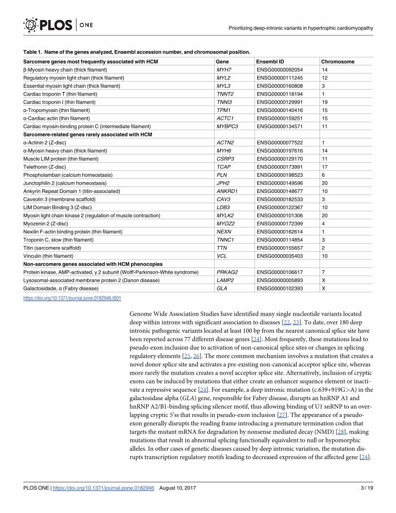
Table 1. Name of the genes analyzed, Ensembl accession number, and chromosomal position.
Sarcomere genes most frequently associated with HCM Gene Ensembl ID Chromosome
β-Myosin heavy chain (thick filament) MYH7 ENSG00000092054 14
Regulatory myosin light chain (thick filament) MYL2 ENSG00000111245 12
Essential myosin light chain (thick filament) MYL3 ENSG00000160808 3
Cardiac troponin T (thin filament) TNNT2 ENSG00000118194 1
Cardiac troponin I (thin filament) TNNI3 ENSG00000129991 19
α-Tropomyosin (thin filament) TPM1 ENSG00000140416 15
α-Cardiac actin (thin filament) ACTC1 ENSG00000159251 15
Cardiac myosin-binding protein C (intermediate filament) MYBPC3 ENSG00000134571 11
Sarcomere-related genes rarely associated with HCM
α-Actinin 2 (Z-disc) ACTN2 ENSG00000077522 1
α-Myosin heavy chain (thick filament) MYH6 ENSG00000197616 14
Muscle LIM protein (thin filament) CSRP3 ENSG00000129170 11
Telethonin (Z-disc) TCAP ENSG00000173991 17
Phospholamban (calcium homeostasis) PLN ENSG00000198523 6
Junctophilin 2 (calcium homeostasis) JPH2 ENSG00000149596 20
Ankyrin Repeat Domain 1 (titin-associated) ANKRD1 ENSG00000148677 10
Caveolin 3 (membrane scaffold) CAV3 ENSG00000182533 3
LIM Domain Binding 3 (Z-disc) LDB3 ENSG00000122367 10
Myosin light chain kinase 2 (regulation of muscle contraction) MYLK2 ENSG00000101306 20
Myozenin 2 (Z-disc) MYOZ2 ENSG00000172399 4
Nexilin F-actin binding protein (thin filament) NEXN ENSG00000162614 1
Troponin C, slow (thin filament) TNNC1 ENSG00000114854 3
Titin (sarcomere scaffold) TTN ENSG00000155657 2
Vinculin (thin filament) VCL ENSG00000035403 10
Non-sarcomere genes associated with HCM phenocopies
Protein kinase, AMP-activated, γ 2 subunit (Wolff-Parkinson-White syndrome) PRKAG2 ENSG00000106617 7
Lysosomal-associated membrane protein 2 (Danon disease) LAMP2 ENSG00000005893 X
Galactosidade, α (Fabry disease) GLA ENSG00000102393 X
https://doi.org/10.1371/journal.pone.0182946.t001
Prioritizing deep-intronic variants in hypertrophic cardiomyopathy
PLOS ONE | https://doi.org/10.1371/journal.pone.0182946 August 10, 2017 3 / 19
In this study, we used targeted high throughput sequencing and computational approaches
to identify deep intronic variants that may contribute to HCM phenotype.
Methods
Patients
The study population comprised 16 unrelated consecutively evaluated patients (8 males, 8
females) referred to the Cardiology Department at University Hospital Santa Maria. For all
probands, the personal and family history, physical examination, ECG and echocardiography
were consistent with a diagnosis of HCM according to international criteria [3]. Patients were
genetically tested at a mean age of 49 years. In addition, family members of two selected pro-
bands were clinically and genetically tested. Before blood collection, all patients and relatives
provided written informed consent for DNA analysis and received genetic counselling in
accordance with guidelines [3]. DNA samples used in this study were residual after conven-
tional diagnostic screening by targeted exome and Sanger sequencing. The project was
approved by the Lisbon Academic Medical Center Ethics Committee.
Targeted gene enrichment and sequencing
Blood samples (5–8 mL) were collected into EDTA tubes at routine clinic visits, and DNA was
isolated from peripheral blood lymphocytes using standard methods. The study was designed
to screen the full genomic DNA sequence of 26 genes indicated in Table 1. These genes are
included in many commercially available testing panels. A capture library was designed using
SureSelect (Agilent) and target regions were sequenced (paired-end) on an Illumina HiSeq
platform with 30–97 base read length. Highly repetitive sequences were excluded. Sample
preparation was carried out as recommended by the manufacturer. Relatives were genotyped
for selected variants by Sanger sequencing.
Bioinformatic data analysis
Raw sequencing paired-end reads (in.fastq format) were aligned using BWA software (version
0.7.12) [29] on the human reference genome (GRCh37) using quality score calibration and
Illumina adapter trimming. Following the exclusion of duplicate reads using Picard MarkDu-
plicates tool (version 1.96) (http://broadinstitute.github.io/picard/), regions around insertion-
deletions (indels) were realigned and each base quality score was recalibrated. For variant call-
ing, we used four distinct tools: GATK-UnifiedGenotyper (version 3.4–46) [30] and SAMtools
mpileup (version 1.2) [31], which use alignment-based approaches, and GATK-HaplotypeCal-
ler (version 3.4–46) [30] and FreeBayes (version 0.9.21.26) [32], which use haplotype-based
approaches. By comparing the performance of each tool against a standard reference
(NA12878, published by Genome in a Bottle consortium [33], we observed a concordance of
~85% (S1 Fig). To take advantage of the strengths of the different tools, we selected variants
that were independently called by at least two of them. This strategy showed a better sensitivity
(~97%) and precision (~98%) compared to analysis using a single tool (S1 Fig). Variants that
were independently selected by at least two tools, and presented a read depth of 20 or more in
the targeted genes, were annotated with ANNOVAR [34] (Fig 1).
For analysis of the clinical impact of coding variants we used the NCBI ClinVar database
(http://www.ncbi.nlm.nih.gov/clinvar/) [35] and classified the variants according to the Amer-
ican College of Medical Genetics and Genomics (ACMG) guidelines [36]. Prediction of patho-
genicity was performed with SIFT [37], PolyPhen2 HVAR [38], Human Splicing Finder
(version 3.0) [39], Mutation taster [40], UMD-predictor [41], PROVEAN [42] and FATHMM
Prioritizing deep-intronic variants in hypertrophic cardiomyopathy
PLOS ONE | https://doi.org/10.1371/journal.pone.0182946 August 10, 2017 4 / 19
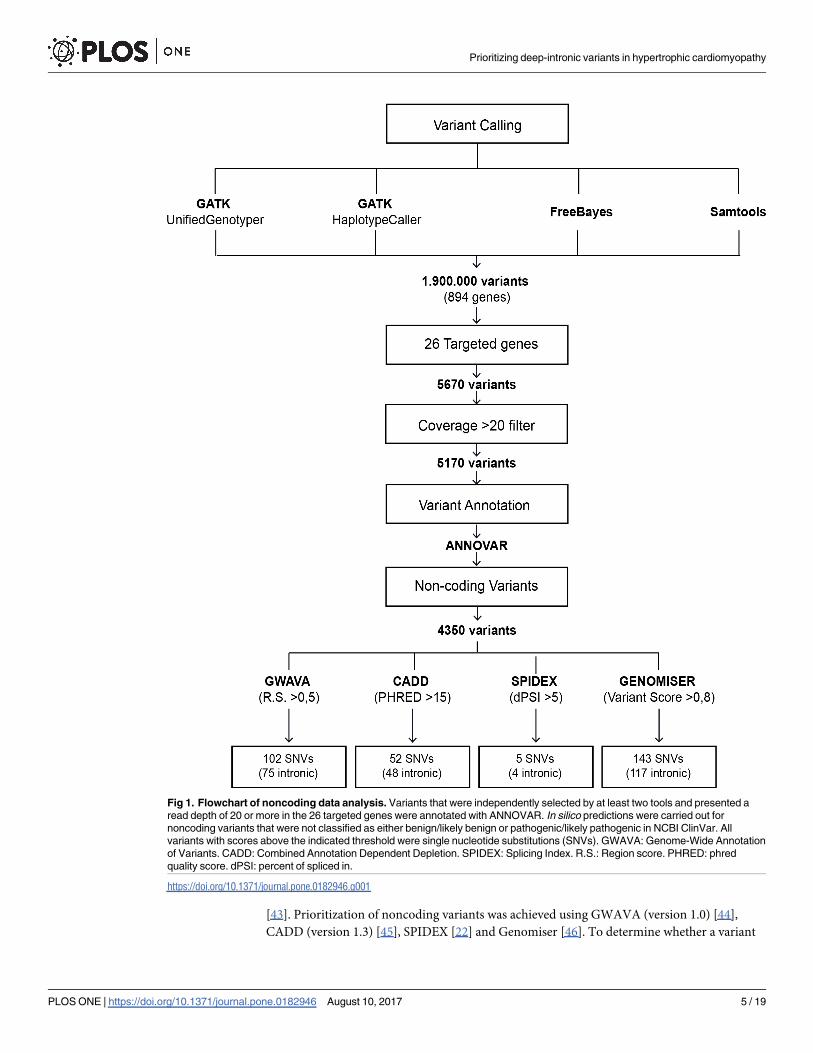
[43]. Prioritization of noncoding variants was achieved using GWAVA (version 1.0) [44],
CADD (version 1.3) [45], SPIDEX [22] and Genomiser [46]. To determine whether a variant
Fig 1. Flowchart of noncoding data analysis. Variants that were independently selected by at least two tools and presented a
read depth of 20 or more in the 26 targeted genes were annotated with ANNOVAR. In silico predictions were carried out for
noncoding variants that were not classified as either benign/likely benign or pathogenic/likely pathogenic in NCBI ClinVar. All
variants with scores above the indicated threshold were single nucleotide substitutions (SNVs). GWAVA: Genome-Wide Annotation
of Variants. CADD: Combined Annotation Dependent Depletion. SPIDEX: Splicing Index. R.S.: Region score. PHRED: phred
quality score. dPSI: percent of spliced in.
https://doi.org/10.1371/journal.pone.0182946.g001
Prioritizing deep-intronic variants in hypertrophic cardiomyopathy
PLOS ONE | https://doi.org/10.1371/journal.pone.0182946 August 10, 2017 5 / 19
may disrupt splicing motifs we used Human Splicing Finder (version 3.0), a tool that predicts
potential splice sites, branch points and enhancer/silencer splicing motifs [39]; RegRNA (ver-
sion 2.0), which searches for enhancer/silencer splicing motifs [47]; and Regulatory Genomics:
Branch point analyser, that predicts the presence of branch points and respective polypyrimi-
dine tracts [48].
As deep intronic mutations may result in altered gene expression through either cryptic
splicing or disruption of transcription regulatory motifs [24], we investigated whether the
identified intronic variants may disrupt transcription factor binding sites (TFBS). We used
available tracks in the UCSC genome browser [49–51] (Transcription Factor ChIP-seq Uni-
form Peaks from ENCODE/Analysis and HMR Conserved Transcription Factor Binding
Sites), focusing on TFBS predicted to be targets for transcription factors that have been impli-
cated in pathways related to cardiac regulation, development or pathophysiology.
Variant frequency was determined using the allele frequency estimates from the 1000
genomes project [52] and gnomAD [53] databases (accessed on June 2017).
Finally, we searched for the potential association of the candidate deep intronic variants
with cardiac diseases identified through GWAS (https://www.genome.gov/gwastudies/index.
cfm?gene=ESRRG and http://www.ebi.ac.uk/gwas/).
Results
Quality of sequencing data
Analysis of sequencing data yielded an average of 96.64% confidently mapped reads per gene.
For 69% of the targeted genes the average read depth was above 200, and for the remaining
genes the average read depth ranged between 130 and 200 (S2A Fig). The average read depth
was slightly lower over noncoding regions (S2B Fig). The average percentage of covered base
pairs was higher than 90 for 85% of the genes, and the lowest coverage was 76% for both cod-
ing and noncoding regions (S2 Fig). Following alignment to the reference genome (GRCh37)
and variant calling, we removed variants that were off-target, or had an average read depth
below 20 (Fig 1). Single nucleotide substitutions and insertions or deletions of a few bases
were identified and considered for further analysis.
Spectrum of exonic and splice site variants
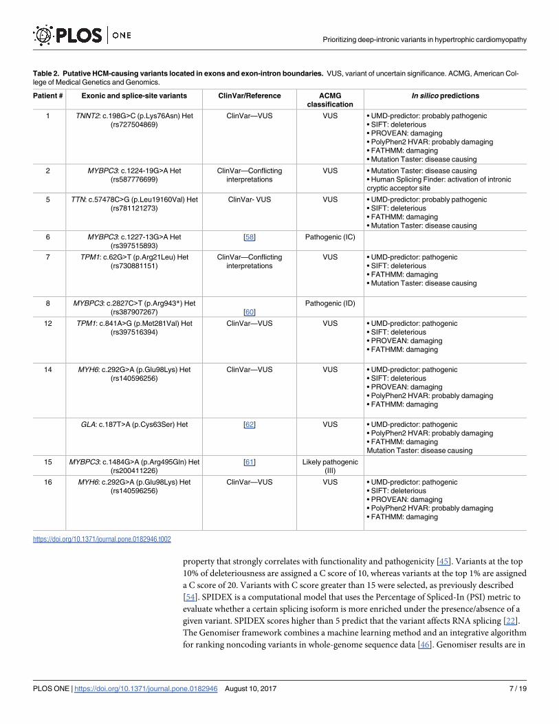
Previously described disease-causing variants in the MYBPC3 gene were detected in 3 patients
(Table 2). Rare variants classified in the NCBI ClinVar database and according to the ACMG
guidelines as of uncertain significance were additionally detected in the TNNT2, MYBPC3,
TTN,TPM1 and MYH6 genes; all these scored as likely pathogenic according to multiple in sil-ico prediction tools (Table 2). Noteworthy, one of the patients harboured, in addition to
MYH6 variant rs140596256, a novel variant in the GLA gene that is not listed in online data-
bases but is predicted to be pathogenic by multiple prediction tools (Table 2).
Assessment of deep intronic variants
The noncoding variants were prioritized using GWAVA [44], CADD [45], SPIDEX [22], and
Genomiser [46]. The genome-wide annotation of variants (GWAVA) is a computational
approach that integrates a wide range of available genomic and epigenomic annotations to pre-
dict the functional impact of variants. GWAVA results are in the range 0–1, with higher values
indicating variants predicted as more likely to be functional. Variants with a GWAVA score
above 0.5 were classified as functional, as in previous studies [44]. The Combined Annotation-
Dependent Depletion (CADD) method provides a metric (C score) for deleteriousness, a
Prioritizing deep-intronic variants in hypertrophic cardiomyopathy
PLOS ONE | https://doi.org/10.1371/journal.pone.0182946 August 10, 2017 6 / 19
property that strongly correlates with functionality and pathogenicity [45]. Variants at the top
10% of deleteriousness are assigned a C score of 10, whereas variants at the top 1% are assigned
a C score of 20. Variants with C score greater than 15 were selected, as previously described
[54]. SPIDEX is a computational model that uses the Percentage of Spliced-In (PSI) metric to
evaluate whether a certain splicing isoform is more enriched under the presence/absence of a
given variant. SPIDEX scores higher than 5 predict that the variant affects RNA splicing [22].
The Genomiser framework combines a machine learning method and an integrative algorithm
for ranking noncoding variants in whole-genome sequence data [46]. Genomiser results are in
Table 2. Putative HCM-causing variants located in exons and exon-intron boundaries. VUS, variant of uncertain significance. ACMG, American Col-
lege of Medical Genetics and Genomics.
Patient # Exonic and splice-site variants ClinVar/Reference ACMG
classification
In silico predictions
1 TNNT2: c.198G>C (p.Lys76Asn) Het
(rs727504869)
ClinVar—VUS VUS • UMD-predictor: probably pathogenic
• SIFT: deleterious
• PROVEAN: damaging
• PolyPhen2 HVAR: probably damaging
• FATHMM: damaging
• Mutation Taster: disease causing
2 MYBPC3: c.1224-19G>A Het
(rs587776699)
ClinVar—Conflicting
interpretations
VUS • Mutation Taster: disease causing
• Human Splicing Finder: activation of intronic
cryptic acceptor site
5 TTN: c.57478C>G (p.Leu19160Val) Het
(rs781121273)
ClinVar- VUS VUS • UMD-predictor: probably pathogenic
• SIFT: deleterious
• FATHMM: damaging
• Mutation Taster: disease causing
6 MYBPC3: c.1227-13G>A Het
(rs397515893)
[58] Pathogenic (IC)
7 TPM1: c.62G>T (p.Arg21Leu) Het
(rs730881151)
ClinVar—Conflicting
interpretations
VUS • UMD-predictor: pathogenic
• SIFT: deleterious
• FATHMM: damaging
• Mutation Taster: disease causing
8 MYBPC3: c.2827C>T (p.Arg943*) Het
(rs387907267) [60]
Pathogenic (ID)
12 TPM1: c.841A>G (p.Met281Val) Het
(rs397516394)
ClinVar—VUS VUS • UMD-predictor: pathogenic
• SIFT: deleterious
• PROVEAN: damaging
• FATHMM: damaging
14 MYH6: c.292G>A (p.Glu98Lys) Het
(rs140596256)
ClinVar—VUS VUS • UMD-predictor: pathogenic
• SIFT: deleterious
• PROVEAN: damaging
• PolyPhen2 HVAR: probably damaging
• FATHMM: damaging
GLA: c.187T>A (p.Cys63Ser) Het [62] VUS • UMD-predictor: pathogenic
• PolyPhen2 HVAR: probably damaging
• FATHMM: damaging
Mutation Taster: disease causing
15 MYBPC3: c.1484G>A (p.Arg495Gln) Het
(rs200411226)
[61] Likely pathogenic
(III)
16 MYH6: c.292G>A (p.Glu98Lys) Het
(rs140596256)
ClinVar—VUS VUS • UMD-predictor: pathogenic
• SIFT: deleterious
• PROVEAN: damaging
• PolyPhen2 HVAR: probably damaging
• FATHMM: damaging
https://doi.org/10.1371/journal.pone.0182946.t002
Prioritizing deep-intronic variants in hypertrophic cardiomyopathy
PLOS ONE | https://doi.org/10.1371/journal.pone.0182946 August 10, 2017 7 / 19
the range 0–1, with values higher than 0.6–0.9 indicating variants more likely to be pathogenic
[46].
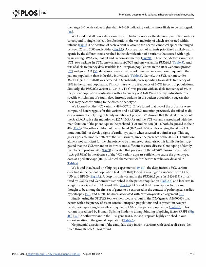
We found that all noncoding variants with higher scores for the different prediction metrics
correspond to single nucleotide substitutions, the vast majority of which are located within
introns (Fig 1). The position of each variant relative to the nearest canonical splice site ranged
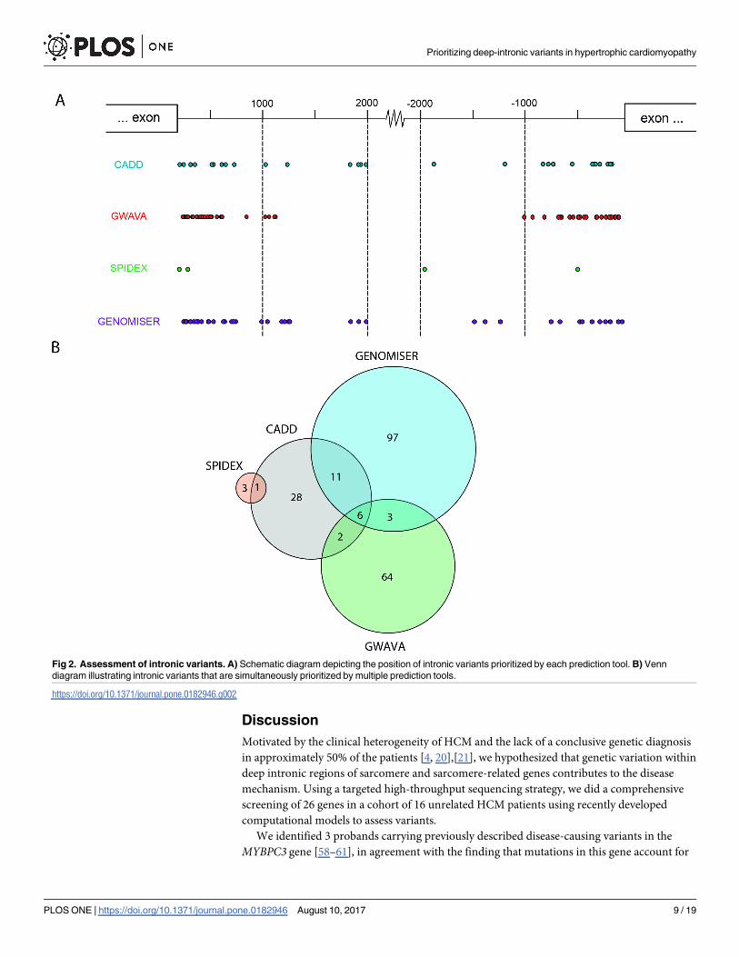
between 20 and 2000 nucleotides (Fig 2A). A comparison of variants prioritized as likely path-
ogenic by the different tools resulted in the identification of 6 variants that scored with high
values using GWAVA, CADD and Genomiser metrics (Fig 2B). These include two variants in
VCL, two variants in TTN, one variant in ACTC1 and one variant in PRKAG2 (Table 3). Anal-
ysis of allele frequency data available for European populations in the 1000 Genomes project
[52] and gnomAD [53] databases reveals that two of these variants are more frequent in the
patient population than in healthy individuals (Table 3). Namely, the VCL variant c.499+-
367T>C (rs113195070) was detected in 6 probands, corresponding to an allele frequency of
19% in the patient population. This contrasts with a frequency of 6–7% in control populations.
Similarly, the PRKAG2 variant c.1234-317T>G was present with an allele frequency of 3% in
the patient population contrasting with a frequency of 0.1–0.3% in healthy individuals. Such
specific enrichment of certain deep intronic variants in the patient population suggests that
these may be contributing to the disease phenotype.
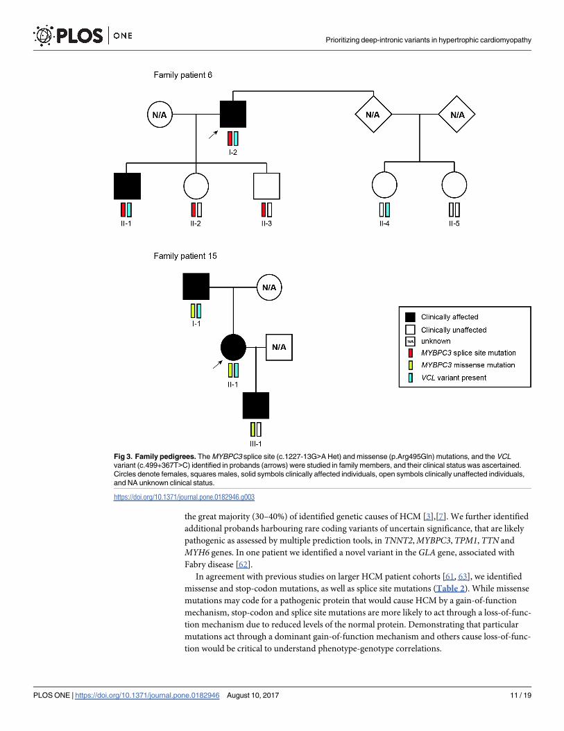
We focused on the VCL variant c.499+367T>C. We found that two of the probands were
compound heterozygous for this variant and a MYBPC3 mutation previously described as dis-
ease-causing. Genotyping of family members of proband #6 showed that the dual presence of
the MYBPC3 splice site mutation (c.1227-13G>A) and the VCL variant is associated with the
manifestation of the phenotype in the proband (I-2) and his son (II-1), both diagnosed in their
40s (Fig 3). The other children of the proband (II-2 and II-3), while carrying the MYBPC3mutation, did not develop signs of cardiomyopathy when assessed at a similar age. This sug-
gests a possible modifier effect of the VCL variant, since the presence of the MYBPC3 mutation
alone is not sufficient for the phenotype to be manifested. Analysis of this family further sug-
gested that the VCL variant on its own is not sufficient to cause disease. Genotyping of family
members of proband #15 (Fig 3) indicated that presence of the MYBPC3 missense mutation
(p.Arg495Gln) in the absence of the VCL variant appears sufficient to cause the phenotype,
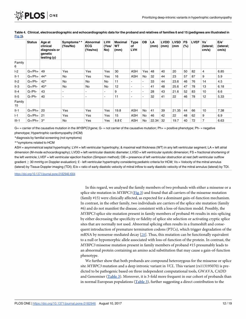
even at a pediatric age (III-1). Clinical characteristics for the two families are detailed in
Table 4.
We found that, based on Chip-seq experiments [49, 50], the deep intronic VCL variant
enriched in the patient population (rs113195070) localizes in a region associated with FOS,
JUN and EP300 (Fig 4A). A deep intronic variant in the PRKAG2 gene (rs114394151) priori-
tized by CADD and Genomiser is enriched in the patient population (Table 3) and localizes in
a region associated with FOS and JUN (Fig 4B). FOS and JUN transcription factors are
thought to be among the first set of genes to be expressed in the context of pathological cardiac
hypertrophy [55], and EP300 has been associated with cardiomyocyte enlargement [56].
Finally, using the SPIDEX tool we identified a variant in the TTN gene (rs72650063) that
occurs with a frequency of 2% in control European populations and is present in two pro-
bands, corresponding to an allele frequency of 6% in the patient population (Table 3). This
variant is predicted by Human Splicing Finder to disrupt binding of splicing factor SRSF1 (Fig
4C) [57]. Another variant in the TTN gene (rs142156368) appears highly enriched in our
cohort relative to the general population (Table 3).
No potential association of the candidate deep intronic variants with cardiac diseases iden-
tified through GWAS was found.
Prioritizing deep-intronic variants in hypertrophic cardiomyopathy
PLOS ONE | https://doi.org/10.1371/journal.pone.0182946 August 10, 2017 8 / 19
Discussion
Motivated by the clinical heterogeneity of HCM and the lack of a conclusive genetic diagnosis
in approximately 50% of the patients [4, 20],[21], we hypothesized that genetic variation within
deep intronic regions of sarcomere and sarcomere-related genes contributes to the disease
mechanism. Using a targeted high-throughput sequencing strategy, we did a comprehensive
screening of 26 genes in a cohort of 16 unrelated HCM patients using recently developed
computational models to assess variants.
We identified 3 probands carrying previously described disease-causing variants in the
MYBPC3 gene [58–61], in agreement with the finding that mutations in this gene account for
Fig 2. Assessment of intronic variants. A) Schematic diagram depicting the position of intronic variants prioritized by each prediction tool. B) Venn
diagram illustrating intronic variants that are simultaneously prioritized by multiple prediction tools.
https://doi.org/10.1371/journal.pone.0182946.g002
Prioritizing deep-intronic variants in hypertrophic cardiomyopathy
PLOS ONE | https://doi.org/10.1371/journal.pone.0182946 August 10, 2017 9 / 19
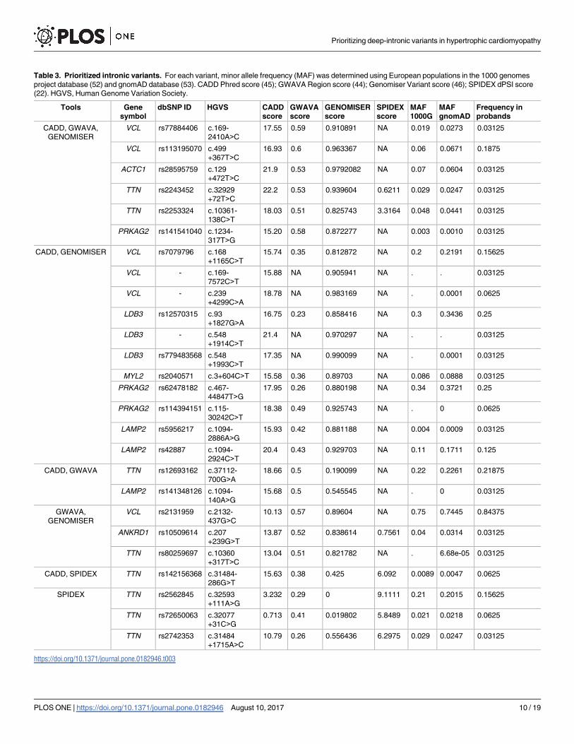
Table 3. Prioritized intronic variants. For each variant, minor allele frequency (MAF) was determined using European populations in the 1000 genomes
project database (52) and gnomAD database (53). CADD Phred score (45); GWAVA Region score (44); Genomiser Variant score (46); SPIDEX dPSI score
(22). HGVS, Human Genome Variation Society.
Tools Gene
symbol
dbSNP ID HGVS CADD
score
GWAVA
score
GENOMISER
score
SPIDEX
score
MAF
1000G
MAF
gnomAD
Frequency in
probands
CADD, GWAVA,
GENOMISER
VCL rs77884406 c.169-
2410A>C
17.55 0.59 0.910891 NA 0.019 0.0273 0.03125
VCL rs113195070 c.499
+367T>C
16.93 0.6 0.963367 NA 0.06 0.0671 0.1875
ACTC1 rs28595759 c.129
+472T>C
21.9 0.53 0.9792082 NA 0.07 0.0604 0.03125
TTN rs2243452 c.32929
+72T>C
22.2 0.53 0.939604 0.6211 0.029 0.0247 0.03125
TTN rs2253324 c.10361-
138C>T
18.03 0.51 0.825743 3.3164 0.048 0.0441 0.03125
PRKAG2 rs141541040 c.1234-
317T>G
15.20 0.58 0.872277 NA 0.003 0.0010 0.03125
CADD, GENOMISER VCL rs7079796 c.168
+1165C>T
15.74 0.35 0.812872 NA 0.2 0.2191 0.15625
VCL - c.169-
7572C>T
15.88 NA 0.905941 NA . . 0.03125
VCL - c.239
+4299C>A
18.78 NA 0.983169 NA . 0.0001 0.0625
LDB3 rs12570315 c.93
+1827G>A
16.75 0.23 0.858416 NA 0.3 0.3436 0.25
LDB3 - c.548
+1914C>T
21.4 NA 0.970297 NA . . 0.03125
LDB3 rs779483568 c.548
+1993C>T
17.35 NA 0.990099 NA . 0.0001 0.03125
MYL2 rs2040571 c.3+604C>T 15.58 0.36 0.89703 NA 0.086 0.0888 0.03125
PRKAG2 rs62478182 c.467-
44847T>G
17.95 0.26 0.880198 NA 0.34 0.3721 0.25
PRKAG2 rs114394151 c.115-
30242C>T
18.38 0.49 0.925743 NA . 0 0.0625
LAMP2 rs5956217 c.1094-
2886A>G
15.93 0.42 0.881188 NA 0.004 0.0009 0.03125
LAMP2 rs42887 c.1094-
2924C>T
20.4 0.43 0.929703 NA 0.11 0.1711 0.125
CADD, GWAVA TTN rs12693162 c.37112-
700G>A
18.66 0.5 0.190099 NA 0.22 0.2261 0.21875
LAMP2 rs141348126 c.1094-
140A>G
15.68 0.5 0.545545 NA . 0 0.03125
GWAVA,
GENOMISER
VCL rs2131959 c.2132-
437G>C
10.13 0.57 0.89604 NA 0.75 0.7445 0.84375
ANKRD1 rs10509614 c.207
+239G>T
13.87 0.52 0.838614 0.7561 0.04 0.0314 0.03125
TTN rs80259697 c.10360
+317T>C
13.04 0.51 0.821782 NA . 6.68e-05 0.03125
CADD, SPIDEX TTN rs142156368 c.31484-
286G>T
15.63 0.38 0.425 6.092 0.0089 0.0047 0.0625
SPIDEX TTN rs2562845 c.32593
+111A>G
3.232 0.29 0 9.1111 0.21 0.2015 0.15625
TTN rs72650063 c.32077
+31C>G
0.713 0.41 0.019802 5.8489 0.021 0.0218 0.0625
TTN rs2742353 c.31484
+1715A>C
10.79 0.26 0.556436 6.2975 0.029 0.0247 0.03125
https://doi.org/10.1371/journal.pone.0182946.t003
Prioritizing deep-intronic variants in hypertrophic cardiomyopathy
PLOS ONE | https://doi.org/10.1371/journal.pone.0182946 August 10, 2017 10 / 19
the great majority (30–40%) of identified genetic causes of HCM [3],[7]. We further identified
additional probands harbouring rare coding variants of uncertain significance, that are likely
pathogenic as assessed by multiple prediction tools, in TNNT2, MYBPC3, TPM1, TTN and
MYH6 genes. In one patient we identified a novel variant in the GLA gene, associated with
Fabry disease [62].
In agreement with previous studies on larger HCM patient cohorts [61, 63], we identified
missense and stop-codon mutations, as well as splice site mutations (Table 2). While missense
mutations may code for a pathogenic protein that would cause HCM by a gain-of-function
mechanism, stop-codon and splice site mutations are more likely to act through a loss-of-func-
tion mechanism due to reduced levels of the normal protein. Demonstrating that particular
mutations act through a dominant gain-of-function mechanism and others cause loss-of-func-
tion would be critical to understand phenotype-genotype correlations.
Fig 3. Family pedigrees. The MYBPC3 splice site (c.1227-13G>A Het) and missense (p.Arg495Gln) mutations, and the VCL
variant (c.499+367T>C) identified in probands (arrows) were studied in family members, and their clinical status was ascertained.
Circles denote females, squares males, solid symbols clinically affected individuals, open symbols clinically unaffected individuals,
and NA unknown clinical status.
https://doi.org/10.1371/journal.pone.0182946.g003
Prioritizing deep-intronic variants in hypertrophic cardiomyopathy
PLOS ONE | https://doi.org/10.1371/journal.pone.0182946 August 10, 2017 11 / 19
In this regard, we analysed the family members of two probands with either a missense or a
splice site mutation in MYBPC3 (Fig 3) and found that all carriers of the missense mutation
(family #15) were clinically affected, as expected for a dominant gain-of-function mechanism.
In contrast, in the other family, two individuals are carriers of the splice site mutation (family
#6) and do not manifest the disease, consistent with a loss-of-function model. Possibly, the
MYBPC3 splice site mutation present in family members of proband #6 results in mis-splicing
by either decreasing the specificity or fidelity of splice site selection or activating cryptic splice
sites that are normally not used. Abnormal splicing often results in a frameshift and conse-
quent introduction of premature termination codons (PTCs), which trigger degradation of the
mRNA by nonsense-mediated decay [28]. Thus, this mutation can be functionally equivalent
to a null or hypomorphic allele associated with loss-of-function of the protein. In contrast, the
MYBPC3 missense mutation present in family members of proband #15 presumably leads to
an abnormal protein containing an amino acid substitution that may cause a gain-of-function
phenotype.
We further show that both probands are compound heterozygous for the missense or splice
site MYBPC3 mutation and a deep intronic variant in VCL. This variant (rs113195070) is pre-
dicted to be pathogenic based on three independent computational tools, GWAVA, CADD
and Genomiser (Table 3). Moreover, it is 3-fold more frequent in our cohort of probands than
in normal European populations (Table 3), further suggesting a direct contribution to the
Table 4. Clinical, electrocardiographic and echocardiographic data for the proband and relatives of families 6 and 15 (pedigrees are illustrated in
Fig 3).
Status Age at
clinical
diagnosis or
genetic
testing (y)
Symptoms**(Yes/No)
Abnormal
ECG
(Yes/no)
LVH
(Yes/
No)
Maximal
WT
(mm)
Type
of
LVH
OB LA
(mm)
LVDD
(mm)
LVSD
(mm
FS
(%)
LVEF Vs´
(lateral;
cm/s)
E/e’
(lateral;
cm/s)
Family
6
I-2 G+/Ph+ 49 Yes Yes Yes 30 ASH Yes 48 40 20 50 82 4 6.85
II-1 G+/Ph+ 44* No Yes Yes 16 ASH No 32 44 23 57 87 9 5.9
II-2 G+/Ph- 42* No No No 11 - - 33 44 23.6 46 76 14 4.5
II-3 G+/Ph- 40* No No No 12 - - 41 48 25.6 47 78 13 6.18
II-4 G-/Ph- 43 - - - 9 - - 28 43 21.6 52 83 10 6.6
II-5 G-/Ph- 40 - - - 11 - - 32 41 22 46 78 12 5.33
Family
15
II-1 G+/Ph+ 20 Yes Yes Yes 19.8 ASH No 41 39 21.35 44 66 10 7.38
I-1 G+/Ph+ 21 Yes Yes Yes 15 ASH No 46 42 22 48 62 9 6.9
III-1 G+/Ph+ 3* No Yes Yes 8.8 £ ASH No 22.34 32 19.7 40 72 7 6.63
G+ = carrier of the causative mutation in the MYBPC3 gene; G- = not carrier of the causative mutation; Ph+ = positive phenotype; Ph- = negative
phenotype; Hypertrophic cardiomyopathy (HCM)
*diagnosis by familial screening (not symptoms)
**symptoms related to HCM
ASH = asymmetrical septal hypertrophy; LVH = left ventricular hypertrophy; & maximal wall thickness (WT) in any left ventricular segment; LA = left atrial
dimension (M-mode echocardiography); LVDD = left ventricular diastolic diameter; LVSD = left ventricular systolic dimension; FS = fractional shortening of
the left ventricle; LVEF = left ventricular ejection fraction (Simpson method); OB = presence of left ventricular obstruction at rest (left ventricular outflow
gradient� 30 mmHg on Doppler evaluation); £ - left ventricular hypertrophy considering pediatric criteria for HCM; Vs = Velocity of the mitral annulus
(lateral) by Tissue Doppler imaging (TDI); E/e = ratio of early diastolic velocity of mitral inflow to early diastolic velocity of the mitral annulus (lateral) by TDI.
https://doi.org/10.1371/journal.pone.0182946.t004
Prioritizing deep-intronic variants in hypertrophic cardiomyopathy
PLOS ONE | https://doi.org/10.1371/journal.pone.0182946 August 10, 2017 12 / 19
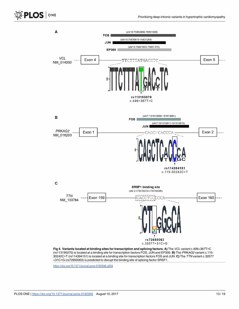
Fig 4. Variants located at binding sites for transcription and splicing factors. A) The VCL variant c.499+367T>C
(rs113195070) is located at a binding site for transcription factors FOS, JUN and EP300. B) The PRKAG2 variant c.115-
30242C>T (rs114394151) is located at a binding site for transcription factors FOS and JUN. C) The TTN variant c.32077
+31C>G (rs72650063) is predicted to disrupt the binding site of splicing factor SRSF1.
https://doi.org/10.1371/journal.pone.0182946.g004
Prioritizing deep-intronic variants in hypertrophic cardiomyopathy
PLOS ONE | https://doi.org/10.1371/journal.pone.0182946 August 10, 2017 13 / 19
disease phenotype. The variant, which consists of a single nucleotide substitution located at
position 367 from the nearest canonical splice site (c.499+367T>C), can potentially disrupt
the binding of transcription factors that have been reported as implicated in pathways related
to cardiac regulation, development or pathophysiology such as FOS, JUN and EP300 [55],[56].
By interfering with the binding of transcription regulatory factors, the variant is expected to
alter the transcription rate of the VCL gene. Consistent with this view, sequence elements
located within introns of large human genes have been shown to act as transcriptional enhanc-
ers [64], and a recent study reported an IRF4 gene variant located in intron 4 that strongly
affects IRF4 transcription through disruption of an enhancer element [65].
Analysis of family #6 reveals that the presence of the VCL variant or the MYBPC3 mutation
in isolation is not sufficient to cause disease phenotype. Indeed, the two clinically affected indi-
viduals in this family are compound heterozygous for the VCL variant and the MYBPC3 splice
site mutation (Fig 3), suggesting that the combination of the two mutations triggers the disease.
A loss-of-function mechanism for the MYBPC3 mutation could explain why in family #6 only
compound heterozygous members manifest the disease, whereas the presence of the heterozy-
gous gain-of-function mutation in family #15 would be sufficient to cause disease. Complex
genotypes, including individuals that carry 2 or more variants in the same or different sarco-
mere-related genes, have been reported in 8% of HCM patients [58], and there is evidence indi-
cating that patients with complex genotype and multiple simultaneous mutations may have
more severe or early disease expression [66]. However, complex genotype-phenotype correla-
tions focusing specifically on carriers of splice site mutations remain to be investigated.
We further identified two single nucleotide substitutions in the titin gene (rs142156368 and
rs72650063) that are 3 to 6-fold more frequent in our cohort of probands than in normal Euro-
pean populations (Table 3). These variants are located in the PEVK domain that plays a role in
extensibility of the sarcomere and contractility of the titin protein [67, 68]. Titin is prone to
extensive alternative splicing that can change its size and its elastic/stiffness properties; associa-
tions have been established between the ratio of expression levels for the main cardiac isoforms
(N2BA and N2B) and genetic and non-genetic forms of cardiac diseases [69, 70]. If these vari-
ants do interfere with titin splicing, as predicted by the SPIDEX computational model, they are
likely to contribute to HCM phenotype, particularly in combination with other HCM-associ-
ated alleles. Supporting this view, titin-truncating splicing isoforms, which are encountered in
approximately 1% of the general population, are sufficient to induce molecular and physiologi-
cal effects on the heart [71].
In conclusion, this study provides a framework for scrutinizing variation along the com-
plete sequence of HCM-associated genes and prioritizing candidates for further analysis. Our
data suggest that deep intronic variation contributes to HCM phenotype. Translation of
genetic information found in an individual to clinical decision taking requires a precise under-
standing of the molecular mechanisms underlying the disease phenotype. To date, mechanistic
and functional studies have been largely restricted to animal models in part due to difficulties
in obtaining human tissue from patients. However, the recent emergence of patient-derived
induced pluripotent stem cells (iPSCs) that can be differentiated into functional cardiomyo-
cytes recapitulating HCM-specific characteristics [72, 73] holds great promise as an exciting
new approach to study how gene mutations relate to clinical outcomes and might be applied to
test our hypothesis-generating data.
Supporting information
S1 Fig. Comparison of variant calling strategies. A) Variants identified by each individual
tool and variants that were independently called by at least two tools (combined) were
Prioritizing deep-intronic variants in hypertrophic cardiomyopathy
PLOS ONE | https://doi.org/10.1371/journal.pone.0182946 August 10, 2017 14 / 19
compared to a standard reference (NA12878, (33)). Concordant or true positive (TP) variants
are defined as those present in the reference and identified by the indicated calling tool. Dis-
cordant extra or false positive (FP) variants are variants not detected in the reference but iden-
tified by the calling tool. Discordant missing or false negative (FN) variants are those present
in the reference but undetected by the calling tools. (B) Sensitivity was assessed by calculating
the ratio between TP/(TP+FN). Precision was assessed by calculating the ratio between TP/
(TP+FP).
(TIF)
S2 Fig. Characterization of sequence data. (A) Box plots show the read-depths across the tar-
geted genes and the average percentage of covered base pairs per gene is depicted in red. (B)
Box plots show the read-depths in coding (orange) and noncoding (grey) regions. The average
percentage of covered base pairs in each region per gene is depicted in red.
(PDF)
Acknowledgments
We thank Vladimir Benes and the EMBL GeneCore sequencing team for generous advice and
technical assistance. We are also grateful to Rute Marcelino and Ana Coutinho from GenoMed
for assistance in genotyping analysis.
This work was supported by Fundacão para a Ciência e Tecnologia, Portugal (Scholarship
BD/82023/2011 to R.M.A, grant EXPL/BIM-MEC/0201/2013 to S.M., grant “Programa de
Actividades Conjuntas n˚ 016394 to M.C.-F), and an unrestricted grant from Merck Sharp &
Dohme to M.C.-F.
Author Contributions
Conceptualization: Joana Tavares, Sandra Martins, Maria Carmo-Fonseca, Luıs Rocha Lopes.
Data curation: Rita Mendes de Almeida, Joana Tavares, Teresa Carvalho, Dulce Brito, Maria
Carmo-Fonseca, Luıs Rocha Lopes.
Formal analysis: Rita Mendes de Almeida, Joana Tavares, Sandra Martins, Teresa Carvalho,
Dulce Brito, Maria Carmo-Fonseca, Luıs Rocha Lopes.
Funding acquisition: Rita Mendes de Almeida, Sandra Martins, Maria Carmo-Fonseca.
Investigation: Rita Mendes de Almeida, Joana Tavares, Teresa Carvalho, Dulce Brito, Maria
Carmo-Fonseca, Luıs Rocha Lopes.
Methodology: Rita Mendes de Almeida, Sandra Martins, Maria Carmo-Fonseca, Luıs Rocha
Lopes.
Project administration: Sandra Martins, Maria Carmo-Fonseca.
Supervision: Sandra Martins, Teresa Carvalho, Francisco J. Enguita, Maria Carmo-Fonseca,
Luıs Rocha Lopes.
Validation: Teresa Carvalho, Dulce Brito, Maria Carmo-Fonseca, Luıs Rocha Lopes.
Visualization: Rita Mendes de Almeida, Joana Tavares, Francisco J. Enguita, Dulce Brito,
Maria Carmo-Fonseca, Luıs Rocha Lopes.
Writing – original draft: Rita Mendes de Almeida, Joana Tavares.
Writing – review & editing: Sandra Martins, Teresa Carvalho, Francisco J. Enguita, Dulce
Brito, Maria Carmo-Fonseca, Luıs Rocha Lopes.
Prioritizing deep-intronic variants in hypertrophic cardiomyopathy
PLOS ONE | https://doi.org/10.1371/journal.pone.0182946 August 10, 2017 15 / 19
References1. Maron BJ, Gardin JM, Flack JM, Gidding SS, Kurosaki TT, Bild DE. Prevalence of hypertrophic cardio-
myopathy in a general population of young adults. Echocardiographic analysis of 4111 subjects in the
CARDIA Study. Coronary Artery Risk Development in (Young) Adults. Circulation. 1995; 92(4):785–9.
PMID: 7641357
2. Arndt AK, MacRae CA. Genetic testing in cardiovascular diseases. Curr Opin Cardiol. 2014; 29(3):235–
40. https://doi.org/10.1097/HCO.0000000000000055 PMID: 24717670
3. Authors/Task Force, Elliott PM, Anastasakis A, Borger MA, Borggrefe M, Cecchi F, et al. 2014 ESC
Guidelines on diagnosis and management of hypertrophic cardiomyopathy: The Task Force for the
Diagnosis and Management of Hypertrophic Cardiomyopathy of the European Society of Cardiology
(ESC). Eur Heart J. 2014; 35(39):2733–79. https://doi.org/10.1093/eurheartj/ehu284 PMID: 25173338
4. Sen-Chowdhry S, Jacoby D, Moon JC, McKenna WJ. Update on hypertrophic cardiomyopathy and a
guide to the guidelines. Nat Rev Cardiol. 2016; 13(11):651–75. https://doi.org/10.1038/nrcardio.2016.
140 PMID: 27681577
5. Geisterfer-Lowrance AA, Kass S, Tanigawa G, Vosberg HP, McKenna W, Seidman CE, et al. A molecu-
lar basis for familial hypertrophic cardiomyopathy: a beta cardiac myosin heavy chain gene missense
mutation. Cell. 1990; 62(5):999–1006. PMID: 1975517
6. McNally Elizabeth M, Barefield David Y, Puckelwartz Megan J. The Genetic Landscape of Cardiomyop-
athy and Its Role in Heart Failure. Cell Metabolism. 2015; 21(2):174–82.
7. Ho CY, Charron P, Richard P, Girolami F, Van Spaendonck-Zwarts KY, Pinto Y. Genetic advances in
sarcomeric cardiomyopathies: state of the art. Cardiovasc Res. 2015; 105(4):397–408. https://doi.org/
10.1093/cvr/cvv025 PMID: 25634555
8. Walsh R, Buchan R, Wilk A, John S, Felkin LE, Thomson KL, et al. Defining the genetic architecture of
hypertrophic cardiomyopathy: re-evaluating the role of non-sarcomeric genes. Eur Heart J. 2017. Epub
ahead of print
9. Geier C, Perrot A, Ozcelik C, Binner P, Counsell D, Hoffmann K, et al. Mutations in the human muscle
LIM protein gene in families with hypertrophic cardiomyopathy. Circulation. 2003; 107(10):1390–5.
PMID: 12642359
10. Wang L, Zuo L, Hu J, Shao H, Lei C, Qi W, et al. Dual LQT1 and HCM phenotypes associated with tet-
rad heterozygous mutations in KCNQ1, MYH7, MYLK2, and TMEM70 genes in a three-generation Chi-
nese family. Europace. 2016; 18(4):602–9. https://doi.org/10.1093/europace/euv043 PMID: 25825456
11. Osio A, Tan L, Chen SN, Lombardi R, Nagueh SF, Shete S, et al. Myozenin 2 is a novel gene for human
hypertrophic cardiomyopathy. Circulation research. 2007; 100(6):766–8. https://doi.org/10.1161/01.
RES.0000263008.66799.aa PMID: 17347475
12. Wang H, Li Z, Wang J, Sun K, Cui Q, Song L, et al. Mutations in NEXN, a Z-disc gene, are associated
with hypertrophic cardiomyopathy. Am J Hum Genet. 2010; 87(5):687–93. https://doi.org/10.1016/j.
ajhg.2010.10.002 PMID: 20970104
13. Veltri T, Landim-Vieira M, Parvatiyar MS, Gonzalez-Martinez D, Dieseldorff Jones KM, Michell CA,
et al. Hypertrophic Cardiomyopathy Cardiac Troponin C Mutations Differentially Affect Slow Skeletal
and Cardiac Muscle Regulation. Front Physiol. 2017; 8:221. https://doi.org/10.3389/fphys.2017.00221
PMID: 28473771
14. LeWinter MM, Granzier HL. Cardiac titin and heart disease. J Cardiovasc Pharmacol. 2014; 63(3):207–
12. https://doi.org/10.1097/FJC.0000000000000007 PMID: 24072177
15. Vasile VC, Ommen SR, Edwards WD, Ackerman MJ. A missense mutation in a ubiquitously expressed
protein, vinculin, confers susceptibility to hypertrophic cardiomyopathy. Biochem Biophys Res Com-
mun. 2006; 345(3):998–1003. https://doi.org/10.1016/j.bbrc.2006.04.151 PMID: 16712796
16. Arimura T, Bos JM, Sato A, Kubo T, Okamoto H, Nishi H, et al. Cardiac ankyrin repeat protein gene
(ANKRD1) mutations in hypertrophic cardiomyopathy. J Am Coll Cardiol. 2009; 54(4):334–42. https://
doi.org/10.1016/j.jacc.2008.12.082 PMID: 19608031
17. Hayashi T, Arimura T, Ueda K, Shibata H, Hohda S, Takahashi M, et al. Identification and functional
analysis of a caveolin-3 mutation associated with familial hypertrophic cardiomyopathy. Biochem Bio-
phys Res Commun. 2004; 313(1):178–84. PMID: 14672715
18. Rapezzi C, Arbustini E, Caforio AL, Charron P, Gimeno-Blanes J, Helio T, et al. Diagnostic work-up in
cardiomyopathies: bridging the gap between clinical phenotypes and final diagnosis. A position state-
ment from the ESC Working Group on Myocardial and Pericardial Diseases. Eur Heart J. 2013; 34
(19):1448–58. https://doi.org/10.1093/eurheartj/ehs397 PMID: 23211230
Prioritizing deep-intronic variants in hypertrophic cardiomyopathy
PLOS ONE | https://doi.org/10.1371/journal.pone.0182946 August 10, 2017 16 / 19
19. Mardis ER. Next-generation DNA sequencing methods. Annu Rev Genomics Hum Genet. 2008;
9:387–402. https://doi.org/10.1146/annurev.genom.9.081307.164359 PMID: 18576944
20. Lopes LR, Rahman MS, Elliott PM. A systematic review and meta-analysis of genotype-phenotype
associations in patients with hypertrophic cardiomyopathy caused by sarcomeric protein mutations.
Heart. 2013; 99(24):1800–11. https://doi.org/10.1136/heartjnl-2013-303939 PMID: 23674365
21. Ingles J, Burns C, Barratt A, Semsarian C. Application of Genetic Testing in Hypertrophic Cardiomyopa-
thy for Preclinical Disease Detection. Circ Cardiovasc Genet. 2015; 8(6):852–9. https://doi.org/10.1161/
CIRCGENETICS.115.001093 PMID: 26671970
22. Xiong HY, Alipanahi B, Lee LJ, Bretschneider H, Merico D, Yuen RK, et al. RNA splicing. The human
splicing code reveals new insights into the genetic determinants of disease. Science. 2015; 347
(6218):1254806. https://doi.org/10.1126/science.1254806 PMID: 25525159
23. Hsiao YH, Bahn JH, Lin X, Chan TM, Wang R, Xiao X. Alternative splicing modulated by genetic vari-
ants demonstrates accelerated evolution regulated by highly conserved proteins. Genome Res. 2016;
26(4):440–50. https://doi.org/10.1101/gr.193359.115 PMID: 26888265
24. Vaz-Drago R, Custodio N, Carmo-Fonseca M. Deep intronic mutations and human disease. Hum
Genet. 2017. Epub ahead of print
25. Romano M, Buratti E, Baralle D. Role of pseudoexons and pseudointrons in human cancer. Int J Cell
Biol. 2013; 2013:810572. https://doi.org/10.1155/2013/810572 PMID: 24204383
26. Dhir A, Buratti E. Alternative splicing: role of pseudoexons in human disease and potential therapeutic
strategies. FEBS J. 2010; 277(4):841–55. https://doi.org/10.1111/j.1742-4658.2009.07520.x PMID:
20082636
27. Palhais B, Dembic M, Sabaratnam R, Nielsen KS, Doktor TK, Bruun GH, et al. The prevalent deep intro-
nic c. 639+919 G>A GLA mutation causes pseudoexon activation and Fabry disease by abolishing the
binding of hnRNPA1 and hnRNP A2/B1 to a splicing silencer. Mol Genet Metab. 2016; 119(3):258–69.
https://doi.org/10.1016/j.ymgme.2016.08.007 PMID: 27595546
28. Popp MW, Maquat LE. Organizing principles of mammalian nonsense-mediated mRNA decay. Annu
Rev Genet. 2013; 47:139–65. https://doi.org/10.1146/annurev-genet-111212-133424 PMID: 24274751
29. Li H, Durbin R. Fast and accurate short read alignment with Burrows-Wheeler transform. Bioinformatics.
2009; 25(14):1754–60. https://doi.org/10.1093/bioinformatics/btp324 PMID: 19451168
30. DePristo MA, Banks E, Poplin R, Garimella KV, Maguire JR, Hartl C, et al. A framework for variation dis-
covery and genotyping using next-generation DNA sequencing data. Nat Genet. 2011; 43(5):491–8.
https://doi.org/10.1038/ng.806 PMID: 21478889
31. Li H. A statistical framework for SNP calling, mutation discovery, association mapping and population
genetical parameter estimation from sequencing data. Bioinformatics. 2011; 27(21):2987–93. https://
doi.org/10.1093/bioinformatics/btr509 PMID: 21903627
32. Garrison EM, G. Haplotype-based variant detection from short-read sequencing. Genomics (q-bioGN);
Quantitative Methods (q-bioQM). 2012.
33. Zook JM, Chapman B, Wang J, Mittelman D, Hofmann O, Hide W, et al. Integrating human sequence
data sets provides a resource of benchmark SNP and indel genotype calls. Nat Biotech. 2014; 32
(3):246–51.
34. Wang K, Li M, Hakonarson H. ANNOVAR: functional annotation of genetic variants from high-through-
put sequencing data. Nucleic Acids Res. 2010; 38(16):e164. https://doi.org/10.1093/nar/gkq603 PMID:
20601685
35. Landrum MJ, Lee JM, Benson M, Brown G, Chao C, Chitipiralla S, et al. ClinVar: public archive of inter-
pretations of clinically relevant variants. Nucleic Acids Res. 2016; 44(D1):D862–8. https://doi.org/10.
1093/nar/gkv1222 PMID: 26582918
36. Richards S, Aziz N, Bale S, Bick D, Das S, Gastier-Foster J, et al. Standards and guidelines for the inter-
pretation of sequence variants: a joint consensus recommendation of the American College of Medical
Genetics and Genomics and the Association for Molecular Pathology. 2015; 17(5):405–24. https://doi.
org/10.1038/gim.2015.30 PMID: 25741868
37. Kumar P, Henikoff S, Ng PC. Predicting the effects of coding non-synonymous variants on protein func-
tion using the SIFT algorithm. Nat Protoc. 2009; 4(7):1073–81. https://doi.org/10.1038/nprot.2009.86
PMID: 19561590
38. Adzhubei IA, Schmidt S, Peshkin L, Ramensky VE, Gerasimova A, Bork P, et al. A method and server
for predicting damaging missense mutations. Nat Methods. 2010; 7(4):248–9. https://doi.org/10.1038/
nmeth0410-248 PMID: 20354512
39. Desmet FO, Hamroun D, Lalande M, Collod-Beroud G, Claustres M, Beroud C. Human Splicing Finder:
an online bioinformatics tool to predict splicing signals. Nucleic Acids Res. 2009; 37(9):e67. https://doi.
org/10.1093/nar/gkp215 PMID: 19339519
Prioritizing deep-intronic variants in hypertrophic cardiomyopathy
PLOS ONE | https://doi.org/10.1371/journal.pone.0182946 August 10, 2017 17 / 19
40. Schwarz JM, Cooper DN, Schuelke M, Seelow D. MutationTaster2: mutation prediction for the deep-
sequencing age. Nat Methods. 2014; 11(4):361–2. https://doi.org/10.1038/nmeth.2890 PMID:
24681721
41. Salgado D, Desvignes JP, Rai G, Blanchard A, Miltgen M, Pinard A, et al. UMD-Predictor: A High-
Throughput Sequencing Compliant System for Pathogenicity Prediction of any Human cDNA Substitu-
tion. Hum Mutat. 2016; 37(5):439–46. https://doi.org/10.1002/humu.22965 PMID: 26842889
42. Choi Y, Chan AP. PROVEAN web server: a tool to predict the functional effect of amino acid substitu-
tions and indels. Bioinformatics. 2015; 31(16):2745–7. https://doi.org/10.1093/bioinformatics/btv195
PMID: 25851949
43. Shihab HA, Gough J, Cooper DN, Stenson PD, Barker GL, Edwards KJ, et al. Predicting the functional,
molecular, and phenotypic consequences of amino acid substitutions using hidden Markov models.
Hum Mutat. 2013; 34(1):57–65. https://doi.org/10.1002/humu.22225 PMID: 23033316
44. Ritchie GR, Dunham I, Zeggini E, Flicek P. Functional annotation of noncoding sequence variants. Nat
Methods. 2014; 11(3):294–6. https://doi.org/10.1038/nmeth.2832 PMID: 24487584
45. Kircher M, Witten DM, Jain P, O’Roak BJ, Cooper GM, Shendure J. A general framework for estimating
the relative pathogenicity of human genetic variants. Nat Genet. 2014; 46(3):310–5. https://doi.org/10.
1038/ng.2892 PMID: 24487276
46. Smedley D, Schubach M, Jacobsen JO, Kohler S, Zemojtel T, Spielmann M, et al. A Whole-Genome
Analysis Framework for Effective Identification of Pathogenic Regulatory Variants in Mendelian Dis-
ease. Am J Hum Genet. 2016; 99(3):595–606. https://doi.org/10.1016/j.ajhg.2016.07.005 PMID:
27569544
47. Chang TH, Huang HY, Hsu JB, Weng SL, Horng JT, Huang HD. An enhanced computational platform
for investigating the roles of regulatory RNA and for identifying functional RNA motifs. BMC Bioinformat-
ics. 2013; 14 Suppl 2:S4.
48. Corvelo A, Hallegger M, Smith CW, Eyras E. Genome-wide association between branch point proper-
ties and alternative splicing. PLoS Comput Biol. 2010; 6(11):e1001016. https://doi.org/10.1371/journal.
pcbi.1001016 PMID: 21124863
49. Kent WJ, Sugnet CW, Furey TS, Roskin KM, Pringle TH, Zahler AM, et al. The human genome browser
at UCSC. Genome Res. 2002; 12(6):996–1006. https://doi.org/10.1101/gr.229102 PMID: 12045153
50. Rosenbloom KR, Armstrong J, Barber GP, Casper J, Clawson H, Diekhans M, et al. The UCSC
Genome Browser database: 2015 update. Nucleic Acids Res. 2015; 43(Database issue):D670–81.
https://doi.org/10.1093/nar/gku1177 PMID: 25428374
51. Rosenbloom KR, Sloan CA, Malladi VS, Dreszer TR, Learned K, Kirkup VM, et al. ENCODE data in the
UCSC Genome Browser: year 5 update. Nucleic Acids Res. 2013; 41(Database issue):D56–63. https://
doi.org/10.1093/nar/gks1172 PMID: 23193274
52. Genomes Project C, Abecasis GR, Auton A, Brooks LD, DePristo MA, Durbin RM, et al. An integrated
map of genetic variation from 1,092 human genomes. Nature. 2012; 491(7422):56–65. https://doi.org/
10.1038/nature11632 PMID: 23128226
53. Lek M, Karczewski KJ, Minikel EV, Samocha KE, Banks E, Fennell T, et al. Analysis of protein-coding
genetic variation in 60,706 humans. Nature. 2016; 536(7616):285–91. https://doi.org/10.1038/
nature19057 PMID: 27535533
54. Mather CA, Mooney SD, Salipante SJ, Scroggins S, Wu D, Pritchard CC, et al. CADD score has limited
clinical validity for the identification of pathogenic variants in noncoding regions in a hereditary cancer
panel. Genet Med. 2016; 18(12):1269–75. https://doi.org/10.1038/gim.2016.44 PMID: 27148939
55. Carreno JE, Apablaza F, Ocaranza MP, Jalil JE. [Cardiac hypertrophy: molecular and cellular events].
Rev Esp Cardiol. 2006; 59(5):473–86. PMID: 16750145
56. Yanazume T, Hasegawa K, Morimoto T, Kawamura T, Wada H, Matsumori A, et al. Cardiac p300 is
involved in myocyte growth with decompensated heart failure. Mol Cell Biol. 2003; 23(10):3593–606.
https://doi.org/10.1128/MCB.23.10.3593-3606.2003 PMID: 12724418
57. Cartegni L, Wang J, Zhu Z, Zhang MQ, Krainer AR. ESEfinder: a web resource to identify exonic splic-
ing enhancers. Nucleic Acids Research. 2003; 31(13):3568–71. PMID: 12824367
58. Alfares AA, Kelly MA. Results of clinical genetic testing of 2,912 probands with hypertrophic cardiomy-
opathy: expanded panels offer limited additional sensitivity. 2015; 17(11):880–8. https://doi.org/10.
1038/gim.2014.205 PMID: 25611685
59. Morita H, Rehm HL, Menesses A, McDonough B, Roberts AE, Kucherlapati R, et al. Shared genetic
causes of cardiac hypertrophy in children and adults. N Engl J Med. 2008; 358(18):1899–908. https://
doi.org/10.1056/NEJMoa075463 PMID: 18403758
Prioritizing deep-intronic variants in hypertrophic cardiomyopathy
PLOS ONE | https://doi.org/10.1371/journal.pone.0182946 August 10, 2017 18 / 19
60. Tajsharghi H, Leren TP, Abdul-Hussein S, Tulinius M, Brunvand L, Dahl HM, et al. Unexpected myopa-
thy associated with a mutation in MYBPC3 and misplacement of the cardiac myosin binding protein C. J
Med Genet. 2010; 47(8):575–7. https://doi.org/10.1136/jmg.2009.072710 PMID: 19858127
61. Van Driest SL, Vasile VC, Ommen SR, Will ML, Tajik AJ, Gersh BJ, et al. Myosin binding protein C
mutations and compound heterozygosity in hypertrophic cardiomyopathy. J Am Coll Cardiol. 2004; 44
(9):1903–10. https://doi.org/10.1016/j.jacc.2004.07.045 PMID: 15519027
62. Brito D, Miltenberger-Miltenyi G, Moldovan O, Navarro C, Madeira HC. Cardiac Anderson-Fabry dis-
ease: lessons from a 25-year-follow up. Rev Port Cardiol. 2014; 33(4):247 e1–7.
63. Millat G, Bouvagnet P, Chevalier P, Dauphin C, Jouk PS, Da Costa A, et al. Prevalence and spectrum
of mutations in a cohort of 192 unrelated patients with hypertrophic cardiomyopathy. Eur J Med Genet.
2010; 53(5):261–7. https://doi.org/10.1016/j.ejmg.2010.07.007 PMID: 20624503
64. Ott CJ, Blackledge NP, Kerschner JL, Leir S-H, Crawford GE, Cotton CU, et al. Intronic enhancers coor-
dinate epithelial-specific looping of the active CFTR locus. Proceedings of the National Academy of Sci-
ences. 2009; 106(47):19934–9.
65. Visser M, Palstra RJ, Kayser M. Allele-specific transcriptional regulation of IRF4 in melanocytes is medi-
ated by chromatin looping of the intronic rs12203592 enhancer to the IRF4 promoter. Hum Mol Genet.
2015; 24(9):2649–61. https://doi.org/10.1093/hmg/ddv029 PMID: 25631878
66. Biagini E, Olivotto I, Iascone M, Parodi MI, Girolami F, Frisso G, et al. Significance of sarcomere gene
mutations analysis in the end-stage phase of hypertrophic cardiomyopathy. Am J Cardiol. 2014; 114
(5):769–76. https://doi.org/10.1016/j.amjcard.2014.05.065 PMID: 25037680
67. Granzier HL, Labeit S. The giant protein titin: a major player in myocardial mechanics, signaling, and
disease. Circ Res. 2004; 94(3):284–95. https://doi.org/10.1161/01.RES.0000117769.88862.F8 PMID:
14976139
68. Linke WA, Kulke M, Li H, Fujita-Becker S, Neagoe C, Manstein DJ, et al. PEVK domain of titin: an entro-
pic spring with actin-binding properties. J Struct Biol. 2002; 137(1–2):194–205. https://doi.org/10.1006/
jsbi.2002.4468 PMID: 12064946
69. Weeland CJ, van den Hoogenhof MM, Beqqali A, Creemers EE. Insights into alternative splicing of sar-
comeric genes in the heart. J Mol Cell Cardiol. 2015; 81:107–13. https://doi.org/10.1016/j.yjmcc.2015.
02.008 PMID: 25683494
70. Yin Z, Ren J, Guo W. Sarcomeric protein isoform transitions in cardiac muscle: a journey to heart failure.
Biochim Biophys Acta. 2015; 1852(1):47–52. https://doi.org/10.1016/j.bbadis.2014.11.003 PMID:
25446994
71. S Schafer S, de Marvao A, Adami E, Fiedler LR, Ng B, Khin E, et al. Titin-truncating variants affect heart
function in disease cohorts and the general population. Nat Genet 2017; 49(1):46–53. https://doi.org/
10.1038/ng.3719 PMID: 27869827
72. Buikema JW, Wu SM. Untangling the Biology of Genetic Cardiomyopathies with Pluripotent Stem Cell
Disease Models. Curr Cardiol Rep. 2017; 19(4):30. https://doi.org/10.1007/s11886-017-0842-1 PMID:
28315121
73. Ross SB, Fraser ST, Semsarian C. Induced pluripotent stem cells in the inherited cardiomyopathies:
From disease mechanisms to novel therapies. Trends Cardiovasc Med. 2016; 26(8):663–72. https://
doi.org/10.1016/j.tcm.2016.05.001 PMID: 27296521
Prioritizing deep-intronic variants in hypertrophic cardiomyopathy
PLOS ONE | https://doi.org/10.1371/journal.pone.0182946 August 10, 2017 19 / 19