vitrification of porcine articular cartilage
TRANSCRIPT
Vitrification of Porcine Articular Cartilage
Kelvin, G.M. Brockbank1,2, Zhen Z. Chen1, and Ying, C. Song11Cell & Tissue Systems, Inc., North Charleston, SC, USA.2The Georgia Tech/Emory Center for The Engineering of Living Tissues, Georgia Institute ofTechnology, Atlanta, GA, USA.
AbstractThe limited availability of fresh osteochondral allograft tissues necessitates the use of banking forlong-term storage. A vitrification solution containing a 55% cryoprotectant formulation, VS55,previously studied using rabbit articular cartilage, was evaluated using porcine articular cartilage.Specimens ranging from 2–6mm in thickness were obtained from 6mm distal femoral cartilage coresand cryopreserved by vitrification or freezing. The results of post-rewarming viability assessmentsemploying alamar-Blue demonstrated a large decrease (p<0.001) in viability in all 3 sizes of cartilagespecimen vitrified with VS55. This is in marked contrast with prior experience with full thickness,0.6mm rabbit cartilage. Microscopic examination following cryosubstitution confirmed iceformation in the chondrocytes of porcine cartilage vitrified using VS55. Experiments using a moreconcentrated vitrification formulation (83%), VS83, showed a significant treatment benefit for largersegments of articular cartilage. Differences between the VS55 and the VS83 treatment groups weresignificant at p < 0.001 for 2mm and 4mm plugs, and at p < 0.01 for full thickness, 6mm plugs. Thepercentage viability in fresh controls, compared to VS55 and VS83, was 24.7% and 80.7% in the2mm size group, 18.2% and 55.5% in the 4mm size group, and 5.2% and 43.6% in the 6mm group,respectively. The results of this study continue to indicate that vitrification is superior to conventionalcryopreservation with low concentrations of dimethyl sulfoxide by freezing for cartilage. Thevitrification technology presented here may, with further process development, enable the long-termstorage and transportation of living cartilage for repair of human articular surfaces.
KeywordsCartilage; cryopreservation; vitrification; osteochondral grafts
IntroductionAdvances in low temperature biology have produced high viability preservation methods forcells and tissues [33]. However, in general the development of preservation methods is notstraightforward and methods that work for many cells in suspension and connective tissues donot work for certain cell types and tissues, including chondrocytes in intact articular cartilage[reviewed, 30]. Process development requires the optimization of chemical and thermal
© 2009 Elsevier Inc. All rights reserved.Corresponding Author #: Dr. Kelvin GM Brockbank, President & Chief Science Officer, Cell & Tissue Systems, Inc., 2231 TechnicalParkway, Suite A, North Charleston, SC 29406, USA, Telephone: 843-514-6164, Facsimile: 843-722-6657,[email protected]'s Disclaimer: This is a PDF file of an unedited manuscript that has been accepted for publication. As a service to our customerswe are providing this early version of the manuscript. The manuscript will undergo copyediting, typesetting, and review of the resultingproof before it is published in its final citable form. Please note that during the production process errors may be discovered which couldaffect the content, and all legal disclaimers that apply to the journal pertain.
NIH Public AccessAuthor ManuscriptCryobiology. Author manuscript; available in PMC 2011 April 1.
Published in final edited form as:Cryobiology. 2010 April ; 60(2): 217–221. doi:10.1016/j.cryobiol.2009.12.003.
NIH
-PA Author Manuscript
NIH
-PA Author Manuscript
NIH
-PA Author Manuscript

treatments to achieve maximal survival and stability. In a recent editorial 11] the need for ice-free cryopreservation methods was emphasized. The consensus opinion was that viable tissuessuch as blood vessels, corneas and cartilage that have proven refractory to cryopreservation byconventional freezing methods, despite decades of intense research by many investigators, canonly be successfully preserved if steps are taken to prevent or control the ice that forms duringcooling and warming. Mathematical modeling may ultimately improve our ability to optimizefreezing procedures for tissues [13], but has not yet contributed to significant advances. Icefree tissue cryopreservation using vitrification have been shown to be effective incardiovascular tissues [31;3;26;27]. Vitrification, an amorphous solidification of a supercooledliquid, can be achieved by adjusting the solute composition, the cooling rate and warming ratesuch that nucleation and growth of ice crystals is essentially prevented [30].
More recently we have extended our vitrification studies to musculoskeletal tissues employinga rabbit articular cartilage model and obtained excellent in vitro and in vivo results [25;28].Articular cartilage is generally considered to be an immunologically privileged tissue and mustcontain living cells at the time of transplantation. Although, fresh osteochondral allografts haveproven to be effective and functional for transplantation, the limited availability of freshallograft tissues necessitates the use of osteoarticular allograft banking with long-term storage[1;16;17;21]. Conventional cryopreservation by means of freezing employing 1-2M Me2SOresults in death of 80–100% of the chondrocytes in articular cartilage plus extracellular matrixdamage due to ice formation [25;28;4]. These detrimental effects are major obstaclespreventing successful clinical utilization of osteochondral allografts [21;29;32] and for futuretissue engineered cartilage products in development. However, human articular cartilage is atleast six times thicker than the rabbit cartilage we have previously used (~0.6mm), thereforeporcine femoral condyle articular cartilage was employed in this study because it approximatesthe thickness of human knee joint cartilage.
The tissue vitrification technology described here employs 55% or 83% weight/volumecryoprotective agents. The original formulation (VS55, a mixture of dimethylsulfoxide[Me2SO], formamide, and propylene glycol) and method of use was licensed from theAmerican Red Cross, where it was intended for organ preservation [6;7;18]. The tissuevitrification methods were then further developed [14;15] and correlations with matrixpreservation investigated [24;4]. In this report the vitrification solution, VS55, and method thatresulted in ~80% chondrocyte preservation in our previous studies employing full thicknessrabbit cartilage [25;28] and VS83 were evaluated using porcine articular cartilage specimensof varying thickness as a model of human articular cartilage.
Materials and MethodsFemoral cartilage was obtained aseptically from the femoral weight bearing condyles ofsexually mature domestic Yorkshire cross pigs weighing between 25 and 30 Kg. These pigswere skeletally immature, maturity is achieved at weights >200 Kg and two years of age. Noanimals were sacrificed for these studies. Bona fide excess tissues from other approved studiesat the Medical University of South Carolina were employed. In this study, three differentthicknesses of porcine cartilage plug (Table 1), 6mm osteochondral cores obtained using abiopsy punch, were harvested post-mortem, randomized to avoid bias and vitrified or frozenas described below. The bone was trimmed away resulting in full thickness articular cartilagecores of ~6mm.
Conventional CryopreservationCartilage specimens were cryopreserved in polyethylene vials using slow rate (−1°C /min)cooling with 1.4M ME2SO in DMEM culture medium plus 10% fetal bovine serum from −4°C to −80°C [2]. The cryopreserved tissue specimens were then stored at −160°C in vapor phase
Brockbank et al. Page 2
Cryobiology. Author manuscript; available in PMC 2011 April 1.
NIH
-PA Author Manuscript
NIH
-PA Author Manuscript
NIH
-PA Author Manuscript
nitrogen for a minimum of 24 hours. Thawing was accomplished in two steps, whereupon thecontainers were transferred to an ice-bath for elution of the cryoprotectant. This was achievedin one step in which the tissue samples were transferred to DMEM.
Vitrification ProtocolThe cartilage specimens were gradually infiltrated with precooled vitrification formulations ofME2SO, formamide and 1,2-propanediol in EuroCollins solution at 4°C in six steps consistingof 0, 12.5, 25, 50, 75 and 100% of each formulation to achieve final cryoprotectantconcentrations of either 55 or 83%. The final cryoprotectant concentrations were 3.10 or 4.6MME2SO, 3.10 or 4.6M formamide, and 2.21 or 3.3M propylene glycol, respectively. Finally,the cartilage specimens were placed in glass scintillation vials (Dia. × H, 25mm × 60mm)containing 2 ml of pre-cooled vitrification solution. The top of the vitrification solution wasthen covered with 0.7 ml of 2-methylbutane (isopentane, freezing point: −160° C, density:0.62) at 4° C to prevent direct air contact. A thermocouple was inserted into a separate dummysample of the same vitrification solution and its output monitored via a digital thermometerthroughout the cooling process. Samples were cooled rapidly (43°C/min) to −100°C by placingthe samples in a precooled bath containing isopentane in a - 135°C mechanical storage freezer.Upon achieving −100°C the specimens were removed from the bath and stored at −135°C inthe mechanical storage freezer, which resulted in slow cooling (3 C/min) to − 135°C. Thesamples were held at −135°C for a minimum of 24 hours. Vitrified cartilage was rewarmed intwo stages, first, slow warming to −100°C (~30°C/min) at the top of the mechanical storagefreezer and then rapidly warmed to melting (~225°C/min) in a 30% ME2SO in water bath atroom temperature. After rewarming, the vitrification solution was removed in seven sequential15 minute steps at 4°C into DMEM culture medium as previously described [27;25;28].
Viability AssessmentViability assessments were initiated within an hour of completion of the rewarming andcryoprotectant elution protocol. Cartilage specimens were incubated under cell cultureconditions in media containing alamarBlue. The alamarBlue™ assay utilizes a water solublefluorometric viability indicator based on the detection of metabolic activity, specifically, anoxidation-reduction (REDOX) indicator which both fluoresces and changes color in responseto chemical reduction of the growth medium caused by cell metabolism. Samples were readon a spectrofluorometer at 590 nm. The data is expressed as relative fluorescent units/mg dryweight of tissue.
Histology MethodsCryosubstitution was utilized to visualize the presence of ice in cryopreserved specimens[25;28]. Cryosubstitution was performed on thin thickness, 2mm cartilage specimens usingchilled (−90°C) 1% osmium tetroxide in 100% methanol in high-density polyethylenescintillation vials containing cryopreserved specimens at −90°C. The tissues were dehydratedwith cryosubstitution medium over a period of several days at −90°C. The vials were thenplaced in a −20°C storage freezer overnight, followed by 4°C for 1 hour, and then finallybrought to room temperature. Finally, the tissues were transferred to 100% acetone, infiltratedwith Araldite® resin (Electron Microscopy Sciences, Fort Washington, PA), polymerized,sectioned and stained with Toluidine blue for viewing by light microscopy. Selected blockswere then thin sectioned (75 nm) for transmission electron microscopy. The sections weredouble-stained with uranyl acetate followed by lead citrate and viewed in a JEM-1210transmission electron microscope (JEOL USA, Inc., Peabody, MA) at an accelerating voltageof 80 kV.
Brockbank et al. Page 3
Cryobiology. Author manuscript; available in PMC 2011 April 1.
NIH
-PA Author Manuscript
NIH
-PA Author Manuscript
NIH
-PA Author Manuscript

Statistical MethodsOne-way ANOVA was employed to compare experimental groups with post hoc testing usingTukey's method. P-values < 0.05 were considered significantly different.
ResultsThe vitrification solution, VS55, previously tested on rabbit articular cartilage, was evaluatedusing porcine articular cartilage. Three different sizes of cartilage were obtained from pigs(Table 1) and vitrified using our standard protocol with VS55. Comparisons between sizegroups demonstrated that there was a significant difference for thin versus full sized plugs(p<0.01), but not for thin versus thick and thick versus full VS55 treated groups (Fig. 1).
Microscopic examination following cryosubstitution confirmed ice formation in thechondrocytes of porcine cartilage vitrified using VS55 verified the presence of ice in thesespecimens (Fig. 2). Light microscopy of vitrified and cryosubstituted pig cartilage (thin size)showed the irregular shape of chondrocytes with considerable cytoplasmic disruption (Fig 2,A). Electron microscopy demonstrated chondrocytes with nuclear disruption and largesecretory vacuoles. Cytoplasmic projections have a “spiked” appearance that may be due toice formation (Fig 2, C). Light microscopy of thin sized pieces of cartilage vitrified with VS83appeared to be free of ice (Fig. 2, B), however small structures were observed that we interpretas due to ice crystals using electron microscopy (Fig. 2,D) similar to our previous observationsemploying VS55 in rabbit cartilage [28].
We then conducted a series of experiments using a higher concentration of 83% vitrificationsolution, VS83, a concentrated version of VS55. One-way ANOVA showed a significanttreatment benefit of VS83 on cartilage tissues (Fig. 3A–C). Post hoc tests using Tukey's methoddetermined that the difference between the VS55 and the VS83 treatment was significant at p< 0.001 in thin and thick size plugs, and at p < 0.01 in full size plugs. The percentage viabilityin fresh controls, compared to VS55 and VS83, was 24.7% and 80.7% in the thin size group,18.2% and 55.5% in the thick size group, and 5.2% and 43.6% in the full size group,respectively. There was also a significant difference between the thin versus thick (p<0.01)and thin versus full thickness (p<0.001) VS83 treated groups, but not the thick versus fullthickness group (not shown on figure).
DiscussionIsolated chondrocytes are relatively easy to cryopreserve in suspensions [22]. However, incontrast, most studies using a variety of animal articular cartilage models [17;21;28;34] andhuman cartilage biopsies [29] have revealed no more than 20% chondrocyte viability followingconventional cryopreservation by freezing procedures employing low concentrations of eitherME2SO or glycerol as cryoprotectants. Ohlendorf et al [21] used a bovine articular cartilage,osteochondral plug model to develop a clinical cryopreservation protocol. This protocolemployed slow rate cooling and 8% ME2SO as the cryoprotectant. They observed loss ofviability in all chondrocytes except those in the most superficial layer at the articular surface.In marked contrast, Muldrew et al [19] previously investigated chondrocyte survival in a similarsheep model. These researchers observed ~50% cell survival post-cryopreservation,predominantly close to the articular surface and deep at the bone/cartilage interface. The middlelayer was devoid of viable cells. More recently, Muldrew et al. demonstrated improved resultsusing a step-cooling cryopreservation protocol, achieving ~62% chondrocytes recovery, butcell survival post-transplantation was poor and again there was significant loss of cells in themid-portion of the graft [20]. The reason for lack of cell survival deeper than the superficiallayers of articular cartilage is most likely multifactorial [12]. Surface cells freeze and thawmore rapidly than cells located deep within the matrix. This phenomenon could result in a
Brockbank et al. Page 4
Cryobiology. Author manuscript; available in PMC 2011 April 1.
NIH
-PA Author Manuscript
NIH
-PA Author Manuscript
NIH
-PA Author Manuscript
greater opportunity for ice to form, both within cells and in the extracellular matrix, deeperwithin the articular cartilage. We observed ice deeper in the tissue in our prior studies of rabbitarticular cartilage cryopreservation [28]. Furthermore, typically employed concentrations ofME2SO (8–20%) may not penetrate adequately to limit intracellular ice formation. Recent datafrom Jomha et al. [10] demonstrated that increasing ME2SO concentrations to 6M can resultin higher overall cell survival (40%) after cryopreservation. Whether or not these investigatorswere achieving vitrification or partial vitrification within the tissues is not clear.
In agreement with the majority of the literature, we have found that cryopreservation byfreezing results in very poor chondrocyte preservation [25;28]. The results of the present studycontinue to indicate that vitrification strategies are superior to conventional cryopreservationby freezing. However, VS55 produced much lower levels of chondrocyte viability in 2 to 6mmthick porcine cartilage samples (Fig. 2) than in our previous studies with ~0.6 mm thick rabbitcartilage where ~80% viability was observed [28]. This observation of very low viability withVS55 using full thickness large animal cartilage is in agreement with Johma et al. [9].Employing VS83 we obtained ~80% chondrocyte preservation in 2mm cartilage specimens(~80% viability) and about 55% viability in 4mm specimens indicating that this vitrificationapproach should be excellent for small cartilage specimens for chondrocyte culture or cartilagebiopsies (Fig. 3). Cryosubstitution studies revealed that ice formation occurred in porcinecartilage preserved with VS55 (Fig. 2). A more concentrated vitrification solution (VS83)resulted in significantly better preservation of the chondrocytes in porcine articular cartilagethan VS55 (Fig. 2), although ultrastructural ice was still observed. However, when VS83 wasemployed on full thickness cartilage (~6mm) only ~43% chondrocyte viability was observed.We believe that this low outcome was due to poor cryoprotectant permeation resulting in smalldestructive ice crystals (Fig. 2D) and not cytotoxicity because much higher levels of cellviability were observed in thinner cartilage specimens which were exposed to thecryoprotectants for the same length of time (Fig. 3A). Alternative vitrification protocols thatmay improve survival of chondrocytes in full thickness grafts are being developed employinglonger incubation times and lower sub-zero incubation temperatures for the final steps inaddition and removal of cryoprotectants to minimize the risks of cryoprotectant cytotoxicitywith longer incubation times. Alternative formulations, such as VS442 [35] and 40% ethyleneglycol with 0.6M sucrose [8], which are less cytotoxic and that do not require the cumbersomemultistep addition and removal procedures employed with VS55 and VS83 are also needed.Further support for a vitrification approach to preservation of articular cartilage was recentlyreported by Pegg et al. [23] in which cryoprotectants were added in a step-wise manner duringcryopreservation employing a 'liquidus-tracking' method that completely avoids thecrystallization of ice and does not require rapid warming.Very thin cartilage specimens (~1mm)preserved in this way incorporated sulphate (35S) into newly synthesized glycosaminoglycansand approached 70% of that of fresh control cartilage. The author indicated that this processis far from ideal [23] and application to thicker cartilage specimens is required to effectivelycompare this method with the literature. Despite the warming rate advantage this method wouldbe difficult to perform routinely as an aseptic process for human cartilage preservation due tothe necessity of continuous addition of cryoprotectants during the cooling process [23],although further research may prove this opinion wrong.
In contrast, the preservation technology presented here can be performed aseptically in amanner similar to frozen products such as heart valves. The major technical limitations of thisvitrification strategy being the rapid warming rates and high cryoprotectant concentrationsrequired to prevent ice growth during re-warming. The duration of post-rewarmingcryoprotectant elution may also be stressful for orthopedic surgeons employing vitrifiedcartilage for transplantation. Strategies to overcome these limitations are being developed.Concern has previously been expressed regarding one of the vitrification formulationcomponents, formamide, being a potential mutagen [5]. This issue may require that vitrified
Brockbank et al. Page 5
Cryobiology. Author manuscript; available in PMC 2011 April 1.
NIH
-PA Author Manuscript
NIH
-PA Author Manuscript
NIH
-PA Author Manuscript

tissue product labeling excludes implantation in pregnant women if formamide is employed.Studies of the collagen matrix have demonstrated better preservation in vitrified than in frozencryopreserved porcine articular cartilage [4]. Biomechanics studies of vitrified cartilage stillneed to be performed.
ConclusionsA more concentrated vitrification formulation was required for preservation of relativelyporcine articular cartilage compared with our earlier experience with rabbit articular cartilage.Further process development employing the new VS83 formulation may enable the long-termstorage and transportation of full thickness living cartilage for surgical repair of human articularsurfaces. This cartilage may be in the form of either osteochondral allografts or, prospectively,tissue-engineered cartilage constructs.
AcknowledgmentsThis study was supported by a U.S. Public Health Grant from the National Institute of Arthritis and Musculoskeletaland Skin Diseases, Grant # R44 AR472731.
References1. Bakay A, Csonge L, Papp G, et al. Osteochondral resurfacing of the knee joint with allograft. Clinical
analysis of 33 cases. Int Orthop 1998;22(277):281.2. Brockbank, KGM. Method for Cryopreserving Musculoskeletal Tissues. US patent. 5,131,850. 1992.3. Brockbank KGM, Lightfoot FG, Song YC, Taylor MJ. Interstitial ice formation in cryopreserved
homografts: A possible cause of tissue deterioration and calcification in vivo. Journal of Heart ValveDisease 2000;9(2):200–206. [PubMed: 10772037]
4. Brockbank KGM, MacLellan WR, Xie J, Hamm-Alvarez SF, Chen ZZ, Schenke-Layland K.Quantitative Second Harmonic Generation Imaging of Cartilage Damage. Cell and Tissue Banking2008;9:299–308. [PubMed: 18431689]
5. Brockbank, KGM.; Walsh, JR.; Song, YC.; Taylor, MJ. Encyclopedia of Biomaterials and BiomedicalEngineering. Vol. 24. New York: Marcel Dekker; 2003. Vitrification: Preservation of CellularImplants; p. 1-26.
6. Fahy, GM. Vitrification. In: McGrath, JJ.; Diller, KR., editors. Low Temperature Biotechnology:Emerging Applications and Engineering Contributions. New York: American Society of MechanicalEngineers; 1988. p. 113-146.
7. Fahy GM, Saur J, Williams RJ. Physical problems with vitrification of large systems. Cryobiology1990;27:492–510. [PubMed: 2249453]
8. Hayashi M, Tsuchiya H, Otoi T, Agung B, Yamamoto N, Tomita K. Influence of freezing with liquidnitrogen on whole-knee joint grafts and protection of cartilage from cryoinjury in rabbits. Cryobiology2009;59:28–35. [PubMed: 19362085]
9. Johma NM, Anoop PC, Bagnall K, McGann LE. Comparison of high cryoprotectant concentrationsfor cryopreservation of porcine articular cartilage. Cell Preservation Technology 2003;1(3):201–206.
10. Jomha NM, Anoop PC, Bagnall K, McGann LE. Effects of Increasing Concentrations of DimethylSulfoxide During Cryopreservation of Porcine Articular Cartilage. Cell Preservation Technology2002;1(2):111.
11. Kaiser J. New Prospects of Putting Organs on Ice. Science 2002;295(5557):1015. [PubMed:11834818]
12. Karlsson JOM, Toner M. Long-term storage of tissues by cryopreservation: critical issues.Biomaterials 1994;17:243–256. [PubMed: 8745321]
13. Karlsson JOM. Cryopreservation: Freezing and Vitrification. Science 2002;296:655–656. [PubMed:11985355]
14. Khirabadi, BS.; Song, YC.; Brockbank, KGM. Method of cryopreservation of tissues by vitrification.United States Patent. #6,740,484. 2004.
Brockbank et al. Page 6
Cryobiology. Author manuscript; available in PMC 2011 April 1.
NIH
-PA Author Manuscript
NIH
-PA Author Manuscript
NIH
-PA Author Manuscript
15. Khirabadi, BS.; Song, YC.; Brockbank, KGM. Method of cryopreservation of tissues by vitrification.United States Patent. #7,157,222. 2007.
16. Malinin TI, Martinez OV, Brown MD. Banking of massive osteoarticular and intercalary boneallografts-12 years' experience. Clin Orthop 1985;197:44–57. [PubMed: 3893831]
17. Marco F, Leon C, Lopez-Oliva F, et al. Intact articular cartilage cryopreservation. In Vivo evaluation.Clin Orthop 1992;283:11–20. [PubMed: 1395233]
18. Mehl PM. Nucleation and crystal growth in a vitrification solution tested for organ cryopreservationby vitrification. Cryobiology 1993;30:509–518. [PubMed: 11987991]
19. Muldrew K, Hurtig M, Schachar N, McGann LE. Localization of freezing injury in articular cartilage.Cryobiology 1994;31:31–38. [PubMed: 8156798]
20. Muldrew K, Novak K, Studholme C, Wohl G, Zernicke R, Schachar N, McGann LE. Transplantationof articular cartilage following a step-cooling cryopreservation protocol. Cryobiology 2001;43:260–267. [PubMed: 11888219]
21. Ohlendorf C, Tomford WW, Mankin HJ. Chondrocyte survival in cryopreserved osteochondralarticular cartilage. J Orthop Res 1996;14:413–416. [PubMed: 8676254]
22. Pegg DE, Wusteman MC, Wang L. Cryopreservation of articular cartilage. Part 1: conventionalcryopreservation methods. Cryobiology 2006;52(3):335–346. [PubMed: 16524570]
23. Pegg DE, Wang L, Vaughan D. Cryopreservation of articular cartilage. Part 3: the liquidus-trackingmethod. Cryobiology 2006;52(3):360–368. [PubMed: 16527263]
24. Schenke-Layland K, Xie J, Haydarkhan-Hagvall S, Hamm-Alvarez SF, Stock UA, Brockbank KGM,MacLellan WR. Optimized preservation of extracellular matrix damage in cardiac tissues:Implications for long-term graft durability. Annals of Thoracic Surgery 2007;83:1641–1650.[PubMed: 17462373]
25. Song YC, An YH, Kang QK, Li C, Boggs JM, Chen ZZ, Taylor MJ, Brockbank KGM. Vitreouspreservation of articular cartilage grafts. Journal of Investigative Surgery 2004;17:65–70. [PubMed:15204712]
26. Song YC, Hagen PO, Lightfoot FG, Taylor MJ, Smith AC, Brockbank KGM. In vivo evaluation ofthe effects of a new ice-free cryopreservation process on autologous vascular grafts. Journal ofInvestigative Surgery 2000;13(5):279–288. [PubMed: 11071564]
27. Song YC, Khirabadi BS, Lightfoot FG, Brockbank KGM, Taylor MJ. Vitreous cryopreservationmaintains the function of vascular grafts. Nature Biotechnology 2000;18:296–299.
28. Song YC, Lightfoot FG, Chen Z, Taylor MJ, Brockbank KGM. Vitreous preservation of rabbitarticular cartilage. Cell Preservation Technology 2004;2(1):67–74.
29. Stone BB, Defranzo BE, Dicesare C, et al. Cryopreservation of human articular cartilage forautologous chondrocyte transplantation. Cryobiology 1998;37:445–446. (abstract).
30. Taylor, MJ.; Song, YC.; Brockbank, KGM. Vitrification in Tissue Preservation: New Developments.In: Benson, E.; Fuller, B.; Lane, N., editors. Life in the Frozen State. Vol. vol. 22. London: Taylorand Francis Books; 2004. p. 603-641.
31. Taylor MJ, Song YC, Kheirabadi BS, Lightfoot FG, Brockbank KGM. Vitrification fulfills its promiseas an approach to reducing freeze-induced injury in a multicellular tissue. Advances in Heat and MassTransfer in Biotechnology, Volume number HTD-Vol 363/BED-Vol 44 1999:93–102.
32. Tomford WW, Fredericks GR, Mankin HJ. Studies on cryopreservation of articular cartilagechondrocytes. J Bone Joint Surg Am 1984;66:253–259. [PubMed: 6693452]
33. Walsh, JR.; Taylor, MJ.; Brockbank, KGM. Storage and Transport Issues for Tissue EngineeredMedical Products. In: Picciolo, GL.; Schutte, E., editors. Tissue Engineered Medical Products(TEMPs), ASTM STP 1452. West Conshohocken, PA: ASTM International; 2003.
34. Wu FJ, Davisson TH, Pegg DE. Preservation of tissue-engineered articular cartilage. Cryobiology1998;37:410.
35. Yin H, Cui L, Liu G, Cen L, Cao Y. Vitreous cryopreservation of tissue engineered bone composedof bone marrow Mesenchymal stem cells and partially demineralized bone matrix. Cryobiology2009;59:180–187. [PubMed: 19576196]
Brockbank et al. Page 7
Cryobiology. Author manuscript; available in PMC 2011 April 1.
NIH
-PA Author Manuscript
NIH
-PA Author Manuscript
NIH
-PA Author Manuscript

Figure 1.Pig cartilage viability using alamarBlue. Cartilage in different sizes vitrified using VS55 andcompared with fresh controls. Data presented as the mean ±1 standard error, n≥6.
Brockbank et al. Page 8
Cryobiology. Author manuscript; available in PMC 2011 April 1.
NIH
-PA Author Manuscript
NIH
-PA Author Manuscript
NIH
-PA Author Manuscript
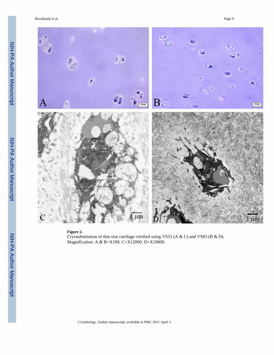
Figure 2.Cryosubstitution of thin size cartilage vitrified using VS55 (A & C) and VS83 (B & D).Magnification: A & B=X100; C=X12000; D=X10000.
Brockbank et al. Page 9
Cryobiology. Author manuscript; available in PMC 2011 April 1.
NIH
-PA Author Manuscript
NIH
-PA Author Manuscript
NIH
-PA Author Manuscript
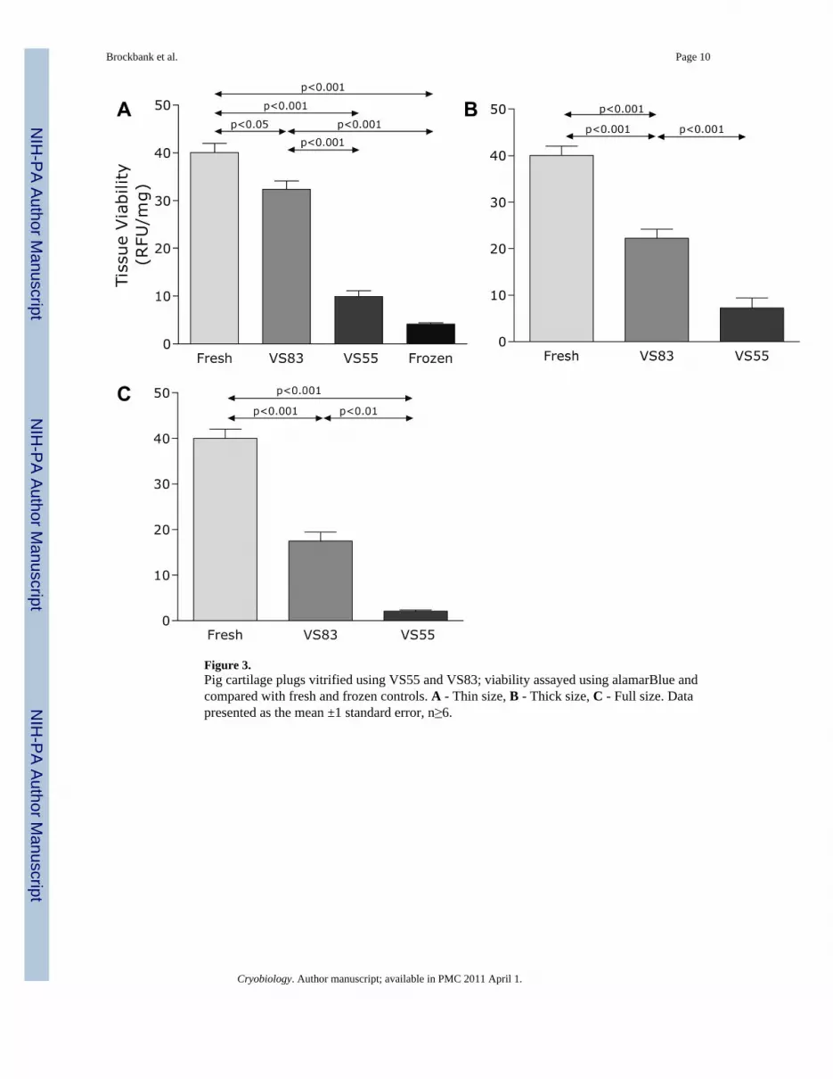
Figure 3.Pig cartilage plugs vitrified using VS55 and VS83; viability assayed using alamarBlue andcompared with fresh and frozen controls. A - Thin size, B - Thick size, C - Full size. Datapresented as the mean ±1 standard error, n≥6.
Brockbank et al. Page 10
Cryobiology. Author manuscript; available in PMC 2011 April 1.
NIH
-PA Author Manuscript
NIH
-PA Author Manuscript
NIH
-PA Author Manuscript

NIH
-PA Author Manuscript
NIH
-PA Author Manuscript
NIH
-PA Author Manuscript
Brockbank et al. Page 11
Table 1
Pig Cartilage Sample Size
Cartilage Size Tissue Dimension
Thin Size Plug 6mm Diam. × 2mm Depth
Thick Size Plug 6mm Diam. × 4mm Depth
Full Size Plug 6mm Diam. × 6mm Depth
Cryobiology. Author manuscript; available in PMC 2011 April 1.