ovariectomy alters the structural and biomechanical properties of ovine femoro-tibial articular...
TRANSCRIPT
OsteoArthritis and Cartilage (2005) 13, 1066e1075
ª 2005 OsteoArthritis Research Society International. Published by Elsevier Ltd. All rights reserved.doi:10.1016/j.joca.2005.07.001
Ovariectomy alters the structural and biomechanical properties of ovinefemoro-tibial articular cartilage and increases cartilage iNOS1
M. A. Cake Ph.D.y*, R. C. Appleyard Ph.D.z, R. A. Read Ph.D.y, M. M. Smith Ph.D.x,G. A. C. Murrell M.D., Ph.D.z and P. Ghosh Ph.D.xySchool of Veterinary and Biomedical Sciences, Murdoch University, WA, AustraliazOrthopaedic Research Institute, St. George Hospital, University of New South Wales, NSW, Australiax Institute of Bone and Joint Research, Royal North Shore Hospital, University of Sydney, NSW, Australia
Summary
Objective: To examine the effect of oestrogen depletion produced by surgical ovariectomy on the structural and biomechanical properties ofovine femoro-tibial articular cartilage (AC), and the production of inducible nitric oxide synthase (iNOS) and nitrotyrosine by these tissues.
Methods: Six aged ewes were surgically ovariectomised (OVX), while six were used as unoperated controls. Dynamic biomechanicalindentation testing of tibial plateau AC was performed at 26 weeks post-op. Histological sections of medial tibial plateau and lateral tibialplateau (LTP), medial and lateral femoral condyles (MFC, LFC) and patellar AC were examined for histopathology, toluidine blue stainingintensity, and patterns of collagen birefringence intensity. Immunoreactivity for iNOS and nitrotyrosine was assessed in full-thickness biopsyplugs of LFC and patellar AC, and patellar AC explants were cultured to determine in vitro NO release.
Results: Phase lag was reduced overall in LTP-AC of OVX sheep (10.9G 2.2( vs 12.1G 2.3(; P! 0.0001). Cartilage thickness was reducedin the LTP of OVX sheep (PZ 0.0002), in association with localised changes in dynamic shear modulus. Toluidine blue staining intensity wasreduced in the patella, LFC, and MFC. Histological examination revealed greater histopathology scores in the MFC of OVX animals, andaltered collagen birefringence intensity plots in the LTP. Immunostaining for iNOS was increased in patella AC (PZ 0.008), whilst nitrotyrosineimmunoreactivity was increased in patella (PZ 0.03) and LFC (P! 0.0001) AC. NO release by patellar AC explants was also elevated.
Conclusions: Oestrogen depletion induced by OVX caused regional thinning of femoro-tibial cartilage, with biomechanical and histologicalchanges suggestive of a disturbance in the content and/or structural organisation of the proteoglycan and collagen macromolecular assembly.The observed up-regulation of cartilage iNOS suggests a possible mechanism for these matrix changes.ª 2005 OsteoArthritis Research Society International. Published by Elsevier Ltd. All rights reserved.
Key words: Ovariectomy, Sheep, Cartilage biomechanics, Nitric oxide radical.
InternationalCartilageRepairSociety
Introduction
A growing body of epidemiological, clinical, and experimen-tal evidence suggests that ovarian steroids, particularlyoestrogen, have the potential to modify the metabolicactivity of joint tissues. In particular, it has been proposedthat the withdrawal of ovarian hormones at the time ofmenopause may contribute to the steep increase in theincidence and prevalence of osteoarthritis (OA) in womenover the age of 501. Conversely, oestrogen replacementtherapy in post-menopausal women may be associated withreduced risk of radiographic hip and knee OA2,3. A recentmagnetic resonance imaging-based study found that usersof oestrogen replacement therapy had greater tibialcartilage volume than non-users4. This difference persistedafter exclusion of women with OA, suggesting that
1This study was supported by an Australian Research CouncilSmall Grant. MAC was supported by the Murdoch UniversityVeterinary Trust and Boehringer Ingelheim Vetmedica. RCA wassupported in part by the St. George Hospital Sydney, South EastSydney Area Health Service, Australia.*Address correspondence and reprint requests to: Dr Martin
Cake, School of Veterinary and Biomedical Sciences, MurdochUniversity, Perth 6105, Australia. Tel: 61-8-9360-2175; Fax: 61-8-9310-4144; E-mail: [email protected] 24 January 2004; revision accepted 2 July 2005.
10
oestrogen may affect the structure of cartilage in theabsence of any clinically detectable degenerative changes.To date, conflicting experimental evidence exists re-
garding the direct influence of oestrogen on articularcartilage (AC). Animal experiments have shown thatexogenous oestrogen depresses proteoglycan (PG) syn-thesis by chondrocytes5,6 and induces detrimental changesin AC in vivo6,7; however, such studies have not alwaysbeen confined to physiological doses of the hormone.Challenging this evidence of a chondrosuppressive role foroestrogen, Turner et al.8 found that ovariectomy of agedewes adversely affected the biomechanical properties ofcartilage, and that this response was preventable byoestrogen replacement. More recently, oestrogen replace-ment therapy has been shown to increase chondrocyte PGsynthesis and reduce the histological severity of spontane-ous OA lesions in ovariectomised (OVX) cynomolgusmacaques9,10. Recent studies by our research group haveshown alterations in cartilage histological structure11 andcalcified cartilage vascularisation12 following ovariectomy ofadult sheep. These findings are supportive of epidemiolog-ical data suggesting that oestrogen is necessary to maintainthe structural integrity of cartilage in older women2.Oestrogens have been shown inmany tissues to influence
production of nitric oxide (NO), a highly reactive free radicalwhich is emerging as an important pathophysiological
66
1067Osteoarthritis and Cartilage Vol. 13, No. 12
mediator in OA13. While oestradiol is generally supportive ofconstitutive (calcium-dependent) nitric oxide production inmost tissues14,15 and is closely correlated to plasma levelsof NO in women16, oestrogens may instead suppress tissueexpression of inducible nitric oxide synthase (iNOS),especially that stimulated by cytokine challenge17,18. Botharticular chondrocytes and synoviocytes generate NOprincipally via iNOS expression, which is increased inOA19,20. While the effect of excess NO production in jointtissues is not entirely clear, it is generally thought to bedetrimental to cartilage, via mechanisms including suppres-sion of PG synthesis, increased matrix metalloproteasesynthesis, and chondrocyte apoptosis21,22.The aim of the present study was to further characterise
the structural and biomechanical alterations induced byovariectomy, and thus oestrogen depletion, in ovinefemoro-tibial AC, as well as to examine the expression ofiNOS and ex vivo production of NO (as its metabolite NO2
�)by AC derived from the joints of these animals.
Methods
ANIMALS
Twelve aged (7 years old) Merino ewes were obtainedfrom a single source and selected for uniformity of size,conformation, body condition, and absence of lameness.Half the sheep were OVX under general anaesthesia viaa small midline laparotomy incision, while half remained asnon-operated controls (NOC). Following a brief recoveryperiod, sheep were maintained on irrigated pasture withoutsupplementary feeding. Body weights were monitored atapproximately monthly intervals. All animal procedureswere approved by the Murdoch University Animal EthicsCommittee.Venous plasma samples were collected twice pre-
operatively, at 4 and 10 weeks post-operatively, and atsacrifice. At each time point, plasma 17b-oestradiolconcentrations were measured using the method of Webbet al.23, involving an affinity chromatography extractionprocedure followed by double-antibody radioimmunoassay.
TISSUE PROCESSING
Sheep were euthanased by intravenous injection ofpentobarbitone sodium [300 mg/ml; Valabarb, Pitman-Moore, Australia] between 28 and 30 weeks post-ovariec-tomy, along with control sheep. One joint (random left/right)was immediately dissected under sterile conditions toobtain cartilage for culture. At this time, samples were fixedfor histology and all joint surfaces were scored for cartilageerosion and osteophyte formation using a 4-point scale24.Tibiae for biomechanical testing were transported on ice for24 h before dissection and removal of the entire proximalepiphyseal region of the tibia using a bandsaw. Eachsample was wrapped in gauze soaked in phosphate-buffered saline (PBS) [Baxter Healthcare, Australia] andthen stored frozen (�20(C) for less than 2 weeks beforebiomechanical testing. Freezeethaw of cartilage has beenshown to have no effect on its biomechanical properties25.
CARTILAGE EXPLANT CULTURE
Four cartilage explant discs were removed from theproximal articular surface of the patella using a disposable3 mm biopsy punch [Stiefel, Germany]. After rinsing in
sterile PBS, the explants were transferred to individual wellsof 24-well multiwell culture plates [Techno Plastic Products,Switzerland]. Explants were cultured in 500 ml of Hams-F12medium [Sigma, USA] supplemented with 1 mM L-glutamine[Sigma, USA], 50 mg/ml gentamicin [David Bull Laborato-ries, Australia], and 10% v/v foetal calf serum (FCS) [TraceScientific, Australia], pH 7.2. In order to assess the effect ofovariectomy on both basal and cytokine-stimulated NOproduction, media were replaced after 24 h of culture (37(C/5% CO2) by 500 ml of fresh Hams-F12/10% FCS, supple-mented with 100 pg/ml ovine interleukin (IL)-1b in two of thefour wells. Ovine recombinant IL-1b26 was kindly suppliedby the Centre for Animal Biotechnology, University ofMelbourne, Australia. After a further 48 h of incubation,the explants and aliquots of conditioned media wereimmediately frozen at �20(C awaiting assay.In vitro nitric oxide release was assayed colorimetrically
as the nitrite content of conditioned media using the Greissreaction27,28 and compared with a standard curve of NaNO3
[Sigma, USA] assayed in parallel. Results were correctedfor explant DNA content as determined using Hoescht33258 (bis-benzimide)29 following papain digestion.
INDENTATION TESTING
Use of the dynamic indentation device has been de-scribed previously24,30 and has been shown to be bothreliable and reproducible31. It incorporates a handle witha stainless steel tube (120 mm long! 4 mm externaldiameter) extending from one end. Enclosed within theend of the tube is a cantilevered piezo-electric beam, witha small non-porous cylindrical probe (0.5 mm diameter)attached to its free end. In its unloaded state the probeprotrudes out the side of the tube by 0.25 mm and oscillateswith a sinusoidal displacement amplitude of G0.03 mm.When brought into contact with the cartilage, the compliantbeam is deflected backwards causing the probe to bepushed back into the tube by an amount dependent on thestatic stiffness of the material being tested. Additionally, thedynamic amplitude is reduced by an amount proportional tothe dynamic stiffness of the material being tested. Unlikea rigid benchtop system where a predetermined static anddynamic strain can be set, this is not possible with ourhandheld system. We have however investigated the staticand dynamic strain imposed on the cartilage using thishandheld system, and found it on average approximates10%. Readings where the strain is above 20% aredisregarded due to the associated non-linear behaviour ofthe cartilage under large deformation. In addition to thestiffness response, phase lag (f) between the supply andresponse voltage signals to the vibration unit is alsomonitored to determine the energy dissipation character-istics of the cartilage.The instrument was used to dynamically indent the AC by
aligning the probe normal to the cartilage surface, andapplying a downward force on the instrument of 2.5 N, asmonitored with a pressure transducer located within thehandle. A single frequency sinusoidal waveform wasapplied to the device causing the probe to vibrate at20 Hz. Constant dynamic stiffness and phase readingswere achieved after a few seconds, at which time data werecollected and saved. Dynamic shear modulus (G*) wascalculated using the theory published by Hayes et al.32;
G*ZDP
Dw
ð1� nÞð4kaÞ

1068 M. A. Cake et al.: Ovariectomy modifies ovine cartilage
where G* is the magnitude of the dynamic shear modulus(MPa), DP is the change in applied force (N), Dw is thechange in displacement (mm), constant a is the radius ofindentor (mm), and k is a geometric scaling factor de-pendent on the area aspect ratio of a/h where hZ cartilagethickness in mm. A number of studies have demonstratedthat the Poisson’s ratio of cartilage is not constant (rangingfrom 0.2 to 0.5)33. However, it has been demonstrated thatwhen AC is indented at frequencies above 1 Hz, thedynamic shear modulus (G*) is relatively constant, due tothe cartilage behaving in a near incompressible manner34.As such, the Poisson’s ratio was assumed to be 0.5.Testing was conducted across a 3! 3 array marked on
both the lateral and medial tibial plateau (LTP, MTP)(nZ 18 locations). Three separate indentation runs weremade across all 18 locations, and the dynamic stiffness andphase lag calculated from the mean of the three measuresat each location. On completion of the indentation assess-ment, the thickness (h) of the AC was determined at eachindentation location so that dynamic shear modulus G*could be calculated. AC thickness was measured usinga needle penetration method similar to that describedpreviously35.Results of biomechanical testing at individual locations
are pooled into zonal subsets corresponding to theequivalent outer (O), middle (M), and inner (I) zones ofthe lateral (L) or medial (M) plateau (six zones: MO, MM, MI;LO, LM, LI), as described previously24.
HISTOLOGICAL METHODS
Histological sections were prepared from the MTP andLTP and medial and lateral femoral condyles (MFC, LFC).Slices (approximately 3 mm thick) were taken from eachregion by bandsaw cuts in the medio-lateral plane,perpendicular to the articular surface, by a single operatorusing a standard protocol. Cuts were positioned in thefemoral condyles at the point of tibial contact in normalstance, and in the tibial plateau at the level of theintercondylar eminences. Toluidine blue- and Masson’sTrichrome-stained histological sections were prepared asdescribed previously36. Semi-quantitative histopathologicalgrading was performed according to a published modifiedMankin’s scoring system36,37. In each joint region, fourzones were scored: inner, middle, outer, and outer marginalzones. Scoring was done by a single observer, accordingto a 6-point scale (structure (0e10), cellularity (0e4),chondrocyte cloning (0e4), territorial toluidine blue stain-ing (0e4), interterritorial toluidine blue staining (0e4),and calcified cartilage pathology (0e4))37, and a meanaggregate score determined as the average of these fourzones. Additionally, vascular index was measured as thenumber of blood vessels invading the length of the calcifiedcartilage layer, excluding marginal regions of osteophyticremodelling12.Computer-assisted histomorphometric analysis was com-
pleted as described previously36. Sections were scanneddirectly using a slide scanner [Microtek Slidescanner 35tplus; Model No. PTS-1950] and analysed using imageanalysis software [ImagePro Plus v3.0.1; Media Cybernet-ics, USA]. Spatial calibration was performed usinga 10! 10 mm high precision graticule. Uncalcified cartilage(UCC) was determined from Masson’s Trichrome-stainedsections, using a radial thickness algorithm within thesoftware to generate mean thickness measurementsbetween lines delimiting the cartilage surface and most
advanced tidemark after dividing the cartilage visually intothirds, such that each arc segment comprised approx-imately one-third of the total arc length, termed outer(abaxial), middle, and inner (axial) zones. PG content wasdetermined as the mean grey-scale pixel intensity(whiteZ 0, blackZ 255) of toluidine blue-stained sections,in each of the above zones of the MFC and LFC, and in full-thickness 5 mm biopsy plugs of patellar cartilage. Collagenbirefringence was quantified using a method similar to thatof Arokoski et al.38 as described elsewhere30. Briefly, thecartilage section was placed between two polarising lenseswith the surface orientated at 45( to the direction ofpolarisation. Digital images of Picro Sirius red-stainedsections were obtained under monochromatic polarisedlight (l 550 nm), from which full-thickness light-intensityprofiles were plotted using image analysis software. Theprofile plot was then normalised by depth (0Z cartilagesurface, 1Z boundary of calcified cartilage) to allowcomparative analysis of cartilage of different thickness.Intensity profiles were determined in the inner, middle andouter zones of the MTP and LTP, and in full-thicknesscartilage plugs obtained from the middle zone of the LFC.
IMMUNOHISTOCHEMISTRY
Immunostaining was performed on 4 mm sections of full-thickness cartilage biopsy plugs obtained from the patellaand LFC and fixed in 10% neutral buffered formalin.Sections were pre-treated with bovine testicular hyaluron-idase [Sigma, USA] (0.5 mg/ml in 0.1 M phosphate buffer,pH 7.4, at 37(C for 1 h) prior to incubation at 4(C overnightwith 1:400 rabbit anti-iNOS IgG [Upstate Biotechnology] or1:1000 rabbit polyclonal anti-iNOS serum [Cayman Chem-ical]. Bound antibody was detected by incubation withbiotinylated goat anti-rabbit secondary antibody, followedby horseradish peroxidase-conjugated streptavidin andDAB reagent. Normal non-immune rabbit serum was usedas a negative control. Sections were examined by a singleblinded observer to determine the percentage of positivelyimmunostained chondrocytes. Cells in the superficial(topmost 250 mm) and deeper levels of cartilage wereassessed separately.
STATISTICAL ANALYSIS
Statistical comparisons were generated using specialistsoftware [Statview 5.0, SAS Institute Inc., USA], performingone-way analysis of variance to analyse variance acrossgroups, and Fisher’s protected least significant differencetest to compare group means between NOC and OVXsheep. Results of biomechanical testing were grouped forstatistical comparison to two ‘levels’ e the entire tibialplateau (all 18 test points, reported as ‘overall’ changes),and by zone (LO, LM, LI, MI, MM, MO); in each case testlocation ID was included as a second independent variable.Results for thickness and toluidine blue staining derived byimage analysis were compared statistically by region (LTP,MFC, etc.) using mean data for each, with zone (inner,middle, outer) included as a second independent variable.Calcified cartilage pathology and neovascularisation werealso compared statistically on a ‘whole-joint’ basis, includingjoint region as a second independent variable. Birefrin-gence intensity profiles were compared by Student’s t testwithin each percentile of normalised depth (0Z cartilagesurface, 100Z calcified cartilage boundary). A significancelevel of PZ 0.05 was used throughout.
1069Osteoarthritis and Cartilage Vol. 13, No. 12
Results
There was no statistically significant difference in bodyweights between OVX and NOC sheep throughout theduration of the trial. Plasma 17b-oestradiol levels weresignificantly reduced in OVX sheep at 5 weeks and allsubsequent time points post-ovariectomy (data not shown).Gross joint pathology was minimal in all sheep, thoughslight softening and fibrillation of the inner region of the MTPwas noted in most sheep. This appears to be a normalfinding in sheep of this age30.
DYNAMIC BIOMECHANICAL TESTING
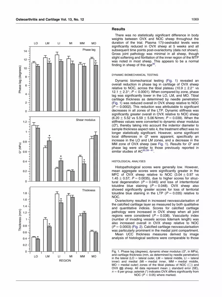
Dynamic biomechanical testing (Fig. 1) revealed anoverall reduction in phase lag in cartilage of OVX sheeprelative to NOC, across the tibial plateau (10.9G 2.2( vs12.1G 2.3(; P! 0.0001). When compared by zone, phaselag was significantly lower in the LO, LM, and MO. Tibialcartilage thickness as determined by needle penetration(Fig. 1) was reduced overall in OVX sheep relative to NOC(PZ 0.0002). This reduction was attributable to significantdecreases in all zones of the LTP. Dynamic stiffness wassignificantly greater overall in OVX relative to NOC sheep(6.20G 5.52 vs 5.59G 5.06 N/mm; PZ 0.039). When thestiffness values were converted to dynamic shear modulus(G*), thereby taking into account the indentor diameter tosample thickness aspect ratio k, the treatment effect was nolonger statistically significant. However, some significantlocal differences in G* were apparent, specifically anincrease in the LO and LM zones, and a decrease in theMM zone of OVX sheep (see Fig. 1). Results for G* andphase lag were similar to those previously reported insimilar studies of AC30,39e41.
HISTOLOGICAL ANALYSES
Histopathological scores were generally low. However,mean aggregate scores were significantly greater in theMFC of OVX sheep relative to NOC (3.04G 0.97 vs1.45G 0.57; PZ 0.0035), due to higher scores for struc-tural degeneration (PZ 0.045) and loss of interterritorialtoluidine blue staining (PZ 0.048). OVX sheep alsoshowed significantly greater scores for loss of territorialtoluidine blue staining in the LTP (PZ 0.035) relative toNOC.Ovariectomy resulted in increased neovascularisation of
the calcified cartilage layer as measured by both qualitativeand quantitative indices. Scores for calcified cartilagepathology were increased in OVX sheep when all jointregions were considered (PZ 0.038). Vascularity index(number of invading vessels across tidemark length) wasalso increased overall in OVX sheep relative to NOC(PZ 0.0003) (Fig. 2). Calcified cartilage neovascularisationwas particularly prominent in the medial joint compartment.Mean UCC thickness measures derived by image
analysis of histological sections were comparable to those
Fig. 1. Phase lag (degrees), dynamic shear modulus (G*, in MPa),and cartilage thickness (mm, as determined by needle penetration)in the lateral (LOZ lateral outer, LMZ lateral middle, LIZ lateralinner) and medial (MIZmedial inner, MMZmedial middle,MOZmedial outer) zones of the tibial plateau of NOC (,) andOVX ( ) sheep. All data represent meanG standard error (SE),nZ 6 per group; asterisk (*) indicates OVX differs significantly from
NOC (P! 0.05) where marked.

1070 M. A. Cake et al.: Ovariectomy modifies ovine cartilage
determined directly by the needle probe method. OVXsheep possessed thinner UCC across the LTP(0.84G 0.41 mm vs 0.99G 0.49 mm; PZ 0.006) withsimilar but not statistically significant results in the LFC(0.58G 0.25 vs 0.69G 0.26 mm; PZ 0.07). Mean grey-scale pixel intensity of toluidine blue-stained sections(Fig. 3) was significantly lower in the LFC (PZ 0.0005)and MFC (PZ 0.002) of OVX animals relative to NOC. Asimilar reduction in toluidine blue staining intensity was alsoseen in full-thickness biopsy plugs of patellar cartilage(mean grey-scale pixel intensity 88.8G 13.1 vs107.5G 15.0; PZ 0.023). Collagen light-intensity profileplots (Fig. 4) revealed a statistically significant increase insuperficial collagen organisation in the middle region of the
Fig. 2. Number of vessels invading the calcified cartilage layeracross the length of 4 mm transverse sections of the LFC, MFC andLTP, MTP of NOC (,) and OVX ( ) sheep. Data representmeanG standard error (SE), nZ 6 per group; asterisk (*) indicatesOVX differs significantly from NOC (P! 0.05) where marked.
Fig. 3. PG content, measured as the mean grey-scale pixel intensity(whiteZ 0, blackZ 255) of toluidine blue-stained histologicalsections of the LFC and MFC of NOC (,) and OVX ( ) sheep.Data represent meanG standard error (SE), nZ 6 per group;asterisk (*) indicates OVX differs significantly from NOC (P! 0.05).
LTP. A similar, statistically significant increase in superficiallight intensity was observed in full-thickness biopsy plugstaken from the LFC (results not shown). However, in theinner and outer regions of the tibial plateau, collagen light-intensity plots appeared to have a ‘flatter’ profile in OVXsheep, with less distinct peaks in superficial light intensity.
IMMUNOHISTOCHEMISTRY
iNOS and nitrotyrosine immunoreactivity in NOC sheepwas predominantly confined to superficial chondrocytes,with only occasional staining of deeper cells. OVX sheeppossessed a greater percentage of iNOS-positive cells inthe deep (O250 mm depth) chondrocytes of patellarcartilage (PZ 0.008) (Table I). Similarly, the percentageof cells staining positively for nitrotyrosine was significantlygreater in both the superficial (P! 0.0001) and deep(P! 0.0001) chondrocytes of LFC cartilage, and in thedeep chondrocytes of patellar cartilage (PZ 0.03). Repre-sentative sections are shown in Fig. 5.
CARTILAGE EXPLANT CULTURE
NO release by patellar cartilage explants, measured asnitrite content (nmol nitrite/mg DNA) of 48-h conditionedmedia, was increased approximately twofold by exposure toovine IL-1b (Fig. 6). NO production was significantly greaterby OVX explants under both unstimulated (PZ 0.005) andIL-1b-stimulated (P! 0.0001) conditions.
Discussion
The results of this study have shown that oestrogendepletion in sheep, produced by means of ovariectomy,provoked significant changes to the structural, biomechan-ical, and metabolic properties of femoro-tibial AC, at 6months post-surgery. Turner et al.8 previously demonstratedthat the biomechanical integrity of ovine cartilage isadversely affected by ovariectomy, as evidenced by a de-crease in aggregate modulus (compressive stiffness) andshear modulus, but not cartilage thickness or permeability.Studies in young rabbits42 and rats43e45 have shown anincrease in cartilage thickness following ovariectomy.However, due to species and age differences these latterfindings may not be applicable to the present study, whichdemonstrated thinner cartilage in the LTP of OVX sheep.Moreover, biomechanical studies showed a generalisedreduction in tibial cartilage phase lag, and localised differ-ences in dynamic shear modulus (G* increased in the innerregions of the LTP, and decreased in the middle of theMTP).In the absence of gross or microscopically detectable
erosion, cartilage thinning may be due to two processes.Firstly, net reduction in matrix production may have inducedan ‘atrophic’ cartilage state. Our analysis of toluidine bluestaining of histological sections showed a net loss of PGfrom both patellar and femoral condyle AC. This finding wasconsistent with recent related studies11, which foundevidence of abnormal cartilage PG synthesis followingovariectomy in sheep. Secondly, advancement of thetidemark may have led to endochondral ossification in thedeeper layers of AC, which would be expected to lead toa reduction in UCC thickness. The observed increase incalcified cartilage neovascularisation in joints of OVXsheep, as also reported by Hwa et al.12, is also consistentwith active remodelling and thickening of this region.

1071Osteoarthritis and Cartilage Vol. 13, No. 12
Fig. 4. Normalised collagen birefringence profile plots of the inner, middle, and outer zones of the LTP and MTP of NOC ( ) and OVX ( )sheep. Profiles represent the mean of nZ 6 sheep per group; asterisk (*) indicates OVX differs significantly from NOC (P! 0.05) at this
cartilage level (Student’s t test).
While the static compressive stiffness of cartilage isreported to be proportional to PG content25,41, dynamicmeasures such as G* have generally been shown tocorrelate more with collagen content40,46,47. In particular, LeRoux et al.47 noted a correlation between G* and themagnitude of superficial collagen birefringence in caninecartilage. The increase in G* in the middle and inner testinglocations of the LTP of OVX animals is therefore likely toreflect a proportional increase in collagen content. Thesechanges were mirrored by an increase in superficialcollagen birefringence in the middle zone of the LTP,suggesting an increase in the number and/or level oforganisation of collagen fibres arranged perpendicular tothe surface within the superficial zone of cartilage. Althoughthe zonal variation in polarisation light-intensity patterns isnot consistently correlated with variation in G*, it should benoted that the locations of collagen light intensity and
indentation testing differed spatially in that the former wasundertaken using a single transverse histological sectionfrom each joint, while the latter was the summed resultsfrom an array of testing locations24.While the relationship between the structural organisation
of cartilage and its phase properties is complex, thegeneralised reduction in tibial cartilage phase lag observedin OVX animals might also reflect changes in collagencontent or organisation. Lower phase lag values reflecta change in viscoelastic behaviour consistent with greaterelasticity and reduced dissipation of shear forces. Asdiscussed elsewhere24,30, this may be consistent with eithera proportional reduction in PG content, and/or a ‘tightening’of the collagenePG matrix arising from alterations in thedensity or organisation of the collagen network. Ovariectomyis known to induce changes in collagen structure in othertissues; for example, ovariectomy of adult rats increases the
Table IImmunohistochemical staining for iNOS and nitrotyrosine in full-thickness UCC plugs from the patella and LFC. Figures indicate thepercentage of positively-stained superficial (S; !250 mm deep) and deep (D; O250 mm deep) chondrocytes (meanG SD) in samples from
NOC (nZ 6) and OVX (nZ 6) sheep. Asterisks indicate OVX differs significantly from NOC (*PZ 0.03; **PZ 0.008; ***P! 0.0001)
Antigen iNOS Nitrotyrosine
Zone S D S D
Patella NOC 46.7G 22.3 11.2G 19.7 54.9G 16.2 29.9G 15.6OVX 69.3G 17.9 45.7G 20.3** 70.1G 14.3 63.6G 21.5*
LFC NOC 59.7G 20.7 35.2G 24.9 33.2G 9.4 10.9G 8.5OVX 76.8G 7.6 56.9G 26.8 81.1G 5.0*** 76.9G 6.6***

1072 M. A. Cake et al.: Ovariectomy modifies ovine cartilage
Fig. 5. Nitrotyrosine immunoreactivity in full-thickness biopsies of LFC cartilage. Two representative sections from NOC (above) and OVX(below) ewes are shown.
collagen content of temporomandibular joint discs48, andreduces the fibril diameter of type I collagen in bones andskin49.Several mechanisms have been proposed to explain how
ovariectomy might disturb chondrocyte metabolism, includ-ing interaction with the post-receptor mechanisms of IL-1activity through the AP-1 promoter pathway50,51, anddisturbance of the anabolic insulin-like growth factor-1axis52,53. Alternatively, the current findings suggest thatthe effects of ovariectomy on cartilage may be related toincreased local release of NO. Patellar cartilage explantsfrom OVX sheep produced significantly more nitrite com-pared to non-OVX animals, and immunostaining for iNOS(in patellar sections) and nitrotyrosine (in LFC and patella)was similarly increased. Though generally reported to havethe opposite effect on constitutive nitric oxide produc-tion14,15, this and other studies suggest that oestradiol isnormally acts to suppress iNOS expression. For example,Kauser et al.54 demonstrated that OVX rats experiencea greater increase in plasma nitrate levels followingendotoxin treatment than entire animals, and that thisincrease can be attenuated by pre-treatment with oestradiol.17b-Oestradiol has also been demonstrated to inhibit iNOSexpression in rat aorta17,55, and murine macrophages18.Similarly, the reduction in AC iNOS expression observedduring pregnancy in multiparous rabbits suggests a sup-pressive effect of ovarian hormones56, though the hormoneresponsible was not identified in that study.As nitric oxide is thought to decrease PG content21,22,57
and alter collagen synthesis58, increased cartilage iNOS
expression following ovariectomy may be a possiblemechanism for many of the cartilage changes observed inour study. Yoon et al.59 noted a comparable increase inNOS expression in rabbit clitoris and vagina followingovariectomy, associated with an increase in collagencontent in these tissues. Further evidence of a possiblecausal link is provided by the similarity of cartilageabnormalities (reduced thickness and phase lag of tibialcartilage, increased superficial collagen light intensity, anddecreased toluidine blue staining intensity in femoralcondyle cartilage) reported following topical treatment withan exogenous NO donor, glyceryl trinitrate, in a parallelstudy using related animals24.In conclusion, this study provides evidence that depletion
of oestrogen from aged ewes by means of ovariectomy wasassociated with an array of localised changes to tibial AC(principally of the LTP) including reduced AC thickness andphase lag, locally increased dynamic shear modulus andaltered collagen birefringence light-intensity profile. Thesefindings are suggestive of a disturbance in the proportionalcontent and/or structural organisation of the collagen andPG components of AC, while the reduction in cartilagethickness is consistent with prolonged suppression of matrixsynthesis. While no changes in the gross appearance ofjoint surfaces were evident, histological examination sug-gested a slight deterioration of structure and loss of PG inthe MFC, and this was mirrored by a reduction in theintensity of toluidine blue staining in femoral condyle andpatellar cartilage. The observation of increased neovascu-larisation of the UCC layer is potentially significant, given

1073Osteoarthritis and Cartilage Vol. 13, No. 12
that this change is one of the classical hallmarks of OAreported in early pathological studies60. However, it shouldbe noted that the biomechanical changes observed in thisstudy after ovariectomy are not clearly degenerative, sincein experimental animal models OA is generally associatedwith reduced cartilage shear modulus, increased phase lag,and reduced superficial birefringence41,61. The cartilagechanges observed in the present study may be betterinterpreted as ‘atrophic’, although some histological evi-dence of degeneration was evident.These findings confirm the importance of ovarian
hormones for the structural and metabolic homeostasis offemoro-tibial AC in the ewe and presumably other species.Furthermore, we report here for the first time an increase incartilage iNOS expression and NO release followingovariectomy, which may represent a possible mediator ofsome of the changes observed.
Acknowledgements
The authors would like to thank Ms Susan Smith for hercompetent preparation of the histological and immunos-tained sections, and Mrs Diana Pethick for her assistancewith animal procedures.
References
1. Spector T, Campion G. Generalized osteoarthritis isa hormonally mediated disease. Ann Rheum Dis 1989;48:523e7.
2. Nevitt M, Felson D. Sex hormones and the risk ofosteoarthritis in women: epidemiologic evidence. AnnRheum Dis 1996;55:673e6.
Fig. 6. Nitric oxide release, measured as the nitrite content (nmol/mgDNA) of 48-h conditioned media, by unstimulated and IL-1b-stimulated patellar cartilage explants from NOC (,) and OVX ( )sheep. Data represent meanG standard error (SE) from duplicatesamples, nZ 6 per group; asterisk (*) indicates OVX differs
significantly from NOC (P% 0.005).
3. Spector T, Nandra D, Hart D, Doyle D. Is hormonereplacement therapy protective for hand and kneeosteoarthritis in women: the Chingford study. AnnRheum Dis 1997;56:432e4.
4. Wluka A, Davis S, Bailey M, Stuckey S, Cicuttini F.Users of oestrogen replacement therapy have moreknee cartilage than non-users. Ann Rheum Dis 2001;60:332e6.
5. Mackintosh D, Mason R. Pharmacological actions of17b-oestradiol on articular cartilage chondrocytes andchondrosarcoma chondrocytes in the absence ofoestrogen receptors. Biochim Biophys Acta 1988;964:295e302.
6. Rosner I, Goldberge V, Moskowitz R. Estrogens andosteoarthritis. Clin Orthop 1986;213:77e83.
7. Tsai C-L, Liu T-K. Estradiol-induced knee osteoarth-rosis on ovariectomized rabbits. Clin Orthop 1993;291:295e302.
8. Turner A, Athanasiou K, Zhu C-F, Alvis M, Bryant H.Biomechanical effects of estrogen on articular carti-lage in ovariectomized sheep. Osteoarthritis Cartilage1997;5:63e9.
9. Ham K, Loeser R, Lindgren B, Carlson C. Effects oflong-term estrogen replacement therapy on osteoar-thritis severity in cynomolgus monkeys. ArthritisRheum 2002;46:1956e64.
10. RichmondR,CarlsonC,Register T, ShankerG, LoeserR.Functional estrogen receptors in adult articularcartilage: estrogen replacement therapy increaseschondrocyte synthesis of proteoglycans and insulin-likegrowth factor binding protein-2. Arthritis Rheum 2000;43:2081e90.
11. Parker D, Hwa S-Y, Sambrook P, Ghosh P. Estrogenreplacement therapy mitigates the loss of jointcartilage proteoglycans and bone mineral densityinduced by ovariectomy and osteoarthritis. APLAR JRheumatol 2003;6:116e27.
12. Hwa S, Smith M, Burkhardt D, Ghosh P. Oestrogendepletion provokes significant vascular and matrixchanges in the calcified cartilage and subchondralbone of the femoraletibial joints of aged ewes. TransOrthop Res Soc 2001:0373.
13. Jang D, Murrell G. Nitric oxide in arthritis. Free RadicBiol Med 1998;24:1511e9.
14. Armour K, Ralston S. Estrogen upregulates endothelialconstitutive nitric oxide synthase expression inhuman osteoblast-like cells. Endocrinology 1998;139:799e802.
15. Weiner C, Lizasoain I, Baylis S, Knowles R, Charles I,Moncada S. Induction of calcium-dependent nitricoxide syntheses by sex hormones. Proc Natl AcadSci U S A 1994;91:5212e6.
16. Rosselli M, Imthurn B, Macas E, Keller P, Dubey R.Circulating nitrite/nitrate levels increase with folliculardevelopment: indirect evidence for estradiol mediatedNO release. Biochem Biophys Res Commun 1994;1543e52.
17. Kauser K, Sonnenberg D, Diel P, Rubanyi GM. Effect of17beta-oestradiol on cytokine-induced nitric oxideproduction in rat isolated aorta. Br J Pharmacol1998;123:1089e96.
18. Hayashi T, Yamada K, Esaki T, Muto E, Chaudhuri G,Iguchi A. Physiological concentrations of 17beta-estradiol inhibit the synthesis of nitric oxide synthasein macrophages via a receptor-mediated system.J Cardiovasc Pharmacol 1998;31:292e8.

1074 M. A. Cake et al.: Ovariectomy modifies ovine cartilage
19. Stadler J, Stefanovic-Racic M, Billiar T, Curran R,McIntyre L, Georgescu H, et al. Articular chondrocytessynthesize nitric oxide in response to cytokines andlipopolysaccharide. J Immunol 1991;147:3915e20.
20. McInnes L, Leung B, Field M, Wei X, Huang F, SturrockR, et al. Production of nitric oxide in the synovialmembrane of rheumatoid and osteoarthritis patients.J Exp Med 1996;184:1519e24.
21. Taskiran D, Stefanovic-Racic M, Georgescu H, Evans C.Nitric oxide mediates suppression of cartilage pro-teoglycan synthesis by interleukin-1. Biochem BiophysRes Commun 1994;200:142e8.
22. Murrell G, Jang D, Williams R. Nitric oxide activatesmetalloprotease enzymes in articular cartilage. Bio-chem Biophys Res Commun 1995;206:15e21.
23. Webb R, Baxter G, McBride D, Nordblom G, Shaw M.The measurement of testosterone and oestradiol-17busing iodinated tracers and incorporating an affinitychromatography extraction procedure. J Steroid Bio-chem 1985;23:1043e51.
24. Cake M, Appleyard R, Read R, Ghosh P, Swain M,Murrell G. Topical administration of the nitric oxidedonor glyceryl trinitrate modifies the structural andbiomechanical properties of ovine articular cartilage.Osteoarthritis Cartilage 2003;11:872e8.
25. Athanasiou K, Rosenwasser M, Buckwalter J, Malinin T,Mow V. Interspecies comparisons of in situ intrinsicmechanical properties of distal femoral cartilage.J Orthop Res 1991;9:330e40.
26. Seouw H, Rothel J, Wood P. Expression and purifica-tion of recombinant interleukin-1b from Escherichiacoli. Vet Immunol Immunopathol 1994;41:229e39.
27. Green L, Wagner D, Glogowski J, Skipper P, Wishnok J,Tannenbaum S. Analysis of nitrate, nitrite and [15N]ni-trate in biological fluids. Anal Biochem 1982;126:131e8.
28. Guevara I, Iwanejko J, Dembinska-Kiec A, PankiewiczJ, Wanat A, Anna P, et al. Determination of nitrite/ni-trate in human biological material by the simple Greissreaction. Clin Chim Acta 1998;177e88.
29. Kim Y-J, Sah R, Doong J-Y, Grodzinsky A. Fluoromet-ric assay of DNA in cartilage explants using Hoechst33258. Anal Biochem 1988;174:168e76.
30. Appleyard R, Burkhardt D, Ghosh P, Read R, Cake M,Swain M, et al. Topographical analysis of thestructural, biochemical, and dynamic biomechanicalproperties of cartilage in an ovine model of osteoar-thritis. Osteoarthritis Cartilage 2003;11:65e77.
31. Appleyard R, Swain M, Khanna S, Murrell G. Theaccuracy and reliability of a novel handheld dynamicindentation probe for analysing articular cartilage.Phys Med Biol 2001;46:541e50.
32. Hayes W, Keer L, Herrmann G, Mockros L. Amathematical analysis for indentation tests of articularcartilage. J Biomech 1972;5:541e51.
33. Jurvelin J, Buschmann M, Hunziker E. Optical andmechanical determination of Poisson’s ratio of adultbovine humeral articular cartilage. J Biomech 1997;30:235e41.
34. Lee R, Frank E, Grodinsky A, Roylance D. Oscillatorycompressional behavior of articular cartilage and itsassociated electromechanical properties. J BiomechEng 1981;103:280e92.
35. Swann A, Seedhom B. Improved techniques formeasuring the indentation and thickness of articularcartilage. Proc Inst Mech Eng [H] 1989;203:143e50.
36. Cake M, Read R, Guillou B, Ghosh P. Modification ofarticular cartilage and subchondral bone pathology inan ovine meniscectomy model of osteoarthritis byavocado and soya unsaponifiables (ASU). Osteoar-thritis Cartilage 2000;8:404e11.
37. Little C, Smith S, Ghosh P, Bellenger C. Histomorpho-logical and immunohistochemical evaluation of jointchanges in a model of osteoarthritis induced by lateralmeniscectomy in sheep. J Rheumatol 1997;24:2199e209.
38. Arokoski J, Hyttinen M, Lapvetelainen T, Takacs P,Kosztaczky B, Modis L, et al. Decreased birefringenceof the superficial zone collagen network in the canineknee (stifle) articular cartilage after long distancerunning training, detected by quantitative polarisedlight microscopy. Ann Rheum Dis 1996;55:253e64.
39. Setton L, Mow V, Howell D. Mechanical behavior ofarticular cartilage in shear is altered by transection ofthe anterior cruciate ligament. J Orthop Res 1995;13:473e82.
40. Zhu W, Mow V, Koob T, Eyre D. Viscoelastic shearproperties of articular cartilage and the effects ofglycosidase treatments. J Orthop Res 1993;11:771e81.
41. Setton L, Elliott D, Mow V. Altered mechanics ofcartilage with osteoarthritis: human osteoarthritis andan experimental model of joint degeneration. Osteo-arthritis Cartilage 1999;7:2e14.
42. Rasanen T, Mesner K. Articular cartilage compressivestiffness following oophorectomy or treatment with17b-estradiol in young postpubertal rabbits. ActaObstet Gynecol Scand 1999;78:357e62.
43. Okuda T, Yasuoka T, Nakashima M, Oka N. The effectof ovariectomy on the temporomandibular joints ofgrowing rats. J Oral Maxillofac Surg 1996;54:1201e10.
44. Yasuoka T, Nakashima M, Okuda T, Tatematsu N.Effect of estrogen replacement on temporomandibularjoint remodelling in ovariectomised rats. J OralMaxillofac Surg 2000;58:189e96.
45. Yamashiro T, Takano-Yamamoto T. Differential re-sponses of mandibular condyle and femur to oestro-gen deficiency in young rats. Arch Oral Biol 1998;43:191e5.
46. Bader D, Kempson G, Egan J, Gilbey W, Barrett A. Theeffects of selective matrix degradation on the short-term compressive properties of adult human articularcartilage. Biochim Biophys Acta 1992;1116:147e54.
47. Le Roux M, Arokoski J, Vail T, Guilak F, Hyttinen M,Kiviranta I, et al. Simultaneous changes in themechanical properties, quantitative collagen organiza-tion, and proteoglycan concentration of articularcartilage following meniscectomy. J Orthop Res2000;18:383e92.
48. Abubaker A, Hebda P, Gunsolley J. Effects of sexhormones on protein and collagen content of thetemporomandibular joint disc of the rat. J OralMaxillofac Surg 1996;54:721e7.
49. Kafantari H, Kounadi E, Fatouros M, Milonakis M,Tzaphlidou M. Structural alterations in rat skin andbone collagen fibrils induced by ovariectomy. Bone2000;26:349e53.
50. Cutolo M, Sulli A, Barone A, Seriolo B, Accardo S. Sexhormones, proto-oncogene expression and apoptosis:their effects on rheumatoid synovial tissue. Clin ExpRheumatol 1996;14:87e94.

1075Osteoarthritis and Cartilage Vol. 13, No. 12
51. Ruh M, Bi Y, Dalonzo R, Bellone C. Effect of estrogenson IL-1-beta promoter activity. J Steroid Biol 1998;66:203e10.
52. Goya L, Garcia-Segura L, Ramos S, Pascual-Leone A,Argente J, Martin M, et al. Interaction betweenmalnutrition and ovarian hormones on the systemicIGF-I axis. Eur J Endocrinol 2002;147:417e24.
53. Fernihough J, RichmondR,CarlsonC,Cherpes T, Holly J,Loeser R. Estrogen replacement therapy modulation ofthe insulin-like growth factor system in monkey kneejoints. Arthritis Rheum 1999;42:2103e11.
54. Kauser K, Sonnenberg D, Tse J, Rubanyi G. 17bestradiol attenuates endotoxin-induced excessive ni-trix oxide production in ovariectomized rats in vivo. AmJ Physiol 1997;42:H506e9.
55. Tamura K, Yamaguchi K, Kogo H. 17Beta-estradiolinhibits ovariectomy-induced expression of induciblenitric oxide synthase in rat aorta in vivo. Life Sci 2000;66:PL259e64.
56. Hellio Le Graverand M, Reno C, Hart D. Influence ofpregnancy on gene expression in rabbit articularcartilage. Osteoarthritis Cartilage 1998;6:341e50.
57. Studer R, Jaffurs D, Stefanovic-Racic M, Robbins P,Evans C. Nitric oxide in osteoarthritis. OsteoarthritisCartilage 1999;377e9.
58. Cao M, Westerhausen-Larson A, Niyibizi C, Kavalko-vich K, Georgescu H, Rizzo C, et al. Nitric oxideinhibits the synthesis of type-II collagen withoutaltering Col2A1 mRNA abundance: prolyl hydroxylaseas a possible target. Biochem J 1997;324:305e10.
59. Yoon H, Chun W, Park Y, Shim B, Han W, Kwon S.Effects of estrogen on nitric oxide synthase andhistological composition in the rabbit clitoris andvagina. Int J Impot Res 2001;13:205e11.
60. Mankin H, Dorfman H, Lippiello L, Zarins A. Bio-chemical and metabolic abnormalities in articularcartilage from osteoarthritic human hips. II Correlationof morphology with biochemical and metabolic data.J Bone Joint Surg 1971;53A:523e37.
61. Appleyard R, Ghosh P, Swain M. Biomechanical,histological and immunohistochemical studies ofpatellar cartilage in an ovine model of osteoarthritisinduced by lateral meniscectomy. Osteoarthritis Car-tilage 1999;7:281e94.