virus type 1 controllers of human immunodeficiency from
TRANSCRIPT
Published Ahead of Print 29 October 2008. 2009, 83(1):140. DOI: 10.1128/JVI.01471-08. J. Virol.
Heckerman and Bruce D. WalkerBrian L. Block, Arne Schneidewind, Todd M. Allen, DavidChanson J. Brumme, Florencia Pereyra, Alicja Trocha, Toshiyuki Miura, Mark A. Brockman, Zabrina L. Brumme, Virus Type 1 Controllers of Human Immunodeficiency
from Elitegag-proteaseCarrying Capacity of Chimeric NL4-3 Viruses HLA-Associated Alterations in Replication
http://jvi.asm.org/content/83/1/140Updated information and services can be found at:
These include:
REFERENCEShttp://jvi.asm.org/content/83/1/140#ref-list-1at:
This article cites 44 articles, 31 of which can be accessed free
CONTENT ALERTS more»articles cite this article),
Receive: RSS Feeds, eTOCs, free email alerts (when new
http://journals.asm.org/site/misc/reprints.xhtmlInformation about commercial reprint orders: http://journals.asm.org/site/subscriptions/To subscribe to to another ASM Journal go to:
on February 28, 2014 by P
EN
N S
TA
TE
UN
IVhttp://jvi.asm
.org/D
ownloaded from
on F
ebruary 28, 2014 by PE
NN
ST
AT
E U
NIV
http://jvi.asm.org/
Dow
nloaded from
JOURNAL OF VIROLOGY, Jan. 2009, p. 140–149 Vol. 83, No. 10022-538X/09/$08.00�0 doi:10.1128/JVI.01471-08Copyright © 2009, American Society for Microbiology. All Rights Reserved.
HLA-Associated Alterations in Replication Capacity of ChimericNL4-3 Viruses Carrying gag-protease from Elite Controllers of
Human Immunodeficiency Virus Type 1�
Toshiyuki Miura,1,2,3 Mark A. Brockman,1,2 Zabrina L. Brumme,1,2 Chanson J. Brumme,1Florencia Pereyra,1,2 Alicja Trocha,1,3 Brian L. Block,1 Arne Schneidewind,1,2 Todd M. Allen,1,2
David Heckerman,4 and Bruce D. Walker1,2,3*Partners AIDS Research Center, Massachusetts General Hospital, 13th St., BLD149, Charlestown, Massachusetts 021291;
Harvard University Center for AIDS Research, Boston, Massachusetts 021152; Howard Hughes Medical Institute,Chevy Chase, Maryland 208153; and Microsoft Research, Redmond, Washington 980524
Received 14 July 2008/Accepted 16 October 2008
Human immunodeficiency virus type 1 (HIV-1)-infected persons who maintain plasma viral loads of <50 copiesRNA/ml without treatment have been termed elite controllers (EC). Factors contributing to durable control of HIVin EC are unknown, but an HLA-dependent mechanism is suggested by overrepresentation of “protective” class Ialleles, such as B*27, B*51, and B*57. Here we investigated the relative replication capacity of viruses (VRC)obtained from EC (n � 54) compared to those from chronic progressors (CP; n � 41) by constructing chimericviruses using patient-derived gag-protease sequences amplified from plasma HIV RNA and inserted into an NL4-3backbone. The chimeric viruses generated from EC displayed lower VRC than did viruses from CP (P < 0.0001).HLA-B*57 was associated with lower VRC (P � 0.0002) than were other alleles in both EC and CP groups. Chimericviruses from B*57� EC (n � 18) demonstrated lower VRC than did viruses from B*57� CP (n � 8, P � 0.0245).Differences in VRC between EC and CP were also observed for viruses obtained from individuals expressing nodescribed “protective” alleles (P � 0.0065). Intriguingly, two common HLA alleles, A*02 and B*07, were associatedwith higher VRC (P � 0.0140 and 0.0097, respectively), and there was no difference in VRC between EC and CPsharing these common HLA alleles. These findings indicate that cytotoxic T-lymphocyte (CTL) selection pressureon gag-protease alters VRC, and HIV-specific CTLs inducing escape mutations with fitness costs in this region maybe important for strict viremia control in EC of HIV.
Human immunodeficiency virus type 1 (HIV-1)-infected in-dividuals who control viremia to below the limit of detection(�50 RNA copies/ml plasma) without antiviral therapy havebeen termed elite controllers (EC) (16). Unraveling the mech-anisms associated with this rare and remarkable phenotype,which has been documented in some persons infected for 30years and counting, should provide important insights regard-ing our understanding of HIV pathogenesis and could provideimportant insights for vaccine development.
Host genetics, host innate and adaptive immune responses,and viral sequence variation have all been suggested to influ-ence the rate of disease progression in HIV-1 infection (re-viewed in reference 16). Recently, we reported that virusessequenced from plasma and peripheral blood mononuclearcells from EC do not share a common ancestor, do not containshared amino acid changes that are specific to this phenotype,and do not typically harbor gross viral genetic defects (36).However, such studies do not address functional properties ofthe encoded proteins, such as viral replication capacity (VRC).Reductions in VRC have been observed following exposure toantiretroviral drug selection pressure (17, 25, 31, 34) and there-fore may also occur in response to immune selection pressure.
Focused studies of VRC in long-term nonprogressors/long-term survivors with detectable viremia have suggested thattheir viruses have reduced VRC compared to those of chronicprogressors (CP) (7, 37, 39).
Due to the difficulties in isolating viruses from EC, investi-gating VRC in this rare group has been very challenging.Blankson et al. reported isolation of replication-competentviruses from 4 of 10 EC studied (8). On the other hand, thesame group reported a case of a B*27/57 EC whose virus hadreduced replication capacity compared to those of laboratoryreference strains and the virus in the transmitting donor (4).However, to date, there have been no studies comparing VRCbetween EC and CP.
Factors that influence VRC are beginning to be defined.Several studies have concluded that the envelope protein is amajor determinant for overall fitness of HIV-1 (6, 32, 40, 44).However, little is known about the influence on viral replica-tion of the Gag protein, whose gene sequence is much moreconserved than that of envelope. Not only is the Gag proteinessential for viral replication, but it is known to be a preferredtarget for HIV-specific cytotoxic T lymphocytes (CTL) and issubject to mutations driven by CTL selection pressure (9, 21,28, 35, 38, 45). Although it has been firmly established that invivo polymorphisms in the conserved pol gene selected underantiretroviral drug therapy can markedly impair viral replica-tion (25, 34), the consequences of immune-driven mutationsfor VRC remain incompletely understood.
Recent studies have indicated that escape mutations in Gag
* Corresponding author. Mailing address: Partners AIDS ResearchCenter, Massachusetts General Hospital, 149 13th St., Room 5212,Charlestown, MA 02129. Phone: (617) 724-8332. Fax: (617) 726-4691.E-mail: [email protected].
� Published ahead of print on 29 October 2008.
140
on February 28, 2014 by P
EN
N S
TA
TE
UN
IVhttp://jvi.asm
.org/D
ownloaded from
CTL epitopes restricted by HLA B*27 or B*57, each of whichis a known protective HLA class I allele, result in a fitness cost(10, 15, 30, 33, 41, 42). Moreover, CTL targeting epitopes withescape mutations were associated with low plasma viral load(28, 35). Furthermore, relative viremia control was observed inthe early phase of infection in persons who acquired virusesfrom donors with protective HLA alleles that target multipleepitopes within Gag (14, 22), suggesting fitness costs incurredas a result of CTL escape in the donor. These considerationsmotivated us to examine the influence of the gag gene on VRC.
In the present study, we generated gag-protease chimericviruses on an HIV-1 NL4-3 virus backbone using PCR prod-ucts amplified from plasma of 54 EC and 41 CP and comparedVRC by using an infectible cell line with a green fluorescentprotein (GFP) reporter to monitor viral replication in vitro.Since there are multiple protease cleavage sites within the Gagprotein, the patient-derived protease gene was included in thechimeric constructs to preserve Gag processing by the autolo-gous protease. We observed marked differences in VRC whencomparing EC and CP and a differential impact on VRC bycertain HLA class I alleles. This is the first study to compareVRC between EC and CP and also the first study to investigateassociations between HLA alleles and in vitro VRC of HIV-1.
MATERIALS AND METHODS
Study subjects and plasma collection. Fifty-four untreated EC (�50 copies/mlplasma) and 41 untreated CP (median viral load, 79,900 [interquartile range,33,600 to 225,000] copies/ml) were included in the present study. Three of the ECsubsequently blipped during clinical follow-up (on average, 813, 183, and 498RNA copies/ml, respectively) but unless otherwise indicated are grouped withthe EC in this analysis. This cohort has been described in detail elsewhere (38).Informed consent was obtained from all participants. Plasma was obtained bystandard procedures and stored at �80°C until use.
Viral RNA isolation and nested RT-PCR amplification of gag-protease fromplasma. For the majority of EC, the first-round reverse transcription (RT)-PCRproducts from plasma containing the entire gag and protease genes were obtainedin the context of a previous study (36), and additionally some EC were added tothe current study by using exactly the same methods. The primers used to amplifythe first-round products were as follows: forward, AAATCTCTAGCAGTGGCGCCCGAACAG (HXB2 nucleotides 623 to 649), and reverse, TAACCCTGCGGGATGTGGTATTCC (2849 to 2826). For CP, viral RNA was isolated from0.5 ml of plasma following the same procedures as those used for EC forgeneration of first-round PCR products. Second-round PCR was performedusing 100-mer primers that completely matched the pNL4-3 sequence usingTakara EX Taq DNA polymerase, Hot Start version (catalog no. RR006; TakaraBio Inc., Shiga, Japan). One hundred microliters of reaction mixture was com-posed of 10 �l of 10� EX Taq buffer, 4 �l of deoxynucleoside triphosphate mix(2.5 mM each), 6 �l of 10 �M forward primer (GAC TCG GCT TGC TGA AGCGCG CAC GGC AAG AGG CGA GGG GCG GCG ACT GGT GAG TACGCC AAA AAT TTT GAC TAG CGG AGG CTA GAA GGA GAG AGATGG G, 695 to 794) and reverse primer (ATG CTT TTA TTT TTT CTT CTGTCA ATG GCC ATT GTT TAA CTT TTG GGC CAT CCA TTC CTG GCTTTA ATT TTA CTG GTA CAG TCT CAA TAG GAC TAA TGG G, 2646 to2547), 0.5 �l of enzyme, and 2 �l of first-round PCR product and water. Theforward primer overlapped the gag coding sequence by five bases (underlined),and the reverse primer ended just one base downstream of the protease gene.Thermal cycler conditions were as follows: 95°C for 2 min, followed by 40 cyclesof 94°C for 30 s, 65°C for 30 s, and 72°C for 2 min and then followed by 7 minof 72°C. PCR products were purified with the Purelink PCR purification kit(catalog no. K3100-002; Invitrogen, California) and eluted in 50 �l of DNase-RNase-free water.
Generation of chimeric viruses. gag-protease-deleted pNL4-3 was developed byinserting the unique restriction enzyme site BstEII at the 5� end of the gag geneand then 45 bases downstream from the 3�end of the protease gene of pNL4-3 bysite-directed mutagenesis using QuikChange II (Stratagene, La Jolla, CA). Thegag-protease gene was then deleted by BstEII digestion (New England Biolabs,Ipswich, MA), resulting in self-ligation of the remaining plasmid. A large amount
of vector was prepared using the Hispeed plasmid maxi kit (catalog no.12663;Qiagen, Maryland), eluted in 1 ml Tris-EDTA buffer, and stored at �20°C untiluse. This vector was then linearized by BstEII digestion at 60°C for 2 h andpurified (Purelink PCR purification kit). Ten micrograms of linearized vectorand 5 to 10 �g of purified PCR product were cotransfected into 2.5 � 106 cellsof the tat-driven GFP reporter T-cell line (GXR cells, CEM origin [11]) in 800�l of R10 Plus medium (RPMI medium with 10% fetal calf serum containingpenicillin and streptomycin) by electroporation (exponential protocol, 300 V, 500�F). Cells were incubated for 45 min at room temperature and transferred toT25 flasks in 10 ml of R10 Plus medium. GFP expression was monitored by flowcytometry (FACSCalibur; BD Biosciences, San Jose, CA) every 1 to 2 days afterday 5. Supernatants were harvested after GFP expression reached 15% amongthe viable cells and were stored at �80°C as viral stocks for subsequent replica-tion capacity experiments. Virus titration and VRC assays were performed asdescribed previously (10, 36, 41) at a multiplicity of infection (MOI) of 0.002.Replication capacity assays were performed in duplicate, using gating strategiesas previously described (10, 11, 36, 41, 42). The slope of the natural log of percentGFP-expressing cells was calculated between days 2 and 6. The natural log wasused for the slope calculation, as appropriate for an exponential growth curve.The MOI of 0.002 was chosen based on initial experiments with this cell line andthe chimeric viruses showing that, at this MOI, a robust and clear exponentialcurve could be plotted before reaching a plateau level of 30 to 40% at 7 to 8 dayspostinfection, which represents the saturation level in this system. In addition, foreach viral stock, viral RNA was extracted from 30 �l of stock supernatant usingthe ChargeSwitch EasyPlex viral kit (catalog no. CS12281-04m; Invitrogen, Cal-ifornia) following the manufacturer’s instructions, followed by nested RT-PCR,as described above, to provide product for sequencing.
Sequencing and phylogenetic analysis. Plasma and viral stock sequencing wasperformed as described previously (36). The origin of gag-protease genes fromchimeric NL4-3 was confirmed by the maximum-likelihood method (DNAml,PHYLIP). Protein amino acid sequence trees were drawn by the maximum-likelihood method as well (Proml, PHYLIP).
Statistical analysis. VRC was compared between the grouped pairs using theMann-Whitney U test. The association between (i) VRC and the presence orabsence of HLA alleles and (ii) VRC and the presence or absence of individualviral amino acid residues was analyzed by Mann-Whitney U test. To account formultiple tests, q values were computed, with cutoffs for significance as indicatedelsewhere (43).
Nucleotide sequence accession numbers. Viral sequences which had not beenreported before were submitted to GenBank (accession numbers EU864042-114and EU873002-005).
RESULTS
Generation and validation of gag-protease chimeric NL4-3viruses. Cotransfection of the amplified patient-derived gag-protease genes with the linearized �gag-protease NL4-3 plasmidinto a GFP reporter cell line resulted in production of infec-tious progeny viruses, with expression of GFP in at least 15%of the cells within a median of 14 days (range, 7 to 28 days) inall 54 EC and 41 CP. The entire gag region of the chimericviruses was then sequenced from the resultant chimeric viralstocks and compared to the original source plasma (Fig. 1). Amedian of 0.598% (interquartile range, 0.199 to 1.397%) dif-ference at the protein level was observed between plasma HIVRNA and chimeric virus, and there was a significant differencebetween EC and CP viruses (0.299% [0.150 to 1.199] versus0.796% [0.399 to 1.796], P � 0.003). However, the vast majorityof the differences were observed at a codon in which we de-tected mixtures in the sequences, and as expected these weremore frequent in persons with higher plasma viral loads. Ifamino acid mixtures were excluded from analysis, there was nodifference between EC and CP (0.0% [0.0 to 0.120] versus0.0% [0.0 to 0.120], P � 0.887). These data indicated thatalthough there may have been some selection of chimeric vi-ruses during recombination and/or culture, which was partic-ularly evident for CP, probably due to higher intraindividual
VOL. 83, 2009 CHIMERIC gag-protease VIRUSES FROM ELITE CONTROLLERS 141
on February 28, 2014 by P
EN
N S
TA
TE
UN
IVhttp://jvi.asm
.org/D
ownloaded from
diversity than that for EC, Gag-Protease of chimeric virusesrepresented the dominant form of plasma viruses in both ECand CP, and selection during culture was unlikely to affect theoverall analysis. For subsequent analyses, sequences deriveddirectly from the chimeric viruses were used.
Comparison of VRCs of chimeric NL4-3 between EC andCP. In order to quantitatively compare the in vitro VRCs ofchimeric viruses from EC and CP, we infected the GFP re-porter cell line with the chimeric viral stocks at an MOI of
0.002 and measured VRC by performing flow cytometric quan-titation of the percentage of cells expressing GFP from days 2to 6 after infection (Fig. 2A). gag-protease chimeric virusesderived from EC displayed significantly reduced replicationcapacity compared to those derived from CP. All but two ECwere infected with clade B viruses; significant differences re-mained (P � 0.0001) even after removal of these two non-cladeB viruses (Fig. 2B). Three of the EC were subsequently deter-mined to have had blips in viral load at the time at which the
FIG. 1. Validation of obtained chimeric NL4-3 sequences by phylogenetic analysis. The tree was drawn by the maximum-likelihood method(Proml, PHYLIP) using entire Gag protein sequences. Blue taxa indicate sequences from chimeric viral stocks. Red taxa indicate the originalplasma viral sequence. Note that all viral stocks and plasma viral sequences from the same subjects shared the same branches.
142 MIURA ET AL. J. VIROL.
on February 28, 2014 by P
EN
N S
TA
TE
UN
IVhttp://jvi.asm
.org/D
ownloaded from
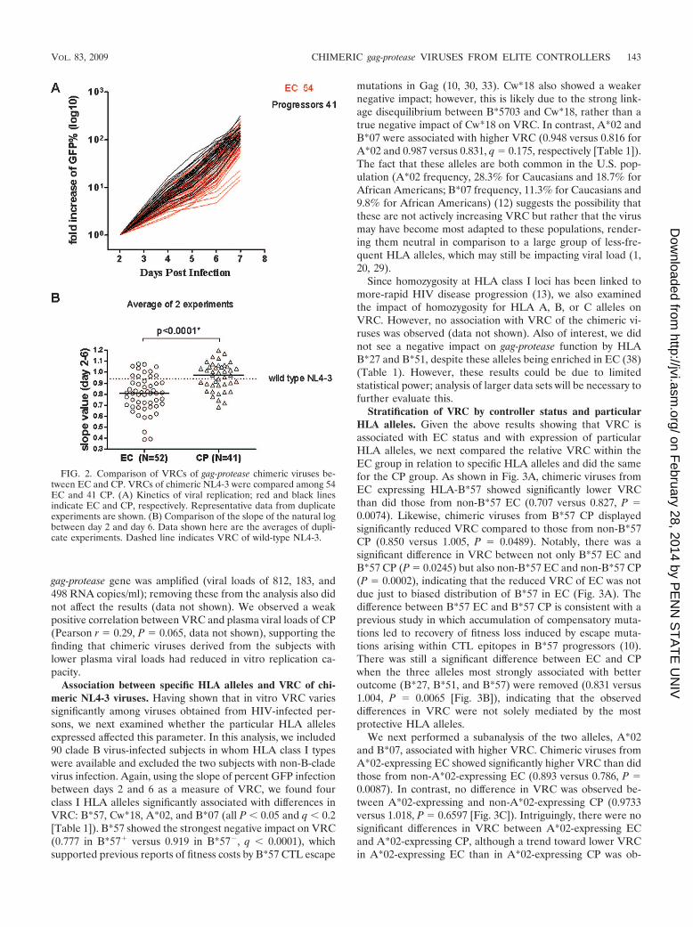
gag-protease gene was amplified (viral loads of 812, 183, and498 RNA copies/ml); removing these from the analysis also didnot affect the results (data not shown). We observed a weakpositive correlation between VRC and plasma viral loads of CP(Pearson r � 0.29, P � 0.065, data not shown), supporting thefinding that chimeric viruses derived from the subjects withlower plasma viral loads had reduced in vitro replication ca-pacity.
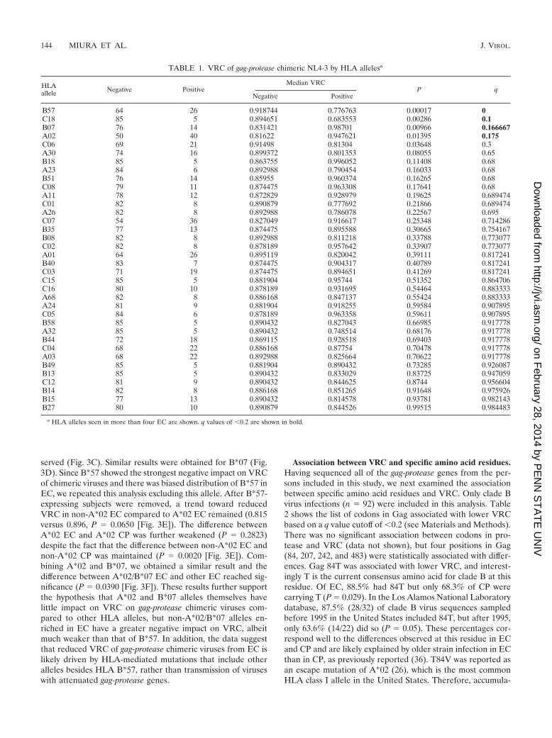
Association between specific HLA alleles and VRC of chi-meric NL4-3 viruses. Having shown that in vitro VRC variessignificantly among viruses obtained from HIV-infected per-sons, we next examined whether the particular HLA allelesexpressed affected this parameter. In this analysis, we included90 clade B virus-infected subjects in whom HLA class I typeswere available and excluded the two subjects with non-B-cladevirus infection. Again, using the slope of percent GFP infectionbetween days 2 and 6 as a measure of VRC, we found fourclass I HLA alleles significantly associated with differences inVRC: B*57, Cw*18, A*02, and B*07 (all P � 0.05 and q � 0.2[Table 1]). B*57 showed the strongest negative impact on VRC(0.777 in B*57� versus 0.919 in B*57�, q � 0.0001), whichsupported previous reports of fitness costs by B*57 CTL escape
mutations in Gag (10, 30, 33). Cw*18 also showed a weakernegative impact; however, this is likely due to the strong link-age disequilibrium between B*5703 and Cw*18, rather than atrue negative impact of Cw*18 on VRC. In contrast, A*02 andB*07 were associated with higher VRC (0.948 versus 0.816 forA*02 and 0.987 versus 0.831, q � 0.175, respectively [Table 1]).The fact that these alleles are both common in the U.S. pop-ulation (A*02 frequency, 28.3% for Caucasians and 18.7% forAfrican Americans; B*07 frequency, 11.3% for Caucasians and9.8% for African Americans) (12) suggests the possibility thatthese are not actively increasing VRC but rather that the virusmay have become most adapted to these populations, render-ing them neutral in comparison to a large group of less-fre-quent HLA alleles, which may still be impacting viral load (1,20, 29).
Since homozygosity at HLA class I loci has been linked tomore-rapid HIV disease progression (13), we also examinedthe impact of homozygosity for HLA A, B, or C alleles onVRC. However, no association with VRC of the chimeric vi-ruses was observed (data not shown). Also of interest, we didnot see a negative impact on gag-protease function by HLAB*27 and B*51, despite these alleles being enriched in EC (38)(Table 1). However, these results could be due to limitedstatistical power; analysis of larger data sets will be necessary tofurther evaluate this.
Stratification of VRC by controller status and particularHLA alleles. Given the above results showing that VRC isassociated with EC status and with expression of particularHLA alleles, we next compared the relative VRC within theEC group in relation to specific HLA alleles and did the samefor the CP group. As shown in Fig. 3A, chimeric viruses fromEC expressing HLA-B*57 showed significantly lower VRCthan did those from non-B*57 EC (0.707 versus 0.827, P �0.0074). Likewise, chimeric viruses from B*57 CP displayedsignificantly reduced VRC compared to those from non-B*57CP (0.850 versus 1.005, P � 0.0489). Notably, there was asignificant difference in VRC between not only B*57 EC andB*57 CP (P � 0.0245) but also non-B*57 EC and non-B*57 CP(P � 0.0002), indicating that the reduced VRC of EC was notdue just to biased distribution of B*57 in EC (Fig. 3A). Thedifference between B*57 EC and B*57 CP is consistent with aprevious study in which accumulation of compensatory muta-tions led to recovery of fitness loss induced by escape muta-tions arising within CTL epitopes in B*57 progressors (10).There was still a significant difference between EC and CPwhen the three alleles most strongly associated with betteroutcome (B*27, B*51, and B*57) were removed (0.831 versus1.004, P � 0.0065 [Fig. 3B]), indicating that the observeddifferences in VRC were not solely mediated by the mostprotective HLA alleles.
We next performed a subanalysis of the two alleles, A*02and B*07, associated with higher VRC. Chimeric viruses fromA*02-expressing EC showed significantly higher VRC than didthose from non-A*02-expressing EC (0.893 versus 0.786, P �0.0087). In contrast, no difference in VRC was observed be-tween A*02-expressing and non-A*02-expressing CP (0.9733versus 1.018, P � 0.6597 [Fig. 3C]). Intriguingly, there were nosignificant differences in VRC between A*02-expressing ECand A*02-expressing CP, although a trend toward lower VRCin A*02-expressing EC than in A*02-expressing CP was ob-
FIG. 2. Comparison of VRCs of gag-protease chimeric viruses be-tween EC and CP. VRCs of chimeric NL4-3 were compared among 54EC and 41 CP. (A) Kinetics of viral replication; red and black linesindicate EC and CP, respectively. Representative data from duplicateexperiments are shown. (B) Comparison of the slope of the natural logbetween day 2 and day 6. Data shown here are the averages of dupli-cate experiments. Dashed line indicates VRC of wild-type NL4-3.
VOL. 83, 2009 CHIMERIC gag-protease VIRUSES FROM ELITE CONTROLLERS 143
on February 28, 2014 by P
EN
N S
TA
TE
UN
IVhttp://jvi.asm
.org/D
ownloaded from
served (Fig. 3C). Similar results were obtained for B*07 (Fig.3D). Since B*57 showed the strongest negative impact on VRCof chimeric viruses and there was biased distribution of B*57 inEC, we repeated this analysis excluding this allele. After B*57-expressing subjects were removed, a trend toward reducedVRC in non-A*02 EC compared to A*02 EC remained (0.815versus 0.896, P � 0.0650 [Fig. 3E]). The difference betweenA*02 EC and A*02 CP was further weakened (P � 0.2823)despite the fact that the difference between non-A*02 EC andnon-A*02 CP was maintained (P � 0.0020 [Fig. 3E]). Com-bining A*02 and B*07, we obtained a similar result and thedifference between A*02/B*07 EC and other EC reached sig-nificance (P � 0.0390 [Fig. 3F]). These results further supportthe hypothesis that A*02 and B*07 alleles themselves havelittle impact on VRC on gag-protease chimeric viruses com-pared to other HLA alleles, but non-A*02/B*07 alleles en-riched in EC have a greater negative impact on VRC, albeitmuch weaker than that of B*57. In addition, the data suggestthat reduced VRC of gag-protease chimeric viruses from EC islikely driven by HLA-mediated mutations that include otheralleles besides HLA B*57, rather than transmission of viruseswith attenuated gag-protease genes.
Association between VRC and specific amino acid residues.Having sequenced all of the gag-protease genes from the per-sons included in this study, we next examined the associationbetween specific amino acid residues and VRC. Only clade Bvirus infections (n � 92) were included in this analysis. Table2 shows the list of codons in Gag associated with lower VRCbased on a q value cutoff of �0.2 (see Materials and Methods).There was no significant association between codons in pro-tease and VRC (data not shown), but four positions in Gag(84, 207, 242, and 483) were statistically associated with differ-ences. Gag 84T was associated with lower VRC, and interest-ingly T is the current consensus amino acid for clade B at thisresidue. Of EC, 88.5% had 84T but only 68.3% of CP werecarrying T (P � 0.029). In the Los Alamos National Laboratorydatabase, 87.5% (28/32) of clade B virus sequences sampledbefore 1995 in the United States included 84T, but after 1995,only 63.6% (14/22) did so (P � 0.05). These percentages cor-respond well to the differences observed at this residue in ECand CP and are likely explained by older strain infection in ECthan in CP, as previously reported (36). T84V was reported asan escape mutation of A*02 (26), which is the most commonHLA class I allele in the United States. Therefore, accumula-
TABLE 1. VRC of gag-protease chimeric NL4-3 by HLA allelesa
HLAallele Negative Positive
Median VRCP q
Negative Positive
B57 64 26 0.918744 0.776763 0.00017 0C18 85 5 0.894651 0.683553 0.00286 0.1B07 76 14 0.831421 0.98701 0.00966 0.166667A02 50 40 0.81622 0.947621 0.01395 0.175C06 69 21 0.91498 0.81304 0.03648 0.3A30 74 16 0.899372 0.801353 0.08055 0.65B18 85 5 0.863755 0.996052 0.11408 0.68A23 84 6 0.892988 0.790454 0.16033 0.68B51 76 14 0.85955 0.960374 0.16265 0.68C08 79 11 0.874475 0.963308 0.17641 0.68A11 78 12 0.872829 0.928979 0.19625 0.689474C01 82 8 0.890879 0.777692 0.21866 0.689474A26 82 8 0.892988 0.786078 0.22567 0.695C07 54 36 0.827049 0.916617 0.25348 0.714286B35 77 13 0.874475 0.895588 0.30665 0.754167B08 82 8 0.892988 0.811218 0.33788 0.773077C02 82 8 0.878189 0.957642 0.33907 0.773077A01 64 26 0.895119 0.820042 0.39111 0.817241B40 83 7 0.874475 0.904317 0.40789 0.817241C03 71 19 0.874475 0.894651 0.41269 0.817241C15 85 5 0.881904 0.95744 0.51352 0.864706C16 80 10 0.878189 0.931695 0.54464 0.883333A68 82 8 0.886168 0.847137 0.55424 0.883333A24 81 9 0.881904 0.918255 0.59584 0.907895C05 84 6 0.878189 0.963358 0.59611 0.907895B58 85 5 0.890432 0.827043 0.66985 0.917778A32 85 5 0.890432 0.748514 0.68176 0.917778B44 72 18 0.869115 0.928518 0.69403 0.917778C04 68 22 0.886168 0.87754 0.70478 0.917778A03 68 22 0.892988 0.825664 0.70622 0.917778B49 85 5 0.881904 0.890432 0.73285 0.926087B13 85 5 0.890432 0.833029 0.83725 0.947059C12 81 9 0.890432 0.844625 0.8744 0.956604B14 82 8 0.886168 0.851265 0.91648 0.975926B15 77 13 0.890432 0.814578 0.93781 0.982143B27 80 10 0.890879 0.844526 0.99515 0.984483
a HLA alleles seen in more than four EC are shown. q values of �0.2 are shown in bold.
144 MIURA ET AL. J. VIROL.
on February 28, 2014 by P
EN
N S
TA
TE
UN
IVhttp://jvi.asm
.org/D
ownloaded from
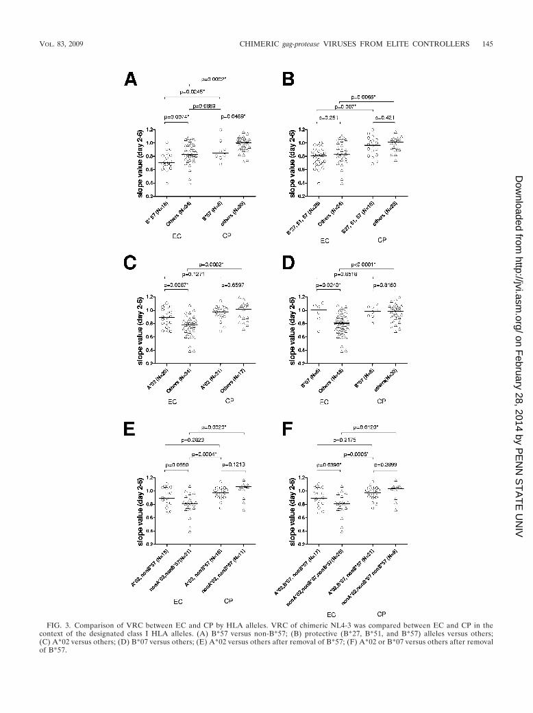
FIG. 3. Comparison of VRC between EC and CP by HLA alleles. VRC of chimeric NL4-3 was compared between EC and CP in thecontext of the designated class I HLA alleles. (A) B*57 versus non-B*57; (B) protective (B*27, B*51, and B*57) alleles versus others;(C) A*02 versus others; (D) B*07 versus others; (E) A*02 versus others after removal of B*57; (F) A*02 or B*07 versus others after removalof B*57.
VOL. 83, 2009 CHIMERIC gag-protease VIRUSES FROM ELITE CONTROLLERS 145
on February 28, 2014 by P
EN
N S
TA
TE
UN
IVhttp://jvi.asm
.org/D
ownloaded from
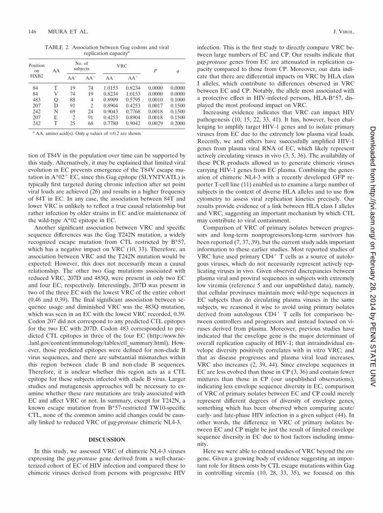
tion of T84V in the population over time can be supported bythis study. Alternatively, it may be explained that limited viralevolution in EC prevents emergence of the T84V escape mu-tation in A*02� EC, since this Gag epitope (SLYNTVATL) istypically first targeted during chronic infection after set pointviral loads are achieved (26) and results in a higher frequencyof 84T in EC. In any case, the association between 84T andlower VRC is unlikely to reflect a true causal relationship butrather infection by older strains in EC and/or maintenance ofthe wild-type A*02 epitope in EC.
Another significant association between VRC and specificsequence differences was the Gag T242N mutation, a widelyrecognized escape mutation from CTL restricted by B*57,which has a negative impact on VRC (10, 33). Therefore, anassociation between VRC and the T242N mutation would beexpected. However, this does not necessarily mean a causalrelationship. The other two Gag mutations associated withreduced VRC, 207D and 483Q, were present in only two ECand four EC, respectively. Interestingly, 207D was present intwo of the three EC with the lowest VRC of the entire cohort(0.46 and 0.39). The final significant association between se-quence usage and diminished VRC was the 483Q mutation,which was seen in an EC with the lowest VRC recorded, 0.39.Codon 207 did not correspond to any predicted CTL epitopesfor the two EC with 207D. Codon 483 corresponded to pre-dicted CTL epitopes in three of the four EC (http://www.hiv.lanl.gov/content/immunology/tables/ctl_summary.html). How-ever, those predicted epitopes were defined for non-clade Bvirus sequences, and there are substantial mismatches withinthis region between clade B and non-clade B sequences.Therefore, it is unclear whether this region acts as a CTLepitope for these subjects infected with clade B virus. Largerstudies and mutagenesis approaches will be necessary to ex-amine whether these rare mutations are truly associated withEC and affect VRC or not. In summary, except for T242N, aknown escape mutation from B*57-restricted TW10-specificCTL, none of the common amino acid changes could be caus-ally linked to reduced VRC of gag-protease chimeric NL4-3.
DISCUSSION
In this study, we assessed VRC of chimeric NL4-3 virusesexpressing the gag-protease gene derived from a well-charac-terized cohort of EC of HIV infection and compared these tochimeric viruses derived from persons with progressive HIV
infection. This is the first study to directly compare VRC be-tween large numbers of EC and CP. Our results indicate thatgag-protease genes from EC are attenuated in replication ca-pacity compared to those from CP. Moreover, our data indi-cate that there are differential impacts on VRC by HLA classI alleles, which contribute to differences observed in VRCbetween EC and CP. Notably, the allele most associated witha protective effect in HIV-infected persons, HLA-B*57, dis-played the most profound impact on VRC.
Increasing evidence indicates that VRC can impact HIVpathogenesis (10, 15, 22, 33, 41). It has, however, been chal-lenging to amplify target HIV-1 genes and to isolate primaryviruses from EC due to the extremely low plasma viral loads.Recently, we and others have successfully amplified HIV-1genes from plasma viral RNA of EC, which likely representactively circulating viruses in vivo (3, 5, 36). The availability ofthese PCR products allowed us to generate chimeric virusescarrying HIV-1 genes from EC plasma. Combining the gener-ation of chimeric NL4-3 with a recently developed GFP re-porter T-cell line (11) enabled us to examine a large number ofsubjects in the context of diverse HLA alleles and to use flowcytometry to assess viral replication kinetics precisely. Ourresults provide evidence of a link between HLA class I allelesand VRC, suggesting an important mechanism by which CTLmay contribute to viral containment.
Comparison of VRC of primary isolates between progres-sors and long-term nonprogressors/long-term survivors hasbeen reported (7, 37, 39), but the current study adds importantinformation to these earlier studies. Most reported studies ofVRC have used primary CD4� T cells as a source of autolo-gous viruses, which do not necessarily represent actively rep-licating viruses in vivo. Given observed discrepancies betweenplasma viral and proviral sequences in subjects with extremelylow viremia (reference 5 and our unpublished data), namely,that cellular proviruses maintain more wild-type sequences inEC subjects than do circulating plasma viruses in the samesubjects, we reasoned it wise to avoid using primary isolatesderived from autologous CD4� T cells for comparison be-tween controllers and progressors and instead focused on vi-ruses derived from plasma. Moreover, previous studies haveindicated that the envelope gene is the major determinant ofoverall replication capacity of HIV-1; that intraindividual en-velope diversity positively correlates with in vitro VRC; andthat as disease progresses and plasma viral load increases,VRC also increases (2, 39, 44). Since envelope sequences inEC are less evolved than those in CP (3, 36) and contain fewermixtures than those in CP (our unpublished observations),indicating less envelope sequence diversity in EC, comparisonof VRC of primary isolates between EC and CP could merelyrepresent different degrees of diversity of envelope genes,something which has been observed when comparing acute/early- and late-phase HIV infection in a given subject (44). Inother words, the difference in VRC of primary isolates be-tween EC and CP might be just the result of limited envelopesequence diversity in EC due to host factors including immu-nity.
Here we were able to extend studies of VRC beyond the envgene. Given a growing body of evidence suggesting an impor-tant role for fitness costs by CTL escape mutations within Gagin controlling viremia (10, 28, 33, 35), we focused on this
TABLE 2. Association between Gag codons and viralreplication capacitya
Positionon
HXB2AA
No. ofsubjects VRC
P q
AA� AA� AA� AA�
84 T 19 74 1.0153 0.8234 0.0000 0.000084 V 74 19 0.8234 1.0153 0.0000 0.0000483 Q 88 4 0.8909 0.5795 0.0010 0.1000207 D 91 2 0.8904 0.4253 0.0017 0.1500242 N 69 24 0.9043 0.7768 0.0018 0.1500207 E 2 91 0.4253 0.8904 0.0018 0.1500242 T 25 68 0.7780 0.9042 0.0029 0.2000
a AA, amino acid(s). Only q values of �0.2 are shown.
146 MIURA ET AL. J. VIROL.
on February 28, 2014 by P
EN
N S
TA
TE
UN
IVhttp://jvi.asm
.org/D
ownloaded from
protein. Since it is not known whether the diversity of the Gagprotein sequence correlates with its function, there remains thepossibility that the observed differences in VRC between ECand CP might reflect distinct interindividual diversity of theGag protein. We also note that there is still considerable over-lap in VRC between EC and CP (Fig. 2B). Therefore, theobserved reduction of Gag-Protease function in EC is not thesole factor to explain elite control but rather one of the factorscontributing to suppression of viremia.
The novelty of the present study is not only in scale or use ofEC samples but also in showing a differential impact on viralreplication by different HLA class I alleles. Although onlyB*57 showed a significant negative impact on VRC, indirectevidence that common HLA alleles like A*02 and/or B*07 hadlittle impact on VRC suggests that other HLA class I alleleslikely had some negative impact on VRC, albeit to a lesserextent than B*57. This finding and additional observations ofsignificantly reduced VRC in non-A*02/B*07/B*57 EC com-pared to non-A*02/B*07/B*57 CP indicate that reduced VRCin non-A*02/B*07/B*57 EC is likely mediated by HLA class Ialleles enriched in EC. These findings further support recentstudies suggesting the importance of CTL targeting Gagepitopes and inducing escape mutations with fitness costs (22,28, 35).
Although this study implies a contribution of altered VRC toviral control in persons with HLA-B*57, it does not link otherwell-known protective HLA alleles like B*27 and B*51 toaltered VRC. Since there are no known B*51 CTL epitopeswithin the Gag-protease (19), it is perhaps not surprising thatwe did not see any impact by this allele. On the other hand,B*27 is known to contain an immunodominant CTL epitopewithin Gag protein, and escape from this CTL response isfrequently observed (18, 23, 27) and has been demonstrated tocompromise VRC (41, 42). However, these mutations typicallydo not arise until late-stage infection (24, 41), and it has beendemonstrated that the dominant CTL escape form R264K Gagrequires an upstream mutation (S173A) which fully compen-sates the fitness cost and is required prior to complete escape(41). Thus, the lack of an effect of B*27 on gag-protease chi-meric virus replication capacity in the present study does notcontradict previous findings. These results indicate more het-erogeneous mechanisms of elite control even among HIV-specific CTL responses.
Another important finding of this study is the observationthat B*57 EC showed significantly reduced VRC compared toB*57 CP. This might be explained by an additional effect ofother protective HLA alleles in B*57 EC that may not bepresent in B*57 CP. However, as reported before, accumula-tion of compensatory mutations that recover reduced VRC byB*57 CTL escape mutations also is a feasible explanation (10,15). However, since we observed rare escape variants in manyof the B*57 EC (unpublished data), the mechanism of atten-uation of viruses in B*57 EC seems more complex. Additionalstudies are required to reveal the role of such rare mutations inVRC in B*57 EC. Likewise, the difference between non-A*02/B*07/B*57 EC and non-A*02/B*07/B*57 CP might be due toaccumulation of compensatory mutations that recover fitnesscosts by escape mutations, or unique mutations in EC, ratherthan different HLA allele distribution between EC and CP.Larger data sets may allow this question to be answered.
It has been suggested that EC might acquire viruses withreduced replication capacity at the time of transmission, ratherthan having these arise in vivo. However, this would not ex-plain the HLA-associated differences in VRC observed in thepresent study. Therefore, we conclude that the diminishedVRC of EC viruses is achieved after transmission rather thantransmitted from donors. However, very recently, it was sug-gested that the subjects considered to have obtained virusesfrom donors with protective HLA alleles could maintain lowerlevels of viremia, at least temporarily, after infection (22). It isquite possible that controllers who have strong immune re-sponses obtain further benefit when an attenuated virus istransmitted, and this may further contribute to elite control. Inorder to clarify this point, longitudinal studies of large numbersof persons in acute infection with diverse set point viral loadswill be necessary.
Although gag-protease was the focus of the current study,there remains the possibility that there is dysfunction of otherHIV-1 genes that may be associated with elite control. Enrich-ment in EC of B*51, which contains no Gag CTL epitopes, isa potential example of an allele that modifies VRC via im-mune-selected mutations in another viral gene. Evaluation offunction of other HIV-1 genes is warranted to further under-stand the mechanisms of elite control.
In conclusion, we observed significantly reduced VRC ofchimeric NL4-3 viruses carrying gag-protease genes from HIVEC and found this to be associated with particular HLA classI allele expression. HLA-B*57 displayed the most profoundeffect on VRC, whereas the more common HLA alleles A*02and B*07 had the least impact. Non-A*02/B*07 HLA allelesenriched in EC also appear to be able to attenuate Gag-pro-tease function. This differential impact by HLA alleles on VRCof chimeric viruses also indicates that attenuation of gag-pro-tease genes is achieved following transmission. It will be im-portant to generate longitudinal data on acute infection andexpand these data sets to further define the role of fitnessdefects in disease outcome and the potential contribution ofclass I-restricted selection pressure on other coding regions toviral fitness, which has potential important implications forvaccine design.
ACKNOWLEDGMENTS
We gratefully acknowledge the efforts of the clinical staff of TheInternational HIV Controllers Study: Erik Berg, Emily Cutrell,Priscilla Padilla, Almas Rathod, Rachel Rosenberg, Sue Bazner, ChrisBirch, and Kristin Moss. Additionally, we recognize and thank thelaboratory staff: Brett Baker, Alissa Rothchild, Ildiko Toth, AishaDarrah, Brooke LaTour, and Agnes Sarkozi. Finally, we acknowledgethe hundreds of providers and researchers from around the world whohave contributed samples to The International HIV Controllers Study.Success in this research depends on the efforts and support of health-care professionals and researchers who refer subjects from around theworld. A complete list of contributors and collaborators can be foundat http://www.hivcontrollers.org/index.php?q�contributor/alphalist/A.We thank Zixin Hu and Daniel Kuritzkes (Brigham and Women’sHospital) for advice regarding construction of the gag-protease-deletedNL4-3 vector.
This work was supported by grants AI028568 and AI030914 from theNIAID/NIH, the Howard Hughes Medical Institute, the Harvard Uni-versity Center for AIDS Research (CFAR), and a gift from the Markand Lisa Schwartz Foundation.
The ideas and opinions expressed in the manuscript are solely theresponsibility of the authors and are not necessarily shared by the NIH
VOL. 83, 2009 CHIMERIC gag-protease VIRUSES FROM ELITE CONTROLLERS 147
on February 28, 2014 by P
EN
N S
TA
TE
UN
IVhttp://jvi.asm
.org/D
ownloaded from
or other funding sources, Massachusetts General Hospital, or its affil-iates. The authors declare no conflicts of interest related to this study.
REFERENCES
1. Altfeld, M., T. M. Allen, E. T. Kalife, N. Frahm, M. M. Addo, B. R. Mothe,A. Rathod, L. L. Reyor, J. Harlow, X. G. Yu, B. Perkins, L. K. Robinson, J.Sidney, G. Alter, M. Lichterfeld, A. Sette, E. S. Rosenberg, P. J. Goulder, C.Brander, and B. D. Walker. 2005. The majority of currently circulatinghuman immunodeficiency virus type 1 clade B viruses fail to prime cytotoxicT-lymphocyte responses against an otherwise immunodominant HLA-A2-restricted epitope: implications for vaccine design. J. Virol. 79:5000–5005.
2. Arien, K. K., G. Vanham, and E. J. Arts. 2007. Is HIV-1 evolving to a lessvirulent form in humans? Nat. Rev. Microbiol. 5:141–151.
3. Bailey, J. R., K. G. Lassen, H. C. Yang, T. C. Quinn, S. C. Ray, J. N.Blankson, and R. F. Siliciano. 2006. Neutralizing antibodies do not mediatesuppression of human immunodeficiency virus type 1 in elite suppressors orselection of plasma virus variants in patients on highly active antiretroviraltherapy. J. Virol. 80:4758–4770.
4. Bailey, J. R., K. O’Connell, H. C. Yang, Y. Han, J. Xu, B. Jilek, T. M.Williams, S. C. Ray, R. F. Siliciano, and J. N. Blankson. 2008. Transmissionof human immunodeficiency virus type 1 from a patient who developedAIDS to an elite suppressor. J. Virol. 82:7395–7410.
5. Bailey, J. R., T. M. Williams, R. F. Siliciano, and J. N. Blankson. 2006.Maintenance of viral suppression in HIV-1-infected HLA-B*57� elite sup-pressors despite CTL escape mutations. J. Exp. Med. 203:1357–1369.
6. Ball, S. C., A. Abraha, K. R. Collins, A. J. Marozsan, H. Baird, M. E.Quinones-Mateu, A. Penn-Nicholson, M. Murray, N. Richard, M. Lobritz,P. A. Zimmerman, T. Kawamura, A. Blauvelt, and E. J. Arts. 2003. Com-paring the ex vivo fitness of CCR5-tropic human immunodeficiency virustype 1 isolates of subtypes B and C. J. Virol. 77:1021–1038.
7. Blaak, H., M. Brouwer, L. J. Ran, F. de Wolf, and H. Schuitemaker. 1998. Invitro replication kinetics of human immunodeficiency virus type 1 (HIV-1)variants in relation to virus load in long-term survivors of HIV-1 infection.J. Infect. Dis. 177:600–610.
8. Blankson, J. N., J. R. Bailey, S. Thayil, H. C. Yang, K. Lassen, J. Lai, S. K.Gandhi, J. D. Siliciano, T. M. Williams, and R. F. Siliciano. 2007. Isolationand characterization of replication-competent human immunodeficiency vi-rus type 1 from a subset of elite suppressors. J. Virol. 81:2508–2518.
9. Borghans, J. A., A. Molgaard, R. J. de Boer, and C. Kesmir. 2007. HLAalleles associated with slow progression to AIDS truly prefer to presentHIV-1 p24. PLoS ONE 2:e920.
10. Brockman, M. A., A. Schneidewind, M. Lahaie, A. Schmidt, T. Miura, I.Desouza, F. Ryvkin, C. A. Derdeyn, S. Allen, E. Hunter, J. Mulenga, P. A.Goepfert, B. D. Walker, and T. M. Allen. 2007. Escape and compensationfrom early HLA-B57-mediated cytotoxic T-lymphocyte pressure on humanimmunodeficiency virus type 1 Gag alter capsid interactions with cyclophilinA. J. Virol. 81:12608–12618.
11. Brockman, M. A., G. O. Tanzi, B. D. Walker, and T. M. Allen. 2006. Use ofa novel GFP reporter cell line to examine replication capacity of CXCR4-and CCR5-tropic HIV-1 by flow cytometry. J. Virol. Methods 131:134–142.
12. Cao, K., J. Hollenbach, X. Shi, W. Shi, M. Chopek, and M. A. Fernandez-Vina. 2001. Analysis of the frequencies of HLA-A, B, and C alleles andhaplotypes in the five major ethnic groups of the United States reveals highlevels of diversity in these loci and contrasting distribution patterns in thesepopulations. Hum. Immunol. 62:1009–1030.
13. Carrington, M., G. W. Nelson, M. P. Martin, T. Kissner, D. Vlahov, J. J.Goedert, R. Kaslow, S. Buchbinder, K. Hoots, and S. J. O’Brien. 1999. HLAand HIV-1: heterozygote advantage and B*35-Cw*04 disadvantage. Science283:1748–1752.
14. Chopera, D. R., Z. Woodman, K. Mlisana, M. Mlotshwa, D. P. Martin, C.Seoighe, F. Treurnicht, D. A. de Rosa, W. Hide, S. A. Karim, C. M. Gray, andC. Williamson. 2008. Transmission of HIV-1 CTL escape variants providesHLA-mismatched recipients with a survival advantage. PLoS Pathog.4:e1000033.
15. Crawford, H., J. G. Prado, A. Leslie, S. Hue, I. Honeyborne, S. Reddy, M. vander Stok, Z. Mncube, C. Brander, C. Rousseau, J. I. Mullins, R. Kaslow, P.Goepfert, S. Allen, E. Hunter, J. Mulenga, P. Kiepiela, B. D. Walker, andP. J. Goulder. 2007. Compensatory mutation partially restores fitness anddelays reversion of escape mutation within the immunodominant HLA-B*5703-restricted Gag epitope in chronic human immunodeficiency virustype 1 infection. J. Virol. 81:8346–8351.
16. Deeks, S. G., and B. D. Walker. 2007. Human immunodeficiency virus con-trollers: mechanisms of durable virus control in the absence of antiretroviraltherapy. Immunity 27:406–416.
17. de la Carriere, L. C., S. Paulous, F. Clavel, and F. Mammano. 1999. Effectsof human immunodeficiency virus type 1 resistance to protease inhibitors onreverse transcriptase processing, activity, and drug sensitivity. J. Virol. 73:3455–3459.
18. Ferrari, G., W. Neal, A. Jones, N. Olender, J. Ottinger, R. Ha, M. J. Mc-Elrath, P. Goepfert, and K. J. Weinhold. 2001. CD8 CTL responses invaccines: emerging patterns of HLA restriction and epitope recognition.Immunol. Lett. 79:37–45.
19. Frahm, F., C. Linde, and C. Brander. 2007. Identification of HIV-derived,HLA class I restricted CTL epitopes: insights into TCR repertoire, CTLescape and viral fitness, p. 3–28. In B. T. Korber et al. (ed.), HIV molecularimmunology 2006/2007. Theoretical Biology and Biophysics Group, LosAlamos National Laboratory, Los Alamos, NM.
20. Furutsuki, T., N. Hosoya, A. Kawana-Tachikawa, M. Tomizawa, T.Odawara, M. Goto, Y. Kitamura, T. Nakamura, A. D. Kelleher, D. A. Coo-per, and A. Iwamoto. 2004. Frequent transmission of cytotoxic-T-lymphocyteescape mutants of human immunodeficiency virus type 1 in the highly HLA-A24-positive Japanese population. J. Virol. 78:8437–8445.
21. Geldmacher, C., J. R. Currier, E. Herrmann, A. Haule, E. Kuta, F.McCutchan, L. Njovu, S. Geis, O. Hoffmann, L. Maboko, C. Williamson, D.Birx, A. Meyerhans, J. Cox, and M. Hoelscher. 2007. CD8 T-cell recognitionof multiple epitopes within specific Gag regions is associated with mainte-nance of a low steady-state viremia in human immunodeficiency virus type1-seropositive patients. J. Virol. 81:2440–2448.
22. Goepfert, P. A., W. Lumm, P. Farmer, P. Matthews, A. Prendergast, J. M.Carlson, C. A. Derdeyn, J. Tang, R. A. Kaslow, A. Bansal, K. Yusim, D.Heckerman, J. Mulenga, S. Allen, P. J. Goulder, and E. Hunter. 2008.Transmission of HIV-1 Gag immune escape mutations is associated withreduced viral load in linked recipients. J. Exp. Med. 205:1009–1017.
23. Goulder, P. J., C. Brander, Y. Tang, C. Tremblay, R. A. Colbert, M. M. Addo,E. S. Rosenberg, T. Nguyen, R. Allen, A. Trocha, M. Altfeld, S. He, M. Bunce,R. Funkhouser, S. I. Pelton, S. K. Burchett, K. McIntosh, B. T. Korber, andB. D. Walker. 2001. Evolution and transmission of stable CTL escape mu-tations in HIV infection. Nature 412:334–338.
24. Goulder, P. J., R. E. Phillips, R. A. Colbert, S. McAdam, G. Ogg, M. A.Nowak, P. Giangrande, G. Luzzi, B. Morgan, A. Edwards, A. J. McMichael,and S. Rowland-Jones. 1997. Late escape from an immunodominant cyto-toxic T-lymphocyte response associated with progression to AIDS. Nat. Med.3:212–217.
25. Harrigan, P. R., S. Bloor, and B. A. Larder. 1998. Relative replicative fitnessof zidovudine-resistant human immunodeficiency virus type 1 isolates invitro. J. Virol. 72:3773–3778.
26. Iversen, A. K., G. Stewart-Jones, G. H. Learn, N. Christie, C. Sylvester-Hviid, A. E. Armitage, R. Kaul, T. Beattie, J. K. Lee, Y. Li, P. Chotiyarnwong,T. Dong, X. Xu, M. A. Luscher, K. MacDonald, H. Ullum, B. Klarlund-Pedersen, P. Skinhoj, L. Fugger, S. Buus, J. I. Mullins, E. Y. Jones, P. A. vander Merwe, and A. J. McMichael. 2006. Conflicting selective forces affect Tcell receptor contacts in an immunodominant human immunodeficiencyvirus epitope. Nat. Immunol. 7:179–189.
27. Kelleher, A. D., C. Long, E. C. Holmes, R. L. Allen, J. Wilson, C. Conlon, C.Workman, S. Shaunak, K. Olson, P. Goulder, C. Brander, G. Ogg, J. S.Sullivan, W. Dyer, I. Jones, A. J. McMichael, S. Rowland-Jones, and R. E.Phillips. 2001. Clustered mutations in HIV-1 gag are consistently requiredfor escape from HLA-B27-restricted cytotoxic T lymphocyte responses. J.Exp. Med. 193:375–386.
28. Kiepiela, P., K. Ngumbela, C. Thobakgale, D. Ramduth, I. Honeyborne, E.Moodley, S. Reddy, C. de Pierres, Z. Mncube, N. Mkhwanazi, K. Bishop, M.van der Stok, K. Nair, N. Khan, H. Crawford, R. Payne, A. Leslie, J. Prado,A. Prendergast, J. Frater, N. McCarthy, C. Brander, G. H. Learn, D. Nickle,C. Rousseau, H. Coovadia, J. I. Mullins, D. Heckerman, B. D. Walker, andP. Goulder. 2007. CD8� T-cell responses to different HIV proteins havediscordant associations with viral load. Nat. Med. 13:46–53.
29. Leslie, A., D. Kavanagh, I. Honeyborne, K. Pfafferott, C. Edwards, T. Pillay,L. Hilton, C. Thobakgale, D. Ramduth, R. Draenert, S. Le Gall, G. Luzzi, A.Edwards, C. Brander, A. K. Sewell, S. Moore, J. Mullins, C. Moore, S.Mallal, N. Bhardwaj, K. Yusim, R. Phillips, P. Klenerman, B. Korber, P.Kiepiela, B. Walker, and P. Goulder. 2005. Transmission and accumulationof CTL escape variants drive negative associations between HIV polymor-phisms and HLA. J. Exp. Med. 201:891–902.
30. Leslie, A. J., K. J. Pfafferott, P. Chetty, R. Draenert, M. M. Addo, M. Feeney,Y. Tang, E. C. Holmes, T. Allen, J. G. Prado, M. Altfeld, C. Brander, C.Dixon, D. Ramduth, P. Jeena, S. A. Thomas, A. St. John, T. A. Roach, B.Kupfer, G. Luzzi, A. Edwards, G. Taylor, H. Lyall, G. Tudor-Williams, V.Novelli, J. Martinez-Picado, P. Kiepiela, B. D. Walker, and P. J. Goulder.2004. HIV evolution: CTL escape mutation and reversion after transmission.Nat. Med. 10:282–289.
31. Lucas, G. M. 2005. Antiretroviral adherence, drug resistance, viral fitnessand HIV disease progression: a tangled web is woven. J. Antimicrob. Che-mother. 55:413–416.
32. Marozsan, A. J., D. M. Moore, M. A. Lobritz, E. Fraundorf, A. Abraha, J. D.Reeves, and E. J. Arts. 2005. Differences in the fitness of two diverse wild-type human immunodeficiency virus type 1 isolates are related to the effi-ciency of cell binding and entry. J. Virol. 79:7121–7134.
33. Martinez-Picado, J., J. G. Prado, E. E. Fry, K. Pfafferott, A. Leslie, S. Chetty,C. Thobakgale, I. Honeyborne, H. Crawford, P. Matthews, T. Pillay, C.Rousseau, J. I. Mullins, C. Brander, B. D. Walker, D. I. Stuart, P. Kiepiela,and P. Goulder. 2006. Fitness cost of escape mutations in p24 Gag inassociation with control of human immunodeficiency virus type 1. J. Virol.80:3617–3623.
34. Martinez-Picado, J., A. V. Savara, L. Sutton, and R. T. D’Aquila. 1999.
148 MIURA ET AL. J. VIROL.
on February 28, 2014 by P
EN
N S
TA
TE
UN
IVhttp://jvi.asm
.org/D
ownloaded from
Replicative fitness of protease inhibitor-resistant mutants of human immu-nodeficiency virus type 1. J. Virol. 73:3744–3752.
35. Matthews, P. C., A. Prendergast, A. Leslie, H. Crawford, R. Payne, C.Rousseau, M. Rolland, I. Honeyborne, J. Carlson, C. Kadie, C. Brander, K.Bishop, N. Mlotshwa, J. I. Mullins, H. Coovadia, T. Ndung’u, B. D. Walker,D. Heckerman, and P. J. Goulder. 2008. Central role of reverting mutationsin HLA associations with human immunodeficiency virus viral set point.J. Virol. 82:8548–8559.
36. Miura, T., M. A. Brockman, C. J. Brumme, Z. L. Brumme, J. M. Carlson, F.Pereyra, A. Trocha, M. M. Addo, B. L. Block, A. C. Rothchild, B. M. Baker,T. Flynn, A. Schneidewind, B. Li, Y. E. Wang, D. Heckerman, T. M. Allen,and B. D. Walker. 2008. Genetic characterization of human immunodefi-ciency virus type 1 in elite controllers: lack of gross genetic defects orcommon amino acid changes. J. Virol. 82:8422–8430.
37. Navis, M., I. Schellens, D. van Baarle, J. Borghans, P. van Swieten, F.Miedema, N. Kootstra, and H. Schuitemaker. 2007. Viral replication capac-ity as a correlate of HLA B57/B5801-associated nonprogressive HIV-1 in-fection. J. Immunol. 179:3133–3143.
38. Pereyra, F., M. M. Addo, D. E. Kaufmann, Y. Liu, T. Miura, A. Rathod, B.Baker, A. Trocha, R. Rosenberg, E. Mackey, P. Ueda, Z. Lu, D. Cohen, T.Wrin, C. J. Petropoulos, E. S. Rosenberg, and B. D. Walker. 2008. Geneticand immunologic heterogeneity among persons who control HIV infectionin the absence of therapy. J. Infect. Dis. 197:563–571.
39. Quinones-Mateu, M. E., S. C. Ball, A. J. Marozsan, V. S. Torre, J. L.Albright, G. Vanham, G. van Der Groen, R. L. Colebunders, and E. J. Arts.2000. A dual infection/competition assay shows a correlation between ex vivohuman immunodeficiency virus type 1 fitness and disease progression. J. Vi-rol. 74:9222–9233.
40. Rangel, H. R., J. Weber, B. Chakraborty, A. Gutierrez, M. L. Marotta, M.Mirza, P. Kiser, M. A. Martinez, J. A. Este, and M. E. Quinones-Mateu.
2003. Role of the human immunodeficiency virus type 1 envelope gene inviral fitness. J. Virol. 77:9069–9073.
41. Schneidewind, A., M. A. Brockman, R. Yang, R. I. Adam, B. Li, S. Le Gall,C. R. Rinaldo, S. L. Craggs, R. L. Allgaier, K. A. Power, T. Kuntzen, C. S.Tung, M. X. LaBute, S. M. Mueller, T. Harrer, A. J. McMichael, P. J.Goulder, C. Aiken, C. Brander, A. D. Kelleher, and T. M. Allen. 2007. Escapefrom the dominant HLA-B27-restricted cytotoxic T-lymphocyte response inGag is associated with a dramatic reduction in human immunodeficiencyvirus type 1 replication. J. Virol. 81:12382–12393.
42. Schneidewind, A., M. A. Brockman, J. Sidney, Y. E. Wang, H. Chen, T. J.Suscovich, B. Li, R. I. Adam, R. L. Allgaier, B. R. Mothe, T. Kuntzen, C.Oniangue-Ndza, A. Trocha, X. G. Yu, C. Brander, A. Sette, B. D. Walker, andT. M. Allen. 2008. Structural and functional constraints limit options forcytotoxic T-lymphocyte escape in the immunodominant HLA-B27-restrictedepitope in human immunodeficiency virus type 1 capsid. J. Virol. 82:5594–5605.
43. Storey, J. D., and R. Tibshirani. 2003. Statistical significance for genomewidestudies. Proc. Natl. Acad. Sci. USA 100:9440–9445.
44. Troyer, R. M., K. R. Collins, A. Abraha, E. Fraundorf, D. M. Moore, R. W.Krizan, Z. Toossi, R. L. Colebunders, M. A. Jensen, J. I. Mullins, G. Van-ham, and E. J. Arts. 2005. Changes in human immunodeficiency virus type 1fitness and genetic diversity during disease progression. J. Virol. 79:9006–9018.
45. Zuniga, R., A. Lucchetti, P. Galvan, S. Sanchez, C. Sanchez, A. Hernandez,H. Sanchez, N. Frahm, C. H. Linde, H. S. Hewitt, W. Hildebrand, M. Altfeld,T. M. Allen, B. D. Walker, B. T. Korber, T. Leitner, J. Sanchez, and C.Brander. 2006. Relative dominance of Gag p24-specific cytotoxic T lympho-cytes is associated with human immunodeficiency virus control. J. Virol.80:3122–3125.
VOL. 83, 2009 CHIMERIC gag-protease VIRUSES FROM ELITE CONTROLLERS 149
on February 28, 2014 by P
EN
N S
TA
TE
UN
IVhttp://jvi.asm
.org/D
ownloaded from