vet pathol march 2010 vol47 245 253
TRANSCRIPT
http://vet.sagepub.com/
Veterinary Pathology Online
http://vet.sagepub.com/content/early/2010/01/28/0300985809358036The online version of this article can be found at:
DOI: 10.1177/0300985809358036
published online 31 December 2009Vet PatholKennedy, F. Forster, B. Iulini, E. Bozzetta and C. Casalone
G. Di Guardo, U. Proietto, C.E. Di Francesco, F. Marsilio, A. Zaccaroni, D. Scaravelli, W. Mignone, F. Garibaldi, S.Coast of Italy
Cerebral Toxoplasmosis in Striped Dolphins (Stenella coeruleoalba) Stranded Along the Ligurian Sea
Published by:
http://www.sagepublications.com
On behalf of:
American College of Veterinary Pathologists
can be found at:Veterinary Pathology OnlineAdditional services and information for
http://vet.sagepub.com/cgi/alertsEmail Alerts:
http://vet.sagepub.com/subscriptionsSubscriptions:
http://www.sagepub.com/journalsReprints.navReprints:
http://www.sagepub.com/journalsPermissions.navPermissions:
by guest on March 5, 2010vet.sagepub.comDownloaded from
Wildlife
Cerebral Toxoplasmosis in Striped Dolphins(Stenella coeruleoalba) Stranded Along theLigurian Sea Coast of Italy
G. Di Guardo1, U. Proietto1, C. E. Di Francesco1, F. Marsilio1,
A. Zaccaroni2, D. Scaravelli2, W. Mignone3, F. Garibaldi5,
S. Kennedy6, F. Forster6, B. Iulini4, E. Bozzetta4, and C. Casalone4
Abstract
This article reports the results of necropsy, parasitologic, microbiologic, histopathologic, immunohistochemical, indirect
immunofluorescence, biomolecular, and serologic investigations on 8 striped dolphins (Stenella coeruleoalba) found stranded
from August to December 2007 on the Ligurian Sea coast of Italy. Severe, nonsuppurative meningoencephalitis was found in4 animals, as characterized by prominent perivascular mononuclear cell cuffing and macrophage accumulations in neuropil.
These lesions were associated with mild lymphocytic–plasmacytic infiltration of choroid plexuses in 1 dolphin. Toxoplasma
gondii cysts and zoites, confirmed by immunohistochemical labeling, were scattered throughout the brain parenchyma of 2 of
the 4 dolphins. No viral inclusions were seen in the brain of any animal. Other findings included severe bronchointerstitial
pneumonia and pulmonary atelectasis, consolidation, and emphysema. Parasites were identified in a variety of organs, including
lung (Halocerchus lagenorhynchi). Microbiologic and serologic examinations for Brucella spp were negative on all 8 dolphins. The
4 animals with meningoencephalitis had serum antibodies against T gondii (titers ranging from 1:80 to 1:320) but not against
morbillivirus. In contrast, the other 4 dolphins were seropositive for morbillivirus (with titers ranging from 1:10 to 1:40) butseronegative for T gondii. No morbillivirus antigen or nucleic acid was detected in the tissues of any dolphin. It is concluded
that the severe lung and brain lesions were the cause of death and that T gondii was the likely etiologic agent of the cerebral
lesions. Morbillivirus infection was not considered to have contributed to death of these animals.
Keywords
striped dolphins, pathology, morbillivirus, Toxoplasma gondii, Brucella spp, Italy
Toxoplasma gondii, a protozoan agent infecting a range of mam-
malian species worldwide, has been implicated as a cause of
abortion and lethal systemic disease in several sea mammal spe-
cies.11Although the potential of this protozoan to affect cetacean
populations has not been thoroughly investigated thus far, a
number of reports clearly suggest that T gondii infection is of
potential concern to cetacean health and conservation.9,11,32
T gondii is commonly believed to be an opportunistic patho-
gen for aquatic mammals,24,26 as was the case in striped dolphins
(Stenella coeruleoalba) during the dolphin morbillivirus (DMV)
epidemic in the Mediterranean Sea from 1990 to 1992.7,21
Between late 2006 and early 2007 another morbillivirus
epidemic was reported in pilot whales (Globicephala melas)
in the Mediterranean Sea near Gibraltar12 and, in the following
months, in pilot whales and striped dolphins along the Spanish
coast.28 Apart from its less dramatic scale, this more recent
event shared many similarities with the Mediterranean striped
dolphin die-off in this area from 1990 to 1992.6,21 Nucleotide
sequence analysis indicated that the DMV strain causing the
recent mortality episodes in pilot whales and striped dolphins
is closely related to that involved in the 1990–1992 die-off.12,28
1Department of Comparative Biomedical Sciences, Faculty of Veterinary
Medicine, University of Teramo, Teramo, Italy2Department of Veterinary Public Health and Animal Pathology, Faculty of
Veterinary Medicine, University of Bologna, Ozzano Emilia, Bologna, Italy3 Istituto Zooprofilattico Sperimentale del Piemonte, Liguria e Valle d’Aosta,
Imperia, Italy4 Istituto Zooprofilattico Sperimentale del Piemonte, Liguria e Valle d’Aosta,
Torino, Italy5Dip.Te.Ris., University of Genova, Genova, Italy6Agri-Food and Biosciences Institute for Northern Ireland, Stormont, Belfast,
Northern Ireland
Corresponding Author:
Giovanni Di Guardo, DVM, Dipl ECVP, Department of Comparative
Biomedical Sciences, Faculty of Veterinary Medicine, University of Teramo,
Piazza Aldo Moro, 45-64100, Teramo, Italy
Email: [email protected]
Veterinary Pathology
000(00) 1-9
ª The Author(s) 2010
Reprints and permission:
sagepub.com/journalsPermissions.nav
DOI: 10.1177/0300985809358036
http://vet.sagepub.com
1
Veterinary Pathology OnlineFirst, published on January 29, 2010 as doi:10.1177/0300985809358036
by guest on March 5, 2010vet.sagepub.comDownloaded from
As in the earlier event,4,5,21 unusually high striped dolphin
mortality was recorded along the coast of Italy during
this recent morbillivirus epidemic, including the Ligurian
coast of northwestern Italy from August to December 2007
(F. Garibaldi and W. Mignone, personal communication). The
aim of this study was to investigate possible causes of death of
these animals, with emphasis on four cases of T gondii infection.
Materials and Methods
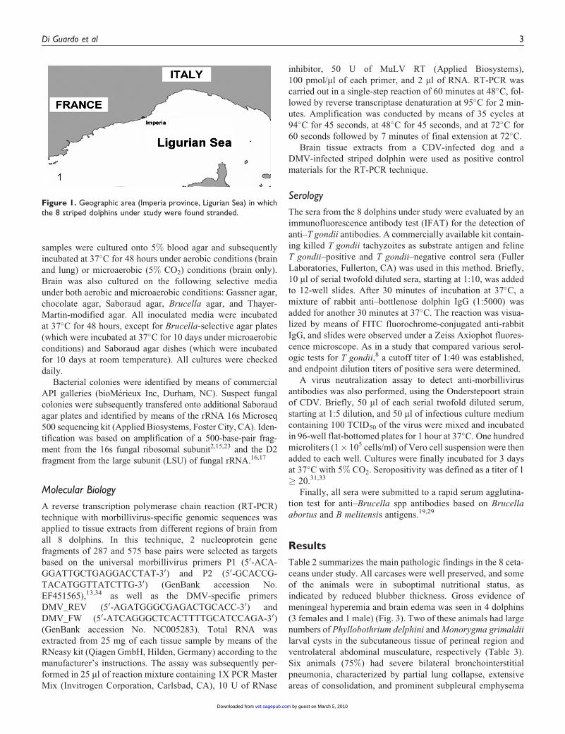
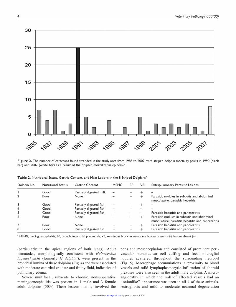
The studywas carried out on 8 striped dolphins (Table 1) that had
been found dead along the Ligurian Sea coast of Italy (Imperia
province) from August to December 2007 (Fig. 1). The annual
mean number of cetaceans found stranded during the previous
22 years (1985–2006) in this region is 4, with S coeruleoalba
being the most commonly stranded species. The 2007 stranding
figures show an increase over previous years (Fig. 2).
At necropsy, tissue samples were collected from a range of
organs (lung, heart, stomach, intestine, liver, pancreas, kidney,
adrenal gland, testis, ovary, uterus, spleen, mesenteric, tracheo-
bronchial and pulmonary lymph nodes, skeletal muscle, and
skin), along with the entire brain from each animal. Tissues
were fixed in 10% neutral-buffered formalin or stored at 4�C
and –20�C until used. At necropsy, a blood sample was col-
lected from the heart chambers of each dolphin, and serum was
removed by immediate centrifugation at 1,000 to 1,500 revolu-
tions per minute for 15 minutes.
All tissues were embedded in paraffin wax, cut into 5-mm-
thick sections, and stained with hematoxylin and eosin for light
microscopic examination. Systematic tissue sampling was car-
ried out on the brain of each animal with sections prepared
from the telencephalon, diencephalon, mesencephalon, pons,
cerebellum, and medulla oblongata. Periodic acid–Schiff
(PAS) histochemical staining was performed on selected brain
sections, whereas Gram staining was carried out on brain slices
from 2 of the 8 dolphins under study.
Immunohistochemical and Indirect
Immunofluorescence Labeling
Immunohistochemical (IHC) labeling of T gondii was carried
out on brain and lung tissues from all dolphins. Antigen retrie-
val was performed by exposing poly-L-lysine-coated slides to a
Tris buffer solution (pH 7.6) containing 0.1% trypsin and 0.1%
calcium chloride. Endogenous peroxidases were inhibited by
transfering sections to a solution of hydrogen peroxide in
methanol, and nonspecific antigen binding sites were blocked
by applying normal horse serum. Sections were then incubated
with a commercially available goat polyclonal anti–T gondii
antiserum solution (1:500; VMRD Inc, Pullman, WA). A sec-
ondary biotinylated horse anti-goat immunoglobulin G (IgG)
solution (Sigma-Aldrich Inc, Saint Louis, MO) was applied
to sections, followed by signal amplification with avidin-
biotin-conjugated peroxidase. The reaction was visualized by
means of 3,30-diaminobenzidine (DAB) chromogen solution
(Sigma-Aldrich Inc), followed by counterstaining with Mayer
hematoxylin.
T gondii–infected ovine brain and canine lung were used as
positive control tissues. For negative controls, the IHC tech-
nique was applied to these tissues but with omission of the pri-
mary antiserum or to sections of normal or pathologically
unrelated ovine, canine, and dolphin tissues.
Indirect immunofluorescence (IIF) and IHC labeling for
morbillivirus antigen was carried out on brain, lung, spleen,
and lymph nodes from each animal. For IHC staining, antigen
retrieval was performed by exposing poly-L-lysine-coated
slides to a citrate buffer solution (pH 6). Endogenous per-
oxidases were inhibited by transferring sections to a solution
of hydrogen peroxide in methanol, and nonspecific antigen
binding sites were blocked by applying normal goat serum.
Sections were subsequently incubated with a commercially
available mouse monoclonal antibody (MoAb) solution
(1:500) against canine distemper virus (CDV) nucleoprotein
antigen (VMRD Inc) that recognizes the same epitope from
different members of the Morbillivirus genus, including
DMV.28,34 A secondary biotinylated goat anti-mouse IgG solu-
tion (DAKO Corp, Carpinteria, CA) was applied to sections,
followed by signal amplification with avidin-biotin-
conjugated peroxidase. The reaction was visualized by means
of DAB chromogen solution (Sigma-Aldrich Inc), and slides
were counterstained with Mayer hematoxylin.22,30 For IIF
labeling of morbillivirus antigen, the same primary MoAb
(1:100 dilution) was applied to tissue sections, followed by
incubation for 3 hours with a goat anti-mouse IgG secondary
antibody conjugated with fluorescein isothiocyanate (FITC)
(KPL Inc, Gaithersburg, MD) and observation under a Zeiss
Axiophot fluorescence microscope.
For both IHC and IIF staining, morbillivirus-positive brain,
lung, spleen, and lymph node samples were used as control tis-
sues. These included CDV-infected dog tissues, DMV-infected
striped dolphin tissues, and phocine (phocid) distemper virus–
infected common seal tissues. For negative controls, the IHC
and IIF techniques were carried out on uninfected striped
dolphin tissues and on tissues from known DMV-infected
striped dolphins but with omission of the primary MoAb.
Microbiology
Detailed microbiologic investigations were carried out on brain
and lung tissues of all 8 dolphins. Representative tissue
Table 1. Details of the 8 Striped Dolphins
Dolphin No. Sex Age Class Date of Stranding
1 M Newborn 22 August 20072 F Adult 10 September 20073 F Adult 22 September 20074 F Subadult 25 September 20075 F Adult 4 November 20076 F Adult 18 November 20077 F Adult 1 December 20078 M Adult 6 December 2007
2 Veterinary Pathology 000(00)
2 by guest on March 5, 2010vet.sagepub.comDownloaded from
samples were cultured onto 5% blood agar and subsequently
incubated at 37�C for 48 hours under aerobic conditions (brain
and lung) or microaerobic (5% CO2) conditions (brain only).
Brain was also cultured on the following selective media
under both aerobic and microaerobic conditions: Gassner agar,
chocolate agar, Saboraud agar, Brucella agar, and Thayer-
Martin-modified agar. All inoculated media were incubated
at 37�C for 48 hours, except for Brucella-selective agar plates
(which were incubated at 37�C for 10 days under microaerobic
conditions) and Saboraud agar dishes (which were incubated
for 10 days at room temperature). All cultures were checked
daily.
Bacterial colonies were identified by means of commercial
API galleries (bioMerieux Inc, Durham, NC). Suspect fungal
colonies were subsequently transfered onto additional Saboraud
agar plates and identified by means of the rRNA 16s Microseq
500 sequencing kit (Applied Biosystems, Foster City, CA). Iden-
tification was based on amplification of a 500-base-pair frag-
ment from the 16s fungal ribosomal subunit2,15,23 and the D2
fragment from the large subunit (LSU) of fungal rRNA.16,17
Molecular Biology
A reverse transcription polymerase chain reaction (RT-PCR)
technique with morbillivirus-specific genomic sequences was
applied to tissue extracts from different regions of brain from
all 8 dolphins. In this technique, 2 nucleoprotein gene
fragments of 287 and 575 base pairs were selected as targets
based on the universal morbillivirus primers P1 (50-ACA-
GGATTGCTGAGGACCTAT-30) and P2 (50-GCACCG-
TACATGGTTATCTTG-30) (GenBank accession No.
EF451565),13,34 as well as the DMV-specific primers
DMV_REV (50-AGATGGGCGAGACTGCACC-30) and
DMV_FW (50-ATCAGGGCTCACTTTTGCATCCAGA-30)
(GenBank accession No. NC005283). Total RNA was
extracted from 25 mg of each tissue sample by means of the
RNeasy kit (Qiagen GmbH, Hilden, Germany) according to the
manufacturer’s instructions. The assay was subsequently per-
formed in 25 ml of reaction mixture containing 1X PCR Master
Mix (Invitrogen Corporation, Carlsbad, CA), 10 U of RNase
inhibitor, 50 U of MuLV RT (Applied Biosystems),
100 pmol/ml of each primer, and 2 ml of RNA. RT-PCR was
carried out in a single-step reaction of 60 minutes at 48�C, fol-
lowed by reverse transcriptase denaturation at 95�C for 2 min-
utes. Amplification was conducted by means of 35 cycles at
94�C for 45 seconds, at 48�C for 45 seconds, and at 72�C for
60 seconds followed by 7 minutes of final extension at 72�C.
Brain tissue extracts from a CDV-infected dog and a
DMV-infected striped dolphin were used as positive control
materials for the RT-PCR technique.
Serology
The sera from the 8 dolphins under study were evaluated by an
immunofluorescence antibody test (IFAT) for the detection of
anti–T gondii antibodies. A commercially available kit contain-
ing killed T gondii tachyzoites as substrate antigen and feline
T gondii–positive and T gondii–negative control sera (Fuller
Laboratories, Fullerton, CA) was used in this method. Briefly,
10 ml of serial twofold diluted sera, starting at 1:10, was added
to 12-well slides. After 30 minutes of incubation at 37�C, a
mixture of rabbit anti–bottlenose dolphin IgG (1:5000) was
added for another 30 minutes at 37�C. The reaction was visua-
lized by means of FITC fluorochrome-conjugated anti-rabbit
IgG, and slides were observed under a Zeiss Axiophot fluores-
cence microscope. As in a study that compared various serol-
ogic tests for T gondii,8 a cutoff titer of 1:40 was established,
and endpoint dilution titers of positive sera were determined.
A virus neutralization assay to detect anti-morbillivirus
antibodies was also performed, using the Onderstepoort strain
of CDV. Briefly, 50 ml of each serial twofold diluted serum,
starting at 1:5 dilution, and 50 ml of infectious culture medium
containing 100 TCID50 of the virus were mixed and incubated
in 96-well flat-bottomed plates for 1 hour at 37�C. One hundred
microliters (1� 105 cells/ml) of Vero cell suspension were then
added to each well. Cultures were finally incubated for 3 days
at 37�C with 5% CO2. Seropositivity was defined as a titer of 1
� 20.31,33
Finally, all sera were submitted to a rapid serum agglutina-
tion test for anti–Brucella spp antibodies based on Brucella
abortus and B melitensis antigens.19,29
Results
Table 2 summarizes the main pathologic findings in the 8 ceta-
ceans under study. All carcases were well preserved, and some
of the animals were in suboptimal nutritional status, as
indicated by reduced blubber thickness. Gross evidence of
meningeal hyperemia and brain edema was seen in 4 dolphins
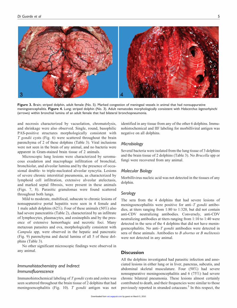
(3 females and 1 male) (Fig. 3). Two of these animals had large
numbers of Phyllobothrium delphini andMonorygma grimaldii
larval cysts in the subcutaneous tissue of perineal region and
ventrolateral abdominal musculature, respectively (Table 3).
Six animals (75%) had severe bilateral bronchointerstitial
pneumonia, characterized by partial lung collapse, extensive
areas of consolidation, and prominent subpleural emphysema
Figure 1. Geographic area (Imperia province, Ligurian Sea) in whichthe 8 striped dolphins under study were found stranded.
Di Guardo et al 3
3 by guest on March 5, 2010vet.sagepub.comDownloaded from
(particularly in the apical regions of both lungs). Adult
nematodes, morphologically consistent with Halocerchus
lagenorhynchi (formerly H delphini), were present in the
bronchial lumina of these dolphins (Fig. 4) and were associated
with moderate catarrhal exudate and frothy fluid, indicative of
pulmonary edema.
Severe multifocal, subacute to chronic, nonsuppurative
meningoencephalitis was present in 1 male and 3 female
adult dolphins (50%). These lesions mainly involved the
pons and mesencephalon and consisted of prominent peri-
vascular mononuclear cell cuffing and focal microglial
nodules scattered throughout the surrounding neuropil
(Fig. 5). Macrophage accumulations in proximity to blood
vessels and mild lymphoplasmacytic infiltration of choroid
plexuses were also seen in the adult male dolphin. A micro-
angiopathy in which the wall of affected vessels had an
‘‘onionlike’’ appearance was seen in all 4 of these animals.
Astrogliosis and mild to moderate neuronal degeneration
Figure 2. The number of cetaceans found stranded in the study area from 1985 to 2007, with striped dolphin mortality peaks in 1990 (blackbar) and 2007 (white bar) as a result of the dolphin morbillivirus epidemic.
Table 2. Nutritional Status, Gastric Content, and Main Lesions in the 8 Striped Dolphinsa
Dolphin No. Nutritional Status Gastric Content MENG BP VB Extrapulmonary Parasitic Lesions
1 Good Partially digested milk – þ þ –2 Poor None – þ þ Parasitic nodules in subcutis and abdominal
musculature; parasitic hepatitis3 Good Partially digested fish – þ þ –4 Good Partially digested fish – þ – –5 Good Partially digested fish þ – – Parasitic hepatitis and pancreatitis6 Poor None þ – þ Parasitic nodules in subcutis and abdominal
musculature; parasitic hepatitis and pancreatitis7 Poor None þ þ þ Parasitic hepatitis and pancreatitis8 Good Partially digested fish þ þ þ Parasitic hepatitis and pancreatitis
a MENG, meningoencephalitis; BP, bronchointerstitial pneumonia; VB, verminous bronchopneumonia; lesions present (þ), lesions absent (–).
4 Veterinary Pathology 000(00)
4 by guest on March 5, 2010vet.sagepub.comDownloaded from
and necrosis characterized by vacuolation, chromatolysis,
and shrinkage were also observed. Single, round, basophilic
PAS-positive structures morphologically consistent with
T gondii cysts (Fig. 6) were scattered throughout the brain
parenchyma of 2 of these dolphins (Table 3). Viral inclusions
were not seen in the brain of any animal, and no bacteria were
apparent in Gram-stained brain tissue of 2 animals.
Microscopic lung lesions were characterized by seromu-
cous exudation and macrophage infiltration of bronchial,
bronchiolar, and alveolar lumina and by the presence of occa-
sional double- to triple-nucleated alveolar syncytia. Lesions
of severe chronic interstitial pneumonia, as characterized by
lymphoid cell infiltration, extensive alveolar atelectasis,
and marked septal fibrosis, were present in these animals
(Figs. 7, 8). Parasitic granulomas were found scattered
throughout both lungs.
Mild to moderate, multifocal, subacute to chronic lesions of
nonsuppurative portal hepatitis were seen in 4 female and
1 male adult dolphins (62%). Four of these animals (50%) also
had severe pancreatitis (Table 2), characterized by an infiltrate
of lymphocytes, plasmacytes, and eosinophils and by the pres-
ence of extensive hemorrhagic and necrotic foci. Many
metazoan parasites and ova, morphologically consistent with
Campula spp, were observed in the hepatic and pancreatic
(Fig. 9) parenchyma and ductal lumina of all 5 of these dol-
phins (Table 3).
No other significant microscopic findings were observed in
any animal.
Immunohistochemistry and Indirect
Immunofluorescence
Immunohistochemical labeling of T gondii cysts and zoites was
seen scattered throughout the brain tissue of 2 dolphins that had
meningoencephalitis (Fig. 10). T gondii antigen was not
identified in any tissue from any of the other 6 dolphins. Immu-
nohistochemical and IIF labeling for morbilliviral antigen was
negative on all dolphins.
Microbiology
Several bacteria were isolated from the lung tissue of 3 dolphins
and the brain tissue of 2 dolphins (Table 3). No Brucella spp or
fungi were recovered from any animal.
Molecular Biology
Morbillivirus nucleic acid was not detected in the tissues of any
dolphin.
Serology
The sera from the 4 dolphins that had severe lesions of
meningoencephalitis were positive for anti–T gondii antibo-
dies, at titers ranging from 1:80 to 1:320, but did not contain
anti-CDV neutralizing antibodies. Conversely, anti-CDV
neutralizing antibodies at titers ranging from 1:10 to 1:40 were
detected in the sera of the 4 dolphins that did not have menin-
goencephalitis. No anti–T gondii antibodies were detected in
sera of these animals. Antibodies to B abortus or B melitensis
were not detected in any animal.
Discussion
All the dolphins investigated had parasitic infection and asso-
ciated lesions in either lung or in liver, pancreas, subcutis, and
abdominal skeletal musculature. Four (50%) had severe
nonsuppurative meningoencephalitis and 6 (75%) had severe
chronic bilateral pneumonia. These lesions almost certainly
contributed to death, and their frequencies were similar to those
previously reported in stranded cetaceans.5 In this respect, the
Figure 3. Brain; striped dolphin, adult female (No. 5). Marked congestion of meningeal vessels in animal that had nonsuppurativemeningoencephalitis. Figure 4. Lung; striped dolphin (No. 3). Adult nematodes morphologically consistent with Halocerchus lagenorhyinchi
(arrows) within bronchial lumina of an adult female that had bilateral bronchopneumonia.
Di Guardo et al 5
by guest on March 5, 2010vet.sagepub.comDownloaded from
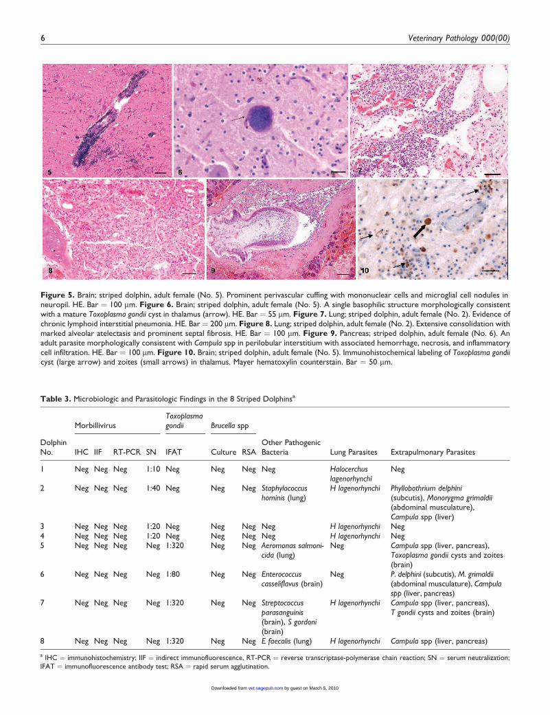
Figure 5. Brain; striped dolphin, adult female (No. 5). Prominent perivascular cuffing with mononuclear cells and microglial cell nodules inneuropil. HE. Bar ¼ 100 mm. Figure 6. Brain; striped dolphin, adult female (No. 5). A single basophilic structure morphologically consistentwith a mature Toxoplasma gondii cyst in thalamus (arrow). HE. Bar ¼ 55 mm. Figure 7. Lung; striped dolphin, adult female (No. 2). Evidence ofchronic lymphoid interstitial pneumonia. HE. Bar ¼ 200 mm. Figure 8. Lung; striped dolphin, adult female (No. 2). Extensive consolidation withmarked alveolar atelectasis and prominent septal fibrosis. HE. Bar ¼ 100 mm. Figure 9. Pancreas; striped dolphin, adult female (No. 6). Anadult parasite morphologically consistent with Campula spp in perilobular interstitium with associated hemorrhage, necrosis, and inflammatorycell infiltration. HE. Bar ¼ 100 mm. Figure 10. Brain; striped dolphin, adult female (No. 5). Immunohistochemical labeling of Toxoplasma gondiicyst (large arrow) and zoites (small arrows) in thalamus. Mayer hematoxylin counterstain. Bar ¼ 50 mm.
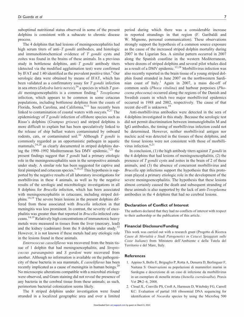
Table 3. Microbiologic and Parasitologic Findings in the 8 Striped Dolphinsa
MorbillivirusToxoplasma
gondii Brucella spp
DolphinNo. IHC IIF RT-PCR SN IFAT Culture RSA
Other PathogenicBacteria Lung Parasites Extrapulmonary Parasites
1 Neg Neg Neg 1:10 Neg Neg Neg Neg Halocerchus
lagenorhynchi
Neg
2 Neg Neg Neg 1:40 Neg Neg Neg Staphylococcus
hominis (lung)H lagenorhynchi Phyllobothrium delphini
(subcutis), Monorygma grimaldii
(abdominal musculature),Campula spp (liver)
3 Neg Neg Neg 1:20 Neg Neg Neg Neg H lagenorhynchi Neg4 Neg Neg Neg 1:20 Neg Neg Neg Neg H lagenorhynchi Neg5 Neg Neg Neg Neg 1:320 Neg Neg Aeromonas salmoni-
cida (lung)Neg Campula spp (liver, pancreas),
Toxoplasma gondii cysts and zoites(brain)
6 Neg Neg Neg Neg 1:80 Neg Neg Enterococcus
casseliflavus (brain)Neg P. delphini (subcutis), M. grimaldii
(abdominal musculature), Campulaspp (liver, pancreas)
7 Neg Neg Neg Neg 1:320 Neg Neg Streptococcus
parasanguinis
(brain), S gordoni
(brain)
H lagenorhynchi Campula spp (liver, pancreas),T gondii cysts and zoites (brain)
8 Neg Neg Neg Neg 1:320 Neg Neg E faecalis (lung) H lagenorhynchi Campula spp (liver, pancreas)
a IHC ¼ immunohistochemistry; IIF ¼ indirect immunofluorescence, RT-PCR ¼ reverse transcriptase-polymerase chain reaction; SN ¼ serum neutralization;IFAT ¼ immunofluorescence antibody test; RSA ¼ rapid serum agglutination.
6 Veterinary Pathology 000(00)
by guest on March 5, 2010vet.sagepub.comDownloaded from
suboptimal nutritional status observed in some of the present
dolphins is consistent with a subacute to chronic disease
process.
The 4 dolphins that had lesions of meningoencephalitis had
high serum titers of anti–T gondii antibodies, and histologic
and immunohistochemical evidence of T gondii cysts and
zoites was found in the brains of these animals. In a previous
study in bottlenose dolphins, anti–T gondii antibody titers
(detected via the modified agglutination test) were confirmed
by IFAT and 1:40 identified as the prevalent positive titer.8Our
serologic data were obtained by means of IFAT, which has
been validated as a confirmatory assay for T gondii infection
in sea otters (Enhydra lutris nereis),26 a species in which T gon-
dii meningoencephalitis is a common finding.3 Toxoplasma
infection, which appears to be common in some cetacean
populations, including bottlenose dolphins from the coasts of
Florida, South Carolina, and California,9,11 has recently been
linked to contamination of coastal waters with oocysts.9,27 The
epidemiology of T gondii infection of offshore species such as
Risso’s dolphins (Grampus griseus) and striped dolphins is
more difficult to explain but has been speculatively linked to
the release of ship ballast waters contaminated by onboard
rodents, cats, or contaminated soil.32 Although T gondii is
commonly regarded as an opportunistic pathogen in aquatic
mammals,24,26 as clearly documented in striped dolphins dur-
ing the 1990–1992 Mediterranean Sea DMV epidemic,7,21 the
present findings suggest that T gondii had a primary etiologic
role in the meningoencephalitis seen in the seropositive animals
under study. A similar role has been suggested for captive and
feral pinniped and cetacean species.9,10,25 This hypothesis is sup-
ported by the negative results of all laboratory investigations for
morbillivirus in these 4 animals, as well as by the negative
results of the serologic and microbiologic investigations in all
8 dolphins for Brucella infection, which has been associated
with meningoencephalitis in cetaceans, including striped dol-
phins.14,19 The severe brain lesions in the present dolphins dif-
fered from those associated with Brucella infection in that
meningitis was less prominent. In contrast, the severity of ence-
phalitis was greater than that reported in Brucella-infected ceta-
ceans.14,19Relatively high concentrations of immunotoxic heavy
metals were measured in tissues from the liver (mercury, lead)
and the kidney (cadmium) from the 8 dolphins under study.35
However, it is not known if these metals had any etiologic role
in the lesions found in these animals.
Enterococcus casseliflavus was recovered from the brain tis-
sue of 1 dolphin that had meningoencephalitis, and Strepto-
coccus parasanguinis and S gordoni were recovered from
another. Although no information is available on the pathogeni-
city of these bacteria in sea mammals, E casseliflavus has been
recently implicated as a cause of meningitis in human beings.20
No microscopic alterations compatible with a microbial etiology
were observed, and Gram staining did not reveal the presence of
any bacteria in the cerebral tissue from these animals; as such,
perimortem bacterial colonization seems likely.
The 8 striped dolphins under investigation were found
stranded in a localized geographic area and over a limited
period during which there was a considerable increase
in reported strandings in that region (F. Garibaldi and
W. Mignone, personal communication). These observations
strongly support the hypothesis of a common source exposure
as the cause of the increased striped dolphin mortality during
2007 in the Ligurian Sea. A similar pattern occurred in 2007
along the Spanish coastline in the western Mediterranean,
where dozens of striped dolphins and several pilot whales died
as a result of a DMV epidemic.12,28 Morbillivirus infection was
also recently reported in the brain tissue of a young striped dol-
phin found stranded in June 2007 on the northwestern Sardi-
nian coast of Italy.1 Again in 2007, a mass die-off of
common seals (Phoca vitulina) and harbour porpoises (Pho-
coena phocoena) occurred along the regions of the Danish and
Swedish coasts in which two major morbilliviral epidemics
occurred in 1988 and 2002, respectively. The cause of that
recent die-off is unknown.18
Anti-morbillivirus antibodies were detected in the sera of
4 dolphins investigated in this study. Because the serologic test
did not permit discrimination between immunoglobulin M and
IgG antibodies, the timing of morbillivirus infection could not
be determined. However, neither morbilliviral antigen nor
nucleic acid was detected in the tissues of these dolphins, and
the tissue lesions were not consistent with those of morbilli-
virus infection.6,21
In conclusion, (1) the high antibody titers against T gondii in
the 4 dolphins that had lesions of meningoencephalitis, (2) the
presence of T gondii cysts and zoites in the brain of 2 of these
animals, and (3) the absence of concurrent morbillivirus and
Brucella spp infections support the hypothesis that this proto-
zoan played a primary etiologic role in the development of the
severe meningoencephalitis. The hypothesis that these lesions
almost certainly caused the death and subsequent stranding of
these animals is also supported by the lack of anti-Toxoplasma
antibodies in the 4 dolphins that had no cerebral lesions.
Declaration of Conflict of Interest
The authors declared that they had no conflicts of interest with respect
to their authorship or the publication of this article.
Financial Disclosure/Funding
This work was carried out with a research grant (Progetto di Ricerca
Cause di Mortalit�a e Studi Patogenetici in Cetacei Spiaggiati sulle
Coste Italiane) from Ministero dell’Ambiente e della Tutela del
Territorio e del Mare, Italy.
References
1. Appino S, Bollo E, Briguglio P, Rotta A, Denurra D, Berlinguer F,
Naitana S: Osservazioni su popolazioni di mammiferi marini in
Sardegna e descrizione di un caso di infezione da morbillivirus
in un esemplare di stenella striata (Stenella coeruleoalba). Praxis
Vet 29:2–6, 2008.
2. Cloud JL, Conville PS, Croft A, Harmsen D, Witebsky FG, Carroll
KC: Evaluation of partial 16S ribosomial DNA sequencing for
identification of Nocardia species by using the MicroSeq 500
Di Guardo et al 7
7 by guest on March 5, 2010vet.sagepub.comDownloaded from
system with an expanded database. J Clin Microbiol 42:578–584,
2004.
3. Cole RA, Lindsay DS, Howe DK, Roderick CL, Dubey JP, Tho-
mas NJ, Baeten LA: Biological and molecular characterizations
of Toxoplasma gondii strains obtained from southern sea otters
(Enhydra lutris nereis). J Parasitol 86:526–530, 2000.
4. Di Guardo G, Agrimi U, Amaddeo D, McAliskey M, Kennedy S:
Morbillivirus infection in a striped dolphin (Stenella coeru-
leoalba) from the coast of Italy. Vet Rec 130:579–580, 1992.
5. Di Guardo G, Agrimi U, Morelli L, Cardeti G, Terracciano G,
Kennedy S: Post mortem investigations on cetaceans found
stranded on the coasts of Italy between 1990 and 1993. Vet Rec
136:439–442, 1995.
6. Di Guardo G, Marruchella G, Agrimi U, Kennedy S: Morbilli-
virus infections in aquatic mammals: A brief overview. J Vet Med
A Physiol Pathol Clin Med 52:88–93, 2005.
7. Domingo M, Visa J, Pumarola M, Marco AJ, Ferrer L, Rabanal R,
Kennedy S: Pathologic and immunocytochemical studies of mor-
billivirus infection in striped dolphins (Stenella coeruleoalba).
Vet Pathol 29:1–10, 1992.
8. Dubey JP, Fair PA, Bossart GD, Hill D, Fayer R, Sreekumar C,
Kwok OCH, Thulliez PA: Comparison of several serologic tests
to detect antibodies to Toxoplasma gondii in naturally
exposed bottlenose dolphins (Tursiops truncatus). J Parasitol
91:1074–1081, 2005.
9. Dubey JP, Fair PA, Sundar N, Velmurugan G, Kwok OC, McFee
WE, Majumdar D, Su C: Isolation of Toxoplasma gondii
from bottlenose dolphins (Tursiops truncatus). J Parasitol
94:821–823, 2008.
10. Dubey J P, Mergel J, Gehring E, Sundar N, Velmurugan G,
Kwok O, Grigg M, Su C, Martineau D: Toxoplasmosis in captive
dolphins (Tursiops truncatus) and walrus (Odobenus rosmarus).
J Parasitol 95:82–85, 2009.
11. Dubey JP, Zarnke R, Thomas NJ, Wong SK, Van Bonn W,
Briggs M, Davis JW, Ewing R, Mensea M, Kwok OCH,
Romand S, Thulliez P: Toxoplasma gondii, Neospora caninum,
Sarcocystis neurona, and Sarcocystis canis-like infections in
marine mammals. Vet Parasitol 116:275–296, 2003.
12. Fernandez A, Esperon F, Herraez P, Espinoza de Los
Monteros A, Clavel C, Bernabe A, Sanchez-Vizcaino JM,
Verborgh P, DeStefanis R, Toledano F, Bayon A: Morbilli-
virus and pilot whale deaths, Mediterranean Sea. Emerg Infect
Dis 14:471–473, 2008.
13. Frisk AL, Konig M, Moritz A, Baumgartner W: Detection of dis-
temper nucleoprotein RNA by reverse transcription-PCR using
serum, whole blood, and cerebrospinal fluid from dogs with dis-
temper. J Clin Microbiol 37:3634–3643, 1999.
14. Gonzalez L, Patterson IA, Reid RJ, Foster G, Barberan M, Blasco
JM, Kennedy S, Howie FE, Godfroid J, MacMillan AP, Shock A,
Buxton D: Chronic meningoencephalitis associated with Brucella
sp infection in live-stranded striped dolphins (Stenella coeru-
leoalba). J Comp Pathol 126:147–152, 2002.
15. Hall L, Doerr KA, Wohlfiel SL, Roberts GD: Evaluation of the
MicroSeq system for identification of mycobacteria by 16S riboso-
malDNAsequencing and its integration intoa routine clinicalmyco-
bacteriology laboratory. J Clin Microbiol 41:1447–1453, 2003a.
16. Hall L, Wohlfiel S, Roberts GD: Experience with the MicroSeq
D2 large-subunit ribosomal DNA sequencing kit for identification
of commonly encountered, clinically important yeast species.
J Clin Microbiol 41:5099–5102, 2003b.
17. Hall L, Wohlfiel S, Roberts GD: Experience with the MicroSeq
D2 large-subunit ribosomal DNA sequencing kit for identification
of filamentous fungi encountered in the clinical laboratory. J Clin
Microbiol 42:622–626, 2004.
18. Harkonen T, Backlin BM, Barrett T, Bergman A, Corteyn M,
Dietz R, Harding KC, Malmsten J, Roos A, Teilmann J: Mass
mortality in harbour seals and harbour porpoises caused by an
unknown pathogen. Vet Rec 162:555–556, 2008.
19. Hernandez-Mora G, Gonzalez-Barrientos R, Morales JA, Chaves-
Olarte E, Guzman-Verri C, Baquero-Calvo E, De-Miguel MJ,
Marın CM, Blasco JM, Moreno E: Neurobrucellosis in stranded
dolphins, Costa Rica. Emerg Infect Dis 14:1430–1433, 2008.
20. Iaria C, Stassi G., Costa GB, Di Leo R, Toscano A, Cascio A:
Enterococcal meningitis caused by Enterococcus casseliflavus:
first case report. BMC Infect Dis 5:3, 2005.
21. Kennedy S: Morbillivirus infections in aquatic mammals. J Comp
Pathol 119:201–225, 1998.
22. Kennedy S, Smyth JA, Cush PF, McAliskey M, McCullough SJ,
Rima BK: Histopathologic and immunocytochemical studies of
distemper in harbor porpoises. Vet Pathol 28:1–7, 1991.
23. Mellmann A, Cloud JL, Andrees S, Blackwood K, Carroll KC,
Kabani A, Roth A, Harmsen D: Evaluation of RIDOM, MicroSeq,
and Genbank services in the molecular identification of Nocardia
species. Int J Med Microbiol 293:359–370, 2003.
24. Migaki G, Sawa TR, Dubey JP: Fatal disseminated toxoplasmosis
in a spinner dolphin (Stenella longirostris). Vet Pathol 27:463–
464, 1990.
25. Miller MA, Sverlow K, Crosbie PR, Barr BC, Lowenstine LJ,
Gulland FM, Packham A, Conrad PA: Isolation and characteriza-
tion of two parasitic protozoa from a pacific harbor seal (Phoca
vitulina richardsi) with meningoencephalomyelitis. J Parasitol
87:816–822, 2001.
26. Miller MA, Gardner IA, Packham A, Mazet JK, Hanni KD,
Jessup D, Estes J, Jameson R, Dodd E, Barr BC, Lowenstine
LJ, Gulland FM, Conrad PA: Evaluation of an indirect fluores-
cent antibody test (IFAT) for demonstration of antibodies to
Toxoplasma gondii in the sea otter (Enhydra lutris). J Parasitol
88:594–599, 2002.
27. Miller MA, Miller WA, Conrad PA, James ER, Melli AC,
Leutenegger CM, Dabritz HA, Packham AE, Paradies D, Harris
M, Ames J, Jessup DA, Worcester K, Grigg ME: Type X
Toxoplasma gondii in a wild mussel and terrestrial carnivores from
coastal California: New linkages between terrestrial mammals, run-
off and toxoplasmosis of sea otters. Int J Parasitol 38:1319–1328,
2008.
28. Raga J-A, Banyard A, Domingo M, Corteyn M, Van Bressem
M-F, Fernandez M, Aznar F-J, Barrett T: Dolphin morbillivirus
epizootic resurgence, Mediterranean Sea. Emerg Infect Dis
14:471–473, 2008.
29. Randall LZ, Jeremiah TS, Alastair PM, Simon DB, Claire ED, Jay
MVH, Kathryn JF, Robert JS: Serological survey for Brucella
spp., phocid herpervirus-1, phocid herpesvirus-2, and phocine
8 Veterinary Pathology 000(00)
8 by guest on March 5, 2010vet.sagepub.comDownloaded from
distemper virus in harbour seals from Alaska, 1976–1999. J Wildl
Dis 42:290–300, 2006.
30. Stanton JB, Givens L, Evermann JF, Brown CC: Immunohisto-
chemical analysis of two strains of lion (Panthera leo)-adapted
canine distemper virus in ferrets (Mustela putorius furo). Vet
Pathol 40:464–467, 2003.
31. Van BressemM-F, Waerebeek KV, Jepson PD, Raga JA, Duignan
PJ, Nielsen O, Di Beneditto AP, Siciliano S, Ramos R, Kant
W, Peddemors V, Kinoshita R, Ross PS, Lopez-Fernandez A,
Evans K, Crespo E, Barrett T: An insight into the epidemiology
of dolphin morbillivirus worldwide. Vet Microbiol 81:287–304,
2001.
32. Van Bressem M-F, Raga JA, Di Guardo G, Jepson P, Duignan P,
Siebert U, Barrett T, De Oliveira Santos MC, Moreno I,
Siciliano S, Aguilar A, Van Waerebeek K: Emerging infectious
diseases in cetaceans worldwide and the possible role of environ-
mental stressors. Dis Aquat Organ 86:143–157, 2009.
33. Visser IK, Van Bressem M-F, De Swart RL, Van De Bildt MW,
Vos HW, Van Der Heijden RW, Saliki JT, Orvell C, Kitching P,
Kuiken T: Characterization of morbilliviruses isolated from dol-
phins and porpoises in Europe. J Gen Virol 74:631–641, 1993.
34. Wohlsein P, Puff C, Kreutzer M, Siebert U, Baumgartner W:
Distemper in a dolphin. Emerg Infect Dis 13:1959–1961, 2007.
35. Zaccaroni A, Scaravelli D, Proietto U, Marsilio F, Mignone W,
Vivaldi B, Caroggio P, Casalone C, Bozzetta E, Garibaldi F, Di
Francesco CE, Di Guardo G: Morbillivirus could not be the major
factor impacting the health of striped dolphins along the Ligurian
Sea coast of Italy. In: 39th Annual Conference of the International
Association for Aquatic Animal Medicine—2008, pp. 86–89.
Omnipress, Madison, WI, 2008.
Di Guardo et al 9
9 by guest on March 5, 2010vet.sagepub.comDownloaded from