vacuum assisted closure: recommendations for use
TRANSCRIPT

Vacuum assisted closure:recommendations for useA consensus document
An International Consensus Initiative
FINAL FOR APPROVAL
DRAFTPRINCIPLES OFBEST PRACTICE

FOREWORDThis timely initiative draws on both the research evidence and the consensusopinion of an international group of experts (see below) to provide guidance onthe successful integration of vacuum assisted closure therapy (V.A.C.® Therapy)into clinical practice. The document specifically reviews its potential use in thefollowing selected indications*: diabetic foot ulcers, complex leg ulcers, pressureulcers, dehisced sternal wounds, open abdominal wounds and traumatic wounds.In addition, it considers quality of life and cost-effectiveness, both of which aregaining importance when evaluating treatment. This document highlights questionsfor future research and is designed to be practical and adaptable for local use incountries worldwide.
Professor Keith Harding
EXPERT WORKING GROUPJan Apelqvist, University Hospital of Malmö, Division of Clinical Sciences,University of Lund (Sweden)David G Armstrong, Rosalind Franklin University of Medicine and Science, Illinois (US)Matthias Augustin, University Clinics of Hamburg (Germany)Mona Baharestani, East Tennessee State University, Tennessee; James HQuillen Veterans Administration Medical Centre, Tennessee (US)Paul Banwell, Queen Victoria Hospital NHS Foundation Trust, EastGrinstead (Co-Chair, UK)Luca Dalla Paola, Abano Terme Hospital, Padova (Italy)Anand Deva, Liverpool Hospital, New South Wales (Australia)William Ennis, University of Illinois, Chicago (US)Joel Fish, Sunnybrook Health Sciences Centre, Toronto (Canada)Wilhelm Fleischmann, Krankenhaus, Bietigheim (Germany)Subhas Gupta, Loma Linda University, California (US)Ronny Gustafsson, Lund University Hospital (Sweden)Keith Harding, Wound Healing Research Unit, Cardiff University (Co-Chair, UK)Raymond E Horch, Friedrich-Alexander-University of Erlangen-Nuremberg(Germany)Richard Ingemansson, Lund University Hospital (Sweden)Gerrolt Jukema, VU University Medical Centre, Amsterdam (The Netherlands)James Mahoney, St Michael’s Hospital, Toronto (Canada)Chantal Mouës, Erasmus University Medical Center, Rotterdam (TheNetherlands)Patricia Price, School of Medicine, Cardiff University (UK)Javier Soldevilla Ágreda, EUE University of La Rioja, Logroño (Spain)Colin Song, Singapore General Hospital (Singapore)Luc Téot, Hospital Lapeyronie, Montpellier (France)Paul Trueman, University of York (UK)Kathryn Vowden, Bradford Teaching Hospitals NHS Foundation Trust andUniversity of Bradford (UK)Peter Vowden, Bradford Teaching Hospitals NHS Foundation Trust andUniversity of Bradford (UK)Thomas Wild, Medical University of Vienna (Austria)
MANAGING EDITOR:Suzie Calne
DEPUTY EDITOR:Rachel Wheeler
EDITORIAL PROJECTMANAGER:Kathy Day
MANAGING DIRECTOR:Jane Jones
SENIOR EDITOR:Paul Banwell
DESIGNER:Jane Walker
PRINTED BY:Printwells, Kent, UK
FOREIGNTRANSLATIONS:RWS Group, London, UK
PUBLISHED BY:Medical EducationPartnership (MEP) LtdOmnibus House39-41 North RoadLondon N7 9DP, UK Tel: + 44 (0)20 7715 0390Fax: +44 (0)20 7715 0391Email: [email protected]: www.mepltd.co.uk
© MEP Ltd 2008
Supported by an unrestrictededucational grant from KCIEurope Holding BV. Theviews expressed in thisdocument are those of theexpert working group and donot necessarily reflect thoseof KCI.
World Union of WoundHealing SocietiesCurrent President: Professor Luc TéotChair, Industrial LiaisonCommittee:Professor Keith HardingFor further details visit: www.wuwhs.org
How to cite this document: World Union of WoundHealing Societies (WUWHS).Principles of best practice.Vacuum assisted therapy:recommendations for use. Aconsensus document.London: MEP Ltd, 2008.
* This document provides recommendations for the use of VAC therapy in six selected wound types. It should benoted, however, that VAC therapy has a role in other wound types, which are not included in this document.

RECOMMENDATIONS FOR USE
VACUUM ASSISTED CLOSURE: RECOMMENDATIONS FOR USE | 1
Vacuum assisted closure (VAC) therapy has helped to improve wound care outcomes and hasled to a number of dramatic changes in clinical practice over the last decade1,2. VAC therapymust be used as part of an individualised, comprehensive treatment plan and is indicated forboth acute and chronic wounds.
PLANNING TREATMENTIn all situations the underlying wound aetiology and comorbidities must first be addressed andtreated. It is essential to optimise all aspects of the patient’s physical, nutritional andpsychosocial wellbeing to ensure treatment is suitable and of maximum benefit.
Before starting VAC therapy it is important to define treatment aims, objectives and clinicalendpoints1. In some circumstances the objective will be to avoid further complications and tocontrol symptoms, rather than to influence time to healing. Examples of clinical endpoints for VACtherapy include 50% volume reduction3, 80% granulation tissue formation or complete closure.
In general, the key aims are to:■ remove exudate and reduce periwound oedema■ increase local microvascular blood flow/test vascularity ■ promote formation of granulation tissue ■ reduce complexity/size of the wound ■ optimise the wound bed prior to and following surgery ■ reduce complexity of surgical wound closure procedures4.
In addition, the application of the VAC dressing system creates a closed, moist woundenvironment, which may act as a barrier to bacteria and patient/caregiver interference. VAC therapy may also help to promote patient independence, mobility and comfort.
Identifying responders to VAC therapyIn chronic wounds, it may be helpful to use the factors listed in Table 1 to assess whether thewound is likely to have a positive response to VAC therapy. It must be noted, however, that inmany circumstances the patient/wound will not exhibit these attributes and yet VAC may stillhave an important role. A good example of this, is the diabetic foot ulcer (see page 3) where thepatient often has multiple comorbidities and the wound has a poor blood supply.
For acute wounds, it is important to adequately debride the wound and followrecommended guidelines for specific wound types (eg dehisced sternal wounds) beforecommencing therapy.
1. European WoundManagement Association.Position Document:Topical negative pressurein wound management.London: MEP Ltd, 2007.
2. Banwell P. Topical negativepressure therapy in woundcare. J Wound Care 1999;8(2): 79-84.
3. Ennis WJ, Lee C, VargasM, Meneses O. Woundoutcomes from a singlepractice at a subacutewound care unit and twohospital based outpatientwound clinics. Wounds2004; 16(5): 164-72.
4. Beier JP, Horch R. Surgicalmanagement of pressureulcers. In: VacuumAssisted ClosureTM
Therapy: Science andpractice. Banwell P,Harding K (Eds). London:MEP Ltd, 2006.
There may be benefits to starting VAC therapy as early as possible. Delaying therapymay allow the wound to deteriorate before being treated effectively !
Table 1 | Factors that may increase success of therapy
■ Wound has good blood supply ■ Patient has been maximally medically ■ Wound has healthy, granular bed stabilised (eg nutrition, blood pressure, blood■ Wound as been freshly debrided (as glucose, fluid balance, infection)
recommended*) ■ Patient has few or well-controlled comorbidities■ Wound produces high levels of exudate ■ Patient is comfortable (eg not in pain)■ Wound is greater than 2cm wide ■ Patient is adherent with therapy * NB Occasionally, in some chronic wounds, surgical debridement may not be appropriate. Prior to starting VAC therapy it isimportant to ensure that the wound has a clean wound bed and that it does not contain necrotic tissue or excessive debris.
Wound factors Patient factors
Figure 1 | An example ofan acute wound(abdomen) and chronicwound (diabetic foot)suitable for VAC therapy.

2 | PRINCIPLES OF BEST PRACTICE
EVALUATING TREATMENTIt is important to review progress regularly. This will involve an accurate and reproduciblemethod of wound measurement5. If there is a reduction in wound area (eg around 15%) afterone or two weeks6, strong consideration should be given to continuing VAC therapy withongoing clinical evaluation. Reassess again after a further week of therapy. If there is noimprovement, discontinue VAC therapy and begin an alternative treatment. VAC therapy maybe reconsidered at a later stage.
In chronic wounds, an effective general assessment measure is to: ■ examine the wound margins for inflammation after the first application of VAC therapy. If
there is increased inflammation consider discontinuing treatment■ re-examine the wound margins for a thin white epithelium after the second and subsequent
applications; this indicates healing■ evaluate the overall appearance of the wound bed. A beefy, granular appearance is a
positive outcome, while a dusky bed indicates inadequate tissue perfusion. Granulationtissue should increase by around 3–5% per day.
Under ideal conditions (especially in the absence of infection), well perfused wounds willrespond quickly (ie within one week) with evidence of granulation tissue formation. Thiscan be used to test vascularity and suitability of VAC therapy to avoid disappointment.
Adverse reactions have, on occasion, been reported (eg adherence to deep tissuestructures). These can usually be avoided by following recommendations (see boxbelow), involving appropriately trained staff and by developing effective communicationstrategies. Specialist involvement will be required in certain situations
VAC THERAPY AND WOUND INFECTIONVAC therapy is not recommended as a stand-alone treatment for wound infection. However, itmay be used with extra caution in infected wounds as long as this is in addition to appropriatetreatment of the infection (see box opposite).
In the presence of persistent infection or deterioration, or in wounds exhibiting no clinicalprogress towards healing (ie odour continues or becomes apparent), perform a thoroughpatient and wound reassessment (including microbiological investigations), discontinue VACtherapy and change treatment. Always consider whether systemic antibiotic therapy and/orappropriate debridement is required and treat the wound infection according to local protocols.
If infection develops during therapy, consider systemic antibiotic treatment and discontinueVAC therapy to allow monitoring of the wound. On specific occasions, an advancedmodification of VAC therapy (VAC-Instill®) may be considered for use in severely infectedwounds (eg infected hip and knee implants and orthopaedic hardware). This involves instillingan appropriate fluid into the wound bed, such as a topical antibacterial solution7.
5. Mouës CM, Vos MC, Jan-Gert CM, et al. Bacterialload in relation to vacuum-assisted closure woundtherapy: A prospectiverandomised trial. WoundRep Reg 2004; 12: 11-17.
6. Attinger CE, Janis JE,Steinburg J, et al. Clinicalapproach to wounds:debridement and woundbed preparation includingthe use of dressings andwound-healing adjuvants.Plast Reconstr Surg 2006;117 (7 Suppl): 72s-109s.
7. Plikaitis CM, Molnar JA.Subatmospheric pressurewound therapy and thevacuum-assisted closuredevice: basic science andcurrent clinical success.Expert Rev Med 2006; 3(2):175-84.
■ Debridement■ Antibiotic therapy■ Patient optimisation■ Frequent
patient/woundassessments
■ More frequentdressing changes
■ Appropriatepressure settings
■ Periwound skinprotection
■ Fenestrated antimicrobialdressings
Factors to considerin the presence ofinfection:
To date almost all published clinical trials on topical negative pressure therapy2 have used the V.A.C.® Therapy system. Thisfully integrated system incorporates a polyurethane (V.A.C.® GranuFoam® and V.A.C. GranuFoam Silver®) or polyvinylalcohol (V.A.C.® WhiteFoam Dressing) foam dressing and a microprocessor controlled unit that establishes a uniformdistribution of pressure across the entire wound. It is this specific system that is referred to throughout this document. Forfurther information on the safe use of this system (including appropriate pressure and therapy settings) and relevant patientsafety information, please go to www.kci-medical.com or contact your local KCI representative.
!
VAC THERAPY IN PRACTICE

VACUUM ASSISTED CLOSURE: RECOMMENDATIONS FOR USE | 3
DIABETIC FOOT ULCERS
Clinicians may sometimes wrongly consider all diabetic foot ulcers to be the same for treatmentpurposes. In fact, there is considerable variation and the decision to use VAC therapy willdepend on the wound subtype. VAC therapy can be considered for deep complex wounds, forpost-surgery wounds and, occasionally, for superficial wounds in addition to standardtreatments (see Application to practice box below). For patients with ischaemic wounds,referral to a vascular surgeon should be considered prior to VAC therapy.
DEEP COMPLEX DIABETIC FOOT WOUNDSVAC therapy can be used in a number of ways to manage the complex diabetic foot wound:■ Reduce complexity/size – ie simplify the wound. In non-infected, non-ischaemic, deep
complex diabetic foot ulcers, VAC therapy can be used to reduce the surface area of thewound by encouraging granulation tissue formation over exposed bone, tendon or tissue.This may help to avoid the need for skin grafting and/or flaps or to reduce the complexity ofthe subsequent surgical closure procedure8,9. A special dressing technique should be used toprevent further pressure damage in plantar wounds when applying VAC therapy10.
■ Promote deep healing – Experience has shown that on occasions VAC therapy can beused for longer periods in combination with other treatment modalities (eg systemicantibiotics) to allow thorough, complete healing of underlying osteomyelitis before skinclosure. This avoids the problem of ulcer recurrence with residual osteomyelitis (ie where skinheals before the underlying bone).
In poorly perfused wounds where revascularisation is not possible, using VAC therapy fora trial period allows the clinician to observe the response to therapy and assess the viability ofthe tissue. Even when a positive outcome is unlikely, VAC therapy used in this way has shownunexpected and encouraging results. The clinician should strive for the most distal amputationlevel that achieves healing and a functional outcome8.
Planning treatmentThe planned duration of therapy for diabetic foot wounds will depend on the specific treatmentgoal. In many cases an initial one- to two-week period of therapy is recommended. After thistime, the wound should be evaluated for progress or deterioration, and: ■ if progress is good – ie there is a daily increase in healthy granulating tissue formation,
decreasing wound depth, a good blood supply and no infection – continue VAC therapy untilthe treatment goal is achieved
■ if progress is poor or there is deterioration, consider alternative treatments or breaks (‘time-outs’) in VAC therapy. During this time the clinician should re-evaluate perfusion, focus onoptimising medical therapy and use other wound modalities until the tissue quality improves.Often at this time VAC therapy can be successfully reinstated.
APPLICATION TO PRACTICEUse VAC therapy only after any underlying disease has been diagnosed andmanaged and after appropriate debridement of non-viable tissueVAC therapy can be an effective adjunct to revascularisation in diabetic foot wounds VAC therapy should be used only after surgical drainage of any infection withconconmitant systemic antibiotic therapy according to local protocolsVAC therapy should be combined with effective offloading and good wound care
VAC therapy is not recommended if the tissue is grossly infected; is ischaemic onpresentation; or in the presence of untreated osteomyelitis
8. Armstrong D, Lavery L.Negative pressure woundtherapy after partialdiabetic foot amputation: amulticentre, randomisedcontrolled trial. Lancet2005; 366: 1704-10.
9. Blume PA, Walters J,Payne W, et al.Comparison of negativepressure wound therapyutilizing vacuum-assistedclosure to advanced moistwound therapy in thetreatment of diabetic footulcers. A multicenter,randomized controlledtrial. Diabetes Care2007[Epub ahead of print].
10. Edmonds ME, Doxford M.Practical management ofdiabetic foot ulcers. In:Vacuum AssistedClosureTM Therapy:Science and practice.Banwell P, Harding K (Eds).London: MEP Ltd, 2007.
!
NB: There are currently nostudies on using VACtherapy in poorly perfusedwounds

Study Interventions Design Selection criteria Clinical outcomes
Armstrong DG, VAC therapy vs Multicentre, Diabetic foot amputation VAC therapy achieved wound healing in 56% vs Lavery LA. modern moist wound randomised to transmetatarsal level, 39% (p=0.04) of patients with a median wound bedLancet 2005; therapy for 16 weeks controlled trial with adequate perfusion preparation time of 42 days vs 84 days (p=0.02)366; 1704-10. n=162 compared with controls
Eginton MT, VAC therapy for 2 Randomised Large diabetic foot wounds VAC therapy reduced wound volume and depthet al. Ann Vasc weeks vs conventional controlled with adequate perfusion, by 59% vs 0% (p<0.005) and 49% vs 8% (p<0.05)Surg 2003; 17: moist gauze dressings crossover trial sharply debrided before respectively, when compared with controls over645-49. for 2 weeks n=10 entry the observation period
McCallon SK, VAC therapy vs Randomised Non-healing (>1 month) VAC therapy produced an average decrease inet al. Ostomy saline-moistened controlled pilot postoperative diabetic foot wound surface area of 28.4% vs a 9.5% increaseWound Manage gauze study wounds, surgically debrided for controls, and an average time to satisfactory2000; 46: 28-34. n=10 before treatment. Patients wound healing of 22.8 days vs 42.8 days for
with venous disease, active controls. Delayed primary closure was achievedinfection or coagulopathy in four of five patients with VAC therapy comparedwere excluded with two of five controls (p values not given)
Note: Although traditional gauze has been used as the comparator in many trials, the largest of these studies (Armstrong and Lavery, 2005) used awide variety of moist wound dressings in the control group. Further studies using modern wound products vs VAC therapy have been published8
or are in progess. For further information about SIGN levels of evidence visit www.sign.ac.uk
Summary of key (SIGN level 1) studies of VAC therapy in diabetic foot ulcers
4 | PRINCIPLES OF BEST PRACTICE
POST-SURGERY DIABETIC FOOT WOUNDSRandomised controlled trials (RCTs) support the use of VAC therapy for the following:■ after open partial foot amputation (from open toe/ray/to metatarsal level)8■ to aid fixation or bolstering of skin grafts11.
Split-skin grafting and bioengineered tissue replacements, particularly acellular matrices, havebeen used in combination with VAC therapy as a practical alternative to flap closure in deepcomplex wounds11. VAC therapy promotes vascular perfusion, which has been shown toenhance skin graft take12.
Planning treatmentIt is not always appropriate to start VAC therapy immediately following surgery and it may bebeneficial to observe the wound for 1–2 days prior to application13. The decision to select VACtherapy will depend on: ■ viability of the skin edge and the tissue immediately below it■ whether there is capillary bleeding■ whether infection has been addressed and necrotic tissue has been removed■ treatment goals and patient factors.
VAC therapy should be stopped after the clinical endpoint is achieved (eg an appropriatereduction in volume or adequate wound bed preparation for subsequent skin grafting).
SUPERFICIAL DIABETIC FOOT WOUNDSVAC therapy is not recommended as a first-line treatment in superficial wounds. However it maybe considered along with other advanced therapies where there has been a poor response toother treatments (ie effective offloading, management of infection and local dressings).
Use VAC therapy with caution if the TcpO2 is between 20 and 30mmHg and there isimpaired sensation (in such cases use lower pressure settings) !
11. Moisidis E, Heath T, BoorerC, et al. A prospective,blinded, randomized,controlled clinical trial oftopical negative pressureuse in skin grafting. PlastReconstr Surg 2004; 114:917-22.
12. Jeschke MG, Rose C,Angele P, et al.Development of newreconstructive techniques:use of Integra incombination with fibringlue and negative-pressure therapy forreconstruction of acuteand chronic wounds. PlastReconstr Surg 2004;113(2): 525-30.
13. Wu S, Armstrong DG.Surgical management ofdiabetic foot ulcers. In:Vacuum AssistedClosureTM Therapy:Science and practice.Banwell P, Harding K (Eds).London: MEP Ltd, 2007.

COMPLEX LEG ULCERS
It is recognised that compression therapy is regarded as the first-line treatment for venous legulcers14. However, there is a role for VAC therapy in inflammatory or complex therapy-resistantleg ulcers that are unsuitable for compression. The use of portable VAC systems may also allowambulatory patients to be treated at home and reduces the need for hospitalisation.
For complex leg ulcers it is important to thoroughly assess the wound using bacterial cultureand biopsy to confirm the diagnosis. Surgical debridement should be performed prior to theapplication of VAC therapy to increase the potential for success.
INFLAMMATORY ULCERSIn patients with inflammatory ulcers, VAC therapy can be used to enhance wound bedpreparation before definitive surgical closure or delayed secondary healing. These patientshistorically have hard-to-heal wounds with high rates of skin graft failure. Ulcers may occur inthe following situations:■ scleroderma■ systemic lupus erythematosus■ hypercoagulation disorders■ rheumatoid arthritis■ vasculitic conditions.
If the underlying clinical condition is resistant or inadequately treated, inflammatory ulcers willusually not heal despite optimal wound management. In addition, as treatment usually involvesnon-steroidal anti-inflammatory drugs, healing may be further impaired. In non-infected ulcers, ashort trial of VAC therapy can be considered to determine whether it is likely to be beneficial.VAC therapy should be applied for 1–3 days and then removed while the response is evaluated.
Note: in a non-healing chronic ulcer in which other treatments have not been successful,granulation tissue may not be seen for up to two weeks when using VAC therapy.
COMPLEX THERAPY-RESISTANT ULCERSVAC therapy can be considered for complex therapy-resistant leg ulcers including:■ highly exuding ulcers■ anatomically challenging ulcers (where the application and stabilisation of dressings is difficult)■ wounds requiring skin grafting (VAC therapy is used here for preoperative wound bed
preparation and postoperative graft stabilisation).
VACUUM ASSISTED CLOSURE: RECOMMENDATIONS FOR USE | 5
If the wound deteriorates after the first dressing change discontinue VAC therapy
Study Interventions Design Selection criteria Clinical outcomes
Vuerstaek JD, VAC therapy vs Randomised Patients hospitalised VAC therapy achieved a wound bed preparation et al. J Vasc standard wound care controlled trial with complex leg ulcers time of 7 days vs 17 days (p=0.005), a median timeSurg 2006; 44: and compression n=60 (>6 months) after failure to complete healing of 29 days vs 45 days (p=0.0001)1029-37. therapy (including of surgical and extensive and a skin graft take rate of 83% vs 70% (p=0.011)
surgical debridement ambulatory treatment compared with controls. VAC therapy reducedand punch skin graft options. Patients were total nursing time (232 mins vs 386 mins, p=0.001)transplantation in followed-up for a period and treatment costs ($3,881 vs $5,452) comparedboth groups) of 12 months with controls
Note: Further medium and long-term follow-up studies are required to demonstrate ulcer recurrence rates, together with the durability andmaintenance of stable soft tissue cover following successful VAC therapy. The role of VAC therapy in oedema management also requires clarification.
Summary of key (SIGN level 1) studies of VAC therapy in chronic leg ulcers
!
14. European WoundManagement Association(EWMA). PositionDocument. Understandingcompression therapy.London: MEP Ltd, 2003.

PRESSURE ULCERS
6 | PRINCIPLES OF BEST PRACTICE
VAC therapy is recommended as a first-line treatment for grade/stage 3 and 4 pressure ulcers incertain situations15. The main role of VAC therapy in pressure ulcers is in reducing the volume of alarge cavity wound. It is not generally recommended for grade/stage 2 ulcers and should not beused where there is suspected deep tissue injury under intact skin. VAC therapy also has animportant role in promoting comfort (eg reduction in dressing changes, exudate and odour),improving patient quality of life and facilitating the nursing management of these complex wounds.
GRADE/STAGE 3 AND 4 PRESSURE ULCERSVAC therapy should be used as part of a comprehensive treatment plan for grade/stage 3 and 4pressure ulcers. The entire base of the wound should be visible and examined before inserting thefoam. These are often complex wounds with multiple tracts; if appropriate, the wound must bedebrided prior to commencing VAC therapy, with excision of bony osteomyelitis, and be fullyexplored to allow access to all deeper extensions.
Optimising the woundVAC therapy can be used preoperatively to precondition wounds for reconstruction or to allow asmaller and/or less complex flap to be used. This may help to reduce the operative time, post-operative risk and donor site morbidity. The effect of VAC therapy should be assessed continuouslyfor a period of up to two weeks. Duration of VAC therapy will be defined by the initial wound sizeand the available volume of tissue for reconstruction. Post-surgery, VAC therapy may be used tomanage small dehiscences as well as to improve perfusion of a marginally viable flap.
Improving mobility/symptom controlIn patients who develop pressure ulcers following a major life event (eg a traumatic spinal cordinjury in an active patient), frequent dressing changes and long-term bed rest can have a criticalimpact on their sense of wellbeing. VAC therapy may allow patients to mobilise in a wheelchairearlier and to return to rehabilitation programmes more quickly. Further research is required.
Some patients with pressure ulcers, such as those who have had multiple flap reconstructions,benefit from longer periods (eg three weeks) of VAC therapy to control symptoms. This can, forexample, reduce exudate and allow a period of comfort before managing the wound withconservative measures. VAC therapy may also have a palliative role providing improved quality oflife for terminally ill patients with pressure ulcers.
Failure to open subcutaneous wound spaces is a frequent cause of treatment failure
Study Interventions Design Selection criteria Clinical outcomes
Schwien T, et al. VAC therapy vs Retrospective Patients with a stage 3 or 4 Thirty-five percent of patients receiving VAC Ostomy Wound various wound matched group pressure ulcer in the home therapy were hospitalised compared with 48% in Manage 2005; care therapies analysis healthcare setting the control group (p<0.05). Emergent care51: 47-60. (SIGN level 2) for wound-related problems was lower in the
n=60 vs n=2288 VAC therapy group (0% vs 8%; p<0.01)
Joseph E, VAC therapy vs Randomised Open wounds (79% pressure ulcers) VAC therapy achieved a mean wound volume et al. Wounds saline wet-to-moist controlled trial in any location that had not closed reduction of 78% vs 30% (p=0.38) compared with2000; 12: 60-67. gauze dressings (SIGN level 1) or shown signs of healing within controls. VAC therapy was associated with fewer
for 6 weeks n=24 4 weeks despite treatment complications (17% vs 44%; p=0.0028)
Further reading: Baharestani, et al. Consensus Statement: A practical guide for managing preessure ulcers with negative pressure wound therapy. Adv Skin WoundCare 2008; 21(Suppl 1) 1-20. Ford CN, et al. Interim analysis of a prospective, randomized trial of vacuum-assisted closure versus the Healthpoint System in themanagement of pressure ulcers. Ann Plast Surg 2002; 49(1); 55-61: 11-17. Wanner MB, et al. Vacuum-assisted closure for cheaper and more comfortable healing ofpressure sores: a prospective study. Scand J Plast Reconstr Surg Hand 2003; 37: 28-33.
Summary of key studies of VAC therapy in pressure ulcers
Note: Further high-quality, prospective studies are needed to compare VAC with other advanced therapies in this patient group.
!■ VAC therapy is not a
substitute for goodbasic care andshould be combinedwith appropriatepressureredistribution andgood skin care
■ Insertion andremoval of the foamdressing is easier inwounds >2cm
■ For sacral pressureulcers in closeproximity to theanus, application ofVAC therapyrequires additionalexpertise
Practical tips:
15. Gupta S, Baharestani MM,Baranoski S, et al.Guidelines for managingpressure ulcers withnegative pressure woundtherapy. Adv Skin WoundCare 2005; 17(Suppl 2): 1-16.
NB: For further information onstaging/grading of pressureulcers see www.npuap.organd www.epuap.org

VACUUM ASSISTED CLOSURE: RECOMMENDATIONS FOR USE | 7
16.Fuchs U, Zittermann A,Stuettgen B, et al. Clinicaloutcome of patients withdeep sternal woundinfection managed byvacuum-assisted closurecompared to conventionaltherapy with open packing:a retrospective analysis.Ann Thorac Surg 2005;79(2): 526-31.
17.Fleck T, Gustafsson R,Harding K, et al. Themanagement of deepsternal wound infectionsusing vacuum assistedclosure™ (V.A.C.®) therapy.Int Wound J 2006; 3: 273-280. NB: This paper isbased on consensusguidelines for deep sternalwound infections –available from KCI.
18.Gustafsson R, Johnsson P,Algotsson L, et al. Vacuumassisted closure therapyguided by C-reactiveprotein level in patients withdeep sternal woundinfection. J ThoracCardiovasc Surg 2002;123: 895-900.
DEHISCED STERNAL WOUNDS
VAC therapy should be considered as a first-line treatment for dehisced sternal woundsfollowing cardiac surgery16,17. This can be used as a bridge to definitive surgical closure or toachieve delayed primary closure or flap reconstruction and closure. In addition, VAC therapymay have the following benefits:■ stabilises the sternum■ facilitates sternal salvage■ facilitates drainage of the anterior mediastinum■ enables patients to be extubated and mobilised early■ decreases long-term mortality.
PLANNING TREATMENTIn deep infected sternal wounds debridement of bone is essential before applying VAC therapy.In suspected sternal wound infection, prompt action should involve irrigation, debridement,bone biopsy, tissue cultures and antibiotic therapy. It is important to protect underlyingstructures using a non-adherent interposed layer and to position the foam dressing correctly toreduce complications16,17.
VAC therapy can be carried out initially for 48 hours. The viability of the wound tissue andculture results will then guide the decision to continue. Further cultures should be taken at eachdressing change. Daily levels of serum C-reactive protein may also be used to guide therapy18.In most patients 5–12 days of VAC therapy will be appropriate.
Dehisced sternal wounds are complex, involve major organs and complications can be lifethreatening. Involvement of a cardiothoracic surgeon with relevant expertise is essential.VAC therapy must be combined with appropriate use of antibiotics and other treatments
Study Interventions Design Selection criteria Clinical outcomes
Sjögren J, VAC therapy vs Retrospective Patients with post-sternotomy VAC therapy achieved 100% survival compared et al. Ann Thorac conventional treatment controlled study mediastinitis (defined with 85% in controls at 90 days (p<0.01) and Surg 2005; 79: (rewiring, open moist n=101 according to US Centers decreased the need for surgical interventions2049-55. saline gauze dressings, for Disease Control and (0% vs 57.5%). Patients receiving VAC therapy
closed irrigation, Prevention (CDC) guidelines) had a reduced failure rate in response to pectoral muscle flaps first-line treatment (0% vs 37.5% failures; p<0.001)or omentum flaps) compared with controls
Sjögren J, VAC therapy for Retrospective Patients undergoing CABG Patients with mediastinitis post-CABG, who et al. Ann Thorac mediastinitis post- controlled study divided into those developing received VAC therapy demonstrated similar earlySurg 2005; 80: coronary artery n=46 vs post-sternotomy mediastinitis and late survival rates compared with patients 1270-75. bypass grafting n=4781 (defined according to US without mediastinitis post-CABG, although
(CABG) vs cases CDC guidelines) and those this did not achieve statistical significancewithout mediastinitis not developing mediastinitispost-CABG
Kutschka I, VAC therapy for Retrospective Patients with severe Patients with mediastinitis receiving VAC therapyet al. Zentralbl post-sternotomy controlled study post-sternotomy mediastinitis demonstrated increased lung function (51.3% Chir 2004; 129 infection n=10 and sternal bone necrosis forced expiratory volume vs 46.1%; p=0.02, and(Suppl 1): 48.4% vital capacity vs 42.7%; p=0.02) compared S33-34. with controls
Fleck TM, VAC therapy for Retrospective Patients with mediastinitis Complete healing was achieved in all 11 patients.et al. Ann Thorac mediastinitis post- controlled study post-cardiac surgery Patients treated with pectoralis flap closure withSurg 2002; 74: cardiac surgery n=11 (CABG, aortic valve VAC therapy had a shorter intensive care unit 1596-1600. replacement, or ascending stay than those not receiving VAC treatment
aortic replacement) (median 1 day vs 9.5 days) (p values not given)
Summary of key (SIGN level 2) studies of VAC therapy in dehisced sternal wounds
Note: Further high-quality, prospective studies are needed to confirm improved survival rates in this patient group.
!

OPEN ABDOMINAL WOUNDS
8 | PRINCIPLES OF BEST PRACTICE
VAC therapy has revolutionised the treatment of open abdominal wounds, yet historically therehave been obstacles to its use in this challenging group of patients (eg the diverse aetiologies).It can be used to achieve delayed primary closure with fascia or to accelerate granulation tissueformation prior to skin grafting19. VAC therapy may have the following benefits: ■ improves survival■ decreases number of dressing changes■ enables a higher rate of total abdominal wall closure■ decreases the need for secondary surgical reconstruction■ reduces complications (eg incisional hernia, infection).
The complexity of the open abdomen means VAC therapy should only be used by specialists withappropriate training and expertise (see Kaplan et al, 2005).
PLANNING TREATMENTTraining, education and experience in using VAC therapy in the open abdomen all positivelyaffect outcomes. The frequency of dressing changes is also important. Dressings must bechanged every 48–72 hours in the absence of wound infection. However, the exact frequencyis dependent on the individual patient’s circumstances, but ideally should not be less than threetimes a week.
Patients with existing fistulae should be referred to a specialist centre as special techniques arerequired when applying VAC therapy in this situation. These include excluding the fistula beforeapplying negative pressure to the remaining wound, and covering a small fistula with the foamdressing. The choice of technique will be influenced by the type and volume of fluid present aswell as the treatment goal. These methods have been reported only as case studies andhave not been formally tested in clinical trials.
Exposed bowel must be adequately protected using a non-adherent interposed layer toprevent fistula formation or other complications
Study Interventions Design Selection criteria Clinical outcomes
Wild T, VAC abdominal Retrospective Patients with an open VAC therapy was associated with a reduced et al. Zentralbl dressing vs classic controlled study abdomen following surgery mortality rate compared with conventional openChir 2006; 131 VAC therapy vs n=62 for secondary peritonitis packing (14% mortality VAC abdominal dressing (Suppl 1): conventional open group vs 21% classic VAC therapy vs 59% S111-14. therapies (laparostoma) conventional therapy group; p<0.0009).
Kaplan M, VAC therapy vs Data compilation Patients with open VAC therapy achieved a 79% fascial closure rateet al. Wounds other techniques from published abdominal wounds or compared with 58% for the vacuum pack method 2005; 17 (eg polypropylene, literature abdominal compartment (p<0.001), 34% for polypropylene and 18% for the (10 Suppl): 1. polyglactin/polycolic, n=2080 syndrome Bogota bag. VAC therapy also had a lower
Bogota bag and incidence of fistula formation (eg 2.6% vs 7% for the vacuum pack method) vacuum pack method [p=0.034], 13% for the
Bogota bag and 21% for polypropylene)
Kaplan M, VAC therapy vs Retrospective Patients with abdominal VAC therapy achieved primary closure of abdominalet al. Ostomy ‘vac pack’ method controlled study compartment syndrome or wall in 78% vs 12.5% patients, with a median timeWound Manage n=22 at high risk of abdominal to wound closure of 12 vs 23 days compared with 2004; 50 compartment syndrome. controls. VAC therapy also reduced hospital stay (30(11A Suppl): Patients had their abdomen vs 40.75 days) and incidence of acute respiratory20S-25S. open for >48 hours distress syndrome (9% vs 50%) (no p values given)
Summary of key (SIGN level 2) studies of VAC therapy in open abdominal wounds
19.Swan M and Banwell PE.Topical Negative Pressure:Advanced management ofthe open abdomen. OxfordWound Healing Society,2003.
20.Rao M, Burke D, Finan PJ,Sagar PM. The use ofvacuum-assisted closure ofabdominal wounds: a wordof caution. Colerectal Dis2007; 9(3): 266-68.
21. Wild T, Goetzinger P,Telekey B. VAC and fistulaformation. Colerectal Dis2007; 9(6): 572-73.
Note: Retrospective studies have shown some advantage of using VAC therapy in the management of the open abdomen. Further high-qualityprospective studies are needed to confirm its role as the standard of care in this wound type.
!NB: Some authors suggestthat VAC therapy should beused with extra caution inpatients with bowelanastomoses or enterotomyrepairs20; however thetechnique used may beimportant in preventingadverse events21.
VACUUM ASSISTED CLOSURE: RECOMMENDATIONS FOR USE | 9
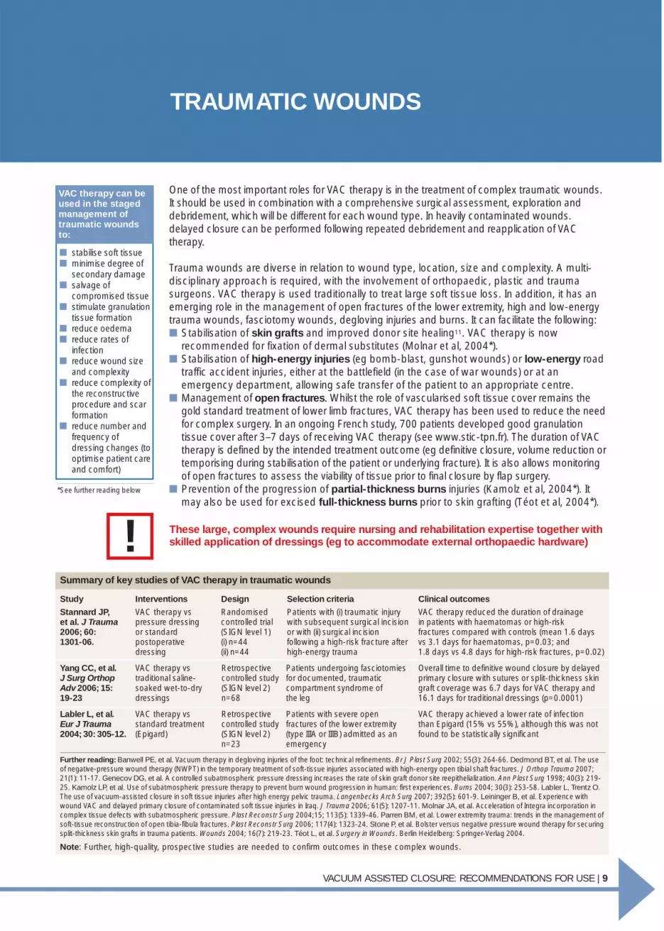
TRAUMATIC WOUNDS
One of the most important roles for VAC therapy is in the treatment of complex traumatic wounds.It should be used in combination with a comprehensive surgical assessment, exploration anddebridement, which will be different for each wound type. In heavily contaminated wounds.delayed closure can be performed following repeated debridement and reapplication of VACtherapy.
Trauma wounds are diverse in relation to wound type, location, size and complexity. A multi-disciplinary approach is required, with the involvement of orthopaedic, plastic and traumasurgeons. VAC therapy is used traditionally to treat large soft tissue loss. In addition, it has anemerging role in the management of open fractures of the lower extremity, high and low-energytrauma wounds, fasciotomy wounds, degloving injuries and burns. It can facilitate the following:■ Stabilisation of skin grafts and improved donor site healing11. VAC therapy is now
recommended for fixation of dermal substitutes (Molnar et al, 2004*).■ Stabilisation of high-energy injuries (eg bomb-blast, gunshot wounds) or low-energy road
traffic accident injuries, either at the battlefield (in the case of war wounds) or at anemergency department, allowing safe transfer of the patient to an appropriate centre.
■ Management of open fractures. Whilst the role of vascularised soft tissue cover remains thegold standard treatment of lower limb fractures, VAC therapy has been used to reduce the needfor complex surgery. In an ongoing French study, 700 patients developed good granulationtissue cover after 3–7 days of receiving VAC therapy (see www.stic-tpn.fr). The duration of VACtherapy is defined by the intended treatment outcome (eg definitive closure, volume reduction ortemporising during stabilisation of the patient or underlying fracture). It is also allows monitoringof open fractures to assess the viability of tissue prior to final closure by flap surgery.
■ Prevention of the progression of partial-thickness burns injuries (Kamolz et al, 2004*). Itmay also be used for excised full-thickness burns prior to skin grafting (Téot et al, 2004*).
Study Interventions Design Selection criteria Clinical outcomes
Stannard JP, VAC therapy vs Randomised Patients with (i) traumatic injury VAC therapy reduced the duration of drainageet al. J Trauma pressure dressing controlled trial with subsequent surgical incision in patients with haematomas or high-risk 2006; 60: or standard (SIGN level 1) or with (ii) surgical incision fractures compared with controls (mean 1.6 days1301-06. postoperative (i) n=44 following a high-risk fracture after vs 3.1 days for haematomas, p=0.03; and
dressing (ii) n=44 high-energy trauma 1.8 days vs 4.8 days for high-risk fractures, p=0.02)
Yang CC, et al. VAC therapy vs Retrospective Patients undergoing fasciotomies Overall time to definitive wound closure by delayedJ Surg Orthop traditional saline- controlled study for documented, traumatic primary closure with sutures or split-thickness skinAdv 2006; 15: soaked wet-to-dry (SIGN level 2) compartment syndrome of graft coverage was 6.7 days for VAC therapy and19-23 dressings n=68 the leg 16.1 days for traditional dressings (p=0.0001)
Labler L, et al. VAC therapy vs Retrospective Patients with severe open VAC therapy achieved a lower rate of infection Eur J Trauma standard treatment controlled study fractures of the lower extremity than Epigard (15% vs 55%), although this was not 2004; 30: 305-12. (Epigard) (SIGN level 2) (type IIIA or IIIB) admitted as an found to be statistically significant
n=23 emergency
Further reading: Banwell PE, et al. Vacuum therapy in degloving injuries of the foot: technical refinements. Br J Plast Surg 2002; 55(3): 264-66. Dedmond BT, et al. The useof negative-pressure wound therapy (NWPT) in the temporary treatment of soft-tissue injuries associated with high-energy open tibial shaft fractures. J Orthop Trauma 2007;21(1): 11-17. Genecov DG, et al. A controlled subatmospheric pressure dressing increases the rate of skin graft donor site reepithelialization. Ann Plast Surg 1998; 40(3): 219-25. Kamolz LP, et al. Use of subatmospheric pressure therapy to prevent burn wound progression in human: first experiences. Burns 2004; 30(3): 253-58. Labler L, Trentz O.The use of vacuum-assisted closure in soft tissue injuries after high energy pelvic trauma. Langenbecks Arch Surg 2007; 392(5): 601-9. Leininger B, et al. Experience withwound VAC and delayed primary closure of contaminated soft tissue injuries in Iraq. J Trauma 2006; 61(5): 1207-11. Molnar JA, et al. Acceleration of Integra incorporation incomplex tissue defects with subatmospheric pressure. Plast Reconstr Surg 2004;15; 113(5): 1339-46. Parren BM, et al. Lower extremity trauma: trends in the management ofsoft-tissue reconstruction of open tibia-fibula fractures. Plast Reconstr Surg 2006; 117(4): 1323-24. Stone P, et al. Bolster versus negative pressure wound therapy for securingsplit-thickness skin grafts in trauma patients. Wounds 2004; 16(7): 219-23. Téot L, et al. Surgery in Wounds. Berlin Heidelberg: Springer-Verlag 2004.
Note: Further, high-quality, prospective studies are needed to confirm outcomes in these complex wounds.
Summary of key studies of VAC therapy in traumatic wounds
■ stabilise soft tissue■ minimise degree of
secondary damage■ salvage of
compromised tissue■ stimulate granulation
tissue formation■ reduce oedema ■ reduce rates of
infection■ reduce wound size
and complexity■ reduce complexity of
the reconstructiveprocedure and scarformation
■ reduce number andfrequency ofdressing changes (tooptimise patient careand comfort)
VAC therapy can beused in the stagedmanagement oftraumatic woundsto:
These large, complex wounds require nursing and rehabilitation expertise together withskilled application of dressings (eg to accommodate external orthopaedic hardware)!
*See further reading below
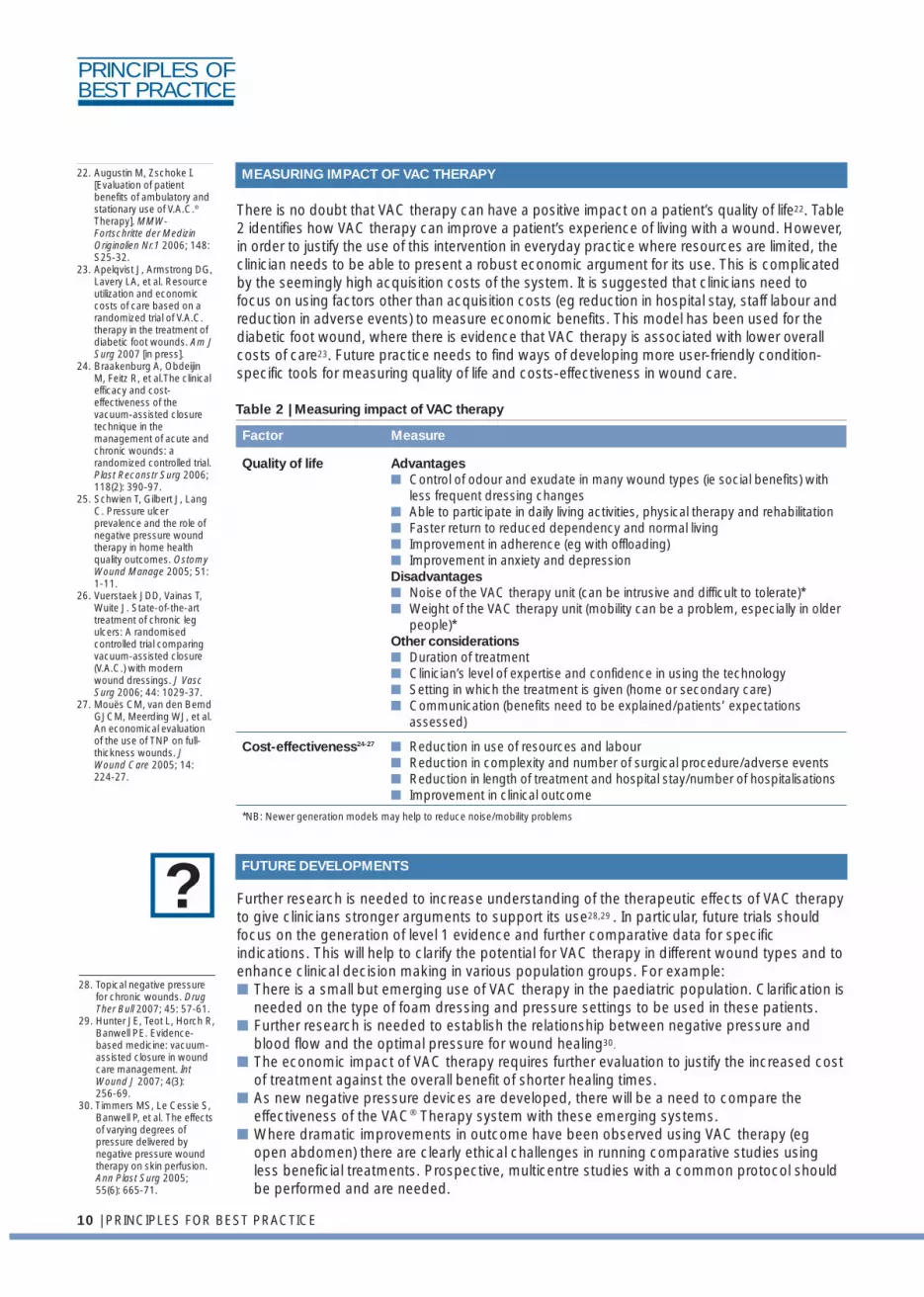
There is no doubt that VAC therapy can have a positive impact on a patient’s quality of life22. Table2 identifies how VAC therapy can improve a patient’s experience of living with a wound. However,in order to justify the use of this intervention in everyday practice where resources are limited, theclinician needs to be able to present a robust economic argument for its use. This is complicatedby the seemingly high acquisition costs of the system. It is suggested that clinicians need tofocus on using factors other than acquisition costs (eg reduction in hospital stay, staff labour andreduction in adverse events) to measure economic benefits. This model has been used for thediabetic foot wound, where there is evidence that VAC therapy is associated with lower overallcosts of care23. Future practice needs to find ways of developing more user-friendly condition-specific tools for measuring quality of life and costs-effectiveness in wound care.
Further research is needed to increase understanding of the therapeutic effects of VAC therapyto give clinicians stronger arguments to support its use28,29 . In particular, future trials shouldfocus on the generation of level 1 evidence and further comparative data for specificindications. This will help to clarify the potential for VAC therapy in different wound types and toenhance clinical decision making in various population groups. For example:■ There is a small but emerging use of VAC therapy in the paediatric population. Clarification is
needed on the type of foam dressing and pressure settings to be used in these patients. ■ Further research is needed to establish the relationship between negative pressure and
blood flow and the optimal pressure for wound healing30.
■ The economic impact of VAC therapy requires further evaluation to justify the increased costof treatment against the overall benefit of shorter healing times.
■ As new negative pressure devices are developed, there will be a need to compare theeffectiveness of the VAC® Therapy system with these emerging systems.
■ Where dramatic improvements in outcome have been observed using VAC therapy (egopen abdomen) there are clearly ethical challenges in running comparative studies usingless beneficial treatments. Prospective, multicentre studies with a common protocol shouldbe performed and are needed.
10 | PRINCIPLES FOR BEST PRACTICE
22. Augustin M, Zschoke I.[Evaluation of patientbenefits of ambulatory andstationary use of V.A.C.®
Therapy]. MMW-Fortschritte der MedizinOriginalien Nr.1 2006; 148:S25-32.
23. Apelqvist J, Armstrong DG,Lavery LA, et al. Resourceutilization and economiccosts of care based on arandomized trial of V.A.C.therapy in the treatment ofdiabetic foot wounds. Am JSurg 2007 [in press].
24. Braakenburg A, ObdeijinM, Feitz R, et al.The clinicalefficacy and cost-effectiveness of thevacuum-assisted closuretechnique in themanagement of acute andchronic wounds: arandomized controlled trial.Plast Reconstr Surg 2006;118(2): 390-97.
25. Schwien T, Gilbert J, LangC. Pressure ulcerprevalence and the role ofnegative pressure woundtherapy in home healthquality outcomes. OstomyWound Manage 2005; 51:1-11.
26. Vuerstaek JDD, Vainas T,Wuite J. State-of-the-arttreatment of chronic legulcers: A randomisedcontrolled trial comparingvacuum-assisted closure(V.A.C.) with modernwound dressings. J VascSurg 2006; 44: 1029-37.
27. Mouës CM, van den BemdGJCM, Meerding WJ, et al.An economical evaluationof the use of TNP on full-thickness wounds. JWound Care 2005; 14:224-27.
Table 2 | Measuring impact of VAC therapy
Quality of life Advantages■ Control of odour and exudate in many wound types (ie social benefits) with
less frequent dressing changes■ Able to participate in daily living activities, physical therapy and rehabilitation■ Faster return to reduced dependency and normal living■ Improvement in adherence (eg with offloading)■ Improvement in anxiety and depressionDisadvantages■ Noise of the VAC therapy unit (can be intrusive and difficult to tolerate)*■ Weight of the VAC therapy unit (mobility can be a problem, especially in older
people)*Other considerations ■ Duration of treatment■ Clinician’s level of expertise and confidence in using the technology■ Setting in which the treatment is given (home or secondary care)■ Communication (benefits need to be explained/patients’ expectations
assessed)
Cost-effectiveness24-27 ■ Reduction in use of resources and labour■ Reduction in complexity and number of surgical procedure/adverse events■ Reduction in length of treatment and hospital stay/number of hospitalisations■ Improvement in clinical outcome
*NB: Newer generation models may help to reduce noise/mobility problems
MEASURING IMPACT OF VAC THERAPY
FUTURE DEVELOPMENTS
Factor Measure
PRINCIPLES OFBEST PRACTICE
?28. Topical negative pressure
for chronic wounds. DrugTher Bull 2007; 45: 57-61.
29. Hunter JE, Teot L, Horch R,Banwell PE. Evidence-based medicine: vacuum-assisted closure in woundcare management. IntWound J 2007; 4(3): 256-69.
30. Timmers MS, Le Cessie S,Banwell P, et al. The effectsof varying degrees ofpressure delivered bynegative pressure woundtherapy on skin perfusion.Ann Plast Surg 2005;55(6): 665-71.