recommendations - european commission
TRANSCRIPT
EUROPEAN COMMISSIONHEALTH & CONSUMER PROTECTION DIRECTORATE-GENERAL
Scientific Steering Committee
OPINION ON
THE SAFETY OF RUMINANT BLOOD WITH RESPECT TO TSE RISKS
ADOPTED BY THE
SCIENTIFIC STEERING COMMITTEE
AT ITS MEETING OF 13-14 APRIL 2000
T_BLOOD.doc 2
OPINION
Animal (incl. ruminant) blood is currently legally fed to animals (including ruminants)
after very gentle processes, spread on pasture as fertiliser, or used for other products that
may reach man or animals (incl. ruminants). There is some concern that animal TSEs
might be spread by these means or through specific blood components or blood based
products that are still permitted to enter the market. It is therefore necessary to establish if
animal TSEs can be transferred via blood and the SSC was requested to:
A. Assess for ruminant blood in general and if possible for each component the risk that
it could harbour the BSE (TSE) agent and hence transfer the disease.
B. Identify the main ruminant blood-based products that are currently on the market,
including products containing ruminant blood or those in which ruminant blood is
used in their manufacture.
C. Assess for each product, the function of the blood component included and the risk
that that product could transfer BSE (TSE) to ruminants.
D. Outline possible measures that could mitigate any identified risk, as far as possible
together with a (qualitative) assessment of the potential impact of the measure on the
risk.
In order to answer these questions, a Working Group was created which delivered a
detailed report upon which this opinion is based (see attachment). On the basis of this
report, the SSC elaborated the following summary account, conclusions and
recommendations.
Remark: The present opinion does not address the possible risks resulting from the
exposure of workers to contaminated material. The SSC is presently preparing Notes on
the safe handling, transport and storage of MBM and other bovine derived materials
which may be contaminated with BSE or other pathogens.
REGARDING EXPERIMENTAL STUDIES ON TSE INFECTIVITY IN BLOOD AND ITS
COMPONENTS.
The majority of assays of infectivity in blood have been carried out in animals or humans
with clinically overt TSE and in consequence there remains substantial ignorance about
the early pathogenetic involvement of the blood, especially in the naturally occurring

T_BLOOD.doc 3
diseases. In BSE, transmission has not been achieved in the majority of studies either in
natural and experimental disease, although recent investigations indicate this possibility.
In experimental scrapie, blood components obtained during both the pre-clinical and
clinical stages of disease from rodents, have revealed the presence of the infectious agent.
The results reviewed in the Working Group Report are consistent with recent data in mice
infected with mouse-adapted human TSE, and in hamsters infected with hamster-adapted
scrapie.
Although reliance upon the results requires an assumption that experimental rodent data
can be extrapolated to the conditions of natural disease in humans and other animal
species, the data from both experimentally-induced and natural TSE suggest that blood
has at least the potential to transmit disease. The reasons for the discrepancy between this
laboratory evidence and the epidemiological evidence that has so far failed to identify any
blood-related cases of TSE, are probably multiple: the absence of significant blood
infectivity until the onset of symptomatic disease, and comparatively low levels of
infectivity during the symptomatic stage of disease: the need for 5 to 10 times more
infectious agent to transmit disease by the intravenous than intracerebral route. For some
blood products, there is a further reduction of infectivity during the course of blood
processing (for example an estimated reduction of 3 logs for the "133°C/20'/3bars"
treatment).
REGARDING SOURCING
Slaughtered cattle are used to source blood for food, feed and a variety of other purposes
including the manufacture of medicinal products and biologicals. Farmed sheep, goats
and deer could supply blood for these purposes. All these species are susceptible to TSE
both naturally and experimentally. BSE as a natural disease has only been reported in
cattle. The possibility of BSE being in sheep and goats cannot be excluded. Furthermore,
one hypothesis for the origin of BSE is from a scrapie-like agent from sheep. It is possible
that such a source still exists or that the BSE agent from cattle has returned to sheep and
goats. If it exists it may no longer be recognisable if natural sub-passage has occurred.
No validated tests exist to detect TSE in live cattle, sheep, goats or deer. Clinical TSE has
never been reported in deer in Europe. A post mortem inspection will not enable detection
of any TSE in any species and will not improve upon the ante mortem inspection. Close
T_BLOOD.doc 4
surveillance for the disease and effective ante mortem clinical inspection of all slaughter
animals therefore remain essential.
Exogenous contamination of blood with CNS material in the form of emboli (and hence
infectivity) is most likely in TSE-infected animals stunned with a stunning pistol that
injects gas into the cranial cavity under pressure. This can also occur if a conventional
cartridge –fired captive bolt is used in combination with pithing. Exogenous
contamination of blood could occur post mortem if SRM are not kept separated from
collected blood.
REGARDING RISK ASSESSMENT
The SSC considers that the most important aspect of risk assessment relates to brain
tissue contamination. The SSC proposes a general approach for the risk assessment for
blood within a given area, which basically involves 3 aspects:
• Slaughterhouse
Basically at the level of each individual slaughterhouse, the following risk factors should
be recorded:
1) number, species and age of slaughtered animals ;
2) number of potentially infected cows being killed and their brain material entering the
bloodstream related to the stunning method used (pneumatic devices, pithing) ;
3) the average amount of blood collected per animal;
4) the dilution by pooling blood from several animals;
5) the amount of such collected blood going to the industry to be processed for human
or animal consumption.
• Geographical BSE risk and surveillance
For the geographical BSE risk and surveillance reference is made to the SSC opinions
adopted sofar by the SSC.
• The use of blood
At present, blood collected hygienically in licensed EU abattoirs can be used for food,
feed and a variety of other purposes without any form of processing. For example, it is
permissible to incorporate fresh untreated plasma into the materials used for the

T_BLOOD.doc 5
production of sausages, and can be spread on land as a fertiliser. Elsewhere, in this
opinion, it is concluded that there could be a risk of the occasional presence of low levels
of TSE infectivity in blood collected in abattoirs. Levels of infectivity which might
represent a risk to animal or human health are not known. Control measures and/or
decontamination standards might need to be developed to potentially TSE-infected blood
collected in abattoirs.
There is insufficient information on the nature of usage of blood and the resulting blood-
products obtained from abattoirs within the EU. The situation is further complicated by
the fact that the names used to describe identical or similar products in different countries
can be quite different. From the limited information available on the manufacturing
processes for blood-products, the most rigorous methods applied would appear to involve
coagulation of the blood at 95-100oC, followed by spray-drying during which the end-
product reaches a temperature of 110oC. Several sets of data indicate that this process is
unlikely to result in any significant reduction in the titre of TSE infectivity. The
alternative is to either apply risk-assessment techniques or, where practicable, adopt the
rule that products should be subjected to the "133oC/20’/3bars" standard. The latter
approach is applicable only to some products.
At the level of processing of blood for human and/or animal consumption, risk can be
further evaluated in respect to
(i) the amount of brain material actually entering the bloodstream following the use of
invasive stunning devices. Neither its volume range nor the range of particle size is
known. Likewise, no quantitative estimates are available on contamination of blood
with SRM materials during the slaughtering process other than by stunning the
animals.
(ii) dilution of CNS material resulting from the emboli and
(iii) the efficacy of the various processing steps in respect to inactivating the BSE agent.
There is little doubt that under certain circumstances, humans or animals could be
exposed to the BSE agent by consuming blood products.
T_BLOOD.doc 6
CONCLUSIONS
The collective data currently available from experimental transmission studies show that
there is uncertainty on the presence of infectivity in the blood of TSE-infected ruminants.
If PrPSc has been detected in the blood of clinically normal sheep from scrapie-susceptible
flocks using a newly-developed and highly sensitive assay system, infectivity of
femtomole amounts remain to be demonstrated.
The relationship between PrPSc and infectivity is not understood. The two do not always
correlate; the presence of PrPSc does not necessarily imply presence of infectivity.
Moreover, the methods for detecting PrPSc need to be validated for the pre-clinical stage.
As far as ruminant blood is concerned, it is considered that the best approach to protect
public health at present is to assume that it could contain low levels of infectivity.
However, even if this is true, it becomes almost irrelevant compared with the level of
contamination that could occur as a result of the methods of stunning used in abattoirs.
These procedures are now recognised to release particles of brain-tissue (potentially
containing high titres of TSE infectivity) into the bloodstream. The frequency at which
this occurs appears to increase with the severity of the stunning process, and this report
recognises that this is an area requiring further research. There are also opportunities for
the contamination of pooled blood as a consequence of the release of brain-tissue from
the hole left by stunning, or with spinal cord during its removal (if a production-line
process is not used). Nevertheless, given the low frequency at which apparently healthy
animals would have TSE infectivity in the CNS at the time of slaughter, it is considered
that the overall potential level of infectivity in pooled blood will be low.
Further specific conclusions are:
- The highest risk of producing CNS emboli follows captive bolt stunning with
compressed air into the cranial cavity.
- Cartridge operated captive bolt stunning-followed-by-pithing presents the next highest
risk.
- There is insufficient knowledge to advise on the degree of risk from the use of
penetrative cartridge-operated stuns without pithing, free bullets or non-penetrative
guns.
- There are no published papers on the effect of various stunning methods on sheep and
goats and in regard to the generation of CNS emboli.

T_BLOOD.doc 7
- More information is required on the possible dissemination of CNS emboli into the
systemic circulation.
- TSE risks may exist as a result of the source of animals for slaughter.
- TSE risks may occur independently of the stunning procedure as a result of TSE-
infected material from SRM for example entering the blood after exit from the body.
- Improved processing procedures could reduce residual TSE risk in the collected
material.
- An accurate ante mortem examination performed at slaughterhouse is helpful to
increase prevention, whereas this is not the case for post-mortem inspection.
1. RECOMMENDATIONS
The Scientific Steering Committee recommends that the present opinion on the safety of
ruminant blood is considered in conjunction with its opinions on "Fallen stock" (June
1999)1 and "Intra-species Recycling"2.
The SSC also recommends that intraspecies recycling of ruminant blood and blood
products should be avoided in situations when a TSE risk exists.
Consideration should further be given to avoiding methods of captive bolt stunning with
compressed air or followed by pithing ruminant food animals that increase the risk of
CNS material entering the blood stream at slaughter wherever there is a significant risk
from TSE3. In addition, sourcing from young4 animals would further reduce the risk.
1 Scientific Opinion of 24-25 June 1999 of the SSC on The risks of non conventional transmissible agents,conventional infectious agents or other hazards such as toxic substances entering the human food oranimal feed chains via raw material from fallen stock and dead animals (including also: ruminants, pigs,poultry, fish, wild/exotic/zoo animals, fur animals, cats, laboratory animals and fish) or via condemnedmaterials. Adopted By the Scientific Steering Committee
2 Scientific Opinion of 24-25 June 1999 of the SSC on the risk born by recycling animal by-products asfeed with regard to propagating TSE in non-ruminant farmed animals.
3 Changing from pneumatic stunning or pithing, to stunning methods that avoid severe brain damagecould go along with an increased risk of physical injury to slaughtermen (particularly during shacklingand bleeding out) if the new methods or building facilities are not properly designed.
4 First infectivity in CNS of cattle is detected in most cases in the last quarter of the incubation period.Defining young animals could be done on the basis of the probability of occurrence of BSE according tothe age. (See for example the annexes 3 and 4 of the Opinion of 28-29 October 1999 of the ScientificSteering Committee on the Scientific Grounds of the Advice of 30 September 1999 of the French FoodSafety Agency (the Agence Française de Sécurité Sanitaire des Aliments, AFSSA), to the FrenchGovernment on the Draft Decree amending the Decree of 28 October 1998 establishing specificmeasures applicable to certain products of bovine origin exported from the United Kingdom.
T_BLOOD.doc 8
(Improved) methods for reducing the risk of cross contaminating blood with CNS or other
SRM post-collection need to be develop or put in place where necessary. Brain spilling
from the bullet hole into the blood tank should be prevented; surveys should check the
absence of brain material in the blood tanks.
Where an element of risk is perceived, this may be reduced or eliminated by (a
combination of) various strategies, as follows:
• Source bovine blood from BSE-free areas or closed herds or other schemes that
reduce to a minimum the probability of an animal being infected;
• Subject the product to a 133oC/3 bar/ 20 minute autoclaving process or equivalent
validated process.
• Pharmaceuticals: including vaccines, are regulated products, and the use of bovine
derived blood products in their manufacture is controlled on a case by case basis. The
basic principles included in the present opinion should of course be respected.
2. NEEDS FOR RESEARCH
Further research is needed:
- to determine the comparative (quantitative) TSE risks from various penetrative and
non-penetrative methods of stunning and pithing in food animal species
- to quantify the possible presence of CNS material in blood following various
stunning methods
- to determine the effect of the "133°C/20'/3bar" treatment on the nutritional value of
blood for animal nutrition and the reduction/elimination of possible infectivity in
blood.
- on possible TSE infectivity of blood cells and other blood components. Furthermore
highly sensitive tests that are presently under development, need to be further
validated.
T_BLOOD.doc 9
REPORT ON
THE SAFETY OF RUMINANT BLOOD WITH RESPECT TO TSE RISKS
SUBMITTED TO
THE SCIENTIFIC STEERING COMMITTEE
AT ITS MEETING OF 13-14 APRIL 2000
…
T_BLOOD.doc 10
DEFINITIONS:
The following definitions are used in the present report:
Blood: fresh whole blood collected from any species of food animal that haspassed an official ante-mortem inspection at a licensed slaughterhouse supervisedby the competent veterinary authority.
Blood products: blood that is treated by physical methods (e.g. separation intocomponents of blood, and/or by heat) or chemical methods such as addition ofanticoagulant.
Blood products for pharmaceutical and cosmetic use: Blood products intendedfor use in biological, medicinal or pharmaceutical products, medical devices orcosmetics.
Blood products for use in food: blood or blood products intended for use inhuman food; post-mortem inspection is mandatory.
Blood products for use in feed: blood or blood products for use in feed for farmanimals (i.e. those species consumed by man).
Blood products for use in petfood: blood or blood products for use in food forcompanion animals.
Blood products for technical use: blood or blood products for uses other thanthose described above, e.g. for fertiliser.
Bloodmeal: a blood product intended for use in animal feed or for technical usesthat has been coagulated by treatment with steam and dried.
Feedingstuffs/Feed material (of animal origin): processed animal protein,rendered fats, gelatine and hydrolysed proteins, milk and milk products, intended tobe used as feed for farmed animals, but excluding petfood.
Laboratory reagents: a packaged product, ready for use by the end user,containing a blood product, and intended for laboratory use as reagent or reagentproduct, whether used alone or in combination.
Processed animal protein (PAP): animal proteins derived entirely from animalby-products, which have been treated so as to render it suitable for direct use asfeedingstuff or as feed material in a feedingstuff for animals or in petfood or asfertilizers. It includes fishmeal, meatmeal, bonemeal, meat-and-bone meal,bloodmeal, dry greaves, feathermeal, [hoofmeal, hornmeal] and other similarproducts including mixture containing these products.
Products used for in vitro diagnosis: a packaged product, ready for use by the enduser, containing a blood product, and used as reagent, reagent product, calibrator,kit or any other system, whether used alone or in combination, intended to be usedin vitro for the examination of samples of human or animal origin, with theexception of donated organs or blood, solely or principally with a view to thediagnosis of a physiological state, state of health, disease or genetic abnormality orto determine safety and compatibility with reagents.
T_BLOOD.doc 11
Technical products: animal by-products intended for purpose other than human oranimal consumption, (such as tanned and treated hides and skins, game trophies,processed wool, hair, bristles, feathers and part of feathers, apicultural products,serum of equidae, blood products other than blood meal, colouring substances forfood, pharmaceutical, bone products for china, gelatine and glue, processedmanure, rendered fats derivatives, organic fertilizers, industrial oil, etc.).
1. MANDATE AND CONTEXT OF THE QUESTION
Animal (incl. ruminant) blood is currently legally fed to animals (includingruminants) after very gentle processes, spread on pasture as fertiliser, or used forother products that may reach man or animals (incl. ruminants). There is someconcern that animal TSEs might be spread via blood or specific blood componentsor blood based products that are still permitted to enter the market. It is thereforenecessary to establish if animal TSEs can be transferred via blood and the SSC wasrequested to:
A. Assess for ruminant blood in general and if possible for each component therisk that it could harbour the BSE (TSE) agent and hence transfer the disease.
B. Identify the main ruminant blood-based products that are currently on themarket, including products containing ruminant blood or those in whichruminant blood is used in their manufacture.
C. Assess for each product, the function of the blood component included and therisk that that product could transfer BSE (TSE) to ruminants.
D. Outline possible measures that could mitigate any identified risk, as far aspossible together with a (qualitative) assessment of the potential impact of themeasure on the risk
2. MATRIX OF PRODUCTS AND USES
Table 2.1. hereafter provides a non exhaustive draft matrix of products and uses ofruminant blood. Table 2.2. is a non-exhaustive draft list of the ruminant bloodderived materials used for human or veterinary medicinal products or for medicaldevices.

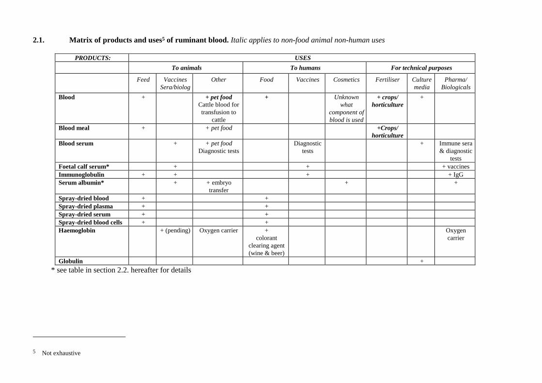
2.1. Matrix of products and uses5 of ruminant blood. Italic applies to non-food animal non-human uses
PRODUCTS: USESTo animals To humans For technical purposes
Feed VaccinesSera/biolog
Other Food Vaccines Cosmetics Fertiliser Culturemedia
Pharma/Biologicals
Blood + + pet foodCattle blood fortransfusion to
cattle
+ Unknownwhat
component ofblood is used
+ crops/horticulture
+
Blood meal + + pet food +Crops/horticulture
Blood serum + + pet foodDiagnostic tests
Diagnostictests
+ Immune sera& diagnostic
testsFoetal calf serum* + + + vaccinesImmunoglobulin + + + + IgGSerum albumin* + + embryo
transfer+ +
Spray-dried blood + +Spray-dried plasma + +Spray-dried serum + +Spray-dried blood cells + +Haemoglobin + (pending) Oxygen carrier +
colorantclearing agent(wine & beer)
Oxygencarrier
Globulin +* see table in section 2.2. hereafter for details
5 Not exhaustive

14
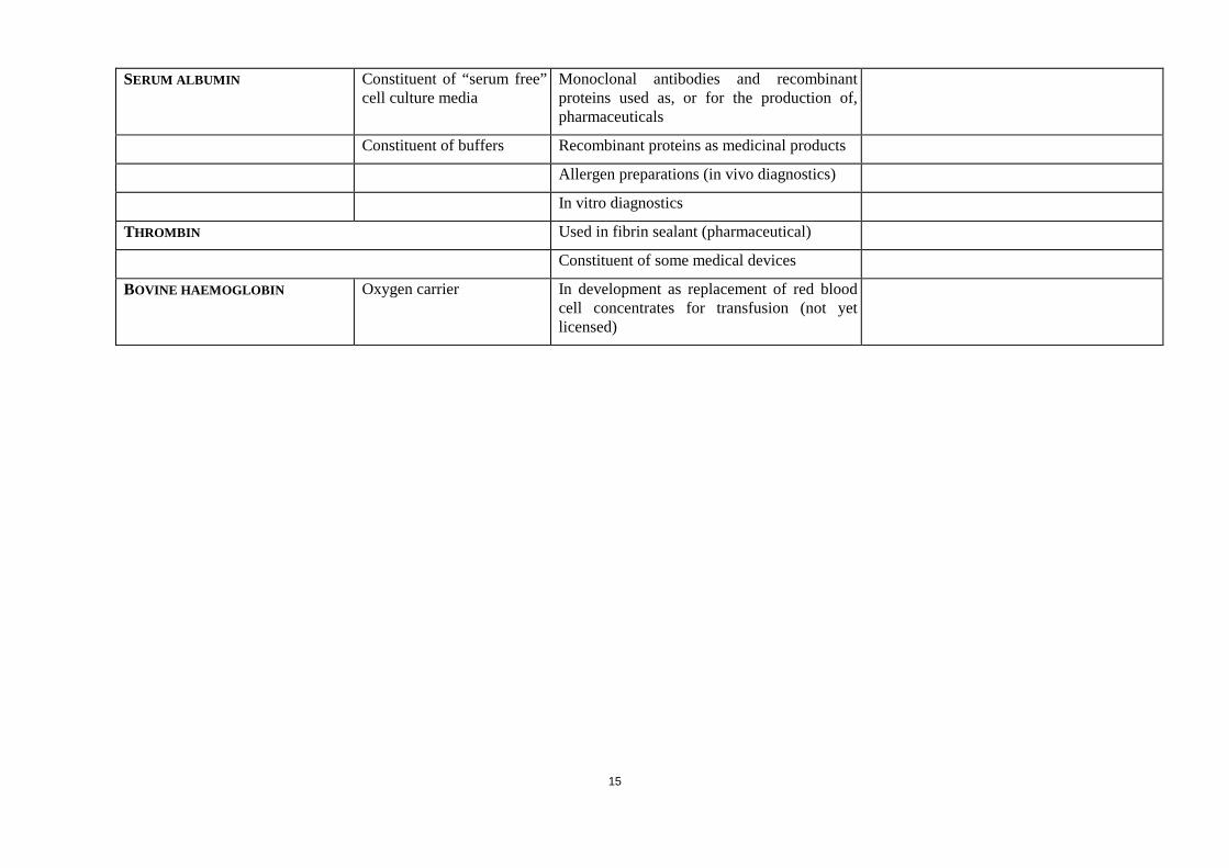
2.2. Ruminant blood derived materials used for human or veterinary medicinal products or for medical devices6
BOVINE BLOOD DERIVEDMATERIAL
GENERAL USE SPECIFIC USE COMMENTS
FOETAL CALF SERUM Cultivation of mammaliancells and, therefore, in theproduction of most cellculture derived medicinalproducts
Veterinary vaccines Used for preparation of cells, propagation ofvaccine strains usually, but not always onlyafter change to serum free medium
Human vaccines Preparation of cells, propagation of vaccinestrains only after change to serum freemedium
Monoclonal antibodies for use in humans Preparation of cells, but increasing use ofserum free media
Monoclonal antibodies, used for purificationof pharmaceutical products (e.g. coagulationfactors)
Preparation of cells, but increasing use ofserum free media
Recombinant proteins from mammalian cellcultures, used as pharmaceuticals
Preparation of cells, but increasing use ofserum free media
Products for gene therapy (preparation ofvectors)
Preparation of cells, but increasing use ofserum free media
Stabiliser in buffers In vitro diagnostics Usually, foetal calf serum is used, but theoccasional use of calf serum or bovineserum cannot be excluded
CALF SERUM Cultivation of mammaliancells
Some stocks of seed viruses (for theproduction of vaccines)
Usually replaced by foetal calf serum
BOVINE SERUM Most often replaced by foetal calf serum
6 not exhaustive
15
SERUM ALBUMIN Constituent of “serum free”cell culture media
Monoclonal antibodies and recombinantproteins used as, or for the production of,pharmaceuticals
Constituent of buffers Recombinant proteins as medicinal products
Allergen preparations (in vivo diagnostics)
In vitro diagnostics
THROMBIN Used in fibrin sealant (pharmaceutical)
Constituent of some medical devices
BOVINE HAEMOGLOBIN Oxygen carrier In development as replacement of red bloodcell concentrates for transfusion (not yetlicensed)

3. EXPERIMENTAL STUDIES ON TSE INFECTIVITY IN BLOOD AND ITS COMPONENTS.This chapter is structured and based in part on the Risk quantification for CJDtransmission via substances of human origin adopted on 21 October 1998 by theScientific Committee on Medicinal Products and Medical Devices.
3.1. GeneralThe transmissible spongiform encephalopathies (TSEs) or prion diseases present assporadic, acquired and inherited disorders which are usually transmissible onexperimental inoculation. Infectivity can be measured only by bioassay. There aretwo common methods of bioassay: end point titration or incubation time intervalassay (Millson et al, 1976, Outram, 1976, Prusiner, 1987, Scott 1993). Bothmethods are slow and costly, especially the former, which has undoubtedlycontributed to the infrequent use of classical titration methods. Tissues or bodyfluids are collected from naturally or experimentally infected animals, andinoculated into healthy inbred assay animals, usually by the intracerebral route(i.c.), but sometimes by other routes - intraperitoneal (i.p), intravenous (i.v.), orsubcutaneous (s.c.), or a combination of i.c. and i.p. It is almost always advised thatacross species barriers i.c. and i.p. should be used. The assay animals are observedover a period of months or years for clinical signs of transmissible spongiformencephalopathy (TSE), which is confirmed by histopathological examination, andin some studies also by Western blot identification of PrPres in the brain.
End point titrations offer the most precise assay of infectivity and are typicallyperformed by the intracerebral injection of ten fold serial dilutions of a tissuesample into groups of rodents. The concentration of infectious agent in the sampleis expressed as the highest dilution capable of transmitting disease to one half ofthe inoculated animals (log10 ID50/g.)
Incubation time assays involve the construction of a dose response curve by endpoint titrations, to enable a titre to be interpolated from the curve for a givendilution of the test specimen. The incubation period assay uses far fewer animals,but its accuracy is dependent upon the skills of the observer in defining the clinicalonset of disease, and on the conditions to which the inocula are subjected (forexample, dose-response curves are significantly extended after exposure ofinfectious samples to partially-inactivating procedures) (Taylor et al, 1999).
Of special importance for measuring the very low concentrations of infectivity inblood is the fact that at limiting dilutions of inoculum the incubation periodbecomes an unreliable measure of the concentration of infectivity, and thus manyanimals (often 30-40) are needed for inoculation of the undiluted specimen in orderto attain a statistically accurate estimate of infectivity.
One factor influencing the sensitivity of TSE bioassay methods concerns thespecies barrier effect, which may result in an underestimate or even absence ofapparent infectivity in the specimen being assayed. Ideally, therefore, infectivityassays should be conducted in the same species as that of the donor animal. Assaysare conducted using a single inoculation of the assay animals. The effects ofrepeated inoculations have not yet been investigated in depth.
The route of exposure is another important variable. For example, inoculation ofblood or a blood product by the i.c. route does not answer the question oftranmissibility, for example by the oral route in feed.
17
Finally, it must always be remembered that unexpected or bizarre experimentalresults require a very cautious evaluation until they can be shown to bereproducible and independently confirmed. Errors of all sorts are possible, fromspecimen mislabelling to laboratory cross-contamination. Some reports ofinfectivity in the blood of humans with CJD must be viewed with circumspectionbecause of aberrant experimental results, or unreproducibility. Tateishi (1980),Manuelidis (1985), Tamai et al (1992).
Blood comprises fluid plasma and cellular elements, and is the body’s essentialtransport medium. Many organs are highly vascularised (e.g. liver, spleen, placenta)and blood represents a significant proportion of their volume. The blood-formingorgans (haemopoietic system) are not only related functionally to the production butalso to the degradation of blood components. The haemopoietic system includes thelymph nodes, tonsil and other submucosal lymphoid nodules, thymus, spleen andbone marrow. The destruction of red blood cells occurs mainly in the spleen. It isnoted that ruminant species have haemal nodes which function to filter blood. Inthe foetus and for a period after birth haemopoiesis has a wider distribution to theliver and meninges in cattle for example.
All experimental transmissions were done by using blood taken from animalswithout brain trauma, that is, living animals.
3.2. Studies of blood infectivity in naturally infected donor species assayed in thesame recipient speciesThere is no literature about same-species assays of blood from cases of naturallyoccurring TSE.
A study undertaken at the Institute for Animal Health, UK, is examining wholeblood and buffy coat from scrapie susceptible Cheviot sheep (PrP genotypeVRQ/VRQ) in the pre-clinical phase of natural scrapie. The samples weresubsequently transfused into susceptible but scrapie-free sheep (PrP genotype:VRQ/VRQ) sourced from New Zealand, and these are being observed for signs ofTSE. The study has been underway for about one year and no clinical cases have sofar developed in the blood or buffy coat recipients (N. Hunter, unpublished data).
3.3. Studies of blood infectivity in naturally infected donor species assayed indifferent recipient species (Table 1)No infectivity was found in serum or in clotted blood of scrapie infected goats andsheep when assayed in Swiss mice (Hadlow et al, 1980, Hadlow et al, 1982). Thesingle report of infectivity in the serum of a naturally infected ram by assay inSwiss mice (Gibbs, 1965) was not confirmed. In natural cases of BSE assayed inRIII mice (Sinc s7), infectivity has been found only in the central nervous system(CNS: brain, spinal cord and retina). No infectivity was found in about 50 othertissues including bone marrow, clotted blood, buffy coat, serum or foetal calf serum(Fraser and Foster, 1994; Bradley, 1999).
3.5. Studies of blood infectivity in experimentally infected donor species assayed indifferent recipient species (Table 2)No infectivity was found by mouse bioassay in blood-clots from scrapie infectedgoats during a time course study (Hadlow et al, 1974).
18
Tissue infectivity in BSE has been comprehensively investigated in cattle in anexperimental pathogenesis study in which cattle were challenged orally with 100ginfected cattle brain from natural cases. Clinical disease was first detected at 35months p.i. (39 months of age). No infectivity by assay in RIII or C57Bl mice wasdetected in buffy coat of orally infected cattle up to the termination of the study at40 months p.i., by which time all remaining animals had become ill. (Wells et al,1996; Wells et al, 1998; G.A.H. Wells unpublished data). Frozen sera and bloodclots have not yet been assayed (Wells et al, 1996).
Studies in progress at the VLA, UK are examining the transmissibility,pathogenesis and phenotype of BSE in Romney and Suffolk sheep after oralexposure to affected cattle brain tissue. Buffy coat, obtained from exposed sheep at6 month intervals p.i. (ARQ/ARQ, PrP genotype) and at 12 month intervals p.i.(ARR homozygous and heterozygous) will be assayed in RIII mice. Infectivity inliver and spleen of the exposed sheep will also be assayed in RIII mice at sequentialtime intervals of 6 or 12 months p.i. (S. Bellworthy, personal communication).
3.5 Studies of blood infectivity in experimentally infected donor species assayed inthe same recipient species (Table 3)Two studies of scrapie (Pattison and Millson, 1962; Pattison, et al 1964) with goatsas donor and indicator/recipient animals did not demonstrate any infectivity inblood (fig. 1).
In two time course studies performed in scrapie infected mice, small amounts ofinfectivity were detected in the blood during later stages of the incubation period inone study (Dickinson and Stamp, 1969), but not in the other (Eklund et al, 1967).This difference may have been due to the use of mouse strains (Dickinson andStamp, 1969). Clarke and Haig (1967) detected infectivity in the blood of diseasedmice and rats infected with the Chandler strain of scrapie, but argued that theamount of infectivity may have resulted from contamination of blood samples withother bodily cells.
Infectivity in the blood of hamsters infected with the hamster adapted 263K strainof scrapie has been investigated by two groups (Diringer, 1984; Casaccia et al,1989) using identical methods to concentrate the scrapie agent from whole blood.Both groups found infectivity in the preparations, but one study was terminated 6weeks after infection, and the other study reported declining levels of infectivityduring the later stages of disease. Using the same 263K hamster scrapie agent,Brown et al (1998) inoculated hamsters with normal human blood that had beenspiked with the agent. Infectivity titres were highest in buffy coat, lower in plasma,and very low to absent in Cohn plasma fractions (see Table 4).
A study undertaken at the Institute for Animal Health, UK, is examining sheep ofthe ARQ/ARQ PrP genotype orally infected with BSE. Whole blood and buffy coatwere taken from the animals at various time points during the incubation period.The samples were subsequently transfused into susceptible but TSE-free sheepsourced from New Zealand and these are being observed for signs of TSE. Thestudy has been underway for about one year and no clinical cases have so fardeveloped in the blood or buffy coat recipients. In continuing studies of thepathogenesis of BSE after experimental oral exposure of cattle (Wells et al, 1996,1998, 1999) assays of pooled buffy coats from cattle at each of the original studytime points (6, 18, 26 and 32 months p.i.) are in progress at the VLA, UK. There is
19
no evidence of transmission to date, but some of these experiments have as yetbeen set up only for one year (G.A.H.Wells, unpublished data)
Two studies in mink (Mustela vison) inoculated with the transmissible minkencephalopathy agent, did not reveal any infectivity in serum (Marsh, 1969) orother blood components (Marsh, 1973). In another study, only a single serumspecimen, taken 28 weeks after inoculation and four weeks before the onset ofclinical signs, showed a small amount of infectivity (Hadlow, 1987). Experimentalmodels of human TSE have also been used in a search for infectivity in blood. Theresult obtained in guinea pigs inoculated with the CJD agent adapted to this speciesis puzzling in that infectivity appeared and disappeared irregularly during theincubation period (Manuelidis, 1978a, see fig. 4). A more consistent picture wasobtained in mice inoculated with the Fukuoka strain of human GSS (Tateishi et al,1980; Kuroda et al, 1983). In this model, low but increasing levels of infectivitywere found in buffy coats obtained during the later stages of disease (fig. 5). In aclosely related system, Doi (1991) could not demonstrate infectivity in whole (ofwhich leukocytes constitute approximately 1% by volume).
The same system has been studied at the NIH, USA (Brown et al, 1998, 1999).Infectivity bioassays were conducted in healthy mice, and the brains of all assayanimals dying during the course of the experiments were examined for the presenceof PrPSc. Infectivity in the blood of animals during the pre-clinical phase of disease(Table 4) occurred in the buffy coat at levels of between 6 and 12 infectious units7
per ml, and was either absent or present in only trace amounts in plasma andplasma fractions. Infectivity rose sharply at the onset of clinical signs to levels ofapproximately 100 infectious units/ml buffy coat, 20 infectious units/ml plasma, 2infectious units/ml cryoprecipitate, and less than 1 infectious unit /ml fractions IVand V. Plasma infectivity was not significantly reduced by either leukodepletionfiltration (Table 5) or high speed (17,000 x g) centrifugation (Table 6).Approximately 7 times more plasma and 5 times more buffy coat were needed totransmit disease by the intravenous than by the intracerebral route (Table 7). Tayloret al (2000) showed small amounts of infectivity can be found in the plasma ofmice when they are infected with mouse-passaged BSE agent. The results are rathersimilar to those obtained by Brown et al (see above) when they looked at the bloodof mice infected with mouse-passaged GSS agent.
3.6. Evidence of infectivity in other organs, tissues and cells with functionalrelationship to blood.Previous work on the tissue distribution of infectivity (detected by mouse bioassay)in sheep and goats with natural scrapie and cattle with BSE has been summarised(OIE, 1996). In scrapie, beginning after 8 months and detected at 10-14 months ofage, low levels of infectivity are detectable in a wide range of lymphoreticular richtissues including the intestine, lymph nodes, spleen and tonsil, and these levelsincrease through the incubation period to clinical disease onset. In clinical cases ofBSE no infectivity has been detected by mouse bioassay in spleen, tonsil, regionallymph nodes, intestine, bone marrow, liver or placenta. Haemal nodes have notbeen tested in regard to TSEs so far.
7 Within the context of Brown et al., (1999) an infectious unit is functionally defined as equivalent to thenumber of transmissions/ml.
20
Data for BSE are based on transmissions attempted from a very small number ofanimals. Nevertheless, the findings are consistent with studies of the pathogenesisof BSE in cattle after oral challenge. In further examinations of the tissues fromthis pathogenesis experiment additional same-species assays by i.c. challenge withblood- associated tissues pooled from selected time points are being conducted bythe VLA, UK. These tissues, with their respective time point derivations, include:spleen (6, 10, 18, 26 months p.i.), liver (6, 18 26, 32 months p.i.) and bone marrow(22, 26, 32, 36 months p.i.). Inoculations of these tissues began in November 1996and continued through March 1999. Consequently the assays have been in progressfor periods of 10-38 months and there is no evidence of transmission to date(January 2000) (G.A.H.Wells, unpublished data).
Experimental parenteral challenge of cattle and RIII mice in three separateexperiments is underway at VLA, UK, with (i) a pool of five brains, (ii) a pool offive spleens and (iii) a pool of lymph nodes from five BSE affected cattle. Thebrain pool has transmitted disease to both species (in cattle, even when diluted 10-7)but neither the spleen pool nor the lymph node pool has transmitted disease toeither species (the cattle study is still incomplete at 84 months p.i. (January 2000)).These experiments have shown that with the increased sensitivity of the same-species assay one can detect about 500 times more infectivity/g of infected materialin cattle than in RIII mice (Bradley 1999, G.A.H.Wells, unpublished data).
Another study conducted by the VLA, UK, to detect possible infectivity in thefoetal membranes and placenta of cattle with clinical BSE, recipient cattle weredosed oronasally with a pooled tissue homogenate from BSE cattle. The recipientswere killed at 24 and 84 months p.i. with no evidence of disease (G.A.H. Wells andS.A.C. Hawkins, unpublished data). Buffy coat, serum and spleen from each of therecipient cattle also gave negative results when assayed in RIII mice (Wells et al1996,1998).
Further studies at VLA, UK, have examined infectivity in pigs exposedexperimentally to the BSE agent. Pigs were inoculated i.c.,i.v., and i.p. with BSEbrain tissue. Mouse bioassays of CNS pools from animals that were clinically andpathologically affected (17-37 months p.i.), or clinically normal but pathologicallyaffected (killed at 24 months p.i.) were positive, but liver and spleen pools werenegative. In another experiment, a group of ten pigs were fed a total of 12 kg BSEbrain divided into three equal amounts and given at 1-2 weekly intervals. Nonedeveloped disease after an 84 month observation period. Spleen, liver, distal ileum,and various lymph nodes from animals killed 24 months and 84 months p.i. wereexamined for infectivity with negative results, although not all assays on tissuesfrom the 84 months p.i. group are completed (G.A.H. Wells and S.A.C. Hawkins,unpublished data).
3.7. Indirect evidence of infectivity in blood from TSE pathogenesis studiesMany studies have been reported in rodent models of scrapie which have beendirected toward an understanding of the pathogenesis of the TSEs after infection byperipheral (non-neural) routes. After peripheral infection a transient viraemiaprobably accounts for the spread of infectivity (or PrPSc as a proxy for infectivity)and resulting in the establishment of infection throughout the lymphoreticularsystem early in the incubation period. The major, if not sole, means by which theagent gains access to the CNS from the periphery appears to be via nerves;however, an accessory hematogenous neuroinvasion cannot be discounted.
21
The role of leukocytes in pathogenesis and neuroinvasion is debated. Inexperiments using mice with genetic defects affecting different functions of theimmune system, an important role was claimed for B lymphyocytes in thepathogenesis of neuroinvasion (Klein et al, 1997). It has also been argued thatperipheral lymphocytes may carry infectivity, and thus account for the presence ofinfectivity in lymphatic organs like peripheral lymph nodes and spleen. However,in animal experiments in which TSE infectivity in blood is demonstrable, thisinfectivity never parallels the infectivity in spleen (Eklund, 1967, Kuroda, 1983,Casaccia, 1989). This lack of correlation between infectivity in spleen andperipheral blood is also supported by the recent observation that lymphocytesisolated from spleens of infected mice carry infectivity, while lymphocytes isolatedfrom the peripheral blood of the same mice do not (Raeber et al, 1999). Identicalresults had previously been obtained in studies of TME infected minks (Marsh,1973).
Other work has implicated follicular dendritic cells and not B lymphocytes as thecritical element for neuroinvasion by at least one strain of scrapie (K. Brown et al,1999). The conflict between such findings may be the result of differences inscrapie strains, and raises the possibility that a viraemic phase of disease may be afunction of agent strain and not obligatory in the pathogenesis of all TSEs.
3.8 Evidence of infectivity in blood and blood associated tissues implied by thepresence of detectable PrPSc
With increasingly sensitive methods for the detection of the disease specific formof PrP in tissues of infected animals has come an increasing use of the PrPSc as aproxy for measuring infectivity to establish distributions of agent after experimentalinoculation. However, the precise relationship between infectivity and PrPSc
concentrations is not fully understood.
Most of the available data suggest that PrPSc is associated with infectivity.However, immunoassay of disease associated PrP does not always correlate withthe level of infectivity. Also, the absence of PrPSc at present levels of detectabilitydoes not necessarily imply absence of infection. (See also: the pre-opinion of 2-3March 2000 of the Scientific Steering Committee on Oral exposure of humans tothe BSE-agent: infective dose and species barrier.)
The application of capillary electrophoresis and similar technologies to measurePrPSc in tissues and blood for which there is accurate infectivity titration datashould help to clarify these discrepancies. Experiments are in the planning stagesat the National Animal Disease Center (NADC, U.S.A.) to infect sheep with bloodfrom scrapie infected sheep and to measure the amount of PrPSc in the blood of therecipients. Similar experiments are underway for hamster blood. As additionalassays are developed with sensitivities in the zeptomole range [10-21], the numberof molecules of PrPSc required to transmit disease may be determined (Schmerr,personal communication).
3.9. ConclusionThe majority of assays of infectivity in blood have been carried out in animals orhumans with clinically overt TSE and in consequence there remains substantialignorance about the early pathogenetic involvement of the blood, especially in thenaturally occurring diseases. In BSE, transmission has not been achieved either in
22
natural and experimental disease. However, not all such experiments are complete.In natural scrapie, attempts to transmit disease from blood have been unsuccessful.Whereas in experimental scrapie, blood components obtained during both the pre-clinical and clinical stages of disease from rodents (but not goats), have revealedthe presence of the infectious agent (Clarke and Haig, 1967; Dickinson and Stamp,1969; Tateishi et al, 1980; Kuroda et al, 1983). These results are consistent withrecent data from time course studies in mice infected with mouse-adapted humanTSE (Brown et al, 1998, 1999), and in hamsters infected with hamster-adaptedscrapie under limiting dilution conditions (Rohwer, personal communication June1998).
Although reliance upon the results requires an assumption that experimental rodentdata can be extrapolated to the conditions of natural disease in humans and otheranimal species, the data from both experimentally-induced and natural TSE suggestthat blood has at least the potential to transmit disease. The reasons for thediscrepancy between this laboratory evidence and the epidemiological evidence thathas so far failed to identify any blood-related cases of TSE, are probably multiple:the absence of significant plasma infectivity until the onset of symptomatic disease,and comparatively low levels of infectivity during the symptomatic stage ofdisease: the need for 5 to 10 times more infectious agent to transmit disease by theintravenous than intracerebral route; and, for plasma products, the further largereductions of infectivity during the course of plasma processing (average reductionof 3 logs for the "133°C/20'/3bars" or equivalent treatment).
4. SOURCING
4.1. SOURCE ANIMALS
4.1.1. Species supplying blood and risks from non ruminant speciesIn theory any food animal species could be a source of blood for human or animalconsumption, for use in medicines, cosmetics, fertilisers or other purposes. Inpractice blood is formally collected, with a view to a specific use as a separate item,from cattle, pigs and poultry. This includes use for human consumption. There isno reason why in principle blood could not be collected from sheep, goats, deer forpurposes other than use as a fertiliser or for rendering. It is noted that if blood is notspecifically collected at slaughter it is likely to be disposed of with abattoir waste.Thus in assessing risks the questions to be asked:- Where does blood from slaughtered animals go?- What processes is it subjected to? and- What is it used for?
In the context of the question (namely risks from TSE) only ruminant species needbe considered as pigs and poultry are not known to be naturally susceptible to TSE.In this context uncontaminated blood from these two species can be assumed tohave a negligible risk. If this was infected with a TSE agent there is a possibility ofcross contamination of the blood post mortem from food in the stomach. Any riskwould depend upon the measures in place and their effective enforcement. If thiswas done risks could be contained to a negligible level. The risk could be avoidedaltogether if feed containing MBM was not fed in the immediate pre-slaughterperiod. Poultry for human consumption may not be fed immediately prior to
23
transport and slaughter therefore any contamination from stomach contents wouldlikely be minimal.
4.1.2. Risks from ruminant species
4.1.2.1. Deer
American deer
Chronic wasting disease (CWD) occurs in several species of Cervidae in NorthAmerica but is for the most part, believed to be geographically localised both inwild and farmed Cervidae (Williams and Young, 1992). The clinical signs aresevere and unlikely to be missed at an ante-mortem inspection. The tissuedistribution of infectivity has not been reported in deer with CWD so there is noknowledge about infectivity in the blood. If the disease followed the pattern ofscrapie, which it does in some respects, any inherent infectivity in blood would beexpected to be at a very low or negligible level.
European deer, as well as reindeer
European deer have never been reported to develop TSE. It is noted that thesurveillance for TSE in deer in Europe is probably sub-optimal for detecting TSEbut if regarded adequate then risks from TSE in European deer would be probablybe very low. If there is a risk of TSE in European deer, then at best guess the riskfrom blood would be expected to be similar to that in sheep and goats with scrapie.The Working Group strongly recommends that research on the possible prevalenceof TSEs in European deer populations should be undertaken.8
4.1.2.2. Sheep and goats
Both species are naturally susceptible to scrapie. Scrapie is commoner in sheep thanin goats. BSE is not known to occur in sheep or goats as a field disease. However,BSE can be transmitted experimentally to sheep and goats, including by the oralroute (Foster, Hope and Fraser, 1993). The tissue distribution of infectivity in sheepand goats with experimental BSE is not known but may resemble that in sheep andgoats with scrapie. This judgement is based on the fact that the spleen and brain ofclinically affected sheep after experimental oral challenge with the BSE agentharbours infectivity (Foster et al, 1996). By contrast there is no detectableinfectivity or PrPSc in the spleen of cattle with natural or experimental BSE. Thusthe pathogenesis of BSE in sheep and goats, after oral challenge, if it were to occur,would perhaps be more likely to follow that of scrapie in sheep than BSE in cattle.This might include a substantial component of maternal and horizontaltransmission. [Maternal transmission of BSE in goats has so far been negative (N.Hunter, personal communication]. For further information on the risks of BSE insheep and goats see the Opinion of 24-25 September 1998 of the SSC on The riskof infection of sheep and goats with Bovine Spongiform Encephalopathy agent.
8 See Commission Decision 98/272/EC
24
Geographical sourcing
To eliminate any risk that there may be from sheep and goat blood, a solution mightbe to source animals from countries or regions where the disease is absent or fromflocks certified to be free from scrapie.
Selection of scrapie ‘resistant’ sheep by PrP genotyping.
This issue is dealt with in detail in the SSC opinion of 22-23 July 1999 on Thepolicy of breeding and genotyping of sheep, i.e. the issue of whether sheep shouldbe bred to be resistant to scrapie.
One hypothesis for the origin of BSE in cattle (Wilesmith et al, 1991) suggests ascrapie-like agent from sheep may be responsible. Thus in countries with scrapiebut without reported BSE, where sheep or goat blood is intended to be collected fora high risk activity without effective post-collection treatment, it may be sensible tooperate to the restriction of use / precautionary measures (as listed in this opinion)as in countries where BSE is known to occur. An alternative might be to onlypermit blood collections for such activities from sheep from scrapie-free countriesor regions or flocks.
4.1.2.3. Cattle
Of all the ruminant species cattle are the most likely to have been exposed to BSEinfectivity but only in certain countries or regions. Even within such at-risk areasthere are herds that are certified as BSE free by a method such as, for example,described in the SSC opinion of 22-23 July 1999 on The conditions related to “BSENegligible risk (closed) bovine herds” or akin to that operated in the UK beefassurance scheme (MAFF, 1996).
Although most cases of BSE occur in dairy animals and in cows rather than bullsthere are sound epidemiological reasons for this. They relate to the different feedingmethods in dairy herds compared with beef suckler herds and to the small size ofthe bull population to the cow population (Bradley and Wilesmith, 1993).
There appears to be no genetic predisposition to BSE in cattle and thus all cattleshould be assumed to be susceptible. Cattle from 20 months to over 18 years havesuccumbed to BSE. However, as most cattle have been infected as calves(Wilesmith et al., 1988) and in view of the small risk of maternal transmission ofBSE it would be wise to consider that all ages of cattle could be infected with theBSE agent.
The distribution of BSE-infectivity in field and experimental cases of BSE is dealtwith in another section of this report as will the possibilities for post mortem crosscontamination of blood collected from cattle.
4.2. TESTING FOR PRPSC
There are currently no practical, validated tests available for testing live cattle forBSE infectivity or PrPSc.
There are validated tests for detecting PrPSc in the brain or spinal cord of cattle withclinical disease post mortem (Moynagh and Schimmel, 1999). Work is continuingon the evaluation of test capability using tissues taken from experimentally infectedanimals before they exhibited clinical signs of BSE. A successful test that can beused on animals in the incubation period depends on its ability to detect low
25
amounts of PrP*, but also on the presence of this marker in the tissue examined.Thus, negative test results could possibly give a false assurance of non-infection inearly cases of the disease.
There are new tests being developed to detect PrPSc in the blood of living animals.In due course they become validated and if they are it may be possible todifferentiate infected from non-infected animals, assuming that positive detectionof PrPSc is a proxy for infectivity. If the non-infected animals are segregated frominfected animals any risks there may be in the blood would be reduced. It will benecessary to be confident that PrPSc negative animals were not infected and remainnegative at repeated subsequent testings.
4.3. EPIDEMIOLOGY / SCREENING AND INCIDENCE
Assessment of Risk for TSE transmission by ruminant blood must obviouslyinclude data on the source of the population used for consumption.
Because of the long incubation period, clinically healthy but infected animals enterthe food chain in areas where TSE occurs. Therefore, it is necessary to determinethe number of such animals.
4.3.1.Diagnostic Assays 4.3.1.1. In vivo.
Clinical signs alone are insufficient to confirm TSE. In regard to BSE, there are noreliable in vivo tests available yet; diagnosis can only be confirmed by post mortemtechniques. In scrapie, biopsies from lymphatic tissues can be used to determineinfection in live animals in the early stages of incubation (Schreuder et al., 1998 ;O’Rourke et al., 1998).
Other and perhaps more sensitive assays have been developed based on antibodydetection of PrPSc e.g. in the blood of sheep with pre-clinical scrapie (Schmerr,1999) such as capillary electrophoresis. However, the suitability of such assays forfast routine diagnosis is not known at present.
The new capillary electrophoresis test on blood A new method has been developed for extracting PrPSc using an organic solventfollowed by a chromatography step. This extraction method concentrates PrPSc. Amethod using capillary electrophoresis using fluorescent peptides and a specificantibody to prion peptides is used in a competitive immunoassay format to detectthe abnormal prion. Although the assay was originally developed using brainmaterial, it has been adapted for blood buffy coats. The sensitivity or specificity ofthe test is not yet known.
The results from the blood assays in sheep show a good correlation with that oflymphoid tissue results, particularly tonsil. When sheep from high risk scrapieflocks were tested, it was found that of the 50 sheep that tested lymphoid tissuepositive, 47 were blood assay positive. It has been observed that when some of thesheep are in the later stages of clinical disease, the blood assay becomes negative.These animals were positive on earlier tests. The three animals that the blood assaydid not correlate were also brain positive. For sheep with negative lymphoid tissuesthat came from the same high risk flocks, there is approximately 5% of theseanimals that were positive on the blood assay including some animals with the
26
polymorphism of QR at codon 171. These sheep will be held and monitored byblood assay.
Sheep were infected orally with scrapie infected brain. Some of the infected sheepbecame positive for PrPSc by blood assay. This experiment is still in progress andthe sheep will be followed for approximately 2 years following the infection.
In order to determine when PrPSc appears in the blood of animals, lambs born toscrapie infected ewes are being followed by blood assay. The lambs were bloodpositive at 4-6 weeks after birth. At 6 months the blood assay was negative forsome of the lambs but became positive again at 8 months of age. The lambs will bekept under observation to verify whether clinical scrapie develops.
In an elk herd that was implicated for infection with chronic wasting disease by a“trace back”, 5 animals were blood positive. One of these animals died in October1999 and was confirmed to have chronic wasting disease. When the herd of 80animals was depopulated, two of the other blood positives were positive in thebrain by immunohistochemistry. Two others are being investigated. Studies areunderway to determine the correlation of the blood assay with Western blot of thelymphoid tissues.
4.3.1.2 Post mortem tests
• Conventional diagnosis of BSE and scrapie is based on histopathologicaldemonstration of spongiform change and neuronal vacuolation in anatomicalpredilection areas (Wells and Whilesmith, 1995). Spongiform change occursmore or less at about the same time or shortly before clinical signs (Wells et al.,1996, 1998).
• Immunohistochemical (IHC) demonstration of PrP accumulation is a veryreliable technique to diagnose TSE (Miller et al., 1994 ; Haritani et al., 1994 ;Graber et al., 1995) even in badly preserved and autolysed tissues (Doherr et al.,2000). The typical appearance and anatomical distribution of the reactionproduct are important criteria. With respect to BSE, and on the basis of thelimited available data, it appears that PrP accumulation is demonstrable for somemonths prior to onset of clinical signs and in advance of microscopic lesions inthe brain (Wells et al., 1996, 1998). IHC is therefore more sensitive thanconventional histology. IHC has also been used to detect PrPSc accumulation inlymphatic tissues derived from sheep infected with scrapie (Schreuder et al.,1998 ; O’Rourke et al., 1998).
• The Western blot (WB)9 (Mohri et al., 1992) also very reliably demonstratesPrPSc accumulation even in severely autolysed tissues. In Western blots,digestion of the PrPC is a critical step. In addition to the positive signal, a shift inposition of the PrP band helps to confirm diagnosis. WB may be able to detectPrPSc in cattle some months prior to clinical disease (Schaller et al, 1999).
• For cattle, ELISA systems have also been developed and have been shown to bereliable for confirmation of a clinical diagnosis of BSE (Moynagh andSchimmel, 1999). Digestion of PrPC can also be a critical step in ELISAprocedures.
9 In some laboratories, scrapie-associated fibril (SAF) detection methods are still applied.

27
• Several more tests are under development (Meyer et al., 1999; (Meloen,personal communication, March 2000).
4.3.1.3. Suitability of diagnostic assays for mass screening
Some of the diagnostic methods described above can be used and have been usedfor mass screening.
• Histological and immunohistochemical procedures are very reliable but costlyand time consuming and therefore not suitable for very large numbers ofsamples.
• Bulk immunoassays with homogenized fresh brain tissues are necessary toprocess large numbers. A commercial Western blot system (Schaller et al, 1999)is capable to examine large numbers of samples within a relatively short time.The technique has been validated with large numbers of samples that were alsoscreened by microscopic examination of the brain and ICH. Sensitivity (ofcourse in respect to detecting clinical cases) and specificity with respect tohistology are near 100%. Positives are confirmed by microscopic examination ofthe brain and immunohistochemistry (Schaller et al, 1999 ; Moynagh andSchimmel, 1999).
• Three microplate immunoassay systems and a Western blotting procedure basedon the detection of the protease-resistant fragment of PrPSc have been evaluatedfor the diagnosis of BSE in cattle for the European Commission. Two of theimmunoassays and the WB procedure showed excellent potential for detectingor confirming clinical BSE for diagnostic purposes or for screening dead orslaughtered animals for such cases. One test (D, CEA) could detect a positivesignal in BSE-affected cattle brain homogenate (103.1 mouse i.c/i.p LD50/g)diluted 130 times. If infectivity parallels PrPSc presence in the bovine brain,though it might not, this would be equivalent to approximately 4 mouse i.c/i.pLD50/g. However the absence of data on the timing of accumulation of PrPSc
and its relationship with infectivity during the development of BSE in bovinesmeans that the sensitivity of these biochemical methods for pre-clinical detectionof infection remains unknown. Recently a BSE ELISA was reported using heatand chemical denaturation to discriminate between abnormal and normal PrP(Meyer et al, 1999). Current initiatives by WHO and OIE to provide standardpreparations of human and animal TSE tissues should facilitate the comparisonand validation of future diagnostic assays.
Remark: The European Commission's test evaluation programme is continuing in2000 with emphasis on early detection and the living animal.
4.3.2.Surveillance10 and modellingA reliable surveillance system to detect TSE cases is essential. A combination ofpassive and active targeted surveillance is probably optimal to determine theestimated or probable incidence of clinical BSE. Based on such data, mathematicalmodels can be applied to determine the number of BSE-infected animals coming toslaughter, from which blood may be sourced, if the model accepts the taking into
10 Surveillance is being dealt with into more detail by a working group of the Scientific SteeringCommittee.
28
account of age. These calculations can then be used to assess the risk of blood inregard to TSE in the area under study.
4.4. The Slaughterhouse: possible exogenous contamination of blood.
4.4.1. General considerations.
Apart from the potential risk that blood from cattle with BSE might contain verylow levels of endogenous infectivity, the question of contamination of blood fromexternal sources must also be addressed. The possible sources are most logicallydivided between the central nervous system (CNS), in which the level of infectivityis high, and all other body organs and tissues, in which the levels of infectivity areeither very low or non-existent. A further reason for considering the CNS separatelyis that because of the procedures used at slaughter, the CNS has a greater likelihoodof contaminating blood than any other tissue or organ.
Most large slaughterhouses are dedicated to a single species, e.g., cattle, sheep, orpoultry; however, smaller facilities may process one predominant species, andaccept other species when they are offered, or may even process all species forwhich they have capability. These non-dedicated plants could provide opportunitiesfor cross-contamination from cattle with unsuspected BSE to blood obtained fromother species. In practice, such risk would be limited to slaughterhouses thatprocessed both cattle and pigs, which are the two preferred species for bloodprocessing (poultry blood, when obtained, is being processed only for non-humanuses).
4.4.2. Slaughterhouse procedures (detailed in annex 3)
Cattle. In the typical situation, arriving cattle are held in lairage until a clinicalantemortem inspection. Only animals passing this inspection are permitted to enterthe slaughterhouse, where they are put into a restraining "knocking" box andstunned into unconsciousness by the slaughterman, using a captive bolt pistol, orless commonly, any one of a number of different methods (vide infra). Immediatelyafterwards, a hind leg is shackled to a chain and the animal is mechanically hoistedto a height convenient for exsanguination by severance of the carotid arteries andjugular veins. Blood gushes into a tank or trough used for the collection of pooledblood from multiple animals. Blood for human consumption is usually collectedhygienically into individual containers to await the result of the post mortemexamination. This blood is not always collected into individual containers, but theblood of 8-10 animals is collected and mixed. Only after passing the post-mortemexamination, the blood is destined to human consumption. In somecountries/abattoirs blood from 8-10 animals may be pooled. Blood, whether from asingle animal or a batch of animals, is diverted for animal consumption if thecarcass of any of these animals fails to pass post mortem examination. The last partof the blood that leaves the carcass is normally only used for animal feed. Thesuspended carcass is skinned and eviscerated and then split in half. After postmortem examination the carcass usually goes to a chiller and is subsequently cut upfor sale in a separate cutting room or at a cutting plant. Edible offals and otherusable products are despatched for sale or to processors. Condemned material andspecified risk materials are despatched separately for appropriate destruction underthe control of the veterinary authority.

29
The details of this procedure can vary from country to country, and in differentslaughterhouses within a single country, depending on national regulations andlocal preferences. They may also vary depending on the size of the slaughterhouse,with smaller facilities often executing the entire procedure in a single room,whereas larger facilities may render animals unconscious and bleed them in onearea, before transferring them to another area for further processing.
Sheep : Sheep are handled in much the same way as cattle, whereas the physicalplants for smaller animals are correspondingly smaller and less complicated. Onemajor variable is the method used to achieve unconsciousness of the animals.Whereas some form of captive bolt stunning is the overwhelming method of choicefor cattle, sheep are usually stunned by electrocution (but may also be stunned bycaptive bolt), and some are pithed. Game and fur-bearing animals are usuallyanaesthetized with carbon dioxide or anaesthetic drugs.
4.4.3. Stunning devicesStun guns can be either penetrating or non-penetrating. The commonest form ofpenetrating guns fire a captive bolt through the cranium into the brain. Guns firing afree bullet are sometimes used. Captive bolt pistols are of three general types:cartridge-fired stunners, pneumatic-powered stunners, and pneumatic-powered airinjection stunners. All three types induce gross cerebral trauma when fired into theskull, and the air-injection variety produces a particularly profound injury. Inaddition, the practice of 'pithing' often accompanies the use of penetrating non-airinjection devices, which involves inserting a rod into the hole made by the bolt andagitating the underlying deep brain structures and cervical spinal cord. Non-penetrating guns stun by concussion from a heavy mushroom-shaped head,potentially minimising but not altogether eliminating the potential for embolisationof cerebral tissue. (see annex 3).
4.4.4. Dissemination of brain tissue emboli during stunning.
It has been known for some time that, in humans, severe brain trauma can befollowed by disseminated intravascular thrombosis due to the coagulating effects ofmicroscopic brain emboli (Ogilvy et al., 1988). In cattle, it has also been shownthat 'marker' bacteria applied to a captive bolt gun can be recovered from spleens,and from spleens and muscle tissue when applied to a pithing rod (Mackey &Derrick, 1979).
Since 1996 there have been several studies on the dissemination of CNS materialinto the bloodstream by different methods of penetrative stunning used to stuncattle slaughtered for human consumption. Garland, Bauer and Bailey (1996) andBauer, Garland and Edwards (1996 Vet Pathol 33,600 – Brain emboli inslaughtered cattle) in the USA reported finding macroscopically visible brain tissuein the left and right branches of the pulmonary artery of 2.5-5.0% of cattle stunnedwith ‘The Knocker’ (by Hantover). This is a pneumatically operated captive boltgun that also injects compressed air into the cranial cavity, thus ‘scrambling’ theCNS tissue. A more recent study (Schmidt et al., 1999) correlated the occurrence oflarge blood clots in 33% of the right ventricle of hearts with the use of this type ofstunner. Some of these clots contained CNS tissue. A modified form of this stunnerthat did not inject air had a lower percentage of clots (12%) compared with 1%where cartridge-fired stunners were used. It was not mentioned if pithing was used.
30
In the UK, Anil et al (1999) confirmed that pneumatic stunning resulted in CNSmaterial being found in the jugular vein and identification of this was facilitated byimmunochemical detection of S100β protein. These authors also compared theresults using a conventional captive bolt pistol (with and without pithing) and anon-penetrative captive bolt gun. Of these three methods only the penetratingcaptive bolt pistol with pithing resulted in the detection of brain material in thejugular vein. Finally, Munro (1997) in the UK, reported a histological study of thepulmonary arteries of 200 slaughter cattle. All but one (stunned with a free bullet)were stunned using a conventional captive bolt pistol. 70% of the latter were pithedas well. No evidence of CNS embolism was found. Some of these studies employedthe use of marker proteins or enzymes to support the morphological findings. Noneof the studies examined other organs or tissues for the presence of brain fragmentemboli.
None of these studies examined other organs or tissues for the presence of brainfragment emboli. Thus, a recent study by Schmidt et al (1999), in which severaldifferent peripheral organs and tissues were examined for the presence of glialfirbrillar acidic protein (GFAP) following stunning is particularly important. NoGFAP was found in muscle from any of the stunned animals. In contrast, as yetunpublished results from a group at Texas A&M have documented microscopicallyvisible brain fragments in liver after stunning - presumably, under the pressure ofinjected air, the fragments are forced into the paravertebral venous plexus, whichhas direct connections to the portal vein system.
Collectively, these studies show that severe brain trauma resulting from the use ofstunners that inject air under pressure in the cranial cavity will produce CNS emboliin a significant number of instances. The practice of pithing, no matter what methodof stunning is used, appears less traumatic but cannot be relied upon to not produceemboli. Penetrative stunning without pithing appears to be safest of the threemethods of stunning in regard to the production of CNS emboli. More research isneeded to determine the effects of penetrative versus non-penetrative stunning insheep, goats and cattle and to what extent CNS tissue can enter into the peripheralblood circulation and consequently cause potential contamination of peripheralorgans.
4.4.5. The potential for environmental contamination of blood.
The slaughterhouse environment is far removed from the sanitised conditions of thelaboratory, and offers continuous opportunities for cross-contamination of tissuesand fluids by common use tools that may be cleaned but not sterilised, or by tissuedebris accumulating in the workplace. If brain emboli were to gain access toperipheral (non-CNS) tissues by virtue of head trauma suffered in the course ofcaptive bolt stunning, then any organ or tissue of the carcass could in principlebecome vulnerable to contamination. Therefore, it is possible that undercircumstances where different steps in the slaughtering process are not conducted indifferent rooms, blood that is siphoned through troughs into collection vessels ofpooled blood could be contaminated by particles or aerosols from other bovinespecified offals being handled by the slaughterhouse personnel (see annex 3).Moreover, contamination might occur from brain tissue escaping from the stunningbolt wound.
31
4.5. CONCLUSIONS1. Slaughtered cattle are used to source blood for food, feed and a variety of other
purposes including the manufacture of medicinal products and biologicals. Farmedsheep, goats and deer could theoretically supply blood for these purposes. All thesespecies are susceptible to TSE both naturally and experimentally. BSE as a naturaldisease has only been reported in cattle. The possibility of BSE being in sheep andgoats cannot be excluded. Furthermore, one hypothesis for the origin of BSE isfrom a scrapie-like agent from sheep. It is possible that such a source still exists orthat the BSE agent from cattle has returned to sheep and goats. If it exists it may nolonger be recognisable if natural sub-passage has occurred.
2. TSE has never been reported in European deer. Surveillance for the disease isrecommended.
3. No validated tests exist to detect TSE in live cattle, sheep, goats or deer.
4. Effective ante mortem clinical inspection of all slaughter animals is essential.
5. Post mortem inspection as currently practised will not enable detection of any TSEin any species and will not improve upon the ante mortem inspection.
6. Exogenous contamination of blood with CNS material in the form of emboli (andhence infectivity) is most likely in TSE-infected animals stunned with a stunningpistol that injects gas into the cranial cavity under pressure. This can also occur if aconventional cartridge–fired captive bolt is used in combination with pithing.
7. Exogenous contamination of blood could theoretically occur post mortem if SRMare not kept separated from collected blood.
5. INACTIVATION OF THE TSE AGENT IN BLOOD-DERIVED PRODUCTS, RECYCLINGAND DISPOSAL.
5.1. IntroductionAt present, blood collected hygienically in licenced EU abattoirs can be used forfood, feed and a variety of other purposes without any form of processing. Forexample, it is permissible to incorporate fresh untreated plasma into the materialsused for the production of sausages, and can be spread on land as a fertiliser.Elsewhere, in this document, it is concluded that there could be a risk of theoccasional presence of low levels of TSE infectivity in blood collected in abattoirs.Because it is not known as to what levels of infectivity might represent a risk toanimal or human health, there is a need to establish an EU-wide policy as to whatcontrol measures and/or decontamination standards might need to be applied topotentially TSE-infected blood collected in abattoirs, regardless of whether it isdiscarded or used for other purposes. The shaping of such a policy requires theavailability of a considerable amount of information to answer a number of keyquestions that are identified below. The answers to a number of these questions areprovided elsewhere in this document, to which reference will be made. Additionalcommentary has been added as appropriate.
32
5.2 The questions5.2.1.For what purposes is blood that is collected in abattoirs used, and how is it
processed?
It is apparent from the tables in section 2.1 and 2.2 of this document, that the natureof usage of blood and the resulting blood-products obtained from abattoirs withinthe EU is wide and incompletely understood. This is compounded by the fact thatthe information that has been made available to the Working Group has beenprovided by a relatively small number of member states. The situation is furthercomplicated by the fact that the names used to describe products in one country canbe quite different from the descriptions, and methods of manufacture, in othercountries. For example, a product known as “cooked blood” in one country isdescribed in another as “blood meal,” and is likely to be produced in other memberstates under other names. It is known that the heating procedures used to produce“cooked blood” are somewhat different from those used to produce “blood meal,”and there are likely to be further variations in the methods used to producecomparable products (but with different names) in other member states. From thelimited information available on the manufacturing processes for blood-products,the most rigorous methods applied would appear to involve coagulation of theblood at 95-100oC, followed by spray-drying11 during which the end-productreaches a temperature of 110oC. Several sets of data indicate that even this processis unlikely to result in any significant reduction in the titre of TSE infectivity(Brown et al., in press; Brown et al, 1990; Taylor & Diprose, 1996 ; Taylor, 1996;Steele et al, 1999a & 1999b). Given this, and the other imponderables, there can beno prescriptive process that sets separate standards for the effective TSEdecontamination of individual products. The alternative is to either apply risk-assessment techniques or introduce a generic rule that all such products should besubjected to the 133oC/20’/3 bars autoclaving procedure12 now used to rendermammalian protein within the EU when it is to be fed to non-ruminant species. Thelatter approach would be likely to result in a number of products beingunsustainable. Although individual manufacturers could opt to apply the 133oCautoclaving process, risk-assessment would appear to be the more general wayforward. Nevertheless, when this autoclaving procedure is applied to bloodproducts or raw blood, these should be regarded as containing a negligible risklevel regardless of the region from which the donor animals were sourced andpassed fit for slaughter.
5.2.2 Is there evidence that the blood of apparently healthy ruminants might containinfectivity at the time of slaughter if they are incubating TSEs?
Earlier discussions have indicated that there is no proof of this at present, butsuggest that this might prove to be the case when applying the more sensitivemethods of detection that are becoming available. It is considered that the best
11 One type of blood meal production is the spray system. During this process, blood undergoes differentphases: homogenisation and filtration; concentration under vacuum at T=30-40°C for 2' ; spray dryingwith air injection at 450-500°C for 30'. At the end of this phase, the meal that exits of the spray towerhas a temperature of about 110°C. During the whole process, the system is hermetically closed. There isalso the roller system, which comprises different phases: 90° C with steam, centrifugation, drying at100° C during 90’. One company prepares bovine and porcine blood for animal feed uses by spraydrying of plasma at 230°C for >20 secs input and 90°C output.
12 1999/534/EC

33
course at present is to assume that such blood could be infected at very low levels.Experimental studies have shown that the detection of infectivity in the blood ofTSE-infected rodents becomes increasingly more successful as one reaches thestage of overt clinical neurological disease (Brown et al, 1999). This suggests thatthe age at which ruminants are slaughtered could have a significant effect onwhether or not infectivity might be present in the bloodstream. However, there is nouniform policy within the EU as to the age at which different ruminant speciesshould be sent to abattoirs. For its own reasons, relating to BSE, the UK currentlyrequires that products for human and animal consumption must be obtained fromcattle that are slaughtered before the age of thirty months (with the exception ofcattle in the beef assurance scheme that can be killed up to 40 months of age).Given that the average incubation period for BSE is around five years, this measureaims at eliminating as far as possible the likelihood that the organs (and blood) ofpotentially BSE-infected cattle will have no detectable infectivity at the time ofslaughter. However, this policy is confined at present to the UK, and othercountries slaughter their bovines at whatever age seems appropriate.
As far as scrapie-infected sheep (and probably goats) are concerned, PrPSc can bedetected in the lymphoreticular tissues of natural cases as early as four months ofage, suggesting that infectivity may also be present in the blood of very younganimals (Schmerr, personal communication, 2000). This indicates that the risk ofscrapie infectivity being present in the blood of slaughtered sheep and goats has tobe considered, regardless of their age at slaughter.
5.2.3 Are the TSE-related risks associated with the collection of blood in abattoirsconfined to the blood of TSE-susceptible ruminant species?
The initial remit was to consider TSE-related risks associated with the usage ordisposal of ruminant-derived blood. However, it would appear that the problem isnot confined to ruminant blood. Although the TSEs that currently exist within theEU are confined to cattle, sheep and goats, it is recognised that the blood ofcurrently-unaffected ruminant or monogastric species such as deer and pigs couldbecome contaminated in pooled blood collected in abattoirs that slaughter a varietyof species.
5.2.4 Even if the blood of TSE-affected ruminants is infected only at a low level, arethere abattoir practices that could enhance the titre of infectivity in the collectedblood?
As discussed, it is now evident that the various combinations of stunning andpithing that are commonly used during the slaughter of cattle can result in particlesof brain-tissue being released into the bloodstream, and this has been highlighted asan area that needs further research. Although this problem has only been studied sofar in cattle, there is no reason to suppose that it does not apply also to sheep orgoats. Thus, in all of these species, there is the possibility of TSE infectivityinvading the bloodstream as a result of the slaughtering procedures if there isinfectivity in the brain.
As has already been discussed, there are further risks of blood becomingcontaminated if the complete slaughtering process, including bleeding, is carriedout within a single area in the abattoir, rather than in a progressive production-linefashion where bleeding is carried out in one area but other parts of the meat-production process are carried out downstream in physically separate areas of theabattoir. Under such confined conditions there could be a somewhat greater degree
34
of contamination of blood. This could occur through the potential release of centralnervous system tissue as a result of skull-splitting [prohibited in the UK as the headis SRM] and spinal cord trauma, in addition to removing lymphoreticular tissues ofsheep and goats such as spleen (the lymphoreticular tissues of cattle with BSE –with the possible exception of that in the distant ileum- appear to be uninfected). Asa result of blood collection procedures, the contamination of collected blood, withbrain tissue that leaks out of the captive bolt wound in the skull, is possible. Incontrast, there is evidence that the lymphoreticular tissues of sheep and goatsinfected with scrapie are likely to be infected at a relatively early stage in thescrapie disease process. This means that significant numbers of young, andapparently healthy, sheep and goats could be slaughtered at a time when thelymphoreticular system (but not the brain or spinal cord) is infected. However, thesize and discrete nature of the spleen (the largest lymphoreticular organ) is suchthat it is likely to be removed intact after slaughter. What is unknown is the degreeof trauma (leading to contamination) that might be sustained by infected lymph-nodes during the process of carcass preparation but this is thought to be minimal.Consequently, even under the worst conditions, the titre of infectivity in pooledblood will be modest compared with that in the brain-tissue of clinically affectedanimals.
5.2.5 What is the current status of scrapie in sheep and goats with regard to humanhealth considerations?
The risk of BSE being present in small ruminants has been addressed in the SSCopinion of 24-25.09.98 on The risk of infection of sheep and goats with BSE agent.
There is long history of dietary and occupational exposure of humans to the scrapieagent, and no evidence that this has ever resulted in human disease. However, thereis experimental evidence that the BSE agent can be transmitted to sheep and goatsby the oral route (Foster et al, 1996). The resulting disease is indistinguishable fromscrapie, and this has posed the question as to whether sheep and goats that were fedmeat and bone meal obtained from BSE-affected countries (before exportation wasprohibited) could have become infected with the BSE agent. At present, this is anopen question, and studies are in progress to determine whether or not this hasactually occurred. Thus, until the results of ongoing studies become available, itwould be sensible to assume that the BSE agent could be present in the sheep andgoat populations of countries where scrapie occurs if sheep and goats have been fedimported meat and bone meal (or home-produced meal in the case of countries withindigenous BSE) since 1980.
5.2.6 Should there be restrictions on the source of blood used to prepare commercialproducts supplied to laboratories?
Companies that supply animal serum for incorporation into viral culture media arealready well aware of the problems associated with the TSEs; this is also true forcompanies that manufacture bacterial culture media that contain animal blood or itscomponents. Scientific advice and provision of guidelines for sourcing materialshas already meant that such companies have probably already taken or consideredto take measures to ensure that their products are (as far as posible) free from TSE-associated risks. With regard to serological products, the small amounts of TSEinfectivity that might be present in any of these products has to be viewed asrepresenting a minimal risk to human health. Any such minimal risk would befurther reduced by the careful modus operandi that should prevail in laboratories

35
that handle biological agents. There is also an obligation for such laboratories tocarry out risk-assessments relating to the procedures that they carry out, andintroduce measures for the protection of laboratory personnel as appropriate. It istherefore considered that the commercial (or private) manufacture of laboratoryreagents derived from animal blood collected in abattoirs does not need to be thesubject of control measures.
6. RISK ASSESSMENT
The Working Group considers that the most important aspect of risk assessmentrelates to brain tissue contamination and proposes the following general approachfor the risk assessment for blood within a given area:
Because of a large number of risk factors involved, risk assessment for ruminantblood needs to be made on a regional base. A distinction may have to be madebetween BSE and Scrapie.
The assessment involves basically 3 levels:
6.1. SlaughterhouseOn a regional base, basically at the level of each individual slaughterhouse, thefollowing risk factors can be objectively evaluated:1) species and age of slaughtered animal ;2) frequency of infected cows being killed and brain material entering the
bloodstream related to the stunning method used (pneumatic devices,pithing);
3) the average amount of blood collected per animal;4) the dilution factor by pooling blood from several animals;5) the amount of such collected blood going to the industry to be processed for
human or animal consumption.
6.2. Geographical BSE risk and surveillanceThe geographical BSE risk and surveillance are covered in detail in a number ofSSC opinions adopted since early 1998. In addition, a working group of theScientific Steering Committee is presently addressing ways to further improveTSE surveillance systems.
An essential risk factor which can be quantified on a country by country basis(using appropriate surveillance data and calculation models) is the incidence ofBSE in the population, which allows to estimate the number of BSE infectedanimals of each age category entering the slaughtered population.
6.3. Use of bloodAt the level of processing of blood for human and/or animal consumption, riskcan be further evaluated in respect to (i) further dilution of CNS material and (ii)the efficacy of the various processing steps in respect to inactivating the BSEagent.
There is little doubt that under certain circumstances, humans or animals could beexposed to the BSE agent by consuming blood products. The frequency of suchexposure could be estimated from case to case on a highly regional base. Anessential risk factor, which cannot be quantified on the base of present knowledge,

36
is the amount of brain material actually entering the bloodstream following theuse of invasive stunning devices. Neither its volume range nor the range ofparticle size is known. Likewise, no quantitative estimates are available oncontamination of blood with SRM materials during the slaughtering process otherthan by stunning the animals.
A more general qualitative approach to risk assessment could be proposed assummarised in the following table:
Increased risk Decreased risk
high incidence of BSE in slaughter animals low incidence of BSE in slaughteranimals
animals > certain age slaughtered Animals < certain age slaughteredinvasive stunning devices non-invasive stunning devicessmall slaughterhouses large slaughterhousesblood frequently collected blood infrequently collectedamount of blood pooled * amount of blood pooled *large amount used for consumption small amounts used for consumptionmild processing of blood products severe processing of blood productsFeeding to same species (no speciesbarrier)
Feeding to humans/different species(species barrier)
* A high degrees of dilution would result in a lower residual infectivity per unit of volume, butmay also imply that a much higher number of individuals be exposed to (lower) infectivity. Interms of possible infections, the outcome may eventually appear to be the same (see also thePre-opinion of 2-3 March of the Scientific Steering Committee on Oral exposure of humans tothe BSE agent: infective dose and species barrier.)
7. CONCLUSIONS AND RECOMMENDATIONS
7.1 GeneralAs has been discussed, the collective data that are currently available fromexperimental transmission studies suggest that there may be no infectivity in theblood of TSE-infected ruminants. In contrast, low levels of PrPSc have beendetected in the blood of clinically normal sheep from scrapie-susceptible flocksusing a newly-developed and highly sensitive assay system. However, the preciserelationship between PrPSc and infectivity is not understood; the two do not alwayscorrelate quantitatively, and the absence of PrPSc does not necessarily mean theabsence of infectivity. As far as ruminant blood is concerned, it is considered thatthe best approach at present is to assume that it could contain low levels ofinfectivity. However, even if this is true, it becomes almost irrelevant comparedwith the level of contamination that could occur as a result of the methods ofstunning used in abattoirs. These procedures are now recognised to release particlesof brain-tissue (potentially containing high titres of TSE infectivity) into thebloodstream; the frequency at which this occurs appears to increase with theseverity of the stunning process, and this report recognises that this is an arearequiring further research. There are also opportunities for the contamination ofpooled blood as a consequence of the release of brain-tissue from the hole left bystunning, or with spinal cord during its removal (if a production-line process is notused). Nevertheless, given the low frequency at which apparently healthy animals

37
would have TSE infectivity in the CNS at the time of slaughter, it is considered thatthe overall potential level of infectivity in pooled blood will be low.
Tables 2.1 and 2.2 show that ruminant blood and blood-products are used in avariety of fashions for feed, food, and other purposes. For reasons discussed in thereport, it is considered necessary at present to conclude that bovine blood mightoccasionally facilitate foodborne transmission of BSE to animals or humans inhigh-risk areas. It was also concluded that the blood of other ruminants and pigscould pose a similar problem if collected in abattoirs that also process cattle. Rawblood is incorporated into feed and food-products, and even the heating proceduresused to process some blood-products appear not to have any great capacity toinactivate the BSE agent. It is therefore considered appropriate to attach conditionsto the use of blood for various purposes depending on the BSE-risk status of thecountry of origin. With regard to restrictions on the use of ruminant blood or itsderivatives in the manufacture of cosmetics and pharmaceuticals, this is alreadycovered by existing international guidelines. This Working Group recommendsadherence to such guidelines.
A particular problem has been to determine whether BSE might have infected sheepand/or goats as a result of including (potentially BSE-infected) meat and bone mealin their feed. Experimental data have demonstrated that sheep are susceptible toBSE by the oral route, and that the resulting disease is clinically andneurohistopathologically indistinguishable from scrapie. It is difficult to resolvewhether transmission of BSE to sheep or goats might have occurred in countrieswithin which scrapie, but not BSE in cattle, is endemic, and where imported meatand bone meal might have been fed to sheep and/or goats. The situation is evenmore complicated in countries where both endemic BSE and scrapie occur if sheepand goats were fed meat and bone meal. In the UK, where such a situation exists,there are a number of ongoing studies that are designed to determine whether theBSE agent has infected sheep but these may not produce unequivocal data. Onefurther clue might be provided by strain-typing the agent/s that have caused a fewsingle cases of scrapie in sheep-flocks in Switzerland during the 1990s. Therelevance of such a study is that Switzerland appeared to be scrapie-free before theemergence of its BSE epidemic. Also, it would be extremely unusual for only singleindividuals to succumb to scrapie in any given flock of sheep. It can be seen in thereport that precautionary measures have been recommended that take account of thepossibility that BSE has already infected sheep and goats, and is masquerading asscrapie.
7.2 Specific conclusions and recommendations.- There is an increased risk of producing emboli of CNS material by use of
stunners that inject gas under pressure into the cranial cavity, or by the use ofpithing rods following any method of stunning.
- The highest risk of producing CNS emboli follows captive bolt stunning withcompressed air into the cranial cavity.
- Cartridge operated captive bolt stunning followed by pithing presents the nexthighest risk.
- A lower risk occurs following use of other methods of penetrative stunning,including cartridge-operated stun guns without pithing.
38
- There is insufficient knowledge to advise on the degree of risk from the use ofpenetrative cartridge-operated stuns without pithing, free bullets or non-penetrative guns.
- More information is required on the possible dissemination of CNS emboli intothe systemic circulation.
- TSE risks may exist as a result of the source of animals for slaughter. The lowestrisk results from the slaughter of animals from TSE free countries, regions,herds or flocks.
- TSE risks may occur independently of the stunning procedure as a result of TSEinfected material from SRM for example entering the blood after exit from thebody.
- Processing procedures could reduce residual TSE risk in the collected material.- The greatest risk of dissemination of infected material, if present in the final
product, would be by exposure of the same species as the donor species.- Risks would be greatest when efficient routes were used and the dose was
greater than a minimum infective dose.- Whereas ante-mortem examination is an essential prelude to slaughter, post-
mortem inspection performed in the slaughterhouse as currently conducted isirrelevant to blood safety in regard to TSE, because it will not detect BSE orscrapie that escaped detection during the ante-mortem inspection.
RECOMMENDATIONS
- Consideration should be given to prohibiting methods of stunning ruminant foodanimals that increase the risk of CNS material entering the bloodstream atslaughter wherever there is a risk from TSE. In addition, sourcing from young13
animals would further reduce the risk.- Develop methods for reduding the risk of cross contaminating blood with CNS
or other SRM post-collection.- Brain may often spill from the bullet hole via the gutter into the blood tank.
Given the low temperatures at which bloodmeal has been prepared and isprepared, it is not excluded that contaminated blood would have contributed andcontributes to the spread of BSE. It is therefore recommended that surveys aredone to verify the presence of brain material in the bloodtanks.
- A change from pneumatic stunning or pithing, to stunning methods that avoidsevere brain damage could go along with an increased risk of physical injury toslaughtermen (particularly during shackling and bleeding out) if the newmethods or building facilities are not properly designed.
13 First infectivity in CNS of cattle is detected in most cases in the last quarter of the incubation period.Defining young animals could be done on the basis of the probability of occurrence of BSE according tothe age. (See for example the annexes 3 and 4 of the Opinion of 28-29 October 1999 of the ScientificSteering Committee on the Scientific Grounds of the Advice of 30 September 1999 of the French FoodSafety Agency (the Agence Française de Sécurité Sanitaire des Aliments, AFSSA), to the FrenchGovernment on the Draft Decree amending the Decree of 28 October 1998 establishing specificmeasures applicable to certain products of bovine origin exported from the United Kingdom.
39
FURTHER RISK REDUCTION
Depending upon their geographic source, blood and blood-products obtained fromhealthy ruminants declared fit for slaughter may, or may not, be considered safe fora variety of purposes depending upon their nature of usage. Where an element ofrisk is perceived, this may be reduced or eliminated by (a combination of) variousstrategies, as follows:1. Source bovine blood from closed herds or equivalent schemes.3. Subject the product to a 133oC/3 bar/20 minutes autoclaving process or
equivalent validated process.4. Source blood for manufacturing cosmetics and pharmaceuticals only according
to corresponding international recognised standards.
These strategies should be implemented by taking also into account also otheropinions of the Scientific Steering Committee, e.g., on "Fallen Stock" (June 1999)and "Intra-species recycling" (September 1999).
RESEARCH
Further research is needed to:- Determine the comparative (quantitative) TSE risks from various penetrative
and non-penetrative methods of stunning in food animal species.- Quantify the possible presence of CNS material in blood following various
stunning methods.- Determine the effects of various stunning procedures used in sheep and goats in
regard to the production of CNS emboli.- Determine the effect of the "133°C/20'/3bar" treatment on the nutritional value
of blood for animal nutrition and the reduction/elimination of possibleinfectivity in blood.
- Investigate the possible TSE infectivity of blood cells and other bloodcomponents. Furthermore highly sensitive tests that are presently underdevelopment, need to be further validated.
8. ACKNOWLEDGEMENTS
The scientific report was prepared by a working group chaired by Prof.Dr.H.Budka.The other members of the working group were: Prof.Dr.M.Pocchiari (rapporteur),Prof.Dr.A.Aguzzi, Dr.R.Bradley, Dr.P.Brown, Prof.Dr.D.Dormont, Dr.J.Hope,Dr.N.Hunter, Prof.Dr.K.Jones, Prof.DrJ.Loewer, Prof.Dr.A.Osterhaus,Prof.Dr.G.Piva, Dr.M.J.Schmerr, Dr D.Taylor, Dr.Urlings, Prof.Dr.M.Vandevelde,Prof.Dr.G.Wells, Prof.Dr.R.Will.

40
9. LITERATURE
Anderson R.M.; Donnelly C.A.; Ferguson N.M.; Woolhouse M.E.; Watt C.J.; Udy H.J.;MaWhinney S.; Dunstan S.P.; Southwood T.R.; Wilesmith J.W.; Ryan J.B.; HoinvilleL.J.; Hillerton J.E.; Austin A.R., and Wells G.A. 1996, Transmission dynamics andepidemiology of BSE in British cattle. Nature, 382:779-788.
Anil, M.H., Love, S., Williams, S., Shand, A., McKinstry, J.L., Helps, C.R., Waterman-Pearson, A., Seghatchian, J. and Harbour, D.A., 1999. Potential contamination of beefcarcases with brain tissue at slaughter. Vet. Rec., 145: 460-462.
Asher, D.M., Gibbs, C.J. Jr, Gajdusek, D.C., 1976. Pathogenesis of subacute spongiformencephalopathies. Ann.clin.lab.sci. 6: 84-103
Baron, T.G.M., Madec, J-Y. & Calavas, D., 1999. Similar signatures of the prion protein innatural scrapie and bovine spongiform encephalopathy-linked diseases. Journal of ClinicalMicrobiology 37, 3700-3704.
Blackmore, D.K. and Delany, M.W., 1988. Slaughter of stock. Publ. No. 118. Vet. Con. Ed.Massey University. Palmerston North, New Zealand.
Bradley, R., (1999). BSE: transmission studies with particular reference to blood. In: Brown F.,Cartwright T., Horaud F., Spieser J.M. (eds): Animal sera, animal sera derivatives andsubstuitutes used in the manufacture of pharmaceuticals: Viral safety and regulatory aspects,Dev. Bio. Stand. Basel, Karger, Vol 99 pp 35-40.
Bradley, R., Wilesmith, J.W., (1993). Epidemiology and control of bovine spongiformencephalopathy (BSE). Br. Med. Bull. 49, 932-959.
Brown, K.L., Stewart K., Ritchie, D.L., Mabbott, N.A., Williams, A., Fraser, H., Morrison,W.I., Bruce, M.E., 1999. Scrapie replication in lymphoid tissues depends on prion protein-expressing follicular dendritic cells. Nature Med 5: 1308-1312.
Brown, P., Cervenakova, L., McShane, L.M. et al (1999) Further studies of blood infectivityin an experimental model of transmissible spongiform encephalopathy, with an explanation ofwhy blood components do not transmit Creutzfeldt-Jakob disease in humans. Transfusion 39,1169-1178.
Brown, P., Gibbs, C.J. Jr, Rodgers-Johnson, P., Asher, D.M., Sulima, M.P., Bacote, A.,Goldfarb, L.G., Gajdusek, D.C., 1994. Human spongiform encephalopathy: the NationalInstitutes of Health series of 300 cases of experimentally transmitted disease. Ann.Neurol. 35:513-529.
Brown, P., Liberski, P.P., Wolff, A. & Gajdusek, D.C., 1990. Resistance of scrapie agent tosteam autoclaving after formaldehyde fixation and limited survival after ashing at 360°C:practical and theoretical implications. Journal of Infectious Diseases, 161: 467-472.
Brown, P., Rau, E.H., Johnson, B.K., Bacote, A.E., Gibbs, C.J., Gajdusek, D.C., 2000. Newstudies on the heat resistance of hamster-adapted scrapie agent: threshold survival afterashing at 600°C suggests an inorganic template of replication. PNAS, 97: 3418-3421.
Brown, P., Rohwer, R.G., Dunstan, B.C., MacAuley, C., Gajdusek, D.C., Drohan, W.N.,1998b. The distribution of infectivity in blood components and plasma derivatives inexperimental models of transmissible spongiform encephalopathy. Transfusion 38: 810-816.
Casaccia, P., Ladogana, A., Xi, Y.G., Pocchiari, M., 1989. Levels of infectivity in the bloodthroughout the incubation period of hamsters peripherally injected with scrapie. Arch.Virol.108: 145-149.
Chandler, R.L., 1961. Encephalopathy in mice produced by inoculation with scrapie brainmaterial. Lancet i: 107-108.
Clarke, M.C., Haig, D.A., 1967. Presence of the transmissible agent of scrapie in the serum ofaffected mice and rats. Vet.Rec. 80: 504-504.
Créange, A., Gray.F., Cesaro, P., Adle-Biassette, H., Duvoux, C., Cherqui, D., Bell, J.,Parchi, P., Gambetti, P., Degos, J-D., 1995. Creutzfeldt-Jakob disease after livertransplantation. Ann.Neurol. 38: 269-272.
Deslys, J.P., Lasmézas, C., Dormont, D., 1994. Selection of specific strains in iatrogenicCreutzfeldt-Jakob disease (letter). Lancet 343: 848-849.
41
Dickinson, A.G., Meikle, V.M.H., Fraser, H., 1968. Identification of a gene which controls theincubation period of some strains of scrapie agent in mice. J. Comp. Path. 78: 293-299
Dickinson, A.G., Stamp, J.T., 1969. Experimental scrapie in Cheviot and Suffolk sheep.J.Comp.Pathol. 79: 23-26.
Diringer, H., 1984. Sustained viremia in experimental hamster scrapie. Arch.Virol. 82: 105-109.Diringer, H., Roehmel, J., Beekes, M., 1998. Effect of repeated oral infection of hamsters with
scrapie. J.Gen.Virol. 79: 609-612Doherr M.G.; Oesch B.; Moser B.; Vandevelde M., and Heim D. Targeted surveillance for
bovine spongiform encephalopathy (BSE). Veterinary Record, 1999; 145, 672Doherr MG; Heim D; Vandevelde M, and Fatzer R. Modelling the expected numbers of
preclinical and clinical cases of bovine spongiform encephalopathy in Switzerland. Vet Rec.1999; 145(6):155-60.
Doi, T., 1991. Correlation between sequential organ distribution of infectivity in mice inoculatedwith Creutzfeldt-Jakob pathogenic agent and prion protein gene expresion (in Japanese,translation by the European Commission). Nagasaki Igakkai Zasshi 66: 104-114.
Donnelly C.A.; Ghani A.C.; Ferguson N.M.; Wilesmith J.W., and Anderson R.M. Analysisof the bovine spongiform encephalopathy maternal cohort study: evidence for direct maternaltransmission. Appl. Statist. 1997; 46(3):321-344.
E.C. (European Commission), 1993. Council Directive 93/119/EC of 22 December 1993 on theprotection of animals at the time of slaughter or killing. Official Journal L.340, 31.12.93: 21-34.
E.C. (European Commission), 1998. The Scientific Committee on Veterinary Measuresrelating to Public Health. "Opinion on the Safety of slaughter practices and methods: risk ofspread of BSE infectivity through cross-contamination of different tissues by using pneumaticstunning during the slaughtering process of ruminants" - 17 February 1998.
E.C. (European Commission), 1999. The Scientific Steering Committee. Scientific Report onthe Risks of non conventional transmissible agents, conventional infectious agents or otherhazards such as toxic substances entering the human food or animal feed chains via rawmaterial from fallen stock and dead animals (including also: ruminants, pigs, poultry, fish,wild/exotic/zoo animals, fur animals, cats, laboratory animals and fish) or via condemnedmaterials. submitted to the SSC at its meeting of 24-25/06/99 (containing updates, 13/07/99).
Eklund, C.M., Kennedy, R.C., Hadlow, W.J., 1967. Pathogenesis of scrapie virus infection inthe mouse. J.Inf.Dis. 117: 15-22.
Esmonde, T.F.G., Will, R.G., Slattery, J.M., Knight, R., Harries-Jones, R., de Silva, R.,Matthews, W.B.,1993. Creutzfeldt-Jakob disease and blood transfusion. Lancet 341: 205-207.
European Union, 1993. Council Directive, 1993 on the protection of animals at the time ofslaughter or killing. Official J. Eur. Comm. No 1.340/21.
Field, E.J., Caspary, E.A., Joyce, G., 1968. Scrapie agent in blood. Vet.Rec. 83: 109-110.Fraser H, (1983). A survey of primary transmission of Icelandic scrapie (rida) to mice. In: Virus
nonconventionels et affections du système nerveux central. LA Court & F Cathala Eds.Masson, Paris. Pp34-46
Fraser, H., Foster, J.D., 1994 Transmission to mice, sheep and goats and bioassay of bovinetissues, In: Transmissimble Spongiform Encephalopathies (Eds. R Bradley & B Marchant).Proc. of a Consultation on BSE with the Scientific Veterinary Commission of the EuropeanCommunities; Brussels, 14-15 September 1993. Commission of the European Communities,Brussels, 1994, pp 145-159.
Gajdusek, D.C., Gibbs, C.J. Jr, Alpers, M., 1966. Experimental transmission of a Kuru-likesyndrome to chimpanzees. Nature 209: 794-796.
Garland, T., Bauer, N., Bailey, M., 1996. Brain emboli in the lung of cattle after stunning, TheLancet, 348: 610.
Gibbs, C.J. Jr, Gajdusek, D.C., Asher, D.M., Alpers, M.P., Beck, E., Daniel, P.M.,Matthews, W.B., 1968. Creutzfeldt-Jakob disease (spongiform encephalopathy):transmission to the chimpanzee. Science 161: 388-389.
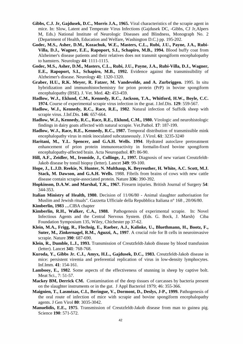
42
Gibbs, C.J. Jr, Gajdusek, D.C., Morris J.A., 1965. Viral characteristics of the scrapie agent inmice. In: Slow, Latent and Temperate Virus Infections (Gajdusek DC, -Gibbs, CJ Jr,AlpersM, Eds.) National Institute of Neurologic Diseases and Blindness, Monograph No. 2(Department of Health, Education and Welfare, Washington D.C.) pp. 195-202.
Godec, M.S., Asher, D.M., Kozachuk, W.E., Masters, C.L., Rubi, J.U., Payne, J.A., Rubi-Villa, D.J., Wagner, E.E., Rapoport, S.I., Schapiro, M.B., 1994. Blood buffy coat fromAlzheimer's disease patients and their relatives does not transmit spongiform encephalopathyto hamsters. Neurology 44: 1111-1115.
Godec, M.S., Asher, D.M., Masters, C.L., Rubi, J.U., Payne, J.A., Rubi-Villa, D.J., Wagner,E.E., Rapoport, S.I., Schapiro, M.B., 1992. Evidence against the transmissibility ofAlzheimer's disease. Neurology 41: 1320-1320.
Graber, H.U., R.K. Meyer, R. Fatzer, M. Vandevelde, and A. Zurbriggen. 1995. In situhybridization and immunohistochemistry for prion protein (PrP) in bovine spongiformencephalopathy (BSE). J. Vet. Med. 42: 453-459.
Hadlow, W.J., Eklund, C.M., Kennedy, R.C., Jackson, T.A., Whitford, H.W., Boyle, C.C.1974. Course of experimental scrapie virus infection in the goat. J.Inf.Dis. 129: 559-567.
Hadlow, W.J., Kennedy, R.C., Race, R.E., 1982. Natural infection of Suffolk sheep withscrapie virus. J.Inf.Dis. 146: 657-664.
Hadlow, W.J., Kennedy, R.C., Race, R.E., Eklund, C.M., 1980. Virologic and neurohistologicfindings in dairy goats affected with natural scrapie. Vet.Pathol. 17: 187-199.
Hadlow, W.J., Race, R.E., Kennedy, R.C., 1987. Temporal distribution of transmissible minkencephalopathy virus in mink inoculated subcutaneously. J.Virol. 61: 3235-3240
Haritani, M., Y.L. Spencer, and G.A.H. Wells. 1994. Hydrated autoclave pretreatmentenhancement of prion protein immunoreactivity in formalin-fixed bovine spongiformencephalopathy-affected brain. Acta Neuropathol. 87: 86-90.
Hill, A.F., Zeidler, M., Ironside, J., Collinge, J., 1997. Diagnosis of new variant Creutzfeldt-Jakob disease by tonsil biopsy (letter). Lancet 349: 99-100.
Hope, J., L.J.D. Reekie, N. Hunter, N. Multhaup, K. Beyreuther, H. White, A.C. Scott, M.J.Stack, M. Dawson, and G.A.H. Wells. 1988. Fibrils from brains of cows with new cattledisease contain scrapie-associated protein. Nature 336: 390-392.
Hopkinson, D.A.W. and Marshal, T.K., 1967. Firearm injuries. British Journal of Surgery 54:344-353.
Italian Ministry of Health, 1980. Decision of 11/06/80 - Animal slaughter authorisation forMuslim and Jewish rituals". Gazzetta Ufficiale della Repubblica Italiana n° 168 , 20/06/80.
Kimberlin, 1983 …CIBA chapterKimberlin, R.H., Walker, C.A., 1988. Pathogenesis of experimental scrapie. In: Novel
Infectious Agents and the Central Nervous System. (Eds. G. Bock, J. Marsh) CibaFoundation Symposium 135, Wiley, Chichester pp 37-62.
Klein, M.A., Frigg, R., Flechsig, E., Raeber, A.J., Kalinke, U., Bluethmann, H., Bootz, F.,Suter, M., Zinkernagel, R.M., Aguzzi, A., 1997. A crucial role for B cells in neuroinvasivescrapie. Nature 390: 687-690.
Klein, R., Dumble, L.J., 1993. Transmission of Creutzfeldt-Jakob disease by blood transfusion(letter). Lancet 341: 768-768.
Kuroda, Y., Gibbs Jr. C.J., Amyx, H.L., Gajdusek, D.C., 1983. Creutzfeldt-Jakob disease inmice: persistent viremia and preferential replication of virus in low-density lymphocytes.Inf.Imm. 41: 154-161.
Lambooy, E., 1982. Some aspects of the effectiveness of stunning in sheep by captive bolt.Meat Sci., 7: 51-57.
Mackey BM, Derrick CM. Contamination of the deep tissues of carcasses by bacteria presenton the slaughter instruments or in the gut. J Appl Bacteriol 1979; 46: 355-366.
Maignien, T., Lasmézas, C.I., Beringue, V., Dormont, D., Deslys, J-P., 1999. Pathogenesis ofthe oral route of infection of mice with scrapie and bovine spongiform encephalopathyagents. J Gen Virol 80: 3035-3042.
Manuelidis, E.E., 1975. Transmission of Creutzfeldt-Jakob disease from man to guinea pig.Science 190: 571-572.

43
Manuelidis, E.E., de Figueiredo, J.M., Kim, J.H., Fritch W.W., Manuelidis, L., 1988.Transmission studies from blood of Alzheimer disease patients and healthy relatives.Proc.Natl.Acad.Sci.USA 85: 4898-4901.
Manuelidis, E.E., Gorgacz, E.J., Manuelidis, L., 1978a. Viremia in experimental Creutzfeldt-Jakob disease. Science 200: 1069-1071.
Manuelidis, E.E., Kim, J.H., Mericangas J.R., Manuelidis, L., 1985. Transmission to animalsof Creutzfeldt-Jakob disease from human blood. Lancet ii: 896-897.
Manuelidis, E.E., Manuelidis, L., 1993. A transmissible Creutzfeldt-Jakob disease-like agent isprevalent in the human population. Proc.Natl.Acad.Sci.USA 90: 7724-7728.
Manuelidis,E.E., Manuelidis, L., Pincus, J.H., Collins, W.F., 1978b. Transmission, from manto hamster, of Creutzfeldt-Jakob disease with clinical recovery. Lancet ii: 40-42.
Marsh, R.F., Burger, D., Hanson, R.P., 1969. Transmissible mink encephalopathy: behavior ofthe disease agent in mink. Amer.J.Vet.Res. 30: 1637-1642.
Marsh, R.F., Hadlow, W.J., 1992. Transmissible mink encephalopathy. Rev. sci. tech. Off.int. Epiz. 11(2): 539-550.
Marsh, R.F., Miller, J.M., Hanson, R.P., 1973. Transmissible mink encephalopathy: studies onthe peripheral lymphocyte. Infect.Immun. 7: 352-355.
Meyer, R. K., B. Oesch, R. Fatzer, A. Zurbriggen, M. Vandevelde, 1999. Detection ofBovine Spongiform Encephalopathy-Specific PrP(Sc) by Treatment with Heat and GuanidineThiocyanate. J. Virol. 73:9386-9392.
Mickwitz, G. von and Leach, T.M., 1977. Review of pre-slaughter stunning in the EC.Commission of the European Communities “Information on Agriculture” no. 30.
Miller, J. M., A. L. Jenny, W. D. Taylor, R. E. Race, D. B, Ernst, J. B. Kotz, and R.Rubenstein. 1994. Detection of prion protein in formalin-fixed brain by hydrated autoclavingimmunohistochemistry for the diagnosis of scrapie in sheep. J. Vet. Diagn. lnvest. 5: 366-368.
Millson, G.C., Hunter, G.D., Kimberlin, R.H., 1976. The physico-chemical nature of thescrapie agent. In: Slow Virus Diseases of Animals and Man. (Ed. R H Kimberlin) North-Holland Research Monographs Frontiers of Biology Vol. 44. North-Holland PublishingCompany, Amsterdam.
Moynagh J. and Schimmel H. Tests for BSE evaluated. Nature. 1999; 400(6740):105.Munro, R., 1997. Neural tissue embolism in cattle. Vet. Rec., 140, 536.Ogilvy SC, McKee AC, Newman NJ, Donnelly SM Kiwak KJ. Embolism of cerebral tissue
to lungs: report of two cases and review of the literature. Neurosurgery 1988; 23: 511-516.OIE (1999a). International Animal Health Code draft chapter on scrapie. OIE Paris.Operskalski, E.A., Mosley, J.W., 1995. Pooled plasma derivatives and Creutzfeldt-Jakob
disease [letter]. Lancet 346: 1224.Order, (1996). The Fresh Meat (Beef control) (No2) Regs 1996 (SI 1996/2097) and SR 1996 No
404 in Northern Ireland. The Stationery Office, London.O'Rourke K.I.; Baszler T.V.; Parish S.M., and Knowles D.P. Preclinical detection of PrPSc in
nicitating membrane lymphoid tissue of sheep. Veterinary Record. 1998; 142:489-491.Outram, G.W. 1976 The pathogenesis of scrapie in mice. In: Slow virus diseases of animals
and man. (Ed. R. H. Kimberlin) North Holland Research Monographs Frontiers of BiologyVol 44, North-Holland Publishing Company, Amsterdam, pp.325-357.
Pattison, I.H., Millson, G.C., 1960. Further observation on the experimental production ofscrapie in goats and sheep. J.Comp.Path. 70: 182-193.
Pattison, I.H., Millson, G.C., 1962. Distribution of scrapie agent in the tissues ofexperimentally inoculated goats. J.Comp.Pathol. Ther. 72: 233-244.
Pattison, I.H., Millson, G.C., Smith, K., 1964. An examination of the action of whole blood,blood cells or serum on the goat scrapie agent. Res.vet.Sci. 5: 116-121.
Prusiner, S.B., 1987. Bioassays of Prions. In: Prions: Novel Infectious Pathogens CausingScrapie and Creutzfeldt-Jakob Disease. (Eds. Stanley B. Prusines, Michael P. McKinley)Academic Press Inc. San Diego.
Race, R., Chesebro, B., 1998. Scrapie infectivity found in resistant species. Nature 392: 770-771.

44
Raeber, A.J., Klein, M.A., Frigg, R., Flechsig, E., Aguzzi, A., Weissmann, C., 1999. PrP-dependent association of prions with splenic but not circulating lymphocytes of scrapie-infected mice. EMBO Journal 18 (10): 2702-2706.
Robinson, M.M., Cheevers, W.P., Burger, D., Gorham, J.R. 1990. Organ-specificmodification of the dose-response relationship of scrapie infectivity. J. Infect. Dis. 161: 783-786.
Rohwer, R.G., 1992. Alzheimer's disease transmission: possible artifact due to intercurrentillness. Neurology 42: 287-288.
Safar, J., H. Wille, V. Itri, D. Groth, H. Serban, M. Torchia, F.E. Cohen, and S.B. Prusiner.1998. Eight prion strains have PrPSc molecules with different conformations. NatureMedicine 4: 1157-1165.
Schaller O.; Fatzer R.; Stack M.; Clark J.; Cooley W.; Biffiger K.; Egli S.; Doherr M.;Vanedevelde M.; Heim D.; Oesch B., and Moser M. Validation of a Westernimmunoblotting procedure for bovine PrPSC detection and its use as a rapid surveillancemethod for the diagnosis of bovine spongiform encephalopathy (BSE). ActaNeuropathologica. 1999; 98(5):437-443.
Schardt, D. Schmidt, S., 1997. Mad about BSE. Nutrition Action Healthletter, US edition -July/August 1997.
Schartz, J., 1997. Stunning scatters brain tissue through body. Listserve Commentary onWashington Post/AP/Wall Street Journal of 25/07/1997.
Schmerr, M J. An analytical method based on capillary electrophoresis to detect the prionprotein. Encyclopedia of Separation Science, (2000).
Schmerr, M. J.; Jenny, A.L.; Bulgin, M. S., Miller, J. M., Hamir, A. N.; Cutlip, R. C. andGoodwin, K R... The Use of Capillary Electrophoresis and Fluorescent Labeled Peptides toDetect the Abnormal Prion Protein in the Blood of Animals that Are Infected with aTransmissible Spongiform Encephalopathy. J. of Chromatogr. A, 853 207-214 (1999).
Schmerr, M.J. and Jenny A.L. A Diagnostic test for scrapie infected sheep using a capillaryelectrophoresis immunoassay with fluorescent labeled peptides. Electrophoresis, 19 409-414(1998).
Schmidt, G.R.,. Hossner, K.L., Yemm, R.S, Gould, D.H., 1999a. "Potential for disruption ofcentral nervous system tissue in beef cattle by different types of captive bolt stunners", J.Food Prot., 62 (4): 390-393.
Schmidt, G.R.,. Hossner, K.L., Yemm, R.S, Gould, D.H., O'Callaghan, J.P. 1999b. Anenzyme-linked immunosorbent assay for glial fibrillary acidic protein as an indicator of thepresence of brain or spinal cord in meat", J. Food Prot., 62(4): 394-397.
Schreuder B.E.C., van Keulen L.J.M., Vromans M.E.V., Langeveld J.P.M., Smits M.A.,1998. Tonsillar biopsy and PrPSc detection in the preclinical diagnosis of Scrapie. VeterinaryRecord 1998, 142, 564-568.
Scott, J.R., 1993. Scrapie pathogenesis. In: Spongiform Encephalopathies. British MedicalBulletin 49(4), (Allen, I.V. Ed.) Churchill Livingstone, Edinburgh, pp778-791.
Steele, P.J., Taylor, D.M. & Fernie, K. (1999) Survival of BSE and scrapie agents at 200oC.Abstracts of a Meeting of the Association of Veterinary Teachers and Research Workers.Scarborough, 29-31 March 1999; p. 21.
Steele, P.J., Taylor, D.M. & Fernie, K., 1999a. Survival of BSE and scrapie agents at 200oC.Abstracts of a Meeting of the Association of Veterinary Teachers and Research Workers.Scarborough, 29-31 March 1999: 21.
Steele, P.J., Taylor, D.M., Fernie, K. & McConnell, I. (1999) Persistence of TDE infectivity:Dry heat treatments. Abstracts of a Symposium on the Characterisation and Diagnosis ofPrion Diseases of Man and Animals, (poster). Tubingen; 23-25 September 1999.
Steele, P.J., Taylor, D.M., Fernie, K., Mcconnell, I., 1999b. Persistence of TDE infectivity:Dry heat treatments. Abstracts of a Symposium on the Characterisation and Diagnosis ofPrion Diseases of Man and Animals, (poster). Tubingen; 23-25 September 1999.
Stekel D.J.; Nowak M.A., and Southwood T.R.E. Prediction of future BSE spread. Nature.1996; 381(6578):119.
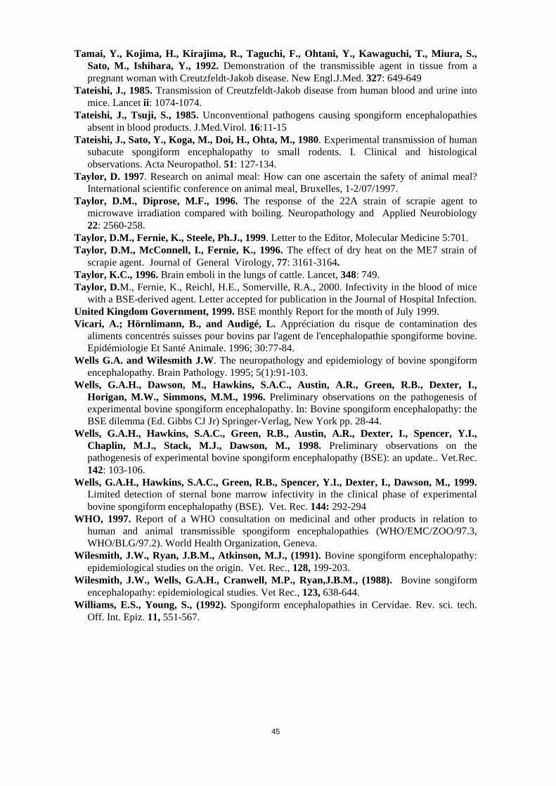
45
Tamai, Y., Kojima, H., Kirajima, R., Taguchi, F., Ohtani, Y., Kawaguchi, T., Miura, S.,Sato, M., Ishihara, Y., 1992. Demonstration of the transmissible agent in tissue from apregnant woman with Creutzfeldt-Jakob disease. New Engl.J.Med. 327: 649-649
Tateishi, J., 1985. Transmission of Creutzfeldt-Jakob disease from human blood and urine intomice. Lancet ii: 1074-1074.
Tateishi, J., Tsuji, S., 1985. Unconventional pathogens causing spongiform encephalopathiesabsent in blood products. J.Med.Virol. 16:11-15
Tateishi, J., Sato, Y., Koga, M., Doi, H., Ohta, M., 1980. Experimental transmission of humansubacute spongiform encephalopathy to small rodents. I. Clinical and histologicalobservations. Acta Neuropathol. 51: 127-134.
Taylor, D. 1997. Research on animal meal: How can one ascertain the safety of animal meal?International scientific conference on animal meal, Bruxelles, 1-2/07/1997.
Taylor, D.M., Diprose, M.F., 1996. The response of the 22A strain of scrapie agent tomicrowave irradiation compared with boiling. Neuropathology and Applied Neurobiology22: 2560-258.
Taylor, D.M., Fernie, K., Steele, Ph.J., 1999. Letter to the Editor, Molecular Medicine 5:701.Taylor, D.M., McConnell, I., Fernie, K., 1996. The effect of dry heat on the ME7 strain of
scrapie agent. Journal of General Virology, 77: 3161-3164.Taylor, K.C., 1996. Brain emboli in the lungs of cattle. Lancet, 348: 749.Taylor, D.M., Fernie, K., Reichl, H.E., Somerville, R.A., 2000. Infectivity in the blood of mice
with a BSE-derived agent. Letter accepted for publication in the Journal of Hospital Infection.United Kingdom Government, 1999. BSE monthly Report for the month of July 1999.Vicari, A.; Hörnlimann, B., and Audigé, L. Appréciation du risque de contamination des
aliments concentrés suisses pour bovins par l'agent de l'encephalopathie spongiforme bovine.Epidémiologie Et Santé Animale. 1996; 30:77-84.
Wells G.A. and Wilesmith J.W. The neuropathology and epidemiology of bovine spongiformencephalopathy. Brain Pathology. 1995; 5(1):91-103.
Wells, G.A.H., Dawson, M., Hawkins, S.A.C., Austin, A.R., Green, R.B., Dexter, I.,Horigan, M.W., Simmons, M.M., 1996. Preliminary observations on the pathogenesis ofexperimental bovine spongiform encephalopathy. In: Bovine spongiform encephalopathy: theBSE dilemma (Ed. Gibbs CJ Jr) Springer-Verlag, New York pp. 28-44.
Wells, G.A.H., Hawkins, S.A.C., Green, R.B., Austin, A.R., Dexter, I., Spencer, Y.I.,Chaplin, M.J., Stack, M.J., Dawson, M., 1998. Preliminary observations on thepathogenesis of experimental bovine spongiform encephalopathy (BSE): an update.. Vet.Rec.142: 103-106.
Wells, G.A.H., Hawkins, S.A.C., Green, R.B., Spencer, Y.I., Dexter, I., Dawson, M., 1999.Limited detection of sternal bone marrow infectivity in the clinical phase of experimentalbovine spongiform encephalopathy (BSE). Vet. Rec. 144: 292-294
WHO, 1997. Report of a WHO consultation on medicinal and other products in relation tohuman and animal transmissible spongiform encephalopathies (WHO/EMC/ZOO/97.3,WHO/BLG/97.2). World Health Organization, Geneva.
Wilesmith, J.W., Ryan, J.B.M., Atkinson, M.J., (1991). Bovine spongiform encephalopathy:epidemiological studies on the origin. Vet. Rec., 128, 199-203.
Wilesmith, J.W., Wells, G.A.H., Cranwell, M.P., Ryan,J.B.M., (1988). Bovine songiformencephalopathy: epidemiological studies. Vet Rec., 123, 638-644.
Williams, E.S., Young, S., (1992). Spongiform encephalopathies in Cervidae. Rev. sci. tech.Off. Int. Epiz. 11, 551-567.

46
ANNEX 1
FIGURES
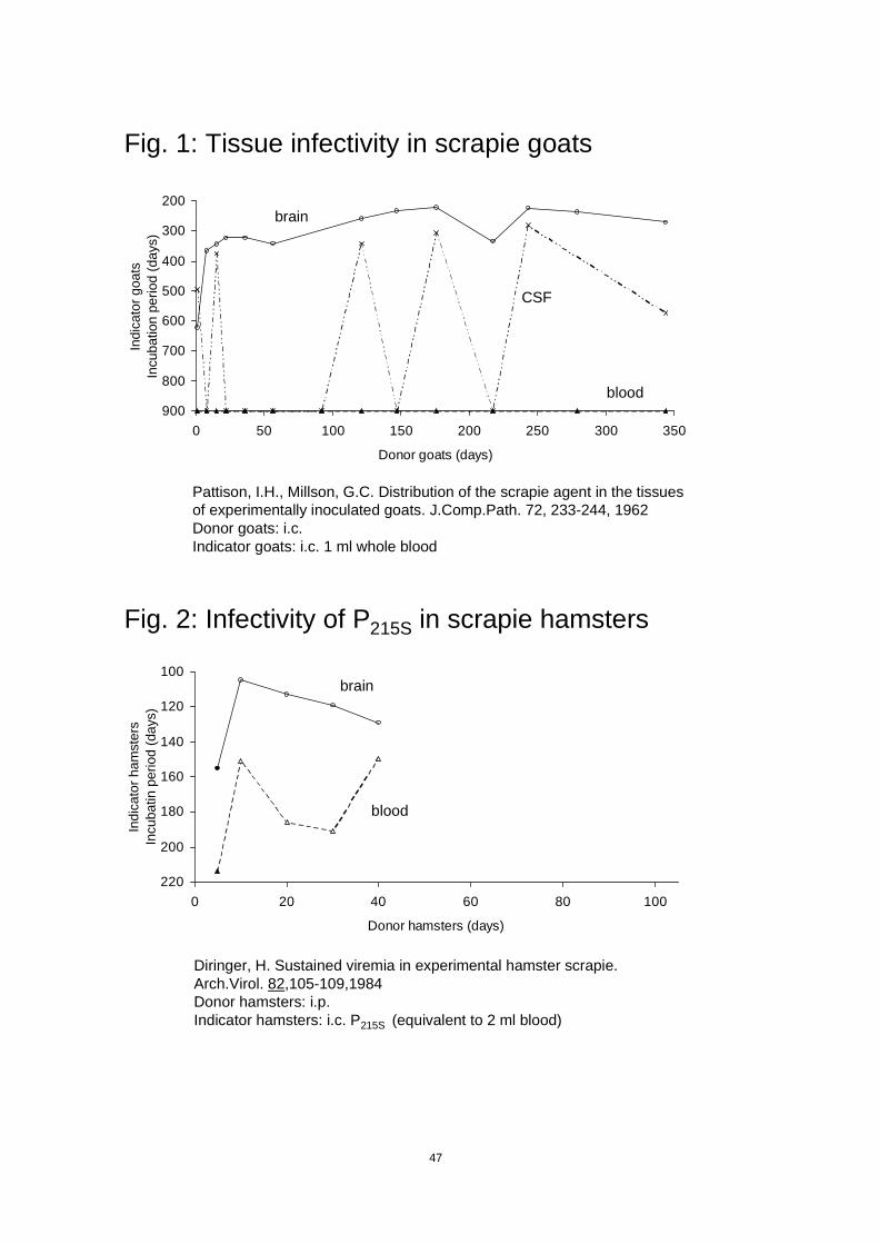
47
Fig. 1: Tissue infectivity in scrapie goats
Pattison, I.H., Millson, G.C. Distribution of the scrapie agent in the tissuesof experimentally inoculated goats. J.Comp.Path. 72, 233-244, 1962Donor goats: i.c.Indicator goats: i.c. 1 ml whole blood
200
300
400
500
600
700
800
9000 50 100 150 200 250 300 350
Donor goats (days)
Indi
cato
r goa
tsIn
cuba
tion
perio
d (d
ays)
brain
CSF
blood
Fig. 2: Infectivity of P215S in scrapie hamsters
Diringer, H. Sustained viremia in experimental hamster scrapie.Arch.Virol. 82,105-109,1984Donor hamsters: i.p.Indicator hamsters: i.c. P215S (equivalent to 2 ml blood)
100
120
140
160
180
200
2200 20 40 60 80 100
Donor hamsters (days)
Indi
cato
r ham
ster
sIn
cuba
tin p
erio
d (d
ays)
blood
brain
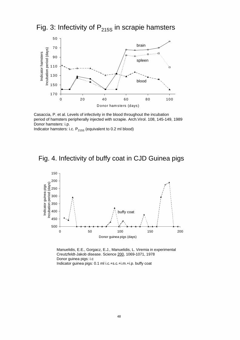
48
Fig. 3: Infectivity of P215S in scrapie hamsters
Casaccia, P. et al. Levels of infectivity in the blood throughout the incubationperiod of hamsters peripherally injected with scrapie. Arch.Virol. 108, 145-149, 1989Donor hamsters: i.p.Indicator hamsters: i.c. P215S (equivalent to 0.2 ml blood)
50
70
90
110
130
150
1700 20 40 60 80 100
D onor ham sters (days)
Indi
cato
r ham
ster
sIn
cuba
tion
perio
d (d
ays)
brain
spleen
blood
Fig. 4. Infectivity of buffy coat in CJD Guinea pigs
Manuelidis, E.E., Gorgacz, E.J., Manuelidis, L. Viremia in experimentalCreutzfeldt-Jakob disease. Science 200, 1069-1071, 1978Donor guinea pigs: i.cIndicator guinea pigs: 0.1 ml i.c.+s.c.+i.m.+i.p. buffy coat
150
200
250
300
350
400
450
5000 50 100 150 200
Donor guinea pigs (days)
Indi
cato
r gui
nea
pigs
Incu
batio
n pe
riod
(day
s)
buffy coat
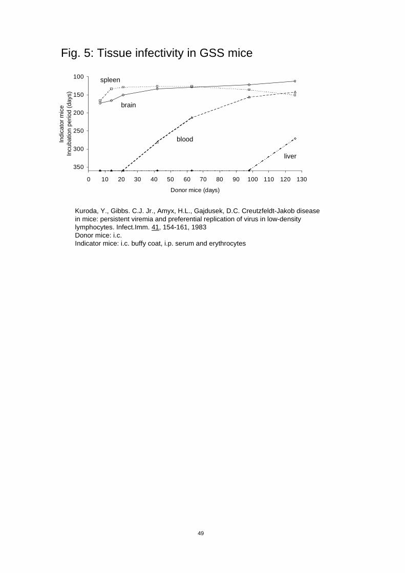
49
Fig. 5: Tissue infectivity in GSS mice
100
150
200
250
300
350
0 10 20 30 40 50 60 70 80 90 100 110 120 130
Donor mice (days)
Indi
cato
r mic
eIn
cuba
tion
perio
d (d
ays)
spleen
brain
blood
liver
Kuroda, Y., Gibbs. C.J. Jr., Amyx, H.L., Gajdusek, D.C. Creutzfeldt-Jakob diseasein mice: persistent viremia and preferential replication of virus in low-densitylymphocytes. Infect.Imm. 41, 154-161, 1983Donor mice: i.c.Indicator mice: i.c. buffy coat, i.p. serum and erythrocytes

T_BLOOD.doc
Rue de la Loi 200, B-1049 Bruxelles/Wetstraat 200, B-1049 Brussel - Belgium - Office: B232 (6/74).Telephone: direct line (+32-2)29 58554, switchboard 299.11.11. Fax: 29 96301 / 96299.
Internet: [email protected]
ANNEX 2
TABLES
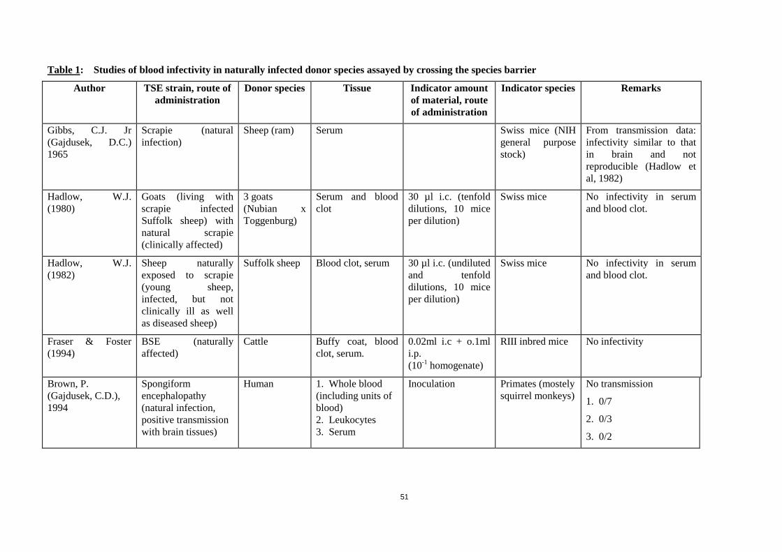
51
Table 1: Studies of blood infectivity in naturally infected donor species assayed by crossing the species barrier
Author TSE strain, route ofadministration
Donor species Tissue Indicator amountof material, routeof administration
Indicator species Remarks
Gibbs, C.J. Jr(Gajdusek, D.C.)1965
Scrapie (naturalinfection)
Sheep (ram) Serum Swiss mice (NIHgeneral purposestock)
From transmission data:infectivity similar to thatin brain and notreproducible (Hadlow etal, 1982)
Hadlow, W.J.(1980)
Goats (living withscrapie infectedSuffolk sheep) withnatural scrapie(clinically affected)
3 goats(Nubian xToggenburg)
Serum and bloodclot
30 µl i.c. (tenfolddilutions, 10 miceper dilution)
Swiss mice No infectivity in serumand blood clot.
Hadlow, W.J.(1982)
Sheep naturallyexposed to scrapie(young sheep,infected, but notclinically ill as wellas diseased sheep)
Suffolk sheep Blood clot, serum 30 µl i.c. (undilutedand tenfolddilutions, 10 miceper dilution)
Swiss mice No infectivity in serumand blood clot.
Fraser & Foster(1994)
BSE (naturallyaffected)
Cattle Buffy coat, bloodclot, serum.
0.02ml i.c + o.1mli.p.(10-1 homogenate)
RIII inbred mice No infectivity
Brown, P.(Gajdusek, C.D.),1994
Spongiformencephalopathy(natural infection,positive transmissionwith brain tissues)
Human 1. Whole blood(including units ofblood)2. Leukocytes3. Serum
Inoculation Primates (mostelysquirrel monkeys)
No transmission
1. 0/7
2. 0/3
3. 0/2
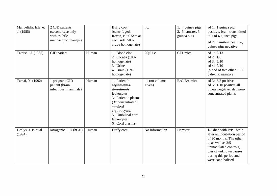
52
Manuelidis, E.E. etal (1985)
2 CJD patients(second case onlywith “subtlemicroscopic changes)
Buffy coat(centrifuged,frozen, cut 0.5cm ateach side, 50%crude homogenate)
i.c. 1. 4 guinea pigs2. 5 hamster, 5guinea pigs
ad 1: 1 guinea pigpositive, brain transmittedto 1 of 6 guinea pigs.
ad 2: hamsters positive,guinea pigs negative
Tateishi, J. (1985) CJD patient Human 1. Blood clot2. Cornea (10%homogenate)3. Urine4. Brain (10%homogenate)
20µl i.c. CF1 mice ad 1: 2/13ad 2: 1/6ad 3: 5/10ad 4: 7/10(blood of two other CJDpatients: negative)
Tamai, Y. (1992) 1 pregnant CJDpatient (braininfectious in animals)
Human 1. Patient’serythrocytes.2. Patient’sleukocytes3. Patient’s plasma(3x concentrated)4. Corderythrocytes.5. Umbilical cordleukocytes6. Cord plasma
i.c (no volumegiven)
BALB/c mice ad 3: 3/8 positivead 5: 1/10 positive allothers negative, also non-concentrated plams
Deslys, J.-P. et al(1994)
Iatrogenic CJD (hGH) Human Buffy coat No information Hamster 1/5 died with PrP+ brainafter an incubation periodof 20 months. The other4, as well as 3/5uninoculated controls,dies of unknown causesduring this period andwere cannibalised
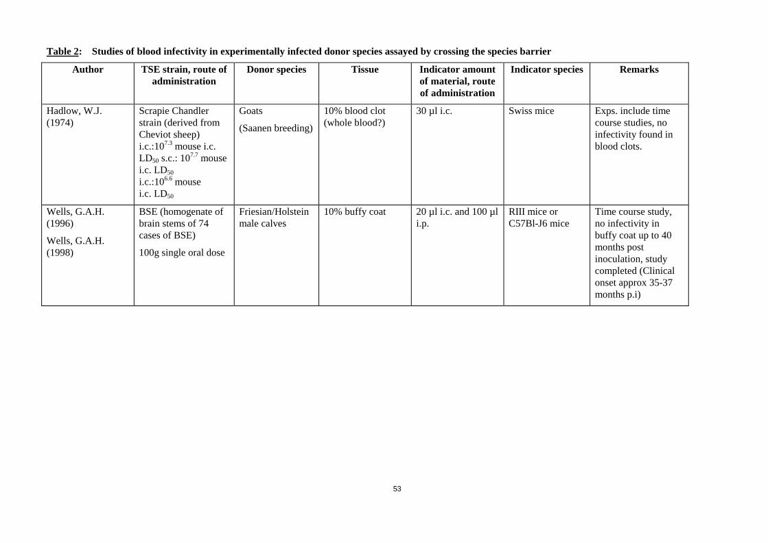
53
Table 2: Studies of blood infectivity in experimentally infected donor species assayed by crossing the species barrier
Author TSE strain, route ofadministration
Donor species Tissue Indicator amountof material, routeof administration
Indicator species Remarks
Hadlow, W.J.(1974)
Scrapie Chandlerstrain (derived fromCheviot sheep)i.c.:107.3 mouse i.c.LD50 s.c.: 107.7 mousei.c. LD50i.c.:106.6 mousei.c. LD50
Goats
(Saanen breeding)
10% blood clot(whole blood?)
30 µl i.c. Swiss mice Exps. include timecourse studies, noinfectivity found inblood clots.
Wells, G.A.H.(1996)
Wells, G.A.H.(1998)
BSE (homogenate ofbrain stems of 74cases of BSE)
100g single oral dose
Friesian/Holsteinmale calves
10% buffy coat 20 µl i.c. and 100 µli.p.
RIII mice orC57Bl-J6 mice
Time course study,no infectivity inbuffy coat up to 40months postinoculation, studycompleted (Clinicalonset approx 35-37months p.i)

54
Table 3: Studies with experimentally infected donor species assayed without crossing species barriers
Author TSE strain, route ofadministration
Donor species Tissue Indicator amount ofmaterial, route of
administration
Indicatorspecies
Remarks
Pattison, I.H. (1962) Goat adapted scrapie(passaged 3 x ingoats)(1 ml sup. (1500 rpm,15 min) 10% brainsusp.) i.c.
Goat Whole blood 1 ml i.c. Goat Time course study, notransmission with blood.(See fig 1)
Pattison, I.H. (1964) Scrapie affected foats(no further details, asin Pattison 1962?)
Cross bredgoat
1. Whole blood.2. Blood cells (mainlyred blood cells)3. Serum (each poolsof two animals)
1a. 1 ml (0.5ml?) i.c.1b. 3 x 5 ml s.c.(weekly intervals)2. 1 ml (0.5 ml?) i.c.3. 1 ml (0.5ml?) i.c.
Cross bredgoat
No transmission
Marsh, R.F. (1969) TME1. 1 ml 10% brain ofnaturally infectedmink (Hayward,Wis.) i.m.2. 0.1 ml 10% brain(2nd passage) i.c.
Mink(Genetics -Dep., Univ. ofWisconsin)
Serum 1 ml s.c. (diluted inten-fold steps)
Mink No infectivity in serum,but infectivity in a seriesof tissues includingspleen.
Marsh, R.F. (1973) TME1. Late clinical stage.2. 105 LD50 i.c.
Mink 1. Whole blood,plasma, 10% red bloodcells, 10% platelets,white blood cells(1.7x10⁷/ml), culturedlymphocytes(1.5x10⁷/ml), PHAstimulatedlymphocytes(1.5x10⁷/ml).2. Lymphocytes(2x10⁷/ml)
0.1 ml i.c. Mink 1. No infectivity in listedpreparations.2. Time course study, noinfectivity inlymphocytes, but inspleen and peripherallymph nodes.
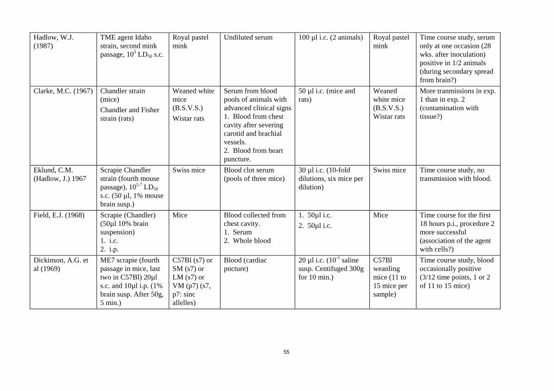
55
Hadlow, W.J.(1987)
TME agent Idahostrain, second minkpassage, 103 LD50 s.c.
Royal pastelmink
Undiluted serum 100 µl i.c. (2 animals) Royal pastelmink
Time course study, serumonly at one occasion (28wks. after inoculation)positive in 1/2 animals(during secondary spreadfrom brain?)
Clarke, M.C. (1967) Chandler strain(mice)Chandler and Fisherstrain (rats)
Weaned whitemice(B.S.V.S.)Wistar rats
Serum from bloodpools of animals withadvanced clinical signs1. Blood from chestcavity after severingcarotid and brachialvessels.2. Blood from heartpuncture.
50 µl i.c. (mice andrats)
Weanedwhite mice(B.S.V.S.)Wistar rats
More tranmissions in exp.1 than in exp. 2(contamination withtissue?)
Eklund, C.M.(Hadlow, J.) 1967
Scrapie Chandlerstrain (fourth mousepassage), 105.7 LD50s.c. (50 µl, 1% mousebrain susp.)
Swiss mice Blood clot serum(pools of three mice)
30 µl i.c. (10-folddilutions, six mice perdilution)
Swiss mice Time course study, notransmission with blood.
Field, E.J. (1968) Scrapie (Chandler)(50µl 10% brainsuspension)1. i.c.2. i.p.
Mice Blood collected fromchest cavity.1. Serum2. Whole blood
1. 50µl i.c.2. 50µl i.c.
Mice Time course for the first18 hours p.i., procedure 2more successful(association of the agentwith cells?)
Dickinson, A.G. etal (1969)
ME7 scrapie (fourthpassage in mice, lasttwo in C57Bl) 20µls.c. and 10µl i.p. (1%brain susp. After 50g,5 min.)
C57Bl (s7) orSM (s7) orLM (s7) orVM (p7) (s7,p7: sincallelles)
Blood (cardiacpncture)
20 µl i.c. (10-1 salinesusp. Centifuged 300gfor 10 min.)
C57Blweanlingmice (11 to15 mice persample)
Time course study, bloodoccasionally positive(3/12 time points, 1 or 2of 11 to 15 mice)
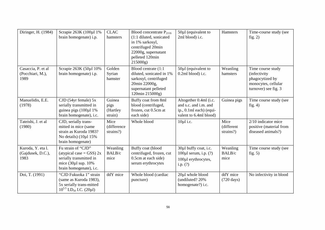
56
Diringer, H. (1984) Scrapie 263K (100µl 1%brain homogenate) i.p.
CLAChamsters
Blood concentrate P215S(1:1 diluted, sonicatedin 1% sarkosyl,centrifuged 20min22000g, supernatantpelleted 120min215000g)
50µl (equivalent to2ml blood) i.c.
Hamsters Time-course study (seefig. 2)
Casaccia, P. et al(Pocchiari, M.),1989
Scrapie 263K (50µl 10%brain homogenate) i.p.
GoldenSyrianhamster
Blood centrate (1:1diluted, sonicated in 1%sarkosyl, centrifuged20min 22000g,supernatant pelleted120min 215000g)
50µl (equivalent to0.2ml blood) i.c.
Weanlinghamsters
Time course study(infectivityphagocytized bymonocytes, cellularturnover) see fig. 3
Manuelidis, E.E.(1978)
CJD (54yr female) 5xserially transmitted inguinea pigs (100µl 1%brain homogenate), i.c.
Guineapigs(Hartleystrain)
Buffy coat from 8mlblood (centrifuged,frozen, cut 0.5cm ateach side)
Altogether 0.4ml (i.c.and s.c. and i.m. andip., 0.1ml each) (equi-valent to 6.4ml blood)
Guinea pigs Time course study (seefig. 4)
Tateishi, J. et al(1980)
CJD, serially trans-mitted in mice (samestrain as Kuroda 1983?No details) (10µl 15%brain homogenate)
Mice(differencestrains?)
Whole blood 10µl i.c. Mice(differentstrains?)
2/10 indicator micepositive (material fromdiseased animals?)
Kuroda, Y. eta l.(Gajdusek, D.C.),1983
Fu strain of “CJD”(atypical case = GSS) 2xserially transmitted inmice (30µl sup. 10%brain homogenate), i.c.
WeanlingBALB/cmice
Buffy coat (bloodcentrifuged, frozen, cut0.5cm at each side)serum erythrocytes
30µl buffy coat, i.c.100µl serum, i.p. (?)100µl erythrocytes,i.p. (?)
WeanlingBALB/cmice
Time course study (seefig. 5)
Doi, T. (1991) “CJD Fukuoka 1” strain(same as Kuroda 1983),5x serially trans-mitted105.5 LD50 I.C. (20µl)
ddY mice Whole blood (cardiacpuncture)
20µl whole blood(undiluted? 20%homogenate?) i.c.
ddY mice(720 days)
No infectivity in blood

T_BLOOD.doc
Rue de la Loi 200, B-1049 Bruxelles/Wetstraat 200, B-1049 Brussel - Belgium - Office: B232 (6/74).Telephone: direct line (+32-2)29 58554, switchboard 299.11.11. Fax: 29 96301 / 96299.
Internet: [email protected]
Table 4: Infectivity levels in the buffy coat, plasma, and Cohn cryoprecipitate+Fraction I+II+III in groups of mice exsanguinated atintervals of 5, 9, 13, and 18 weeks after inoculation with the Fukuoka-1 strain of TSE. Mice were asymptomatic at 5, 9, and 13weeks, and showed symptoms at 18 weeks
Number of Number of Time after Inoculated inoculated positive Infectious unitsinoculation Specimen1 volume (ml)2 animals animals per ml (CI)3
5 weeks Buffy coat 0.28 35 3 11.3 (3-29)Plasma 0.59 40 1 1.8 (0.1-8)
Cryo+I+II+III 0.16 29 2 0.5 (0.1-1.5)
9 weeks Buffy coat 0.15 29 1 6.8 (0.4-30)Plasma 0.29 38 0 0.0 (0-11)
Cryo+I+II+III 0.14 28 3 1.0 (0.3-2.6)
13 weeks Buffy coat 0.16 28 1 6.4 (0.4-28)Plasma 0.29 38 0 0.0 (0-11)
Cryo+I+II+III 0.15 28 2 0.6 (0.1-1.7)
18 weeks4 Buffy coat 0.14 27 11 106.0 (55-184)Plasma 0.25 33 5 21.9 (8-47)
1. Cryoprecipitate and fraction I+II+III were prepared from 10 ml plasma.2. 0.03 ml volumes were inoculated into groups of mice either undiluted, or at dilutions of 1:2, 1:4, or 1:40. These dilution factors were taken into account whencalculating the total undiluted inoculated volume of each specimen.3. CI = 95% confidence interval (based on a likelihood ratio).4. Cryoprecipitate from this bleeding was not available for inoculation.

T_BLOOD.doc
Rue de la Loi 200, B-1049 Bruxelles/Wetstraat 200, B-1049 Brussel - Belgium - Office: B232 (6/74).Telephone: direct line (+32-2)29 58554, switchboard 299.11.11. Fax: 29 96301 / 96299.
Internet: [email protected]
Table 5: Leukofiltration of plasma from mice inoculated with the Fukuoka-1 strain of TSE
Number of Number ofExperimental Inoculated inoculated positive Infectious unitsConditions Specimen volume (ml)1 animals animals per ml (CI)2
Exp.#1: 16 week plasma pool (symptomatic mice)
Frozen & thawed Pre-filtration 0.47 28 12 34.4 (18-58) (undiluted) Post- filtration 0.33 35 4 13.0 (4-30)
Exp.#2: 13 week plasma pool (asymptomatic mice)
Frozen & thawed Pre-filtration 0.29 38 0 0.0 (0-11)(diluted 1:4) Post-filtration 0.26 35 8 34.6 (16-65)
Exp.#3: 18 week plasma pool (symptomatic mice)
Fresh Pre-filtration 0.25 33 5 21.9 (8-47)(undiluted) Post-filtration 0.23 29 4 19.8 (6-46)
1. 0.03 ml volumes were inoculated intracerebrally at dilutions of 1:2 and 1:4. These dilution factors were taken into account when calculating the total undilutedinoculated volume of each specimen.2. CI = 95% confidence interval (based on a likelihood ratio).
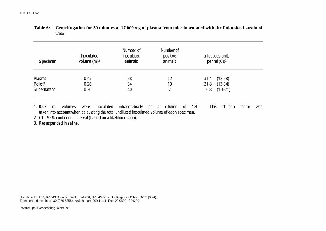
T_BLOOD.doc
Rue de la Loi 200, B-1049 Bruxelles/Wetstraat 200, B-1049 Brussel - Belgium - Office: B232 (6/74).Telephone: direct line (+32-2)29 58554, switchboard 299.11.11. Fax: 29 96301 / 96299.
Internet: [email protected]
Table 6: Centrifugation for 30 minutes at 17,000 x g of plasma from mice inoculated with the Fukuoka-1 strain ofTSE
Number of Number ofInoculated inoculated positive Infectious units
Specimen volume (ml)1 animals animals per ml (CI)2
Plasma 0.47 28 12 34.4 (18-58)Pellet3 0.26 34 19 21.8 (13-34)Supernatant 0.30 40 2 6.8 (1.1-21)
1. 0.03 ml volumes were inoculated intracerebrally at a dilution of 1:4. This dilution factor wastaken into account when calculating the total undiluted inoculated volume of each specimen.
2. CI = 95% confidence interval (based on a likelihood ratio).3. Resuspended in saline.

T_BLOOD.doc
Rue de la Loi 200, B-1049 Bruxelles/Wetstraat 200, B-1049 Brussel - Belgium - Office: B232 (6/74).Telephone: direct line (+32-2)29 58554, switchboard 299.11.11. Fax: 29 96301 / 96299.
Internet: [email protected]
Table 7: Bioassay of infectivity in the plasma and buffy coat of symptomatic mice inoculated16-18 weeks earlier with the Fukuoka-1 strain of TSE: comparison of levels of infectivity detected by intracerebral orintravenous inoculation of specimens into the assay animals
Number of Number of Intravenous toRoute of Inoculated inoculated positive Infectious units intracerebral
Specimen inoculation volume1 animals animals per ml (CI)2 equivalence
Plasma intravenous 4.50 30 16 5.1 (3-8)intracerebral 0.47 28 12 34.4 (18-58) 7:1
Buffy coat intravenous 1.40 28 18 20.6 (12-32)intracerebral 0.14 27 11 106.0 (55-184) 5:1
1. For intracerebral inoculations, 0.03 ml was inoculated at dilutions of 1:2, 1:4, or 1:40. For intravenous inoculations, 0.15 ml was inoculated undiluted(plasma) or 0.2 ml at a dilution of 1:4 (buffy coat). These dilution factors were taken into account when calculating the total undiluted inoculated volume ofeach specimen.2. CI = 95% confidence interval (based on a likelihood ratio).
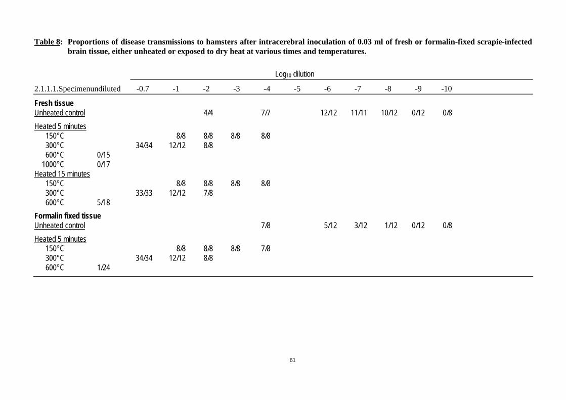
61
Table 8: Proportions of disease transmissions to hamsters after intracerebral inoculation of 0.03 ml of fresh or formalin-fixed scrapie-infectedbrain tissue, either unheated or exposed to dry heat at various times and temperatures.
Log10 dilution
2.1.1.1. Specimenundiluted -0.7 -1 -2 -3 -4 -5 -6 -7 -8 -9 -10
Fresh tissueUnheated control 4/4 7/7 12/12 11/11 10/12 0/12 0/8Heated 5 minutes 150°C 8/8 8/8 8/8 8/8 300°C 34/34 12/12 8/8 600°C 0/15 1000°C 0/17Heated 15 minutes 150°C 8/8 8/8 8/8 8/8 300°C 33/33 12/12 7/8 600°C 5/18Formalin fixed tissueUnheated control 7/8 5/12 3/12 1/12 0/12 0/8Heated 5 minutes 150°C 8/8 8/8 8/8 7/8 300°C 34/34 12/12 8/8 600°C 1/24
T_BLOOD.doc
Rue de la Loi 200, B-1049 Bruxelles/Wetstraat 200, B-1049 Brussel - Belgium - Office: B232 (6/74).Telephone: direct line (+32-2)29 58554, switchboard 299.11.11. Fax: 29 96301 / 96299.
Internet: [email protected]
ANNEX 3
STUNNING, KILLING AND COLLECTION OFBLOOD FROM FOOD ANIMALS FOR HUMAN ANDANIMAL CONSUMPTION AND OTHER PURPOSES
INTRODUCTIONSince blood, for human food or animal feed, is only permitted to be collected fromhealthy animals that have passed an official ante mortem inspection and that are killed ina licensed slaughterhouse, supervised by the Competent Veterinary Authority, only theseanimals will be considered. This report is concerned with TSE risks only. Therefore, intheory, only ruminant species need to be considered. In practice blood is collected fromcattle and pigs and could be collected for various purposes from other ruminant speciesincluding sheep, goats and deer, and theoretically also from poultry. Slaughter methodsvary between species and between different religious sects so this annex will cover allthese aspects.
Killing is accomplished by bleeding. Bleeding out is of considerable importance to thesubsequent keeping quality of meat and acceptability by the consumer. Some methods ofreligious slaughter employ a sharp knife to ‘cut the throat’ without any prior treatment.Welfare considerations conflict with this aspect of slaughter. In the EU, most animals arestunned before they are bled out. The purpose of stunning is to reduce pre-slaughterstress of the animal, to ensure the slaughter is conducted in a humane manner, to improveoperator safety and maintain hygiene to a high standard. In particular, stunning is used toinduce a state of unconsciousness and insensibility of sufficient duration to ensure that theanimal does not recover whilst being bled out (Blackmore & Delany, 1988). Pigs areparticularly prone to pre-slaughter stress. Excessive stress can significantly affect themeat quality of pigs and so produce pale soft and exudative (PSE) meat or dark firm anddry (DFD) meat depending upon the glycogen level in muscles at the time of slaughter.
LEGISLATIONAccording the Council Directive (1993) on the protection of animals at the time ofslaughter or killing, solipeds, ruminants, pigs, rabbits and poultry brought intoslaughterhouses for slaughter shall be a) moved and if necessary lairaged, b) restrainedand c) stunned before slaughter. Animals must be restrained in an appropriate manner, tospare them any avoidable pain, suffering, agitation, injury or contusions. Animals mustnot be suspended before stunning or killing; however, poultry and rabbits may besuspended for slaughter provided that appropriate measures are taken to ensure that theyare in a sufficiently relaxed state for stunning. Permitted methods for stunning are 1)captive bolt pistol, 2) concussion, 3) electro-narcosis and 4) exposure to carbon dioxide.
According the existing EU legislation, (Directive 93/119/EC), animals shall be spared anyavoidable excitement, pain or suffering during movement, lairaging, restraint, stunning,slaughter or killing. The construction, facilities and equipment of slaughterhouses, and

63
their operation, shall be such as to spare animals any avoidable excitement, pain orsuffering.
Instruments, restraint and other equipment and installations used for stunning or killingmust be designed, constructed, maintained and used in such a way as to achieve rapid andeffective stunning or killing in accordance with the provisions of this Directive. Thecompetent authority shall check that the instruments, restraint and other equipment usedfor stunning or killing comply with the above principles and shall check regularly toensure that they are in a good state of repair and will allow the aforementioned objectiveto be attained.
Inspections and controls in slaughterhouses shall be carried out under the responsibility ofthe competent authority, which shall at all times have free access to all parts ofslaughterhouses in order to ascertain compliance with this Directive.
Methods on stunningCaptive bolt stunning is widely used for all farm animals and rabbits. Gunpowder(cartridge), compressed air and spring under tension have been used to drive bolts throughthe skull of animals. The ideal shooting position is frontally on the head. Pithing issometimes used to prolong the period of stunning and to improve operator safety. It is notcompulsory and many large slaughterhouses do not use it. It is not recommended forhygienic reasons.
A study in the EU-Member states in the seventies showed that the captive bolt was usedfor stunning of more than 90% of cattle. A few cases were stunned by concussion.Electrical stunning was mainly used for sheep and in a few cases for bobby calves. Gasstunning was not used for cattle and sheep (Mickwitz & Leach, 1977). It can be expectedthat these figures have not changed dramatically in the last 20 years. Religiousslaughtering (bleeding without stunning) is limited in the Member states and shall operateunder the responsibility of the official veterinarian.
EFFECT ON BRAIN TISSUEIn general, penetration of a missile into the brain can cause injury in the following threeways, depending on its velocity and shape: a) by laceration and crushing at a low velocity(< 100 m/s), b) by shock waves at a high velocity (about 100 to 300 m/s) and c) bytemporary cavitation at a very high velocity (> 300 m/s) (Hopkinson & Marshall, 1967).The velocity of a bolt of a captive bolt pistol is about 100 m/s in the air. This low velocityand shape of the bolt should crush the cortex and deeper parts of the brain either by thebolt itself or by forward shock waves (Lambooij 1982).
When the bolt penetrates the cavity of the skull, the capacity cannot be increased due tothe inflexible bony skull. The consequence is that some brain tissue has to leave thecavity via the hole of the penetrated bolt in the skull. When air pressure is used to limitconvulsions a lot of brain tissue will leave the cavity. Brain tissue was observed in lungsof cattle slaughtered in the US after being stunned with a pneumatic powered stunner(Garland et al, 1996), however, the validity and relevance was questioned (Taylor, 1996;Munro, 1997). To determine the extent of dissemination of CNS tissue in the heartapproximately 2,000 carcasses in commercial beef slaughterhouses in the US werescreened. Visible clots (not necessarily containing CNS emboli) in the right ventriclewere observed in 33%, 12% and 1% when powered air injection stunners, pneumaticpowered stunners and cartridge stunners, respectively, were used (Schmidt et al, 1999).Moreover, multiple fragments of brain tissue were detected in the jugular vein blood of 4

64
out of 15, 1 out of 16 and 0 out of 15 cattle slaughtered after the use of powered airinjection stunners, cartridge penetrating stunner with subsequent pithing and captive boltstunner, respectively. Brain tissue was not observed in the jugular vein blood afterconcussion stunning (Anil et al, 1999).
PHASES OF THE SLAUGHTERING PROCESSThe different slaughtering phases can be summarised as follows:
1. Movement and lairaging of animals in slaughterhouses
2. Restraint of animals before stunning, slaughter or killing- Animals must be restrained in an appropriate manner in such a way as to spare them
any avoidable pain, suffering, agitation, injury or contusions.- Animals which are stunned or killed by mechanical or electrical means applied to
the head must be presented in such a position that the equipment can be applied andoperated easily, accurately and for the appropriate time. The competent authoritymay, however, in the case of solipeds and cattle, authorize the use of appropriatemeans to restrain head movements.
3. StunningNote stunning renders the animal unconscious, bleeding out kills. Stunning must notbe carried out unless it is possible to bleed the animals immediately afterwards. Theperson that is responsible for stunning, shackling, hoisting and bleeding of animalsmust carry out those operations consecutively on one animal before carrying them outon another animal.
4. Bleeding outFor animals which have been stunned, bleeding must be started as soon as possibleafter stunning and be carried out in such a way as to bring about rapid, profuse andcomplete bleeding. In any event, the bleeding must be carried out before the animalregains consciousness.
All animals which have been stunned must be bled by incising at least one of thecarotid arteries or the vessels from which they arise.
Usually, blood destined to human use is collected with a hollow knife. This procedurerequires more time than for conventional bleeding. But this procedure is morehygienic. In the case of severing of arteries, blood is collected more rapidly.
Blood is collected by gravity beneath the hoisted animal into a tank or a canal, to largecollection reservoirs. It is absolutely impossible to separe blood from single animals.
This causes two problems:- the post-mortem inspection on carcasses by the veterinarian is carried out
downstream in the slaughtering chain. In this manner, if the carcass is not declaredfit for human consumption, blood of the same carcass is no longer separable fromblood of the other animals;
- if the captive bolt pistol has not operated correctly and if the carcass of this animalis not seperated, blood with possible cerebral fragments is not separable from therest of the mass.
Individual animal blood collections are not usually done from poultry, swine and cattleso cross-contamination is possible.

65
SLAUGHTER METHODS
1. Captive bolt pistolInstruments must be positioned so as to ensure that the projectile enters the cerebralcortex. In particular, it is prohibited to shoot cattle in the poll position. When using acaptive bolt instrument, the operator must check to ensure that the bolt retracts to itsfull extent after each shot. If it does not so retract, the instrument must not be usedagain until it has been repaired. Animals must not be placed in stunning pens unlessthe operator who is to stun them is ready to do so as soon as the animal is placed in thepen. Animals must not be placed in head restraint until the slaughterman is ready tostun them.
This method is only permitted using a mechanically-operated instrument whichadministers a blow to the skull. The operator must ensure that the instrument is appliedin the proper position and that the correct strength of cartridge is used to produce aneffective stun without fracture of the skull.
2. Electronarcosisa. Electrodes. Electrodes must be so placed that they span the skull, enabling the
current to pass through the brain. Appropriate measures must also be taken toensure that there is good electrical contact, in particular by removing excesswool/hair or wetting skin.
b. Waterbath stunners (for poultry)
3. Exposure to carbon dioxideOnly for pigs
The following specific methods are subject to authorisation by the competent authority,which must ensure in particular that they are used by duly qualified staff and respectingthe animal.
4. Free bullet pistol or rifleFor various species, in particular large farmed game and deer.
5. Decapitation and dislocation of the neck: only for poultry
6. Electrocution and carbon dioxide (see above)
7. Vacuum chamber:This method, which is to be used only for the killing without bleeding of certainanimals for consumption belonging to farmed game species (quail, partridge andpheasant).
OTHER POSSIBLE METHODS FOR SLAUGHTERING MEAT ANIMALSThere is a wide variety of captive bolt stunners available. Non-penetrative stunners have a"mushroom-headed" bolt which impacts with the skull but does not enter the brain,causing the stun due to concussive forces alone. Penetrative stunners cause insensibilitydue to the concussive blow to the skull, and the physical damage resulting from the entryof the bolt into the brain.
There is in addition a particular model of penetrative stunner which provides a blast of airthrough the centre of the bolt following entry into the brain which is intended to have thesame effect as pithing after the stun.

66
These pneumatic stunners with air injection prevail in USA and North Europe whereas inIreland, Italy and the UK no slaughterhouse uses this system.
The use of an injection of high pressure air into the brain produces a significant blowback of brain tissue. This produces a smearing of the head of the animal with liquefiedbrain. The use of this stunner again, following an initial effective stun, leads to a massivequantity of liquefied brain exiting from the original bolt hole (E.C., 1998).
In the case of air introduction in cranial cavity or vertebral canal, there is a possiblepassage of brain tissue in blood circulation (Schmidt, 1999).
ANIMAL SLAUGHTER AUTHORISATION FOR MUSLIM AND JEWISHRITUALS (Italian Health Ministry Decision of 11/06/80)All the precautions able to avoid excitement, pain or suffering of the animals must beadopted. Only slaughtering without stunning is authorised and it must be carried out byqualified people. It is to be done by cutting simultaneously, with a sharp knife, the throat,windpipe and the blood vessels in the neck.
According to information available on Internet, the Muslim ritual consists of cutting (witha sharp knife) the throat, windpipe and the blood vessels in the neck causing death, butwithout cutting the spinal cord. The blood has to be drained before the head is removed.
From the same source (internet) appears that, under the Jewish ritual, the rabbi, using arazor-sharp knife (about 46 cm long by 3.5 cm wide), made a swift cut from side to sideto sever both jugular veins and the two carotid arteries in a single stroke withoutburrowing, tearing or ripping the animal. The head was then raised further as the bloodspurted forth. For beef, the animal is to be cut horizontally across the throat, severing themajor blood vessels, trachea and the oesophagus.
BLOOD COLLECTION
1. For human consumptionBlood for human consumption must be collected only from individual animals so thatif a carcass later fails post mortem examination the blood from that animal can bediscarded. If the carcass passes post mortem inspection the blood can be pooled withblood collected similarly from animals that have also passed post mortem inspection.
2. For pharmaceutical useBlood such as foetal calf blood used to extract foetal calf serum for vaccinemanufacture is collected in a very hygienic and careful manner in order to avoid crosscontamination with maternal blood, placenta and environmental hazards (Shailer andCorin, 1999). Cattle from selected donor herds of high health status can be used(Rolleston, 1999).
3. For animal feedBlood is usually collected into an anticoagulant such as sodium phosphate/sodiumcitrate.
REFERENCES
Rolleston WBR, 1999. Reducing variables through the use of donor herds. Dev BiolStand, 99, 79-86.
Schailer C, Corrin K, 1999 Serum supply: policies and controls operating in NewZealand. Dev Biol Stan, 99, 71-78.