under the hammer: residues resulting from production and microwear on experimental stone tools
TRANSCRIPT

556
L. Byrne, A. Ollé and J. M. Vergès
© University of Oxford, 2006,
Archaeometry
48
, 4 (2006) 549–564
After cleaning, no residues remain and no deformation of the flint surface is visible(Fig. 4 (b)).
It is important to note that some of the technological residues produced by retouching withsoft hammers are very similar to residues generated by use on the same materials (i.e., boneand antler). These deposits have in the past been referred to as ‘polish’, but it is important toreiterate that ‘polish’ and ‘residues or deposits’ are two very different phenomena, which mustbe differentiated in order to gain a clear understanding of microwear mechanisms.
The first part of our experimental programme clearly illustrates this point by examining, onthe one hand, hammerstone residues and, on the other, underlying flint deformation. Althoughthis deformation is most pronounced when hard hammers such as quartz, quartzite, sandstoneand, to a lesser extent, limestone are used, soft hammers such as antler also modify surfacetopography, producing smooth compressed zones associated with multiple fine striations.
Subsequent modification of hammerstone features with use
Having gained a basic knowledge of different types of percussion features, we then conductedseveral short experiments (five of which are presented here) in order to gauge the resistance ofthese percussion smears to use and to record the extent of their modification during wood-working, hide-processing and butchery experiments.
Experiment 1
First, a step-by-step woodworking experiment was conducted on a flint flakeretouched with a limestone hammer. The sidescraper was used to cut fresh juniper (
Juniperusoxycedrus
) for 2.5 min, rinsed under tepid water for 1 min and returned to the ESEM chamber.After careful observation, the tool was reused for a further 2.5 min, rinsed and re-examinedwith the ESEM. The final stage entailed using the tool in the same way for an additional 5min, making a total of 10 min use. During use, the tool was held perpendicularly to the branchand cut through the bark and the fibrous wood. Care was taken to observe and visually recordthe same zones observed before use, although angles can vary very slightly between thedifferent series.
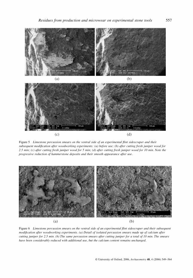
Very little morphological modification of the tool edge was observed after the initial phaseof this short woodworking experiment, although percussion features illustrated above (Figs 5and 6) had undergone substantial modification. In some areas, percussion deposits andstriations appear to have been removed by polish formation and analysis reveals no traces ofcalcium. However, in other areas, percussion features are still visible but their appearance haschanged significantly. In Figures 5 (b) and 6 (a), after use, the percussion smears have a muchsmoother appearance than in Figure 5 (a), and appear to have been compacted and polished.Like the percussion deposits prior to use, these zones contain a high proportion of calcium.
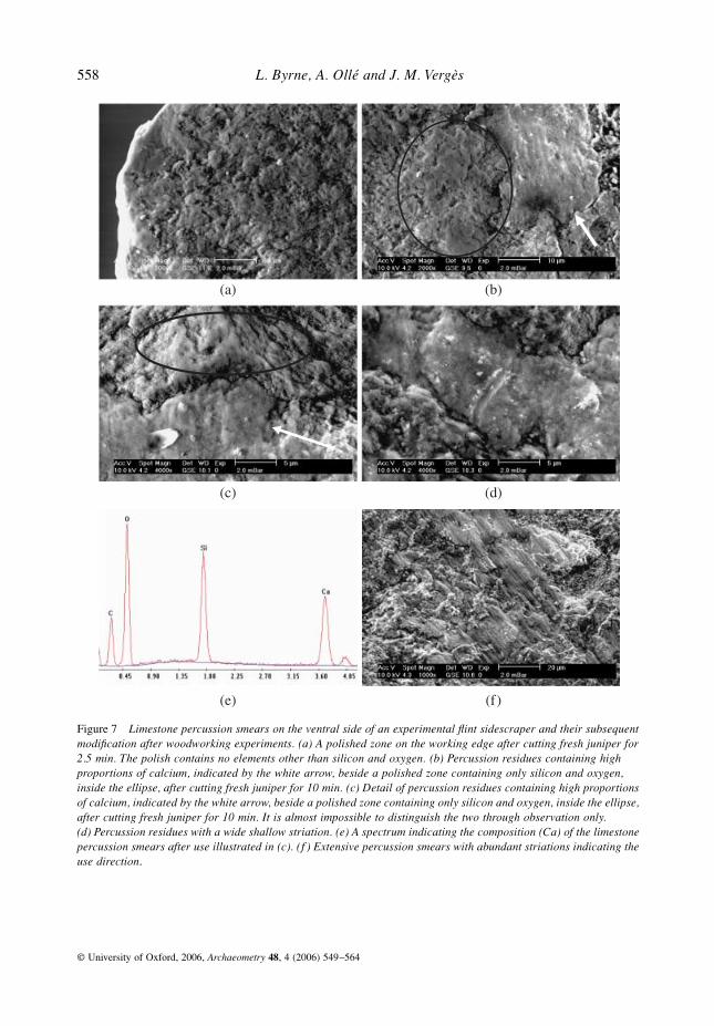
Some polish formation occurred locally after this experiment, as illustrated in Figure 7.Although polish formation processes are beyond the scope of this paper, it is important to notethat the only elements present in polished zones are silicon and oxygen.
After cutting juniper for a further 2.5 min, plant residues are more frequent, although norecognizable juniper tissues were identified. Polish is visible along much of the tool edge. Asabove, EDX analysis of polished areas reveals that the only elements present are silicon andoxygen.
Apart from in cracks and depressions, most of the hammerstone deposits have beenremoved from the proximal end of the tool, probably simply through an abrasive action duringthe woodworking. However, towards the centre and the distal edge of the tool, extensive
Residues from production and microwear on experimental stone tools
557
© University of Oxford, 2006,
Archaeometry
48
, 4 (2006) 549–564
Figure 5 Limestone percussion smears on the ventral side of an experimental flint sidescraper and their subsequent modification after woodworking experiments: (a) before use; (b) after cutting fresh juniper wood for 2.5 min; (c) after cutting fresh juniper wood for 5 min; (d) after cutting fresh juniper wood for 10 min. Note the progressive reduction of hammerstone deposits and their smooth appearance after use.
Figure 6 Limestone percussion smears on the ventral side of an experimental flint sidescraper and their subsequent modification after woodworking experiments. (a) Detail of isolated percussion smears made up of calcium after cutting juniper for 2.5 min. (b) The same percussion smears after cutting juniper for a total of 10 min. The smears have been considerably reduced with additional use, but the calcium content remains unchanged.
558
L. Byrne, A. Ollé and J. M. Vergès
© University of Oxford, 2006,
Archaeometry
48
, 4 (2006) 549–564
Figure 7 Limestone percussion smears on the ventral side of an experimental flint sidescraper and their subsequent modification after woodworking experiments. (a) A polished zone on the working edge after cutting fresh juniper for 2.5 min. The polish contains no elements other than silicon and oxygen. (b) Percussion residues containing high proportions of calcium, indicated by the white arrow, beside a polished zone containing only silicon and oxygen, inside the ellipse, after cutting fresh juniper for 10 min. (c) Detail of percussion residues containing high proportions of calcium, indicated by the white arrow, beside a polished zone containing only silicon and oxygen, inside the ellipse, after cutting fresh juniper for 10 min. It is almost impossible to distinguish the two through observation only. (d) Percussion residues with a wide shallow striation. (e) A spectrum indicating the composition (Ca) of the limestone percussion smears after use illustrated in (c). (f ) Extensive percussion smears with abundant striations indicating the use direction.
Residues from production and microwear on experimental stone tools
559
© University of Oxford, 2006,
Archaeometry
48, 4 (2006) 549–564
hammerstone smears remain (Figs 5 (c) and 6 (b)) and the calcium content of these smears hasnot changed with use. Striations are extensive and parallel to the working edge, indicating thedirection of the cutting motion.
Finally, after sawing juniper for a further 5 min, from the central area to the distal end ofthe working edge, hammerstone smears are still extensive and their elemental configurationremains unchanged. As above, these smears are still made up of calcium. However, theappearance of these hammerstone smears is quite different: many of them are reduced in size(Fig. 6 (c)), while the surface has become much smoother (Fig. 5 (d)) and, in several places,bears longitudinal or diagonal striations (Figs 7 (d) and 7 (f)). It becomes extremely difficultto visually distinguish hammerstone smears from use-wear (Fig. 7 (b)), even at magnificationsof up to 4000× (Fig. 7 (c)). The only feasible means of differentiating the two is throughsystematic elemental analysis. In Figures 7 (b) and 7 (c), the analysis of the zone indicated bythe ellipse does not reveal the presence of any components other than silica, and it is inter-preted as use-related deformation. The arrows point to deposits made up of calcium(Fig. 7 (e)), which are hammerstone smears.
This short woodworking experiment shows that some hammerstone deposits are still presentafter use and that they can mimic use-generated features. It is thus crucial for microwearanalysts to be able to recognize hammerstone deposits before embarking upon microwear orresidue studies, in order to clearly differentiate production processes (technological deforma-tion and residues) from use mechanisms (deformation and residues).
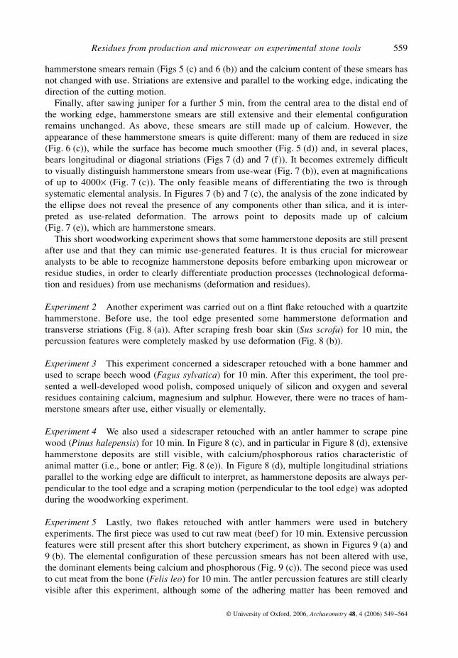
Experiment 2 Another experiment was carried out on a flint flake retouched with a quartzitehammerstone. Before use, the tool edge presented some hammerstone deformation andtransverse striations (Fig. 8 (a)). After scraping fresh boar skin (Sus scrofa) for 10 min, thepercussion features were completely masked by use deformation (Fig. 8 (b)).
Experiment 3 This experiment concerned a sidescraper retouched with a bone hammer andused to scrape beech wood (Fagus sylvatica) for 10 min. After this experiment, the tool pre-sented a well-developed wood polish, composed uniquely of silicon and oxygen and severalresidues containing calcium, magnesium and sulphur. However, there were no traces of ham-merstone smears after use, either visually or elementally.
Experiment 4 We also used a sidescraper retouched with an antler hammer to scrape pinewood (Pinus halepensis) for 10 min. In Figure 8 (c), and in particular in Figure 8 (d), extensivehammerstone deposits are still visible, with calcium/phosphorous ratios characteristic ofanimal matter (i.e., bone or antler; Fig. 8 (e)). In Figure 8 (d), multiple longitudinal striationsparallel to the working edge are difficult to interpret, as hammerstone deposits are always per-pendicular to the tool edge and a scraping motion (perpendicular to the tool edge) was adoptedduring the woodworking experiment.
Experiment 5 Lastly, two flakes retouched with antler hammers were used in butcheryexperiments. The first piece was used to cut raw meat (beef) for 10 min. Extensive percussionfeatures were still present after this short butchery experiment, as shown in Figures 9 (a) and9 (b). The elemental configuration of these percussion smears has not been altered with use,the dominant elements being calcium and phosphorous (Fig. 9 (c)). The second piece was usedto cut meat from the bone (Felis leo) for 10 min. The antler percussion features are still clearlyvisible after this experiment, although some of the adhering matter has been removed and
560 L. Byrne, A. Ollé and J. M. Vergès
© University of Oxford, 2006, Archaeometry 48, 4 (2006) 549–564
Figure 8 Percussion features and their subsequent modification after hide and woodworking experiments. (a) Hammerstone features on a flint flake retouched with a quartzite hammer. The tool edge presents some deformation (inside the ellipse) and transverse striations (white arrows). (b) The same tool edge after scraping fresh boar skins for 10 min. The percussion features have been completely masked by use deformation. (c) A cracked and polished zone on the working edge of a tool retouched with reindeer antler and used to scrape pinewood for 10 min. The polished zones contain only silicon and oxygen, whereas particles trapped in the fissures contain traces of calcium. (d) Antler percussion residues with striations running parallel to the working edge after scraping pinewood for 10 min. EDX analysis yields characteristic proportions of calcium and phosphorus. (e) A spectrum indicating the composition of the antler percussion features (Ca, P and Mg) illustrated in (d).
Residues from production and microwear on experimental stone tools 561
© University of Oxford, 2006, Archaeometry 48, 4 (2006) 549–564
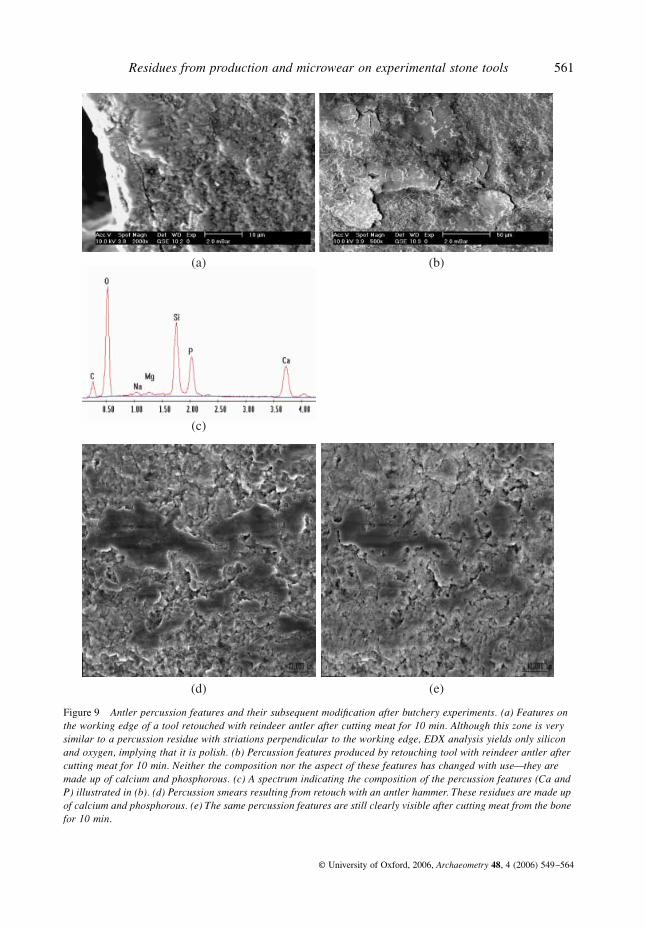
Figure 9 Antler percussion features and their subsequent modification after butchery experiments. (a) Features on the working edge of a tool retouched with reindeer antler after cutting meat for 10 min. Although this zone is very similar to a percussion residue with striations perpendicular to the working edge, EDX analysis yields only silicon and oxygen, implying that it is polish. (b) Percussion features produced by retouching tool with reindeer antler after cutting meat for 10 min. Neither the composition nor the aspect of these features has changed with use—they are made up of calcium and phosphorous. (c) A spectrum indicating the composition of the percussion features (Ca and P) illustrated in (b). (d) Percussion smears resulting from retouch with an antler hammer. These residues are made up of calcium and phosphorous. (e) The same percussion features are still clearly visible after cutting meat from the bone for 10 min.
562 L. Byrne, A. Ollé and J. M. Vergès
© University of Oxford, 2006, Archaeometry 48, 4 (2006) 549–564
some deformation of the flint surface is clearly visible (Figs 9 (d) and 9 (e)). Analysis revealsthe presence of calcium and phosphorous.
DISCUSSION
We have shown in this paper that some hammerstone smears consist of diagnostic elementsand that, in some cases, they are still visible after use. Four of the six retouched stone toolsused here retained percussion features after experiments lasting for 10 min. One of these is atool retouched with a limestone hammer, used in a step-by-step woodworking experiment;another is a tool retouched with an antler hammer, used to scrape pine wood; whilst the twoothers are flakes retouched with antler hammers and subsequently used in butchery experi-ments. Our experiments clearly illustrate that percussion features can undergo significanttransformation during use, to such an extent that, in some cases, the only reliable means ofdistinguishing these percussion features from microwear features is through systematic EDXanalysis.
The implications of these somewhat surprising results for archaeological stone tools areclear; if archaeological tools present microwear features and residues, it is possible that per-cussion features and residues have also been preserved. This is especially pertinent for siteslocated on or beside abundant raw material sources, where tools are used for short periods oftime before being discarded, or where a relatively high proportion of tools is unused. Simi-larly, tools used on soft materials, such as meat, may undergo very little modification with useand may retain extensive technological features, as shown by our butchery experiments withflint tools retouched with antler hammers.
Although is it straightforward to remove most percussion smears from unused experimentalflint tools, following the cleaning procedure outlined above, it appears that technologicalresidues that are resistant to use are more difficult to remove. Although removing technologicalfeatures from unused stone tools is an artificial situation, the aim of this exercise was purelyto record the deformations underlying the hammerstone deposits. The most characteristicdeformations of the flint topography due to retouching are friction marks, which indicate thepercussion direction. These features can be easily confused with linear features resulting fromuse, which are often interpreted as indicative of the direction of use.
These observations should also be taken into consideration by analysts attempting to extractDNA from stone tools, as technological traces of DNA may subsist on tools retouched withbone or antler; for example, particularly in microcracks provoked by the production process.
CONCLUSIONS
The primary aims of this research were to characterize the elemental composition of hammer-stone smears, to assess the subsequent modification of hammerstone smears with use, and toillustrate the potential interference of these hammerstone deposits with deposits from workedmaterials and use-generated surface deformation. Our main conclusions are that differentkinds of hammerstone smears (quartz, limestone, bone, antler and wood) are extensive onexperimental retouched stone tools, that with EDX analysis it is possible to identify the typeof hammerstone used, and that hammerstone smears can be surprisingly resistant to use andremarkably similar to use-generated residues and microwear. The only reliable means of cor-rectly identifying some hammerstone features after use is through systematic EDX analysis,which may explain why these features have, up until now, been largely overlooked.
Residues from production and microwear on experimental stone tools 563
© University of Oxford, 2006, Archaeometry 48, 4 (2006) 549–564
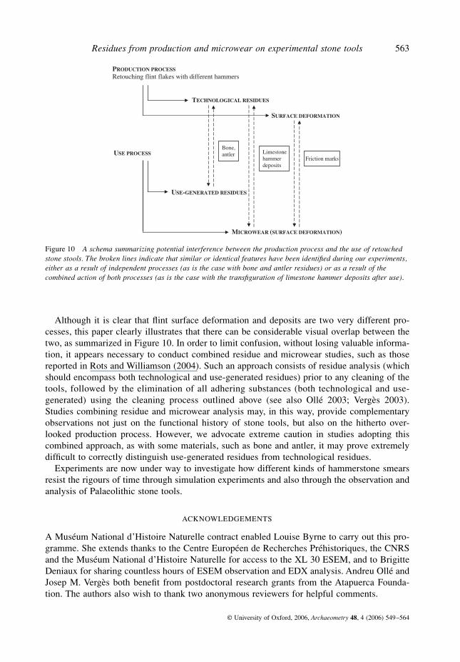
Although it is clear that flint surface deformation and deposits are two very different pro-cesses, this paper clearly illustrates that there can be considerable visual overlap between thetwo, as summarized in Figure 10. In order to limit confusion, without losing valuable informa-tion, it appears necessary to conduct combined residue and microwear studies, such as thosereported in Rots and Williamson (2004). Such an approach consists of residue analysis (whichshould encompass both technological and use-generated residues) prior to any cleaning of thetools, followed by the elimination of all adhering substances (both technological and use-generated) using the cleaning process outlined above (see also Ollé 2003; Vergès 2003).Studies combining residue and microwear analysis may, in this way, provide complementaryobservations not just on the functional history of stone tools, but also on the hitherto over-looked production process. However, we advocate extreme caution in studies adopting thiscombined approach, as with some materials, such as bone and antler, it may prove extremelydifficult to correctly distinguish use-generated residues from technological residues.
Experiments are now under way to investigate how different kinds of hammerstone smearsresist the rigours of time through simulation experiments and also through the observation andanalysis of Palaeolithic stone tools.
ACKNOWLEDGEMENTS
A Muséum National d’Histoire Naturelle contract enabled Louise Byrne to carry out this pro-gramme. She extends thanks to the Centre Européen de Recherches Préhistoriques, the CNRSand the Muséum National d’Histoire Naturelle for access to the XL 30 ESEM, and to BrigitteDeniaux for sharing countless hours of ESEM observation and EDX analysis. Andreu Ollé andJosep M. Vergès both benefit from postdoctoral research grants from the Atapuerca Founda-tion. The authors also wish to thank two anonymous reviewers for helpful comments.
Figure 10 A schema summarizing potential interference between the production process and the use of retouched stone stools. The broken lines indicate that similar or identical features have been identified during our experiments, either as a result of independent processes (as is the case with bone and antler residues) or as a result of the combined action of both processes (as is the case with the transfiguration of limestone hammer deposits after use).
564 L. Byrne, A. Ollé and J. M. Vergès
© University of Oxford, 2006, Archaeometry 48, 4 (2006) 549–564
REFERENCES
Anderson-Gerfaud, P., 1981, Contribution méthodologique à l’analyse des microtraces d’utilisation sur les outilspréhistoriques, Ph.D. thesis, Bordeaux University.
Anderson-Gerfaud, P., 1990, Aspects of behaviour in the Middle Palaeolithic: functional analysis of stone tools fromsouthwest France, in The emergence of modern humans: an archaeological perspective (ed. P. Mellars), 389–418,Edinburgh University Press, Edinburgh.
Christensen, M., 1999, Technologie de l’ivoire au Paléolithique supérieur: caractérisation physico-chimique dumatériau et analyse fonctionnelle des outils de transformation, BAR vol. 751, British Archaeological Reports,Oxford.
Christensen, M., Walter, Ph., and Menu, M., 1992, Usewear characterisation of prehistoric flints with IBA, NuclearInstruments and Methods in Physics Research, B64, 488–93.
Hardy, B. L., 1994, Investigations of stone tool function through use-wear, residue and DNA analyses at the MiddlePaleolithic site of La Quina, France, Ph.D. dissertation, Indiana University.
Hardy, B. L., and Garufi, G. T., 1998, Identification of woodworking on stone tools through residue and use-wearanalyses: experimental results, Journal of Archaeological Science, 25, 177–84.
Ibáñez, J. J., González, J. E., Lagüera, M. A., and Gutiérrez, C., 1990, Knapping traces: their characteristics accordingto the hammerstone and the technique used, Le silex de sa genèse à l’outil. Actes du V° colloque international surle silex, 1987, Cahiers du Quaternaire, 17, 547–53.
Jahren, A. H., Toth, N., Schick, K., Clark, J. D., and Amundson, R. G., 1997, Determining stone tool use: elementaland morphological analyses of residues on experimentally manufactured stone tools, Journal of ArchaeologicalScience, 24, 245–50.
Keeley, L., 1980, Experimental determination of stone tool uses: a microwear analysis, The University of ChicagoPress, Chicago.
Kimura, B., Brandt, S. A., Hardy, B. L., and Hauswirth, W. W., 2001, Analysis of DNA from ethnoarchaeologicalstone scrapers, Journal of Archaeological Science, 28, 45–53.
Loy, T. H., and Dixon, E. J., 1998, Blood residues on fluted points from eastern Beringia, American Antiquity, 63,21–46.
Mansur-Franchomme, E., 1986, Microscopie du matériel lithique préhistorique. Traces d’utilisation, altérationsnaturelles, accidentelles et technologiques. Exemple de Patagonie, Cahiers du Quaternaire n° 9, Editions du CentreNational de la Recherche Scientifique, Paris.
Ollé, A., 2003, Variabilitat i patrons funcionals en els sistemes tècnics de mode 2. Anàlisi de les deformacions d’úsen els conjunts lítics del Riparo Esterno de Grotta Paglicci (Rignano Garganico, Foggia), Áridos (Arganda,Madrid) i Galería-TN (Sierra de Atapuerca, Burgos), Ph.D. thesis, Rovira i Virgili University, Tarragona.
Patou-Mathis, M. (ed.), 2002, Retouchoirs, compresseurs, percuteurs . . . Os à impressions et éraillures, EditionsSociété Préhistorique Française.
Rots, V., and Williamson, B. S., 2004, Microwear and residue analyses in perspective: the contribution of ethnoar-chaeological evidence, Journal of Archaeological Science, 31, 1287–99.
Shanks, O. C., Vella, A. T., Bonnischen, R., and Ream, W., 2001, Recovery of protein and DNA trapped in stone toolmicrocracks, Journal of Archaeological Science, 28, 965–72.
Shanks, O. C., Hodges, L., Tilley, L., Kornfeld, M., Larson, M. L., and Ream, W., 2005, DNA from ancient stonetools and bones excavated at Bugas-Holding, Wyoming, Journal of Archaeological Science, 32, 27–38.
Tuross, N., Barnes, I., and Potts, R., 1996, Protein identification of blood residues on experimental stone tools, Journal ofArchaeological Science, 23, 289–96.
Vergès, J. M., 2003, Caracterització des models d’instrumental lític de mode 1 a partir de les dades de l’anàlisifuncional dels conjunts litotènics d’Aïn Hanech i El-Kherba (Algèria), Monte Poggiolo i Isernia la Pineta (Itàlia),Ph.D. thesis, Rovira i Virgili University, Tarragona.
Wadley, L., Lombard, M., and Williamson, B. S., 2004, The first residue analysis blind tests: results and lessonslearnt, Journal of Archaeological Science, 31, 1491–501.


556
L. Byrne, A. Ollé and J. M. Vergès
© University of Oxford, 2006,
Archaeometry
48
, 4 (2006) 549–564
After cleaning, no residues remain and no deformation of the flint surface is visible(Fig. 4 (b)).
It is important to note that some of the technological residues produced by retouching withsoft hammers are very similar to residues generated by use on the same materials (i.e., boneand antler). These deposits have in the past been referred to as ‘polish’, but it is important toreiterate that ‘polish’ and ‘residues or deposits’ are two very different phenomena, which mustbe differentiated in order to gain a clear understanding of microwear mechanisms.
The first part of our experimental programme clearly illustrates this point by examining, onthe one hand, hammerstone residues and, on the other, underlying flint deformation. Althoughthis deformation is most pronounced when hard hammers such as quartz, quartzite, sandstoneand, to a lesser extent, limestone are used, soft hammers such as antler also modify surfacetopography, producing smooth compressed zones associated with multiple fine striations.
Subsequent modification of hammerstone features with use
Having gained a basic knowledge of different types of percussion features, we then conductedseveral short experiments (five of which are presented here) in order to gauge the resistance ofthese percussion smears to use and to record the extent of their modification during wood-working, hide-processing and butchery experiments.
Experiment 1
First, a step-by-step woodworking experiment was conducted on a flint flakeretouched with a limestone hammer. The sidescraper was used to cut fresh juniper (
Juniperusoxycedrus
) for 2.5 min, rinsed under tepid water for 1 min and returned to the ESEM chamber.After careful observation, the tool was reused for a further 2.5 min, rinsed and re-examinedwith the ESEM. The final stage entailed using the tool in the same way for an additional 5min, making a total of 10 min use. During use, the tool was held perpendicularly to the branchand cut through the bark and the fibrous wood. Care was taken to observe and visually recordthe same zones observed before use, although angles can vary very slightly between thedifferent series.
Very little morphological modification of the tool edge was observed after the initial phaseof this short woodworking experiment, although percussion features illustrated above (Figs 5and 6) had undergone substantial modification. In some areas, percussion deposits andstriations appear to have been removed by polish formation and analysis reveals no traces ofcalcium. However, in other areas, percussion features are still visible but their appearance haschanged significantly. In Figures 5 (b) and 6 (a), after use, the percussion smears have a muchsmoother appearance than in Figure 5 (a), and appear to have been compacted and polished.Like the percussion deposits prior to use, these zones contain a high proportion of calcium.
Some polish formation occurred locally after this experiment, as illustrated in Figure 7.Although polish formation processes are beyond the scope of this paper, it is important to notethat the only elements present in polished zones are silicon and oxygen.
After cutting juniper for a further 2.5 min, plant residues are more frequent, although norecognizable juniper tissues were identified. Polish is visible along much of the tool edge. Asabove, EDX analysis of polished areas reveals that the only elements present are silicon andoxygen.
Apart from in cracks and depressions, most of the hammerstone deposits have beenremoved from the proximal end of the tool, probably simply through an abrasive action duringthe woodworking. However, towards the centre and the distal edge of the tool, extensive
Residues from production and microwear on experimental stone tools
557
© University of Oxford, 2006,
Archaeometry
48
, 4 (2006) 549–564
Figure 5 Limestone percussion smears on the ventral side of an experimental flint sidescraper and their subsequent modification after woodworking experiments: (a) before use; (b) after cutting fresh juniper wood for 2.5 min; (c) after cutting fresh juniper wood for 5 min; (d) after cutting fresh juniper wood for 10 min. Note the progressive reduction of hammerstone deposits and their smooth appearance after use.
Figure 6 Limestone percussion smears on the ventral side of an experimental flint sidescraper and their subsequent modification after woodworking experiments. (a) Detail of isolated percussion smears made up of calcium after cutting juniper for 2.5 min. (b) The same percussion smears after cutting juniper for a total of 10 min. The smears have been considerably reduced with additional use, but the calcium content remains unchanged.
558
L. Byrne, A. Ollé and J. M. Vergès
© University of Oxford, 2006,
Archaeometry
48
, 4 (2006) 549–564
Figure 7 Limestone percussion smears on the ventral side of an experimental flint sidescraper and their subsequent modification after woodworking experiments. (a) A polished zone on the working edge after cutting fresh juniper for 2.5 min. The polish contains no elements other than silicon and oxygen. (b) Percussion residues containing high proportions of calcium, indicated by the white arrow, beside a polished zone containing only silicon and oxygen, inside the ellipse, after cutting fresh juniper for 10 min. (c) Detail of percussion residues containing high proportions of calcium, indicated by the white arrow, beside a polished zone containing only silicon and oxygen, inside the ellipse, after cutting fresh juniper for 10 min. It is almost impossible to distinguish the two through observation only. (d) Percussion residues with a wide shallow striation. (e) A spectrum indicating the composition (Ca) of the limestone percussion smears after use illustrated in (c). (f ) Extensive percussion smears with abundant striations indicating the use direction.
Residues from production and microwear on experimental stone tools
559
© University of Oxford, 2006,
Archaeometry
48, 4 (2006) 549–564
hammerstone smears remain (Figs 5 (c) and 6 (b)) and the calcium content of these smears hasnot changed with use. Striations are extensive and parallel to the working edge, indicating thedirection of the cutting motion.
Finally, after sawing juniper for a further 5 min, from the central area to the distal end ofthe working edge, hammerstone smears are still extensive and their elemental configurationremains unchanged. As above, these smears are still made up of calcium. However, theappearance of these hammerstone smears is quite different: many of them are reduced in size(Fig. 6 (c)), while the surface has become much smoother (Fig. 5 (d)) and, in several places,bears longitudinal or diagonal striations (Figs 7 (d) and 7 (f)). It becomes extremely difficultto visually distinguish hammerstone smears from use-wear (Fig. 7 (b)), even at magnificationsof up to 4000× (Fig. 7 (c)). The only feasible means of differentiating the two is throughsystematic elemental analysis. In Figures 7 (b) and 7 (c), the analysis of the zone indicated bythe ellipse does not reveal the presence of any components other than silica, and it is inter-preted as use-related deformation. The arrows point to deposits made up of calcium(Fig. 7 (e)), which are hammerstone smears.
This short woodworking experiment shows that some hammerstone deposits are still presentafter use and that they can mimic use-generated features. It is thus crucial for microwearanalysts to be able to recognize hammerstone deposits before embarking upon microwear orresidue studies, in order to clearly differentiate production processes (technological deforma-tion and residues) from use mechanisms (deformation and residues).
Experiment 2 Another experiment was carried out on a flint flake retouched with a quartzitehammerstone. Before use, the tool edge presented some hammerstone deformation andtransverse striations (Fig. 8 (a)). After scraping fresh boar skin (Sus scrofa) for 10 min, thepercussion features were completely masked by use deformation (Fig. 8 (b)).
Experiment 3 This experiment concerned a sidescraper retouched with a bone hammer andused to scrape beech wood (Fagus sylvatica) for 10 min. After this experiment, the tool pre-sented a well-developed wood polish, composed uniquely of silicon and oxygen and severalresidues containing calcium, magnesium and sulphur. However, there were no traces of ham-merstone smears after use, either visually or elementally.
Experiment 4 We also used a sidescraper retouched with an antler hammer to scrape pinewood (Pinus halepensis) for 10 min. In Figure 8 (c), and in particular in Figure 8 (d), extensivehammerstone deposits are still visible, with calcium/phosphorous ratios characteristic ofanimal matter (i.e., bone or antler; Fig. 8 (e)). In Figure 8 (d), multiple longitudinal striationsparallel to the working edge are difficult to interpret, as hammerstone deposits are always per-pendicular to the tool edge and a scraping motion (perpendicular to the tool edge) was adoptedduring the woodworking experiment.
Experiment 5 Lastly, two flakes retouched with antler hammers were used in butcheryexperiments. The first piece was used to cut raw meat (beef) for 10 min. Extensive percussionfeatures were still present after this short butchery experiment, as shown in Figures 9 (a) and9 (b). The elemental configuration of these percussion smears has not been altered with use,the dominant elements being calcium and phosphorous (Fig. 9 (c)). The second piece was usedto cut meat from the bone (Felis leo) for 10 min. The antler percussion features are still clearlyvisible after this experiment, although some of the adhering matter has been removed and
560 L. Byrne, A. Ollé and J. M. Vergès
© University of Oxford, 2006, Archaeometry 48, 4 (2006) 549–564
Figure 8 Percussion features and their subsequent modification after hide and woodworking experiments. (a) Hammerstone features on a flint flake retouched with a quartzite hammer. The tool edge presents some deformation (inside the ellipse) and transverse striations (white arrows). (b) The same tool edge after scraping fresh boar skins for 10 min. The percussion features have been completely masked by use deformation. (c) A cracked and polished zone on the working edge of a tool retouched with reindeer antler and used to scrape pinewood for 10 min. The polished zones contain only silicon and oxygen, whereas particles trapped in the fissures contain traces of calcium. (d) Antler percussion residues with striations running parallel to the working edge after scraping pinewood for 10 min. EDX analysis yields characteristic proportions of calcium and phosphorus. (e) A spectrum indicating the composition of the antler percussion features (Ca, P and Mg) illustrated in (d).
Residues from production and microwear on experimental stone tools 561
© University of Oxford, 2006, Archaeometry 48, 4 (2006) 549–564
Figure 9 Antler percussion features and their subsequent modification after butchery experiments. (a) Features on the working edge of a tool retouched with reindeer antler after cutting meat for 10 min. Although this zone is very similar to a percussion residue with striations perpendicular to the working edge, EDX analysis yields only silicon and oxygen, implying that it is polish. (b) Percussion features produced by retouching tool with reindeer antler after cutting meat for 10 min. Neither the composition nor the aspect of these features has changed with use—they are made up of calcium and phosphorous. (c) A spectrum indicating the composition of the percussion features (Ca and P) illustrated in (b). (d) Percussion smears resulting from retouch with an antler hammer. These residues are made up of calcium and phosphorous. (e) The same percussion features are still clearly visible after cutting meat from the bone for 10 min.
562 L. Byrne, A. Ollé and J. M. Vergès
© University of Oxford, 2006, Archaeometry 48, 4 (2006) 549–564
some deformation of the flint surface is clearly visible (Figs 9 (d) and 9 (e)). Analysis revealsthe presence of calcium and phosphorous.
DISCUSSION
We have shown in this paper that some hammerstone smears consist of diagnostic elementsand that, in some cases, they are still visible after use. Four of the six retouched stone toolsused here retained percussion features after experiments lasting for 10 min. One of these is atool retouched with a limestone hammer, used in a step-by-step woodworking experiment;another is a tool retouched with an antler hammer, used to scrape pine wood; whilst the twoothers are flakes retouched with antler hammers and subsequently used in butchery experi-ments. Our experiments clearly illustrate that percussion features can undergo significanttransformation during use, to such an extent that, in some cases, the only reliable means ofdistinguishing these percussion features from microwear features is through systematic EDXanalysis.
The implications of these somewhat surprising results for archaeological stone tools areclear; if archaeological tools present microwear features and residues, it is possible that per-cussion features and residues have also been preserved. This is especially pertinent for siteslocated on or beside abundant raw material sources, where tools are used for short periods oftime before being discarded, or where a relatively high proportion of tools is unused. Simi-larly, tools used on soft materials, such as meat, may undergo very little modification with useand may retain extensive technological features, as shown by our butchery experiments withflint tools retouched with antler hammers.
Although is it straightforward to remove most percussion smears from unused experimentalflint tools, following the cleaning procedure outlined above, it appears that technologicalresidues that are resistant to use are more difficult to remove. Although removing technologicalfeatures from unused stone tools is an artificial situation, the aim of this exercise was purelyto record the deformations underlying the hammerstone deposits. The most characteristicdeformations of the flint topography due to retouching are friction marks, which indicate thepercussion direction. These features can be easily confused with linear features resulting fromuse, which are often interpreted as indicative of the direction of use.
These observations should also be taken into consideration by analysts attempting to extractDNA from stone tools, as technological traces of DNA may subsist on tools retouched withbone or antler; for example, particularly in microcracks provoked by the production process.
CONCLUSIONS
The primary aims of this research were to characterize the elemental composition of hammer-stone smears, to assess the subsequent modification of hammerstone smears with use, and toillustrate the potential interference of these hammerstone deposits with deposits from workedmaterials and use-generated surface deformation. Our main conclusions are that differentkinds of hammerstone smears (quartz, limestone, bone, antler and wood) are extensive onexperimental retouched stone tools, that with EDX analysis it is possible to identify the typeof hammerstone used, and that hammerstone smears can be surprisingly resistant to use andremarkably similar to use-generated residues and microwear. The only reliable means of cor-rectly identifying some hammerstone features after use is through systematic EDX analysis,which may explain why these features have, up until now, been largely overlooked.
Residues from production and microwear on experimental stone tools 563
© University of Oxford, 2006, Archaeometry 48, 4 (2006) 549–564
Although it is clear that flint surface deformation and deposits are two very different pro-cesses, this paper clearly illustrates that there can be considerable visual overlap between thetwo, as summarized in Figure 10. In order to limit confusion, without losing valuable informa-tion, it appears necessary to conduct combined residue and microwear studies, such as thosereported in Rots and Williamson (2004). Such an approach consists of residue analysis (whichshould encompass both technological and use-generated residues) prior to any cleaning of thetools, followed by the elimination of all adhering substances (both technological and use-generated) using the cleaning process outlined above (see also Ollé 2003; Vergès 2003).Studies combining residue and microwear analysis may, in this way, provide complementaryobservations not just on the functional history of stone tools, but also on the hitherto over-looked production process. However, we advocate extreme caution in studies adopting thiscombined approach, as with some materials, such as bone and antler, it may prove extremelydifficult to correctly distinguish use-generated residues from technological residues.
Experiments are now under way to investigate how different kinds of hammerstone smearsresist the rigours of time through simulation experiments and also through the observation andanalysis of Palaeolithic stone tools.
ACKNOWLEDGEMENTS
A Muséum National d’Histoire Naturelle contract enabled Louise Byrne to carry out this pro-gramme. She extends thanks to the Centre Européen de Recherches Préhistoriques, the CNRSand the Muséum National d’Histoire Naturelle for access to the XL 30 ESEM, and to BrigitteDeniaux for sharing countless hours of ESEM observation and EDX analysis. Andreu Ollé andJosep M. Vergès both benefit from postdoctoral research grants from the Atapuerca Founda-tion. The authors also wish to thank two anonymous reviewers for helpful comments.
Figure 10 A schema summarizing potential interference between the production process and the use of retouched stone stools. The broken lines indicate that similar or identical features have been identified during our experiments, either as a result of independent processes (as is the case with bone and antler residues) or as a result of the combined action of both processes (as is the case with the transfiguration of limestone hammer deposits after use).
564 L. Byrne, A. Ollé and J. M. Vergès
© University of Oxford, 2006, Archaeometry 48, 4 (2006) 549–564
REFERENCES
Anderson-Gerfaud, P., 1981, Contribution méthodologique à l’analyse des microtraces d’utilisation sur les outilspréhistoriques, Ph.D. thesis, Bordeaux University.
Anderson-Gerfaud, P., 1990, Aspects of behaviour in the Middle Palaeolithic: functional analysis of stone tools fromsouthwest France, in The emergence of modern humans: an archaeological perspective (ed. P. Mellars), 389–418,Edinburgh University Press, Edinburgh.
Christensen, M., 1999, Technologie de l’ivoire au Paléolithique supérieur: caractérisation physico-chimique dumatériau et analyse fonctionnelle des outils de transformation, BAR vol. 751, British Archaeological Reports,Oxford.
Christensen, M., Walter, Ph., and Menu, M., 1992, Usewear characterisation of prehistoric flints with IBA, NuclearInstruments and Methods in Physics Research, B64, 488–93.
Hardy, B. L., 1994, Investigations of stone tool function through use-wear, residue and DNA analyses at the MiddlePaleolithic site of La Quina, France, Ph.D. dissertation, Indiana University.
Hardy, B. L., and Garufi, G. T., 1998, Identification of woodworking on stone tools through residue and use-wearanalyses: experimental results, Journal of Archaeological Science, 25, 177–84.
Ibáñez, J. J., González, J. E., Lagüera, M. A., and Gutiérrez, C., 1990, Knapping traces: their characteristics accordingto the hammerstone and the technique used, Le silex de sa genèse à l’outil. Actes du V° colloque international surle silex, 1987, Cahiers du Quaternaire, 17, 547–53.
Jahren, A. H., Toth, N., Schick, K., Clark, J. D., and Amundson, R. G., 1997, Determining stone tool use: elementaland morphological analyses of residues on experimentally manufactured stone tools, Journal of ArchaeologicalScience, 24, 245–50.
Keeley, L., 1980, Experimental determination of stone tool uses: a microwear analysis, The University of ChicagoPress, Chicago.
Kimura, B., Brandt, S. A., Hardy, B. L., and Hauswirth, W. W., 2001, Analysis of DNA from ethnoarchaeologicalstone scrapers, Journal of Archaeological Science, 28, 45–53.
Loy, T. H., and Dixon, E. J., 1998, Blood residues on fluted points from eastern Beringia, American Antiquity, 63,21–46.
Mansur-Franchomme, E., 1986, Microscopie du matériel lithique préhistorique. Traces d’utilisation, altérationsnaturelles, accidentelles et technologiques. Exemple de Patagonie, Cahiers du Quaternaire n° 9, Editions du CentreNational de la Recherche Scientifique, Paris.
Ollé, A., 2003, Variabilitat i patrons funcionals en els sistemes tècnics de mode 2. Anàlisi de les deformacions d’úsen els conjunts lítics del Riparo Esterno de Grotta Paglicci (Rignano Garganico, Foggia), Áridos (Arganda,Madrid) i Galería-TN (Sierra de Atapuerca, Burgos), Ph.D. thesis, Rovira i Virgili University, Tarragona.
Patou-Mathis, M. (ed.), 2002, Retouchoirs, compresseurs, percuteurs . . . Os à impressions et éraillures, EditionsSociété Préhistorique Française.
Rots, V., and Williamson, B. S., 2004, Microwear and residue analyses in perspective: the contribution of ethnoar-chaeological evidence, Journal of Archaeological Science, 31, 1287–99.
Shanks, O. C., Vella, A. T., Bonnischen, R., and Ream, W., 2001, Recovery of protein and DNA trapped in stone toolmicrocracks, Journal of Archaeological Science, 28, 965–72.
Shanks, O. C., Hodges, L., Tilley, L., Kornfeld, M., Larson, M. L., and Ream, W., 2005, DNA from ancient stonetools and bones excavated at Bugas-Holding, Wyoming, Journal of Archaeological Science, 32, 27–38.
Tuross, N., Barnes, I., and Potts, R., 1996, Protein identification of blood residues on experimental stone tools, Journal ofArchaeological Science, 23, 289–96.
Vergès, J. M., 2003, Caracterització des models d’instrumental lític de mode 1 a partir de les dades de l’anàlisifuncional dels conjunts litotènics d’Aïn Hanech i El-Kherba (Algèria), Monte Poggiolo i Isernia la Pineta (Itàlia),Ph.D. thesis, Rovira i Virgili University, Tarragona.
Wadley, L., Lombard, M., and Williamson, B. S., 2004, The first residue analysis blind tests: results and lessonslearnt, Journal of Archaeological Science, 31, 1491–501.