unconscious relational encoding depends on hippocampus
TRANSCRIPT
BRAINA JOURNAL OF NEUROLOGY
Unconscious relational encoding depends onhippocampusSimone B. Duss,1,2 Thomas P. Reber,1,2 Jurgen Hanggi,3 Simon Schwab,2,4 Roland Wiest,5
Rene M. Muri,2,6 Peter Brugger,7 Klemens Gutbrod2,6 and Katharina Henke1,2
1 Division of Experimental Psychology and Neuropsychology, Department of Psychology, University of Bern, Bern, Switzerland
2 Centre for Cognition, Learning and Memory, University of Bern, Bern, Switzerland
3 Division Neuropsychology, Institute of Psychology, University of Zurich, Zurich, Switzerland
4 Department of Psychiatric Neurophysiology, University Hospital of Psychiatry, University of Bern, Switzerland
5 Institute of Diagnostic and Interventional Neuroradiology, University Hospital Bern, Bern, Switzerland
6 Division of Cognitive and Restorative Neurology, Department of Neurology, University Hospital Bern, Bern, Switzerland
7 Neuropsychology Unit, Department of Neurology, University Hospital Zurich, Zurich, Switzerland
Correspondence to: Katharina Henke,
Department of Psychology, University of Bern,
Fabrikstrasse 8, 3012 Bern, Switzerland.
E-mail: [email protected].
See Mayes (doi:10.1093/brain/awu284) for a scientific commentary on this article.
Textbooks divide between human memory systems based on consciousness. Hippocampus is thought to support only conscious
encoding, while neocortex supports both conscious and unconscious encoding. We tested whether processing modes, not
consciousness, divide between memory systems in three neuroimaging experiments with 11 amnesic patients (mean
age = 45.55 years, standard deviation = 8.74, range = 23–60) and 11 matched healthy control subjects. Examined processing
modes were single item versus relational encoding with only relational encoding hypothesized to depend on hippocampus.
Participants encoded and later retrieved either single words or new relations between words. Consciousness of encoding
was excluded by subliminal (invisible) word presentation. Amnesic patients and controls performed equally well on the
single item task activating prefrontal cortex. But only the controls succeeded on the relational task activating the hippocampus,
while amnesic patients failed as a group. Hence, unconscious relational encoding, but not unconscious single item encoding,
depended on hippocampus. Yet, three patients performed normally on unconscious relational encoding in spite of am-
nesia capitalizing on spared hippocampal tissue and connections to language cortex. This pattern of results suggests that
processing modes divide between memory systems, while consciousness divides between levels of function within a memory
system.
Keywords: flexible; implicit; nondeclarative; relational; subliminal
IntroductionMemory is not a single faculty nor does it serve a single compu-
tational goal. Humans have several types of memory that are
partially independent, operate within distinct brain circuits, and
pursue separate computational goals (Tulving, 1985, 2002;
Cohen and Eichenbaum, 1993; Squire and Zola, 1996; O’Reilly
and Rudy, 2000; Moscovitch, 2008; Henke, 2010). Evidence for
distinct memory systems came from studies in patients with
amnesia due to hippocampal damage (Squire and Zola, 1996).
Hippocampal patients exhibit impairments of episodic encoding
and retrieval, which refers to the conscious encoding and retrieval
doi:10.1093/brain/awu270 Brain 2014: Page 1 of 16 | 1
Received February 27, 2014. Revised July 2, 2014. Accepted July 31, 2014.� The Author (2014). Published by Oxford University Press on behalf of the Guarantors of Brain.
This is an Open Access article distributed under the terms of the Creative Commons Attribution Non-Commercial License (http://creativecommons.org/licenses/by-nc/4.0/), which permits
non-commercial re-use, distribution, and reproduction in any medium, provided the original work is properly cited. For commercial re-use, please contact [email protected]
Brain Advance Access published October 1, 2014 at W
orld Trade Institute on O
ctober 1, 2014http://brain.oxfordjournals.org/
Dow
nloaded from

of personally experienced events (Tulving, 2002). Nevertheless,
hippocampal patients have preserved unconscious memory
abilities, such as skill acquisition, priming for single items, and
conditioning. These preserved memory abilities do not require con-
sciousness of encoding and retrieval and are supported by extra-
hippocampal brain regions. Because of this pattern of results,
hippocampal processing became firmly associated with conscious-
ness of encoding and retrieval (Squire and Zola, 1996; Tulving,
2002; Moscovitch, 2008).
The different types of memory divide not only on consciousness
but also on processing modes (Cohen and Eichenbaum, 1993;
Squire and Zola, 1996; O’Reilly and Rudy, 2000; Henke, 2010).
The typical processing mode of the hippocampus is the rapid
encoding of new and flexible associations between items within
and across events (Cohen and Eichenbaum, 1993; O’Reilly and
Rudy, 2000; Henke, 2010). Due to their flexible representation,
which owes to the hippocampus, the reactivation of associations
can be triggered by remotely related retrieval cues in situations
that are different from the encoding situation. Inflexible memories,
on the other hand, are thought to depend on the neocortex alone.
Inflexible memories contain unitized (non-relational) information,
whose reactivation is bound to retrieval cues that correspond clo-
sely to items present in the encoding situation. Items can be flex-
ibly stored in the context of an event, if they are constituents of a
network of interrelated items, or they can be stored out-of-con-
text as isolated, distinct, non-relational and rather inflexible repre-
sentations (Mayes et al., 2002; Giovanello et al., 2003; Kan et al.,
2007). Flexible representations are thought to depend on the
hippocampus and its connections with neocortex, while inflexible
representations rely on neocortex alone, as exemplified in priming
or recognition by familiarity.
Experimental designs often confounded processing modes (flex-
ible relational versus rigid non-relational) with levels of conscious-
ness, which made it difficult to pin down the variable that
distinguishes between memory systems—consciousness or
processing modes? Flexible relational encoding/retrieval was
often assessed with tests of episodic memory that require con-
sciousness of retrieval, while rigid non-relational encoding/retrieval
was often assessed with priming tests that do not require con-
sciousness of retrieval. To find out whether processing modes,
i.e. flexible relational versus rigid non-relational, rather than con-
sciousness, would distinguish between memory systems, we
excluded consciousness of encoding/retrieval in this study by pre-
senting all encoding material subliminally, i.e. invisibly. Using a
stringent masking paradigm, we studied unconscious flexible rela-
tional versus unconscious rigid non-relational encoding in 11 pa-
tients with amnesia due to damage in the hippocampal-anterior
thalamic axis and 11 matched control subjects (Table 1). To our
knowledge, amnesic patients have not been tested before with
subliminal protocols.
Based on our notion (Henke, 2010) that processing modes
divide between memory systems with flexible relational encoding
depending on the hippocampus, we hypothesized that damage in
the hippocampal-anterior thalamic axis would impede unconscious
flexible relational but not rigid non-relational encoding and re-
trieval. According to traditional views (Squire and Zola, 1996;
Tulving, 2002; Moscovitch, 2008), unconscious encoding of any
kind of information should remain unaffected by damage in the
hippocampal-anterior thalamic axis. Our second hypothesis postu-
lates a dissociation between conscious and unconscious flexible
relational encoding within the hippocampal memory system.
Although we assume that flexible relational encoding always
depends on the hippocampal memory system, our second
hypothesis predicts that a certain amount of preserved hippocam-
pal tissue and preserved hippocampal connections to neocortex
may suffice unconscious but not conscious flexible relational
encoding. We had hypothesized earlier (Henke, 2010) that uncon-
scious relational encoding might be impaired following large but
not small hippocampal lesions based on neuroimaging data show-
ing that neural networks in the medial temporal lobe (and neo-
cortex) tend to be more sparsely recruited during unconscious
versus conscious relational encoding and retrieval (Henke et al.,
2003a; Degonda et al., 2005; Reber et al., 2012, 2014).
Accordingly, a larger level of functionality in the hippocampal-an-
terior thalamic axis and its connections to neocortex might be
required for conscious versus unconscious relational encoding
and retrieval.
To test these hypotheses, we collected both behavioural data
and functional and structural brain data in patients and controls
using MRI to estimate the location and extent of structural brain
damage as well as the degree of preserved resting-state functional
connectivity between hippocampus and neocortex (Fig. 1). Due to
the neocortical deafferentiation secondary to hippocampal neur-
onal loss, we expected reduced functional connectivity between
hippocampus and neocortex in amnesic patients. Any residual
viable tissue in the hippocampus must dispose of preserved con-
nections with neocortex to support encoding and retrieval. Our
neuroimaging data confirmed medical diagnoses and revealed pre-
served hippocampal tissue and functional connectivity in certain
amnesic patients. We performed also functional MRI during all
memory tasks to image the task-underlying neural network in
controls as well as residual hippocampal memory functions and
functional compensation in amnesic patients. Unfortunately, 5 of
11 patients could not be examined with MRI because of ferrous
implants or claustrophobia; but behavioural data were obtained in
all patients.
Participants underwent three functional MRI experiments that
tested the subliminal processing and later retrieval of single nouns
(one experiment) and pairs of unrelated nouns (two experiments).
All encoding words were presented subliminally with our estab-
lished masking technique (Degonda et al., 2005; Reber and
Henke, 2011; Reber et al., 2012, 2014). Because the masked
encoding words were invisible, the later presented supraliminal
test words could only invoke unconscious retrieval processes.
Compared to controls, we expected patients to exhibit a dimin-
ished performance on the relational task that required the uncon-
scious formation and later retrieval of semantic associations
between two nouns. An equal performance was expected be-
tween groups regarding the processing of single nouns because
a rigid, non-relational single word representation should suffice
this task.
While paradigms were otherwise identical between the two
relational experiments, one relational experiment contained a
single encoding trial, while the other contained nine encoding
2 | Brain 2014: Page 2 of 16 S. B. Duss et al.
at World T
rade Institute on October 1, 2014
http://brain.oxfordjournals.org/D
ownloaded from

trials (Fig. 2 and Supplementary Fig. 1). We presented pattern-
masked pairs of unrelated nouns, such as ‘violin – lemon’, for
semantic relational encoding. Following a 5 min study-test interval,
we presented pairs of new test nouns supraliminally. Because test
nouns were semantic neighbours of encoding nouns, the combin-
ation of the superordinate conceptual categories was retained
from study to test in so-called intact test pairs (encoding ‘violin
– lemon’! retrieval ‘cello – mandarin’) but recombined in so-
called broken test pairs. The use of distinct encoding and retrieval
nouns required a flexible representation of associations. At test,
participants were required to decide whether the two nouns in a
retrieval pair would fit together semantically or not. Based on
previous experiments (Reber and Henke, 2011; Reber et al.,
2014), we expected a successful relational encoding and retrieval
to reflect in a larger number of fit responses to intact versus
broken test pairs.
To test the rigid and non-relational encoding and retrieval of
single nouns, we used a subliminal semantic priming task (Fig. 2).
Semantic priming is a form of memory thought to rely on
prefrontal and temporal neocortices, but not hippocampus
Figure 1 Study overview. There were three sessions that took place on three half-days. In the first session, we examined patients and
controls neuropsychologically. The second and third sessions were devoted to experimentation. Depending on a participant’s MRI
compatibility, the two experimental sessions were conducted in the magnetic resonance scanner or in a behavioural laboratory. The
second session started with anatomical MRI and ended with arterial spin labelling. In-between these scans, participants performed the
functional MRI (fMRI) experiment on unconscious relational memory with one encoding trial and the functional MRI experiment on
unconscious single word memory (semantic word priming). The third session started with the resting state functional MRI scan followed by
the functional MRI experiment on unconscious relational memory with nine encoding trials. This session ended with the objective tests of
stimulus awareness (Supplementary material).
Table 1 Basic data on amnesic patients
Patient Age Aetiology WMS
GM
WMS
LTM
WAIS
verbal IQ
MRI *L hipp.
mm3
% volume
L hipp.
*R hipp.
mm3
% volume
R hipp.
1 47 L AHE and L parahippo-
campus resection
55 72 99 yes 731.9 19.5 3467.7 89.2
2 46 Bilateral fornix dissection 59 53 95 yes 3231.7 86 3231.2 83.1
3 60 Limbic encephalitis 66 52 119 yes 1379.4 36.7 0 0
4 51 Limbic encephalitis 85 50 112 yes 2422.2 64.5 2183.9 56.2
5 45 Anoxia 81 76 128 yes 2786.1 74.2 2402.8 61.8
6 43 Developmental amnesia 88 59 124 yes 1756 46.7 1845.7 47.5
7 48 Herpes-simplex encephalitis 60 50 91 no
8 46 Herpes-simplex encephalitis 73 62 100 no
9 45 Hypoxia: cardiac arrest 67 52 95 no
10 47 Hypoxia: cardiac arrest 75 68 111 no
11 23 Developmental amnesia 50 50 106 no
11 controls
(M � SD)
46.1 � 9.0 - 119.9 � 8.2 120.3 � 6.7 112.2 � 11.4 yes 3756.9 � 352.2 - 3889.0 � 390.9 -
*Manually traced hippocampal volumes corrected for total intracranial volume by covariance.
% volume = relative to the controls’ mean volume.Well performing patients are grey-shaded.AHE = amygdalohippocampectomy; L = left; R = right; WMS = Wechsler Memory Scale; WAIS = Wechsler Adult Intelligence Scale; GM = general memory; LTM = long-term memory; hipp = hippocampus; M = mean; SD = standard deviation.
Unconscious encoding depends on hippocampus Brain 2014: Page 3 of 16 | 3
at World T
rade Institute on October 1, 2014
http://brain.oxfordjournals.org/D
ownloaded from

(Henson, 2003). The semantic priming task required participants to
process subliminal nouns and to judge whether subsequently
presented supraliminal (visible) nouns were subjectively pleasant
or unpleasant in meaning. A supraliminal noun was either pre-
ceded by a semantically related (fisher ! angler) or unrelated
(credit ! hill) subliminal noun. Semantic priming was expected
to reflect in judgement reaction times, which should distinguish
between test nouns that were related versus unrelated to sublim-
inal encoding nouns. Following all functional MRI experiments,
participants took objective tests of stimulus awareness
(Supplementary Fig. 2), which confirmed that all subliminal mater-
ial was presented outside participants’ conscious awareness
(Supplementary material).
Materials and methods
ParticipantsWe examined 11 right-handed patients [mean age = 45.55 years,
standard deviation (SD) = 8.74, range = 23–60] with amnesia due to
encephalitis, hypoxia/anoxia, developmental amnesia, amygdalohippo-
campectomy, and bilateral fornix dissection (Table 1). We also exam-
ined 11 right-handed healthy control participants that were matched
to individual patients regarding gender, age, and education. All par-
ticipants (patients and controls) underwent a full neuropsychological
examination before experimentation (Supplementary Table 1).
Participants’ performance on tests of verbal and visual short- and
long-term memory was assessed using Wechsler Memory Scale–
Revised. Controls outperformed patients on tests of long-term
Figure 2 Designs of the three memory functional MRI experiments. (A) Relational memory. Participants performed two functional MRI
experiments on unconscious relational encoding and retrieval. These experiments had similar designs but one experiment had a single
encoding trial and the other nine. For subliminal encoding, we presented masked pairs of unrelated nouns, while participants performed an
attention task. Encoding and retrieval were separated by 5 min of quiet rest. For unconscious retrieval, we presented participants with
supraliminal (visible) word pairs. Nouns in these pairs were semantic neighbours of subliminal encoding nouns. Combinations of super-
ordinate concepts and hence semantic relations were retained from study to test in ‘intact pairs’, while superordinate concepts were
recombined and semantic relations broken in ‘broken pairs’. Because the study and test format differed in this task, flexibility of memory
representation was required. The indirect retrieval task was to decide whether the two nouns in a pair fit together semantically or not. (B)
Single word memory. One trial comprised the subliminal (masked) presentation of a noun that was immediately followed by a supraliminal
(visible) noun. Subliminal and supraliminal nouns were either related semantically or unrelated. Participants engaged in an attention task
during subliminal presentations and gave subjective pleasant-unpleasant judgments to supraliminal nouns. Due to the repetition of
superordinate concepts (but not stimuli), semantic priming can be expected in the ‘related’ condition. t = time.
4 | Brain 2014: Page 4 of 16 S. B. Duss et al.
at World T
rade Institute on October 1, 2014
http://brain.oxfordjournals.org/D
ownloaded from
memory. Compared to their IQ, patients’ memory performance was
reduced on average by 2.55 SD (min. reduction: 1.80 SD; max. re-
duction: 3.37 SD), which indicates severe anterograde amnesia.
Patients’ verbal IQ and their lexical-semantic access were normative
and equal to that of controls. Although scores on tests of figural and
semantic fluency were significantly better in controls than patients, the
patients’ scores were still average to low-average with respect to norm
values. Patients performed statistically equal to controls on tests of
short-term memory, phonemic fluency, interference control (Victoria
Stroop), working speed, mental flexibility (Trail Making), and concept
finding and shifting (Kramer).
Six of 11 patients fulfilled inclusion criteria for MRI and underwent
structural MRI, arterial spin labelling, and functional MRI. The other
five patients were MRI-incompatible because of ferrous implants
or claustrophobia. Patients, who were not eligible to MRI, performed
the memory experiments behaviourally. Although 6 of 11 patients
underwent MRI measurements, the memory functional MRI data of
only three patients (‘well performing’ patients, see the ‘Results’ sec-
tion) met quality criteria for analyses. In the resting state scan, where
no task was given and patients could just rest, all patients yielded
high-quality data.
All participants were reimbursed for travel expenses and insured
during their participation in the study. All participants gave semi-
informed written consent. They were naıve of subliminal presentations
because they were misinformed that the experiments were about
attention (task given during subliminal presentations) and language
(indirect retrieval task). At the end of the study, participants
were debriefed. This study was approved by the local ethics
committee.
Three experiments on unconsciousencoding and retrievalParticipants either lay in the magnetic resonance scanner with their
heads fixated by foam cushions or—if MRI-incompatible—sat in a
behavioural laboratory with their heads positioned on a chin rest.
The MRI chamber and behavioural laboratory were completely dar-
kened. A digital light processing (DLP BenQ) projector projected the
stimuli on a white screen with a refresh rate of 60 Hz. We used the
software Presentation� (http://www.neurobs.com) for stimulus pres-
entations. Subliminal presentations were highly accurate and synchro-
nized to the vertical refresh of the video signal. The stimulated visual
field spanned 10� (height) � 13� (width). Participants’ responses were
recorded with an MRI-compatible response pad and with a standard
computer mouse in the behavioural laboratory.
Subliminal stimulus presentationOne subliminal encoding trial covered a 6 s time-window and included
12 masked presentations of the same word or word pair (W)
(Degonda et al., 2005). Each word or word pair was flashed for
17 ms flanked by visual noise masks (M), which were presented for
183 ms. Noise masks consisted of random patterns of black and white
pixels (800 � 600). A white central fixation cross or a horizontal or
vertical bar (F) appeared for 233 ms on a black background at a rate of
once per second, i.e. six times during one subliminal encoding trial.
Once in these six presentations and at a random location in the order,
the fixation cross was exchanged by a horizontal or vertical line seg-
ment. Participants were instructed to fixate on the fixation cross and
to indicate the occurrence of a horizontal (left button) and vertical line
segment (right button) by a right-hand key press. This attention task
ensured that participants focused gaze on the middle of the screen and
remained attentive throughout subliminal stimulations. Words in pairs
were projected to the left and right visual half-fields. Single
words were projected to the right visual half-field to be initially pro-
cessed by the left cerebral hemisphere. All participants were right-
handed.
Unconscious relational encoding andretrieval
Design and procedure
We performed two relational memory experiments (Fig. 2 and
Supplementary Fig. 1) using functional MRI. One experiment was de-
signed with a single encoding trial and the other experiment with nine
encoding trials to find out whether amnesic patients would profit from
repeated encoding. Each of the two relational memory experiments
entailed three runs. Each run contained an encoding part and a re-
trieval part.
The encoding part encompassed the presentation of 16 subliminal
word pairs in the experimental condition and 16 subliminal pairs of
consonant strings in the baseline condition. There were 32 pairs of
consonant strings in the baseline condition of the encoding part of
the experiment with 9-fold encoding (Supplementary Fig. 1). Trials
in both experiments were blocked by condition. Condition blocks alter-
nated regularly. One encoding block encompassed two trials, each
lasting 6 s. In the experiment with nine subliminal encoding trials,
word pairs were not repeated immediately but interleaved by other
word pairs and pairs of consonant strings (Supplementary Fig. 1).
Before experimentation, participants viewed all encoding words supra-
liminally to ensure word understanding and allow for familiarization
with words (but not word-word combinations).
The retrieval part of each run entailed four conditions. Stimuli of all
four conditions were presented supraliminally (visibly). Each condition
comprised 16 stimuli, which were presented in blocks of four. Stimuli
were presented for 3.5 s with an interstimulus interval of 1 s. Words
presented in the intact pair condition and the broken pair condition
were semantic neighbours of encoding words. Words in intact pairs
retained the categorical semantic relation acquired during subliminal
encoding (e.g. encoding: violin – lemon; retrieval: cello – mandarin). In
broken pairs, encoded categorical relations were broken by the recom-
bination of concepts (e.g. encoding: violin – lemon . . . . table – car;
retrieval: harp – truck). Both the intact pair and the broken pair con-
dition allowed for semantic word priming. The two other conditions of
the retrieval part were baseline conditions; one baseline condition con-
tained pairs of new words and the other pairs of consonant strings.
Retrieval was instructed indirectly: participants were required to decide
whether the two words in a retrieval pair fit together semantically or
not. Because words in a pair were never close semantically, partici-
pants were asked to relax their response criterion to aim at an equal
number of ‘fit’ and ‘don’t fit’ responses. In baseline blocks where pairs
of consonant strings were presented, participants were instructed to
decide whether the two strings fit together visually, like two pieces of
art. Both instructions fostered a holistic processing of stimuli.
Participants took practice trials before each experiment. In addition,
we gave supraliminal relational encoding and retrieval trials to allow
participants establishing a task set (Reber and Henke, 2011).
Stimuli
We used 96 triplets of nouns for each of the two relational memory
experiments. Nouns in a triplet were members of the same superordin-
ate category (e.g. string instruments: violin, cello, harp; citrus fruit:
Unconscious encoding depends on hippocampus Brain 2014: Page 5 of 16 | 5
at World T
rade Institute on October 1, 2014
http://brain.oxfordjournals.org/D
ownloaded from
lemon, mandarin, grapefruit; vehicles: car, bus, truck). The first nouns
from the first two triplets were combined for an encoding word pair
(e.g. violin – lemon); the second nouns from the first two triplets were
combined for an intact retrieval pair (e.g. cello – mandarin); and the
third noun of the first triplet plus the third noun of the third triplet was
selected to form a broken retrieval pair (e.g. harp – truck) (Fig. 2).
Retrieval words were balanced between the intact and broken pair
condition. Hence, across participants each retrieval word pair was
equally often presented as an intact and as a broken retrieval word
pair. Accordingly, encoding word pairs had to be re-arranged for half
of participants.
For the baseline condition of the encoding part, we created 48 pairs
of consonant strings to be used in the experiment with one encoding
trial and 96 pairs of consonant strings to be used in the experiment
with nine encoding trials (Supplementary Fig. 1). Each consonant string
consisted of the random combination of eight consonants (e.g.
bgtmkhwn – nsdplkmr). For the two baseline conditions of the re-
trieval part of the two relational experiments, we formed two sets
of 48 new word pairs and two sets of 48 additional pairs of consonant
strings.
Unconscious single word encoding andretrieval
Design and procedure
We conducted a subliminal semantic priming experiment using func-
tional MRI to examine unconscious single word encoding and retrieval.
A trial consisted of the subliminal presentation of a noun or a conson-
ant string. This was immediately followed by the supraliminal (3.5 s)
presentation of a noun. An inter-stimulus interval of 1 s separated
trials. The experiment comprised three conditions. A subliminal noun
and the following supraliminal noun were semantically related in the
‘related’ condition and unrelated in the ‘unrelated’ condition. In the
baseline condition, we presented a subliminal consonant string that left
the semantic processing of the following supraliminal noun uninflu-
enced. Participants’ task was to decide whether a supraliminal noun
was subjectively pleasant or unpleasant in meaning. Before experimen-
tation, we gave practice trials with separate stimuli. We asked partici-
pants to establish a classification criterion during the practice trials that
allowed them to give an equal number of pleasant and unpleasant
responses. Participants were instructed to retain their classification cri-
terion during the experiment. Each experimental condition embraced
56 trials. Each trial represented a block in this functional MRI experi-
ment. To avoid tiring, we split the large number of trials into two
functional MRI time-series. We counterbalanced the order of condition
blocks across participants.
Stimuli
We selected 224 pairs of concrete German nouns that were strongly
related semantically (OpenThesaurus, a database-driven website).
Then, we built two stimulus lists for counterbalancing that contained
the same 112 supraliminal nouns. These supraliminal nouns were com-
bined with either a semantically related or a semantically unrelated
subliminal noun. A supraliminal noun was combined with a semantic-
ally related subliminal noun in one list and with a semantically unre-
lated subliminal noun in the other list. Each of the two stimulus lists
was given to half of participants. Another set of 112 nouns was com-
piled for use in the baseline condition. Every baseline noun was com-
bined with a randomly generated consonant string (e.g. rgplksdn) that
was presented as a subliminal prime. Half of nouns were assigned to
one list and the other half of nouns to another list. Each list was given
to half of participants.
Behavioural dataReaction times acquired during the retrieval part of each of the three
memory experiments were z-transformed per participant. Trials with
z-values deviating more than two standard deviations from the
individual mean were excluded. In the subliminal semantic priming
experiment, the dependent variable was mean reaction time per con-
dition (related, unrelated, baseline). In the two relational memory ex-
periments, the dependent variable was percentage of given fit
responses per condition (intact pair, broken pair, new pair, pair of
consonant strings).
NeuroimagingAll MRI data were acquired with one single Siemens MAGNETOM
Verio whole body magnetic resonance system (3 Tesla, Siemens)
equipped with a standard 12-channel head coil.
Structural MRIWe used a 3D T1-weighted magnetization-prepared rapid acquisition
gradient echo (MP-RAGE) sequence for manual hippocampal volume-
try and voxel-based morphometry. Imaging parameters were repeti-
tion time = 7.92 ms, echo time = 2.48 ms, flip angle � = 16�, 176
sagittal slices, original voxel size = 1 mm3, 256 � 224 matrix, no inter-
slice gaps.
Voxel-based morphometryStructural brain images were processed using the VBM8 toolbox
(v433) provided by Christian Gaser (http://dbm.neuro.uni-jena.de/
vbm.html). This toolbox is based on statistical parametric mapping
(SPM8) software (Welcome Department of Imaging Neuroscience
Group, London, UK; http://www.fil.ion.ucl.ac.uk/spm). Structural
brain images were bias-corrected, segmented into grey and white
matter and CSF, and then normalized to a template in MNI space
using linear and non-linear parameters. The template is provided by
the IXI-database (http://www.brain-development.org/) and is derived
from 550 healthy control participants. Normalized grey matter seg-
ments were non-linearly modulated; i.e. they were multiplied by the
Jacobian determinant of the non-linear deformations derived from the
normalization matrix. Hence, we accounted for the fact that some
brain regions were expanded and contracted during spatial normaliza-
tion but still corrected for individual brain size. Finally, the modulated
grey matter segments were smoothed with a Gaussian kernel of 8 mm
full-width at half-maximum.
Some of our amnesic patients had no grey matter or very low grey
matter volumes in temporal or thalamic regions. Therefore, we used
the optimal threshold masking approach proposed by Ridgway et al.
(2009) to ensure that brain regions with zero or low grey matter vol-
umes were also considered for statistical analysis. Ridgway provided a
freely available toolbox (http://www.cs.ucl.ac.uk/staff/g.ridgway/
masking/) to calculate an average image of participants’ grey matter
segments to create a binary mask that was maximally correlated with
the average grey matter image (Ridgway et al., 2009). Statistical ana-
lysis was performed with SPM8. We compared maps of regional grey
matter volumes between amnesic patients and controls with a voxel-
wise independent sample t-test. Equality of variance was assumed,
when comparing the map of regional grey matter volumes of an
6 | Brain 2014: Page 6 of 16 S. B. Duss et al.
at World T
rade Institute on October 1, 2014
http://brain.oxfordjournals.org/D
ownloaded from
individual amnesic patient to the controls’ maps (http://www.mrc-cbu.
cam.ac.uk/people/rik.henson/personal/Henson_Singlecase_06.pdf).
Statistical maps were thresholded at a P = 0.001 (uncorrected for mul-
tiple comparisons) with a cluster extent of k = 20 voxels. We used
thresholds uncorrected for multiple comparisons to test for differences
in grey matter volumes between controls and patients because we
needed to avoid a type II error or a false negative result. It should
be noted that voxel-based morphometry is commonly used to reveal
subtle differences in grey matter volume within the normal range (no
brain lesions) that may originate from genetic or training differences.
Hence, potential differences in grey matter volumes between patients
and controls need not reflect neuropathology.
Manual hippocampal volumetryManual tracing of hippocampus was performed on the raw DICOM
images of patients and controls (n = 17) using the software PMOD
V2.7 (PMOD Technologies Ltd, http://www.pmod.com). The
structural magnetic resonance images had a voxel size of 1 mm3.
Two raters delineated the border of the hippocampus manually on
each coronal 1 mm slice. Anatomical criteria were based on Konrad
et al. (2009). Left and right hippocampal volumes were highly
correlated between raters (rleft = 0.897, rright = 0.929) and did not
significantly differ in size [volumeleft: t(16) = 1.002, P = 0.331;
volumeright: t(16) = 1.110, P = 0.283]. Rater-averaged left and right
hippocampal volumes were corrected for total intracranial volume
using the covariance approach (Free et al., 1995; Buckner et al.,
2004). Total intracranial volume was computed based on participants’
structural images using FreeSurfer (http://surfer.nmr.mgh.harvard.
edu/).
Memory functional MRIThe three memory functional MRI experiments were performed using
a single-shot echo-planar blood oxygen level-dependent sequence. We
acquired 45 slices in interleaved order parallel to the AC-PC axis cover-
ing the whole brain. The measured spatial resolution was
2 � 2 � 2 mm3 with a 1 mm inter slice gap (repetition time = 3 s,
echo time = 20 ms, flip angle � = 90�, 96 � 96 matrix). Participants’
press responses were recorded with a Lumina LP-400 response pad
for functional MRI (Cedrus). We used SPM8 for data preprocessing
and analysis. Data were slice-time corrected, spatially realigned, and
co-registered to participants’ structural reference images. For normal-
ization to the standard MNI space, we applied the deformation fields
of participants’ structural MRI images to their functional MRI images.
These structural image files were created in the process of voxel-based
morphometry, when the tissue segments were normalized to the MNI
template. Functional MRI data were then normalized to MNI space
and smoothed with a Gaussian kernel of 6 mm full-width at half-
maximum.
Three first-level models were estimated using the data of the con-
trols: one model for the encoding part of the two relational memory
experiments, a further model for the retrieval part of the two relational
memory experiments, and a third model for the experiment on sub-
liminal semantic word priming. The three models included regressors
that were created by convolving a canonical haemodynamic response
function and its temporal and spatial derivatives with the box-car func-
tions of the on- and off-sets of the experimental and baseline condi-
tions. The three models also included the six movement regressors that
were estimated during spatial realignment. First level contrast images
of control participants were analysed by means of random-effects
ANOVAs including participants’ mean global activation levels as
covariate. For subliminal relational encoding, we computed first-level
contrast images of relational word encoding versus encoding pairs of
consonant strings (baseline). For unconscious relational retrieval, we
computed first-level contrast images comparing brain activity in
response to intact word pairs, broken word pairs, new word pairs
(baseline condition) and pairs of consonant strings (second baseline
condition). First-level contrasts for subliminal semantic word priming
compared brain activity to pairs of related words with brain activity to
unrelated words. Because one functional imaging block comprised the
presentation of a subliminal word and its corresponding supraliminal
word, the contrast images contained both encoding- and retrieval-
related brain activity. However, since the processing of subliminal
words must have been equal between the ‘related’ and ‘unrelated’
condition, subliminal word encoding was subtracted out leaving
signal differences due to the processing of the supraliminal retrieval
word.
The functional MRI data of the three amnesic patients were ana-
lysed in the same way, however using fixed-effects models due to the
small sample size. We report t-test contrasts. Height threshold was
P = 0.001 (uncorrected for multiple comparisons) and the cluster
extent 10 consecutive voxels. A height threshold of P = 0.005 (uncor-
rected for multiple comparisons) and cluster extent of 5 was applied to
the medial temporal lobe because it was our region of interest. The
reason why we did not correct for multiple comparisons is that
functional MRI signals associated with subliminal stimulus processing
are sparse and weak throughout the brain because the processing is
carried out by small neural assemblies with neurons exhibiting only
short and variable spiking activity (during �30 ms) in response to
subliminal stimuli (Rolls and Tovee, 1994; Kovacs et al., 1995; Rolls
et al., 1999).
Resting state functional MRIThe resting state scan took 6 min. We acquired 240 functional images
using the following parameters: repetition time = 1.5 s, echo
time = 30 ms, flip angle = 90�, 64 � 64 matrix, in-plane reso-
lution = 4 � 4 mm2, slice thickness = 4 mm, inter-slice gap = 1 mm.
Hence, voxel size was 4 � 4 � 5 mm3. Participants were instructed
to close their eyes in order to relax without falling asleep and to let
their thoughts come and go. The preprocessing of the data was iden-
tical to the preprocessing of the memory functional MRI data except
for smoothing, which was done with a 10 mm3 full-width at half-max-
imum Gaussian kernel. Independent component analysis was per-
formed with the data of each individual participant using the GIFT
toolbox (http://icatb.sourceforge.net/). The number of components
to be extracted was estimated by the minimum description length
criteria (Li et al., 2007) as implemented in the GIFT toolbox. We
identified the component that was most prominent in the remaining
tissue of the hippocampus in a fully automated data-driven manner. A
subject-specific template image was derived from a bilateral hippocam-
pal region of interest image (Tzourio-Mazoyer et al., 2002). To
exclude damaged hippocampal tissue in patients, we masked the
hippocampal region of interest image with the grey matter mask,
which was created from each person’s map of regional grey matter
volumes, which we had gained during the preprocessing stages of
voxel-based morphometry. We correlated the z-images of the
extracted components with the hippocampal template image per
person. The component of interest was the component that correlated
the strongest with this template. The selected component was masked
again by each person’s grey matter mask. Images were thresholded at
z-values greater than 1.5 and lower than �1.5.
Unconscious encoding depends on hippocampus Brain 2014: Page 7 of 16 | 7
at World T
rade Institute on October 1, 2014
http://brain.oxfordjournals.org/D
ownloaded from

Excluding the impact of brain damageon functional MRI signalMemory functional MRI data of only three patients passed the quality
check and hence underwent statistical analyses. Hippocampal and thal-
amic volumes were merely reduced in these patients and reductions
were visually imperceptible. Hence, there were no apparent lesion
spaces filled with CSF. Memory-related hippocampal blood oxygen
level-dependent responses did not overlap with areas of reduced
tissue volume as assessed by voxel-based morphometry in each
patient.
For the independent component analysis of the resting-state func-
tional MRI data, individual masks excluded damaged tissue in patients.
Based on each patient’s grey matter volumes, which were gained at
preprocessing stages of voxel-based morphometry, we created individ-
ual hippocampal templates that included only viable hippocampal
tissue. These templates were used to identify the component that
was most prominent in the residual hippocampal tissue. The selected
component was then masked by each patient’s grey matter mask of
reduced tissue volume.
Results
Unconscious relational encoding andretrievalWe computed a repeated-measures analysis of variance (ANOVA)
with the within-subjects factors Retrieval Condition (intact versus
broken retrieval word pairs) and Encoding Intensity (one versus
nine encoding trials). The between-subjects factor was Group
(controls versus patients). The dependent variable was percentage
of fit responses. The ANOVA revealed a significant effect of
Group [F(1, 20) = 4.831, P = 0.040, �2p = 0.194] with patients
giving generally less fit responses than controls because of a strin-
gent response criterion (patients, M � SE = 35.1 � 3.6%; controls,
46.6 � 3.6%). More importantly, the ANOVA yielded a signifi-
cant two-way interaction of Retrieval Condition � Encoding
Intensity [F(1, 20) = 4.675, P = 0.043, �2p = 0.189]. Participants
gave more fit responses to intact (42.7 � 2.9%) than broken
pairs (38.6 � 3.0%) after having gone through nine instead of
just one encoding trial (intact pairs, 39.7 � 3.5%; broken pairs,
42.3 � 3.5%). Post hoc tests clarified that this interaction was
driven by controls (Fig. 3A). Following nine encoding trials, con-
trols gave on average 6.0 � 2.5% more fit responses to intact
(48.0 � 3.2%) than broken pairs (42.0 � 3.8%); this difference
was 40 [t(10) = 2.369, P = 0.039, r2 = 0.359]. In contrast, am-
nesic patients gave only 2.2 � 2.3% more fit responses to intact
(37.4 � 4.5%) than broken pairs (35.2 � 4.5%); this difference
was not 40 [t(10) = 0.956, P = 0.362]. Hence, only controls es-
tablished and retrieved new semantic relations between words.
The ANOVA yielded no further significant results.
Although the difference in performance between patients
and controls suggests a significant three-way interaction of
Retrieval Condition, Encoding Intensity, and Group, this interaction
was not significant [F(1, 20) = 0.308, P = 0.585, �2p = 0.015]. The
reason for this lack of significance was the good performance of
the ad hoc sample of patients (Table 1, Patients 4–6), who were
MRI-compatible and yielded motion-artefact-free MRI images.
This was the group of patients whose memory functional MRI
data were analysed and reported in this article. These patients
are hereafter referred to as ‘well performing’ patients. They
gave on average 8.0 � 0.4% more fit responses to intact
(42.7 � 6.0%) than broken pairs (34.7 � 6.2%); this difference
was 40 [t(2) = 20.184, P = 0.002, r2 = 0.995]. The other eight
patients formed an ad hoc group, of which we cannot report
memory functional MRI data, because these patients were MRI-
incompatible or yielded motion-distorted memory functional MRI
images. These patients are hereafter referred to as ‘poorly per-
forming’ patients. Poorly performing patients could not differenti-
ate between intact (35.4 � 5.9%) and broken pairs (35.5 � 6.0%)
[� � 0.01 � 2.8%; t(7) = �0.004, P = 0.997].
Neuropsychological profiles differed between the group of
poorly performing patients and the group of well performing pa-
tients (Supplementary Table 2). The well performing patients
yielded better scores than the poorly performing patients on
tests of verbal immediate memory; i.e. on the verbal memory
index of Wechsler Memory Scale–Revised (WMS-R),
t(9) = 3.917, P = 0.004, r2 = 0.630 [subtests Verbal Paired
Associates I and Logical Memory I: both t(9)52.761, both
P = 0.022, both r25 0.459]. The well performing patients yielded
also better scores than the poorly performing patients on a test of
verbal intelligence, namely Wechsler Adult Intelligence Scale–
Revised (WAIS-R) [t(9) = 3.114, P = 0.012, r2 = 0.519], and on
the Boston Naming Test (BNT), t(7) = 3.055, P40.018,
r2 = 0.571 (df corrected because equality of variances not
assumed). However, the well performing patients’ long-term
memory score (WMS-R long-term memory index) was equal to
the poorly performing patients’ score [t(9) = 0.640, P = 0.538],
which confirms the diagnosis of severe amnesia. The well perform-
ing patients’ WMS-R indexes of immediate (general) and long-
term memory (delayed recall) were far below the controls’ [both
t(12)54.346, P40.001, r250.611]. Hence, compared to poorly
performing patients, the well performing patients exhibited better
abilities in verbal immediate memory (verbal memory index of
WMS-R), semantic memory (BNT, WAIS-R), and verbal intelli-
gence (WAIS-R) suggesting superior functions in left temporal
and prefrontal cortices.
The Verbal Paired Associates subtest of the WMS-R requires the
conscious relational encoding and retrieval of words and hence
matches the computational demands of our word pair task that
assesses unconscious relational encoding and retrieval. As men-
tioned, the well performing patients yielded better scores than
the poorly performing patients on the immediate recall part of
the Verbal Paired Associates subtest [well performing patients:
M (SD) = 16.67 (2.31); poorly performing patients: M
(SD) = 10.75 (3.37); t(9) = 2.761, P = 0.022, r2 = 0.459].
Nevertheless, the well performing patients still scored lower than
the controls on this test [controls: M (SD) = 21.64 (1.80);
t(12) = 4.021, P = 0.002, r2 = 0.574]. As expected, conscious and
unconscious verbal associative encoding and retrieval were mar-
ginally correlated. This was true for the entire sample consisting of
controls and patients (n = 22; r = 0.349, Pone-tailed = 0.056; Fig. 3B)
and for the patients alone (n = 11; r = 0.442, Pone-tailed = 0.087). In
fact, patients were driving this correlation; controls exhibited little
8 | Brain 2014: Page 8 of 16 S. B. Duss et al.
at World T
rade Institute on October 1, 2014
http://brain.oxfordjournals.org/D
ownloaded from
variance due to ceiling performance on conscious associative
encoding/retrieval. These results conform to the view that the
hippocampal memory system serves both conscious and uncon-
scious encoding/retrieval.
Furthermore, the entire sample’s scores of unconscious relational
encoding/retrieval correlated also with scores of verbal intelligence
(WAIS, verbal IQ) (r = 0.407, Ptwo-tailed P = 0.060, r2 = 0.166) and
with scores on the Boston Naming Test (r = 0.663, Ptwo-
tailed = 0.002, r2 = 0.440). When only the patients’ data were
included in these correlations, solely the correlation with the
scores on the Boston Naming Test reached significance (Ptwo-
tailed = 0.873, P = 0.0004, r2 = 0.762). None of these scores
(Verbal Paired Associates subtest of the WMS-R, WAIS verbal
IQ, Boston Naming Test, unconscious relational encoding/re-
trieval) correlated with unconscious single item encoding/retrieval
[controls and patients (n = 22): all r40.296, all P50.181; pa-
tients (n = 11): all r40.391, all P50.234].
In conclusion, amnesic patients as a group failed on unconscious
relational encoding and retrieval. Nevertheless, the ad hoc
subgroup of well performing patients exhibited intact unconscious
relational encoding and retrieval. The following sections clarify the
neural mechanisms underlying the well performing patients’
residual encoding and retrieval capacity.
Hippocampal and neocortical activitysupports unconscious relationalencodingAlthough 6 of 11 patients fulfilled inclusion criteria for MRI, the
memory functional MRI data of only three patients—namely, the
three well performing patients—fulfilled quality criteria for statis-
tical analyses, i.e. no image artefacts. The resting state functional
MRI data were relatively artefact-free in all six patients because
patients were not engaged in tasks and could just lay still.
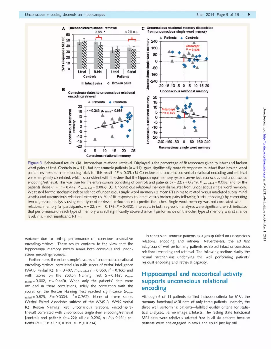
Figure 3 Behavioural results. (A) Unconscious relational retrieval. Displayed is the percentage of fit responses given to intact and broken
word pairs at test. Controls (n = 11), but not amnesic patients (n = 11), gave significantly more fit responses to intact than broken word
pairs; they needed nine encoding trials for this result. *P5 0.05. (B) Conscious and unconscious verbal relational encoding and retrieval
were marginally correlated, which is consistent with the view that the hippocampal memory system serves both conscious and unconscious
encoding/retrieval. This was true for the entire sample consisting of controls and patients (n = 22; r = 0.349, Pone-tailed = 0.056) and for the
patients alone (n = ; r = 0.442, Pone-tailed = 0.087). (C) Unconscious relational memory dissociates from unconscious single word memory.
We tested for the stochastic independence of unconscious single word memory (� mean RTs in ms to related versus unrelated supraliminal
words) and unconscious relational memory (� % of fit responses to intact versus broken pairs following 9-trial encoding) by computing
two regression analyses using each type of retrieval performance to predict the other. Single word memory was not correlated with
relational memory (all participants, n = 22, r = �0.176, P = 0.432). Intercepts in both regression analyses were significant, which indicates
that performance on each type of memory was still significantly above chance if performance on the other type of memory was at chance
level. n.s. = not significant. RT = .
Unconscious encoding depends on hippocampus Brain 2014: Page 9 of 16 | 9
at World T
rade Institute on October 1, 2014
http://brain.oxfordjournals.org/D
ownloaded from

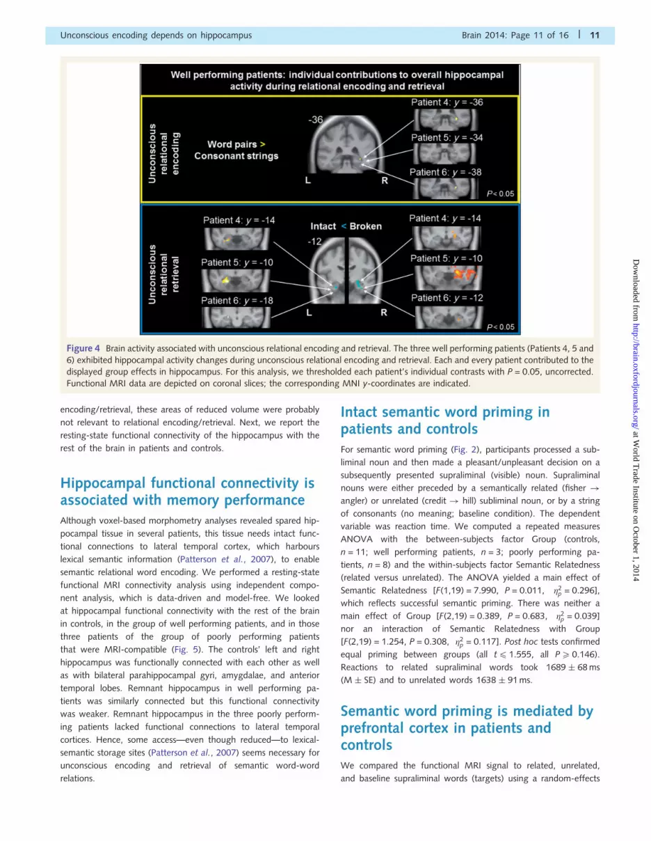
The three well performing patients’ brain activity was compared
between relational word encoding (first encoding trial) and the
processing of pairs of consonant strings (baseline) using a fixed-
effects model suited for small sample sizes. The 11 controls’ first-
level contrast images were analysed using a random-effects
ANOVA. T-test contrasts showed increased neuronal activity to
subliminal word pairs in bilateral hippocampus of controls and in
the right hippocampus of patients. Each individual patient ex-
hibited enhanced signal in this right hippocampal region (Fig. 4).
That this hippocampal region actually comprises grey matter to
support encoding was confirmed in each patient by voxel-based
morphometry. Both patients and controls exhibited additional
signal enhancement to relational word encoding in bilateral para-
hippocampal gyrus and areas mediating semantic processing
(Patterson et al., 2007; Binder et al., 2009) in the left hemisphere,
namely the superior, middle and inferior frontal gyrus, superior
and middle temporal gyrus, and cingulate gyrus.
Hippocampal and neocortical activitysupports unconscious relationalretrievalHippocampal activity distinguished between intact and broken re-
trieval word pairs in both controls and in the three well performing
patients following nine encoding trials (Fig. 4). Interestingly, this
signal difference went into opposite directions between patients
and controls. Controls increased signal to intact versus broken
word pairs in bilateral hippocampus and right thalamus, while pa-
tients increased signal to broken versus intact word pairs in bilat-
eral hippocampus and parahippocampal gyrus. We assume that
controls focused primarily on detecting preserved semantic rela-
tions between words (match detection), while patients focused on
the semantic mismatch of words in broken pairs (Kumaran and
Maguire, 2007). Both match and mismatch detection operate in
the hippocampus, presumably relying on distinct hippocampal sub-
fields, namely CA3 and CA1, respectively (Chen et al., 2011;
Duncan et al., 2012). Each individual patient exhibited signal
changes in those left and right hippocampal areas that reached
significance in group statistics (Fig. 4). Each patient disposed of
grey matter in these activated zones.
Controls exhibited further activity increases in response to intact
versus broken pairs in bilateral medial prefrontal cortices and the
anterior cingulate. These areas support relational reasoning and
the retrieval and encoding of schema congruent new information
(Krawczyk, 2012; van Kesteren et al., 2012). Hence, medial pre-
frontal cortex may have mediated the reactivation of subliminally
encoded semantic relations (schemas) and the detection of analo-
gous semantic relations in intact retrieval word pairs (van Kesteren
et al., 2012). Controls increased activity in response to broken
versus intact pairs within left inferior and right middle frontal
gyrus, bilateral superior temporal gyri, right parahippocampal
gyrus including uncus, and left posterior hippocampus. This left-
sided hippocampal deactivation (reverse contrast) was 2.4 cm
behind the left-sided activation found in controls (reported
above). The controls’ right parahippocampal signal increase to
broken pairs conforms to the patients’ bilateral parahippocampal
signal increase to broken pairs. These parahippocampal activations
in response to recombined concepts, i.e. new combinations of
concepts, were located in the rhinal cortex. Together with the
hippocampal signal increases displayed by both groups in response
to broken versus intact pairs, these rhinal signal increases might
underlie the spontaneous relational encoding of the rearranged
words in broken pairs. Patients exhibited further activity increases
to broken versus intact pairs in left lateral prefrontal cortex, bilat-
eral temporal lobe, angular gyrus, superior parietal lobule, precu-
neus, bilateral precuneus, and several occipital areas.
Structural differences betweenpatients and controls: voxel-basedmorphometryWe compared maps of regional grey matter volumes between
amnesic patients and controls as well as between well performing
patients and poorly performing patients using voxel-wise inde-
pendent sample t-tests. The voxel-based morphometry analysis
revealed areas of larger grey matter volume in controls versus
the six magnetic resonance-scanned patients, but not reversed
(Table 2). Controls exhibited extended areas of larger grey
matter volume in the left hippocampus, left parahippocampal
gyrus, left amygdala, bilateral thalamus, left putamen and palli-
dum as well as left lingual/fusiform gyrus and left middle/inferior
temporal gyrus. Hence, besides the expected medial temporal and
diencephalic regions, there were areas of reduced neocortical
volume in patients versus controls within the left lingual/fusiform
gyrus and left middle/inferior temporal gyrus. The inspection of
each patient’s voxel-based morphometry contrast (versus controls)
revealed no neocortical overlap of reduced tissue volume between
all of the six patients. At the contrary, the obtained statistical dif-
ferences in neocortical volume were driven by certain patients. The
reduced volume in the left lingual/fusiform gyrus was due to two
well performing patients (Patients 5 and 6; see Table 1) and two
poorly performing patients (Patients 1 and 3). The reduced volume
in the left middle/inferior temporal gyrus was due to one well
performing patient (Patient 6). Consequently, volume reductions
in these neocortical regions were not associated with poor uncon-
scious relational encoding/retrieval. This conclusion was corrobo-
rated by the direct comparison of grey matter volumes between
well performing patients and poorly performing patients. This
comparison yielded no significant result indicating that well per-
forming patients exhibited no area of increased tissue volume, not
even in the hippocampus. The lack of a hippocampal difference
between patient groups owed to the fact that Patient 2 of the
group of poorly performing patients had a bilateral fornix dissec-
tion but relatively preserved hippocampal volumes. The fornix pa-
tients’ large hippocampal volumes compensated for the other poor
performers’ small residual hippocampal volumes eliminating the
effect of larger hippocampal volumes in well versus poorly per-
forming patients. The reversed contrast revealed that well per-
forming patients had smaller tissue volumes than poorly
performing patients in the right posterior cingulate gyrus, right
cuneus, left middle occipital gyrus, and cerebellum. Given that
the well performing patients excelled in unconscious relational
10 | Brain 2014: Page 10 of 16 S. B. Duss et al.
at World T
rade Institute on October 1, 2014
http://brain.oxfordjournals.org/D
ownloaded from
encoding/retrieval, these areas of reduced volume were probably
not relevant to relational encoding/retrieval. Next, we report the
resting-state functional connectivity of the hippocampus with the
rest of the brain in patients and controls.
Hippocampal functional connectivity isassociated with memory performanceAlthough voxel-based morphometry analyses revealed spared hip-
pocampal tissue in several patients, this tissue needs intact func-
tional connections to lateral temporal cortex, which harbours
lexical semantic information (Patterson et al., 2007), to enable
semantic relational word encoding. We performed a resting-state
functional MRI connectivity analysis using independent compo-
nent analysis, which is data-driven and model-free. We looked
at hippocampal functional connectivity with the rest of the brain
in controls, in the group of well performing patients, and in those
three patients of the group of poorly performing patients
that were MRI-compatible (Fig. 5). The controls’ left and right
hippocampus was functionally connected with each other as well
as with bilateral parahippocampal gyri, amygdalae, and anterior
temporal lobes. Remnant hippocampus in well performing pa-
tients was similarly connected but this functional connectivity
was weaker. Remnant hippocampus in the three poorly perform-
ing patients lacked functional connections to lateral temporal
cortices. Hence, some access—even though reduced—to lexical-
semantic storage sites (Patterson et al., 2007) seems necessary for
unconscious encoding and retrieval of semantic word-word
relations.
Intact semantic word priming inpatients and controlsFor semantic word priming (Fig. 2), participants processed a sub-
liminal noun and then made a pleasant/unpleasant decision on a
subsequently presented supraliminal (visible) noun. Supraliminal
nouns were either preceded by a semantically related (fisher !
angler) or unrelated (credit ! hill) subliminal noun, or by a string
of consonants (no meaning; baseline condition). The dependent
variable was reaction time. We computed a repeated measures
ANOVA with the between-subjects factor Group (controls,
n = 11; well performing patients, n = 3; poorly performing pa-
tients, n = 8) and the within-subjects factor Semantic Relatedness
(related versus unrelated). The ANOVA yielded a main effect of
Semantic Relatedness [F(1,19) = 7.990, P = 0.011, �2p = 0.296],
which reflects successful semantic priming. There was neither a
main effect of Group [F(2,19) = 0.389, P = 0.683, �2p = 0.039]
nor an interaction of Semantic Relatedness with Group
[F(2,19) = 1.254, P = 0.308, �2p = 0.117]. Post hoc tests confirmed
equal priming between groups (all t41.555, all P5 0.146).
Reactions to related supraliminal words took 1689 � 68 ms
(M � SE) and to unrelated words 1638 � 91 ms.
Semantic word priming is mediated byprefrontal cortex in patients andcontrolsWe compared the functional MRI signal to related, unrelated,
and baseline supraliminal words (targets) using a random-effects
Figure 4 Brain activity associated with unconscious relational encoding and retrieval. The three well performing patients (Patients 4, 5 and
6) exhibited hippocampal activity changes during unconscious relational encoding and retrieval. Each and every patient contributed to the
displayed group effects in hippocampus. For this analysis, we thresholded each patient’s individual contrasts with P = 0.05, uncorrected.
Functional MRI data are depicted on coronal slices; the corresponding MNI y-coordinates are indicated.
Unconscious encoding depends on hippocampus Brain 2014: Page 11 of 16 | 11
at World T
rade Institute on October 1, 2014
http://brain.oxfordjournals.org/D
ownloaded from

one-way ANOVA in controls and fixed-effects pairwise compari-
sons in the group of well performing patients. Both the group of
controls and the group of patients enhanced activity in response to
related versus unrelated supraliminal words within left inferior,
middle, and superior frontal gyri and the anterior cingulate.
Hence, the semantic proximity of supraliminal words to previously
flashed subliminal words increased computational demands in pre-
frontal language areas (Henson, 2003; Segaert et al., 2013).
Particularly the left inferior frontal gyrus is known to subserve
the detection of semantic relatedness between words. Only the
controls exhibited repetition suppression of the functional MRI
signal when processing related supraliminal words within temporal
and parietal areas. This repetition suppression may not have been
essential to task performance because patients manifested no
repetition suppression but performed equally well.
Relational retrieval and single wordretrieval are uncorrelatedWe tested for the stochastic independence of unconscious rela-
tional and single word encoding/retrieval by computing two re-
gression analyses. We used the experimental measures of word
priming and relational retrieval to predict each other. The intercept
of the regression reveals whether performance on the predicted
task (y-axis) is greater than zero if performance on the predictor
task (x-axis) is at zero (Greenwald et al., 1995) (Fig. 3C). Word
priming was not correlated with relational retrieval (all participants,
n = 22, r = �0.176, P = 0.432). Intercepts in both regression ana-
lyses were significant [predicted: word priming, y-axis inter-
cept = 43.23, SE = 17.93, t(20) = 2.411, P = 0.026, r2 = 0.225]
[predicted: relational retrieval, y-axis intercept = 4.81, SE = 1.90,
t(20) = 2.474, P = 0.022, r2 = 0.234].
DiscussionWe performed three neuroimaging experiments in amnesic pa-
tients and controls to find out whether processing modes rather
than consciousness would divide between memory systems,
namely hippocampal versus neocortical processing. Examined pro-
cessing modes were flexible relational and rigid non-relational
(single item) encoding and retrieval. We hypothesized that only
relational encoding would depend on the hippocampal memory
system and would therefore be affected in amnesic patients.
Table 2 Differences in grey matter volume (voxel-based morphometry)
Peak voxel Cluster extent BA Side MNI-coordinates k t z
x y z
Controls4 patients
Parahippocampal gyrus Left thalamus, hippocampus, lingual gyrus,parahippocampal gyrus, precuneus, rightthalamus, posterior cingulate gyrus
27, 28, 30, 35 L �18 �36 �3 1747 7.46 4.75
Lingual gyrus �8 �37 1 7.44 4.75
Thalamus �5 �30 6 5.68 4.09
Hippocampus Hippocampus, putamen, pallidum L �30 �15 �12 361 4.49 3.52
Pallidum �24 �4 �6 4.46 3.5
Amygdala Hippocampus, amygdala,parahippocampal gyrus
34, 28 L �17 �3 �14 270 4.97 3.76
Lingual gyrus,parahippocampal gyrus
Lingual gyrus, fusiform gyrus 19 L �23 �60 �9 172 5.2 3.87
Lingual gyrus �20 �69 �8 4.33 3.43
Middle temporal gyrus Middle temporal gyrus, inferiortemporal gyrus
20, 21 L �53 �31 �15 72 5.24 3.89
Paracentral lobule Paracentral lobule, postcentral gyrus 3, 4 R 9 �36 73 65 5.35 3.94
Patients4 controls
No suprathreshold clusters
Well performing patients4 poorly performing patients
No suprathreshold clusters
Poorly performing patients4well performing patients
Posterior cingulate gyrus Posterior cingulate gyrus, precuneus 31, 23 R 8 �57 30 49 22.8 4.24
Middle occipital gyrus Middle occipital gyrus 19 L �26 �94 13 23 12.9 3.7
Cuneus Cuneus 18, 31 R 6 �76 21 24 14.9 3.85
Cerebellum Cerebellum lobule VI, vermis R 12 �78 �20 143 23.8 4.28
Cerebellum L �4 �66 �27 18.4 4.05
Cerebellum Cerebellum lobule VI L �28 �67 �21 34 16.2 3.93
Height threshold P = 0.001; extent threshold = 20 voxels.Local maxima are 48 mm apart. MNI-coordinates stand for the peak voxel within the cluster of voxels exhibiting differential probabilistic grey matter volume.k = extent of significant cluster (number of voxels); BA = Brodmann area; L = left; R = right.
12 | Brain 2014: Page 12 of 16 S. B. Duss et al.
at World T
rade Institute on October 1, 2014
http://brain.oxfordjournals.org/D
ownloaded from
Consciousness of encoding and retrieval was excluded by the sub-
liminal (invisible) presentation of all encoding material.
Unconscious encoding provided for a fair comparison of memory
performance between amnesic patients and controls because it
eliminated the confounding effect of conscious memory.
Subliminal encoding also provided for a powerful demonstration
of unconscious memory processing. The addition of functional
MRI to behavioural testing allowed for determination of the
memory system(s) that supported residual memory functions in
amnesic patients. Functional MRI investigations in amnesic pa-
tients are rare and mostly include one single patient (Maguire
et al., 2001, 2005; Caulo et al., 2005) because of several MRI
contraindications—a problem that also affected our study.
Our patients and controls performed equally well on uncon-
scious single word encoding and retrieval. But the relational task
separated the two groups. While controls succeeded on uncon-
scious relational encoding and retrieval activating their hippocam-
pus, amnesic patients failed as a group. This is the first
demonstration, to our knowledge, that unconscious encoding in
the human depends on the hippocampal-anterior thalamic axis
and its connections to neocortex. An ad hoc sample of amnesic
patients succeeded on unconscious relational encoding capitalizing
on residual hippocampal tissue and functional connections to the
lateral temporal lobes. Although these well performing patients
showed intact unconscious relational encoding, their conscious re-
lational encoding was severely impaired.
Importantly, voxel-based morphometry revealed that our pa-
tients exhibited no damage in an overlapping neocortical region
that could be the seat of unconscious relational encoding and
retrieval. Although group statistics revealed smaller volumes in
the left lingual and fusiform gyri as well as the left middle and
inferior temporal gyri in patients versus controls, these reductions
were due to few patients, who came from both the group of
poorly and the group of well performing patients. Hence,
volume reductions in these regions were not associated with a
poor performance on unconscious relational encoding and
retrieval. Moreover, the group of well performing patients ex-
hibited no area of increased neocortical tissue volume relative to
the group of poorly performing patients suggesting that poor task
performance was not associated with neocortical damage. We
therefore assume that damage in the hippocampal-anterior thal-
amic axis and its disconnection from the neocortex had caused the
patients’ performance deficits on conscious and unconscious rela-
tional encoding and retrieval.
We draw the following conclusions that we discuss below: (i)
separate memory systems mediate relational versus single item
memory; (ii) the hippocampal memory system operates with and
without consciousness; and (iii) is necessary for unconscious rela-
tional encoding and retrieval; and (iv) conscious versus uncon-
scious memory formation may require a greater level of function
within the hippocampal-neocortical network.
In the realm of conscious stimulus processing both at the time
of encoding and retrieval, there is good evidence in support of a
dissociation of memory systems regarding relational versus single
item processing in amnesic patients (Mayes et al., 2002;
Giovanello et al., 2003; Kan et al., 2007). These studies controlled
for differences in memory load and difficulty between tasks. Still,
retrieval performance of hippocampal patients was only impaired
on tests of relational memory. Brain activation studies in healthy
participants point to the same direction reporting hippocampal
activity increases during association formation versus single item
encoding or retrieval (Henke, 2010). The current results in amnesic
patients extend the relational/single item dissociation to the realm
of unconscious encoding and retrieval processes.
But not all forms of associative memory are flexible and hippo-
campal-dependent and not all forms of single item memory are
inflexible and hippocampal-independent. In the following, we
define the concept of ‘flexibility’ more precisely and explain why
our word pair task requires a larger degree of flexibility of memory
representation than our single word task. Flexible memory repre-
sentations can be activated through many, even remotely related
retrieval cues in situations that have little or no overlap with the
encoding situation. On the other hand, inflexible, unitized mem-
ories can only be reactivated by a cue that was present at encod-
ing such as ‘face . . .’ for the word Facebook; the cue ‘nose . . .’
would not trigger reactivation. Flexible memories can be reacti-
vated intrinsically, i.e. even without any external retrieval cue,
through self-cueing. In this process, one can deliberately choose
to reactivate all facets of a memory or only certain facets. Each
facet of a flexible memory representation is both related to other
facets of the same representation and to facets of other memory
Figure 5 Resting state functional connectivity. Results of the
independent component analyses performed on the resting state
functional MRI data are depicted in axial slices at MNI z = �24
to �12 in steps of 4 mm. Independent component analyses
were performed separately for each participant. The resulting
images reflect the degree to which the component that is most
prominent in the hippocampus contributes to the signal in other
parts of the brain. Z-transformed images were averaged across
groups of interest (controls, n = 11; well performing patients,
n = 3; MRI-compatible subgroup of the group of poorly per-
forming patients, n = 3).
Unconscious encoding depends on hippocampus Brain 2014: Page 13 of 16 | 13
at World T
rade Institute on October 1, 2014
http://brain.oxfordjournals.org/D
ownloaded from

representations. Hence, the flexibility of a memory reflects in (i)
diverse access options; (ii) selective reactivation options; and (iii)
relational binding. Accordingly, not all associative memories are
flexible (some are fused like Facebook) and not all single item
memories are inflexible because some are relationally integrated
into a context as part of an episodic memory. The design of our
single word task does not require a flexible representation for
successful performance because a retrieval cue is provided,
which is a semantic neighbour of the encoding word. Hence, se-
mantic priming, which is inflexible, suffices task requirements. An
additional relational integration of the encoding word into the
encoding context is unnecessary. The subliminal prime word
‘fisher’ may activate this word’s concept node in the temporal
neocortex with activation spreading over to the concept node
‘angler’ that gets primed, or the activation of the concept node
‘angler’ at test may trigger the reactivation of its primed semantic
neighbour ‘fisher’ through spreading activation. The results in am-
nesic patients confirm that a purely neocortical route suffices
normal task performance on the single word task. Yet, an inflex-
ible memory representation does not suffice normal performance
on our word pair task because this task requires that a new rela-
tion be established between two concept nodes (and their seman-
tic neighbours).
We have earlier reported successful subliminal relational encod-
ing and a flexible retrieval of face-word and word-word combin-
ations in behavioural (Duss et al., 2011; Reber and Henke, 2011,
2012) and functional MRI studies (Henke et al., 2003a, b;
Degonda et al., 2005; Reber et al., 2012, 2014) in normal par-
ticipants using the same masking paradigm as used in the current
study. New semantic word-word associations were established un-
consciously even across time points (Reber and Henke, 2012;
Reber et al., 2012). The integration of associative memories
over time was possible both within (Reber and Henke, 2012;
Reber et al., 2012) and across levels of consciousness (Henke
et al., 2013). Our neuroimaging data demonstrated that relational
encoding and retrieval as well as the relational integration of in-
formation across time-points were assisted by hippocampus
(Henke et al., 2003a, b; Degonda et al., 2005; Reber et al.,
2012, 2014). However, studies in normal participants cannot clar-
ify whether the hippocampus is necessary for unconscious memory
formation or merely co-activated but not essential for the task.
The present result of impaired unconscious relational processing in
amnesic patients speaks to the necessity of the hippocampus for
unconscious relational encoding. This new finding questions the
current textbook account of memory systems that attributes to
the hippocampus only a role in conscious encoding (Squire and
Zola, 1996; Tulving, 2002; Moscovitch, 2008). Instead, this new
finding supports the relational memory account (Cohen and
Eichenbaum, 1993) and the processing account of memory sys-
tems (Henke, 2010).
Remarkably, three amnesic patients succeeded on unconscious
relational encoding and retrieval relying on residual hippocampal
tissue and functional connections to lateral temporal cortex. These
patients had retained hippocampal tissue reaching from 46–74%
of normal values. Hence, their volume loss was 560%, which was
regarded a critical threshold for preserved relational memory capa-
cities in a study of animal spatial encoding (Kleinknecht et al.,
2012). Although the well performing patients were severely im-
paired on all standard tests of long-term memory, they scored
higher on tests of immediate verbal memory and verbal intelli-
gence compared to the other patients. Both their rich semantic
knowledge and residual hippocampal tissue and hippocampal con-
nections to temporal language storage sites may have enabled
their normal performance on the test of unconscious relational
encoding and retrieval (Kan et al., 2009).
These findings settle the debate on intact versus impaired un-
conscious memory for word pairs in amnesic patients (Schacter,
1998). Behavioural studies in amnesic patients suggested that the
severity of amnesia was related to the performance of patients on
indirect tests of unconscious memory for word pairs. The word
pairs had been presented visibly for conscious encoding but
could not be consciously remembered due to amnesia (Schacter
and Graf, 1986; Shimamura and Squire, 1989). One reason for
this association between severity of amnesia and test performance
could be the contamination of unconscious with residual conscious
memory in patients with lighter forms of amnesia (McKone and
Slee, 1997; Smith and Squire, 2008). Another reason for this re-
lation may be spared functionality in the hippocampal-anterior
thalamic axis in lighter forms of amnesia that suffices unconscious
but not conscious memory processing. The current findings sup-
port this second interpretation because our design excluded con-
sciousness as a confounding variable. Our data suggest that the
spared functionality in well performing patients concerned both
the hippocampal-anterior thalamic axis and its connections to neo-
cortical language areas. The possibility that processing in neocor-
tical language sites alone had generated unconscious relational
memories in these well performing patients can be excluded be-
cause all amnesic patients, also those that failed on the relational
task, exhibited normal cognitive functions and normal semantic
priming for single words that require neocortical language sites.
Hence, if language cortex alone supported relational word
processing, all patients should have yielded normal results. But
this was clearly not the case. Furthermore, relational and single
word retrieval were not correlated (Fig. 3C), which would be ex-
pected if both tasks relied on neocortical language sites alone. In
light of these data, the finding (Verfaelllie et al., 2012) of in-
tact performance of a group of amnesic patients on an implicit
semantic word-word priming task may be attributable to a sub-
group of patients with speared functionality in the hippocampal-
anterior thalamic axis and its connections to neocortical language
areas.
The fact that conscious relational memory was impaired while
unconscious relational memory was intact in well performing pa-
tients suggests that levels of consciousness of memory processing
require different resources within a memory system. A larger level
of functionality in the hippocampal-anterior thalamic axis and its
connections to neocortex seems required for conscious versus un-
conscious relational encoding and retrieval. This assumption is sup-
ported by the functional connectivity of hippocampus with the
anterior temporal lobe, which was strong in controls, weaker in
the well performing patients and absent in the other patients
(Fig. 5). We assume that consciousness of encoding is a correlate
of the strength and coherence of activity in the task-relevant
memory system. This is a notion, which is in line with quantitative
14 | Brain 2014: Page 14 of 16 S. B. Duss et al.
at World T
rade Institute on October 1, 2014
http://brain.oxfordjournals.org/D
ownloaded from
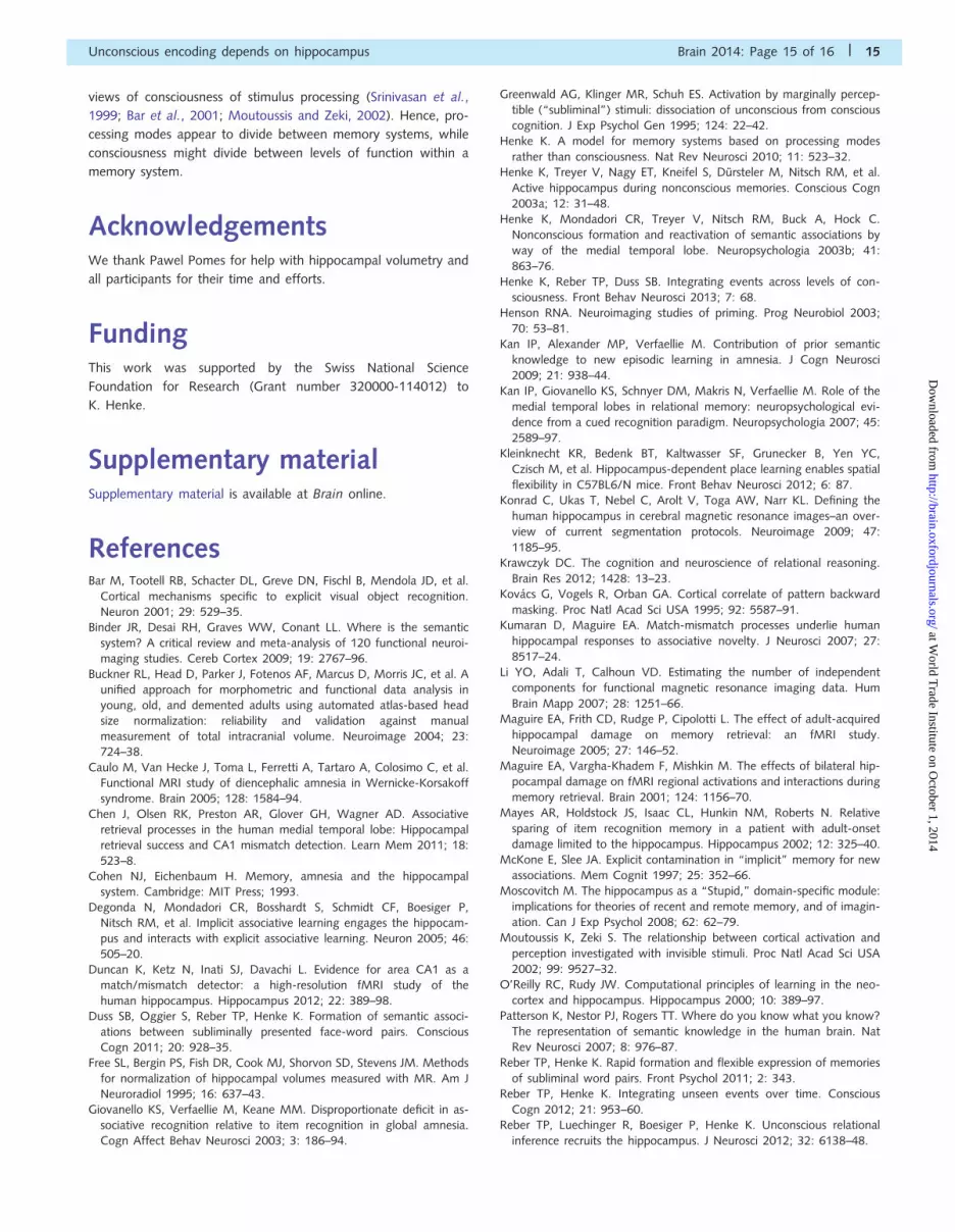
views of consciousness of stimulus processing (Srinivasan et al.,
1999; Bar et al., 2001; Moutoussis and Zeki, 2002). Hence, pro-
cessing modes appear to divide between memory systems, while
consciousness might divide between levels of function within a
memory system.
AcknowledgementsWe thank Pawel Pomes for help with hippocampal volumetry and
all participants for their time and efforts.
FundingThis work was supported by the Swiss National Science
Foundation for Research (Grant number 320000-114012) to
K. Henke.
Supplementary materialSupplementary material is available at Brain online.
ReferencesBar M, Tootell RB, Schacter DL, Greve DN, Fischl B, Mendola JD, et al.
Cortical mechanisms specific to explicit visual object recognition.
Neuron 2001; 29: 529–35.Binder JR, Desai RH, Graves WW, Conant LL. Where is the semantic
system? A critical review and meta-analysis of 120 functional neuroi-
maging studies. Cereb Cortex 2009; 19: 2767–96.
Buckner RL, Head D, Parker J, Fotenos AF, Marcus D, Morris JC, et al. A
unified approach for morphometric and functional data analysis in
young, old, and demented adults using automated atlas-based head
size normalization: reliability and validation against manual
measurement of total intracranial volume. Neuroimage 2004; 23:
724–38.
Caulo M, Van Hecke J, Toma L, Ferretti A, Tartaro A, Colosimo C, et al.
Functional MRI study of diencephalic amnesia in Wernicke-Korsakoff
syndrome. Brain 2005; 128: 1584–94.
Chen J, Olsen RK, Preston AR, Glover GH, Wagner AD. Associative
retrieval processes in the human medial temporal lobe: Hippocampal
retrieval success and CA1 mismatch detection. Learn Mem 2011; 18:
523–8.Cohen NJ, Eichenbaum H. Memory, amnesia and the hippocampal
system. Cambridge: MIT Press; 1993.
Degonda N, Mondadori CR, Bosshardt S, Schmidt CF, Boesiger P,
Nitsch RM, et al. Implicit associative learning engages the hippocam-
pus and interacts with explicit associative learning. Neuron 2005; 46:
505–20.Duncan K, Ketz N, Inati SJ, Davachi L. Evidence for area CA1 as a
match/mismatch detector: a high-resolution fMRI study of the
human hippocampus. Hippocampus 2012; 22: 389–98.Duss SB, Oggier S, Reber TP, Henke K. Formation of semantic associ-
ations between subliminally presented face-word pairs. Conscious
Cogn 2011; 20: 928–35.Free SL, Bergin PS, Fish DR, Cook MJ, Shorvon SD, Stevens JM. Methods
for normalization of hippocampal volumes measured with MR. Am J
Neuroradiol 1995; 16: 637–43.Giovanello KS, Verfaellie M, Keane MM. Disproportionate deficit in as-
sociative recognition relative to item recognition in global amnesia.
Cogn Affect Behav Neurosci 2003; 3: 186–94.
Greenwald AG, Klinger MR, Schuh ES. Activation by marginally percep-
tible (“subliminal”) stimuli: dissociation of unconscious from conscious
cognition. J Exp Psychol Gen 1995; 124: 22–42.
Henke K. A model for memory systems based on processing modes
rather than consciousness. Nat Rev Neurosci 2010; 11: 523–32.Henke K, Treyer V, Nagy ET, Kneifel S, Dursteler M, Nitsch RM, et al.
Active hippocampus during nonconscious memories. Conscious Cogn
2003a; 12: 31–48.
Henke K, Mondadori CR, Treyer V, Nitsch RM, Buck A, Hock C.
Nonconscious formation and reactivation of semantic associations by
way of the medial temporal lobe. Neuropsychologia 2003b; 41:
863–76.
Henke K, Reber TP, Duss SB. Integrating events across levels of con-
sciousness. Front Behav Neurosci 2013; 7: 68.
Henson RNA. Neuroimaging studies of priming. Prog Neurobiol 2003;
70: 53–81.
Kan IP, Alexander MP, Verfaellie M. Contribution of prior semantic
knowledge to new episodic learning in amnesia. J Cogn Neurosci
2009; 21: 938–44.
Kan IP, Giovanello KS, Schnyer DM, Makris N, Verfaellie M. Role of the
medial temporal lobes in relational memory: neuropsychological evi-
dence from a cued recognition paradigm. Neuropsychologia 2007; 45:
2589–97.
Kleinknecht KR, Bedenk BT, Kaltwasser SF, Grunecker B, Yen YC,
Czisch M, et al. Hippocampus-dependent place learning enables spatial
flexibility in C57BL6/N mice. Front Behav Neurosci 2012; 6: 87.Konrad C, Ukas T, Nebel C, Arolt V, Toga AW, Narr KL. Defining the
human hippocampus in cerebral magnetic resonance images–an over-
view of current segmentation protocols. Neuroimage 2009; 47:
1185–95.Krawczyk DC. The cognition and neuroscience of relational reasoning.
Brain Res 2012; 1428: 13–23.Kovacs G, Vogels R, Orban GA. Cortical correlate of pattern backward
masking. Proc Natl Acad Sci USA 1995; 92: 5587–91.Kumaran D, Maguire EA. Match-mismatch processes underlie human
hippocampal responses to associative novelty. J Neurosci 2007; 27:
8517–24.
Li YO, Adali T, Calhoun VD. Estimating the number of independent
components for functional magnetic resonance imaging data. Hum
Brain Mapp 2007; 28: 1251–66.Maguire EA, Frith CD, Rudge P, Cipolotti L. The effect of adult-acquired
hippocampal damage on memory retrieval: an fMRI study.
Neuroimage 2005; 27: 146–52.
Maguire EA, Vargha-Khadem F, Mishkin M. The effects of bilateral hip-
pocampal damage on fMRI regional activations and interactions during
memory retrieval. Brain 2001; 124: 1156–70.
Mayes AR, Holdstock JS, Isaac CL, Hunkin NM, Roberts N. Relative
sparing of item recognition memory in a patient with adult-onset
damage limited to the hippocampus. Hippocampus 2002; 12: 325–40.
McKone E, Slee JA. Explicit contamination in “implicit” memory for new
associations. Mem Cognit 1997; 25: 352–66.
Moscovitch M. The hippocampus as a “Stupid,” domain-specific module:
implications for theories of recent and remote memory, and of imagin-
ation. Can J Exp Psychol 2008; 62: 62–79.
Moutoussis K, Zeki S. The relationship between cortical activation and
perception investigated with invisible stimuli. Proc Natl Acad Sci USA
2002; 99: 9527–32.
O’Reilly RC, Rudy JW. Computational principles of learning in the neo-
cortex and hippocampus. Hippocampus 2000; 10: 389–97.
Patterson K, Nestor PJ, Rogers TT. Where do you know what you know?
The representation of semantic knowledge in the human brain. Nat
Rev Neurosci 2007; 8: 976–87.
Reber TP, Henke K. Rapid formation and flexible expression of memories
of subliminal word pairs. Front Psychol 2011; 2: 343.Reber TP, Henke K. Integrating unseen events over time. Conscious
Cogn 2012; 21: 953–60.Reber TP, Luechinger R, Boesiger P, Henke K. Unconscious relational
inference recruits the hippocampus. J Neurosci 2012; 32: 6138–48.
Unconscious encoding depends on hippocampus Brain 2014: Page 15 of 16 | 15
at World T
rade Institute on October 1, 2014
http://brain.oxfordjournals.org/D
ownloaded from
Reber TP, Luechinger R, Boesiger P, Henke K. Detecting analogies un-consciously. Front Behav Neurosci 2014; 8: 9.
Ridgway GR, Omar R, Ourselin S, Hill DL, Warren JD, Fox NC. Issues
with threshold masking in voxel-based morphometry of atrophied
brains. Neuroimage 2009; 44: 99–111.Rolls ET, Tovee MJ. Processing speed in the cerebral cortex and the
neurophysiology of visual masking. Proc Biol Sci 1994; 257: 9–15.
Rolls ET, Tovee MJ, Panzeri S. The neurophysiology of backward visual
masking: information analysis. J Cogn Neurosci 1999; 11: 300–11.Schacter DL. Memory and awareness. Science 1998; 280: 59–60.
Schacter DL, Graf P. Preserved learning in amnesic patients - perspectives
from research on direct priming. J Clin Exp Neuropsychol 1986; 8:727–43.
Segaert K, Weber K, de Lange FP, Petersson KM, Hagoort P. The sup-
pression of repetition enhancement: a review of fMRI studies.
Neuropsychologia 2013; 51: 59–66.Shimamura AP, Squire LR. Impaired priming of new associations in am-
nesia. J Exp Psychol Learn Mem Cogn 1989; 15: 721–8.
Smith CN, Squire LR. Experience-dependent eye movements reflect
hippocampus-dependent (aware) memory. J Neurosci 2008; 28:12825–33.
Squire LR, Zola SM. Structure and function of declarative and nonde-
clarative memory systems. Proc Natl Acad Sci USA 1996; 93:
13515–22.
Srinivasan R, Russell DP, Edelman GM, Tononi G. Increased synchron-
ization of neuromagnetic responses during conscious perception.
J Neurosci 1999; 19: 5435–48.
Tulving E. How many memory-systems are there. Am Psychol 1985; 40:
385–98.
Tulving E. Episodic memory: From mind to brain. Annu Rev Psychol
2002; 53: 1–25.Tzourio-Mazoyer N, Landeau B, Papathanassiou D, Crivello F, Etard O,
Delcroix N, et al. Automated anatomical labeling of activations in
SPM using a macroscopic anatomical parcellation of the MNI MRI
single-subject brain. Neuroimage 2002; 15: 273–89.
van Kesteren MTR, Ruiter DJ, Fernandez G, Henson RN. How schema
and novelty augment memory formation. Trends Neurosci 2012; 35:
211–9.Verfaelllie M, LaRocque KF, Keane MM. Intact implicit verbal relational
memory in medial temporal lobe amnesia. Neuropsychologia 2012; 50:
2100–6.
16 | Brain 2014: Page 16 of 16 S. B. Duss et al.
at World T
rade Institute on October 1, 2014
http://brain.oxfordjournals.org/D
ownloaded from