ucl discovery - university college london
TRANSCRIPT
PATHOPHYSIOLOGICAL MECHANISMS IN SUBARACHNOID HAEMORRHAGE
A Study of the Neuropharmacological, Physiological and Morphological Changes that Occur in a Model of
Subarachnoid Haemorrhage Developed in the Laboratory Rat
by
Andre' Jackowski BSc, MB.BS, FRCS.
A dissertation submitted for the degree of Doctorate of Medicine
at the University of London
Cerebral Oedema Research Group Institute of Neurology The National Hospitals Queen Square London WC1N 3BG
Department of Anatomy and Developmental Biology and Centre for Neuroscience University College London London WC1E 6BT
London 1990

ProQuest Number: U542428
All rights reserved
INFORMATION TO ALL USERS The quality of this reproduction is dependent upon the quality of the copy submitted.
In the unlikely event that the author did not send a com p le te manuscript and there are missing pages, these will be noted. Also, if material had to be removed,
a note will indicate the deletion.
uestProQuest U542428
Published by ProQuest LLC(2017). Copyright of the Dissertation is held by the Author.
All rights reserved.This work is protected against unauthorized copying under Title 17, United States C ode
Microform Edition © ProQuest LLC.
ProQuest LLC.789 East Eisenhower Parkway
P.O. Box 1346 Ann Arbor, Ml 48106- 1346
ABSTRACT
This thesis reviews current knowledge regarding the problems created by, possible causal factors, and present management of the disordered pathophysiology that may arise following subarachnoid haemorrhage (SAH) in man. Mechanisms responsible for the normal regulation of the cerebral vasculature are also reviewed. The present investigation explores how various neuropharmacological, physiological and morphological aspects of the major cerebral arteries are affected by such haemorrhage in a small animal model of experimental SAH developed in the laboratory rat.Preliminary immunohistochemical studies revealed that contrary to earlier reports, the extraparenchymal cerebral arteries do not receive a significant serotonergic innervation. Serotonin was only rarely present under normalcircumstances in the neural plexus, and when found wasinvariably contained within nerves also identified ascatecholaminergic in nature. Following SAH, majoralterations in the neurotransmitter content of the cerebrovasculature occurred. The perivascular sympathetic nerves of major cerebral vessels rapidly accummulated serotonin, while a coincident depletion of neuropeptide Y took place. Using cortically implanted platinum-wire microelectrodes, with measurement of cerebral blood flow (CBF) by hydrogen-clearance, the timecourse of the global reduction in CBF that develops acutely in this model was documented. A 50% reduction in blood flow persisted for up to 3 hours posthaemorrhage, at 24 hours this was restored
to 85% of normal, and recovered fully by 48 hours. The fall in CBF developed independently of concommitant changes in intracranial and cerebral perfusion pressure, and it would appear likely that early vasospasm secondary to released blood products, rather than pressure changes per-se, is responsible for the acute cerebral ischaemia that develops. Electron microscopic studies, demonstrated the delayed development of a mild cerebral vasculopathy, comprising focal areas of subintimal medial necrosis. Transformation of cells derived from the pia-arachnoid into macrophages, occurred on the second day post haemorrhage. These cells were then largely responsible for a rapid phagocytic removal of the subarachnoid blood clot. Previous findings obtained by workers using experimental models of SAH are reviewed, and suggestions as to the nature and aetiology of delayed cerebral ischaemia and vasospasm arising after SAH deduced.
CONTENTSPage No.
INTRODUCTION1.1 The Clinical Problem 7.1 .2 Present Management of Subarachnoid
Haemorrhage 28.1 .3 Cerebral Arteries and Control of the
Cerebral Vasculature 42.1 .4 Aims of the Study 67.
GENERAL METHODS2.1 Immunofluorescence and Immunoelectron
microscopy 69.2.2 High performance liquid chromatography 70.2.3 Hydrogen clearance measurement of LCBF 70.2.4 Acid-etch Scanning electron microscopy 72.
EXPERIMENTAL CHAPTER I:ULTRASTRUCTURE OF SEROTONIN-CONTAINING NERVE FIBRES IN THE MIDDLE CEREBRAL ARTERY OF THE RAT AND EVIDENCE FOR ITS LOCALISATION WITHIN CATECHOLAMINE CONTAINING NERVE FIBRES BY IMMUNOELECTRON MICROSCOPY3.1 Introduction 77.3.2 Materials and methods 79.3.3 Results 82.3.4 Discussion 84.3.5 Figures 1 - 2 87-90.
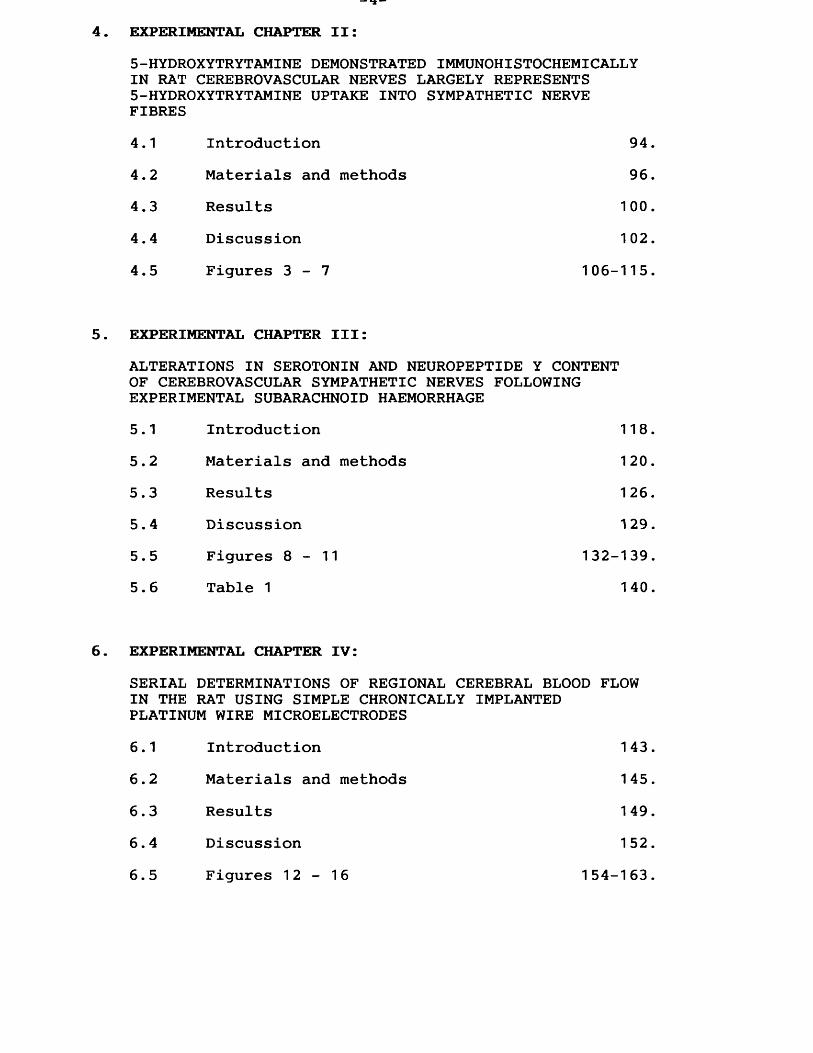
EXPERIMENTAL CHAPTER II:5-HYDROXYTRYTAMINE DEMONSTRATED IMMUNOHISTOCHEMICALLY IN RAT CEREBROVASCULAR NERVES LARGELY REPRESENTS 5-HYDROXYTRYTAMINE UPTAKE INTO SYMPATHETIC NERVEFIBRES4.1 Introduction 944.2 Materials and methods 964.3 Results 1004.4 Discussion 1024.5 Figures 3 - 7 106-115
EXPERIMENTAL CHAPTER III:ALTERATIONS IN SEROTONIN AND NEUROPEPTIDE Y CONTENT OF CEREBROVASCULAR SYMPATHETIC NERVES FOLLOWING EXPERIMENTAL SUBARACHNOID HAEMORRHAGE5.1 Introduction 1185.2 Materials and methods 1205.3 Results 1265.4 Discussion 1295.5 Figures 8 - 1 1 132-1395.6 Table 1 140
EXPERIMENTAL CHAPTER IV:SERIAL DETERMINATIONS OF REGIONAL CEREBRAL BLOOD FLOW IN THE RAT USING SIMPLE CHRONICALLY IMPLANTED PLATINUM WIRE MICROELECTRODES6.1 Introduction 1436.2 Materials and methods 1456.3 Results 1496.4 Discussion 1526.5 Figures 1 2 - 1 6 154-163
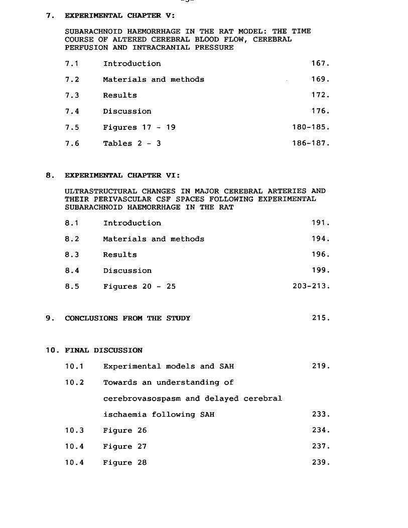
7. EXPERIMENTAL CHAPTER V:SUBARACHNOID HAEMORRHAGE IN THE RAT MODEL: THE TIME COURSE OF ALTERED CEREBRAL BLOOD FLOW, CEREBRAL PERFUSION AND INTRACRANIAL PRESSURE7.1 Introduction 167.7.2 Materials and methods 169.7.3 Results 172.7.4 Discussion 176.7.5 Figures 17 - 19 180 -185.7.6 Tables 2 - 3 186--187.
EXPERIMENTAL CHAPTER VI:ULTRASTRUCTURAL CHANGES IN MAJOR CEREBRAL ARTERIES AND THEIR PERIVASCULAR CSF SPACES FOLLOWING EXPERIMENTAL SUBARACHNOID HAEMORRHAGE IN THE RAT8.1 Introduction 191 .8.2 Materials and methods 194.8.3 Results 196.8.4 Discussion 199.8.5 Figures 20 - 25 203 -213.
CONCLUSIONS FROM THE STUDY 215.
FINAL DISCUSSION10.1 Experimental models and SAH 219.10.2 Towards an understanding of
cerebrovasospasm and delayed cerebral ischaemia following SAH 233.
10.3 Figure 26 234.10.4 Figure 27 237.10.4 Figure 28 239.

11. ACKNOWLEDGEMENTS AND STATEMENT OF ORIGINALITY 240.
12. REFERENCES 242.
13. PUBLICATIONS ARISING 338.
1. INTRODUCTION
1.1 THE CLINICAL PROBLEM
Epidemiology and Aetiology:
Subarachnoid haemorrhage (SAH) constitutes a major health problem. In England and Wales alone, an average of 3,000 deaths annually are registered as having resulted directly from SAH (OPCS, 1989). Placed into context by a comparison of the annual fatality figures; SAH accounts for 128 deaths per million of population per year, malignant neoplasms of the brain for 102, cardiac dysrhythmias for 67, and pulmonary emboli for 49 (OPCS, 1985). The incidence of fatalities from SAH rises steadily with age and there is generally a 2:1 female to male predominance (OPCS, 1985), except in the very young where this ratio is reversed (Meyer et al., 1989).
Ruptured intracranial aneurysms account for 75 to 80%, and arteriovenous malformations for around 5%, of all cases of diagnosed haemorrhage into the subarachnoid spaces (McKissock and Paine, 1959; Pakarinen, 1967; Locksey, 1969; Phillips et al., 1980). The aetiology of SAH also varies with age, in the over 30 years age group, by far the commonest predisposing cause is a ruptured aneurysm, however in patients under 20 years of age it is more likely to be due to a congenital arteriovenous malformation. Other causes of SAH include head injury, bleeding disorders, primary or secondary tumours, while in approximately 15% of cases the causation remains unknown despite extensive investigation (Hayward, 1977; Shephard, 1984). The prevalence of
intracranial berry aneursyms detected at autopsy varies at between 0.2 - 9.6% in different series, with the average lying around 5% (Crawford and Sarner, 1965; Pakarinen, 1967; Phillips et al., 1980; Sekar and Heros, 1981). Between 20 - 80% of such autopsy diagnosed aneurysms also reveal evidence to suggest that they have undergone leak or rupture in the past (Wilson et al., 1954; Wiebers et al., 1981). These figures would suggest that intracranial aneurysms and subarachnoid haemorrhage may be a more frequent occurrence than an examination of hospital admission and mortality statistics alone might suggest. Intracranial aneurysms include the saccular or berry aneurysm, fusiform dilatations, mycotic, traumatic and dissecting aneurysms, as well as microaneurysms of Charcot and Bouchard (Ross Russell, 1963).
By far the most frequent cause underlying a SAH however, is the saccular or berry type of aneurysm. Some explanation of the structure of cerebral arteries and aneurysms is first necessary, in order to understand the mechanisms proposed for the formation of the latter. Cerebral arteries are composed of three layers - an outer collagenous adventitia, a circular smooth muscle media, and an inner intimal layer (Dahl, 1973). The intima is composed of a single layer of endothelial cells, resting upon a fenestrated internal elastic lamina. These fenestrations have a mean diameter of 2 urn generally, but at vessel bifurcations there are a smaller number of larger (7 urn) fenestrations that constitute a potential wall weakness (Campbell and Roach, 1981). The tunica media consists of several layers of
circularly arranged smooth muscle cells that diminish in number as the vessels decrease in size. The media at arterial branchings commonly shows defects or discontinuities in the muscle layer (Forbus, 1930). Although similar gaps occur in arteries elsewhere in the body (Hassler, 1963), no external elastic lamina as possessed by the extracranial arteries, is present in cerebral arteries (Crompton, 1976). Such defects, presumably must at least partly account for the prediliction of berry aneurysms to occur at branch points (Crompton, 1966). In the presence of an intact internal elastic lamina however, medial defects alone, do not appear to compromise the strength of the arterial wall (Glynn, 1940). Additional degeneration or weakness of the internal elastic lamina must therefore arise for aneurysm formation to occur. A saccular cerebral aneurysm arising at the apex of such an arterial bifurcation is generally thin walled, on histological examination consisting of an intima and adventitia alone and characterised by the virtual absence of any medial smooth muscle layer. The internal elastic lamina is always grossly diminished, usually being thinned and severely fragmented (Stehbens, 1975; Sekhar and Heros, 1981).Predisposing Factors
Arterial hypertension increases haemodynamic stress and aggrevates degenerative changes within the arterial wall. In clinical studies, conflicting findings were initially found regarding the effects of hypertension upon cerebral aneurysm formation and rupture (Black and Hicks, 1952; McCormick and Schmalstieg, 1977; Wiebers et al., 1981). Most of these
series are open to criticism, in that evidence of preictal hypertension was obtained by retrospective means, such as evidence of cardiac hypertrophy on ECG or at autopsy. In the Framingham study (Sacco et al., 1984) however, in which a defined population was studied prospectively, hypertension was found to be a definite risk factor, both for aneurysmal rupture and fatality of outcome.Smoking The risk of aneurysmal SAH in smokers versus non-smokers, has been analysed both for men and women. In the case of the latter, this risk has also been examined in relation to use of oral contraceptives. Heavy cigarette smoking of >20/day appears to be significantly associated with SAH as a risk factor in women, although not in men (Bell and Symon, 1979; Sacco et al., 1984). Concurrent usage of cigarettes and oral contraceptives may synergistically raise the risk of SAH (Petitti, 1978; Sacco et al., 1984).Other diseases Intracranial aneurysms have not infrequently been reported in association with polycystic renal disease, coarctation of the aorta, and connective tissue disorders such as Marfans, Ehlers-Danlos and pseudoxanthoma elasticum (Dixon, 1951; Bigelow, 1953; Stehbens, 1962; Rubenstein and Cohen, 1964; Stehbens et al., 1989). Such associations do not necessarily imply a causal predisposition, and no significant increase in incidence has actually been demonstrated in the above conditions. In some disorders, such as polycystic kidneys, any increased incidence might simply reflect the higher incidence of hypertension that occurs in this condition.
Connective tissue defects The fibrous structures of collagen, elastin and reticulin play an important role in the load-bearing capacity of the arteries. It has been reported that a deficiency may exist, both in levels of type III collagen and the amounts of reticular fibers, present within cerebral vessels from some patients suffering from ruptured intracranial saccular aneurysms (Neil-Dwyer et al., 1987; Ostergaard and Oxlund, 1987; Ostergaard et al., 1987; de Paepe et al., 1988). The cerebral vessels from such patients may also demonstrate altered mechanical properties with significant increases in arterial wall extensibility (Ostergaard and Oxlund, 1987).Genetic factors A low familial incidence (0.7 - 3.2%) has been observed for intracranial saccular aneurysms (Carroll and Haddon, 1964; Hashimoto, 1977; Fukawa and Aihara, 1987). Within such families, the frequency of occurence suggests an autosomal dominant mode of inheritance for some prediposing factor. Few investigations of associations between the major histocompatibility complex and saccular aneurysms in the general population have been performed. An increased occurence of the HLA-DR2 antigen and C3-F gene has however been reported in patients with diagnosed ruptured intracranial aneurysms (Ostergaard et al., 1986a; Ostergaard et al., 1986b).Experimental induction
Interesting insights into the pathogenesis of intracranial aneurysms, have been provided by a series of experimental studies in which aneurysm formation was successfully provoked, both in the monkey and in rats
(Hashimoto et al., 1978; Suzuki et al., 1980; Hashimoto et al., 1987). Cerebral aneurysms were produced by a combination of inducing systemic hypertension, ligating the common carotid artery on one side to produce a flow change, together with feeding on beta-aminoproprionitrile which causes defects in the linkages between the elastin and collagen of tissues.Summary
Analysis of clinical observations and experimental studies, now strongly suggests that intracranial saccular aneurysms are acquired as the result of a complex interplay of predisposing factors and conditions. The most important of these would appear to be haemodynamic stress, arterial hypertension, and a derrangement of the connective tissues - particularly affecting the internal elastic lamina. Because the media must also be absent or weakened for an aneurysm to develop, these tend naturally therefore to form at arterial branch points, where a discontinuity in the media and weakness in the elastica is already present.
Pathophysiology and Natural History:
Release of blood into the subarachnoid space from a ruptured vessel will result in an acute rise in intracranial pressure (ICP), and if bleeding continues, will reach a point within one to two minutes where the intracranial pressure equals diastolic blood pressure and effective cerebral perfusion is lost (Nornes, 1973). Unless a large haematoma is formed, ICP will then start to fall, and providing a fibrin-platelet plug has formed at the leak
point so bleeding does not recommence, perfusion can resume, averting immediate cerebral death. Severe primary brain damage may still have been occasioned however, by pressure effects upon the midbrain and brain stem or more directly from parenchymal disruption and haematoma formation within brain tissues adjacent to the point of blood release. Survival and morbidity thus far, will largely depend upon the severity and location of the initial bleed.
An accurate assessment of the natural history of subarachnoid haemorrhage is not easily attained, that which exists relates largely to the outcome of aneurysmal haemorrhage, and series based upon patients reaching neurosurgical centres will grossly underestimate the overall mortality and morbidity of the condition. It is necessary therefore, to look instead at defined population studies.One large community study in Finland (Pakarinen, 1967), found that 43% of patients died shortly after haemorrhage, and in those surviving their initial bleed another 35% died within one year. Combining the findings of a number of other published series, it appears that approximately 12% of patients that bleed from a saccular aneursym will be dead before reaching hospital. Without treatment, the one month mortality in those surviving the initial ictus is approximately 50% with a significant morbidity of about 25%, while at the end of one year, mortality rises to around 60% (Crawford and Sarner, 1965; Jane et al., 1977; Phillips et al., 1980; Ljunggren et al., 1984; Rosenorn et al., 1987a).
In a large number of cases therefore, it appears to be the potentially preventable complications and
pathophysiological changes that can develop subsequent to a SAH, rather than the primary bleed itself, that will determine eventual patient outcome - both in terms of survival and of lasting disability. In the past, rebleeding was considered as practically the sole cause of delayed neurological deterioration in patients that had recently suffered an aneursymal SAH. More recently, evidence whilst confirming that approximately two-thirds of sudden deteriorations are due to rebleeds, has also shown both that a number of acute deteriorations and the majority of cases in which neurological deterioration develops more gradually, are in fact due to the development of cerebral ischaemia (Maurice-Williams, 1982; Mickey et al., 1984; Mohsen et al., 1984; Vermeulen et al., 1984a).
RebleedingApproximately 20% of patients surviving an aneurysmal SAH
will suffer a further bleed within two weeks, and up to 60% within the six months after the first haemorrhage, unless surgically treated (Locksey, 1966; Pakarinen, 1967; Jane et al., 1977; Kassel and Torner, 1983). Such repeated aneurysmal subarachnoid bleeding is associated with a greatly worsened prognosis, the overall mortality rate being nearly double that of those patients that do not rebleed (Rosenorn et al., 1987). The peak incidence for such rebleeds has been the subject of controversy. It is generally reported that the highest risk of such rebleeding occurs around the end of the first week following the initial event (Locksley, 1969; Nibblelink et al., 1975; Vermeulen et al., 1984; Rosenorn et al., 1987b). However,
two other studies have found that the highest rate of rebleeding occurs on the same day as the initial haemorrhage, with no later peak being identified (Kassel and Torner, 1983; Jane et al., 1985). Most authors however are in agreement, that the incidence of rerupture is highest in the first three weeks and, that after six months the risk of rebleeding for an untreated aneurysm levels off at around 3% per year (Winn et al., 1977; Kassell and Torner, 1983; Jane et al., 1985). After the first decade, this has been reported to fall further, to just under 1% per year, which is the same as for that of an unruptured incidental aneurysm (Nishioka et al., 1984). The risk of rebleeding from an arteriovenous malformation has been less extensively studied, but would appear to be approximately 6% in the first year then around 2 - 3 % per year subsequently (Graf et al., 1983).
Certain diagnosis of rebleeding as the cause of an acute deterioration requires care. Although serial CT scans would appear to be of considerable value here in excluding other causes for deterioration, and confirming the correct diagnosis (Van Gijn and Van Dongen, 1980).
Cerebral Ischaemia. Blood Flow and AutoregulationClinically, cerebral ischaemia developing post-SAH is
heralded by a gradual worsening in the patients level of consciousness, together with the onset of new focal neurological signs. Approximately 30% of all patients will develop delayed cerebral ischaemia post-SAH, the peak incidence being at around seven to ten days after the initial bleed (Pickard et al., 1989). The risk in the
individual patient appears to be directly related to the amount of blood released, and rises to about 60% for those patients with the most blood on CT scanning (Davis et al., 1980; Mizukami et al., 1980; Mohsen et al., 1984). Delayed cerebral ischaemia has been considered by some authors as actually causing more death and disability in patients admitted to neurosurgical units following SAH, than rebleeding itself (Illingworth, 1979; Maurice-Williams,1982; Kassel et al., 1985). Repeat computed tomography in such cases usually demonstrates areas of low attenuation, and measurements of cerebral blood flow (CBF) reveal areas of well defined regional flow decrease (Ferguson et al., 1972; Symon et al.,1972; Grubb et al., 1977; Ischii, 1979; Mohsen et al., 1984; Mickey et al., 1984). Overall, CBF tends to decline in the first week after SAH and to remain low over the next two weeks. The reduction in flow is usually most marked in the older patients and in those that are drowsy or in poor clinical grades (Meyer et al., 1982; Meyer et al., 1983; Mickey et al., 1984). The exact mechanism whereby such a decreased CBF and delayed cerebral ischaemia arises is uncertain, although it is likely to be multifactorial in aetiology. One major factor would appear to be the development of angiographically detectable luminal arterial narrowing or "vasospasm" (vide infra). Delayed hypoperfusion is however also clearly associated with raised intraventricular pressure and ventricular dilatation (Ferguson et al., 1972; Ishii, 1979), falls in patient blood pressure, hypovolaemia, and surgical manipulation (Symon, 1978; Pickard et al., 1980; Solomon et al., 1984)
After subarachnoid haemorrhage a loss of the normal autoregulatory capacity of the cerebral ciculation may arise. In health, cerebrovascular reflexes are capable of maintaining a constant blood flow to the cerebral tissues despite variations in perfusion pressure of between 60 - 150 mmHg (Lassen, 1959; Harper, 1966; Symon, 1978). Cerebral autoregulation to arterial hypotension appears though to be disturbed, even in mild cases of SAH. Generally the degree of vasoparalysis is related to the patients clinical grade, and becomes increasingly impaired in those patients in poorer neurological condition (Alvund et al., 1972; Voldby, 1988; Tenjin et al., 1988). As well as affecting autoregulation to pressure, subarachnoid haemorrhage may also impair cerebrovascular reactivity and blood flow changes produced by alterations in arterial pCC^ Unlike autoregulation to pressure, this effect appears to be confined only to those patients in the worst clinical grades, with severe tissue acidosis. Here both modes of reactivity are affected - so called total vasoparalysis (Voldby et al., 1985a; Dernbach et al., 1988; Voldby,1988).
Cerebral VasospasmA persistent constriction of cerebral arteries that
followed SAH, was first described about 1950 (Robertson, 1949; Ecker and Riemenschneider, 1951). Considerable clinical and radiographic evidence now exists, to link the onset of such arterial narrowing with the development of delayed cerebral ischaemia (Allcock and Drake, 1965; Loach and De Azevedo-Filho, 1976; Fischer et al., 1977; Ishii,
1979; Voldby et al., 1985b). Although it was initially reported that vasospasm appeared relatively frequently, early in the period following SAH (Du Boulay, 1963; Allcock and Drake, 1965), more recent studies using greater patient numbers have shown it to be predominantely a delayed phenomena (Saito et al., 1977; Weir et al., 1978; Kwak et al., 1979). It is rarely seen within three days of a presenting bleed, has its onset about the fourth or fifth day after SAH, is maximal throughout the second week and largely gone by the end of the third week. A degree of delayed vasospasm is seen in approximately 50% of all cases of SAH from aneursym rupture, however severe spasm, defined as a reduction in vessel luminal diameter of greater than 50% from normal, appears to affect only about 10% of patients (Boullin, 1985; Kassell, 1985).
The term vasospasm, readily came to be employed to describe such angiographically demonstrated delayed arterial luminal narrowing, as early investigators naturally assumed that it represented an active contraction of the medial smooth muscle of the vessels concerned (Simeone et al.,1968; Sundt et al., 1977). Such arterial narrowing has proven remarkably resistant to attempts to produce effective lasting reversal or prevention of such spasm, despite the use of a wide variety of pharmacological agents with known vasodilatory actions (Wilkins, 1980). In the light of these findings, and on the basis of a number of histopathological studies upon fresh and postmortem material, an alternative hypothesis proposes that delayed luminal narrowing represents a proliferative vasculopathy (Crompton, 1964;
Conway and McDonald, 1972; Mizukami et al., 1976; Peerless et al., 1980; Smith et al., 1985). These investigators and many others, having observed the occurrence of an early phase of medial necrosis followed by later subintimal thickening and fibrosis within affected vessels.
Vasospasm in experimental animal models commonly displays a biphasic pattern. An initial phase of marked constriction over hours, being followed by a phase of less intense but more prolonged narrowing (vide infra). An early phase of immediate vasospasm has been documented in man during angiography, when aneurysm rupture occurred under the procedure (Liliequist et al., 1976; Mohr and Kase, 1983). Odom (1975) however, saw no immediate vasospasm in four cases of aneurysm rupture during angiography, while Weir et al. (1978) found no spasm in 106 patients who had angiographic studies performed within 24 hours of their SAH. It is likely therefore, that an initial phase of acute vasospasm, unlike delayed vasospasm, is either uncommon or lasts for a few hours only in man.
Cerebral MetabolismUnder normal physiological conditions, CBF and cerebral
metabolism are closely coupled, increases in cerebral oxygen utilisation (CMRC^) leading to an increased blood flow. Conversely, a reduction in CBF will be compensated for by a rise in oxygen extraction and the arteriovenous difference of oxygen (AVDC^) will increase (Lassen, 1959; Voldby et al., 1985). Subarachnoid haemorrhage produces a global decrease in CMRC^ in all patients, but this is more marked in those with severe neurological deficits (Fein, 1975;
Grubb et al., 1977; Martin et al., 1984). Studies in which CBF, CMRC> 2 and AVDC> 2 have been simultaneously measured, reveal that this fall represents a true suppression of the oxidative metabolic rate, and that an uncoupling of CBF from cerebral metabolism occurs (Voldby et al., 1985b; Volby,1988).
Cerebral Blood VolumeA striking finding post-SAH is the major increase in
cerebral blood volume (CBV), up to nearly 60% above normal, that is seen in patients in poor clinical grades with severe neurological deficits and marked cerebral vasospasm on angiography. Such changes have been reported to be more consistently related to clinical progress, than measurements of cerebral blood flow (Eversden et al., 1977; Grubb et al., 1977). Using positron emission tomography, Martin et al. (1984) have demonstrated that patients without cerebral arterial narrowing show little change in CBV as compared with normal subjects, while patients with vasospasm consistently’demonstrate' marked increases' in'CBV.' The l'afge increases seen, suggests that while cerebral vasospasm may consist of a constriction of the large radiographically visible extraparenchymal vessels, that it is accompanied also by a massive dilation of intraparenchymal vessels (Grubb et al., 1977). Experimental studies have confirmed that a persistent elevation of CBV develops post-SAH, and have also revealed that a transient acute reduction in CBV occurs in the period immediately following the actual haemorrhage itself (Kuyama et al., 1984).
Hydrocephalus and raised Intracranial PressureRelease of blood into the subarachnoid spaces, may lead
to the development both of immediate acute hydrocephalus, and a more delayed chronic persistant hydrocephalus (Heros RC, 1984; Ojemann et al., 1987). Until quantitative methods for the assessment of ventricular dilation had been employed, the reported incidence of hydrocephalus post-SAH varied widely between 14 and 85 percent (Foltz and Ward, 1956; Vassilouthis and Richardson, 1979; Wenig et al., 1979; Kassel et al., 1984). However, more recent studies in which ventricular dilation has been carefully defined, using for example the bicaudate index on CT scanning, have shown the incidence of hydrocephalus on initial CT to be in the order of 20% (Van Gijn et al., 1985; Milhorat, 1987; Hasan et al.,1989). The pathophysiology of the disordered CSF dynamics that arise post-SAH are complex. In those patients in whom an intraventricular clot forms, an obvious mechanically obstructive element may occur immediately. Similarly, extensive clot in and around the basal cisterns can acutely obstruct both ventricular outflow and flow around the hemispheres. More distally, obstruction to CSF outflow and reabsorption at the level of the arachnoid villi, caused by an accummulation of erythrocytes within them, has been described (Ellington and Margolis, 1969). In some 10% of patients, a delayed hydrocephalus of a chronic communicating type will develop despite the clearance of all visible blood clot from the CSF containing spaces on CT scanning (Yasargil et al., 1973). In this latter instance it would appear that varying degrees of subarachnoid fibrosis develop, resulting
in persistant CSF outflow obstruction and impairment of reabsorption (Kibler et al., 1961; Ischii et al., 1979). In this context, it is relevant to note that treatment with the antifibrinolytic agent e-aminocaproic acid actually appears to increase the incidence of such hydrocephalus (Park, 1979; Kassell et al., 1984).
Blood Brain BarrierThe blood brain barrier (BBB) is an essential structure
in the maintenance of a constant environment for the normal functioning of the tissues of the central nervous system. Blood-arterial wall barrier damage, and increases in capillary permeability can occur in a variety of conditions including; hypertension, ischaemia, trauma, seizures, and raised intracranial pressure (Rinder et al., 1968; Haggendal et al., 1970; Howse et al., 1974; O'Brien et al., 1974). Following subarachnoid haemorrhage, abnormal post-contrast enhancement can be seen in the region of the basal subarachnoid cisterns and occasionally far more extensively over the leptomeningeal surface (Moran et al., 1978 Davis et al., 1980; Sobel et al., 1981). Such changes have been considered to represent BBB breakdown with an increased permeability of the cerebral arterial wall. The actual degree of post-contrast enhancement seen being found by a number of authors, to be variously correlated with the development of neurological deficits, the occurrence of cerebral vasospasm, and poor patient outcome (Hirata et al., 1982; Tazawa et al., 1983; Doczi et al., 1984).
Hyponatraemia, Hypovolaemia amd Central Neurendocrine Changes
Following subarachnoid haemorrhage, and in a variety of other intracranial disorders, patients are prone to the development of disturbances both in their sodium metabolism and circulating blood volume. The hyponatraemia seen in such cases was considered originally to represent a state of cerebral salt wasting. An impaired ability of the kidney to reabsorb sodium, despite a depressed concentration of sodium in the plasma, was thought to perhaps be due to the loss of some as yet unknown, cerebral salt conserving factor (Peters et al., 1950; Welt et al., 1952; Cort, 1954). The description however, by Schwartz et al. (1957) of a syndrome of inappropriate secretion of antidiuretic hormone (SIADH) that caused hyponatraemia and persistant natriuresis in patients with bronchogenic carcinoma, was followed by a general acceptance that SIADH was the probable cause also in many neurosurgical instances including SAH (Joynt et al., 1965; Hald et al., 1967; Maroon et al., 1970; Fox et al., 1971; Wise, 1978;'Dbczi et‘ al.,’ 1981 j.’ Indeed, in a number of such patients, actual measurement of the levels of the central neuroendocrine antidiuretic hormone - vasopressin, revealed an increased secretion of hypothalamic neuron derived vasopressin into the blood and CSF following SAH (Maroon and Nelson, 1979; Mather et al., 1981).
The important difference between these two aetiologies, is that in cerebral salt wasting there will be progessive sodium and extracellular volume contraction, whereas in SIADH there should be extracellular volume expansion. Some patients with SAH would appear to meet the diagnostic
criteria for SIADH; with hyponatraemia, hypo-osmolar plasma, hyperosmolar urine, and urinary sodium concentrations of >25 Meq/L in the presence of normovolaemia. However, latterly other studies upon patients with hyponatraemia following SAH, have clearly shown that a state of hypovolaemia with reductions in plasma volume (PV), red cell volume (RCV), and total circulating blood volume (TCBV), are in fact considerably more common (Maroon and Nelson, 1979; Nelson et al., 1981; Nelson et al., 1987). This would suggest therefore that in the majority of patients, SIADH is not the cause for the low sodium observed, and explains no doubt, why a regimen of fluid restriction to attempt to correct hyponatraemia post-SAH has been strongly associated with an increased incidence of cerebral infarction (Wijdicks et al., 1985).
The primary defect in the cerebral sodium wasting syndrome is as yet unknown. The central nervous system can affect sodium balance via neural, and by central neuroendocrine mechanisms. Both renin release and sodium reabsorption from the proximal tubule of the kidney can be altered by stimulation of the renal nerve (DiBona, 1985).The hypothalamus and associated nuclei control the thirst response, regulate ADH release and more recently have been shown to possess neurons containing atrial natriuretic polypeptide (Kawata et al., 1985; Standaert et al., 1986). Atrial natriuretic polypeptide (ANP) appears to play a role both in central and peripheral regulation of sodium homeostasis, and is largely produced and released from the atrial myocardium (Thibault et al., 1983). Recently, it has
been demonstrated that substantial elevations in plasma and CSF ANP levels, may arise in patients following SAH (Diringer et al., 1988: Doczi et al., 1988).
Autonomic DysfunctionIndirect evidence of altered autonomic nervous system
dysfunction following subarachnoid haemorrhage, is frequently seen. Arterial hypertension is extremely common, cardiac arrhythmias may occur and electrocardiographic alterations are well described. Direct evidence of sympathetic overactivity was first obtained, following the demonstration of an increased urinary excretion of free catecholamines and their metabolites in patients after SAH (Meyer et al., 1973; Neil-Dwyer et al., 1974). Elevated levels of circulating catecholamines and monoamine metabolites have been a uniform finding, some authors also documenting a correlation between their plasma concentrations, clinical grade and eventual outcome (Peerless and Griffiths, 1975; Benedict and Loach, 1978; Mine'gishi' et'al.’, ‘1987). Similarly, local amine levels in the cerebrospinal fluid have also been shown to be elevated following SAH (Cummins and Lothian, 1973), the increases being most marked in those patients with delayed cerebral ischaemia (Shigeno, 1982). Although considerable evidence now exists regarding the central sympathetic changes in animal studies, little direct information has to date been obtained in man. Tsukahara and his coworkers (1987) however, have shown that the noradrenaline content of human cerebral arteries is greatly reduced and also demonstrated concommitant alterations in cerebrovascular smooth muscle
adrenergic-receptor affinity following SAH.Cardiovascular manifestations of subarachnoid haemorrhage
have been recognised since 1947, when Byer et al. described large upright T waves and long Q-T intervals in a patient following SAH. Subsequent studies have confirmed that electrocardiographic (ECG) abnormalities are seen in at least 50% of patients with aneursymal SAH, the commonest being broad or inverted T waves, Q-T prolongation, S-T segment elevation or depression and prominent U waves (Marion et al., 1986). Post-mortem studies and measurements of CPK isoenzymes indicate that SAH can actually result in subendocardial ischaemia and focal areas of necrosis (Koskelo et al., 1964; Doshi and Neil-Dwyer, 1977). Arrhythmias, both ventricular and supraventricular may develop (Wong and Cooper, 1969; Galloon et al., 1972) and can be life threatening (Parizel, 1973; Estanol and Marin, 1975). The mechanism by which these cardiac changes occur is less clearly understood, although the alterations in catecholamine levels are widely assumed to be implicated. It is certainly the case that an increased systemic catecholamine output post-SAH correlates well both with ECG changes and myocardial damage (Reichenbach and Benditt,1970; Feibel et al., 1976).
As well as cardiac changes, acute pulmonary oedema has frequently been documented as developing in association with SAH (Ducker, 1968; Ciongoli et al., 1972). The actual incidence of pulmonary oedema following fatal SAH from aneurysm rupture is particularly high, at around 30% (Crompton, 1964; Weir, 1978). Acute pulmonary oedema is a
common event in those patients with acutely elevated ICP.The underlying cause is probably due to an inability, in some cases, of the heart to cope with the central neurogenically driven, sudden surge in sympathetic activity that leads to massive increases in systemic blood pressure, peripheral and pulmonary capillary resistance. While assisting the perfusion of the compromised brain, these extreme changes may lead to an acute failure in cardiac output, followed by left atrial distension and a transmitted rise in pulmonary venous pressure (Sarnoff and Sarnoff,1952; Ducker and Simmons, 1968; Theodore and Robin, 1976).
1.2 PRESENT MANAGEMENT OF SUBARACHNOID HAEMORRHAGE
Diagnosis:
The management of patients surviving SAH, will naturally first include a confirmation of the correct clinical diagnosis having been made. Computed tomography (CT) is at present the initial investigation of choice, it will reveal blood within the subarachnoid spaces in approximately 90% of patients who have suffered a recent SAH (Scotti et al., 1977), as well as suggesting both the cause and the site of the bleed. In cases of uncertainty a lumbar puncture will show evenly bloodstained CSF, with a xanthochromic supernatant present in addition, if performed 12 to 24 hours after the bleed (Walton, 1956). Subarachnoid blood is rapidly removed however, and even a few days delay may make the diagnosis less certain since both the CT and CSF may now be clear. Persisting xanthochromia is a useful diagnostic aid here, even moderate degrees remain readily detectable by spectrophotometry, although it may also be detected by the naked eye alone for an average of 23 days (Walton, 1956; Van der Meulen, 1966). Subsequent angiography and visualisation of the vessels of the cerebral circulation, remains at present the essential investigation in order to identify the source of the bleed prior to definitive treatment.
Surgical Treatment:
Historical backgroundTreatment of SAH, due to aneursym rupture and most other
causes, is primarily a neurosurgical one. The procedures
employed aim to isolate the relevant pathology from the normal cerebral circulation, in order to prevent further bleeding. The first surgical procedure carried out on a patient suffering from SAH, due to a ruptured intracranial aneurysm, was performed in 1931 by Norman Dott at Edinburgh who successfully wrapped the aneurysm with muscle (Dott, 1933). Then in 1937, Walter Dandy foreshadowed the modern management of aneurysmal SAH, when, by using a McKenzie silver clip, he was the first person to selectively occlude the neck of an intracranial aneurysm (Dandy, 1938). Dandy and others (Dandy, 1944; Falconer, 1950; Norlen and Olivecrona, 1953) went on to promote a more widespread policy of neurosurgical intervention, with the use of both clipping and wrapping of intracranial aneurysms, for the prevention of further bleeding after SAH. Despite the enthusiasm of these early pioneers, there was little evidence at this time to indicate that the outcome following intracranial surgery, was in any way superior to that of a period of bed-rest alone (McKissock et al., 1958). It was only in 1960, that a controlled trial, comparing the results of conservative against surgical treatment, in SAH due to ruptured interal carotid artery aneurysms, was published (McKissock et al., 1960). This study demonstrated for the first time, that a lowering of morbidity and mortality, could be achieved in such cases, through surgical intervention to prevent rebleeding. Subsequently, the introduction of the operating microscope with application of microsurgical techniques to such operations (Pool and Colton, 1966; Krayenbuhl et al., 1972; Yasargil and Fox,
1975), together with the development of self-closing crossed action arterial clips (McFadden - review, 1989), has led to further improvements in the results of surgical treatments over conservative therapy. The mortality ascribed to operative intervention for aneurysmal SAH having been reduced as a consequence of such advances, to below 3% since the end of the seventies (Yasargil, 1984).
Timing of surgical interventionThe timing of surgical intervention for aneurysmal SAH
has been, and continues to be, the subject of great interest and debate (Drake, 1978; Sano and Saito, 1979; Kassel and Drake, 1982; Flamm, 1986; Ljunggren and Brandt, 1986; Maurice-Williams, 1987). Early surgery obviates the risk of rebleeding, allows the removal of the subarachnoid clot, and permits the use of aggressive volume expansion and hypertensive therapy. Against this must be weighed the greater technical risks posed by brain swelling, and an increased risk of intraoperative aneurysm rupture. Later surgery carries a lower perioperative"mortality,' but a' greater patient wastage may occur from rebleeding and other complications during the period of delay. In assessing the results of surgery in SAH performed at different surgical timings, it is important to apply the concept of total management morbidity and mortality, as opposed to surgical morbidity and mortality alone (Lougheed, 1969). The International Cooperative Study on the Timing of Aneurysm Surgery, was set up in an attempt to resolve the issue, but failed to demonstrate a clear advantage for either modality (Adams, 1986). There have been few non-randomised
retrospective, and only one prospective randomised trial of different timing policies, to date. Chyatte et al. (1988) in a retrospective analysis of Mayo Clinic records, found there was little difference in either morbidity or mortality. Ljunggren et al. (1988) comparing the results of an early surgical treatment policy carried out in Sweden against later surgical intervention in Denmark, found a similar morbidity but lower mortality with earlier treatment. In the one prospective study published (Ohman and Heiskanen, 1989), 216 patients were randomised to either acute (0-3 days), intermediate (4-7 days) or late surgery ( >8 days ). This analysis revealed no significant differences in the mortality rates between the operation groups, although morbidity was probably higher in the intermediate group. The surgeons dilemma therefore has apparently not changed since Maurice-Williams (1987) wrote, that "Until there is a large scale randomised trial of different timing policies applied to a single patient population the matter is likely to remain unsettled".
Evacuation of intracerebral and subarachnoid clotA special group of patients constitute the 4-17% of all
cases of aneurysmal SAH in which a clinically significant intracerebral haematoma (ICH) occurs (Bohm and Hugosson,1978; Sano, 1979; Tapaninaho et al., 1988). The surgical treatment of such cases has been contentious, some authors reporting the results of acute surgical intervention under such circumstances as differing little from those of conservative management (Pia, 1979). Others have recommended urgent evacuation of the haematoma as the sole procedure
(Lougheed and Marshall, 1973; Symon, 1988) while a further body of opinion favours a policy of acute evacuation of ICH and clipping of aneurysm at the same time (Ljunggren et al., 1981; Wheelock et al., 1983; Tapaninaho et al., 1988). Mortality in the group of patients treated either conservatively or by evacuation of ICH alone, has been reported as between 69-100%. In those patients treated by evacuation of ICH with simultaneous clipping, the mortality rate has varied widely between 0-100%. One randomised prospective study (Heiskanen et al., 1988) that compared the results of conservative therapy against emergency ICH evacuation and definitive clipping, found a mortality rate of 80% in the conservative group and 27% in the surgical group, the difference being statistically significant. Clearly however, it is not only mortality figures alone that should be considered when assessing patient outcome from such therapies, but also the quality of life in those that survive. This has been seldom analysed or commented upon in papers on this subject. In the series of Wheelock et al. (1983), eleven moribund patients underwent combined clipping of their aneurysm and evacuation of an associated ICH. Although there were six survivors, only one patient recovered sufficiently to be discharged home. Differences in survival outcome appear to exist, depending upon the vessel of origin of the aneurysm from which ICH arises. Auer (1985) reported four patients in each of whom a symptomatic ICH arose from the anterior cerebral circulation. Despite emergency evacuation of clot and successful clipping of the aneurysm all cases died. More encouraging results would seem
achievable however, in the salvage of patients moribund from temporal lobe ICH due to middle cerebral artery aneurysm rupture. Brandt et al. (1987) reported a series of four patients, with abnormal respirations and decerebrate posturing, in whom CT scan revealed SAH with a large ICH characteristic for a middle cerebral artery aneurysm. Rather than delay surgery by performing angiography, these cases were taken immediately to theatre for evacuation of haematoma and clipping of the expected underlying aneurysm. In this series three of the four patients did recover sufficently to return home.
The volume and location of actual subarachnoid blood clot as revealed by CT scanning, has been shown to be directly related to the risk of subsequently developing delayed cerebral arterial narrowing and cerebral ischaemia (Davis et al., 1980; Mizukami et al., 1980; Mohsen et al., 1984). A number of surgeons therefore, have advocated early surgical evacuation of subarachnoid haematoma as a potential means of preventing such cpmplications (Johnson et al., 1958; Dolenc et al., 1982; Mizukami et al., 1982). The feasibility of achieving a significant, early removal of subarachnoid blood has been demonstrated both by cisternal irrigation with artificial CSF (Kennady, 1966) and by using a blunt-tip suction tube (Wakabayashi and Fujita, 1984). Experimental evidence, obtained in a primate model of SAH, has suggested that a thorough removal of subarachnoid clot within 48 hours after SAH may help to prevent vasospasm (Nosko et al., 1982; Handa et al., 1987). At present however, there is no clinical evidence to suggest that such procedures
significantly lessen the risk of vasospasm or ischaemia in patients, and an extensive application of the procedure may indeed actually worsen acute cerebral swelling (Drake, 1981; Ohta et al., 1982).
HydrocephalusSome degree of asymptomatic ventricular enlargement is
commonly seen following SAH, it is important however to recognise two groups of patients in whom either acute or delayed ventricular enlargement occurs, and which if left untreated by shunting, results in further neurological deterioration. It is worth noting here, that the intracranial pressure/volume relationship may be greatly altered acutely following SAH, and some patients do not tolerate well even small rises in ICP (Brock and Jane,1980). An immediately beneficial effect upon the conscious level, of early CSF drainage in cases of acute hydrocephalus post-SAH, has now been well documented by a number of authors (Kusske et al., 1973; Suzuki et al., 1974; Hartmann et al., 1977; Hasan et al., 1989). The overall outlook'in ’ these patients, that present immediately following SAH with acute hydrocephalus, remains however, extremely unfavourable (Van Gijn et al., 1985; Doczi et al., 1983). A policy of reducing ICP by ventricular drainage before control of the source of the bleeding, for example prior to aneursym clipping, appears to be associated with an increased risk of early rebleeding of around 40%, and an incidence of ventriculitis of approximately 50% after 3 days drainage (Brock and Jane, 1980; Hasan et al., 1989). In 10% of SAH patients, chronic hydrocephalus of the communicating type
may persist or develop after a delay. Unsurprisingly, in these patients, the results of shunting are usually good, with significant improvements both in their mental state and CBF following ventricular shunting procedures (Vasargil et al., 1973; Vassilouthis and Richardson, 1979; Mickey et al.,1984).
Medical treatment:
General measuresPatients when first seen are rapidly assessed as to their
airway, and adequacy of ventilation and oxygenation ensured. If the patient is unable to maintain these parameters adequately, or if there are signs of increasing intracranial pressure with tentorial herniation, then intubation and hyperventilation is indicated. The blood pressure should initially be monitored continuously, and subsequently at frequent intervals. Arterial hypotension should be corrected vigorously as it may result in cerebral ischaemia in the presence of impaired autoregulation and raised intracranial pressure. The temptation to reduce the commonly found transient arterial hypertension should probably be resisted also for the same reasons, although some authors have advocated treatment to lower blood pressure if excessively high, to reduce the risk of rebleeding. Patients are placed upon strict bed-rest, stimuli minimised and the numbers of visitors restricted. In those patients in the better clinical grades, severe headache and neck pain will require adequate analgesia with codeine phosphate being given as necessary. Stool softeners should also be given regularly
so as to avoid the development of constipation, with resultant excessive straining. Poorer grade and incontinent patients will require the insertion of a closed-system continuous catheter drainage system, while a careful fluid-balance record should be kept in all cases. Prophlaxis of venous thromboembolism is performed by active and passive leg movement, together with the wearing of graded compression stockings.
Volume expansion, haemodilution. induced hypertensionHypervolaemic haemodilution, combined in some instances
with induced hypertension, has been widely recognised as a valuable adjunct in the prevention and treatment of delayed cerebral ischaemia arising following SAH. There is now good evidence available to show that SAH results in many patients in a contraction of the extracellular and vascular spaces, perhaps via a cerebral salt-wasting syndrome (Maroon and Nelson, 1979; Kudo et al., 1981; Pritz, 1984). Volume expansion is primarily aimed at correcting this contraction, thereby leading to an improvement in cardiac output and cerebral perfusion (Pritz et al., 1978; Finn et al., 1986). In addition, volume expansion using crystalloid or colloid agents can be employed to achieve a haemodilution, aiming at a haematocrit of 30-35%. Such haemodilution results in a lowering of blood viscosity (Stone et al., 1968; Wood and Kee, 1985), and leads to improved blood flow in the cerebral microcirculation (Sundt and Waltz, 1967; Grotta et al., 1982). The optimum haematocrit to maximize oxygen delivery to the cerebral tissue has been estimated at around 33% (Thomas, 1985; Wood and Kee, 1985). Kosnick and Hunt (1976)
were the first to report, that ischaemic neurological deficits that arose after intracranial aneurysm surgery could be successfully reversed by induced hypertension, raising the mean arterial pressure by some 20-40 mmHg above pretreatment values. Subsequent authors have confirmed the effectiveness of such therapies, and that approximately 60% of ischaemic deficits following SAH can be reversed using hypervolaemic haemodilution therapy combined with induced hypertension; aiming for a haematocrit of 33-38%, a central venous pressure of 10-12 mmHg (or a pulmonary wedge pressure of 15-18 mmHg) and a systolic arterial pressure of 160-200 mmHg (Symon, 1978; Kassell et al., 1982; Awad et al., 1986). An obvious risk of such therapy is rebleeding from an uncontrolled aneurysm, and most authors would reserve or modify this therapy in the non-operated patient. More recently, it has been reported that early treatment with fludrocortisone acetate may result in a reduction in the incidence of those patients that develop volume depletion after aneurysmal SAH (Wijdick et al., 1988).
Antifibrinolvtic agentsSurgical intervention in patients that have recently
suffered aneurysmal SAH, is primarily aimed at the prevention of rebleeding. It was natural therefore, that medical therapy with antifibrinolytic agents should have been introduced, in an attempt to achieve a similar aim. After initial favourable reports (Gibbs and O'Gorman, 1967; Mullan and Dawley, 1968), the widespread adoption of such agents by neurosurgeons worldwide soon followed. Mostly, two chemically related agents came into common usage - epsilon
aminocaproic acid (EACA) and tranexamic acid (TEA). Both act by similar mechanisms. Firstly by inhibiting the conversion of plasminogen to plasmin, and also by partially blocking the action of plasmin upon fibrin clot (Kassell et al, 1986). Early studies using these agents, appeared to confirm their clinical efficacy in the setting of aneurysmal SAH, with a reduction in rebleeding rates of up to fifty percent being reported (Nibbelink et al., 1975; Adams et al., 1981). Subsequent studies however, in which the overall mortality outcome was examined, have since shown that any reduction obtained in the mortality from rebleeding, is equally offset by a concommitant increase in cerebral ischaemia and infarction (Kassell et al., 1984b; Vermeulen et al., 1984). At present therefore, antifibrinolytic agents would appear to be of no overall benefit, and to have no place in the current management of patients following rupture of an intracranial aneurysm (Lindsay, 1987).
Autonomic blockadeA considerable body of evidence has demonstrated that
following SAH a state of increased sympathetic outflow exists, with elevated levels of central and circulating catecholamines (Meyer et al., 1973; Neil-Dwyer et al., 1974; Benedict and Loach, 1978; Minegishi et al., 1987). Cardiac manifestations that can arise following SAH, include ventricular and supraventricular arrhythmias, as well as actual injury in the form of subendocardial necrosis (Marion et al., 1986). Attempts have been made to modify this response pharmacologically. A beneficial effect of
adrenergic blockade, especially beta-blockade with propanolol, being reported by some authors in the overall management of patients after aneurysmal SAH (Weider, 1974; Walter et al., 1982; Neil-Dwyer, 1986). Cardiac dysrhythmias as a cause of sudden death in such circumstances however, are probably most common immediately post-haemorrhage (Secher-Hansen, 1964; Estanol and Marin, 1975). Prophylaxis against these early events is clearly difficult, while the importance of later occuring events as a significant contribution to morbidity and mortality in patients attending hospital has not in fact been established (Marion et al., 1986).
Prevention and treatment of cerebral vasospasm and ischaemia It remains unclear whether the delayed cerebral arterial narrowing with a partly resultant cerebral ischaemia, following SAH, represents an active contraction together with a failure of relaxation of cerebrovascular smooth muscle, or whether it instead results from a proliferative inflammatory vasculopathy of this vessel wall itself. Many attempts, using varied pharmacological agents, have been aimed at preventing or reversing such changes. The results of such therapies have been extensively reviewed previously (Wilkins, 1980; Wilkins, 1986). Wilkins concluding that "the numerous attempts made, ingenious though they have been, have largely been unsuccessful". Recently however, some encouraging results have been obtained in reducing the incidence of cerebral ischaemia after SAH, by early treatment with calcium channel blockers; particularly those of the dihydropyridine family. Calcium channel blockers
inhibit the influx of calcium through receptor- or voltage- operated channels in cell membranes. They have been widely used, both for prophylaxis against, and in the treatment of cardiac arrhythmias and angina (Triggle and Swamy, 1980; Braunwald, 1982). Dihydropyridines such as nimodipine readily bind to human brain membranes and selectively affect cerebrovascular smooth muscle to a greater extent than they do systemic arterial smooth muscle (Kazda and Towart, 1982; Peroutka and Allen, 1983). Their experimental administration can result in moderate cerebral vasodilation, which is most pronounced in the penetrating arterioles (Harper et al., 1981; Takayasu et al., 1988). However, they do not appear to reduce the frequency or severity of cerebral vasospasm following SAH (Espinosa et al., 1984a; Nosko et al., 1985).
The first multicentre controlled trial of nimodipine therapy in aneurysmal SAH was conducted by Allen et al (1983), and involved 121 patients. Cerebral arterial vasospasm was not reduced, nor was eventual recovery from ischaemic neurological deficits affected. Temporary neurological deficits did however occur with greater frequency in the placebo group. Two subsequent uncontrolled studies, and a further somewhat smaller controlled study, suggested that nimodipine therapy reduced both the incidence of delayed ischaemic dysfunction and permanent neurological deficits (Ljunggren et al., 1984a; Auer et al., 1986; Philippon et al., 1986). It was only in 1989 however, that the results of a substantial (540 patients), multicentre, double-blind, placebo-controlled trial on the efficacy of oral nimodipine therapy in this condition was published
(Pickard et al., 1989). This study showed a significant effect of treatment for all clinical grades of patients, both before and after operation, when started within 96 hours after the ictus. Oral nimodipine given to patients as 60mg four hourly, was shown to significantly reduce the incidence of cerebral infarction by 34%, and to reduce the incidence of poor outcome by 40%, as compared with placebo therapy.
The exact mechanism of action of these compounds in reducing cerebral ischaemia after SAH remains unclear. As previously indicated, dihydropyridines do not appear to actually prevent cerebral vasopasm and do not in themselves lead to consistent increases in cerebral blood flow (Neil-Dwyer, 1987; Schmidt et al., 1988; Young and Chien, 1989). In animal models of cerebral ischaemia however, nimodipine has been shown to reduce both infarct size and mortality, and to improve eventual neurological outcome (Harris et al., 1982; Kazda, 1983; Bederson et al., 1985; Steen et al., 1985; Gotoh et al., 1986b). Ischaemia leads to a rapid influx of calcium across neural membranes, and to an impaired mitochondrial ATP production that directly results in water accumulation within cells due to loss of ATP-dependent Na+/K+ membrane pump function (Siesjo, 1981; Hossman et al., 1983; Mohamed et al., 1985). Calcium channel antagonists such as nimodipine, by inhibiting neuronal calcium entry, may prevent or reduce secondary metabolic changes, thereby preserving neuronal ATP during the course of an ischaemic insult (Mabe et al., 1985; Kucharczyk et al., 1989)
1.3 CEREBRAL ARTERIES AND CONTROL OF THE CEREBRAL CIRCULATION
Anatomy:The blood' supply to the brain in man is predominantly
derived from the two internal carotid and two vertebral arteries which lie within the subarachnoid space (Warwick and Williams, 1973). This simple arrangement is not uniform throughout the mammalian species, a point often overlooked in animal studies of experimental SAH. Primates and laboratory rodents, share with man, a virtual isolation of the intracranial and extracranial blood supply, with negligible anastomoses between them. In the dog and cat however, it is the external carotid artery that supplies the larger share of blood to the brain, via the rete mirabile a network of anastomoses that exists between extracerebral and intracerebral arterial branches (Kety, 1972).
The carotid and vertebrobasilar arteries communicate with each other at the base of the brain, through the cerbral arterial circle named' after Thomas Willis' (1621-1675)', who' published in 1664 a work based upon brain dissections he had performed, illustrated by Christopher Wren. The circle of Willis is regarded as providing a potential anastomotic circle, allowing collateral circulation in the event of interruption of one of the main input vessels (Fields et al., 1965).
The larger cerebral vessels branch to form successively smaller cerebral arteries, some also being given off directly. In contrast to some other regional circulations, the large arteries, including internal carotid and basilar
arteries, circle of Willis, and their direct branches, are important resistance vessels, accounting for almost half of total cerebral vascular resistance (Heistad et al., 1978; Abboud, 1981). The smaller arteries reach the neural surface in the pia mater, dividing to form muscular arterioles that together with the smallest arteries penetrate perpendicularly into the neural tisue (Warwick and Williams, 1973). The intracerebral arterioles within the neuropil are surrounded by a perivascular space, bounded by a basement membrane. Nerve fibers derived from the extracerebral perivascular plexus may penetrate as far as this space. As arteriolar calibre is reduced, the perivascular space narrows and is eventually obliterated by fusion of the vascular and parenchymal basement membranes. Smooth muscle cells are replaced by pericytes at this point, and the vessel becomes a true capillary (Jones, 1970;Rennels and Nelson, 1975).
Structure:Cerebral arteries, * arterioles arid * capillaries', conform in
general terms to the same plan as that for vessels in the remainder of the body. Intracranial arteries are composed of the usual three layers - named from within outwards; the tunica intima, tunica media, and tunica adventitia (Dahl, 1973). The intima consists of a luminal lining composed of a single layer of endothelial cells, resting upon a fenestrated internal elastic lamina. These fenestrations have a mean diameter of 2 urn, but at vessel bifurcations there are a smaller number of larger (7 um) fenestrations (Campbell and Roach, 1981). The tunica media is composed
of several layers of circularly arranged smooth muscle cells, that become fewer in number as the vessels decrease in size. In all vessels, membrane contacts between smooth muscle cells are found, these appear to allow to a limited extent the direct spread of electrical activity (Keatinge, 1978; Dahl, 1986). The media at cerebral arterial branchings commonly shows defects or discontinuities in the muscle layer (Forbus, 1930). Unlike the extracranial arteries, apart also from the ovarian artery, the cerebral arteries do not possess an external elastic lamina (Crompton, 1976). The adventitial layer is largely composed of collections of collagen bundles, between which fibroblasts are found.Within this adventitial layer, run bundles of nerve fibers forming a neural plexus that extends inwards to supply the surface of the smooth muscle media layer, in a manner typical of that observed for the autonomic innervation of vascular beds (Dahl and Nelson, 1964; Nelson et al., 1972; Burnstock, 1977). Vasa vasorum are largely absent (Scaravilli, 1985). Cerebral arterioles essentially resemble their parent arteries, but the media is now composed of only one to three layers of smooth muscle cells, and the adventitial layer is relatively thin. Cerebral capillaries are of the continuous variety, being composed of a non-fenestrated, complete tube of endothelial cells. Pericytes may partially surround this endothelium, which otherwise is separated only by its basement membrane from the surrounding neuropil (Warwick and Williams, 1973; Dahl, 1986). Pericytes have long been regarded by some workers, on the basis of their cytological features, to be a primitive
or undifferentiated form of vascular smooth muscle, and to subserve a role in capillary contractility (Krogh, 1929; Farquhar and Hartmann, 1956; Maynard et al., 1957). Similarly, on the basis both of cytological and biochemical evidence, it has been suggested that vascular endothelial cells possess a contractile capability (Rostgaard et al., 1972; Yohro and Burnstock., 1973). It is interesting in this context therefore, to note the observations made by a number of workers, of axon terminals directly apposed to and apparently innervating both pericytes and endothelial cells of CNS capillaries directly (Edvinsson et al., 1973;Swanson, 1977; Itakura et al, 1977; Itakura et al, 1985).
Cerebral vessels, especially the small arterioles and capillaries, present both physical and biochemical barriers to the passage of substances from blood to the brain. The physical barrier is formed by the presence of a continuous endothelium with its tight junctions, while the biochemical barrier is due to the presence of degradative enzymes that effectively prevent many substances from reaching the brain in any appreciable concentration (Rapoport, 1967).
Control of the cerebral circulation:The normal CBF to grey matter is in the region of 60-70
mls/100g/min in the adult, and to white matter is approximately 25 mls/1OOg/min. This gives a mean hemisphere flow of around 50-60 mls/1OOg/min (Thomas and Crockard,1985). Symon (1978), has detailed the effects upon the primate brain of a gradually reduced blood flow. At about 20 mls/1OOg/min, electrical activity ceases. At a CBF of around 12-20 mls/1OOg/min, membrane pump function fails, with
potassium losses from the cell together with sodium and water entry leading to cytotoxic oedema. Finally at a CBF of below 12 mls/1OOg/min, irreversible cellular damage is sustained.
In normal health however, the cerebral circulation displays an exquisite responsiveness, maintaining normal levels of flow despite fluctuations in systemic arterial pressure, regulating normal levels of oxygen and carbon dioxide within the brain tissues, and modulating flow locally to the demands of increased neuronal activity. Although a large number of mechanisms may influence the cerebral circulation, they must all ultimately act by producing change in one of two ways, either in the cerebral perfusion pressure - the difference between cerebral arterial and venous pressures, or the resistance to flow of blood through the cerebral vessels themselves.
Factors concerned with the regulation of the cerebral circulation can be considered conveniently under the following headings:
Regulation to changes in systemic arterial pressure Regulation to changes in blood gases Regulation to changes in cerebral metabolism
In addition, it is useful to consider separately,Regulation by neural and vascular endothelial influences
since more is known regarding the actions of the various neurotransmitters upon the cerebral circulation, than is presently understood regarding their exact role in the above situations.
Regulation to changes in systemic arterial pressureThe cerebral circulation maintains the CBF at a
remarkably constant level, over a wide range of systemic arterial pressures. This autoregulatory response is capable of keeping CBF close to normal, between mean arterial pressures of approximately 60-150 mmHg (Harper, 1966). Autoregulation also acts to maintain a constant CBF, when cerebral perfusion pressure (CPP) is similarly reduced by increases in CSF or cerebral venous pressure (Miller et al., 1972). These autoregulatory reponses occur fairly rapidly, within 10-90 seconds (Symon, 1973). Autoregulation appears to be mediated by a vasodilation or vasoconstriction of the cerebral vessels, in response to increases or decreases in CPP respectively (Fog, 1938; Mchedlishvili, 1976). The exact mechanism underlying the responses of cerebral vessels in autoregulation remains unclear, proposed mechanisms have included myogenic, metabolic, neurogenic, and endothelial factors.
In initial studies, it was felt that regulation to pressure change must be a function of the vascular smooth muscle itself, hence the term autoregulation; for a full description of the myogenic hypothesis see Folkow (1964). There is no doubt, that in certain vascular beds that myogenic factors play a significant role. For example in the rat portal vein, stretch or relaxation appears to elicit sustained coordinated electricial and mechanical activity directly (Johansson and Mellander, 1975). In the isolated cat middle cerebral artery, a pressure-dependent reduction in internal diameter occurs, which is mediated by muscle
cell membrane depolarisation with action potential generation, and which continues in the presence of neuronal blockade (Harder, 1984; Harder and Lombard, 1985). The subject of intrinsic myogenic responses in cerebral vessels has been extensively reviewed previously (Orlov, 1979; Mamisashivili, 1979), the evidence indicates that although a short acting direct vessel response may occur, that it does not satisfactorily wholly explain the mechanism of autoregulation.
The metabolic theory, supposes that changes in perfusion pressure may alter the local concentrations of substances affecting vascular tone. Agents commonly proposed include adenosine, K+, and H+ . Adenosine is a well known potent dilator of cerebral vessels (Berne et al., 1974). Its concentration in the brain rises as a result of increasing hypotension within the autoregulatory range (Winn et al., 1980a; Bockman et al., 1981). Theophylline, a methylxanthine that blocks the cerebrovascular effects of adenosine, reduces cerebral vascular resistance at normal perfusion pressure, but actually increases it at decreased perfusion pressure (Oberdorster et al., 1975). Morii et al. (1989) have shown that caffeine, another methylxanthine that blocks adenosine receptors, attenuates the vasodilatory response of cerebral vessels to induced hypotension, further supporting a role for adenosine in CBF autoregulation.
Extracellular K+ and H+ concentrations have profound effects upon cerebral arteriolar diameters, and as a result blood flow (vide infra). It might be expected, that their local concentrations would alter, depending upon
perfusion pressure. However, measurements of perivascular K+ and H+ concentration near pial vessels, when arterial pressure has been varied by 60-200 mmHg, have failed to demonstrate any change despite these vessels constricting or dilating in response to the induced pressure changes (Siesjo and Zwentnow, 1970; Wahl and Kuschinsky, 1979).
The existence of a neural role in autoregulation in man,is suggested by the observation that autoregulation of CBFis altered in patients with severe impairment of their autonomic system, such as occurs in Shy-Drager syndrome (Gotoh et al., 1972). Of the neurogenic influences, the most studied in this context have been that of the sympathetic nerves (Bill and Linder, 1976; Edvinsson et al., 1976a; Strandgaard et al., 1976; Busija et al., 1980; Heistad et al., 1982). From these studies, it now appears that although the sympathetic nerves are not of necessity required for the autoregulatory response of cerebral vessels, that they can however, exert a strong protective effect against increases in CBF during acute hypertension. In effect, shifting theupper limit of CBF autoregulation to higher levels. Aconverse effect has been noted following acute sympathectomy, when the lower limit of autoregulaton is reduced by approximately 30 mmHg (Fitch et al., 1975).
More recently, a significant vascular endothelial contribution towards autoregulation has also been suggested, as it has been shown that contraction of isolated cerebral vessels to stretch or transmural pressure rise is effectively abolished by removal of the endothelial layer (Katusic et al., 1986; Harder 1987).
Regulation to changes in blood gasesCBF is profoundly affected by alterations in arterial
PCO2 . Hypercapnia leading to a reduced cerebrovascular resistance, as a result of arterial dilation, with a consequently increased CBF. Conversely, hypocapnia has precisely the opposite effects (Raper et al., 1971). The cerebrovascular tone is also responsive to changes in arterial pC^/ but less so, than to similar changes in pCC^. When Pa02 falls below 40-50mm Hg, arterial dilation occurs and CBF rises (Borgstrom et al., 1975; Jones et al., 1981). Raising the PaC^ at normal atmospheric pressure, has little effect upon CBF when PaCC^ remains constant, although the use of hyperbaric oxygen will produce a fall in CBF (Lambertsen et al., 1953; Jones et al., 1981).
Cerebrovascular reactivity to arterial pCC> 2 is not due simply to blood pH change, as alterations in arterial pH, provided the PCO2 is kept constant, have little effect upon CBF (Harper and Bell, 1963). However, CO 2 is freely diffusible across the blood brain barrier, and after its passage will inevitably give rise to a change in brain extracellular pH. These alterations in extracellular H+ concentration have a profound effect upon cerebral arteries; decreased pH leading to dilation and augmenting CBF, while increased pH leads to constriction with a reduction in CBF (Betz and Heuser, 1967; Lassen, 1968; Kuschinsky et al., 1972). It is now generally considered that changes in CBF due to alterations in PaCO~ are largely mediated via changes in brain extracellular pH (Skinhoj, 1966; Kontos et al., 1972; Kontos et al., 1977). The concept however, of a
brainstem center that additionally modulates the cerebrovascular response to PaCC^ change, is suggested by studies utilising acute brain sectioning. These have demonstrated the importance of the brainstem at the level of the Pons in moderating this PaCC> 2 response (Shalit et al., 1967; Capon, 1975). More recently, it has shown that stereotactic lesioning of the locus coeruleus, a nucleus within this region that diffusely projects central noradrenergic fibers to the cerebrum, results in a significant reduction in the slope of the CBF - PaCC^ response curve (Reddy et al., 1986).
Hypoxia appears to be capable of directly affecting the responses of cerebrovascular smooth muscle. Isolated cerebral vessels exposed to hypoxia show inhibition both of contractility to transmural pressure change, and vasoactivity to applied agents such as serotonin (Lombard et al., 1986; Vinall and Simeone, 1986). This impairment of reactivity, appears to reflect a reduced calcium uptake into the smooth muscle cell under hypoxic conditions (Ebeigbe et al., 1980; Vinall and Simeone, 1986). It has also been proposed that the rise in CBF induced by hypoxaemia is due to an accumulation of lactic acid and resultant acidosis. However, in hypoxia studies the CBF increase is clearly initiated before lactate rises, and while pH is unchanged (Nordberg and Siesjo, 1975; Astrup et al., 1979; Javaheri,1986). As hypoxia continues however, tissue acidosis develops due to lactic acid accumulation, and this may then significantly contribute to the maintenance of vasodilation (Astrup et al., 1979). A contribution by K+ to the
initiation of hypoxic hyperaemia, is suggested by the finding of a moderate increase in extracellular brain K+ shortly after the onset of hypoxia (Krishner et al., 1975; Astrup et al., 1976). It has also been proposed that adenosine may play a role in the hypoxic response. During hypoxia, adenosine is known to accumulate as a result of the dephosphorylation of adenosine monophosphate (AMP) by 5-nucleotidase (Rubio et al., 1975). Brain tissue and interstitial adenosine concentrations rise up to sixfold, within seconds of hypoxia (Winn et al., 1981; Zetterstrom and Fredholm, 1982). During shorter periods of hypoxia, of up to 30 seconds, these temporal changes in brain tissue adenosine are inversely correlated with the changes in cerebrovascular resistance (Winn et al., 1984). Further support for the role of adenosine, is given by the finding that administration of the adenosine inhibitor theophylline attenuates both the vasodilation and hyperaemic response produced by hypoxia (Emerson and Raymond, 1981; Morii et al., 1987).
Regulation to changes in metabolismsThe concept that cerebral blood flow can be adjusted
either to or by neuronal activity, was elegantly hypothesised towards the end of the last century by Roy and Sherrington (1890) who wrote that "The chemical products of cerebral metabolism contained in the lymph that bathes the walls of the arterioles of the brain can cause variations of the calibre of the cerebral vessels. In this reaction the brain possesses an intrinsic mechanism by which its vascular supply can be varied locally in correspondence with local
variations in functional activity." The expectations of Roy and Sherrington that increased neuronal activity was accompanied by increased metabolism and that an increased CBF would follow, has been amply confirmed in numerous experimental studies (Sokoloff, 1961; Olesen, 1971;Greenberg et al., 1979). However, the precise mechanism by which neuronal function and cerebral perfusion are so closely and intimately coupled, remains less certain (Sokoloff, 1981).
Suggested factors for this coupling have included changes in extracellular pH, PO2 , ^ 2 ' K+' anc* a^enosine. Using the activation of neurons by electrical stimulation, it has been demonstrated that local blood flow increases within 1-2 seconds of enhanced activity, and that the zone of increased CBF does not extend beyond approximately 250 um from the site of increased activity (Moskalenko, 1975; Silver, 1978). Following local neuronal stimulation, hyperaemia is initiated in spite of an initial fall in extracellular H+ concentration and a rise in PO2 (Lubbers and Leniger-Follert, 1978; Silver, 1978). Both these latter authors demonstrated that an immediate rise in local extracellular K+ concentration, of the order of 0.5-5 Meq/L, occurs with such enhanced neuronal activity. The extracellular K+ has similarly been shown, to rise at the onset of bicuculline-induced seizures (Astrup, 1978). Extracellular K+ acts as a potent vasodilator and promotor of increased CBF (Kuschinsky et al., 1972; Cameron and Caronna, 1976). Increases in extracellular K+ in the range demonstrated to occur with increased neuronal activity, will
cause small cerebral arterioles to dilate, by an increase in conductance of K+ channels and resultant hyperpolarisation of smooth muscle cells (Silverberg, 1989).
In neuronal coupling, as in hypoxia and hypotension, again it has been proposed that adenosine may be partly involved in mediating vasodilation. Both electrical stimulation and bicuculline-induced seizures have been shown to produce an increase in brain adenosine concentration (Pull and Mcllwain, 1972; Wahl et al., 1978; Winn et al., 1980b). While extracellular K+ concentrations increase with the activation of neuronal metabolism, extracellular Ca + concentrations show a decrease. These changes in Ca^+ have been observed both with somatosensory cortex activation by peripheral field stimuli (Heinemann et al., 1977) and bicuculline-induced seizures (Heuser, 1978). The reductions in Ca^+ reported from these experiments were however, less than 1mM, which suggests they form only a minor contribution towards the vasodilatory response (Betz et al., 1973).
Recently, attention has become increasingly focused on the role of the neural plexus that surrounds cerebral vessels and microvessels, both in terms of local coupling of neuronal function to blood flow, and in causing a more widespread activation of cerebral metabolism and increased blood flow (vide infra).
Regulation bv neural and vascular endothelial influences Neural effects
The cerebrovascular bed is known to be innervated by a quite extensive system of perivascular nerve fibers, that utilises a wide variety of vasoconstrictor and vasodilator
transmitters. The cerebral vasculature would appear to be unique, in that it receives nerve fibers from both the peripheral autonomic system and central neuronal sources directly. The larger cerebral vessels of the circle of Willis, small arteries and penetrating arterioles, are innervated by nerve fibers that arise from the cervical and cranial ganglia. As these vessels sucessively branch and decrease in diameter, so there is a concommitant reduction in the number of perivascular nerve fibers that accompany them. There are also, significant variations in the density of innervation received by vessels, in different areas of the brain. Approximately 75% of the arterioles of the thalamus, hypothalamus and parietal cortex are innervated, while in the medulla, cerebellum and occipital cortex, only some 25% of vessels receive any identifiable innervation (Edvinsson and Owman, 1977). The intracerebral capillaries in contrast, would appear to receive a completely separate more limited innervation by nerve fibers that arise from the ascending central aminergic pathways of the brainstem. In addition, both capillary endothelial cells and pericytes possess contractile proteins (Owman et al., 1977), and may therefore be capable of reacting directly to the release of neurotransmitters from nearby intracerebral neurons. NORADRENALINE
Cerebral vessels both in humans and in laboratory animals receive a rich innervation of noradrenegic nerve fibers. The arteries of the anterior circulation are largely supplied from the ipsilateral superior cervical ganglia (Nielsen and Owman, 1967; Edvinsson et al., 1972; Edvinsson et al.,
1976b). The arteries of the posterior circulation, receive in addition a contribution from the lower cervical ganglia, and tend to have a greater overlap of innervation between the two sides (Edvinsson, 1975). Detailed immunoelectron microscopic examination both of the hypothalamus and cerebral cortex, has demonstrated that noradrenergic nerve fibers are also found in close association with intracerebral capillaries, in some cases with no glial process between them (Swanson et al., 1977; Papadopoulos et al., 1989). These latter nerve fibers originate centrally, the majority arising from neurons in the locus coeruleus (Fuxe et al., 1968).
Cerebral arteries are however, relatively insensitive to noradrenaline as compared with other vessels (Duckies and Bevan, 1976). Studies though, have indicated the presence on human cerebral vessels of both alpha^- and alpha2 ~ adrenoceptors (Toda, 1983; Tsukahara et al., 1985). Alpha-adrenoceptor function seeming to predominate in the large cerebral arteries, and causing constriction. However, beta2-adrenoceptors appear to predominate in smaller vessels where their activation produces a vasodilation (Edvinsson et al., 1979; Harik et al., 1980; Nathanson, 1980). The noradrenergic sympathetics appear predominantly to play a role in acute hypertension and autoregulation, whilst the central noradrenergic projection of the locus coeruleus may be involved in modulating the hypercapnic response (Vida supra). The role of noradrenergic nerves in the regulation of resting CBF has been more disputed (Heistad and Marcus, 1978; Purves 1978). Overall findings, and more recent
investigations, do lend support to the concept that peripheral noradrenergic sympathetic stimulation can reduce cerebral perfusion, larger pial vessels constricting whilst smaller intraparenchymal vessels dilate (Edvinsson and MacKenzie, 1977; Gotoh et al., 1986a). The effects upon the resting CBF, of lesions made within the locus coeruleus have been largely inconclusive (Bates et al., 1977; Dahlgren et al., 1981a), stimulation however of this nucleus has been reported to result in a global reduction in CBF (Raichle et al., 1975).
NEUROPEPTIDE YNeuropeptide Y (NPY) coexists with noradrenaline in the
great majority of the sympathetic nerves supplying cerebral blood vessels (Edvinsson et al., 1983; Matsuyama et al., 1985). In addition more recently, it has also been shown to coexist in a smaller proportion of the cerebrovascular parasympathetic cholinergic/VIPergic nerves (Cavanagh et al., 1989; Suzuki et al., 1989a). NPY causes potent coricentratibn-dependent' contractions ‘ of cerebral' vessels in man and animals, and does not appear to require an intact endothelium for its action (Edvinsson et al., 1983a; Allen et al., 1984; Mejia et al., 1988). The primary action of NPY upon cerebrovascular smooth muscle would appear to be mediated by a direct stimulation of specific receptors. This is quite unlike the situation in peripheral arteries, where NPY does not cause a direct contraction per-se, but instead potentiates the contractions induced by electrical or aminergic nerve stimulation (Edvinsson, 1985; Edvinsson et al., 1987).
ACETYLCHOLINE / VASOACTIVE INTESTINAL PEPTIDEThe cerebral circulation is endowed with a cholinergic,
and vasoactive intestinal peptide (VlP)ergic innervation that is more diffuse in its origin than that of the noradrenergic innervation. Cholinergic / VIPergic nerves arise both from cranial microganglia, and cells in the otic and sphenopalatine ganglia that project via the greater pertrosal branch of VII to the carotid plexus (Chorobski and Penfield, 1932; Edvinsson et al., 1980; Gibbins et al.,1984; Hara et al., 1985; Saito et al., 1985). A proportion of such nerves and their cell bodies will be either purely cholinergic or VIPergic alone, whilst in others the two neurotransmitters coexist (Suzuki et al., 1989). The density of the innervation by cholinergic and VIPergic nerves appears to be considerably greater in the anterior, as compared with the posterior cerebral circulation (Gibbins et al., 1984; Saito et al., 1985). Stimulation of the greater petrosal / VII pathway or sphenopalatine ganglia directly, produces increases in CBF that occur quite independently of changes in cerebral metabolic activity (D'Alecy and Rose, 1977; Goadsby, 1989; Seylaz et al., 1989). Petrosal denervation studies however, have failed to show any involvement in either hypercapnic or hypoxic responses (Hoff et al., 1977; Busija and Heistad, 1981). Hence the physiological role of these nerves remains unclear at present.
In vitro studies show that cerebral vessels possess cholinergic receptors of the muscarinic type. Application of acetylcholine in low concentrations to isolated vessels
produces vasodilation, while higher concentrations result in vasoconstriction (Edvinsson et al., 1977a; Lee et al.,1980). The dilatory response produced by cholomimetic stimulation, is attenuated following removal of the vascular endothelium. Experiments indicate that in low concentrations, acetylcholine causes vasodilation by inducing the release of an endothelium derived relaxing factor - EDRF (Furchgott and Zawadzski, 1980; Lee, 1982). There is also now evidence for intrinsic cholinergic neuronal activation of the cortical microvasculature. Acetylcholine released by cortical cholinergic neurons as a result of stimulation of the fastigial nucleus of the cerebellum, being capable of effecting a regional increase in CBF without concommitant changes in glucose utilization (Nakai et al., 1983; Iadecola et al., 1986; Arneric et al., 1987).
In vitro, VIP dilates both small and large cerebral arteries, even at quite low concentrations. This response is blocked by indomethacin, a cyclo-oxygenase inhibitor, indicating that it may in part be mediated via local prostaglandin synthesis (Wei et al., 1980a; Edvinsson et al., 1981). It is of some interest therefore, that indomethacin administered in a number of species including humans can produce cerebral vasoconstriction and reduce CBF without affecting cerebral metabolism (Vide infra). Many VIP-containing neurons have now been demonstrated to exist diffusely in the cerebral cortex (Fuxe et al., 1977). The possibility of a direct action of VIP, released from such cells, upon the cerebral microcirculation is lent weight by
the electron microscopic observation of close associations existing between some intracortical blood vessels with nearby VIPergic terminals and neurons (Eckenstein and Baughman, 1984).
TACHYKININS/CGRPThe cerebrovasculature receives a nociceptive/vasomotor
sensory innervation, that largely originates from cell bodies within the trigeminal ganglion (Mayberg et al., 1981; Ruskell and Simons, 1987; Suzuki et al., 1989b). Tachykinins are a family of small peptides that produce cerebral vasodilation. The tachykinins; substance P (SP), neurokinin A (NKA) and neurokinin B (NKB), coexist together with another peptide - calcitonin gene related peptide (CGRP), within these sensory nerve fibers and their parent cell bodies. (Liu-Chen et al., 1983; Uddman et al., 1985; Saito et al., 1987). Few studies involving either trigeminal stimulation or denervation have been performed. One study however, has shown that trigeminal ganglionectomy, but not rhizotomy, attenuates the increase iii CBF induced by severe hypertension by 25-30% (Sakas et al., 1981). CGRP and the tachykinins, produce relaxation of precontracted isolated cerebral arteries in the following order of potency:CGRP> SP>NKA. The relaxation induced by the tachykinins are all endothelial-dependent, while that of CGRP is not (Hanko, 1985; Edvinsson and Jansen, 1987; Mejia et al., 1988).
SEROTONINThe widespread presence of serotonin (5-HT) within the
brain was first demonstrated, by biochemical means, over
thirty years ago (Twarog and Page, 1953). With the development of immunohistochemical staining methods employing specific antibodies raised against 5-HT, it has been made possible to visualise the origin and distribution pattern of this neurotransmitter directly (Steinbusch,1978). The cerebral structures in most species studied, receive a rich monosynaptic innervation that arises from cell bodies located within the dorsal and medial raphe nuclei of the midbrain (Steinbusch, 1981; Lindvall and Bjorklund, 1984; Morrison and Foote, 1986). Detailed immunohistochemical studies upon the raphe nuclei, have demonstrated the occurence of intimate contacts between serotonergic neurons and brainstem microvessels, for which a vasosensorimotor role was postulated (DiCarlo, 1977;DiCarlo, 1984; Kapadia and DeLanerolle, 1984). Biochemical and immunoelectron microscopic studies have also furnished evidence for a more widespread innervation of the cerebral microcirculation by axonal projections from these same nuclei (Reinhard et al., 1979; Itakura et al., 1985; Scatton et al., 1985). More recently, evidence for the existence of a serotonergic innervation of the major cerebral vessels of various species, apparently arising peripherally from the superior cervical ganglia, has been reported (Griffith et al., 1982; Griffith and Burnstock, 1983; Alafaci et al., 1986; Cowen et al., 1987). Although this finding has not been supported by others (Itakura et al., 1985). Stimulation of the raphe nuclei, in intact conscious animals, causes an increase in thalamic and cortical neuronal glucose utilisation, and produces heterogenous increases in blood
flow (Cudennec et al., 1987; Cudennec et al., 1989). As with noradrenaline, serotonin has both vasoconstrictor and vasodilator effects upon the cerebral vessels. The direct effects of 5-HT has been studied both upon isolated vessels in vitro, and by its microapplication to pial vessels in vivo. Vessels with a resting diameter of 200 urn or more constrict, whilst smaller arterioles of less than 70 um dilate (Edvinsson et al., 1977b; MacKenzie et al., 1978).The 5-HT induced vasoconstriction is largely mediated by the 5-HT 2 receptor, and can be inhibited by the specific antagonist ketanserin (Edvinsson et al., 1978; Chang and Owman, 1986). This vasoconstriction is highly dependent upon extracellular calcium, and is more sensitive to calcium antagonists than is the case for peripheral vessels (Allen and Banghart, 1979; Shimizu et al., 1980; Towart, 1981). The vasodilator action of 5-HT upon small arterioles is also dependent upon the mean arterial pressure. The higher the vascular tone, the greater the dilator response to applied 5-HT (Harper and MacKenzie, 1977a). This dilator response is entirely unaffected by the classic 5-HT antagonists, but is however inhibited by propanolol, which suggests that the response is in some way mediated by an interaction with beta-adrenoceptors (Edvinsson et al., 1977b).
ADENOSINE NUCLEOSIDE AND NUCLEOTIDESThese include; adenosine, adenosine 5'-monophosphate
(AMP), adenosine 5 1-diphosphate (ADP), and adenosine 5'-triphosphate (ATP). The potent, and widespread effects of the adenine compounds upon the vasculature have long been recognised (Drury and Szent-Gyorgi, 1929). Their actions
upon vascular smooth muscle are mediated by specific purinoceptors, that can be subclassified, partly on the relative potency of the adenine compounds (Burnstock, 1978; Van Calker et al., 1979; Londos, 1980; Burnstock, 1987). Purinoceptors can be divided into and purinoceptors.P^-purinoceptors can also be subclassified into (or R^) and A 2 (or R& ) receptors, whilst P 2 ~purinoceptors may be further subclassified as P2x” an(* P2y” PurinocePt°rs • T^e vasodilatory action of adenosine and AMP upon cerebral vessels is largely mediated by the A 2 subtype of the P^-purinoceptor. Its activation leads to adenylate cyclase stimulation, and can be antagonised by the methylxanthines (Edvinsson and Fredholm, 1983; McBean et al., 1988). This adenosine induced relaxation is endothelium independent (Hardebo et al., 1985). ATP and ADP act predominantly via the P 2 ~purinoceptor, the occupation of which, in some cases, leads to increased prostaglandin synthesis (Burnstock,1987). In cerebral vessels, ATP exerts effects both by activation of the P 2 x~purinoceptor causing vasoconstriction and via the P 2 y-purinoceptor to produce vasodilation, the latter effect probably being mediated via inhibitory endothelial receptors (Burnstock and Kennedy, 1986; Alborch et al., 1989). Adenosine and its nucleotides share a ubiquitous distribution in all tissues, and are essential for a variety of biochemical and metabolic processes (Bowman and Rand, 1980). Local neuronal release may play an important role in the cerebrovascular responses to changes in perfusion pressure, hypoxia and cerebral metabolism (vide supra). In addition, there is now considerable biochemical
evidence that ATP is stored as a cotransmitter with noradrenaline in sympathetic neurons and noradrenergic nerves (Stjarne and Lishajko, 1966; Geffen and Livett, 1971; Medrum and Burnstock, 1983; Sneddon and Westfall, 1984) including the sympathetic nerves supplying cerebral vessels (Muramatsu et al., 1981).
Endothelial effects
The intimal endothelial layer, lying directly interposed between circulating blood and vascular smooth muscle, is strategically placed to influence vessel responses. Endothelial cells take up and metabolise vasoactive substances such as noradrenaline, serotonin and substance P. In response to neurohumoral mediators and other stimuli, they are capable of releasing both endothelial-derived relaxant and constrictor substances. In addition, endothelial cells can produce and secrete vasoactive prostaglandins, most notably prostacyclin.
ENDOTHELIUM-DEPENDENT RELAXING FACTORSThe necessity for an intact endothelium in the
cholinergic vasodilator response, and the existence of a potent relaxing agent released by vascular endothelial cells in response to acetylcholine, and later termed endothelium-derived relaxing factor (EDRF), was first reported by Furchgott and Zawadski (1980). Subsequently, the presence of an intact endothelium has also been shown to be required for the vasodilation elicited in cerebral vessels, not only by acetylcholine but in addition by ATP, tachykinins and bradykinin (Verrecchia et al., 1986;
Edvinsson and Jansen, 1987; Alborch et al., 1989). EDRF, probably only one of a number of relaxant substances released by the vascular endothelium, is a very labile diffusible substance, readily degraded by superoxide anions. It has recently been shown to be identical with nitric oxide (NO) (Palmer et al., 1987). EDRF appears to act by stimulating the soluble guanylate cyclase of vascular smooth muscle cytosol. This leads to increases in cGMP levels, and relaxation of the vascular smooth muscle (Holzmann, 1982; Furchgott, 1983).
ENDOTHELIUM-DEPENDENT CONTRACTING FACTORSThe development in isolated cerebral arteries of
contractions to either anoxia or changes in transmural pressure, are dependent upon the presence of an intact endothelial layer, and can therefore be explained on the basis of the release of some endothelium-derived contracting factor(s) (Katusic and Vanhoutte, 1986; Harder, 1987). It is quite likely that more than one factor is involved in this endothelium-dependent constriction.‘One such factor is endothelin, a recently identified, 21 amino acid vasoconstictor peptide produced by vascular endothelial cells (Yanagisawa et al., 1988). Endothelin appears to be one of the most vasoconstrictive of substances yet known, and endothelin-induced constriction is both long lasting and extremely resistant to reversal.
Endothelin-like immunoreactivity is present in the endothelium of cerebral vessels. Studies show that the application of endothelin to cerebral vessels both in vivo and in vitro produces a strong sustained vasoconstriction,
that is at least partly calcium-dependent (Edvinsson et al., 1989; Mima et al., 1989; Robinson et al., 1989; Saito et al., 1989).
ENDOTHELIAL-DERIVED EICOSANOIDSThe endothelial cells of the cerebrovasculature, are
known to be capable of synthesising and releasing a variety of arachidonic acid derivatives. These include; prostaglandin E 2 / prostaglandin F 2 and the vasodilator prostacyclin (Boullin et al., 1981; Pickard and Walker, 1984). Information on the physiological effects of these endogenously synthesised eicosanoids, has largely been inferred from studies that attempt to impair their production using the cyclo-oxygenase inhibitor indomethacin. Works by numerous investigators have demonstrated that the inhibition of cyclo-oxygenase activity by indomethacin, induces cerebral vasoconstriction, reducing CBF in man and a variety of animals by 30-50% during normocapnia, without similarly affecting metabolism (Bill, 1979; Pickard, 1981; Dahlgren et al., '1981b; Crockard et'al.', ' 1982; McCulloch et al., 1982; Quintana et al., 1983; Pickles et al., 1984).More controversial, has been a reported role of prostaglandins in increasing CBF during hypercapnia. Of the previously mentioned workers, some have also found that indomethacin reduces the rise in CBF elicited by hypercapnia. In contrast, others (Wei et al., 1980b; Busija and Heistad, 1983; Jackson et al., 1983) have been unable to demonstrate any effect of indomethacin upon the hypercapnic response.
1 .4 AIMS OF THE STUDYThese were to examine the effects of subarachnoid
haemorrhage upon various neuropharmacological, physiological, and morphological aspects of the major cerebral arteries. It was hoped that a productive investigations of changes in the above parameters, of possible relevance to man, could be obtained using a small animal model of experimental SAH developed in the laboratory rat. In particular, it was considered of potential benefit towards achieving a greater understanding of the pathophysiology of the condition to explore the following avenues of research interest.
(1) To elucidate the normal relationship of cerebrovascular 5-HT containing nerve fibers with other nerve fibers, particularly catecholaminergic sympathetics, within the major cerebral arteries.
(2) To characterise and compare any changes that might arise following SAH in the levels of vasoconstrictor neurotransmitters such as 5-HT and NPY contained within these perivascular nerve fibers.
(3) To determine the precise time-course of any alterations in CBF following experimental SAH in the rat, and to attempt to correlate this against any alterations in CPP, ICP, or other changes that might develop over the same time period.
(4) To undertake scanning and transmission electron microscopic studies upon the major cerebral arteries at different time points following experimental SAH, to
establish if changes in ultrastructural morphology similar to those that have been associated with vasospasm in man and larger animals, also develop after SAH in the rat.
(5) To ascertain the mechanism, by which subarachnoid blood clot is so effectively and rapidly cleared from the perivascular and other CSF spaces, following experimental SAH in this animal model.
2. GENERAL METHODS
Details of the experimental methods and materials that were utilised to obtain the data presented and analysed in this thesis, 'are provided in each of the individual experimental chapters that follow. Certain general principles, and points of specific note with regards to some of the techniques employed are first briefly considered.
2.1 IMMUNOFLUORESCENCE AND IMMUNOELECTRON MICROSCOPY
The two-layer, indirect immunofluorescence technique, as described by Coons et al. (1955), was used for the demonstration of immunoreactivity to the neurotransmitters 5-HT and neuropeptide Y at the light microscopic level. By this method, a primary antibody raised against the substance being investigated, is first reacted with the tissue under study. A second-layer antibody, directed against the gamma-globlin of the species in which the first antibody was raised, is then applied. This second-layer antibody, to which a fluorescent label’ such a s •fluorescein-isothiocyanate• (FITC) has firstly been preconjugated, binds to the first antibody, allowing visualisation of the primary antibody-antigen complex location when viewed under an ultraviolet microscope.
Localisation of immunoreactivity for 5-HT at the ultrastructural level, by transmission electron microscopy, was performed by a three-layer peroxidase anti-peroxidase (PAP) technique (Sternberger, 1979). Visualisation of the resulting antibody-antigen complex was achieved using Graham and Karnovsky's (1966) diaminobenzidine (DAB) reaction for
peroxidase. The final reaction product obtained, being made electron-dense following treatment with osmium tetroxide.
2.2 HIGH PERFORMANCE LIQUID CHROMATOGRAPHYThe concentrations of 5-HT and noradrenaline in pooled
samples of cerebral vessels, were determined by HPLC with electrochemical detection, using the method of Reinhard et al. (1980). Tissue samples were homogenised in 0.1 M perchloric acid using a motor-driven glass-glass homogeniser, cell debris being removed by low speed centrifugation with filtration of the resultant supernatant. Aliquots were then injected through a reverse phase column (C-18 microbondapak, Radial Pak, Waters, UK); compounds being separated out on the basis of their overall polarity. Subsequent detection and quantitations were performed with a glassy electrode set at a potential of 0.72V (Roth Metrohm detector, Roth Scientific). Supernatant aliquots used for the determination of noradrenaline concentration were firstly treated by alumina extraction (Keller et al., 1976) followed by the' addition of' sodi'uni heptane' sulphbnate,' then reverse phase ion-pair separation, by passage through the column. Noradrenaline levels were corrected for alumina recovery losses using dihydroxybenzylamine (DHBA) as an internal standard.
2.3 HYDROGEN-CLEARANCE MEASUREMENT OF LOCAL CEREBRAL BLOOD FLOW
The measurement of local blood flow, using implanted platinium electrodes to record tissue hydrogen concentrations following the inhalation of hydrogen gas, was
first described by Aukland et al. (1964). The necessary calculations involved being derived from Kety and Schmidts's (1948) interpretation of the Fick principle (Fick, 1870). Kety and Schmidt recognised that " the amount of an inert gas taken up by tissue per unit of time is equal to the quantity brought to the tissue by the arterial blood minus the quantity carried away in the venous blood ". ie: WdCi = ( Ca - Cvi ) Fdt (1)Where Ci is the concentration in the tissue with volume W,Ca is arterial concentration, Cvi the concentration in venous blood from that particular tissue, and F is the blood flow.
The theoretical principles and validation of the hydrogen clearance method have been fully described (Auckland et al., 1964), and its applicability to the measurement of local cerebral blood flow extensively reviewed (Farrar, 1987). Further technical consideration and an assessment of the suitability of the techique for the obtaining of cerebral blood flow measurements chronically in small animals, forms the subject of experimental chapter IV of this thesis. The basic principle utilised, is that a platinium-wire electrode is inserted into the cerebral tissues under study, then a positive potential created between that electrode and a remote reference electrode. Hydrogen gas administered by inhalation, results in the formation of an electrode current generated by the oxidation of molecular hydrogen to hydrogen ions upon the platinum surface. Electrode current will then be a linear function of tissue hydrogen concentration (Ci), provided other parameters remain stable. Standard clearance
curves for hydrogen can therefore be readily obtained, byrecording the resultant electrode current over time elapsedsince hydrogen inhalation was discontinued. The slope of theresultant tissue desaturation clearance curve when plottedon a semilogarithmic scale against time, permits theobtaining of flows in cc/min/cc of tissue.Since: F = 0.693 (2)
T/2where T/2 is the time in min for tissue hydrogen concentration and the resultant current to be reduced to half of its numerical value.
2.4 ACID-ETCH SCANNING ELECTRON MICROSCOPYThe general principles, and practical application of both
transmission and scanning electron microscopy have been so extensively described as to need no further introduction here. However, to allow the three-dimensional organisation of the perivascular schwann-cell / neural plexus within the adventitia of major cerebral vessels to be visualised, before and after SAH, it was necessary to use an acid-etch microdissection technique. This method, used as a preliminary step in the preparation of specimens prior to scanning electron micrscopy, was originally described by Evan et al. (1976) for the study of cell surfaces. It has subsequently been adapted by Uehara and his coworkers for the study of vascular nerve plexuses (Uehara et al., 1981). In brief, specimens after initial perfusion-fixation then post-fixation in aqueous osmium textroxide and washings in distilled water, are then further treated by exposure to 8N HC1 for 10-30 min at 60°C prior to further washing,

dehydrations and processing through to critical point drying. The addition of the 8N HC1 treatment step results in a partial hydrolysis of the connective tissues. This permits vessels to be mechanically fractured at their adventitial medial border, thereby fully exposing the perivascular neural plexus, prior to final sputter coating with gold for viewing by scanning electron microscopy.

3. EXPERIMENTAL CHAPTER I:
ULTRASTRUCTURE OF SEROTONIN-CONTAINING NERVE FIBRES IN THE MIDDLE CEREBRAL ARTERY OF THE RAT AND EVIDENCE FOR ITS LOCALISATION^WITHIN CATECHOLAMINE-CONTAINING NERVE FIBRES BY IMMUNOELECTRON MICROSCOPY
Summary: This study has examined the ultrastructuralcharacteristics of serotonin (5-HT)-like immunoreactive (5-HT-I) nerves in the perivascular plexus of a major cerebral blood vessel and their relationship with catecholaminergic (CA) nerves. This was achieved by immunohistochemistry for 5-HT used alone and in combination with simultaneous 5-hydroxydopamine (5-OHDA) false transmitter preloading to demonstrate catecholaminergic nerves examined by electron microscopy. In immersion-fixed vessels about 15% of perivascular nerves showed some 5-HT-like immunoreactivity. These nerves were characterised by a predominance of small pleomorphic vesicles with a diameter of 40-50 nm and a few large granular vesicles about 100-150 nm diameter. 5-HT-I nerves were found at all levels of the perivascular plexus, from the outer adventitia where they were formed together with non-reactive nerves into bundles by Schwann cells and their processes, to more singly at the adventitia-media border, sometimes in close apposition to smooth muscle cells, separated by some 125 nm. Examination of animals treated by 5-OHDA preloading revealed that these 5-HT-I nerves were almost invariably identified as catecholaminergic. However, CA nerves did not always show 5-HT-like immunoreactivity, some 25% only, being doubly labelled in this study. Treatment of animals with the catecholaminergic nerve uptake blocker Desmethylimipramine (DMI) resulted in abolition of 5-HT-like immunoreactivity. These results provide direct ultrastructural evidence for the localisation of 5-HT within the perivascular catecholaminergic nerves of a major cerebral vessel and

suggest that the contained 5-HT is mainly derived by uptake rather than synthesis within them.
3.1 INTRODUCTIONThe presence of serotonin as a putative neurotransmitter
in cerebral perivascular nerves was first suggested by autofluorescence studies in a lower vertebrate, the lamprey (Baumgarten, 1972). Subsequent autoradiographic and autofluorescence studies in the rat and monkey supported its presence in the plexus about major cerebral vessels (Chan-Palay, 1976) and the microvasculature of the brainstem raphe region (Di Carlo, 1977). Work followed that suggested a serotonergic innervation of the small intraparenchymal vessels of the rat forebrain (Reinhard et al., 1978;Reinhard et al., 1979) and studies showed depletion of 5-HT from small pial and intraparenchymal vessels after lesions of the raphe nuclei (Reinhard et al., 1979; Mackenzie and Edvinsson, 1980; Edvinsson et al., 1983b). The first immunohistochemical evidence for its existence in a major cerebral vessel was preseted in a light microscopic study of the rabbit vertebral artery (Griffith et al., 1982) and in addition, some of its pharmacological properties characterised. More recently, 5-HT-like immunoreactive nerves have been demonstated in the major cerebral arteries of the gerbil and have been shown to share a common origin, with noradrenergic nerves, from the superior cervical ganglion (Alafaci et al., 1986; Cowen et al., 1986), although it was not possible to say if there were two distinct populations of nerves or the two substances co-existed within the same nerves and their parent neurones.
A complex pharmacological interrelationship exists
between 5-HT and noradrenaline in the vasoconstrictor response elicited by each of these substances released upon stimulation of the perivascular nerves ( Marin et al., 1981; Morotoki and Su, 1981; Kawasaki and Takasaki, 1984). In addition it has been demonstrated that 5-HT may be taken up and accumulated in catecholaminergic nerves of peripheral and cerebral arteries and released by subsequent nerve stimulation (Verbewen et al., 1983; Kawasaki and Takasaki, 1984).
There have been no published immunohistochemical studies upon 5-HT-like immunoreactive (5-HT-I) nerves of a major cerebral vessel at the electron microscopic level. Previous studies have looked at the innervation of the brainstem microvasculature and small intraparenchymal cerebral vessels (Kapadia and De Lanerolle, 1984; Itakura et al., 1985). It was decided therefore to examine 5-HT-I nerves in the perivascular plexus of a major cerebral blood vessel by immunohistochemistry at the electron microscopic level and to investigate a possible interrelationship with catecholaminergic nerve fibres.
3.2 MATERIALS AND METHODSA total of 18 Wistar rats (150-200 g) were used. Ten
animals were subjected to immunohistochemistry utilising anti-5-HT antibody either alone or preabsorbed with a variety of control substances to test the specificity of the antiserum. Four animals were given 5-OHDA (100 mg/kg i.p.) 24 and 12 h before sacrifice to demonstratecatecholaminergic nerves (Dail and Evans, 1978), in addition to being processed for 5-HT immunoreactivity subsequently. Four animals were given the catecholaminergic nerve uptake blocker Desmethylimipramine (DMI) (28.5 umole/kg i.p.) at 48 h, 24 h and 1 h prior to sacrifice and subsequently processed for 5-HT immunoreactivity. All the animals were deeply anaesthetised with ether, and either perfused via the ascending aorta with approximately 200 ml of fixative composed of 4% paraformaldehyde, 0.1% glutaraldehyde, in 0.1 M phosphate buffer pH 7.3, and followed by 500 ml of the same fixative minus glutaraldehyde or the brains were rapidly removed after decapitation and immersed in glutaraldehyde-free fixative for 2h then placed in 0.1 M phosphate buffer overnight at 4 °C . The Circle of Willis was then removed from the brain and the middle cerebral arteries dissected out from their origin to first major divisions and rinsed in 0.1 M phosphate buffer for 1 h. The brains of two of the animals that had been treated with 5-OHDA and the four animals that had been treated with DMI were cut by vibratome into 50 urn thick coronal sections and rinsed in 0.1 M phosphate buffer for 3 h.
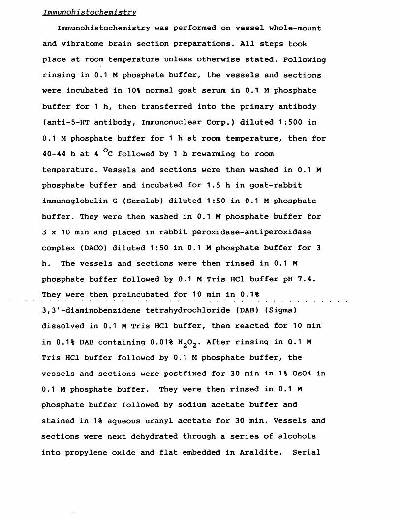
Immunohis tochemi s try
Immunohistochemistry was performed on vessel whole-mount and vibratome brain section preparations. All steps took place at room temperature unless otherwise stated. Following rinsing in 0.1 M phosphate buffer, the vessels and sections were incubated in 10% normal goat serum in 0.1 M phosphate buffer for 1 h, then transferred into the primary antibody (anti-5-HT antibody, Immunonuclear Corp.) diluted 1:500 in 0.1 M phosphate buffer for 1 h at room temperature, then for 40-44 h at 4 °C followed by 1 h rewarming to room temperature. Vessels and sections were then washed in 0.1 M phosphate buffer and incubated for 1.5 h in goat-rabbit immunoglobulin G (Seralab) diluted 1:50 in 0.1 M phosphate buffer. They were then washed in 0.1 M phosphate buffer for 3 x 10 min and placed in rabbit peroxidase-antiperoxidase complex (DACO) diluted 1:50 in 0.1 M phosphate buffer for 3 h. The vessels and sections were then rinsed in 0.1 M phosphate buffer followed by 0.1 M Tris HC1 buffer pH 7.4. They were then preincubated for 10 min in 0.1%3,3'-diaminobenzidene tetrahydrochloride (DAB) (Sigma) dissolved in 0.1 M Tris HC1 buffer, then reacted for 10 min in 0.1% DAB containing 0.01% After rinsing in 0.1 MTris HC1 buffer followed by 0.1 M phosphate buffer, the vessels and sections were postfixed for 30 min in 1 % 0s04 in 0.1 M phosphate buffer. They were then rinsed in 0.1 M phosphate buffer followed by sodium acetate buffer and stained in 1% aqueous uranyl acetate for 30 min. Vessels and sections were next dehydrated through a series of alcohols into propylene oxide and flat embedded in Araldite. Serial
ultrathin sections were cut from the midpoint of the vessels and mounted on 200-mesh grids. The ultrathin sections were stained with aqueous uranyl acetate followed by lead citrate and examined with a Philips-300 electron microscope. The flat embedded vibratome sections were examined by light microscopy and the raphe-nuclei and nucleus of the solitary tract identified. Appropriate regions were marked, cut and remounted on araldite stubs. After examination of semithin sections, these regions were trimmed to allow serial ultrathin sections of portions of the nuclei to be cut, stained and examined as described above.
Immunohistochemical controls
Specificity of the anti-5-HT antibody was tested by_3preabsorption of the primary antiserum with 5-HT (10 M) for
24h at 4 °C prior to use, and omission of the primary antiserum, both of which resulted in abolition of immunostaining of nerves. Cross-reactivity of the anti-5-HT antibody for histamine, dopamine, 5-OHDA and noradrenaline was excluded by preabsorption of' the primary antiserum with’ each of these substances (10~^M) for 24 h at 4 °C when immunostaining of nerves was not abolished.
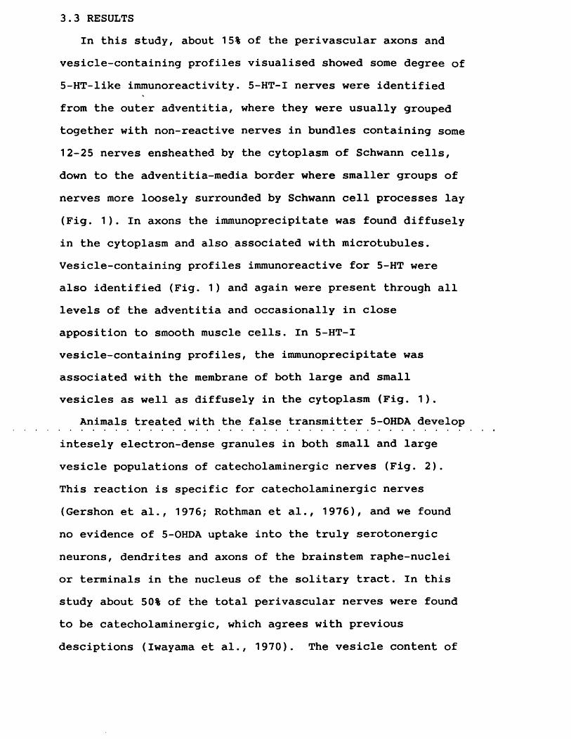
3.3 RESULTSIn this study, about 15% of the perivascular axons and
vesicle-containing profiles visualised showed some degree of 5-HT-like immunoreactivity. 5-HT-I nerves were identified from the outer adventitia, where they were usually grouped together with non-reactive nerves in bundles containing some 12-25 nerves ensheathed by the cytoplasm of Schwann cells, down to the adventitia-media border where smaller groups of nerves more loosely surrounded by Schwann cell processes lay (Fig. 1). In axons the immunoprecipitate was found diffusely in the cytoplasm and also associated with microtubules. Vesicle-containing profiles immunoreactive for 5-HT were also identified (Fig. 1) and again were present through all levels of the adventitia and occasionally in close apposition to smooth muscle cells. In 5-HT-I vesicle-containing profiles, the immunoprecipitate was associated with the membrane of both large and small vesicles as well as diffusely in the cytoplasm (Fig. 1).
Animals treated with the false transmitter 5-OHDA develop intesely electron-dense granules in both small and large vesicle populations of catecholaminergic nerves (Fig. 2). This reaction is specific for catecholaminergic nerves (Gershon et al., 1976; Rothman et al., 1976), and we found no evidence of 5-OHDA uptake into the truly serotonergic neurons, dendrites and axons of the brainstem raphe-nuclei or terminals in the nucleus of the solitary tract. In this study about 50% of the total perivascular nerves were found to be catecholaminergic, which agrees with previous desciptions (Iwayama et al., 1970). The vesicle content of
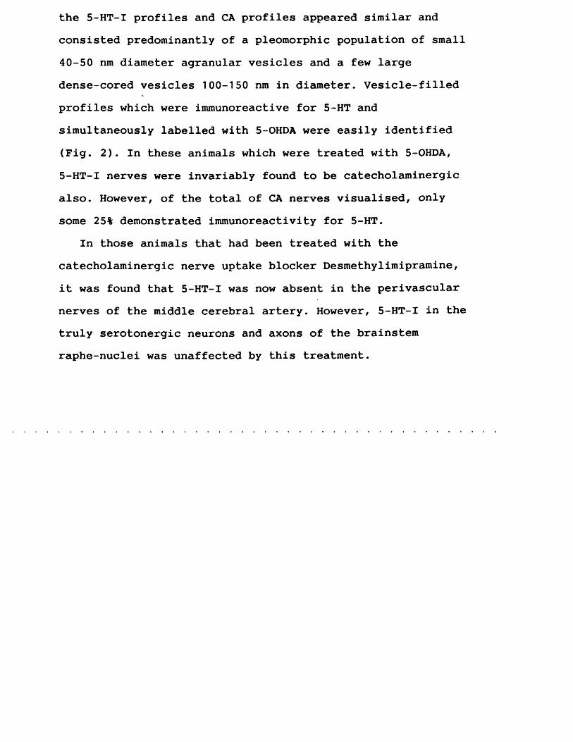
the 5-HT-I profiles and CA profiles appeared similar and consisted predominantly of a pleomorphic population of small 40-50 nm diameter agranular vesicles and a few large dense-cored vesicles 100-150 nm in diameter. Vesicle-filled profiles which were immunoreactive for 5-HT and simultaneously labelled with 5-OHDA were easily identified (Fig. 2). In these animals which were treated with 5-OHDA, 5-HT-I nerves were invariably found to be catecholaminergic also. However, of the total of CA nerves visualised, only some 25% demonstrated immunoreactivity for 5-HT.
In those animals that had been treated with the catecholaminergic nerve uptake blocker Desmethylimipramine, it was found that 5-HT-I was now absent in the perivascular nerves of the middle cerebral artery. However, 5-HT-I in the truly serotonergic neurons and axons of the brainstem raphe-nuclei was unaffected by this treatment.
3.5 DISCUSSION5-HT is a potent and well documented constrictor of a
variety of vessels, including the major cerebral vessels.Its mechanism of action is complex, interacting with that of adrenergic nerves. Exogenous application in low doses to isolated, peripheral and cerebral arteries in vitro causes
2constriction by a direct activation of postjunctional 5-HT receptors (Toda and Fujita, 1973; Allen et al., 1974a; Edvinsson et al., 1978; Medgett et al., 1984). In subcontractile doses, application potentiates the vasoconstrictor response elicited by stimulation of perivascular sympathetic nerves by an identical mechanism (Morotoki and Su, 1981; Van Nueten et al., 1981; Medgett et al., 1984). Application of higher doses causes constriction by a prejunctional mechanism, 5-HT causing displacement release of noradrenaline to act upon alpha - adrenoceptors (Fozard and Mwaluko, 1976; Marin and Sanchez, 1980; Itakura et al., 1985). Griffith et al. (1982) were able to demonstrate that the transmural nerve stimulation evoked neurogenic response in the rabbit basilar artery that persists after adrenoceptor blockade (Lee et al., 1980) could be blocked by the 5-HT2 receptor antagonist ketanserin. In addition to this pharmacological interaction, it is now becoming increasingly apparent that perivascular catecholaminergic nerves in peripheral and cerebral arteries can take up 5-HT and release it subsequently upon nerve stimulation (Verbewen et al., 1983; Kawasaki and Takasaki, 1984).
In this study of rat middle cerebral arteries prepared by
immersion-fixation, 5-HT-I was found to be present only in nerves that were shown also to be catecholaminergic in nature. Approximately 25% of the perivascular catecholaminergic nerves displaying some degree of 5-HT-like immunoreactivity. These findings providing direct ultrastructural evidence to support recent physiological and pharmacological studies which have suggested co-localisation of these transmitter substances in the perivascular nerves of major cerebral vessels.
It is unclear, however, whether the 5-HT-I demonstrated in these perivascular catecholaminergic nerves represents synthesis or simply uptake and storage of 5-HT. A recent study (Sah and Matsumoto, 1987) has shown that some rat superior cervical ganglion sympathetic neurons grown in culture can release 5-HT in a Ca dependent manner. The study also showed that the released 5-HT could be derived both by uptake and by synthesis. When the neurons were grown in 5-HT-containing culture medium, their 5-HT stores were derived from uptake. However, when they grown in a 5-HT-free medium that had been conditioned by cardiac myocytes, some neurones were able to synthesise 5-HT from tryptophan.
To ascertain whether the 5-HT-I observed in the perivascular nerves of major cerebral vessels is derived from uptake or synthesis, rats were examined that had been treated with the catecholaminergic nerve uptake blocker DMI in a dose that has been previously shown to reduce uptake activity by 83% (Wong et al., 1974). Following treatment with DMI 5-HT-I could not be demonstrated in any of the
catecholaminergic nerves of the rat middle cerebral artery, whilst the 5-HT-I of the raphe-nucleus neurons and terminals in the solitary tract nucleus were unaffected. This would support the premise that the 5-HT-I seen in a proportion of perivascular catecholaminergic nerves is due to uptake rather than intrinsic synthesis.
The possibility still remains, however, that if the level of synthesis of 5-HT was low, then by preventing reuptake of released 5-HT with DMI the nerves could be depleted of 5-HT to a level insufficient to be visualised by immunohistochemical methods.
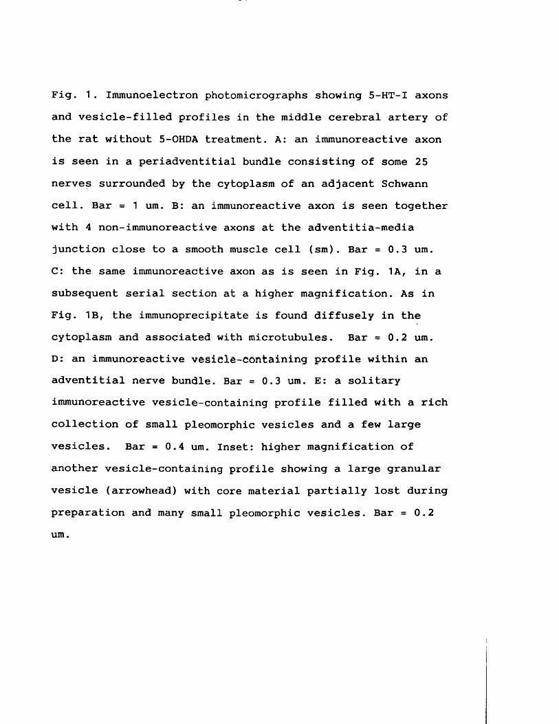
Fig. 1 . Immunoelectron photomicrographs showing 5-HT-I axons and vesicle-filled profiles in the middle cerebral artery of the rat without 5-OHDA treatment. A: an immunoreactive axon is seen in a periadventitial bundle consisting of some 25 nerves surrounded by the cytoplasm of an adjacent Schwann cell. Bar = 1 urn. B: an immunoreactive axon is seen together with 4 non-immunoreactive axons at the adventitia-media junction close to a smooth muscle cell (sm). Bar = 0.3 urn.C: the same immunoreactive axon as is seen in Fig. 1A, in a subsequent serial section at a higher magnification. As in Fig. 1B, the immunoprecipitate is found diffusely in the cytoplasm and associated with microtubules. Bar = 0.2 urn.D: an immunoreactive vegiele-containing profile within an adventitial nerve bundle. Bar = 0.3 urn. E: a solitary immunoreactive vesicle-containing profile filled with a rich collection of small pleomorphic vesicles and a few large vesicles. Bar = 0.4 um. Inset: higher magnification of another vesicle-containing profile showing a large granular vesicle (arrowhead) with core material partially lost during preparation and many small pleomorphic vesicles. Bar = 0.2 um.
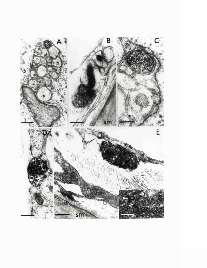
'
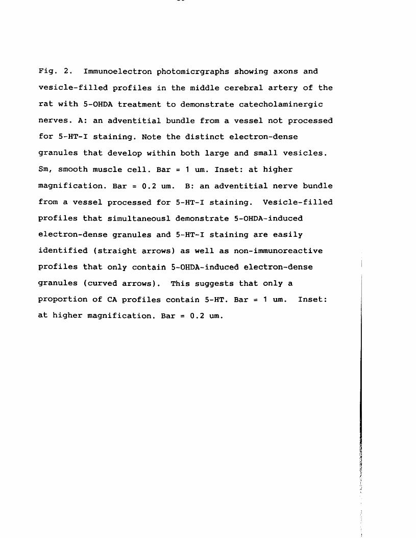
Fig. 2. Immunoelectron photomicrgraphs showing axons and vesicle-filled profiles in the middle cerebral artery of the rat with 5-OHDA treatment to demonstrate catecholaminergic nerves. A: an adventitial bundle from a vessel not processed for 5-HT-I staining. Note the distinct electron-dense granules that develop within both large and small vesicles. Sm, smooth muscle cell. Bar = 1 um. Inset: at higher magnification. Bar = 0.2 um. B: an adventitial nerve bundle from a vessel processed for 5-HT-I staining. Vesicle-filled profiles that simultaneousl demonstrate 5-OHDA-induced electron-dense granules and 5-HT-I staining are easily identified (straight arrows) as well as non-immunoreactive profiles that only contain 5-OHDA-induced electron-dense granules (curved arrows). This suggests that only a proportion of CA profiles contain 5-HT. Bar = 1 um. Inset: at higher magnification. Bar = 0.2 um.
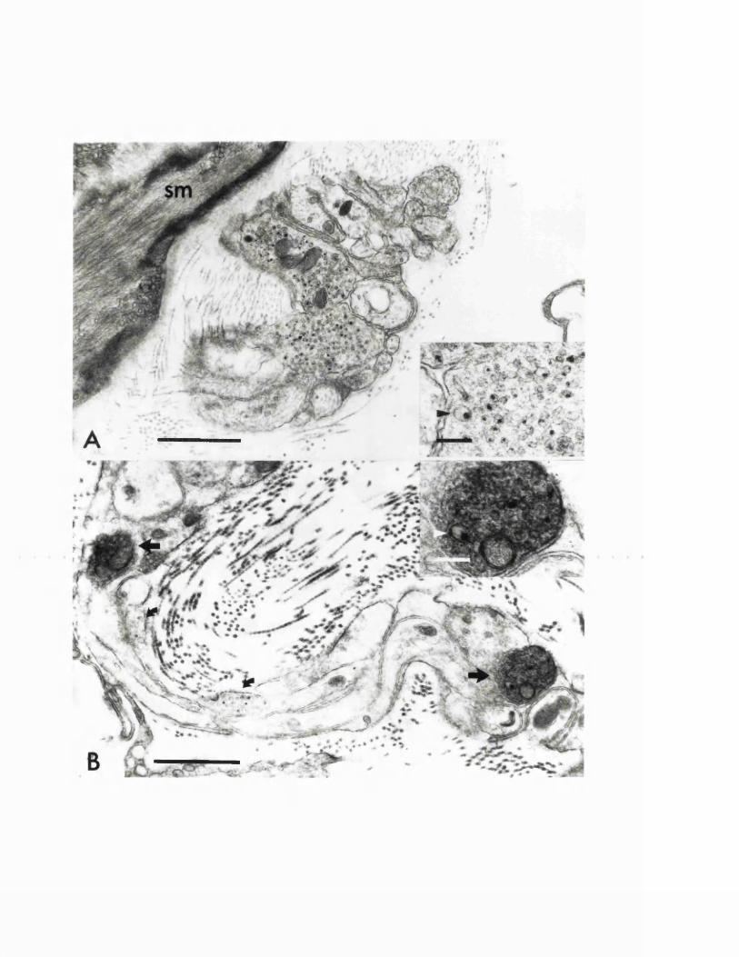
4. EXPERIMENTAL CHAPTER II:
5-HYDROXYTRYPTAMINE DEMONSTRATED IMMUNOHISTOCHEMICALLY IN RAT CEREBROVASCULAR NERVES LARGELY REPRESENTS 5-HYDROXYTRYPTAMINE UPTAKE INTO SYMPATHETIC NERVE FIBRES
Summary: This chapter re-examines by means of fluorescenceimmunohistochemical techniques, a proposed serotonergic innervation of major cerebral vessels in the rat. Earlier studies had demonstrated a dense perivascular plexus of5-hydroxytryptamine immunoreactive nerve fibres upon major cerebral vessels in this and many other species. In this investigation however, it was found that 5-hydroxytryptamine immunoreactive nerve fibres are rarely observed in cerebral vessels prepared by perfusion-fixation in situ, and only form a well-developed plexus in vessels prepared, as in previous authors studies, by immersion-fixation. Prior treatment with a predominantly noradrenergic uptake inhibitor desmethylimipramine but not the serotonergic uptake inhibitor fluoxetine produced a major diminution in the 5-hydroxytryptamine immunoreactive plexus visualised in these immersion-fixed vessels. In addition,5-hydroxytryptamine immunoreactive nerves were only occasionally observed in immersion-fixed vessels from animals that had been pretreated with 6-hydroxydopamine to produce adrenergic denervation. The removal firstly, of vessel-contained blood, by left ventricular perfusion with Krebs, prior to vessel dissection and immersion-fixation, resulted in an absence of 5-hydroxytryptamine immunoreactivity in perivascular nerves. Immunoreactivity could then be restored by briefly incubating vessels in krebs containing either blood or 5-hydroxytryptamine, before fixation. It would appear therefore that5-hydroxytryptamine is rarely present under normal circumstances in the perivascular nerves of major cerebral
vessels and that previous descriptions of a dense serotonergic nerve plexus, represent 5-hydroxytryptamine in blood released during vessel dissection, being taken up via the noradrenaline-uptake system into perivascular sympathetic nerves. The possibility is thus raised that5-hydroxytryptamine uptake and interaction within perivascular adrenergic nerves could occur in those cerebrovascular disorders where blood is released.
4.1 INTRODUCTIONSerotonin is well known for its marked effects upon the
cerebral circulation (Harper and Mackenzie, 1977b; Mackenzie and Edvinsson, 1980). Intracarotid administration reduces cerebral blood flow profoundly by constriction of large calibre vessels and arteriovenous anastomoses, whilst small arterioles dilate (Verdouw et al., 1984; Saxena and Verdouw, 1985). The possibility that perivascular nerves upon the major cerebral vessels are involved in this response was raised, when studies in a variety of species (Baumgarten, 1972; Chan-Palay, 1976; Griffith et al., 1982; Edvinsson et al., 1983b; Chang et al., 1987; Cowen et al., 1987) including man (Griffith and Burnstock, 1983) suggested the presence of a specific serotonergic innervation.
The immunohistochemical studies employed, however, have all utilised immersion-fixation of vessels, and in some cases vessel incubation in vitro with 5-HT or the monoamine oxidase inhibitor pargyline to enhance 5-HT-like immunoreactive (5-HT-I) nerve staining. The virtual disappearance of such nerves following superior cervical ganglionectomy (Marco et al., 1985; Cowen et al., 1986;Cowen et al., 1987), which also results in the loss of noradrenergic sympathetic nerves (Nielson and Owman, 1967; Iwayama et al., 1970), together with recent evidence demonstrating an ability of sympathetic nerves to accumulate exogenous 5-HT in vitro, (Verbeuren et al., 1983; Levitt and Duckies, 1986; Saito and Lee., 1987) has led to two questions being raised. Firstly, are all, or at least a majority of the serotonin-containing nerves demonstrated
previously in major cerebral vessels in fact sympathetic nerves, and secondly what is the origin of the serotonin that has been demonstrated within them.
The rat is an animal in which a serotonergic innervation of major cerebral vessels has been well described, (Edvinsson et al., 1983b; Chang and Owman, 1986; Cowen et al., 1987) it was decided therefore to undertake further studies upon it in order to elucidate these questions. This study was designed to assess whether the 5-HT-I plexus demonstrated in previous studies, was as equally developed in perfusion-fixed material, and if not, whether it could represent 5-HT uptake into sympathetic nerves during tissue dissection and preparation procedures.
4.2 MATERIALS AND METHODSA total of 33 male Sprague-Dawley rats weighing 350-450g
were used in this study. All animals were anaesthetised with 70% nitrous oxide, 30% oxygen and 1.5% halothane and were allowed to breathe spontaneously.
In the first group of experiments, seven rats were sacrificed by perfusion with 150 mis of fixative composed of 4% paraformaldehyde in 0.1 M phosphate buffered saline (PBS) through the left ventricle. The brains were then removed and all branches of the circle of Willis dissected out to yield a single preparation that included the anterior cerebral, middle cerebral, internal carotid, posterior cerebral, superior cerebellar, basilar and vertebral arteries. These vessels were then further fixed in the same fixative for 2 to 24 h.
In the second group, fourteen rats were utilised; all these animals were killed by decapitation under anaesthesia. The brains were rapidly removed, and the circle of Willis dissected out in a Krebs solution composition (in M): NaCl 0.12, KC1 0.1, KH2P04 0.05, MgSC>4 0.03, NaHCC>3 0.12, glucose 0.07, CaCl2 2.5. The vessels were then fixed by immersion in 4% paraformaldehyde in 0.1 M PBS for 2 to 24 h. Within this group of animals, 5 rats received no pretreatment and 9 rats, three in each category, were pretreated with one of the following drugs: the adrenergic neurotoxin6-hydroxydopamine (6-OHDA) (100-150mg/kg) i.v. 72 and 24h before sacrifice, the noradrenergic uptake antagonist desmethylimipramine (28.5 umole/kg) i.p. 1h before sacrifice, or the serotonergic uptake antagonist fluoxetine
(28.5 umole/kg) i.p. 1h before sacrifice.In the third group, 12 rats were utilised. These animals
were killed by perfusion with Krebs through the left ventricle, the brains then rapidly removed and the circle of Willis and cerebral vessels dissected out. The circle of Willis from 3 rats were then immediately fixed by immersion in 4% paraformaldehyde in 0.1M PBS. Those from the remaining 9 animals were incubated at 37°C for 30 minutes in either Krebs alone (3 rats), Krebs containing 5-HT (10”^M), or Krebs containing 10% by volume of blood previously collected from another rat. Following 30 mins incubation, the circle of Willis vessels were fixed by immersion in 4% paraformaldehyde in PBS for 2-24 h. The Krebs solution used in incubation procedures was constantly aerated with a gas mixture of 95% 02 / 5% C02.
Immunohistochemistry
Immunohistochemistry was performed on vessel whole-mount preparations. All steps took place at room temperature.After fixation' all vessels' were’ rinsed in’ PBS. To facilitate antibody penetration the vessels were then rinsed in 80% alcohol for 1h, and given short rinses in PBS with 0.1% Triton X-100. Vessels from one half of each circle of Willis were then incubated for 12-18 h in anti-5-HT antibody (Immunonuclear Corp.) diluted 1:400 with PBS containing 0.1% Triton X-100. After 3 PBS washes, vessels were exposed for 2 h to fluorescein isothiocyanate (FITC)-conjugated goat anti-rabbit IgG (Nordic) at a dilution of 1:50. Vessels were briefly rinsed in PBS then placed in PBS with 0.05% pontamine sky blue and 1% dimethyl sulphoxide for 5-10 mins
in order to reduce background autofluorescence (Cowen et al., 1985). Vessels were next air-dried onto slides and coverslipped in an anti-fade mountant (Citifluor, City University,London, U.K.) for viewing. Vessels from the other half of each circle of Willis, utilised for the demonstration of neuropeptide-Y-like immunoreactive (NPY-I) nerves, were treated similarly but using an anti-NPY antibody (Immunonuclear Corp.) diluted 1:400.
Immunohistochemical controls
Specificity of the primary antibody was tested by preabsorption with excess antigen (10”^M) for 24 h at 4°C prior to use and omission of the primary antiserum, both of which resulted in abolition of immunoreactivity. Cross-reactivity of the anti-5-HT antibody for histamine, dopamine and noradrenaline was excluded by preincubation
_3with each of these substances at 10 M when immunoreactivity was maintained.
6-hvdroxvdoDamine induced adrenergic denervation
6-Hydroxydopamine was used to induce a selective degeneration of adrenergic nerve terminals (Thoenen and Tranzer, 1968; Costa et al., 1982), in order to investigate the effect of the resultant sympathetic denervation, upon the ability to demonstrate 5-HT containing nerves upon circle of Willis vessels. Confirmation of the effectiveness of the adrenergic denervation produced was ascertained as cerebral vessels from 6-OHDA treated and untreated animals were examined for the presence or absence of NPY-I nerves. NPY-I has been demonstrated previously, as localised within
the adrenergic nerves of cerebral vessels (Matsuyama et al., 1985; Edvinsson et al., 1987), and NPY-I in adrenergic nerves is markedly reduced, together with noradrenaline, following 6-OHDA treatment (Morris et al., 1986a; Edvinsson et al., 1987). In those animals in this study that received6-OHDA, there was always a major reduction of NPY-I nerves throughout the circle of Willis whilst untreated animals in all other groups always demonstrated a dense plexus as previously described (Edvinsson et al., 1987).
4.3 RESULTSGroup I Perfusion-fixed vessels: In cerebral vessels
taken from animals killed by perfusion-fixation, 5-HT-I nerves were only inconstantly and most infrequently observed (Fig. 3). In five of the seven animals employed, there were no 5-HT-IR nerves seen throughout any of the vessels comprising the circle of Willis. Whilst in the remaining two animals there were a few faint 5-HT-I nerves seen restricted to the vertebral and basilar arteries, the remaining superior cerebellar, posterior cerebral, internal carotid, middle cerebral and anterior cerebral arteries were otherwise entirely devoid of 5-HT-I nerve fibres.
Group II Immersion-fixed vessels: Within this group of animals killed by decapitation, followed by vessel dissection and subsequent immersion-fixation, the results were affected by the prior treatment received. In all five animals that received no pretreatment, a well defined plexus of 5-HT-I nerves was consistantly demonstrated throughout all thevessels of the circle'of Willis■(Fig: 4)Pretreatment with the serotonergic uptake inhibitor fluoxetine (3 animals), produced only a minor diminution in the extent and intensity of the 5-HT-I nerve plexus (Fig. 5A and 5B). However, pretreatment with an equimolar dose of the noradrenergic uptake inhibitor desmethylimipramine (3 animals) resulted in the virtual abolition of the 5-HT-I nerve plexus (Fig. 5C and 5D). Similarly, in three animals pretreated with 6-OHDA to induce degeneration of adrenergic varicosities, it was found that 5-HT-I nerves were only seldom observed (Fig. 6).
Group III Krebs perfused vessels: In this group of animals perfused with Krebs to remove blood from the vessels prior to dissection, with or without an incubation, followed by immersion-fixation, the results obtained depended upon the incubation procedure. In the three animals whose vessels were immersion-fixed without any incubation, 5-HT-I nerves were never observed. Similarly in those vessels (3 animals) incubated in Krebs alone no 5-HT-I nerves were seen (Fig. 7A and 7B). However, in vessels incubated in either Krebs plus 5-HT (Fig. 7C and 7D) or Krebs plus blood (Fig.7E and 7F) prior to immersion-fixation, the results differed. In these latter vessels, a strongly staining plexus of 5-HT-I nerves was once again seen throughout the circle of Willis.
4.5 DISCUSSIONSerotonin is predominantly synthesised by the chromaffin
cells of the gastrointestinal tract (Erspamer and Testini, 1959). It is then released into the portal circulation, where that fraction that escapes inactivation by hepatocytes and subsequently the pulmonary bed, is taken up and stored within the granules of platelets. Serotonin synthesising neurons have also been demonstrated in the enteric plexus of the gut and the raphe nuclei of the brainstem (Dahlstrom and Fuxe, 1964; Dreyfus et al., 1977; Steinbusch et al., 1978; Costa et al., 1982). On the basis of these and other findings a variety of roles have been suggested for serotonin, in health and disease, in a number of sites. Serotonin released during platelet aggregation has been proposed to act upon the smooth muscle of injured vessels to produce local vasoconstriction and aid haemostasis (Van Zwieten, 1987). In the enteric plexus, serotonergic neurons and their processes would seem to be involved in the regulation of intestinal motility (Gershon and Sherman, 1987). With regard to the cerebral circulation, the serotonergic neurons of the raphe nuclei appear to make somal and dendritic contacts locally with parenchymal blood vessels of the brainstem, and also contact more distal intracerebral parenchymal blood vessels through long projection axons (Scheibel et al., 1975; Reinhard et al., 1979; Di Carlo, 1984; Kapaidia and de Lanerolle, 1984; Scatton et al., 1985). This has led to the suggestion of a possible role for serotonin in cerebral vasomotor control, regulating overall parenchymal cerebrovascular resistance,
in response to changes in brainstem blood flow, as sensed by the raphe neurons.
A completely separate, serotonergic innervation of the circle of Willis and large extraparenchymal cerebral vessels then appeared to exist, following the demonstration by fluorescence immunohistochemistry of a well developed 5-HT-I plexus about such vessels in a number of species, including man. Unlike the innervation of the smaller intraparenchymal cerebral vessels, this plexus was largely lost following removal of the superior cervical ganglia, which are known also to be the major source of adrenergic innervation to these major cerebral vessels. However, from an examination of perfusion-fixed material and by using uptake studies, this experimental chapter now clearly demonstrates that the major cerebral vessels of the rat, are normally only seldom and inconstantly innervated by occasional faint 5-HT-I nerves limited strictly to the vertebrobasilar arteries.When the same vessels are examined from animals that have been killed by decapitation, followed by vessel dissection and immersion-fixation, then a rich network of 5-HT-I nerves is readily demonstrated throughout the circle of Willis.
The hypothesis, that this difference is due to 5-HT from blood, released during dissection, being taken up via the noradrenaline uptake mechanism into sympathetic nerves was tested. Firstly, development of a well-defined 5-HT-I plexus in immersion-fixed material could largely be prevented by pretreatment with a predominantly noradrenergic uptake inhibitor but not with a more selective serotonergic
uptake inhibitor. Similarly, the development of a 5-HT-I plexus could largely be prevented, by prior treatment of animals with 6-OHDA which produces a selective degeneration of adrenergic nerve terminals.
In a separate series of experiments, the careful removal of vessel-contained blood by perfusion with Krebs solution prior to brain removal and vessel dissection, also resulted in the absence of any visible 5-HT-I plexus. Such a plexus was readily restored if vessels were then briefly incubated in Krebs containing either 5-HT (10_^M) or freshly collected blood (10% Vol).
Taken together, these results indicate that the well developed 5-HT-I nerve plexus previously described upon major cerebral vessels largely represents an in vitro uptake phenomenon, serotonin from blood released during vessel dissection being readily taken up into sympathetic nerves where it can subsequently be demonstrated by immunohistochemistry. The very small and inconstant number of 5-HT-I fibres that were occasionally seen in perfusion-fixed animals, largely restricted to the basilar artery, might represent centrally derived serotonergic fibres that have passed out along the perforating arteries to the raphe complex. Another possibility might be that 5-HT in locally elevated levels, derived from nearby raphe neurons, is taken up into adjacent sympathetic nerves.
The reasons why perivascular sympathetic nerves supplying cerebral vessels are capable of avid uptake of 5-HT needs to be examined. For example, is it a protective mechanism to remove potent concentrations of 5-HT arising in platelet
aggregation and other conditions or is it a biological device for reinforcing sympathetic vasoconstriction arising during certain physiological and pathological states. These and other questions need to be resolved in future studies.
The ready demonstration of such an ability in vitro is of interest, in that a similar mechanism of blood-derived 5-HT uptake and interaction in perivascular sympathetics may occur in vivo in cerebrovascular disorders such as following stroke, migraine attacks and in vasospasm following subarachnoid haemorrhage, where serotonin has frequently been implicated in the aetiology (Sicuteri et al., 1961; Anthony et al., 1969; Allen et al., 1974a; Voldby et al., 1982; Tagari et al., 1983; Richardson et al., 1987).
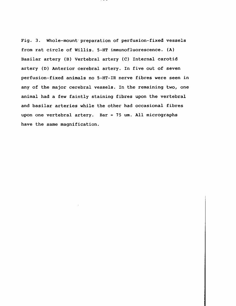
Fig. 3. Whole-mount preparation of perfusion-fixed vessels from rat circle of Willis. 5-HT immunofluorescence. (A) Basilar artery (B) Vertebral artery (C) Internal carotid artery (D) Anterior cerebral artery. In five out of seven perfusion-fixed animals no 5-HT-IR nerve fibres were seen in any of the major cerebral vessels. In the remaining two, one animal had a few faintly staining fibres upon the vertebral and basilar arteries while the other had occasional fibres upon one vertebral artery. Bar = 75 urn. All micrographs have the same magnification.
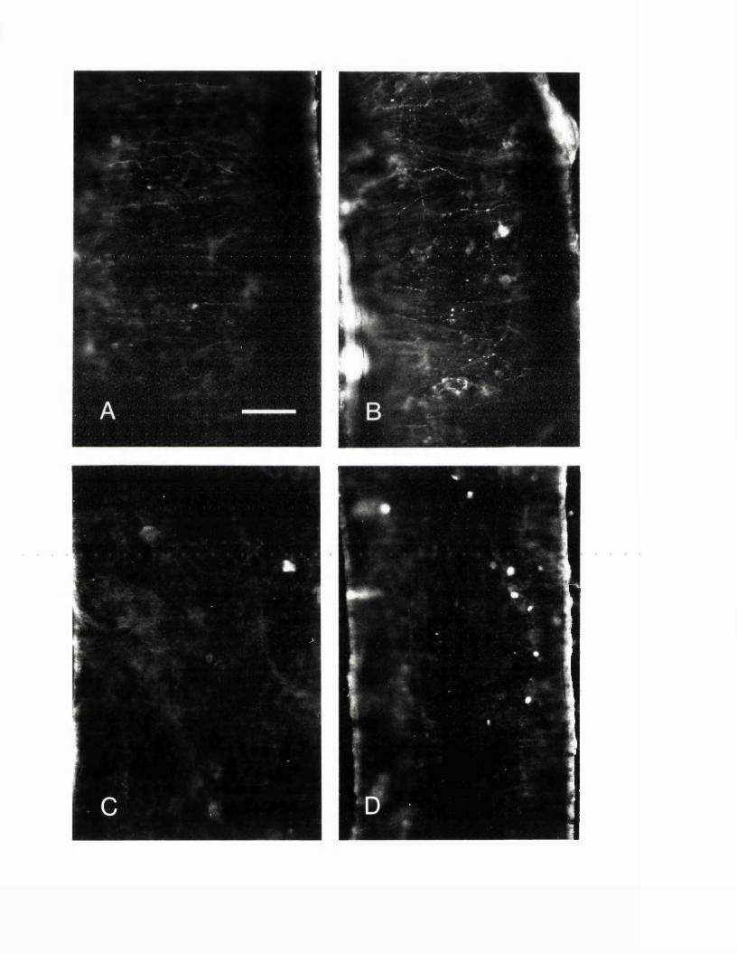
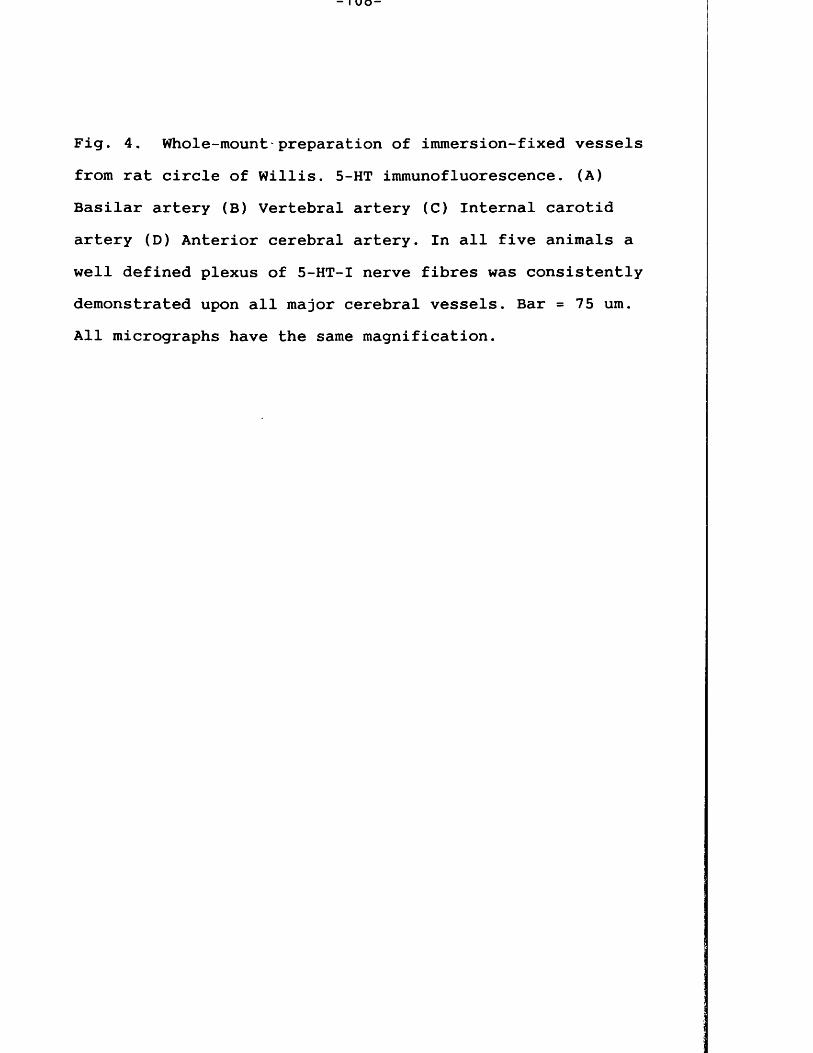
Fig. 4. Whole-mount'preparation of inunersion-fixed vessels from rat circle of Willis. 5-HT immunofluorescence. (A) Basilar artery (B) Vertebral artery (C) Internal carotid artery (D) Anterior cerebral artery. In all five animals a well defined plexus of 5-HT-I nerve fibres was consistently demonstrated upon all major cerebral vessels. Bar = 75 urn. All micrographs have the same magnification.
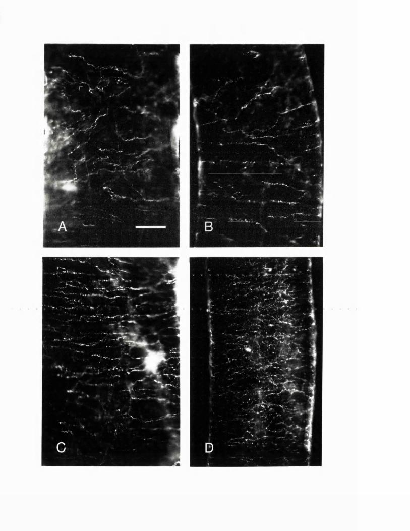
Fig. 5. Immunofluorescence photomicrographs of immersion-fixed cerebral vessel whole-mounts from rats pretreated with fluoxetine (A and B) or desmethylimipramine (C and D). A and C are corresponding portions of vertebral arteries whilst B and D are similar regions of anterior cerebral arteries. Compared as pairs: A against C, and B against D, it can be seen that numerous 5-HT-I fibres persist following fluoxetine administration whilst after desmethylimipramine, only a few fibres remain faintly immunoreactive. Bar = 75 urn. All micrographs have the same magnification.
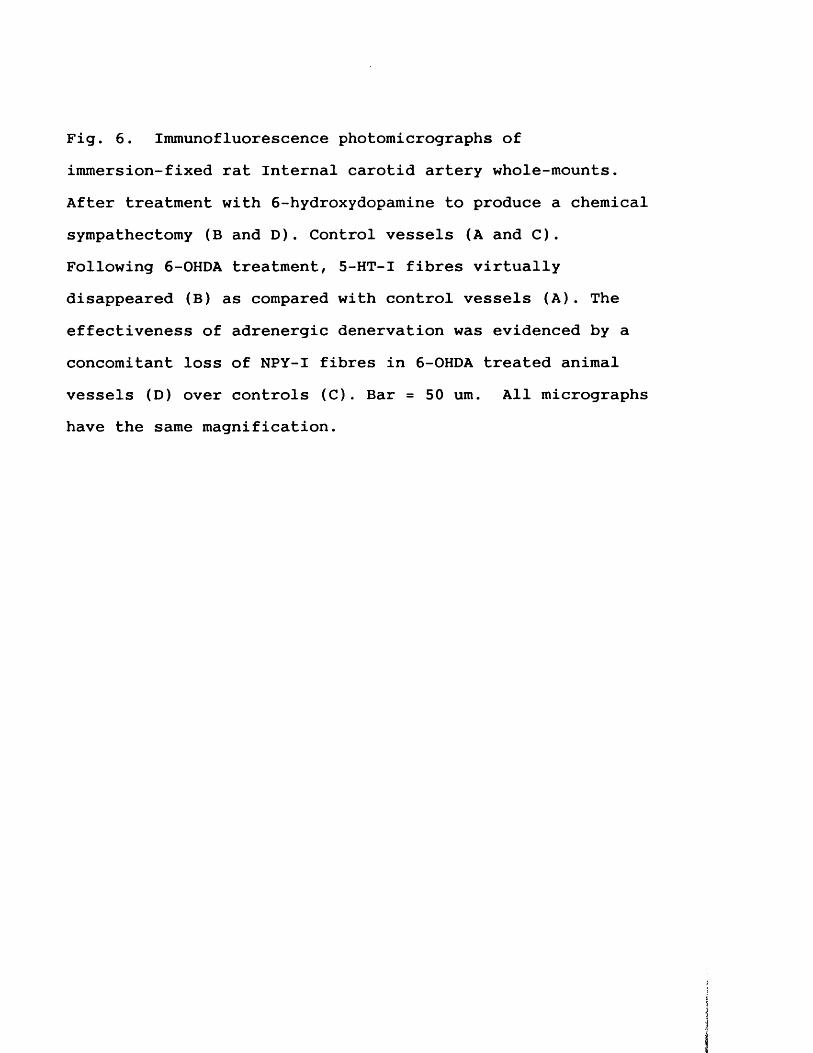
Fig. 6. Immunofluorescence photomicrographs of immersion-fixed rat Internal carotid artery whole-mounts. After treatment with 6-hydroxydopamine to produce a chemical sympathectomy (B and D). Control vessels (A and C).Following 6-OHDA treatment, 5-HT-I fibres virtually disappeared (B) as compared with control vessels (A). The effectiveness of adrenergic denervation was evidenced by a concomitant loss of NPY-I fibres in 6-OHDA treated animal vessels (D) over controls (C). Bar = 50 um. All micrographs have the same magnification.
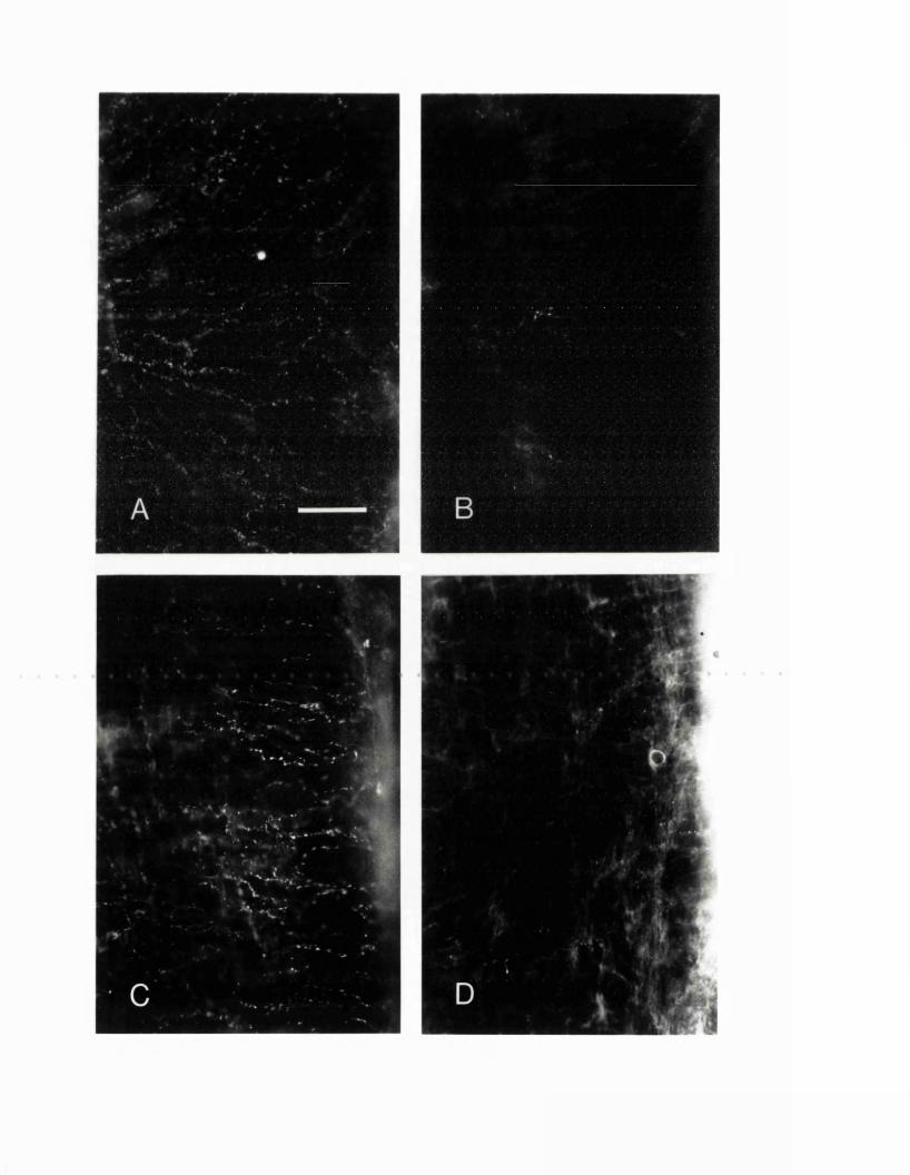
Fig. 7. Whole-mount preparation of circle vessels, perfused with Krebs to remove contained blood, and incubated in either Krebs alone (A and B), Krebs plus 5-HT (C and D) or Krebs plus blood (E and F) prior to immersion-fixation. In (A and B) 5-HT-I fibres were completely absent. Following incubation with 5-HT or blood a well-developed plexus of 5-HT-I nerves fibres was restored. Bar = 100 urn. All micrographs have the same magnification.
5. EXPERIMENTAL CHAPTER III:
ALTERATIONS IN SEROTONIN AND NEUROPEPTIDE Y CONTENT OF CEREBROVASCULAR SYMPATHETIC NERVES FOLLOWING EXPERIMENTAL SUBARACHNOIDvHAEMORRHAGE
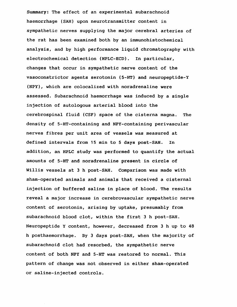
Summary: The effect of an experimental subarachnoid haemorrhage (SAH) upon neurotransmitter content in sympathetic nerves supplying the major cerebral arteries of the rat has been examined both by an immunohistochemical analysis, and by high performance liquid chromatography with electrochemical detection (HPLC-ECD). In particular, changes that occur in sympathetic nerve content of the vasoconstrictor agents serotonin (5-HT) and neuropeptide-Y (NPY), which are colocalized with noradrenaline were assessed. Subarachnoid haemorrhage was induced by a single injection of autologous arterial blood into the cerebrospinal fluid (CSF) space of the cisterna magna. The density of 5-HT-containing and NPY-containing perivascular nerves fibres per unit area of vessels was measured at defined intervals from 15 min to 5 days post-SAH. In addition, an HPLC study was performed to quantify the actual amounts of 5-HT and noradrenaline present in circle of Willis vessels at 3 h post-SAH. Comparison was made with sham-operated animals and animals that received a cisternal injection of buffered saline in place of blood. The results reveal a major increase in cerebrovascular sympathetic nerve content of serotonin, arising by uptake, presumably from subarachnoid blood clot, within the first 3 h post-SAH. Neuropeptide Y content, however, decreased from 3 h up to 48 h posthaemorrhage. By 3 days post-SAH, when the majority of subarachnoid clot had resorbed, the sympathetic nerve content of both NPY and 5-HT was restored to normal. This pattern of change was not observed in either sham-operated or saline-injected controls.
5.1 INTRODUCTIONSubarachnoid haemorrhage (SAH) is known, both clinically
in humans (Grubb et al., 1977; Symon, 1978; Kwak et al., 1979), and from experimental studies in animals (Kuwayama et al., 1972; Petruk et al., 1972; Umansky et al., 1983; Svendgard et al., 1983) to be associated with the development of angiographically defined cerebral arterial spasm, reduced cerebral blood flow, and impaired autoregulation. The neural plexus that surrounds and innervates the major cerebral vessels is one of the factors known to control the calibre of these vessels (Edvinsson and MacKenzie, 1977), with sympathetic noradrenergic nerves making up a major component of this plexus (Iwayama et al., 1970). Studies, both in human SAH and from animal models, have revealed the development posthaemorrhage of sympathetic nerve dysfunction in cerebral vessels, with an impairment of noradrenaline uptake and synthesising ability ( Lobato et al., 1980; Edvinsson et al., 1982; Tsukahara et al., 1988).
It has become clear from recent discoveries that the cerebrovascular sympathetic nerves contain other substances, in addition to noradrenaline, that can act as neurotransmitters and neuromodulators. One of these, the peptide neuropeptide Y (NPY), is known to cause strong concentration-dependent contractions of cerebral vessels (Edvinsson et al., 1987). Cerebrovascular sympathetic nerves in vitro have additionally been shown to readily take up serotonin (5-HT) into nerve terminals (Verbeuren et al., 1983; Levitt and Duckies, 1986; Jackowski et al., 1989a), from whence it can act as a vasoconstrictor false
transmitter (Saito and Lee, 1986). Following rupture of an intracranial aneurysm, 5-HT-containing platelets are released about vessels locally and throughout the subarachnoid space, with elevated 5-HT levels being found in the cerebrospinal fluid (CSF) post-SAH (Voldby et al., 1982; Tagari et al., 1983). In this study, the effects of an induced subarachnoid haemorrhage upon the content of 5-HT and NPY in the sympathetic nerves of major cerebral vessels in the rat, as revealed by immunohistochemistry, was examined for up to 5 days postictus. In addition, the levels of 5-HT and noradrenaline within the circle of Willis vessels in the period immediately following SAH, was measured by high performance liquid chromatography (HPLC) with electrochemical detection (ECD).
5.2 MATERIALS AND METHODS Animal Preparations
One hundred and ninety male Sprague-Dawley rats weighing 350-450 grams were used in the experiments. The animals were anaesthetised with 70% nitrous oxide, 30% oxygen and 1.5% Halothane during operative procedures and perfusions. While cisternal injections were made, the halothane was reduced to 0.75%. All animals were allowed to breathe spontaneously.In each animal the left femoral artery was cannulated and connected to a Statham P50 transducer. The arterial pressure was continously recorded with a Lectromed recorder and blood gases were intermittently monitored.
In addition to a control group of 30 normal animals, the animals were divided into three groups, consisting of one group that underwent experimental subarachnoid haemorrhage (SAH), and two further groups of controls that received either a subarachnoid injection of buffered saline or sham operation only.
Induction of subarachnoid haemorrhageIn 78 animals, SAH was induced by a single injection of
0.3 mis of autologous arterial blood, freshly withdrawn from the femoral cannula and replaced with an equal volume of normal saline. The blood was injected at a rate of 0.1 mis/ minute under operating microscope vision, via a 30 gauge needle passed through the previously exposed atlanto-occipital membrane, into the cisterna magna of the prone head-down animal.
In 50 animals, 0.3 mis of buffered normal saline was injected in place of autologous blood. Buffered normal
saline was prepared by the addition of 25 meq/1 of NaHCO^ and equilibrated at 37°C with a gas mixture of 5% C02/95% O 2
to pH 7.4.In a further 32 animals, a sham operation only was
performed, the rats undergoing the same procedures to those receiving an SAH, except that no injections were given into the cisterna magna.
ImmunohistochemistryImmunohistochemistry was performed on vessel whole-mount
preparations. All steps took place at room temperature. Animals for the immunohistochemical determination of the density of nerves upon major cerebral vessels were killed post-SAH at times 15 min, 1 h, 3 h, 6 h, 1 day, 2 days, 3 days, and 5 days. Groups examined included normal, sham- operated, saline-injected, and SAH rats Under anaesthesia, the left ventricle was perfused with 150 mis of fixative composed of 4% paraformaldehyde in 0.1M phosphate-buffered saline (PBS). The brain was immediately removed and all branches of the circle of Willis dissected out to yield a single preparation that included the anterior cerebral, middle cerebral, internal carotid, posterior cerebral and basilar arteries. Vessels were further fixed for 2 h, then rinsed in 3 changes of PBS for 1 h. To facilitate antibody penetration the vessels were then rinsed in 80% alcohol for 1 h and given short rinses in PBS with 0.1% Triton X-100. Vessels were then incubated for 16 h in anti-5-HT antibody (Immunonuclear Corp.) diluted 1:400 with PBS containing 0.1% Triton X-100. After three PBS washes, vessels were exposed for 2 h to fluorescein isothiocyanate (FITC)-conjugated
goat anti-rabbit IgG (Nordic) at a dilution of 1:50. Vessels were briefly rinsed in PBS, then placed in PBS with 0.05% pontamine sky blue and 1% dimethyl sulphoxide for 5-10 min in order to reduce background autofluorescence (Cowen et al., 1985). Vessels were next air-dried onto slides and coverslipped in an anti-fade mountant (Citifluor, City University,London, U.K.). Vessels used for the immunohistochemical demonstration of NPY content within perivascular nerves were treated similarly but using an anti-NPY antibody (Immunonuclear Corp.) diluted 1:400.Slides were viewed, and selected areas photographed at X 100 using a Zeiss microscope equipped for viewing FITC fluorescence
Specificity of the anti-5-HT and anti-NPY antibody was_3tested by preincubation with 10 M concentration of 5-HT or
NPY, respectively for 24 h at 4°C prior to use, or omission of the primary antibody, both of which resulted in elimination of immunostaining of nerves.
The densities of nerves containing 5-HT or NPY weremeasured from photomicrographs taken at uniform exposure,film speed and development time. In every animal fivestandardised areas were sampled from the circle of Willis:one each from the anterior cerebral, middle cerebral,internal carotid, posterior cerebral, and basilar arteries.The number of nerve fibers seen crossing the diagonal of a
2rectangular area measuring 0.3 x 0.2 mm in each of these five unit areas was counted and averaged per animal. Results are expressed as the overall mean + SEM and data compared using the Student's t test. Significance was accepted for
p<0.05.
6-OHDA and Clomipramine experimentsTo ensure, as seemed probable from previous studies, that
the 5-HT- and NPY-immunoreactive nerve fibers counted following SAH truly represented sympathetic nerves, six animals that had been pretreated with the adrenergic neurotoxin 6-hydroxydopamine (6-OHDA) were also examined. Administration of 6-OHDA has been shown to induce degeneration of adrenergic nerve terminals (Thoenen and Tranzer, 1968) including those upon major cerebral vessels (Matsuyama et al., 1985) and was administered as two doses (100 and 150 mg/Kg i.v.) 72 and 24 h before SAH and/or killing.
To investigate further a supposition that the marked increase post-SAH of 5-HT-containing nerves was a consequence of 5-HT, released from subarachnoid blood clot, being taken up into sympathetic nerves, the effect of the noradrenergic nerve uptake antagonist clomipramine was studied. A single administration of clomipramine in a dose (57 umole/kg i.p.) previously shown to reduce adrenergic uptake activity by 83% (Wong et al., 1974) was administered 1 h prior to SAH in four animals.
High performance liquid chromatographyAnimals for the HPLC determination of the total amounts
of 5-HT and noradrenaline present in circle of Willis vessels were killed at 3h post-SAH. Three groups of animals were examined: normal, saline-injected, and SAH rats. Under anaesthesia the left ventricle was perfused with Krebs
solution to remove contaminating blood, contained within the cerebral vessels. Next, the brains were rapidly removed and transferred into ice-cold Krebs solution and the circle of Willis arteries resected and dissected clean under the operating microscope. Vessel preparations were briefly rinsed in three changes of ice-cold Krebs solution and the excess liquid blotted off before being placed in cryotubes for rapid freezing in liquid nitrogen, with storage at - 20°C until later analysis. Vessels from six animals were pooled for each determination of 5-HT and noradrenaline content by HPLC. The total sample wet weight was approximately 9 mg. Samples were homogenised in 500 ul of ice-cold 0.1M perchloric acid containing 0.4 mM sodium bisulphite and 25 ng of dihydroxybenzylamine (DHBA) using a motor-driven glass-glass homogeniser, and cell debris was removed by low speed centrifugation. Aliquots (100 ul) of the supernatant were injected without further treatment for the determination of 5-HT by HPLC with electrochemical detection (HPLC-ECD) using the method of Reinhard et al. (1980). The remainder of the supernatant was subjected to alumina extraction (Keller et al., 1976). Noradrenaline and DHBA were separated on a 10 u Bondapak C-18 reverse phase column (Radial Pak, Waters, Harrow, U.K.) using a mobile phase of 0.1 M sodium dihydrogen phosphate (pH 5.0) containing 5mM heptane sulphonate, 0.1mMethylenediaminetetraacetic acid, and 13% (vol/vol) methanol (Moyer and Jiang, 1978). Quantitation was performed with a glassy carbon electrode set at a potential of + 0.72 V. Noradrenaline levels were corrected for recovery using the

DHBA internal standard. Results are expressed as the mean + SEM and the data compared using the Student's t test. Significance was accepted for p<0.05.
5.3 RESULTSMacroscopic appearance
In all animals examined shortly following SAH, there was extensive blood clot within the basal cisterns that extended throughout the subarachnoid space and about the circle of Willis (Fig. 8A). In the rat, there appears to be a rapid removal of blood from the CSF-containing spaces and, by the third day after haemorrhage, nearly all clot had disappeared (Fig. 8B). Preparation of clean vessels for examination was facilitated, since separation of overlying meninges resulted in removal of the majority of any adherent clot.
Changes in nerve density of 5-HT- or NPY-containing nerves (Figs. 9A-9C)5-HT
In normal rats, there was as previously described, an extremely low and variable density of 5-HT-I nerve fibers found upon the circle of Willis vessels (Fig. 10A), the mean density being 0.08 + 0.08. Following SAH however, the number of nerves containing 5-HT rose rapidly: to 10.2 + 0.51 at 15 min, reaching a peak of 12.0 +. 0.34 at 1 h post-SAH (Fig. 10B). The numbers declined rapidly over the next 24 h to 1.8 +. 0.18, and once more were an infrequent finding by 3 days post-SAH. In the saline-injected and sham control groups, no significant changes in the density of 5-HT-containing nerves occurred.
NPYIn normal rats (Fig. 11A), the mean density of
NPY-containing nerve fibers found in vessels from the
circle of Willis was 14.6 + 0.35. Following SAH, the number of NPY-containing nerves declined gradually to reach a nadir at 24 h post-SAH (Fig. 11B) of 6.1 + 0.23, a figure representing 42% of normal values. Recovery was seen by day 2, and a return to pre-SAH levels had occurred by day 3 post-SAH. In the sham control group, no significant changes in the density of NPY-containing nerves took place. However, in the saline-injected animals, a similar but far less marked diminution of nerve density occured, with a maximum decrease to 76% of normal values, occurring on day 1 postinjection.
Effects of 6-OHDA or clomipramine treatmentThe prior administration of the sympathetic neurotoxin
6-OHDA effectively prevented the marked increase in 5-HT-containing nerve fibres following SAH that had previously been observed (Fig. 10C). Similarly, in those animals that received the adrenergic nerve uptake inhibitor clomipramine 1h prior to SAH, any increase in 5-HT-containing nerves was also largely prevented (Fig. 10D). Following the administration of 6-OHDA, NPY-containing nerve fibres were only infrequently observed, both in control animals and in animals post-SAH (Fig. 11C and 11D ).
Results of HPLC study (Table 1)The amount of 5-HT present in circle of Willis vessels
from normal animals was low (0.049 + 0.015 ug/g). Experimental SAH caused nearly a threefold increase in the amount of 5-HT present in these vessels at 3 h post-SAH.
However, in saline-injected animals, at 3 h post-injection the level was not found to differ significantly from that of normal animals. The amount of noradrenaline present in circle of Willis vessels from normal animals was 1.80 + 0.03 ug/g. Neither experimental SAH nor saline-injection caused a statistically significant change in the amount of noradrenaline present 3 h postinjection, although there was always a trend towards a slight reduction following SAH.
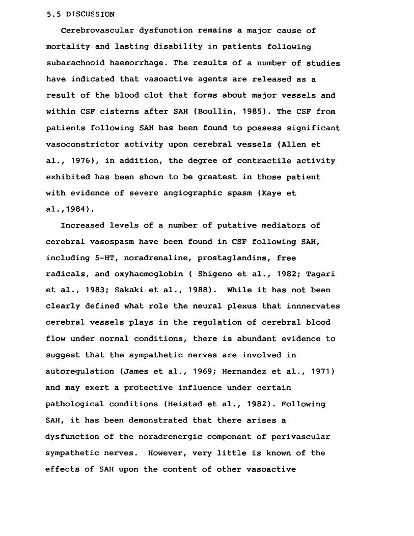
5.5 DISCUSSIONCerebrovascular dysfunction remains a major cause of
mortality and lasting disability in patients following subarachnoid haemorrhage. The results of a number of studies have indicated that vasoactive agents are released as a result of the blood clot that forms about major vessels and within CSF cisterns after SAH (Boullin, 1985). The CSF from patients following SAH has been found to possess significant vasoconstrictor activity upon cerebral vessels (Allen et al., 1976), in addition, the degree of contractile activity exhibited has been shown to be greatest in those patient with evidence of severe angiographic spasm (Kaye et al.,1984).
Increased levels of a number of putative mediators of cerebral vasospasm have been found in CSF following SAH, including 5-HT, noradrenaline, prostaglandins, free radicals, and oxyhaemoglobin ( Shigeno et al., 1982; Tagari et al., 1983; Sakaki et al., 1988). While it has not been clearly defined what role the neural plexus that innnervates cerebral vessels plays in the regulation of cerebral blood flow under normal conditions, there is abundant evidence to suggest that the sympathetic nerves are involved in autoregulation (James et al., 1969; Hernandez et al., 1971) and may exert a protective influence under certain pathological conditions (Heistad et al., 1982). Following SAH, it has been demonstrated that there arises a dysfunction of the noradrenergic component of perivascular sympathetic nerves. However, very little is known of the effects of SAH upon the content of other vasoactive
transmitters and neuromodulators such as NPY and ATP, which are contained also within sympathetic nerves. In addition, no studies to date have examined the consequences of SAH upon the 5-HT content of cerebrovascular sympathetic nerves, which are capable of an efficient uptake of 5-HT through their amine uptake mechanism (Levitt and Duckies, 1986). It is perhaps relevant that a recent study in the dog coronary artery following experimental coronary occlusion, with platelet aggregation, has demonstrated an accummulation of 5-HT into adrenergic nerves with subsequent adrenergic dysfunction (Cohen et al., 1987).
The results of this study clearly demonstrate that, following SAH, the 5-HT content of cerebral vessels increases, and that 5-HT is rapidly taken up via their amine uptake mechanism into sympathetic nerve terminals, from which it can be readily detected by specific immunofluorescence.
Other workers have previously demonstrated that SAH may transiently decrease the noradrenaline content of cerebrovascular sympathetic nerves (Edvinsson et al., 1982; Delgardo et al., 1985). A finding from this study is that the NPY content of sympathetic nerves is similarly decreased, by nearly 60%, in a transient manner following SAH. A smaller, 24% decrease in content following saline-injection was also noted, and may represent a stress- release response, as has been demonstrated to occur peripherally by other workers (Morris et al., 1986b).
It has been suggested that the observed decrease in catecholamine content in sympathetic terminals could be due
to an increased release of contained neurotransmitter and is in some way triggered by SAH. It is well known that 5-HT directly causes vasoconstriction of cerebral vessels by specific receptors (Chang and Owman, 1987). However, in addition, in higher concentration, 5-HT taken up into nerves in vitro can trigger, by a tyramine-like effect, the displacement release of noradrenaline from sympathetic nerves (Marin et al., 1981) and, presumably other, contained vasoactive agents such as NPY and ATP that are also displaced by tyramine (Cheng and Shen, 1987; Kirpatrick, personal communication) and that would then be free to act directly upon the vessel wall.
This study has demonstrated following experimental SAH in vivo in the rat, that 5-HT is indeed taken up into the perivascular sympathetic nerve endings of major cerebral vessels and that coincident with this there is a temporary decrease in NPY content. Whether these two phenomena represent a causal or casual relationship remains to be determined, but is yet further evidence of a transient marked dysfunction of cerebrovascular sympathetic nerves that arises post-SAH.
Fig. 8. Ventral view of brain from animals in which subarachnoid haemorrhage has been induced by the cisterna magna injection of 0.3 ml of autologous arterial blood.A: SAH induced 1h prior to killing showing extensive blood throughout the subarachnoid space and surrounding major cerebral vessels. B: SAH induced 3 days prior to killing showing the rapid resolution of subarachnoid blood clot in the rat.
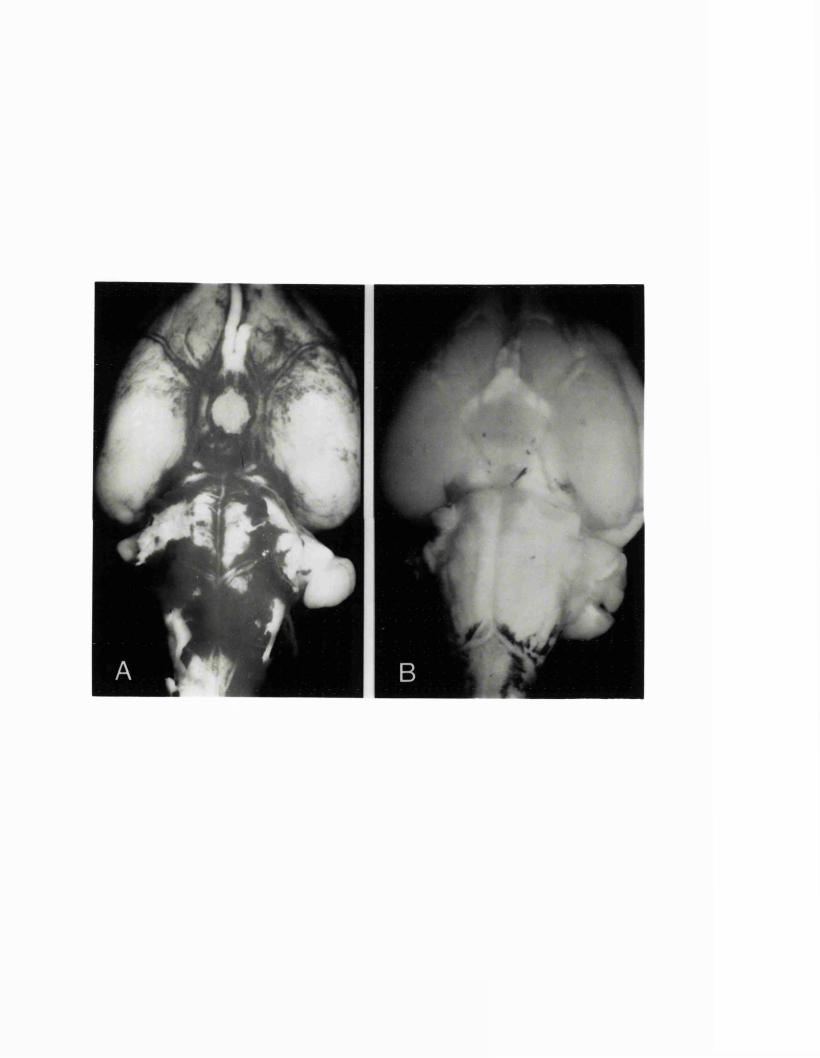
Fig. 9. Graphs showing changes in the mean densities of 5-hydroxytrytamine and neuropeptide Y-containing perivascular nerves of major cerebral vessels up to 5 days following SAH or control procedures. A: Following subarachnoid haemorrhage. B: Following injection of buffered saline in place of blood. C: Following a sham operation only.
ilf ★The vertical bars represent 2 SEM ( p <0.001, p <0.01).
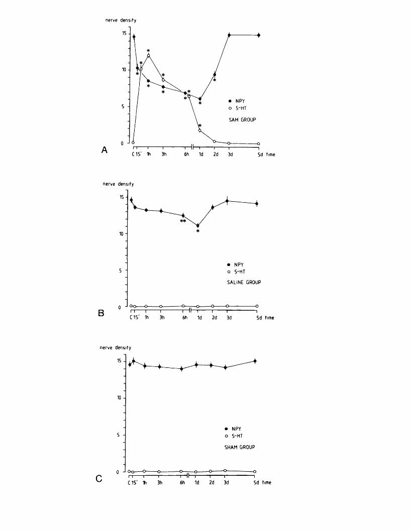
nerve density
15
10
NPY5-HT5
SAH GROUP
C15’ 1h 3h 6h 1d 2d 3d 5d time
nerve density
15
10
• NPY o 5-HT5
SALINE GROUP
0 J ° o -o- —o
C15’ 1h 3h 6h 1d 2d 3d 5d time
nerve density
15
10
• NPY o 5-HT5
SHAM GROUP
—o-0 -I oo o — o-
C15' 1h 6h 1d 2d 3d 5d time3h
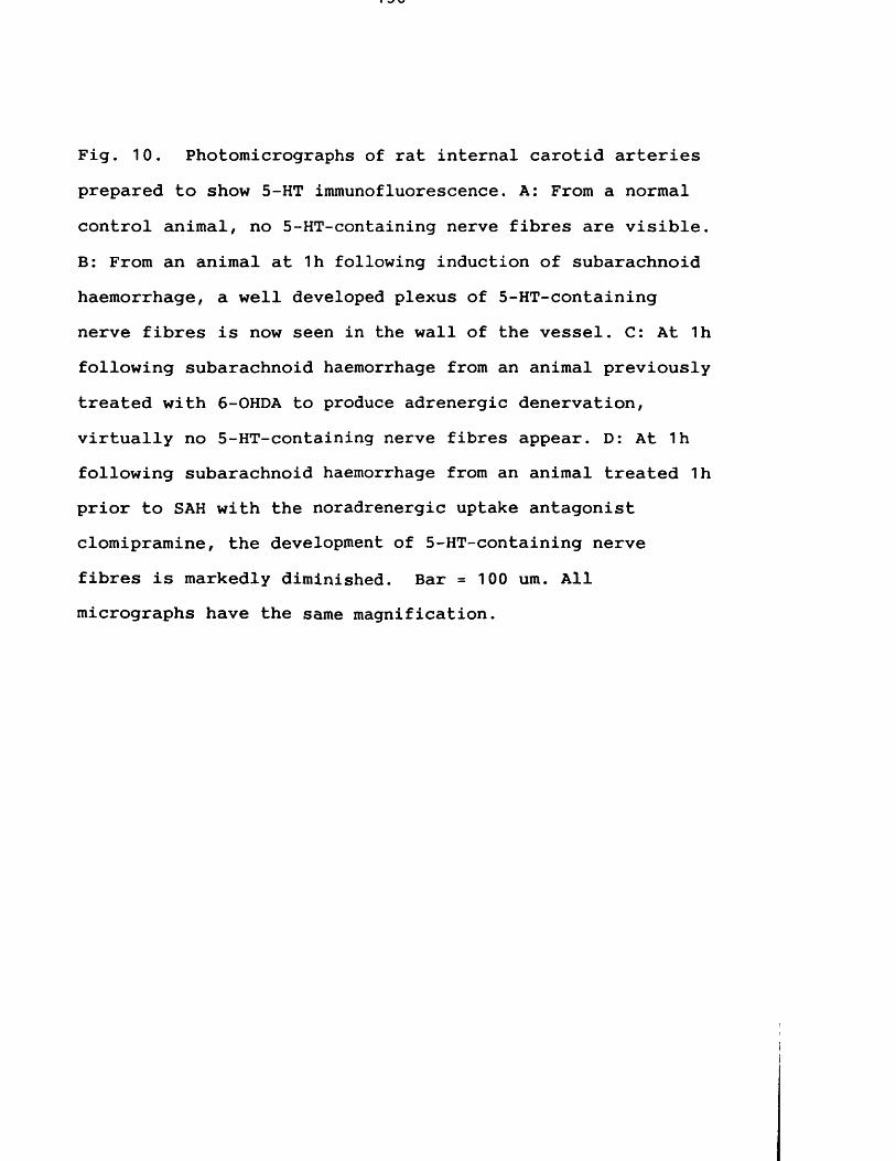
Fig. 10. Photomicrographs of rat internal carotid arteries prepared to show 5-HT immunofluorescence. A: From a normal control animal, no 5-HT-containing nerve fibres are visible. B: From an animal at 1h following induction of subarachnoid haemorrhage, a well developed plexus of 5-HT-containing nerve fibres is now seen in the wall of the vessel. C: At 1h following subarachnoid haemorrhage from an animal previously treated with 6-OHDA to produce adrenergic denervation, virtually no 5-HT-containing nerve fibres appear. D: At 1h following subarachnoid haemorrhage from an animal treated 1h prior to SAH with the noradrenergic uptake antagonist clomipramine, the development of 5-HT-containing nerve fibres is markedly diminished. Bar =100 urn. All micrographs have the same magnification.
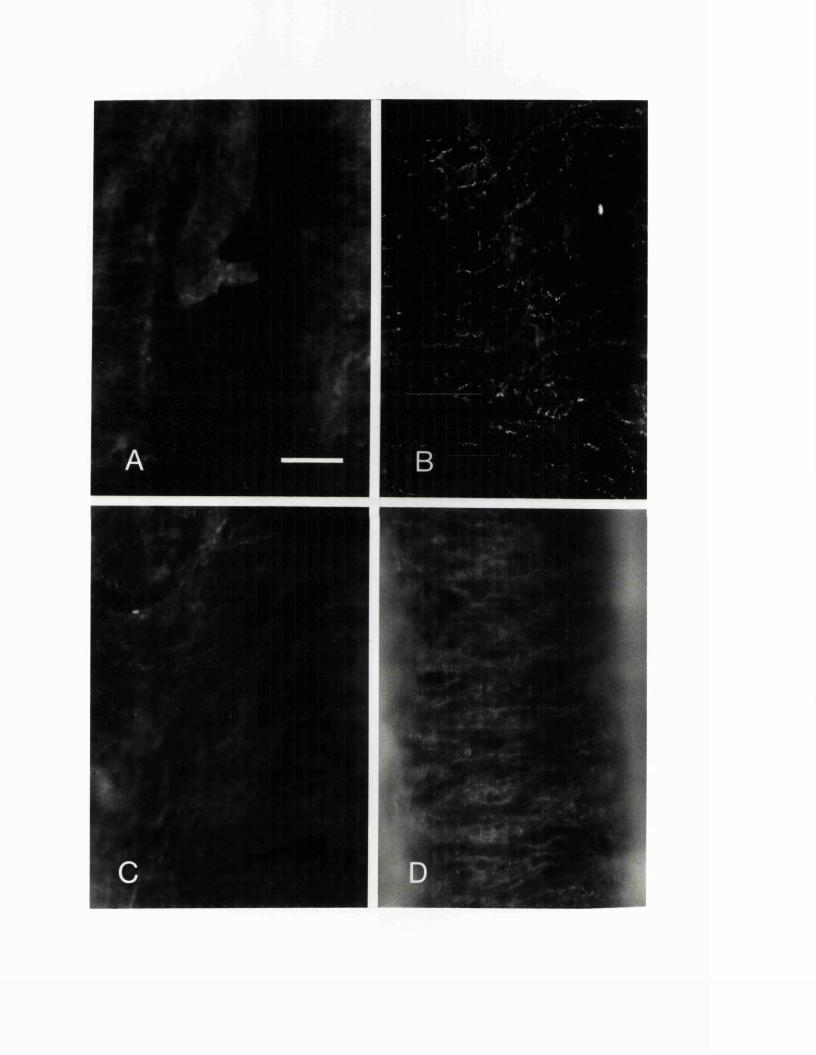
Fig. 11. Photomicrographs of rat internal carotid arteries prepared to show NPY immunofluorescence. A: From a normal control animal, a dense plexus of NPY-containing nerve fibre is always visible. B: From an animal at 1 day following induction of subarachnoid haemorrhage, a plexus of NPY- containing nerves is still apparent but this is markedly reduced both in its density and intensity. C and D: Following pretreatment of animals with 6-OHDA to produce adrenergic denervation, NPY-containing nerve fibres have virtually disappeared both in normal controls (C) and in animals at 1 day post-SAH (D). Bar = 1 0 0 urn. All micrographs have the same magnification.
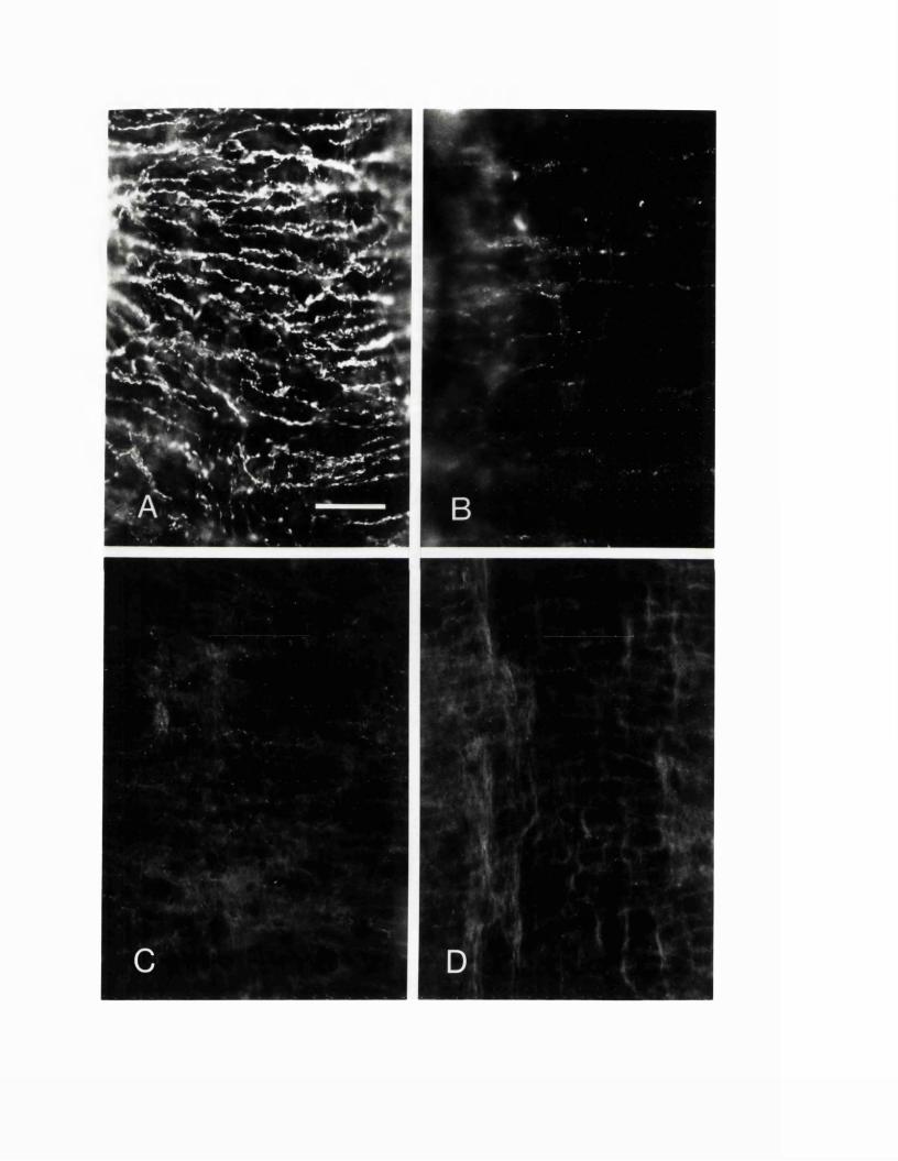
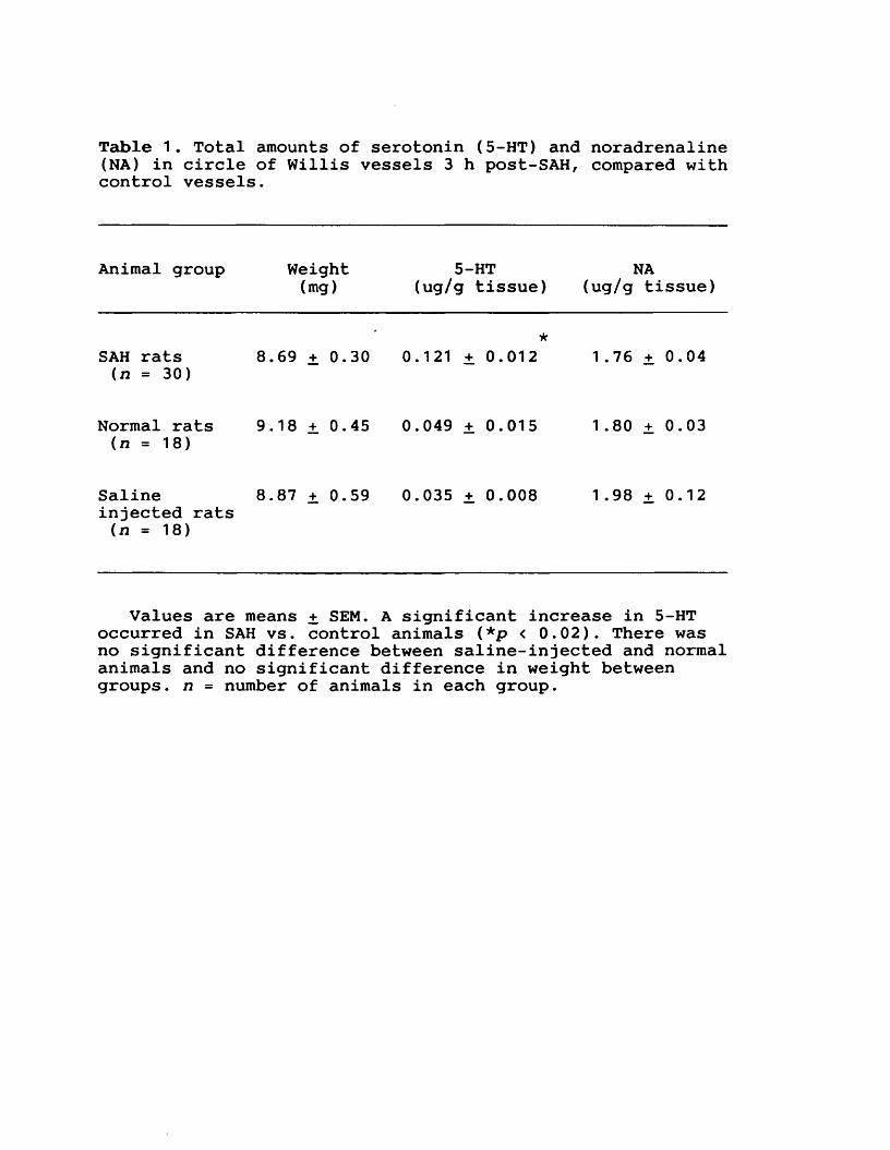
Table 1. Total amounts of serotonin (5-HT) and noradrenaline (NA) in circle of Willis vessels 3 h post-SAH, compared with control vessels.
Animal group Weight(mg)
5-HT (ug/g tissue)
NA(ug/g tissue)
SAH rats (n = 30)
8.69 + 0.30★
0.121 + 0.012 1.76 + 0.04
Normal rats (n = 18)
9.18 + 0.45 0.049 ± 0.015 1.80 ± 0.03
Salineinjected rats (n = 18)
8.87 + 0.59 0.035 + 0.008 1.98 + 0.12
Values are means + SEM. A significant increase in 5-HT occurred in SAH vs. control animals (*p < 0.02). There was no significant difference between saline-injected and normal animals and no significant difference in weight between groups, n = number of animals in each group.

6. EXPERIMENTAL CHAPTER IV:SERIAL DETERMINATIONS OF REGIONAL CEREBRAL BLOOD FLOW IN THE RAT USING SIMPLE CHRONICALLY IMPLANTED PLATINUM WIRE MICROELECTRODES
Summary: The ability to reliably obtain serial determinations of regional cerebral blood flow (rCBF) over time in small animals using simple and inexpensively constructed platinum-wire microelectrodes has been investigated. Repeated measurements of local cortical flow were obtained on a daily basis using the hydrogen clearance technique in a group of 18 animals, in each of which, six electrodes were chronically implanted. These studies have shown the simple microelectrodes utilised to give a low variability of results, as well as being well tolerated and provoking minimal tissue reaction even over prolonged periods of time. Under identical steady state conditions, serial determinations of rCBF were obtained for up to one week, with a maximum variation in mean values of only 12%, a result that compares favourably with the known serial determination error for the technique in the shorter term.
6.1 INTRODUCTIONMethodologies currently employed for the measurement of
cerebral blood flow (CBF) in small animals include theradioactive microspheres technique (Heymann et al., 1977),1 4C-Iodoantipyrine autoradiography (Sakurada et al., 1978), and hydrogen clearance (Auckland et al., 1964). Of these, the autoradiographic technique is unsuitable for repeated studies in the same animal since it requires tissue sections while the microspheres technique is limited by the total number of microspheres that can safely be injected into small animals (Solomon et al., 1985). The hydrogen clearance technique using cortically implanted platinum electrodes, has regularly been used for acute studies of cerebral ischaemia in small animals. It was desired however, to extend the technique to allow repeated determinations of CBF over longer periods of time to be obtained in the same animal.
Platinum microelectrodes have only infrequently been employed before for determining regional CBF (rCBF) in chronic experiments, usually in larger animals such as the cat (Rummer 1984), and no systematic investigation of the reliability of the hydrogen clearance technique in chronic studies has been reported. Although platinum is known to be well tolerated and to provoke minimal tissue reaction when implanted in the brain (Summerlee et al., 1982), it was not known whether tissue trauma over time would create a changing or excessive zone of damaged cells and tissue fluid around the electrode site. Such a zone could potentially create unstable electrical baselines and act as a diffusion
barrier, affecting the sensitivity and shape of the hydrogen clearance curves obtained, rendering the technique impractical for measurements over any prolonged period of time. This study was designed to answer these questions and to assess also the duration of functionally useful electrode survival times.
6.2 MATERIALS AND METHODS Construction of microelectrode assembly
The microelectrode used was constructed from a commerically available electronics printed-circuit board socket (30S/093/V Oxley Developments, Ulverston, Cumbria,UK) to the connector pin of which, a short length of 100 urn diameter teflon-coated platinum-iridium wire (Clarke Electromedical Instruments, Pangbourne, Reading, UK) was soldered. The pin and soldered join was then electrically insulated with quick setting epoxy-resin (RS Components, Corby, Northants, UK) applied to form a tear-drop shape (Fig. 12). When the resin had hardened the platinum-wire was shortened to a final length of 1.5mm, and the distal 0.5mm of the electrode bared of teflon insulation. Physiological preparations and measurement of CBF
A total of 24 male Sprague-Dawley rats weighing between 350-450 g were used in the experiments. Implantation and fixation of the electrode-assembly was performed in animals anaesthetised on a mixture of 70% nitrous oxide: 30% oxygen, with 1.5% halothane added and who were allowed to breathe spontaneously. In each animal, six electrodes were implanted as left and right pairs into parietal, occipital and cerebellar cortical regions, respectively. Following cleansing of the scalp with 70% alcohol, the superior aspect of the skull was exposed via a single midline sagittal incision and the skin edges drawn back. The pericranium was then excised and burr holes made over the specified cortical areas, with saline irrigation to avoid local heating. The electrode assembly, together with a small drop of
cyanoacrylate glue (RS Components) applied to the base of the epoxy-resin tear drop, was then inserted into the burr hole. By making the burr hole and the tear drop of similar diameters, immediate fixation followed contact of the epoxy-resin upon the burr hole shoulder. Each electrode was then firmly secured by running further epoxy-resin onto the tear drop and surrounding bare cortical bone. The scalp edges were approximated with sutures and a small scalpel blade nick made in the skin directly over each electrode to allow the connector sockets to protrude snugly through the scalp, enabling lead connections to be made via a matching male connector plug (30P/093 Oxley Developments). The reference electrode employed was constructed from 320 urn diameter Ag/AgCl wire passed during each set of recordings via a 21 gauge needle subcutaneously into the tissues on the back. CBF was measured at each electrode site by the hydrogen clearance technique as previously detailed (Auckland et al., 1964; Pasztor et al., 1973; Young 1980 ). Hydrogen gas was administered in the inhaled anaesthetic gases to a final concentration of approximately 7% and was given via a parallel circuit to ensure that inspired concentrations fell immediately to zero at onset of desaturation. Hydrogen inhalation times were kept at a constant 3 min for all flow measurements which, for the rates of flow obtained in our animals, should result in a more than 90% saturation and subsequent desaturation of tissues within this time period (Farrar, 1987). CBF was then calculated from the resultant clearance curves using the initial slope method (Olesen et al., 1971; Doyle et al.,
1975) between 30 and 120 sec.For flow recordings, animals were induced and initially
anaesthetised on a mixture of 70% nitrous oxide : 30% oxygen together with 1.5% halothane. After being allowed to settle on this mixture for 30 min, the halothane was then reduced to 0.75% for a further 30 min before flow recordings were undertaken. The electrical apparatus together with the polarising voltage (+400 mv) was turned on for 30 min before recordings, to allow electrode stabilisation to steady baseline values to take place. Three hydrogen clearance recordings were then made separated by 10 min intervals, the CBF being calculated from the final curve obtained. Satisfactory electrode function was defined by: the attainment of a stable electrical baseline at 30 min following the application of the polarising voltage, electrode sensitivity remaining constant over the period of three clearance recordings, and finally, the formation of clearance curves of typical shape and appearance. Only flow recordings from electrodes that fully satisfied these criteria were utilised for the serial determination of cortical flows. Recordings were made for up to 10 days following implantation; the CBF being calculated then expressed as a percentage of each animals initial CBF value, obtained 2h following electrode implantation on day 0.
Histological analysesIn six rats, electrodes were implanted as previously
described and the animals sacrificed subsequently for the microscopic localisation and assessment of electrode tracks at 2h, 1day, 3days, 5days, 7days and 1Odays
postimplantation. Under deep inhalational anaesthesia, animals were perfused through the left ventricle with approximately 150 ml of fixative composed of 4% paraformaldehyde in 0.1M phosphate buffered saline. The overlying skull with microelectrodes still attached was carefully removed and the brains immersed in the same fixative overnight at 4 °C. The brains were then rinsed in 0.1M phosphate buffered saline and processed for embedding in paraffin wax. Sections 7um thick were cut and stained with Haematoxylin and Eosin for viewing by light microscopy. Using a calibrated eyepiece, the diameter of the electrode track, distance and numbers of neurons, reactive glia and macrophages from the electrode track was assessed.
6.3 RESULTS Histological findings
There was only a minimal immediate tissue reaction provoked by the insertion and presence of the platinum-wire microelectrodes; this did not appear to change appreciably with the subsequent passage of time. The resultant track cavity was largely similar in diameter to that of the platinum wire used (100 urn) (Fig. 13). At 2h following implantation, the track boundaries consisted of the brain parenchyma itself together with a small number of extravasated erythrocytes. By the third day postimplantation, fibroblasts appeared in the superficial portion of the track and by seven days, a smooth fibroblast lining in continuity with the overlying meninges had formed. Immediately surrounding the microelectrode track was an annulus of approximately 125 urn in which a mild microglial reaction occurred. Neurons of normal appearance were usually present at a distance of some 50 um from the track boundary.
Electrode toleranceAnimals appeared to tolerate the electrodes well, feeding
and behaving normally. No cases of infection were seen at any electrode site and there were no instances of detectable loosening of the microelectrode assembly up to seven days postimplantation. After this time however, some instances did occur, and after ten days, 24% of electrodes showed evidence of loosening in their attachment to the skull.
Functional electrode survival times108 electrodes were implanted in 18 animals for the
serial determination of regional cortical flow by the hydrogen clearance technique. Out of the total implanted, some 103 microelectrodes (95%) fulfilled our criteria for acceptable electrode function, producing satisfactory hydrogen clearance curves on day 0 at 2h postimplantation. The subsequent time course for continued satisfactory electrode function is detailed in Fig. 14. These results indicate that for periods of study of up to five days, when over 60% of implanted microelectrodes functioned satisfactorily, that a serial assessment of CBF during this time at least is practicable. After five days the numbers of satisfactory electrodes declined more rapidly, with less than 20% of microelectrodes continuing to function satisfactorily into the second week.
CBF variabilityThe mean + SEM values of regional blood flow obtained 2 h
postimplantation were 1 4 8 + 4 ml/100g/min (parietal cortex), 1 5 8 + 5 ml/100g/min (occipital cortex), and 9 3 + 2
ml/100g/min (cerebellar cortex), figures in general agreement with values obtained previously in the rat (Cremer and Seville, 1983; Kangstrom et al. 1983). Subsequent serial determinations of flow under identical steady-state conditions demonstrated that repeated blood flow measurements could be obtained over periods of several days with, in some individual cases, quite remarkably low variability (Fig. 15). The overall results for regional flows measured at increasing times up to ten days from

microelectrode implantation, is detailed in Fig. 16. The results demonstrate, that for periods up to seven days following microelectrode implantation, that it is possible under the same steady-state conditions to obtain mean values of regional cortical flow that vary by a maximum of only 12% from those values obtained shortly after implantation.
6.5 DISCUSSIONrCBF changes may develop immediately, or evolve following
a delay, after a widely varied number of pathophysiological events. Study of such changes for periods greater than a few hours in small animals such as the rat, has generally necessitated the use of a number of matched groups of animals at multiple time points to acquire the same data as would have been obtained by serial flow determinations over time in a single group of animals. The usage of simple chronically implanted platinum-wire microelectrodes as we describe, allows just such a serial determination of CBF to be readily performed. As well as allowing the use of smaller animal numbers to achieve the same data, such a technique should also reduce variability due to normal interanimal flow differences, as the flow data in each individual animal may now be expressed as a percentage of its own control value.
Such theoretical advantages can only be realised, however, if the serial determination of CBF by this method can be shown to be both practical, and capable of achieving a similar degree of accuracy over periods time, as has been demonstrated already in the shorter term. The hydrogen clearance technique has been shown over short term observations to have a serial determination error of up to 10%, other conditions being equal (Pasztor et al., 1973). In this study it has been possible under identical steady-state conditions to achieve serial determinations of regional cortical flow for up to one week with no more than a 12% variation in the mean flow values obtained.

It is felt therefore, that the serial assessment of CBF in small animals using simple platinum-wire microelectrodes is both practical and for periods of study up to one week, possesses a margin of experimental error not dissimilar from that for the technique in the shorter term.
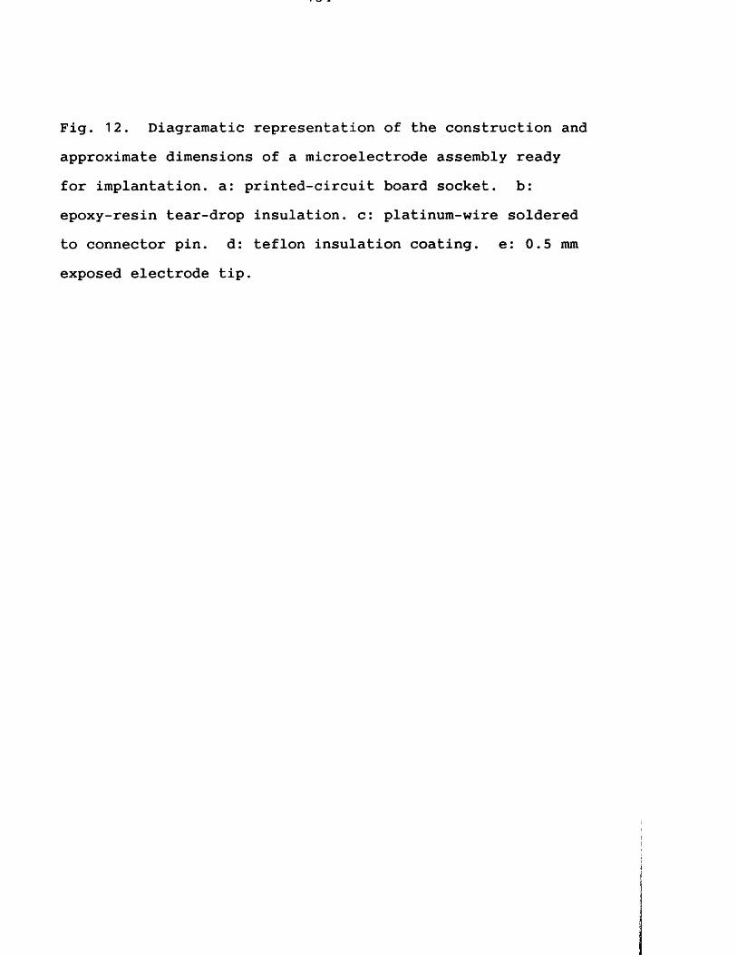
Fig. 12. Diagramatic representation of the construction and approximate dimensions of a microelectrode assembly ready for implantation, a: printed-circuit board socket. b: epoxy-resin tear-drop insulation, c: platinum-wire soldered to connector pin. d: teflon insulation coating. e: 0.5 mm exposed electrode tip.
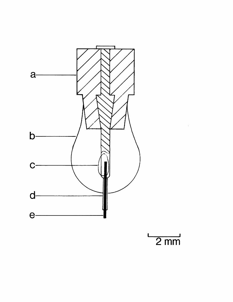
I____________I2 mm
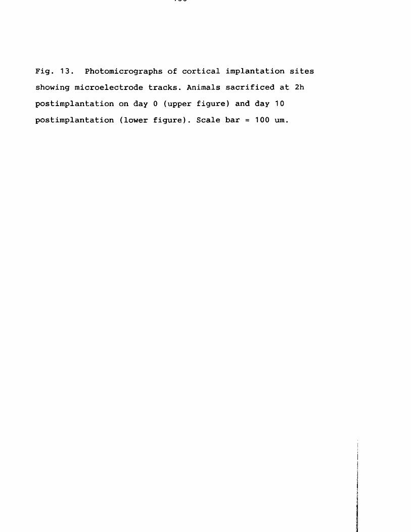
Fig. 13. Photomicrographs of cortical implantation sites showing microelectrode tracks. Animals sacrificed at 2h postimplantation on day 0 (upper figure) and day 10 postimplantation (lower figure). Scale bar = 100 urn.
o'- *#
4£ * •
/ * #*' #
• * * ;^▲ I h ^< ;i f
4 / 1.
t*'/ ♦ - v - ** \iS. v " . i*“ tl \ ’ t *r ** % M < **V
. * »•/*{‘ »* * *-• »: ’ • ‘;. #• • i t '
J ' i L rV r | . /if f • « 1
jr:*f t * * m
b I
r ’ *. • ’ .* 1 * *
*# . % >
M U P ,
\«*£j|Vt »V. - *r ir ' ..Tw* 1 ' >« * . ^ A V / . X *
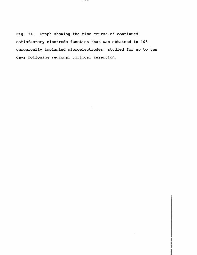
Fig. 14. Graph showing the time course of continued satisfactory electrode function that was obtained in 108 chronically implanted microelectrodes, studied for up to ten days following regional cortical insertion.
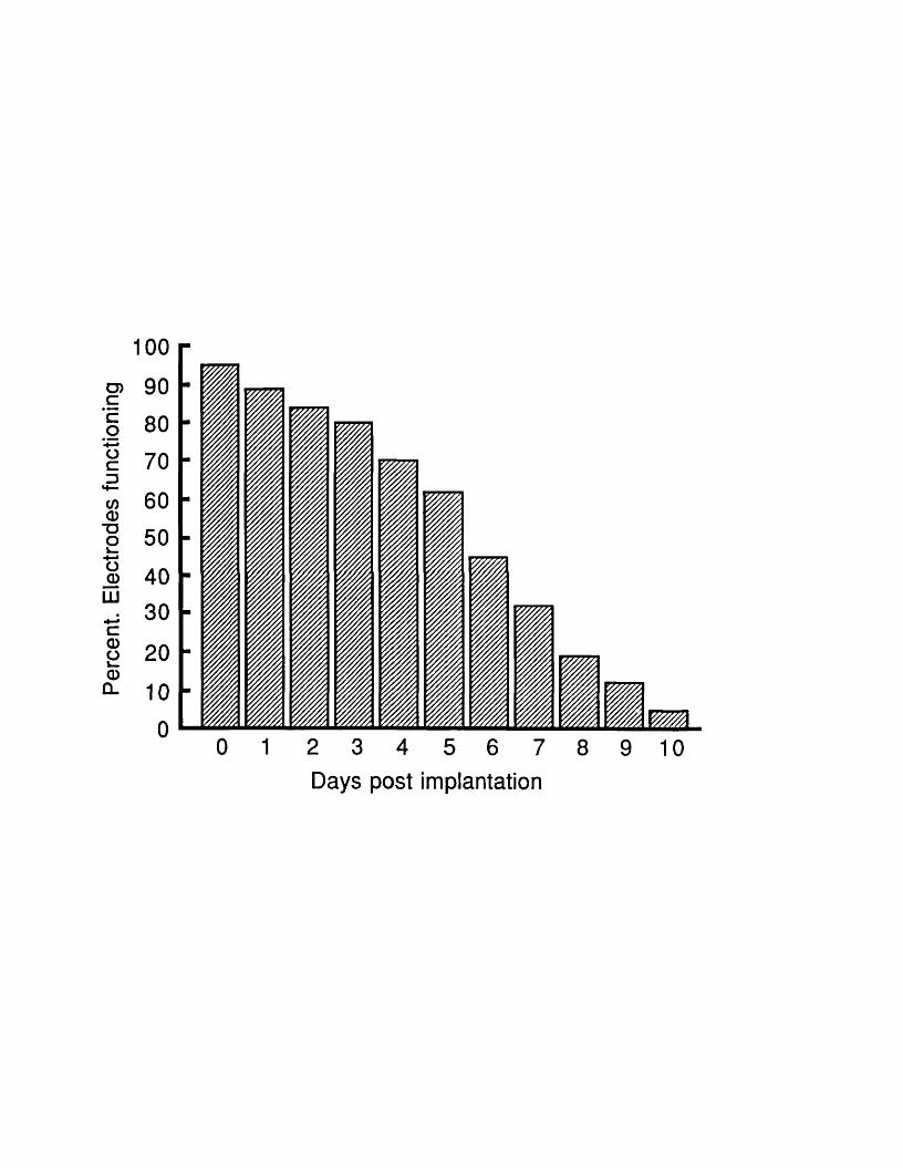
Perc
ent.
Elec
trode
s fu
nctio
ning
100
Days post implantation
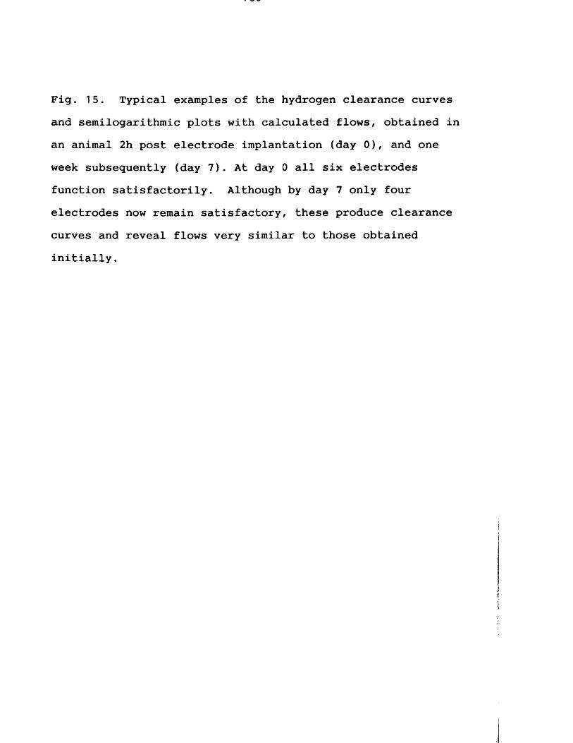
Fig. 15. Typical examples of the hydrogen clearance curves and semilogarithmic plots with calculated flows, obtained in an animal 2h post electrode implantation (day 0), and one week subsequently (day 7). At day 0 all six electrodes function satisfactorily. Although by day 7 only four electrodes now remain satisfactory, these produce clearance curves and reveal flows very similar to those obtained initially.
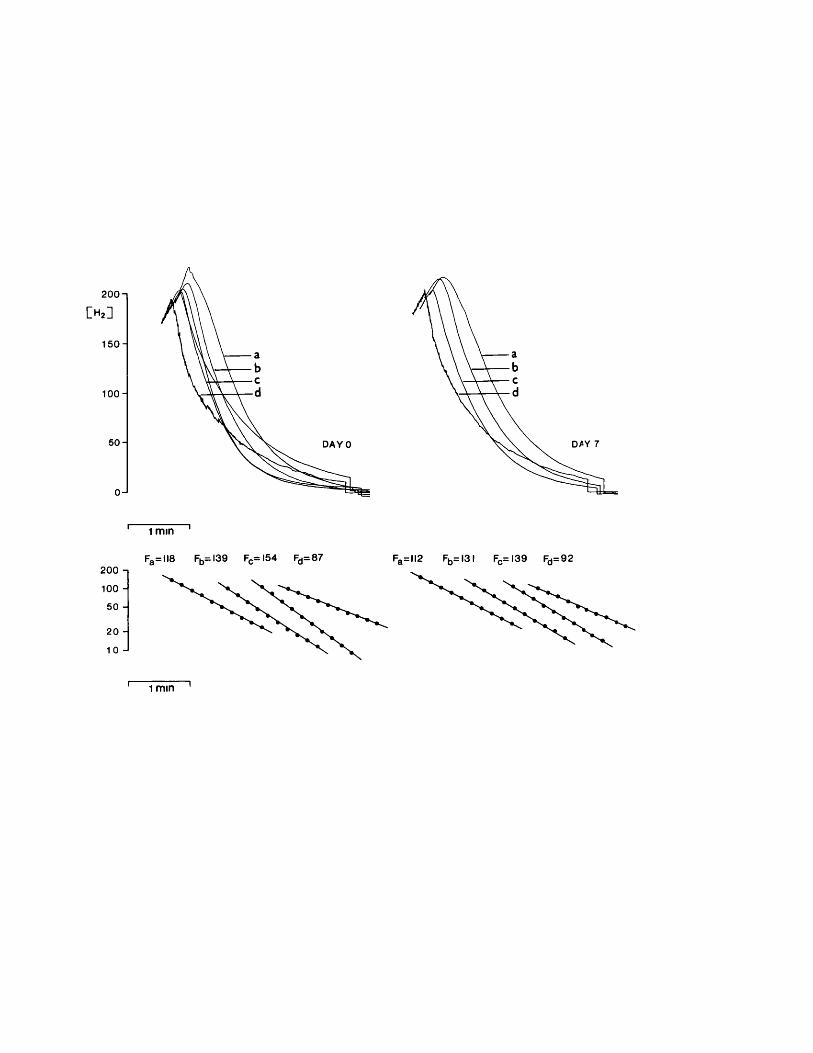
200-1
M150-1
100 H
50 H
0J
DAYO
1 m m
Fa= ll8 Fb= l39 FC= I5 4 Fd= 87 Fa= ll2 Fb= 131 Fc= I39 Fd=92
1 m in
Fig. 16. Regional cortical blood flows measured serially under identical steady-state conditions, plotted for up to 10 days after microelectrode implantation. Flows are expressed as a percentage of 2h postimplantation control blood flow value. ( — •— , parietal cortex; — , occipital cortex; — O — , cerebellar cortex). Values are means + SEM.
Perc
ent,
of co
ntro
l flo
w
125120115110105100
959085
750 1 2 3 4 5 6 7 8 9 10
Days post implantation

7. EXPERIMENTAL CHAPTER V:SUBARACHNOID HAEMORRHAGE IN THE RAT MODEL: THE TIME COURSE OF ALTERED CEREBRAL BLOOD FLOW, CEREBRAL PERFUSION AND INTRACRANIAL PRESSURE
Summary: The rat subarachnoid haemorrhage (SAH) model was studied in order to establish the precise time course of the globally reduced cerebral blood flow (CBF) that follows, and to ascertain whether related concomitant temporal changes in cerebral perfusion pressure (CPP) and intracranial pressure (ICP) take place. Studies were carried out upon spontaneously breathing animals under inhalational anaesthesia. SAH was induced in animals by a single intracisternal injection of 0.3 ml of blood; controls received either 0.3 ml of buffered saline in place of blood or underwent sham procedures only. Serial measurements of regional cortical cerebral blood flow were obtained by the hydrogen clearance technique, both acutely and for up to 5 days subsequently. Experimental SAH in the rat resulted in an immediate 50% global reduction in cortical flows that persisted for up to 3 h post-SAH. At 24 h, flows were still significantly reduced at 85% of their control values (p< 0.05), but by 48 h, flows had regained their normal values and these were maintained up to 5 days post-SAH. ICP rose acutely after haemorrhage to nearly 50 mmHg with C-type pressure-waves being present. ICP then fell slowly, only fully returning to control levels at 72 h. Acute hydrocephalus was observed in autopsy studies of SAH animals but not in controls. Reductions in CPP occurred post-SAH, but only in the order of 15%, which could not alone account for the fall in CBF that took place. These findings indicate that this increasingly utilised small animal model of SAH develops only acute global flow changes, and it is likely that early vasospasm, secondary to released blood products

rather than pressure changes per-se, is responsible for the initial cerebral ischaemia that develops.
7.1 INTRODUCTIONSubarachnoid haemorrhage (SAH) resulting from the rupture
of an intracranial aneurysm in man, may lead to decreased cerebral blood flow (CBF), raised intracranial pressure (ICP) with hydrocephalus and the development of prolonged cerebral arterial spasm. Considerable evidence points to cerebral ischaemia resulting from reduced cerebral perfusion as the major cause of delayed neurological deterioration following SAH (Heilbrun et al., 1972; Ischii, 1979;Kutsuzawa et al., 1983; Voldby et al., 1985b), the peak incidence in man being around seven to ten days after a bleed (Pickard et al., 1989). Cerebral arterial spasm is one factor that has been significantly associated with a reduced CBF, and the presence of vasospasm is also known to be related both to the patient's clinical grade and eventual outcome (Loach and De Azevedo-Filho, 1976; Weir et al., 1978). It is likely however that the cause of the cerebral ischaemia that occurs post-SAH is multifactorial in origin, delayed hypoperfusion having been found to be associated not only with angiographically demonstrated luminal narrowing of vessels but also with the development of episodes of hypovolaemia, raised intraventricular pressure and ventricular dilatation (Ferguson et al., 1972; Ischii, 1979; Wijdicks et al., 1985).
Increasingly, in place of larger animal models such as the dog, cat, and monkey (Brawley et al., 1968; Kuwayama et al., 1972; Petruck et al., 1972; Umansky et al., 1984), the laboratory rat has been employed for experimental studies of SAH (Barry et al., 1979; Lacy and Earle, 1982; Solomon et
al., 1985; Svendgaard et al., 1985). Studies in the rat have demonstrated acute reductions in CBF, a biphasic pattern of vasospasm, and changes in cerebral metabolic activity and perivascular neurotransmitter content, consequent upon the release of homologous arterial blood into the subarachnoid spaces. Surprisingly however, little information exists regarding the subsequent time course of the globally reduced CBF that develops acutely post-SAH in the rat. Similarly, few details are known regarding the temporal development of changes in intracranial and cerebral perfusion pressure (CPP), and their interrelationship with reduced CBF following experimental SAH in the rat model. This study was designed therefore, to precisely determine the exact time course of such changes in the CBF occurring both within minutes and up to several days following the event, together with the obtaining of serial measurements of ICP and CPP over the same period of time.
7.2 MATERIALS AND METHODS Animal Preparations
Experiments were performed on 90 male Sprague-Dawley rats weighing between 350 and 450 g. Animals were allowed to breathe spontaneously on an anaesthetic mixture consisting of 70% nitrous oxide : 30% oxygen, with 1.5% halothane added during operative procedures; reduced to 0.75% halothane 30 min before cisternal injections or measurements of CBF were performed. The left femoral artery was cannulated; arterial blood pressure was monitored with a Statham P50 pressure transducer (Statham Instruments Inc., Oxnard, California) and was recorded on a Lectromed MX6 chart recorder (Ormed Ltd., Welwyn Garden City, U.K.). Arterial blood samples were obtained anaerobically in microhaematocrit tubes (0.1 ml), and systemic P C0o, P_00 and pH determined using an ABL
^ cl ^
30 blood-gas analyser (Radiometer, Copenhagen, Denmark). The haematocrit was measured. Body temperature was maintained at 37 °C by external warming.
Induction of Experimental SAHSAH was induced in 30 rats by a posterior craniocervical
approach using a modification of a technique described previously (Solomon et al., 1985). Briefly, utilising the operating microscope the posterior cervical musculature was separated in the midline, exposing the arch of the atlas, occipital bone and atlanto-occipital membrane. For production of haemorrhage, 0.3 ml of arterial blood was withdrawn by syringe from the femoral arterial line and replaced with an equal volume of normal saline. The freshly withdrawn blood was then injected into the subarachnoid
space via a 30 gauge needle passed into the cisterna magna. Injections were given as six equal aliquots over a period of 3 to 4 minutes. Control groups comprised: one group of 30 rats that received an intracisternal injection of 0.3 ml buffered saline (normal saline with 25 meq/1 NaHCO^ equilibrated at 37 °C with 5% CO 2 : 95% air to pH 7.4), and a second sham-operated group of 30 rats that underwent identical operative procedures including cisternal needle puncture to the other two groups but in which no injections were given.
CBF MeasurementsRegional cortical grey matter CBF (rCBF) was measured by
the hydrogen clearance technique (Auckland et al., 1964; Young, 1980) using a chronically implanted platinum-wire microelectrode array (Jackowski et al., 1989c). In six animals from each treatment group, three pairs of microelectrodes were inserted into the parietal, occipital and cerebellar cortical regions of both left and right-hand hemispheres. In each animal the rCBF was determined pre-injection, after 3, 15, 30, 60, 120, 180 min and 1 to 5 days following SAH, saline or sham operation. The values of rCBF were calculated from the hydrogen clearance curves obtained, using the initial slope method (Oleson et al., 1971; Doyle et al., 1975) between 30 and 120 sec. The regional flows derived were then expressed as a percentage of each animal1s control rCBF value obtained prior to injection on day 0.
ICP CPP and Other MeasurementsICP was measured via a saline-filled 30 gauge cannula
placed into the cisterna magna and connected to a Statham P50 pressure transducer. Data values for ICP, CPP, MABP, haematocrit and blood gases for the times immmediately post-SAH up to 3 h post event were acquired in the same animals as were employed also for the serial assessment of rCBF. Studies of these same parameters at the time points 1 to 5 days post-SAH, were performed in separate groups of animals, each group consisting of six animals, studied at those specified time points alone.
Statistical AnalysisAll values are given as the mean + SEM unless otherwise
stated. Statistical comparison between experimental groups at different time points was performed using a two-tailed t test, p < 0.05 was taken as the level of significance.
7.3 RESULTS rCBF Changes
Regional cortical flow changes were analysed separately for parietal, occipital, and cerebellar areas. The alterations in flow observed however were essentially global, affecting all cortical areas to the same degree (Fig. 17). The regional flow data from each animal have therefore been combined to give a mean cortical flow value that was used for further comparisons and in the statistical analysis between treatment groups. The mean cortical flow changes following experimental SAH, saline-injection, or sham-operation are summarised in Table 2. Following experimental SAH there was an immediate and marked reduction in flow, the overall CBF falling by 15 minutes to 50 + 2% ofpre-injection values (p < 0.001). This reduction persistedup to 3 h post-haemorrhage. At 24 h, flow was just significantly reduced still at 85 + 7% of control values (p < 0.05). Pre-haemorrhage values were regained on the second day and remained normal up to 5 days subsequently. The mean cortical flow change in animals receiving cisternal saline-injection was similar to that obtained in sham-operated animals. Neither underwent rapid or marked changes in flow; in both groups after 30 minutes of the acute study there was a gradual reduction in the mean cortical flow reaching 80-85% of pre-haemorrhage values at 3 h. In both groups of control animals, the mean cortical flow had fully recovered to pre-haemorrhage values by 24 h.
ICP ChangesThe changes in ICP differed markedly in animals that
underwent experimental SAH, from those that received cisternal saline-injection or underwent sham procedures. Differences in response occurred both in the period during and acutely following injection as well as in the time course of subsequent recovery. Administration into the subarachnoid space of 0.3 ml of blood as six aliquots was accompanied by a step-like rise in ICP that reached a maximum value of approximately 50 mmHg and only slowly declined upon completion of the injection (Fig. 18). During the period of acutely raised ICP, pressure-waves were commonly observed, these occurred at a frequency of about 8 per minute and were usually closely related to similar variations in the arterial pressure. Also during this period, an increased cerebrospinal fluid pulse pressure was often evident. In those animals in which 0.3 ml of buffered saline was administered in place of blood, the ICP attained roughly the same immediate peak values, but fell rapidly towards control values both between individual aliquots and upon final completion of injection. The subsequent time course of ICP recovery differed also between the groups (Fig. 19). Following experimental SAH the ICP remained elevated for up to 3 h at a pressure of 16 mmHg compared with a normal value of only 4 mmHg in the rat. At 24 h the ICP was still mildly elevated at 8 mmHg, and it only fully returned to control levels after 48 h. In the saline-injected animals the ICP normalised far more rapidly, falling to 8 mmHg by 15 minutes and returning fully to
control values by 2 h. There were no significant changes in the ICP of sham-operated animals. No formal analysis of ventricular volumes was performed in this study, however on sectioning the brains of animals used in delayed ICP studies it was observed that hydrocephalus occurred in animals 24 h post-SAH, but not following control procedures.
MABP and CPP Changes (Fig. 19)In both the SAH and saline-injected animals there was a
rapid rise in MABP following the injection of an 0.3 ml volume load into the subarachnoid space via the cisterna magna. The pressure reached a mean peak value of around 125 mmHg then fell slowly after 3 minutes, recovering to normal values by 3 h in both groups of animals.
CPP fell immediately by approximately 20 % following either SAH or saline-injection as the rise in MABP produced failed to match the peak ICP that resulted. In the SAH animals, CPP then recovered to normal within 3 minutes only to fall again by 15 % at 1 to 3 h post-SAH as ICP remained elevated in the presence of a now normal MABP. In the saline-injected animals, as ICP returned rapidly to normal values, the CPP after its initial fall became elevated by 20 % only to then fall in parallel with the recovery in MABP.In both SAH and saline-injected animals the MABP and CPP were maintained subsequently at normal levels from 24 h onwards. There were no significant changes in the MABP or CPP of sham-operated animals.
Other Physiological ParamatersThe blood gases and haematocrit for the experimental
groups at different times are given in Table 3. These parameters remained largely constant and approximate values were as follows: the haematocrit 45 %, the P CO, 37 mmHg,
cL 4
the P_0o 139 mmHg and the pH 7.4. There were no significanta £differences in these values between the experimental groups.
7.5 DISCUSSIONA significant global reduction in CBF during the period
acutely following experimental SAH has been demonstrated in animal studies performed in the monkey, dog, cat (Petruck et al., 1972; Brawley et al., 1968; Umansky et al., 1983)' and more recently in the rat (Solomon et al., 1985). However details of the subsequent time course to recovery of such alterations in CBF, and of any subsequent development of delayed ischaemic changes have been described far less commonly. In the monkey, persistent reductions in CBF have been demonstrated up to 3 days post-SAH (Jakubowski et al., 1982; Sahlin et al., 1987), while in the rat a rather uniform 20% reduction in CBF has been reported to occur at 2 days post-haemorrhage (Delgado et al., 1986). The latter authors, when previously reporting their angiographic findings following experimental SAH in the rat, described a biphasic pattern of vasospasm with a maximum acute spasm at 10 minutes and a maximum late spasm at 48 h. By contrast, another earlier study (Barry et al., 1979) documented only an immediate onset of spasm which recovered by day 3 and was followed by vasodilatation from day 5 onwards. The rat has also been used to study delayed changes in cerebral metabolic activity following experimental SAH, one group (Soloman et al., 1987) reporting a significant decrease in metabolic activity of < 34% at 48 h while conversely another found a 30% increase (Delgado et al., 1986).
The measurements of CBF obtained in our study clearly demonstrate the occurrence of an acute decrease in mean cortical flow of approximately 50% for up to 3 h following
haemorrhage. A smaller, but still statistically significant, reduction in CBF persisted up to 24 h post-SAH. However, at 48 h during the period when a second lesser phase of vasospasm has been described, the mean cortical CBF was now fully recovered to normal values. Such a lack of absolute correlation between the time course of a reduced CBF and the previously described angiographic changes in the rat is not altogether surprising. Studies upon SAH in humans have shown the precise relationship of vasospasm to impaired CBF to be uncertain. Many investigators have found the presence of cerebral vasospasm in patients, to be associated with a reduced CBF both as a global and focal phenomena (Heilbrun et al., 1972; Ischii, 1979; Voldby et al., 1985b). Other authors however, have shown a less well-defined relationship (Bergvall et al., 1973; Grubb et al., 1977; Mickey et al., 1984).
Several patient studies have also shown a relationship post-SAH between hydrocephalus, raised ICP and reduced rCBF (Ferguson and Harper, 1971; Hartmann et al., 1977; Ischii, 1979). Similarly, hydrocephalus and vasospasm following SAH have been found to be significantly associated (Black, 1986), and the therapeutic benefits from ventricular drainage in cases of raised ICP, upon CBF and clinical grade have been well described (Kusske et al., 1973; Suzuki et al., 1974; Hartmann, 1980). A finding from this experimental chapter has been to emphasise the differences in ICP response following the administration of either blood or saline into the rat cisterna magna. Injection of 0.3 ml saline caused only a short-lived elevation of ICP to a peak
of approximately 50 mmHg. In contrast, an equal volume of released blood produced the same peak ICP, but the pressure then fell only slowly; remaining elevated four-fold at 3 h, approximately twice normal at 24 h and recovering to normal at 72 h. Concomitant with these changes in ICP, pressure-wave phenomena were observed following SAH, but not after control procedures. These waves most closely resembled Traube-Hering-Mayer related "C" waves, were seen only when the pulse-pressure amplitude was increased in the presence of an elevated ICP and are almost certainly evidence of a greatly reduced intracranial compliance post-SAH.
The changes in CBF that were observed following experimental SAH in the rat cannot simply be explained on the basis of concurrent changes in ICP and CPP alone. In the saline-injected control animals, despite 20% fluctuations in CPP, the CBF remained relatively constant, while in the SAH animals the maximum sustained reduction in CPP was 15%, which, even in the presence of impaired autoregulation, would not alone account for the 50% decline in CBF that took place.
The time course of reduced CBF in the rat does correlate well however with previously made observations (Barry et al., 1979; Jackowski et al., 1989b) that subarachnoid-released blood diminishes and is largely resorbed within 48 h in the rat. This suggests that the reduction in cerebral flow that occurs is closely related to the actual presence of blood and blood breakdown-products within the perivascular CSF spaces themselves. Acting either
directly upon the cerebral vessel wall or perhaps more indirectly via perivascular nerves and central brainstem afferent connections (Svendgaard et al., 1985; Solomon et al., 1987; Jackowski et al., 1989b) to produce an early phase of vasospasm. Although from this study it now appears that significant delayed cerebral ischaemia fails to develop in the rat after haemorrhage, this model still remains a cheap and readily reproducible one, demonstrating many of the pathophysiological changes that occur shortly following SAH in man. Use of the rat for further investigative studies upon the mechanisms arising out of, and treatment of SAH, would appear to be relevent provided the observed differences in the CBF response compared to that obtained in the clinical situation remains recognised.
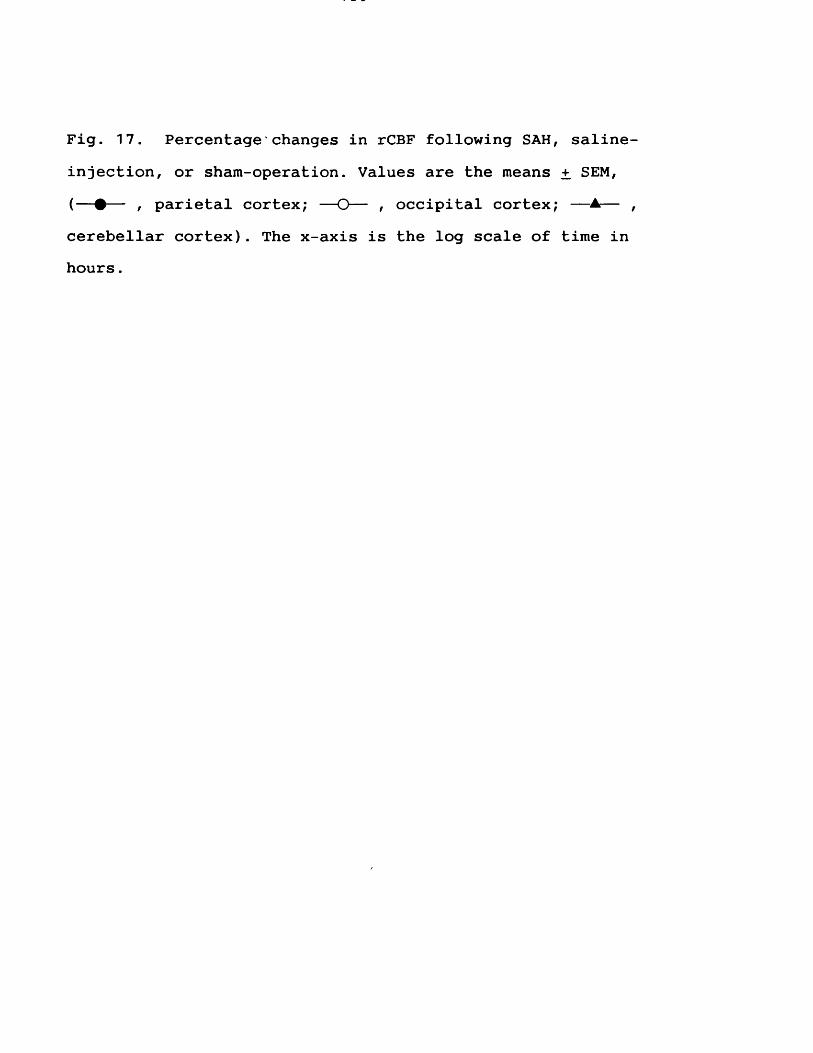
Fig. 17. Percentage * changes in rCBF following SAH, saline injection, or sham-operation. Values are the means + SEM,(— #— , parietal cortex; — O— , occipital cortex; — ▲—
cerebellar cortex). The x-axis is the log scale of time in hours.
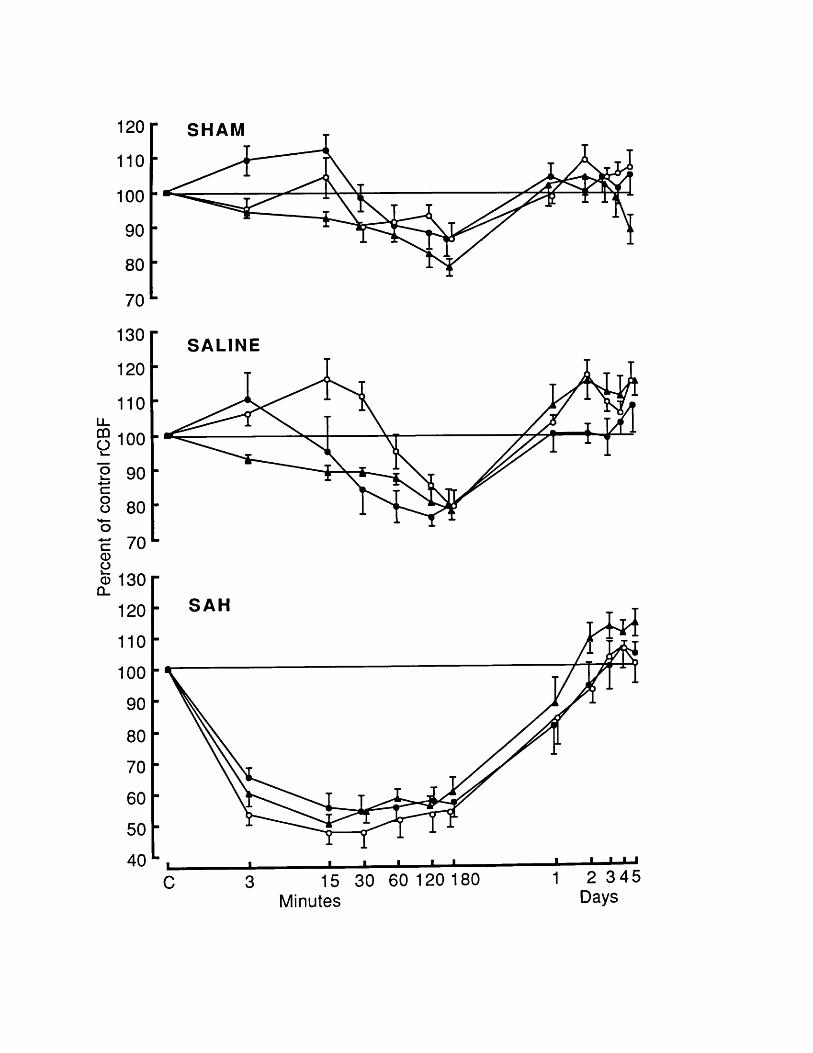
120
110
100
90
80
70
130
120
110
g 1002 90■M
8 80O£ 70<1)© 130Q_
120
110
10090
80
70
60
50
40
SHAM
SALINE
SAH
2 3 4 5 DaysMinutes
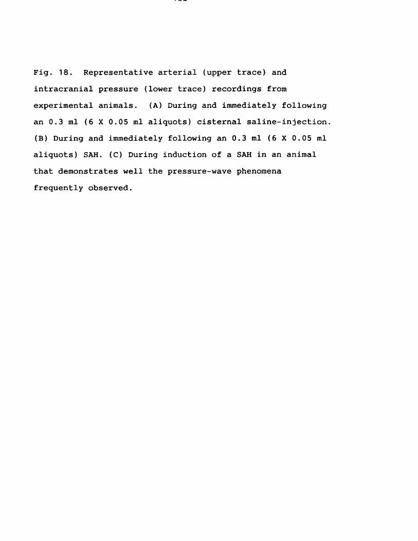
Fig. 18. Representative arterial (upper trace) and intracranial pressure (lower trace) recordings from experimental animals. (A) During and immediately following an 0.3 ml (6 X 0.05 ml aliquots) cisternal saline-injection. (B) During and immediately following an 0.3 ml (6 X 0.05 ml aliquots) SAH. (C) During induction of a SAH in an animal that demonstrates well the pressure-wave phenomena frequently observed.
250 mmHg
^ V/S/VWVWN/^
0 m m Hg
50 mmHg
1 min
0 m mHg
.05 .0 5 .0 5 .05.05 .05
2 5 0 m m Hg
0 m m Hg
50 mmHg
1 min
0 m m Hg
.05 .05 .05 .05 .05 .05
2 5 0 m m Hg
0 5 .05 .05
'A -,,1 min ,
/Hw .—1 A
.05 .05
0 m m Hg
50 mmHg
A.05
0 m mHg
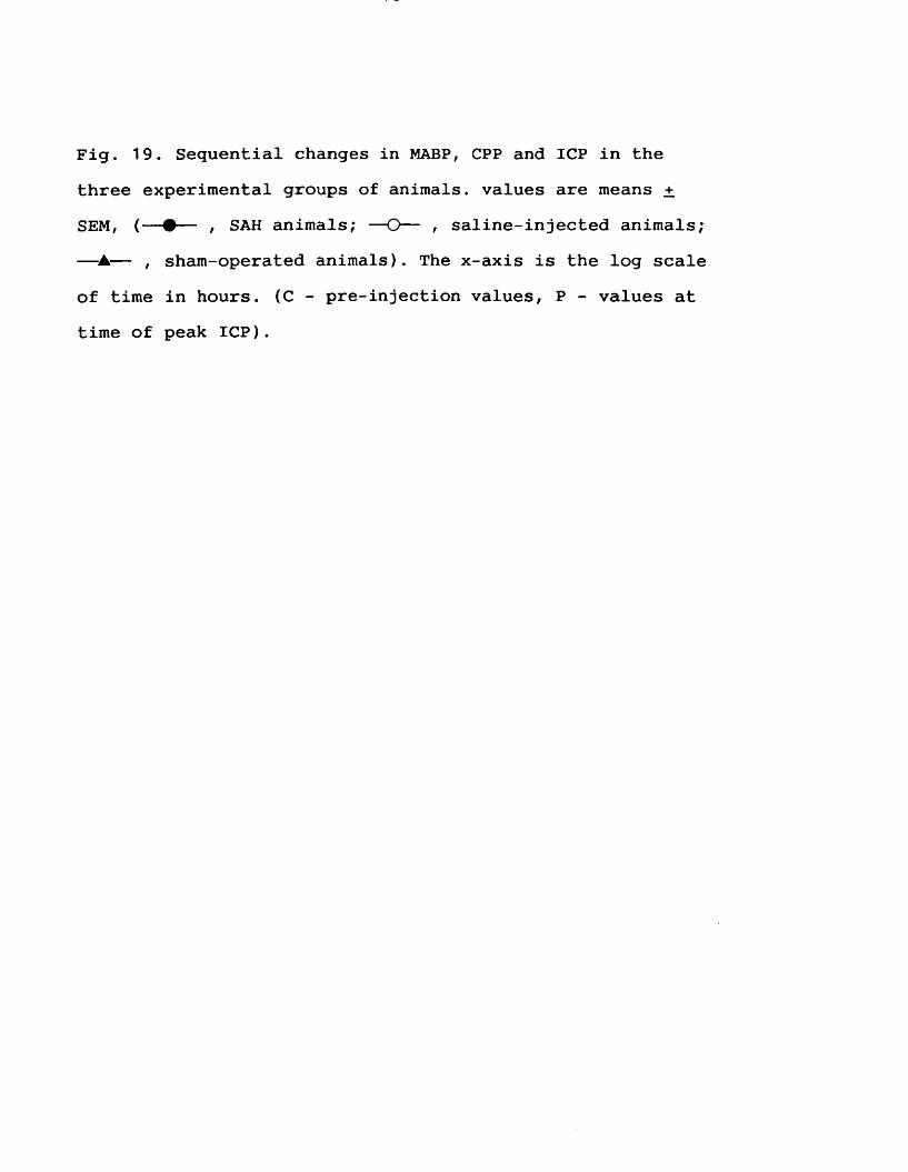
Fig. 19. Sequential changes in MABP, CPP and ICP in the three experimental groups of animals, values are means + SEM, (— • — , SAH animals; — O— , saline-injected animals; — ▲— , sham-operated animals). The x-axis is the log scale of time in hours. (C - pre-injection values, P - values at time of peak ICP).
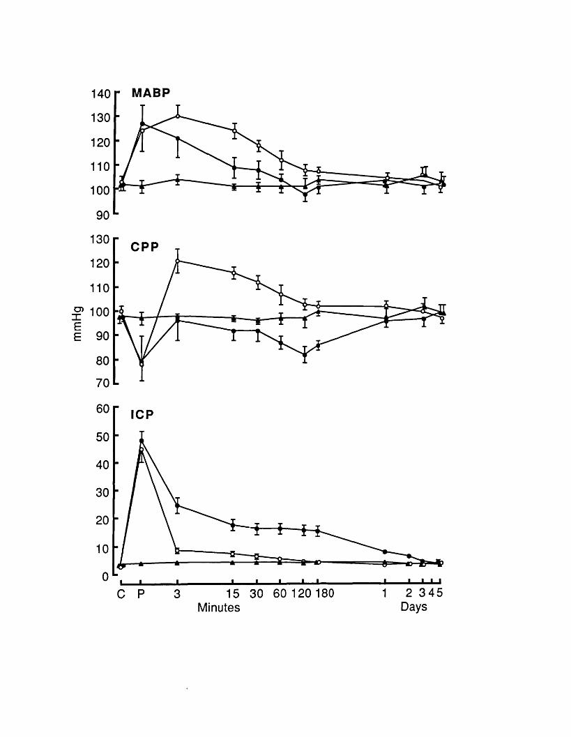
140
130
120110
100
90
130
120
110
10090
80
70
60
50
40
30
2010
0
MABP
CPP
ICP
■ ■ ■15 30 60 120 180
Minutes2 34 5
Days
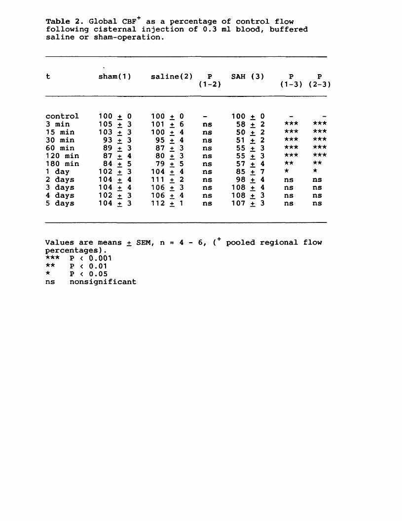
Table 2. Global CBF as a percentage of control flow following cisternal injection of 0.3 ml blood, buffered saline or sham-operation.
t sham(1) saline(2) P(1-2)
SAH (3) P(1-3)
P(2-3
control 100 ± 0 100 ± 0 100 + 03 min 105 ± 3 101 ± 6 ns 58 + 2 ***15 min 103 ± 3 100 ± 4 ns 50 + 2 ★★★30 min 93 ± 3 95 ± 4 ns 51 2 *** ***60 min 89 ± 3 87 ± 3 ns 55 _+ 3 ***120 min 87 ± 4 80 ± 3 ns 55 +_ 3180 min 84 ± 5 79 ± 5 ns 57 ± 4 **1 day 102 ± 3 104 ± 4 ns 85 ± 7 * *2 days 104 ± 4 111 ± 2 ns 98 ± 4 ns ns3 days 104 ± 4 106 ± 3 ns 108 ± 4 ns ns4 days 102 ± 3 106 ± 4 ns 108 ± 3 ns ns5 days 104 ± 3 112 ± 1 ns 107 ± 3 ns ns
Values are means + SEM, n 4 - 6, < + pooled regional flowpercentages). *** P < 0.001** P < 0.01* P < 0.05ns nonsignificant
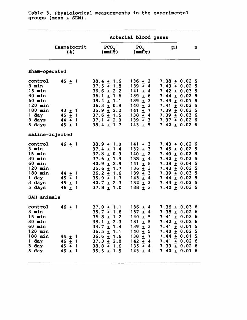
Table 3. Physiological measurements in the experimental groups (mean + SEM).
Arterial blood gasesHaematocrit P C ® 2 n
(%) (mmHg) (mmHg)
sham-operatedcontrol 45 + 1 38.4 + 1 .6 136 + 2 7.38 0.02 53 min 37.5 + 1.8 139 + 4 7.43 + 0.02 51 5 min 36.6 + 2.2 141 + 4 7.42 + 0.03 530 min 36.1 ± 1.6 139 + 6 7.44 + 0.02 560 min 38.4 ± 1.1 139 ± 3 7.43 + 0.01 5120 min 36.3 ± 0.8 140 ± 3 7.41 + 0.02 5180 min 43 +_ 1 35.9 ± 2.2 141 ± 1 7.39 + 0.02 51 day 45 + 1 37.6 ± 1 .5 138 ± 4 7.39 + 0.03 63 days 44 ± 1 37.1 ± 2.0 139 ± 3 7.37 ± 0.02 65 days 45 ± 1 38.4 ± 1.7 143 ± 5 7.42 0.02 6saline-injectedcontrol 46 ± 1 38.9 + 1 .0 141 + 3 7.43 ± 0.02 63 min 37.4 + 1.4 132 + 3 7.45 ± 0.02 51 5 min 37.8 + 0.9 140 + 2 7.40 ± 0.02 530 min 37.6 + 1 .9 138 + 4 7.40 + 0.03 560 min 40.9 + 2.9 141 + 5 7.38 + 0.04 5120 min 35.6 + 1.7 136 ± 3 7.43 ± 0.02 5180 min 44 + 1 36.2 + 1.6 139 + 3 7.39 + 0.03 51 day 45 + 1 35.9 + 1.7 143 ± 4 7.44 + 0.02 53 days 45 + 1 40.7 + 2.3 132 ± 3 7.43 ± 0.02 55 days 46 + 1 37.8 + 1.0 138 ± 3 7.40 ± 0.03 5SAH animalscontrol 46 ± 1 37.0 ± 1.1 136 ± 4 7.36 + 0.03 63 min 35.7 + 1.6 137 ± 4 7.38 + 0.02 61 5 min 36.8 ± 1.2 140 ± 5 7.41 + 0.03 630 min 38.1 ± 2.3 131 ± 5 7.42 + 0.02 660 min 34.7 ± 1 .4 139 + 3 7.41 + 0.01 5120 min 36.5 ± 1.1 140 + 5 7.40 + 0.02 5180 min 44 + 1 36.6 ± 1 .6 138 + 7 7.44 0.01 51 day 46 + 1 37.3 ± 2.0 142 ± 4 7.41 + 0.02 63 day 45 + 1 38.8 ± 1.6 135 ± 4 7.39 ± 0.02 65 day 46 + 1 35.5 ± 1 .5 143 ± 4 7.40 ± 0.01 6

8. EXPERIMENTAL CHAPTER VI:ULTRASTRUCTURAL CHANGES IN MAJOR CEREBRAL ARTERIES AND THEIR PERIVASCULAR CSF SPACES FOLLOWING EXPERIMENTAL SUBARACHNOID HAEMORRHAGE IN THE RAT
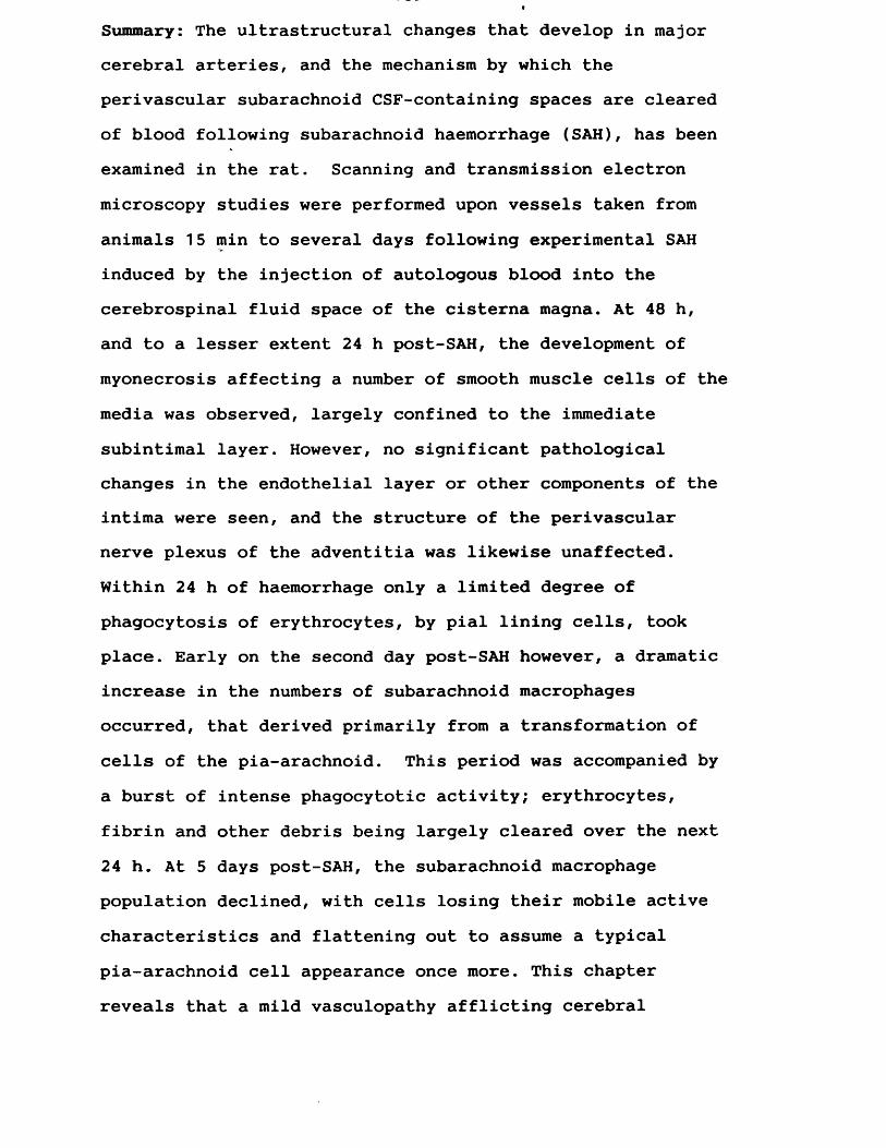
Summary: The ultrastructural changes that develop in major cerebral arteries, and the mechanism by which the perivascular subarachnoid CSF-containing spaces are cleared of blood following subarachnoid haemorrhage (SAH), has been examined in the rat. Scanning and transmission electron microscopy studies were performed upon vessels taken from animals 15 min to several days following experimental SAH induced by the injection of autologous blood into the cerebrospinal fluid space of the cisterna magna. At 48 h, and to a lesser extent 24 h post-SAH, the development of myonecrosis affecting a number of smooth muscle cells of the media was observed, largely confined to the immediate subintimal layer. However, no significant pathological changes in the endothelial layer or other components of the intima were seen, and the structure of the perivascular nerve plexus of the adventitia was likewise unaffected. Within 24 h of haemorrhage only a limited degree of phagocytosis of erythrocytes, by pial lining cells, took place. Early on the second day post-SAH however, a dramatic increase in the numbers of subarachnoid macrophages occurred, that derived primarily from a transformation of cells of the pia-arachnoid. This period was accompanied by a burst of intense phagocytotic activity; erythrocytes, fibrin and other debris being largely cleared over the next 24 h. At 5 days post-SAH, the subarachnoid macrophage population declined, with cells losing their mobile active characteristics and flattening out to assume a typical pia-arachnoid cell appearance once more. This chapter reveals that a mild vasculopathy afflicting cerebral

arteries, and a phase of pronounced macrophage phagocytic activity, are both most marked at around 48 h following SAH in the rat. These two findings may be of further interest, inasmuch as it is also around this time that a phase of delayed cerebral arterial narrowing has been previously documented.
8.1 INTRODUCTIONA major cause of potentially preventable mortality and
morbidity in patients following aneurysmal subarachnoid haemorrhage (SAH) is the cerebral ischaemia that has been associated with the delayed development of cerebral arterial luminal narrowing (Heilbrun et al., 1972; Ischii, 1979; Mickey et al., 1984). Studies of human cerebral vessels obtained at operation and post-mortem in such patients, have revealed the early occurrence in narrowed arterial segments, of myonecrosis affecting a number of the smooth muscle cells of the media. This is followed in some later cases by a phase of intimal thickening and subintimal proliferation (Crompton, 1964; Conway and McDonald, 1972; Hughes and Schianchi, 1978; Peerless et al., 1980; Smith et al., 1985). Pathological studies in experimental SAH, have taken place mainly in larger animals such as the monkey and dog, where some evidence for similar changes arising within the vessel wall have also been described (Tanabe et al., 1978; Peerless et al., 1980; Espinosa et al., 1984b). SAH both in man and from experimental studies has additionally been shown to result in the development post-haemorrhage of a dysfunction of the cerebrovascular sympathetic nerves, with alterations in the levels of their contained neurotransmitters (Lobato et al., 1980; Edvinsson et al., 1982; Tsukahara et al.,1988; Jackowski et al., 1989b). Although whether these latter findings represented the development of actual structural damage to the neural plexus itself, or merely changes in neurochemical expression, has not hitherto been established.
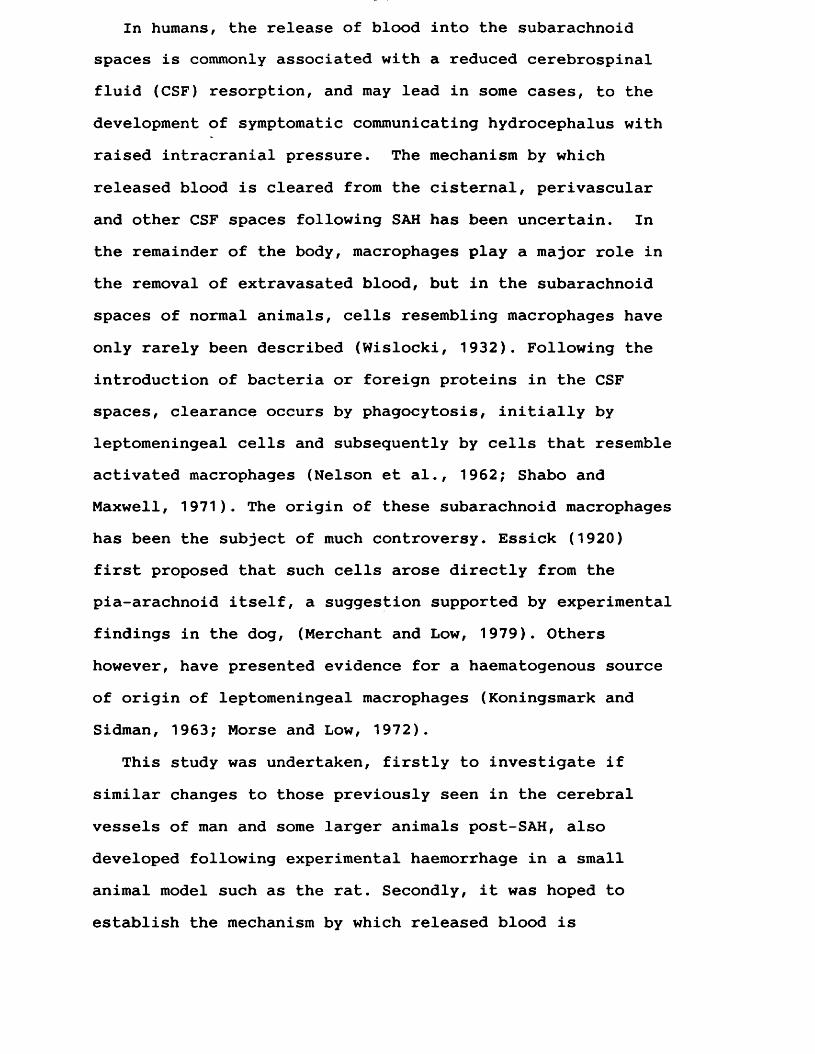
In humans, the release of blood into the subarachnoid spaces is commonly associated with a reduced cerebrospinal fluid (CSF) resorption, and may lead in some cases, to the development of symptomatic communicating hydrocephalus with raised intracranial pressure. The mechanism by which released blood is cleared from the cisternal, perivascular and other CSF spaces following SAH has been uncertain. In the remainder of the body, macrophages play a major role in the removal of extravasated blood, but in the subarachnoid spaces of normal animals, cells resembling macrophages have only rarely been described (Wislocki, 1932). Following the introduction of bacteria or foreign proteins in the CSF spaces, clearance occurs by phagocytosis, initially by leptomeningeal cells and subsequently by cells that resemble activated macrophages (Nelson et al., 1962; Shabo and Maxwell, 1971). The origin of these subarachnoid macrophages has been the subject of much controversy. Essick (1920) first proposed that such cells arose directly from the pia-arachnoid itself, a suggestion supported by experimental findings in the dog, (Merchant and Low, 1979). Others however, have presented evidence for a haematogenous source of origin of leptomeningeal macrophages (Koningsmark and Sidman, 1963; Morse and Low, 1972).
This study was undertaken, firstly to investigate if similar changes to those previously seen in the cerebral vessels of man and some larger animals post-SAH, also developed following experimental haemorrhage in a small animal model such as the rat. Secondly, it was hoped to establish the mechanism by which released blood is

subsequently cleared from the perivascular and cisternal subarachnoid spaces surrounding the major cerebral arteries.
8.2 MATERIALS AND METHODS Animal preparations
Adult male Sprague-Dawley rats weighing 350-450 g were used for the experiments. Subarachnoid heamorrhage was induced in 36 animals anaesthetised with a mixture of 70% nitrous oxide, 30% oxygen, to which 1.5% halothane was added. Animals were allowed to breathe spontaneously at all times. Autologous arterial blood (0.3 ml) freshly withdrawn from a femoral arterial catheter and replaced with an equal volume of normal saline was injected via a 30 gauge needle into the cisterna magna. Control animals comprised a group of 24 animals in which an equal volume of bufffered normal saline (25 mEq/l of NaHCO^ added and equilbrated at 37°C with a gas mixture of 5% C02/95% O 2 to pH 7.4) was injected in place of blood, and a further group of 24 animals in which a sham operation only was performed, with no injections given. Animals were subsequently killed and examined at times 15 min, 1 h, 3 h, 1 day, 2 days, 3 days, 5 days and 7 days post-SAH.
Perfusion-fixation and tissue examinationUnder inhalational anaesthesia, the heart was exposed and
a cannula inserted through the left ventricle into the ascending aorta. After incising the right atrium, the whole animal was perfused with approximately 200 ml of fixative composed of 4% paraformaldehyde, 0.1% glutaraldehyde, in 0.1 M phosphate buffer, pH 7.3, and followed by 500 ml of the same fixative minus glutaraldehyde. The brains were removed shortly after perfusion-fixation and immersed in the same fixative for 2 h, then placed in 0.1 M phosphate buffer
overnight. Under the operating microscope, the circle of Willis was then dissected from the brain and the proximal segment of the basilar artery and a distal segment of the internal carotid artery were taken and prepared for transmission (TEM) and scanning (SEM) electron microscopy. After washing in 0.1 M phosphate buffer, the specimens were post-fixed in 1% osmium tetroxide in 0.1 M phosphate buffer for 30 min then rinsed in distilled water. Specimens for TEM were next stained in 1% aqueous uranyl acetate for 30 min, then dehydrated through graded alcohols into propylene oxide and flat embedded in araldite. Ultrathin sections were cut and mounted on 200-mesh grids to be stained with aqueous uranyl acetate followed by lead citrate and examined with a Philips-300 electron microscope. Specimens for SEM, after post-fixation in osmium tetroxide, were further prepared, both conventionally and following an acid-etch treatment using 8N HC1 (Fujiwara and Uehara, 1984), the latter process allowing an improved visualisation of the perivascular neural plexus and vessel wall three-dimensional structure. Specimens, after dehydration in graded alcohols, were taken through amyl acetate, critical-point dried in a Balzers Union CO 2 bomb, mounted on SEM stubs, sputter coated with gold using a Polaron E 5000 unit and viewed with a Hitachi S-530 microscope.
8.3 RESULTS Endothelium
Scanning electron microscopy revealed the intimal endothelium of arteries taken from animals following experimental SAH, as being largely similar in appearance to the endothelium taken from control animals (Fig. 20). A well defined polygonal pavement-like layer of endothelal cells was always present on the luminal surface, with the long axis of the cells being orientated in the direction of blood flow. Acutely (up to 3 h) following SAH, the endothelial cells tended to be more rounded and possess an increased number of bleb-like and microvillus surface projections compared to those from control animals, although no other evidence of epithelial cell loss or damage was apparent at any time. By 1 day following experimental SAH, the endothelial cell appearance was once again identical to controls.
MediaA striking feature was the development 1 day post-SAH of
a small but significant number of smooth muscle cells, that displayed an overall increased electron density, contained dense-bodies, degenerate mitochondria, and showed loss of their normal internal structure (Fig. 21A). By 2 days the sarcolemma of such necrotic cells had broken down, with the appearance of aggregated myofilament complexes, free ribosomes and membrane debris, lying freely in the extracellular space between smooth muscle cells (Fig. 21B). These changes were most commonly observed in, although not wholely confined to, the medial layers closest to the
elastic lamina.
Perivascular nerves and adventitiaThe schwann-cell/neural plexus was most readily
visualised in the acid-etched SEM preparations (Fig. 22). No differences were apparent between experimental animals and controls, either in the structure of the plexus itself or in the extent of its distribution within the adventitial layer, at any of the times studied.
Subarachnoid spacesImmediately following SAH, the perivascular and cisternal
subarachnoid spaces adjacent to the basilar and internal carotid arteries were occupied by a dense blood-clot that consisted largely of erythocytes trapped within a fibrin mesh (Fig. 23A). Little change was detected on the first day, although occasionally erythrocytes were seen that appeared to have been directly phagocytosed by cells of the pial membrane. Early on the second day post-SAH, large numbers of rounded cells became evident. Fully matured forms displayed a variety of ruffled membranes and microappendages, and possessed the characteristics of subarachnoid macrophages. This emergent cell population appeared to originate principally from the pial, and to a lesser extent the arachnoid membranes. Within these structures, transition zones appeared (Fig. 23B), from which intermediate cell types could be seen in various stages of transformation, initially rounding up then gaining microappendages upon their cell surface. Defects in the normally continuous sheet of overlapping pial cells became
evident, as maturing cells migrated out into the surrounding subarachnoid blood-clot. These cells behaved subsequently in a manner characteristic of activated macrophages, and could be seen avidly phagocytosing erythrocytes and other debris from the subarachnoid spaces (Fig. 24). Already by 3 days post-SAH, the perivascular CSF spaces were largely cleared of blood cells and fibrin. At 5 days post-SAH the macrophage population declined and sites were increasingly observed upon the pia-arachnoid membranes and arachnoid trabeculations, where subarachnoid macrophages appeared to be losing their mobile active characteristics, flattening out to resemble typical pia-arachnoid cells once more (Fig.
8.4 DISCUSSIONThe development of a delayed narrowing of major cerebral
arteries following SAH due to aneursymal rupture, has been known since the orginal angiographic description by Ecker and Riemenschneider (1951). The time course of this constriction in man has been detailed (Kwak et al., 1979), its onset is usually delayed until the fourth or fifth day post-bleed and has a maximum incidence between 10 and 14 days. Experimental studies in the dog (Brawley et al.,1967; Kuwayama et al., 1972; Nagai et al., 1974) revealed that cerebral arteries respond to SAH in a biphasic manner, with an acute reversible phase of narrowing immediately post bleed, followed in some instances by a more persistent phase that develops after a variable delay.
Study of such angiographically observed narrowed segments of cerebral arteries, taken either at operation or at autopsy from patients dying following SAH, have demonstrated the occurence of varying degrees of medial necrosis, subintimal proliferation and other pathological changes (Crompton, 1964; Conway and McDonald, 1972; Hughes and Schianchi, 1978; Peerless et al., 1980; Smith et al., 1985). Whilst investigators of experimental SAH in the monkey have documented similar morphological changes (Peerless et al., 1980; Espinosa et al., 1984b), studies in dogs have been more variable, some authors observing pathological changes in the walls of cerebral vessels (Tanabe et al., 1978; Tani et al., 1978) while others have found few or none at all (Pickard et al., 1985).
Recently the laboratory rat has been increasingly
employed for studies of experimental SAH (Barry et al. ,1979; Lacy and Earle, 1982; Svendgaard et al., 1985; Solomon et al., 1987), its relative cheapness and greater availability allowing its use in larger animal numbers. A biphasic time-course of narrowing in cerebral vessels post-SAH has been reported in the rat (Delgardo et al., 1985), but no pathological changes of rat cerebral vessels after haemorrhage described. In the present study, we have now found evidence for similar changes to those previously described in larger animal models. Myonecrosis of limited but significant numbers of smooth muscle cells of the media of cerebral vessels was observed, that commenced the day following haemorrhage. A number of studies, both of man and in a variety of animals including the rat, have now clearly demonstrated that a dysfunction of both endothelial cell and perivascular nerve related cerebrovascular reactivity develops post-SAH (Pickard and Perry, 1983; Sahlin et al., 1985; Nakagomi et al., 1987; Kim et al., 1988a). In the present study, the ultrastructural morphology of both the endothelium and neural plexus appeared to be largely unaffected following haemorrhage. This would suggest therefore, that these alterations possibly reflect either impaired smooth muscle responsiveness, or qualitative changes in endothelial cell and nerve function, rather than any physical damage to the latter structures themselves.
Clearance of released blood from the subarachnoid spaces in man can take from 6 to 30 days (Tourtellotte et al., 1964), the continued presence of blood clot within these spaces predisposes to impaired CSF resorption,
hydrocephalus, raised intracranial pressure and delayed cerebral ischaemia (Ellington and Margolis, 1969; Vassilouthis and Richardson, 1979; Butler et al., 1980; Mohsen et al., 1984). In man, most erythrocytes rapidly become emeshed within the arachnoid trabeculae of the subarachnoid spaces and it was naturally hypothesised that they were then removed by lysis and phagocytosis (Sprang, 1934; Hammes, 1944). Early studies of erythrocyte clearance in animals however, seemed to suggest that cells might instead be largely removed intact via perineural lymphatic-type channels (Kennady, 1967; McQueen et al., 1974). Separate studies had suggested a system of tubular spaces and pores across the arachnoid villi (Welch and Friedman, 1960) and it was also proposed that whole erythrocytes might pass directly into the blood stream via this route (Simmond, 1953). Ultrastructural studies have since shown the endothelium of the arachnoid villi to be continuous, thereby preventing such a mode of clearance (Alksne and White, 1965). More recently, studies in the dog (Alksne and Lovings, 1972) have suggested that removal of erythrocytes from the subarachnoid spaces occurred mainly by lysis and possibly by phagocytosis of resultant debris by cells of the arachnoid villi. Again in the dog, an increase in the numbers of free subarachnoid macrophages was also shown to occur after SAH by Julow et al. (1979), who observed limited erythrophagocytosis to take place.
In this investigation of the mode of clearance of the perivascular subarachnoid spaces in the rat, erythrocytes immediately following haemorrhage, occasionally underwent
phagocytosis by cells of the pial membrane. With increased survival time, there followed a period characterised by a burst of intense phagocytic activity by mobile cells possessing the typical features of activated macrophages. This greatly increased population of subarachnoid macrophages appeared to be largely derived by the transformation of cells from the pia membrane. Upon clearance of blood-clot from the perivascular subarachnoid spaces, these activated macrophages flattened out, assuming the characteristics of typical pia-arachnoid cells once more.
The development of pathological changes affecting the media of the rat basilar artery and a period of intense macrophage erythrophagocytic activity within perivascular subarachnoid blood-clot, were both maximal at around 48 h post-haemorrhage. Interestingly, this period also coincides with the time at which a delayed onset of basilar arterial luminal narrowing has previously been described (Delgardo et al 1985). Macrophage activity and oxyhaemoglobin autoxidation are both well known to be associated with the generation and release of free radicals (Sutton et al.,1976; Babior, 1978). The findings from this experimental chapter may be of further interest therefore, inasmuch as work by a number of authors now suggests that an excess of free radicals may be involved in the genesis of both arterial wall damage and the angiographically visible delayed cerebral arterial narrowing that may follow SAH (Sakaki et al., 1988; Steinke et al., 1989; Zuccarello et al., 1989)
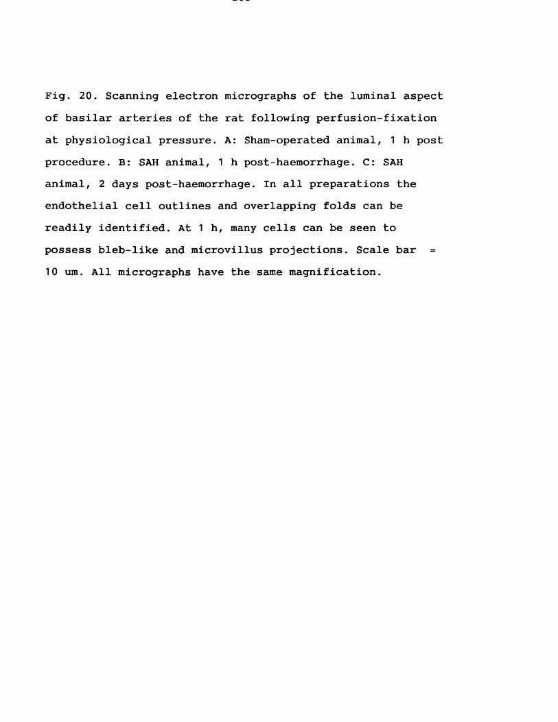
Fig. 20. Scanning electron micrographs of the luminal aspect of basilar arteries of the rat following perfusion-fixation at physiological pressure. A: Sham-operated animal, 1 h post procedure. B: SAH animal, 1 h post-haemorrhage. C: SAH animal, 2 days post-haemorrhage. In all preparations the endothelial cell outlines and overlapping folds can be readily identified. At 1 h, many cells can be seen to possess bleb-like and microvillus projections. Scale bar 10 urn. All micrographs have the same magnification.
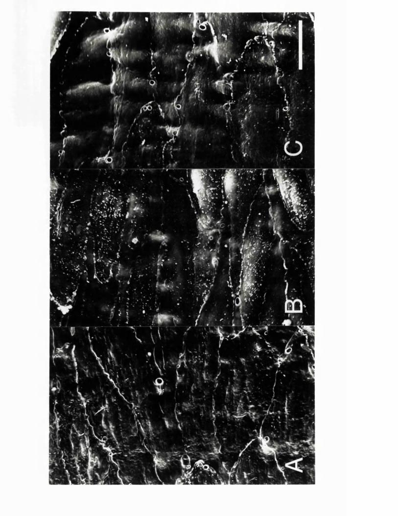
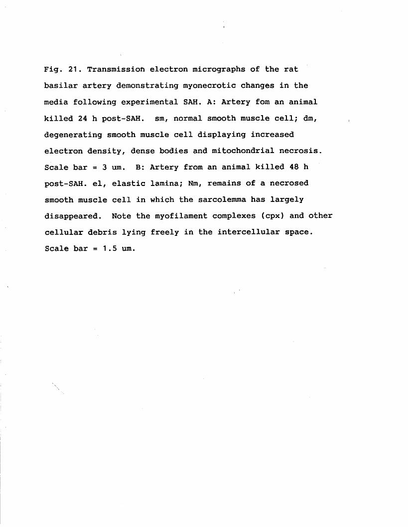
Fig. 21. Transmission electron micrographs of the rat basilar artery demonstrating myonecrotic changes in the media following experimental SAH. A: Artery fom an animal killed 24 h post-SAH. sm, normal smooth muscle cell; dm, degenerating smooth muscle cell displaying increased electron density, dense bodies and mitochondrial necrosis. Scale bar = 3 urn. B: Artery from an animal killed 48 h post-SAH. el, elastic lamina; Nm, remains of a necrosed smooth muscle cell in which the sarcolemma has largely disappeared. Note the myofilament complexes (cpx) and other cellular debris lying freely in the intercellular space. Scale bar = 1.5 urn.
U *> "»V‘-
-v,. a V rvtwr
sm / ji«-
r.>
/ '***-
•X. x
j&dm ■PVf
» * < • • X > pc m
, cpx
-. w < r ’l’v!,.w
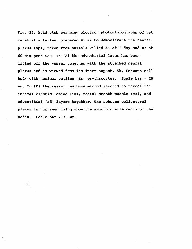
Fig. 22. Acid-etch scanning electron photomicrographs of rat cerebral arteries, prepared so as to demonstrate the neural plexus (Np), taken from animals killed A: at 1 day and B: at 60 min post-SAH. In (A) the adventitial layer has been lifted off the vessel together with the attached neural plexus and is viewed from its inner aspect. Sh, Schwann-cell body with nuclear outline; Er, erythrocytes. Scale bar = 20 urn. In (B) the vessel has been microdissected to reveal the intimal elastic lamina (in), medial smooth muscle (me), and adventitial (ad) layers together. The schwann-cell/neural plexus is now seen lying upon the smooth muscle cells of the media. Scale bar = 30 urn.
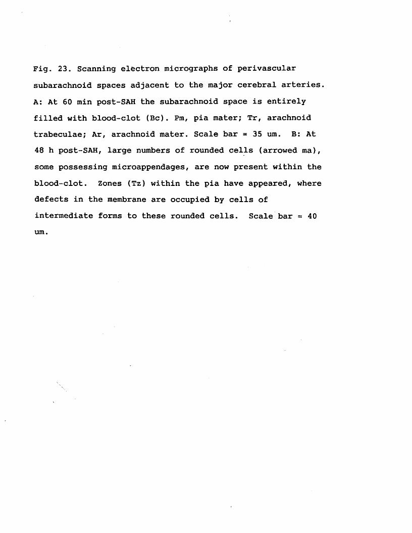
Fig. 23. Scanning electron micrographs of perivascular subarachnoid spaces adjacent to the major cerebral arteries. A: At 60 min post-SAH the subarachnoid space is entirely filled with blood-clot (Be). Pm, pia mater; Tr, arachnoid trabeculae; Ar, arachnoid mater. Scale bar = 35 um. B: At 48 h post-SAH, large numbers of rounded cells (arrowed ma), some possessing microappendages, are now present within the blood-clot. Zones (Tz) within the pia have appeared, where defects in the membrane are occupied by cells of intermediate forms to these rounded cells. Scale bar = 40 um.
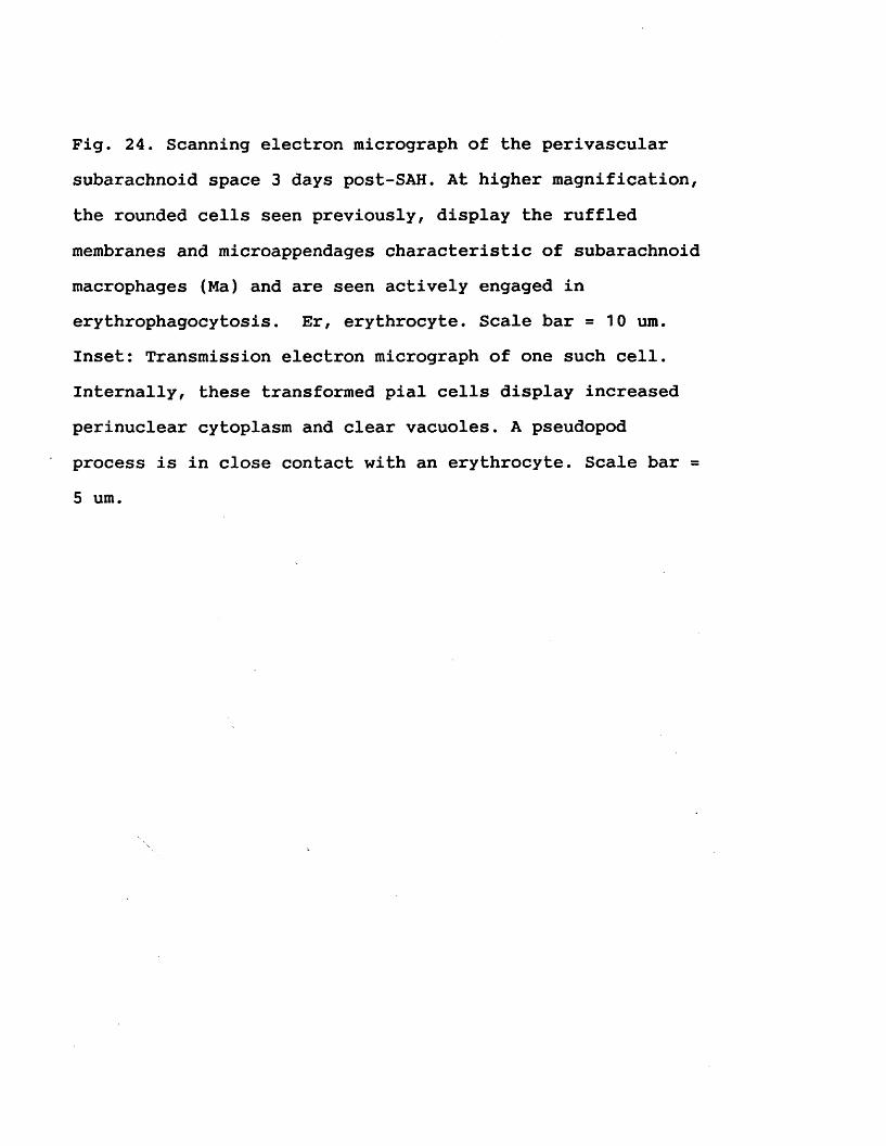
Fig. 24. Scanning electron micrograph of the perivascular subarachnoid space 3 days post-SAH. At higher magnification, the rounded cells seen previously, display the ruffled membranes and microappendages characteristic of subarachnoid macrophages (Ma) and are seen actively engaged in erythrophagocytosis. Er, erythrocyte. Scale bar = 1 0 um. Inset: Transmission electron micrograph of one such cell. Internally, these transformed pial cells display increased perinuclear cytoplasm and clear vacuoles. A pseudopod process is in close contact with an erythrocyte. Scale bar = 5 um.
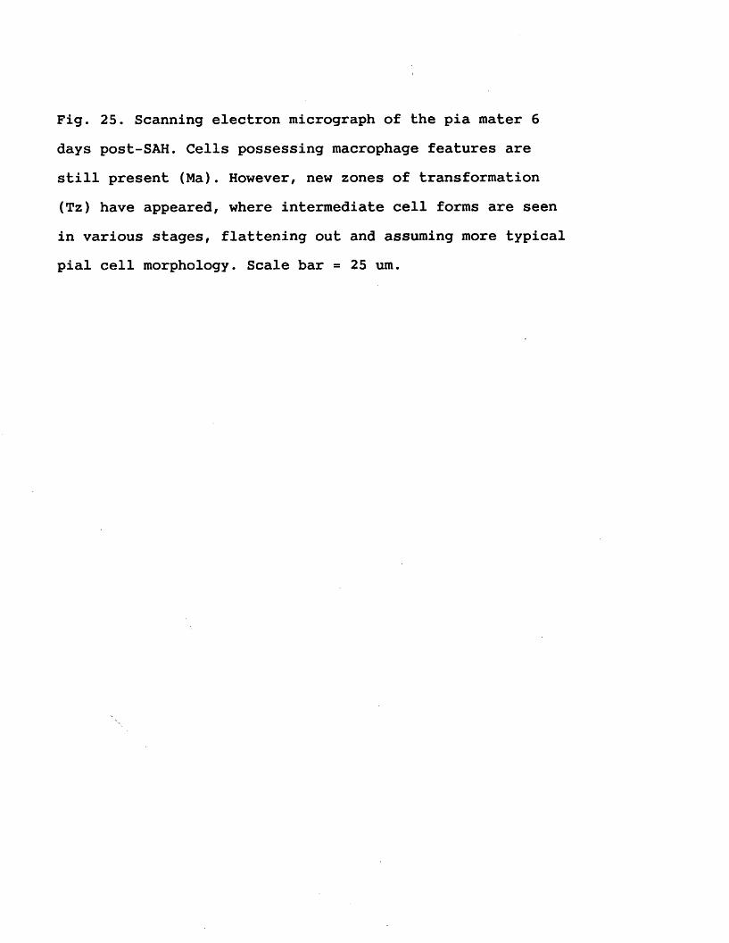
Fig. 25. Scanning electron micrograph of the pia mater 6 days post-SAH. Cells possessing macrophage features are still present (Ma). However, new zones of transformation (Tz) have appeared, where intermediate cell forms are seen in various stages, flattening out and assuming more typical pial cell morphology. Scale bar = 25 urn.
9. CONCLUSIONS FROM THE STUDY
The findings can be summarised in the following way:
(1) A significant discrete serotonergic innervation of the major cerebral arteries in the rat, and possibly other species including man, does not appear to exist, contrary to earlier reports in the literature.
(2) Serotonin, when observed within the perivascular nerves of the large cerebral arteries, is found to occur only in nerves also identified as catecholaminergic sympathetics. When present, it appears to arise largely as a result of a direct uptake of the indole from the surroundings.
(3) After an induced SAH, serotonin was rapidly and readily taken up by the cerebrovascular sympathetic nerves, while a coincident depletion of NPY from these same nerves took place. Once blood clot had been largely resorbed from the CSF, the neurotransmitter content of these nerves was then restored once more to normal.
(4) Experimental SAH in the rat was found to result in an immediate 50% global reduction in cortical CBF that persisted for up to three hours post-haemorrhage. There was a smaller but significant reduction in flow 24 hours post SAH, while CBF was normal between day 2 and 5. The decrease in CBF was clearly most marked at about the same time at which a maximal acute spasm has been observed by other authors, using the same animal model. However, no later reduction in flow was seen at 48 hours, the time at which a delayed phase of vasospasm has previously been described in
the rat. The ICP was increased and the CPP decreased during the first 24 hours after the SAH. The recorded reduction in CPP of 15% was found not to be in accordance with a fall in CBF of 50%. It is suggested that early vasospasm secondary to released blood rather than pressure changes is responsible for the initial cerebral ischaemia observed.
(5) Ultrastructural studies of the cerebral arteries from rats subjected to SAH, revealed some, but not all of the morphological changes found in the vasculopathy associated with delayed cerebral arterial narrowing in man and large animal models after SAH. Subintimal myonecrosis was observed, especially at 48 hours after the SAH, while no intimal or subintimal proliferation developed subsequently. The findings of intact perivascular nerves post SAH are of interest, and suggest that the described disappearance of neurotransmitter content is due to a functional change in the nerve rather than to a chemical neurectomy.
(6) The primary mechanism by which the perivascular subarachnoid spaces were cleared of adjacent blood clot following haemorrhage was by phagocytosis. This took place largely on the second day post-SAH, a period characterised by intense macrophage phagocytic activity. The origin of these subarachnoid macrophages was studied, and it appears that they are derived by a transformation from cells of the pia-arachnoid membranes. In the context of these findings, it is perhaps relevant to note that such phagocytic activity is well known to be associated with the local release of free radicals. These have been strongly implicated by some
authors in one mechanism of cellular damage that may subsequently lead to delayed cerebrovasculopathy and arterial narrowing.
Secondarykdeterioration after SAH in man, other than from proven episodes of rebleeding, is commonly attributable to the onset of delayed cerebral ischaemia. The causes of which are probably multifactorial, but most certainly include: a reduced CBF, impaired cerebrovascular autoregulation, and raised intracranial pressure. The first of these three, has been closely linked with the development of arterial luminal narrowing affecting one or more of the major cerebral arteries. Pathological studies have suggested that such vessel narrowing may represent a reactive vasculopathy induced either by blood-clot directly, or in response to secondarily released products.
The results obtained from this present study indicate, that a simple single-haemorrhage rat model displays a number at least of the features that can arise following SAH either in man or in larger animals after SAH. In particular, the development of a global reduction in CBF, an elevated ICP with reduced intracranial compliance, alterations in neurotransmitter balance within the perivascular sympathetic nerves that normally contribute to autoregulation, and ultrastructural evidence of a delayed cerebrovasculopathy were noted. Although not responding in an entirely identical manner to that of the human response after SAH, the rat model of SAH is revealed to be a worthwhile one for future studies to further investigate and determine how best to manage the disordered pathophysiology that commonly arises

following SAH.
10. FINAL DISCUSSION
10.1 EXPERIMENTAL MODELS AND SAH
In vivo animal studies:A wide variety of animal species have been utilised in
experimental studies of subarachnoid haemorrhage. Baboons, monkeys, dogs, cats, rabbits and rats having all been variously employed for this purpose. The in vivo SAH created in these studies, has largely been simulated either by the subarachnoid injection of the animals own autologous arterial blood, or by inducing rupture of a vessel of the circle of Willis. A considerable number of such animal studies, in which the primary aim was purportedly to investigate the mechanisms causing delayed cerebral vasospasm, have in fact instead been largely concerned with examining the acutely vasoconstrictive effects of subarachnoid blood. This caveat aside, a wealth of useful information has now emerged from such laboratory studies.
Cerebral arterial narrowing - VasospasmEchlin (1965), was one of the first to examine the
effects of experimental SAH, produced in monkeys by the single direct application of arterial blood close to a major cerebral vessel. He demonstrated, that when subarachnoid blood came into contact with large arteries, that a marked vasospasm (30 to 60 percent of prehaemorrhage diameter) developed almost immediately; an effect that he noted generally lasted 10 to 40 minutes but sometimes longer. Brawley et al (1968) subsequently made the important observation in his studies on the dog, that cerebral
arterial narrowing following experimental SAH generally resulted in a biphasic response. In adult mongrel dogs in which the SAH was produced by rupture of an anterior cerebral artery, resultant haemorrhage was initially associated with a directly observed acute phase of arterial constriction that lasted for less than 1 hour. This was followed by a delayed recurrence of vessel narrowing, that developed 3 to 24 hours later and became maximal on the third day post-SAH. A similarly biphasic time-course was also observed, when SAH was instead produced in the dog by a single injection of autologous arterial blood into the cisterna magna (Kuwayama et al., 1972). These latter authors followed their animals by means of serial angiograms, and demonstrated a response pattern that consisted of an acute phase of constriction that had largely recovered by 2 hours. This was followed by a delayed onset of recurrent narrowing at 24 hours with the affected vessels subsequently recovering to their normal diameters by 7 days. A persistant arterial narrowing following experimental SAH, has subsequently been reported as occurring in other smaller animal species. In the rabbit (Edvinsson et al., 1982) and in the rat (Barry et al., 1979), reductions in basilar artery diameters have been shown to be present for periods of up to 9 days and 48 hours post-haemorrhage respectively. Delgado and coworkers (1985) have performed angiographic studies in the rat, both acutely and at different time points up to one week following the SAH produced by a single intracisternal blood injection. In this small animal study, a biphasic time-course, similar to that previously reported
in the dog, was also found. An early phase of marked luminal narrowing (40 percent of prehaemorrhage diameter) was seen at 10 minutes, and had largely recovered by 90 minutes. This was then followed by a second but less marked constriction (25 percent of prehaemorrhage diameter) at 24 and 48 hours, which subsequently resolved fully by 72 hours.
CBF. Autoregulation and Cerebral MetabolismThe acute effects of subarachnoid haemorrhage upon
cerebral blood flow, autoregulation, and reactivity to pCC^have been widely investigated in experimental models, whilstdelayed effects have received somewhat less detailedattention. In adult rhesus monkeys, Petruk et al. (1972)
133measured the CBF by a Xenon clearance technique and showed a significant acute (up to 3 hours) reduction in CBF in 12 out of 14 monkeys subjected to SAH. They also reported finding a close correlation between the degree of impaired flow and severity of neurological deficit. A number of authors have similarly studied the acute changes in CBF that followed experimental SAH in baboons (Jakubowski et al., 1982; Kamiya et al., 1983; Kuyama et al., 1984). Haemorrhage being produced by snare transection of the posterior communicating artery. In these studies, CBF generally became rapidly reduced to 20% - 50% of normal and showed a variable pattern of recovery, depending upon the neurological grade of the animal. In some instances, persistant reductions in CBF of up to 18% at 3 days were observed (Jabubowski et al., 1982; Sahlin et al., 1987). In the cat, experimentally induced SAH, provoked either by a cisternal blood injection (Umansky et al., 1983) or by internal carotid artery
puncture (Trojanowski, 1984) gave rise to similar results. Both regional and global CBF's were found to fall at 10 minutes by 15 - 50% of control values, and these remained depressed for up to 3 hours posthaemorrhage. Soloman (1985) has examined the immediate effects only, of experimental SAH upon CBF in a smaller animal model, the rat, induced by a single intracisternal injection of blood. Measuring blood flow acutely using a labelled microsphere technique, he demonstrated a 40% decrease in CBF over a 60 minute period.
Assessment of autoregulatory function and CO 2 reactivity after experimental SAH has been carried out predominantly using the baboon (Jakubowski et al., 1982; Kamiya et al., 1983; Sahlin et al., 1987). In such studies it was found that autoregulation and cerebrovascular reactivity to CO 2
became depressed in a global fashion, both acutely and for up to 3 days following the induction of SAH.
Whilst in man it has been found that SAH is clearly associated with global reductions in cerebral metabolism, particularly in those patients in the poorest clinical grades, the few results obtained from animal studies are extremely inconsistent. Studies have shown either no change (Sahlin et al., 1987), or a diffuse increase (Delgardo et al., 1986). Although, both Fein (1975) and Soloman (1987) separately found that metabolic activity in the monkey and in rats was globally decreased following experimental SAH.
Blood Brain Barrier / Brain oedemaExperimental studies in animal models, have revealed that
the BBB breakdown observed following SAH, predominantly affects the blood arterial-wall barrier of major cerebral
arteries. Although the resultant changes may partly be explained by the sudden rise in ICP and arterial pressure that occurs post-SAH, they can also be provoked equally well by the presence of blood clot in the subarachnoid space alone (Peterson et al., 1983; Sasaki et al., 1985; Sasaki et al., 1986; Zuccarello et al., 1987)
An increased brain water content, with an accompanying increase in sodium content, is known to develop early following experimental SAH (Kamiya et al., 1983; Kuyama et al., 1984; Doczi, 1985). Interestingly, evidence has emerged to suggest that such brain swelling may be modified by vasopressin (Doczi, 1984), implying that brain water permability and electrolyte balance may in part be regulated by this centrally released neuropeptide.
Peripheral autonomic and Central nervous system changes Following experimentally induced SAH in in vivo animal
models, there is now both direct and indirect evidence that a comparable degree of peripheral sympathetic overactivity to that seen in man, similarly takes place. Elevated levels of plasma catecholamines develop, also an increased release of adrenal medullary catecholamines and enkephalins can be demonstrated (Offerhaus and Van Gool, 1969; Klein et al.,1986). Electrocardiographic abnormalities, similar in variety to those that are seen clinically in patients, have also been observed after experimental SAH (Estanol et al., 1977; Lacy and Earle, 1983). These appear to occur, as a direct result of the effects of a sustained high level of circulating catecholamines upon the myocardium and conducting tissues of the heart (Schenk and Moss, 1966).
As shown in man, again evidence exists from animal studies, performed in this instance in the rat, to show that an elevation of brain noradrenaline concentration develops after experimental SAH (Solomon et al., 1986). Further studies have shown that this elevation reflects an increased synthesis and release of the neurotransmitter, presumably arising from projection axons that originate from neurons of the locus coeruleus (McCormack et al., 1986).
A number of authors have examined the effects of experimental SAH upon the periarterial nerve plexus that surrounds and innervates the major intracranial arteries. These studies have consistently demonstrated the development of a marked reduction, to complete exhaustion, of noradrenaline stores in the nerve terminals about such vessels (Fraser et al., 1970; Peerless and Kendall, 1975; Svendgaard et al., 1977a). Another finding of significance has been that a reduced uptake of noradrenaline by cerebrovascular perivascular nerves, arises post SAH (Lobato et al., 1980; Edvinsson et al., 1982). Such vessels also display a state of denervation supersensitivity to exogenously applied noradrenaline (Duckies et al., 1977; Svendgaard et al., 1977b). A reduction in the neurotransmitter content does not appear to be solely confined to the noradrenergic system. More recently, in studies both upon the dog and in the monkey, it has been reported that a reduction in the numbers of intracranial perivascular nerves displaying VIP, SP and NPY immunoreactivity may also occur following haemorrhage (Hara et al., 1986; Uemura et al., 1987).
Of interest therefore, in view of these observed changes in cerebral noradrenaline content and perivascular neurotransmitter status, have been a series of experiments performed by Svendgaard and his coworkers. They have attempted to elucidate if any role is played by the centrally projecting serotonergic, dopaminergic and catecholaminergic neural pathways, in the development of vasospasm affecting the vertebrobasilar arteries following experimental SAH. Chemical destruction of the central serotonergic and dopaminergic pathways prior to the intracisternal injection of blood, apparently does not affect the occurence of such vasospasm after SAH (Svengaard et al., 1986). These same authors report however, that the lesioning of ascending catecholaminergic fibres in the mesencephalon or medulla either by a stereotactic lesioning with 6-OHDA, or by neonatal systemic 6-OHDA adminstration, effectively prevents the development of vertebrobasilar artery spasm (Svendgaard et al., 1985; Delgado et al.,1987). These findings however, are difficult to reconcile with the fact that there is no known evidence for any central catecholaminergic innervation of the vertebrobasilar system. They have instead therefore proposed, an alternative neurohumoral mechanism, in which overactivity in central catecholaminergic projections to the hypothalamus might cause vasospasm of large cerebral arteries through secondary release of vasopressin. In support of this hypothesis, they have conducted experiments demonstrating that arginine-vasopressin deficient Brattleboro rats fail to develop acute vasospasm after SAH (Delgado et al., 1988).
Against this argument however, must be considered the fact that several other authors have been unable to demonstrate any consistent vasoconstrictor response of the cerebral vessels in response to vasopressin (Allen et al., 1974a; Lassoff and Alturs, 1980; Katusic et al., 1984).
In vitro studies:These studies, employing vessels and CSF obtained either
from patients or in animals following SAH, are all characterised by the investigation of any resultant changes in cerebrovascular responsiveness or pharmacology in an in vitro environment.
Such investigations, have been largely focused upon attempts to unmask a specific substance or series of substances, putatively contained within the CSF in increased amounts following haemorrhage, and which are held responsible for the production of vasospasm leading to cerebral ischaemia. It had been noted early on, that the application of CSF obtained from patients with angiographic evidence of vasospasm following SAH, caused isolated cerebral arteries to contract, as well as sensitising them to many other vasoconstrictor agents (Boullin et al., 1976). It was also found that such vasoconstrictor activity was highest in those patients who had evidence of severe angiographic vasospasm or who developed delayed ischaemic deficits and subsequently died (Boullin, 1980; Kaye et al., 1984).
SerotoninOne of the first substances to be extensively
investigated in this way was 5-hydroxytryptamine. In 1974 in a series of classic experiments, Allen (Allen et al., 1974a; Allen et al., 1974b; Allen et al., 1974c) using the canine basilar artery, examined the contractile activity of human CSF obtained folowing haemorrhage. Largely on the basis of these studies, it was at that time concluded that serotonin was the main agent responsible for the immediate arterial spasm that arose following vessel exposure to bloody CSF obtained post SAH. The subsequent finding that perianeurysmal CSF obtained at the time of operation for ruptured intracranial aneurysms (and shown to contain 5-HT) could be prevented from causing the constriction of isolated human intracranial arteries by concommitant application of the 5-HT antagonist ketaserin (Tagari et al., 1983), would appear to strongly support this argument. Although it remains a prime candidate for the cause of the acute vasospasm commonly seen immediately after experimental SAH in animal models, 5-HT is however, unlikely to alone be responsible for a more chronic vasospasm, or the delayed vasospasm seen in man. In vivo administration of serotonin into the CSF space in animals, indeed produces spasm, but this is short-lived, persisting for some 2-3 hours only (Allen et al., 1974c; Boullin et al., 1978). A point that has been made in man is that although 5-HT concentrations in the CSF will be considerably elevated immediately following release of blood into the subarachnoid space, that there is no consistently found elevation of levels subsequently, during the period of delayed arterial narrowing. These measurements have however, been made predominantly upon
ventricular CSF samples (Vapalahti et al., 1978; Voldby et al., 1982). More recently, evidence has emerged that suggests that significantly elevated concentrations of 5-HT may in fact persist for considerably longer in the CSF contained locally within periarterial cisterns (Tagari et al., 1983; Chehrazi et al., 1989).
Most in vitro studies upon the role of serotonin released from platelets following experimental SAH, have naturally focused upon its known vasoconstrictor actions. Of perhaps more relevance to the aetiology of the chronic arterial narrowing found in human SAH, may be its less well known, stimulatory actions upon vascular smooth muscle (VSM) mitogenesis and deoxyribonucleic acid synthesis. The mitogenic effects of serotonin and for that matter human platelet-derived growth factor (PDGF), have to date, primarily been studied in bovine aortic smooth muscle cells in culture but may well be relevant for VSM cells of the intracranial vessels following SAH. PDGF is another potent mitogen for VSM cells. It is released from the alpha-granules of platelets that have been exposed to vascular connective tissue, and its mitogenic response is synergistically affected by the concommitant presence of serotonin (Nemecek et al., 1986). Serotonin-induced VSM mitogenesis is mediated by specific receptors and is critically dependent upon guanine nucleotide binding proteins while that of the PDGF response is not (Kavanaugh et al., 1988).
Ervthrocvtes / OxvhaemoqlobinErythocyte products appear to be a necessary factor in
the development of a cerebral vasculopathy arising following experimental SAH (Duff et al., 1988). In vitro, the directly vasoconstrictor effects of either haemoglobin or haemolysate have been well described (Handa et al., 1980; Wellum et al., 1980; Boullin et al., 1983). Prolonged exposure to oxyhaemoglobin is known to modify the response of isolated cerebral vessels from various species, to a variety of agents. Relaxations induced by substance P released from vasodilator nerves, the electrogenic sodium pump, endothelium-derived relaxing factor, prostaglandin I2, nitroglycerin, and angiotensin-II are all impaired following exposure to oxyhaemoglobin (Lee et al., 1984; Toda, 1988; Onoue et al., 1989). Conversely, oxyhaemoglobin potentiates both neurogenic vasoconstriction and 5-HT induced vasoconstriction of cerebral vessels (Lee et al., 1984; Tanaka and Chiba, 1988).
Oxygen Free RadicalsActive species of oxygen are liberated upon the
conversion of oxyhaemoglobin to methaemoglobin (Misra and Fridovich, 1972; Sutton et al., 1976). They are also generated by the phagocytic activity of polymorphonuclear leucocytes, monocytes and macrophages (Babior, 1978). Both of these processes will naturally arise in intimate proximity to the walls of the major cerebral vessels, following a SAH. Any excessive formation of oxygen free radicals will tend to overwhelm the naturally occurring enzyme-defence mechanisms subserved by superoxide dismutase (SOD), catalase and glutathione peroxidase. Released free radicals will then be available to participate in harmful
oxidation reactions involving nearby cell structures. Unsaturated membrane fatty acids undergo lipid peroxidation, while DNA and proteins may also be irreversibly damaged, such changes if severe, leading to tissue death (Joyce,1987). After induction of experimental SAH, an elevation in the concentration of lipid peroxides, both in the CSF and walls of major cerebral arteries, has been clearly demonstrated (Sakaki et al., 1988). Early attempts at protection against free radicals using treatment with a 21-aminosteroid with antioxidant properties, have suggested that the degree of vasospasm seen following experimental SAH may be moderated by such agents (Steinke et al., 1989; Zuccarello et al., 1989).
EDRFEndothelium-dependent relaxation in response to ATP,
thrombin, acetylcholine, and other substances, is now known to be significantly impaired in isolated arteries taken from animals following induced SAH (Nagakomi et al., 1987; Hongo et al., 1988; Kim et al., 1988a). In contrast however, the endothelium-dependent contractions in response to arachidonic acid, calicium ionophore A23187, hypoxia and mechanical stretch apparently remain intact (Kim et al., 1988a; Kim et al., 1988b). Measurements, both of normal basal release and of increased release in response to stimuli, have shown that endothelial cell production and release of EDRF is in fact unchanged following SAH (Kim et al., 1989). This would imply that somehow a reduced cerebrovascular smooth muscle responsiveness to EDRF develops, and is itself the primary cause of the impaired
relaxation responses that develop posthaemorrhage.
Arachidonic acid metabolitesElevations in the levels of prostaglandins and
leukotrienes*within the CSF, have been reported in patients following aneurysmal SAH (Paoletti et al., 1988; Chehrazi et al., 1989). Both compounds are the products of arachidonic acid metabolism. Prostaglandins being derived via the cyclo-oxygenase pathway and acting primarily as vasoactive agents; while the leukotrienes formed via the lipoxygenase pathway modulate membrane permability, brain oedema formation and local inflammatory responses (Piper, 1983; Dempsey et al., 1986). Prostaglandin concentrations are also significantly raised in CSF and cerebral blood vessels taken from animals following an experimentally induced SAH (Maeda et al., 1981; Walker et al., 1983). Cyclo-oxygenase inhibitors are known to decrease the directly vasoconstrictive effects of whole blood and haemolystate upon isolated vessels (Okamoto et al., 1984; Lang and Maron,1988). It has been proposed therefore, that an increased synthesis of vasoconstrictor prostaglandins by these vessels may be partly responsible for the blood-induced constrictor response observed in vitro. It has also been reported that cyclo-oxygenase inhibitors may attenuate cerebral vasospasm after experimental SAH (White et al., 1979; White and Robertson, 1983). Parodoxically however, the vasoconstrictor effect of CSF obtained from patients following recent SAH, upon isolated vessels, is enhanced in vessels preincubated with indomethacin (Brandt et al., 1981) suggesting that vasodilator prostacyclin synthesis tends to

predominate in the latter situation. Overall, the results obtained do suggest that an imbalance in the synthesis of contractile and relaxant arachidonic acid metabolites can arise in vessels following SAH, and that this may contribute to the genesis of arterial narrowing.
10.2 TOWARDS AN UNDERSTANDING OF CEREBROVASOSPASM AND DELAYED CEREBRAL ISCHAEMIA FOLLOWING SAH
What then, can we now say regarding the mechanisms that are likely to be responsible for the production of delayed cerebral ischaemia, and of the role of cerebral arterial narrowing or chronic 'vasospasm' in its genesis? Firstly, let us reconsider what is known both of acute and chronic cerebrovasospasm.
Acute vasospasm:In vivo studies of SAH, in which experimental haemorrhage
has been induced by various means in animal models, reveal under such circumstances the universal occurrence of an immediate but transitory phase of reversible physiological cerebral arterial narrowing. Similarly, the application of fresh arterial whole-blood or oxyhaemoglobin in vitro, causes an immediate contractile response in isolated cerebral vessels taken from many species, including it should be noted those of man. Although an immediate phase of short-lived vasoconstriction has on occasions been observed to arise in humans acutely after SAH, a truly biphasic timecourse of vasospasm as described from animal studies, does not appear to be a common occurrence in man.
The major factors thought responsible for the generation of an acute physiological constriction of cerebral arteries after haemorrhage have been discussed earlier, and their interactions are now summarised in Figure 26.
Might it be possible either that a marked acute physiological contraction or state of intense sympathetic
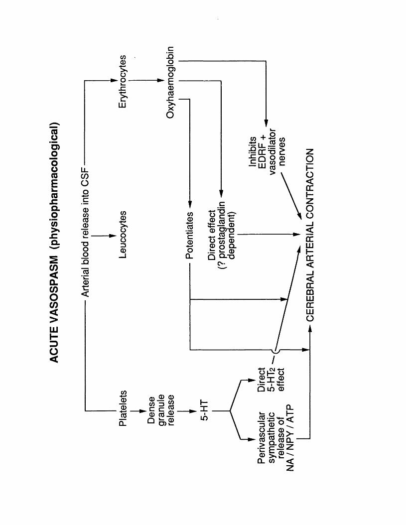
ACUT
E VA
SOSP
ASM
(p
hysi
opha
rmac
olog
ical
)
LLCOOO
CDCOco_a)CD"Ooo_Q
lo"k_CD■c<
COCD>*o- o -V_JZk_
LU
claoO)oECDCO_c>*XO
CO COi o— + m w# L L ^ CD
— OCTS £ Q Oc m w “ LU CO>
COCD>sOOo3CD
COCDCO
c_CDo
CL
a> 5 c3 = — CD3= -=- w CD 0 ) - 0
^ CO c ■ O to CDCD o Q-- ^ CD
Q Q - T 3 C-
CD ^ CD
® — 9CD CO -Hi
V* UJ S'CO S CO CL
- CE
REB
RA
L AR
TERI
AL
CO
NTR
AC
TIO
N

overactivity, could in any way contribute to later vessel wall damage and delayed chronic cerebral arterial narrowing; mediated perhaps via the mechanism of excitotoxic neurotransmitter release? High levels, both of CSF and circulating catecholamines are quite commonly observed in man after SAH, with concommitant depletion of neurotransmitters from within the terminals of cerebrovascular nerve fibers. It has certainly been observed experimentally that production of an intense arterial constriction by the application of such catecholamines in vitro, can result subsequently in the development of recognisable vessel wall damage (Alksne and Greenhoot, 1974; Joris and Majno, 1981).
Chronic vasospasm:Chronic cerebrovasospasm is usually only manifested at
about the fourth day after haemorrhage, it is maximal in incidence in the second week and largely gone by the end of the third week. Despite the application of a quite extensive variety of pharmacological agents, such delayed vasospasm, unlike acute vasospasm, has proven remarkably resistant to all attempts aimed either at preventing or producing lasting reversal of luminal narrowing. In the introduction to experimental chapter VI, evidence was presented obtained both from human studies and from animal models including this one, that strongly suggests that delayed cerebral arterial luminal narrowing represents the development of structural pathological changes in the vessel wall itself, rather than any contractile activity of the cerebrovascular smooth muscle per-se. The occurrence of vessel wall changes
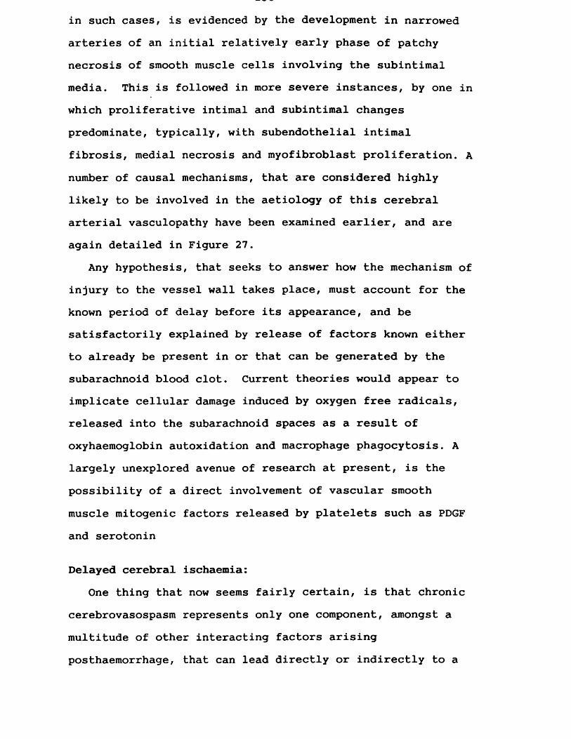
in such cases, is evidenced by the development in narrowed arteries of an initial relatively early phase of patchy necrosis of smooth muscle cells involving the subintimal media. This is followed in more severe instances, by one in which proliferative intimal and subintimal changes predominate, typically, with subendothelial intimal fibrosis, medial necrosis and myofibroblast proliferation. A number of causal mechanisms, that are considered highly likely to be involved in the aetiology of this cerebral arterial vasculopathy have been examined earlier, and are again detailed in Figure 27.
Any hypothesis, that seeks to answer how the mechanism of injury to the vessel wall takes place, must account for the known period of delay before its appearance, and be satisfactorily explained by release of factors known either to already be present in or that can be generated by the subarachnoid blood clot. Current theories would appear to implicate cellular damage induced by oxygen free radicals, released into the subarachnoid spaces as a result of oxyhaemoglobin autoxidation and macrophage phagocytosis. A largely unexplored avenue of research at present, is the possibility of a direct involvement of vascular smooth muscle mitogenic factors released by platelets such as PDGF and serotonin
Delayed cerebral ischaemia:One thing that now seems fairly certain, is that chronic
cerebrovasospasm represents only one component, amongst a multitude of other interacting factors arising posthaemorrhage, that can lead directly or indirectly to a
CHRO
NIC
VASO
SPAS
M
(pat
holo
gica
l)_C
c o oD)‘= O 03EI20 X 03 OS' <3
LJL
— (D□ ® X E <D O -C O ~ CO c/5 m F 0 35 Qji w ®Q> > CL co
CL
0C
EREB
RA
L AR
TERI
AL
. in
flam
mat
ion
LUM
INAL
NA
RRO
WIN
G
& fib
rosi
s
delayed onset of reduced cerebral blood flow with its attendant risk of cerebral ischaemia (Figure 28). Autoregulation of cerebrovascular blood flow is commonly disturbed after SAH, and it becomes increasingly impaired in those patients in the worst clinical grades. Such patients tolerate poorly, even quite modest falls in their arterial blood pressure. More recently, a greater awareness has emerged of the frequency with which patients who develop hyponatraemia after SAH also suffer from hypovolaemia. In these patients there is an urgent need to actually expand the circulating volume, rather than to fluid restrict them and thereby worsen the risk of ischaemia. Similarly, the role of communicating hydrocephalus and raised intracranial pressure developing subsequent to haemorrhage, has become more commonly recognised as one readily reversible cause of a compromised cerebral perfusion. Finally, an interesting finding that has recently emerged from experimental studies, is the suggestion that the sympathetic outflow from the brainstem locus coeruleus (LC) nucleus is also activated following SAH, along with the more well known activation of the peripheral sympathetic system (McCormack et al., 1986; Soloman et al., 1986). Potentially, this is of considerable interest, as it is known from experimental studies that stimulation of the LC nucleus can result in a global reduction in CBF (Raichle et al., 1975). It is possible therefore, although as yet only speculative, that in man a greatly increased activity of the LC nucleus after SAH may be one further mechanism in the pathophysiology of the reduced CBF following SAH.
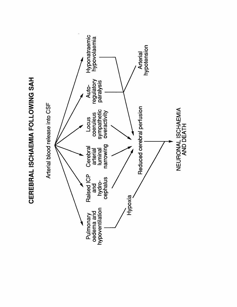
CERE
BRAL
IS
CHAE
MIA
FO
LLOW
ING
SAH
LLCOoOCCDCOco0)0L_
1—a)■c<
e |ilco o c lQ-
03 CO ■JZ c 0 0J o< Q_
CO-j^§ o | - | o d (59° o E |O >s> CO o
Q_
JQ0
■o
O
LU<XoCO
oX3LU
CO 03,03
AND
DEA
TH
11 ACKNOWLEDGEMENTS AND STATEMENT OF ORIGINALITY
The work for this thesis was principally funded by the Medical Research Council who awarded the author an MRC Training Fellowship. Additional financial support was provided by the Welton Trust. I would first and foremost like to thank, and state my appreciation to my joint supervisors; Alan Crockard, Consultant Neurosurgeon at the National Hospital for Nervous Diseases, Queens Square London, and Professor Geoffrey Burnstock, Head of the Department of Anatomy and Developmental Biology and Centre for Neuroscience, University College London. I also wish to express my considerable gratitude to Professor Lieberman,Dr. Parnavelas, and other members of the Anatomy Department at UCL such as Professors Gray, Zeki and Wall, for first inspiring my interest in the field of Neuroscience as a undergraduate. A commitment to Neurosurgery, and appreciation of the need to apply a scientific approach in order to advance the speciality, was subsequently encouraged by my working together with Alan Crockard, David Thomas and Professor L Symon in the Departments of Neurosurgery at the National Hospitals for Nervous Diseases, London. I would also like here to thank Dr Ralph Ross Russell, both for his quiet friendly encouragement and for allowing me the use of his laboratory facilities at the Institute of Neurology. I am extremely grateful to John Parnavelas who patiently taught me the techiques of immunohistochemistry, also to Katherine Allen who together with John Dobbin, advised me both in the techiques of CBF measurement by hydrogen clearance, and small animal inhalational anaesthesia.
All of the experimental studies, observations and technical preparations discussed in this thesis, were designed and performed entirely by the author with the exception of the help received from the following individuals. The analysis by high performance liquid chromatography of homogenised vessel samples for 5-HT and noradrenaline content, for which I must acknowledge the assistance of Jill Lincoln at UCL. Additional thanks are also due both to Yvonne Blankson at the Institute of Neurology and Frank Kristek at UCL, for their help in preparation of histological sections of brains used to demonstrate electrode tracks, and preparation of some of the material used for transmission electron microscopy respectively. The plates illustrating this thesis were produced with the expert assistance of the photographic department of U.C.L. Finally, and most importantly of all I wish to reserve my strongest and warmest thanks to my wife Karen, without whose constant support, encouragement, and practical help with the typing this manuscript would never have been realised.
During the course of performing these studies I held the following appointments:
Honorary Registrar in Neurosurgery, the National Hospitals for Nervous Diseases, Queen Square, London.
MRC Research Fellow, The Institute of Neurology, Queen Square, London.
12. REFERENCES
Abboud FM (1981) Special characteristics of the cerebral circulation. Fed Proc 40:2296-2300
Adams HP (1986) Early management of the patient with recent aneurysmal subarachnoid haemorrhage. Stroke 17:1068-1070
Adams HP, Nibblelink DW, Torner JC, Sahs AL (1981)Antifibrinolytic therapy in patients with aneurysmal subarachnoid haemorrhage. Arch Neurol 38:25-29
Alafaci C, Cowen T, Crockard HA, Burnstock G (1986) Cerebral perivascular serotonergic fibers have a peripheral origin in the gerbil. Brain Res Bull 16:303-304
Alborch E, Torregrosa G, Salom JB, Miranda FJ, Campos V, Alabadi JA (1989) Heterogenicity of P 2 ~purinoceptors in brain circulation. J Cereb Blood Flow Metab 9 (suppl 1 ):S210
Alksne JF, White LE (1965) Electron-microscope study of the effects of increased intracranial pressure on the arachnoid villus. J Neurosurg 22:481-488
Alksne JF, Lovings E (1972) The role of the arachnoid villi in the removal of red blood cells from the subarachnoid space. An electron microscopic study in the dog. J Neurosurg 36:192-200
Alksne JF, Greenhoot JH (1974) Experimental catecholamine induced chronic cerebral vasospasm:myonecosis in vessel wall. J Neurosurg 41:440-445
Allcock J, Drake C (1965) Ruptured intracranial aneurysms - the role of arterial spasm. J Neurosurg 22:21-29
Allen GS, Henderson LM, Chou SN, French LA (1974a) Cerebral arterial spasm. Part 1: In vitro contractile activity of vasoactive agents on canine basilar and middle cerebral arteries. J Neurosurg 40:433-441
Allen GS, Henderson LM, Chou SN, French LA (1974b) Cerebral arterial spasm. Part 2: In vitro contractile activity of serotonin in human serum and CSF on the canine basilar artery, and its blockage by methylsergide and phenoxybenzamine. J Neurosurg 40:442-450
Allen GS, Gold LHA, Chou SN, French LA (1974c) Cerebralarterial spasm. Part 3: In vivo intracisternal production of spasm by serotonin and blood and its reversal by phenoxybenzamine. J Neurosurg 40:451-458
Allen GS, Gross CJ, French LA, Chou SN (1976) In vitrocontractile activity of vasoactive agents including human CSF on human basilar and anterior cerebral arteries. J Neurosurg 44:594-600.
Allen GS, Banghart SB (1979) Cerebral arterial spasm. Part 9: In vitro effects of nifedipine on serotonin-, phenylephrine-, and potassium-induced contractions of basilar and femoral arteries. Neurosurg 4:37-42
Allen JM, Todd N, Crockard HA, Schon F, Yetas JC, Bloom SR (1984) Presence of neuropeptide Y in human circle of Willis and its possible role in cerebral vasospasm.Lancet 2:550-552
Alvund ED, Loeser JD, Bailey WL, Compass MK (1972)Subarachnoid haemorrhage due to ruptured aneurysms. Arch Neurol 27:273-281
Anthony M, Hiterberger H, Lance JW (1969) The possiblerelationship of serotonin to the migraine syndrome. Res Clin Stud Headache 2:29-59
Arneric SP, Iadecola C, Underwood M, Reis D (1987) Local cholinergic mechanisms participate in the increase in cortical cerebral blood flow elicited by electrical stimulation of the fastigial nucleus in the rat. Brain Res 411:212-225
Astrup J, Heuser D, Lasser NA, Nilsson B, Nordberg K, Siesjo BK (1976) Evidence against H+ and K+ as the main factors in the regulation of cerebral blood flow during epileptic discharges, acute hypoxia, amphetamine intoxication, and hypoglycaemia. A microelectrode study. In: Ionic Actions on Vascular Smooth Muscle (Betz E, ed), New York, Springer, pp110-115
Astrup J, Heuser D, Lasser NA, Nilsson B, Nordberg K, Siesjo BK (1979) Evidence against H+ and K+ as main factors for the control of cerebral blood flow. A microelectrode study. In: Cerebral Vascular Smooth Muscle and its Control (Ciba Foundation Symposium 56), Amsterdam, Elsevier, pp313-332
Auckland K, Bower BF, Berliner RW (1964) Measurement of local blood flow with hydrogen gas. Cir Res 14:164-187
Auer LM (1985) The comatose patient at the acute stage - a surgical taboo? In: Timing of aneurysm surgery (Auer LM, ed), Berlin, Walter de Gruyter
Auer LM, Brandt L, Ebeling U, Gilsbach J, Groeger U, Harders A, Ljunggren B, Oppel F, Reulen HJ, Saeveland H (1986) Nimodipine and early aneurysm operation in good condition SAH patients. Acta Neurochir 82:7-13
Awad I, Carter LP, Spetzler RF, Medina M, Williams FW (1986) Clinical vasospasm after subarachnoid haemorrhage. Response to hypervolemic haemodilution and arterial hypertension. Stroke 18:365-372
Babior B (1978) Oxygen-dependent microbial killing by phagocytes. N Engl J Med 298:659-668 and 721-725
Barry KJ, Gogjian MA, Stein BM (1979) A small animal model for the investigation of subarachnoid haemorrhage and cerebral vasospasm. Stroke 10:538-541
Bates D, Weinshilboum RM, Campbell RJ, Sundt TM Jr (1977) The effect of lasions in the locus coeruleus on the physiological responses of the cerebral blood vessels in cats. Brain Res 136:431-443
Baumgarten HG (1972) Biogenic monoamines in the cyclostome and lower vertebrate brain. Prog Histochem Cytochem 4:1-89
Bederson JB, Bartkowski HM, Obana W, Nishimura MC, Tsuji M, Pitts LH (1985) Nimodipine in rat MCA occlusion (MACO). Stroke 16:142
Bell BA, Symon L (1979) Smoking and subarachnoid haemorrhage. Br Med J 1:577-578
Benedict CR, Loach AB (1978) Sympathetic nervous system activity in patients with subarachnoid haemorrhage. Stroke 9:237-244
Bergvall U, Steiner L, Forster DMCL (1973) Early pattern of cerebral circulatory disturbances following subarachnoid haemorrhage. J Neurosurg 5:24-32
Berne RM, Rubio R, Curnish RR (1974) Release of adenosine from ischemic brain: Effect on cerebral vascular resistance and incorporation into cerebral adenine nucleotides. Circ Res 35:262-271
Betz E, Heuser D (1967) Cerebral cortical blood flow during changes of acid-base equilibrium of the brain. J Appl Physiol 23:726-733
Betz E, Enzenross HG, Vlahov V (1973) Interaction of H+ and Ca^+ in the regulation of local pial vascular resistance. Pfluegers Arch 343:79-88
Bigelow NH (1953) The association of polycystic kidneys with intracranial aneurysms and other related disorders. Am J Med Sci 225:485-494
Bill A (1979) Effect of indomethacin on regional blood flow in conscious rabbits. A microsphere study. Acta Physiol Scand 105:437-442
Bill A, Linder J (1976) Sympathetic control of cerebral blood flow in acute arterial hypertension. Acta Physiol Scand 96:114-121
Black PM (1986) Hydrocephalus and vasospasm aftersubarachnoid haemorrhage from ruptured intracranial aneurysms. Neurosurgery 18:12-16
Black BK, Hicks J (1952) The relation of hypertension to arterial aneurysms of the brain. US Armed Services Med J 3:1813-1818
Bockman EL, Fritschka E, Ferguson JL, Spitzer JJ (1981) Increased adenosine in cerebrospinal fluid during the autoregulatory response to mild hypotension. In: Cerebral Microcirculation and Metabolism (Cervos-Navarro J, Fritschka E, eds) New York, Raven Press, pp243-248
Bohm E, Hugosson R (1978) Experiences of surgical treatment of 400 consecutive ruptured cerebral arterial aneurysms. Acta Neurochir (Wein) 40:33-43
Borgstrom L, Johansson H, Siesjo BK (1975) The relationship between arterial p02 and cerebral blood flow in hypoxic hyperaemia. Acta Physiol Scand 93:423-432
Boullin DJ (1980) Cerebral vasospasm. Chichester, Wiley
Boullin D (1985) Cerebral arterial spasm (CerebralVasospasm). In: Neurosurgery The Scientific Basis of Clinical Practice (Crockard A, Hayward R, Hoff JT, eds), Oxford, Blackwell Scientific Publications, pp240-266
Boullin DJ, Mohan J, Grahame-Smith DG (1976) Evidence for the presence of a vasoactive substance (possibly involved in the aetiology of cerebral arterial spasm) in cerebrospinal fluid from patients with subarachnoid haemorrhage. J Neurol Neurosurg Psych 39:756-766
Boullin DJ, Boulay GH, Rogers AT (1978) Aetiology of cerebral arterial spasm following subarachnoid haemorrhage: evidence against a major involvement of 5-hydroxytryptamine in the production of acute spasm. Br J Clin Pharm 6:203-215
Boullin DJ, Blaso WP, Mistry G, DuBoulay GH, Aitken V,Tagari P (1981) Role of prostacyclin in physiological control of the cerebral circulation in health and cerebrovascular disease. In: Cerebral Microcirculation and Metabolism (Cervos-Navarro J, Fritschka E, eds), New York, Raven Press, pp205-211
Boullin DJ, Tagari P, DuBoulay G, Aitken V, Hughes JT (1983) The role of hemoglobin in the etiology of cerebral vasospasm. An in vivo study of baboons. J Neurosurg 59:231-236
Brandt L, Ljunggren B, Andersson KE, Hindfelt B, Uski T (1981) Effects of indomethacin and prostacyclin on isolated human pial arteries contracted by CSF from patients with aneurysmal SAH. J Neurosurg 55:877-883
Brandt L, Sonesson B, Ljunggren B, Saveland H (1987)Ruptured middle cerebral artery aneurysm with intracerebral haemorrhage in younger patients appearing moribund: emergency surgery? Neurosurgery 20:925-929
Braunwald E (1982) Mechanism of action of calcium-channel blocking agents. N Engl J Med 307:1618-1627
Brawley BW, Strandness DE Jr, Kelley WA (1968) The biphasic response of cerebral vasospasm in experimental subarachnoid haemorrhage. J Neurosurg 28:1-8
Brock M, Jane JA (1980) Intracranial haemorrhage. Summary. In: Intracranial Pressure IV (Shulman K, Marmarou A, Miller JD, Becker DP, Hochwald GM, Brock M, eds), Berlin, Springer-Verlag, pp201-202
Burnstock G (1977) Ultrastructure of the vascularneuroeffector apparatus, with special regard to brain vessels. In: Neurogenic Control of the Brain Circulation. (Owman C, Edvinsson L, eds), Oxford, Pergammon Press, pp3-10
Burnstock G (1978) A basis for distinguishing two types of purinergic receptor. In: Cell Membrane Receptors for Drugs and Hormones: A Multidisciplinary Approach (Bolis L, Straub RW, eds), New York, Raven Press, pp107-118
Burnstock G (1987) Present status of purinergicneurotransmission - implications for vascular control.In: Neuronal Messengers in Vascular Function (Nobin A, Owman C, Arneklo-Nobin B, eds), Amsterdam, Elsevier, pp327-340
Burnstock G, Kennedy C (1986) A dual function for adeosine 5 *-triphosphate in the regulation of vascular tone. Circ Res 58:319-330
Busija DW, Heistad DD, Marcus ML (1980) Effect ofsympathetic nerves on cerebral vessels during acute, moderate increases in arterial pressure in dogs and cats. Circ Res 46:696-702
Busija DW, Heistad DD (1981) Effects of cholinergic nerves on cerebral blood flow in cats. Circ Res 48:62-69
Busija DW, Heistad DD (1983) Effects of indomethacin on cerebral blood flow during hypercapnia in cats. Am J Physiol 244:H519-H524
Butler AB, Maffeo CJ, Johnson RN, Bass NH (1980) Impaired absorption of CSF during experimental subarachnoid haemorrhage: Effects of blood components on vesicular transport in arachnoid villi. In: Intracranial pressure IV (Shulman K, Marmarou A, Miller JD, Becker DP, Hochwald GM, Brock M, eds), Berlin, Springer, pp245-248
Byer E, Ashman R, Toth LA (1947) Electrocardiograms with large upright T waves and long Q-T intervals. Am Heart J 33:796-806
Cameron IR, Caronna J (1976) The effect of local changes in potassium and bicarbonate concentration on hypothalamic blood flow in the rabbit. J Physiol (London) 262:415-430
Campbell GJ, Roach MR (1981) Fenestrations in the internal elastic lamina at bifurcations of human cerebral arteries. Stroke 12:489-496
Capon A (1975) Effect of acute sections of the brain stem on hypercapnic vasodilation of cerebral and spinal vessels. In: Blood Flow and Metabolism in the Brain (7th International Symposium on Cerebral Blood Flow and Metabolism), New York, Churchill Livingston, pp1.16-1.17
Carroll RC, Haddon W (1964) Birth characteristics of persons dying of cerebral aneurysms. J Chron Dis 17:705-711
Cavanagh JFR, Mione MC, Burnstock G (1989) Colocalisation of NPY and VIP in cerebrovascular nerves originating from the pterygopalatine ganglion in the rat. J Cereb Blood Flow Metab 9(suppl 1):S677
Chang JY, Owman C (1986) Immunohistochemical andpharmacological studies on serotonergic nerves and receptors in brain vessels. Acta Physiol Scand Suppl 552:49-53
Chang JY, Owman C (1987) Involvement of specific receptors and calcium mechanisms in serotonergic contractile responses of isolated cerebral and peripheral arteries from rat. J Pharmacol Exp Ther 242:629-636
Chang JY, Hardebo JE, Owman C, Sahlin C, Svendgaard NA(1987) Nerves containing serotonin, its interaction with noradrenaline and characterisation of serotonergic receptors in cerebral arteries of monkey. J Auton Pharmacol 7:317-329
Chan-Palay V (1976) Serotonin axons in the supra- and subependymal plexuses and in the leptomeninges; their roles in local alterations of cerebrospinal fluid and vasomotor activity. Brain Res 102:103-130
Chehrazi BB, Giri S, Joy RM (1988) Prostaglandins andvasoactive amines in cerebral vasospasm after aneurysmal subarachnoid haemorrhage. Stroke 20:217-224
Cheng JT, Shen CL (1987) Release of neuropeptide Y (NPY) induced by tyramine in the isolated vas deferens of rat. Naunyn-Schiedebergs Arch Pharmacol 335:255-260
Chorobski S, Penfield W (1932) Cerebral vasodilator nerves and their pathway from the medulla oblongata with observations of the pial and intracerebral vascular plexuses. Arch Neurol 28:1257
Chyatte D, Fode N, Sundt T (1988) Early versus late intracranial aneurysm surgery in subarachnoid haemorrhage. J Neurosurg 69:326-331
Ciongoli AK, Poser CM (1972) Pulmonary edema secondary to subarachnoid haemorrhage. Neurology 22:867-870
Cohen RA, Zitnay KM, Weisbrod RM (1987) Accumulation of 5-hydroxytryptamine leads to dysfunction of adrenergic nerves in canine coronary artery following intimal damage. Circ Res 61:829-833
Conway LW, McDonald LM (1972) Structural changes of theintradural arteries following subarachnoid haemorrhage. J Neurosurg 37:715-723
Coons AH, Leduc EH, Conolly JM (1955) Studies on antibody production 1. A method for the histochemical demonstration of specific antibody and its application to a study of the hyperimmune rabbit. J Exp Med 102:49-60
Cort JH (1954) Cerebral salt wasting. Lancet 1:752-754
Costa M, Furness JB, Cuello AC, Verhofstad AAJ, Steinbusch HWJ, Elde RP (1982) Neurons with 5-hydroxytryptamine-like immunoreactivity in the enteric nervous system: their visualization and reactions to drug treatment. Neuroscience 7:351-363
Cowen T, Haven AJ, Burnstock G (1985) Pontamine sky blue: a counter stain for background autofluorescence in fluorescence and immunofluorescence histochemistry. Histochemistry 82:205-208
Cowen T, Alafaci C, Crockard HA, Burnstock G (1986)5-HT-containing nerves to major cerebral arteries of the gerbil originate in the superior cervical ganglia. Brain Res 384:51-59
Cowen T, Alafaci C, Crockard HA, Burnstock G (1987) Origin and postnatal development of nerves showing 5-hydroxytryptamine-like immunoreactivity supplying major cerebral arteries of the rat. Neurosci Letts 78:121-126
Crawford MD, Sarner M (1965) Ruptured intracranial aneurysm: Community study. Lancet 2:1254-1257.
Cremer JE, Seville MP (1983) Regional blood flow, bloodvolume, and haematocrit values in the adult rat. J Cereb Blood Flow Metab 3:170-182
Crockard HA, Iannotti F, Ladds G (1982) Cerebrovascular effects of prostaglandin inhibitors in the gerbil. J Cereb Blood Flow Metab 2:67-72
Crompton MR (1964) The pathogenesis of cerebral infarction following the rupture of cerbral berry aneurysms. Brain 87:491-510
Crompton MR (1966) The pathogenesis of cerebral aneurysms. Brain Res 89:797-814
Crompton MR (1976) Pathology of degenerative cerebral arterial disease. In: Cerebral Arterial Disease (Ross Russell RW ed), London, Churchill Livingstone, pp40-56
Cudennec A, Duverger D, Mackenzie ET, Scatton B, Serrano A(1987) Serotonergic neuron stimulation modulates thalamocortical glucose use in the conscious rat. J Cereb Blood Flow Metab 7:502-506
Cudennec A, Duverger D, Mackenzie ET (1989) Nature of the regional increases in cerebral blood flow induced by raphe stimulation in the conscious rat. J Cereb Blood Flow Metab 9(suppl 1):S375
Cummins BH, Lothian D (1973) Amine levels in thecerebrospinal fluid after subarachnoid haemorrhage. Br J Surg 60:910
Dahl E (1973) The fine structure of intracerebral vessels. Z Zellforsch 145:577-586
Dahl E (1986) The ultrastructure of cerebral blood vessels in man. Cephalgia 6 (suppl 4):45-48
Dahl E, Nelson E (1964) Electron microscopic observations on human intracranial arteries: II. Innervation. Arch Neurol (Chicago)10:158
Dahlgren N, Lindvall 0, Sakabe T, Siesjo BK (1981a) Cerebral blood flow and oxygen consumption in the rat brain after lesions of the noradrenergic locus coeruleus system.Brain Res 209:11-23
Dahlgren N, Ingvar M, Yokoyama H, Siesjo BK (1981b) Effect of indomethacin on local cerebral blood flow in awake, minimally restrained rats. J Cereb Blood Flow Metab 1:233-236
Dahlstrom A, Fuxe K (1964) Evidence for the existence of monoamine-containing neurons in the central nervous system. I. Demonstration of monoamines in the cell bodies of brainstem neurons. Acta Physiol Scand 62(suppl232): 1-55
Dail WG, Evan AP (1978) Ultrastructure of adrenergic nerve terminals and SIF cells in the superior cervical ganglion of the rabbit. Brain Res 148:469-477
D'Alecy LG, Rose CJ (1977) Parasympathetic cholinergic control of cerebral blood flow in dogs. Circ Res 41:324-331
Dandy WE (1938) Intracranial aneurysm of internal carotid artery, cured by operation. Ann Surg 107:654-659
Dandy WE (1944) Intracranial Arterial Aneurysms. New York, Comstock, pp1-147
Davis JM, Davis KR, Crowell RM (1980) Subarachnoidhaemorrhage secondary to ruptured intracranial aneurysm: Prognostic significance of cranial CT. AJNR 1:17-21
Delgado TJ, Brismar J, Svendgaard NA (1985) Subarachnoid haemorrhage in the rat; Angiography and fluorescence microscopy of the major cerebral arteries. Stroke 16:595-602
Delgado TJ, Arab MAR, Diemer NH, Svendgaard NA (1986)Subarachnoid haemorrhage in the rat: Cerebral blood flow and glucose metabolism during the late phase of cerebral vasospasm. J Cereb Blood Flow Metab 6:590-599
Delgado TJ, Arbab MAR, Rosengren E, Svendgaard NA (1987) Effect of neonatal 6-hydroxydopamine treatment on experimental vasospasm following a subarachnoid haemorrhage in the rat. J Cereb Blood Flow Metab 7:289-294
Delgado TJ, Arbab MAR, Warberg J, Svendgaard NA (1988) The role of vasopressin in acute cerebral vasospasm. Effect on spasm of a vasopressin antagonist or vasopressin antiserum. J Neurosurg 68:266-273
Dempsey RJ, Roy MW, Cowen DE (1986) Lipoxygenase metabolites of arachidonic acid and the development of ischaemic cerebral oedema. Neurol Res 8:53-56
de Paepe A, van Landegen W, de Keyser F, De Reuck J (1988) Association of multiple intracranial aneurysms and collagen type III deficiency. Clin Neurol Neurosurg 90:53-56
Dernbach PD, Little JR, Jones SC, Ebrahim ZY (1988) Altered cerebral autoregulation and CO 2 reactivity after aneurysmal subarachnoid haemorrhage. Neurosurgery 22:822-826
DiBona GF (1985) Neural regulation of renal tubular sodium reabsorption and renin release. Fed Proc 44:2816-2822
DiCarlo V (1977) Histochemical evidence for a serotonergic innervation of the microcirculation in the brainstem. In: Neurogenic Control of the Brain Circulation (Owman C, Edvinsson L, eds), London, Pergamon Press, pp55-58
DiCarlo V (1984) Perivascular serotonergic neurons:Somatodendritic contacts and axonic innervation of blood vessels. Neurosci Letts 51:295-302
Diringer M, Ladenson PW, Stern BJ, Schleimer J, Hanley DF(1988) Plasma atrial natriuretic factor and subarachnoid haemorrhage. Stroke 19:1119-1123
Dixon JM (1951) Angioid streaks and pseudoxanthomaelasticum: With aneurysm of the internal carotid artery. Am J Ophthalmol 34:1322-1323
Doczi T (1984) Involvement of vasopressin in brain oedema formation: Further evidence obtained from the Brattleboro diabetes insipidus rat with experimental subarachnoid haemorrhage. Neurosurgery 14:436-441
Doczi T (1985) The pathogenetic and prognostic significance of blood-brain barrier damage at the acute stage of aneurysmal subarachnoid haemorrhage. Clinical and experimental studies. Acta Neurochirg 77:110-132
Doczi T, Bende J, Huska E, Kiss J (1981) Syndrome ofinappropriate secretion of antidiuretic hormone after subarachnoid haemorrhage. Neurosurgery 4:394-396
Doczi T, Nemessanyi Z, Svegvary Z (1983) Disturbances of cerbrospinal fliud circulation during the acute stage of subarachnoid haemorrhage. Neurosurgery 12:435-438
Doczi T, Ambrose J, O'Laoire S (1984) Significance ofcontrast enhancement in cranial computerized tomography after subarachnoid haemorrhage. J Neurosurg 60:335-342
Doczi T, Joo F, Vecsernyes M, Bodosi M (1988) Increased concentration of atrial natriuretic factor in the cerebrospinal fluid of patients with aneurysmal subarachnoid haemorrhage and raised intracranial pressure. Neurosurg 23:16-19
Dolenc V, Fettich M, Korsic M, Pregelj R, Skrap M, Lamovec Z, Cerk M, Kregar T (1982) Blood clot evacuation in aneurysm surgery in the acute stage (Arguments pros and cons) Acta Neurochir (Wien) 63:105-109
Doshi R, Neil-Dwyer G (1977) Hypothalamic and myocardial lesions after subarachnoid haemorrhage. J Neurol Neurosurg Psych 40:821-826
Dott NM (1933) Intracranial aneurysms: cerebralarterio-radiography, surgical treatment. Edin Med J 40:219-240
Doyle TF, Martins AN, Kobrine AI (1975) Estimating total cerebral blood flow from the initial slope of hydrogen washout curves. Stroke 6:149-152
Drake CG (1978) On the surgical treatment of intracranial aneurysms. Ann R Coll Phys Surg Can 11:185-195
Drake CG (1981) Management of cerebral aneurysms. Stroke 12:273-283
Dreyfus CF, Bornstein MB, Gershon MD (1977) Synthesis of serotonin* by neurons of the myenteric plexus in situ and in organotypic tissue culture. Brain Res 128:125-139
Du Boulay G (1963) Distribution of spasm in the intracranial arteries after subarachnoid haemorrhage. Acta Radiol (Diagn) 1:257-266
Ducker TB (1968) Increased intracranial pressure andpulmonary edema. Part 1: Clinical study of 11 cases. J Neurosurg 28:112-117
Ducker TB, Simmons RL (1968) Increased intracranial pressure and pulmonary edema. Part 2: The hemodynamic response of dogs and monkeys to increased intracranial pressure. J Neurosurg 28:118-123
Duckies SP, Bevan JA (1976) Pharmacological characterization of adrenergic receptors of a rabbit cerebral artery in vitro. J Pharmacol Exp Ther 197:371-378
Duckies SP, Kim J, Bevan RD, Bevan JA (1977) Alteration of innervation and reactivity of monkey cerebral arteries after subarachnoid haemorrhage. Stroke 8:10
Duff TA, Louie J, Feilbach JA, Scott G (1988) Erythocytes are essential for development of cerebral vasculopathy resulting fromsubarachnoid haemorrhage in cats. Stroke 19:68-72
Ebeige AB, Pickard JD, Jennett S (1980) Responses of systemic vascular smooth muscle to hypoxia. Q J Exp Physiol 65:273-292
Echlin FA (1965) Spasm of basilar and vertebral arteries caused by experimental subarachnoid haemorrhage. J Neurosurg 23:1-11
Eckenstein F, Baughman RW (1984) Two types of cholinergic innervation in cortex, one co-localised with vasoactive intestinal polypeptide. Nature 309:153-155
Ecker A, Riemenschneider P (1951) Arteriographicdemonstration of spasm of the intracranial arteries with special reference to saccular arterial aneurysms. J Neurosurg 8:660-667
Edvinsson L (1975) Neurogenic mechanism in thecerebrovascular bed. Autonomic nerves, amine receptors and their effects in cerebral blood flow. Acta Physiol Scand Suppl 427:1-36
Edvinsson L (1985) Functional role of perivascular peptides in the control of cerebral circulation. Trends in Neurosci 8:126-131
Edvinsson L, Owman CH, Rosengren E, West KA (1972)Concentrations of noradrenaline in pial vessels, choroid plexus, and iris during two weeks after sympathetic ganglionectomy or decentralization. Acta Physiol Scand 85:201-206
Edvinsson L, Lindvall M, Nielsen KC, Owman C (1973) Are brain vessels innervated also by central (non-sympathetic) adrenergic neurones? Brain Res 63:496-499
Edvinsson L, Hardebo JE, Owman C (1976a) Influence of the cerebrovascular sympathetic innervation on regional flow, autoregulation, and blood-brain barrier function. In: Cerebral Vascular Smooth Muscle and its Control (Ciba Foundation Symposium 56), Amsterdam, Elsevier, pp69-95
Edvinsson L, Owman C, Sjoberg NO (1976b) Autonomic nerves, mast cells and amine receptors in human brain vessels; A histochemical and pharmacological study. Brain Res 115:337-393
Edvinsson L, MacKenzie ET (1977) Amine mechansims in the cerebral circulation. Pharmacol Rev 28:275-348
Edvinsson L, Owman C (1977) Sympathetic innervation and adrenergic receptors in intraparenchymal cerebral arterioles of baboon. Acta Physiol Scand Suppl 452:57-59
Edvinsson L, Falk B, Owman C (1977a) Possibilities for a cholinergic action on smooth musculature and sympathetic axons in brain vessels mediated by muscarinic and nicotinic receptors. J Pharmacol Exp Ther 200:149-156
Edvinsson L, Hardebo JE, MacKenzie ET, Stewart M (1977b)Dual action of serotonin on pial arterioles in situ and the effect of propanolol on the response. Blood Vessels 14:366-371
Edvinsson L, Hardebo JE, Owman C (1978) Pharmacological analysis of 5-hydroxytryptamine receptors in isolated intracranial and extracranial vessels of the cat and man. Circ Res 42:143-151
Edvinsson L, Lacombe P, Owman C, Reynier-Rebuffel AM, Seylaz J (1979) Quantitative changes in regional cerebral blood flow of rats induced by alpha- or beta- adrenergic stimulants. Arch Physiol Scand 107:289-296
Edvinsson L, Fahrenkrug J, Hanko J, Owman C, Sundler F, Uddman R (1980) VIP (vasoactive intestinal polypeptide) -containing nerves of intracranial arteries in mammals. Cell Tissue Res 208:135-142
Edvinsson L, Fahrenkurg J, Hanko J, McCulloch S, Owman C, Uddman R (1981) Vasoactive intestinal polypeptide: Distribution and effects on cerebral blood flow and metabolism. In: Cerebral Microcirculation and Metabolism (Cervos-Navarro J, Fritschka E, eds), New York, Raven Press, pp147-155
Edvinsson L, Egund N, Owman C, Sahlin C, Svendgaard NA (1982) Reduced noradrenaline uptake and retention in cerebrovascular nerves asssociated with angiographically visible visible vasoconstriction folllowing experimental subarachnoid haemorrhage in rabbits. Brain Res Bull 9:799-805
Edvinsson L, Fredholm BB (1983) Characterisation ofadenosine receptors in isolated cerebral aretries of cat. Br J Pharmac 80:631-637
Edvinsson L, Emson P, McCulloch J, Tatemoto K, uddman R(1983a) Neuropeptide Y: cerebrovascular innervation and vasomotor effects in the cat. Neurosci Lett 43:79-84
Edvinsson L,Dequeurce A, Duverger D, MacKenzie ET, Scatton B (1983b) Central serotonergic nerves project to the pial vessels of the brain. Nature (Lond) 306:55-57
Edvinsson L, Jansen I (1987) Characterisation of tachykinin receptors in isolated basilar arteries of guinea-pig. Br J Pharmacol 90:553-559
Edvinsson L, Copeland J, Emson PC, McCulloch J, Uddman (1987) Nerve fibers containing neuropeptide Y in the cerebrovascular bed: Immunocytochemistry, radioimmunoassay and vasomotor effects. J Cereb Blood Flow Metab 7:45-57
Edvinsson L, Fallgren B, Jansen I, McCulloch J (1989)Effects of endothelin on feline cerebral vessels: Mechanisms of action. J Cereb Blood Flow Metab 9(suppl 1):S452
Ellington E, Margolis G (1969) Block of arachnoid villi by subarachnoid haemorrhage. J Neurosurg 30:651-657
Emerson TE, Raymond RM (1981) Effect of topical adenosine deaminase treatment on the functional hyperemic and hypoxic responses of cerebrocortical microcirculation. J Cereb Blood Flow Metab 4:447-457
Erspamer V, Testini A (1959) Observations on the release and turnover rate of 5-hydroxytryptamine in the gastrointestinal tract. J Pharm Pharmacol 11:618-623
Espinosa F, Weir B, Overton T, Castor W, Grace M, Boisvert D (1984a) A randomised placebo-controlled double-blind trial of nimodipine after SAH in monkeys. Part 1:Clinical and radiological findings. J Neurosurg 60:1167-1175
Espinosa F, Weir B, Shnitka T, Overton T, Boisvert D (1984b) A randomized placebo-controlled double-blind trial of nimodipine after SAH in monkeys. Part 2: Pathological findings. J Neurosurg 60:1176-1185
Essick CR (1920) Formation of macrophages by the cells lining the subarachnoid cavity in response to the stimulus of particulate matter. Carnegie Cont Embryol 42:379-389
Estanol BV, Marin OS (1975) Cardiac arrthymias and sudden death in subarachnoid haemorrhage. Stroke 6:382-386
Estanol BV, Loyo MV, Mateos JH, Foyo E, Cornejo A, Guavara J (1977) Cardiac arrhythmias in experimental subarachnoid haemorrhage. Stroke 8: 440-447
Evan AP, Dail WG, Dammrose D, Palmer C (1976) Scanningelectron microscopy of cell surfaces following removal of extracellular material. Anat Rec 185:433-446
Eversden ID, Morgan M, Lewer Allen M, Richardson AE (1977) Cerebral blood flow and cerebral blood volume in patients with subarachnoid haemorrhage. Acta Neurol Scand (Suppl) 56:340-341
Falconer MA (1950) Surgical treatment of spontaneoussubarachnoid haemorrhage. Preliminary report. Br Med J 1:809-813
Farquhar MG, Hartmann JF (1956) Electron microscopy of cerebral capillaries. Anat Rec 124:288
Farrar JK (1987) Hydrogen clearance technique. In: Cerebral blood flow physiological and clinical aspects (Wood JH, ed), New York, McGraw-Hill book company, pp 275-287
Feibel JH, Campbell RG, Joynt RJ (1976) Myocardial damage and cardiac arrthymias in cerebral infarction and subarachnoid haemorrhage: Correlation with increased systemic catecholamine output. Trans Am Neurol Ass 101:242-244
Fein JM (1975) Cerebral energy metabolism after subarachnoid haemorrhage. Stroke 6:1-8
Ferguson GG, Harper AM, Fitch W, Rowan JO, Jennet B (1972) cerebral blood flow measurements after spontaneous subarachnoid haemorrhage. Europ Neurol 8:15-22
Fick A (1870) Ueber die messung des Blutquantums in denHerzventrikeln. Sitz ber Physik-med ges Wurzburg 2:16-28
Fields WS, Bruetman ME, Weibel J (1965) Collateral circulation of the brain. Baltimore, Williams and Wilkins.
Finn SS, Stephensen SA, Miller CA, Drobnich L, Hunt W (1986) Observations on the perioperative management of aneurysmal subarachnoid haemorrhage. J Neurosurg 65:48-62
Fischer C, Roberson G, Ojemann R (1977) Cerebral vasospasm with ruptured saccular aneurysm - the clinical manifestations. Neurosurgery 1:245-248
Fitch W, MacKenzie ET, Harper AM (1975) Effects ofdecreasing arterial pressure on cerebral blood flow in the baboon: Influence of the sympathetic nervous system. Circ Res 37:550-557
Flamm ES (1986) The timing of aneurysm surgery 1985. Clin Neurosurg 33:147-158
Fog M (1938) The relationship between the blood pressure and the tonic regulation of the pial arteries. J Neurol Psych 1:187-197
Folkow B (1964) Description of the myogenic hypothesis.Circ Res Suppl 15:279-287
Foltz EL, Ward AA (1956) Communicating hydrocephalus from subarachnoid bleeding. J Neurosurg 13:546-566
Forbus WD (1930) On the origin of miliary aneurysms of the superficial cerebral arteries. Bull Johns Hopkins Hosp 47:239-284
Fox JL, Falik JL, Shalhoub RJ (1971) Neurosurgicalhyponatremia: the role of inappropriate antidiuresis. J Neurosurg 34:506-514
Fozard JR, Mwaluko GMP (1976) Mechanism of the indirect sympathomimetic effect of 5-hydroxytryptamine on the isolated heart of the rabbit. Br J Pharmacol 57:115-125
Fraser RAR, Stein BM, Barrett RE (1970) Noradrenergicmediation of experimental cerebrovascular spasm. Stroke 1:356-362
Fujiwara T, Uehara Y (1984) The cytoarchitecture of the wall and the innervation pattern of the microvessels in the rat mammary gland: A scanning electron microscopic observation. Am J Anat 170:39-54
Fukawa O, Aihara H (1987) Familial intracranial aneurysm - report of 20 cases in eight families and review of the literature. No Shinkei Geka (NYV) 15:911-919
Furchgott RF (1983) Role of endothelium in responses of vascular smooth muscle. Circ Res 53:557-573
Furchgott RF, Zawadzski JV (1980) The obligatory role of endothelial cells in the relaxation of arterial smooth muscle by acetylcholine. Nature 288:373-376
Fuxe K, Hamberger B, Hokfelt T (1968) Distribution ofnoradrenaline nerve terminals in cortical areas of the rat. Brain Res 8:125-131
Fuxe K, Hokfelt T, Said SI (1977) Vasoactive intestinalpolypeptide and the nervous system: Immmunohistochemical evidence for localization in central and peripheral neurons, particularly intracortical neurons of the cerebral cortex. Neurosci Lett 5:241-246
Galloon S, Rees GAD, Briscoe CE (1972) Prospective study of electrocardiographic changes associated with subarachnoid haemorrhage. Br J Anaesth 44:511-516
Geffen LB, Livett BG (1971) Synaptic vesicles in sympathetic neurons. Physiol Rev 51:98-157
Gershon MD, Robinson RG, Ross LL (1976) Serotoninaccumulation in the guinea pig myenteric plexus: ion dependence, structure-activty relationship and the effect of drugs. J Pharmacol Exp Ther 198:548-561
Gershon MD, Sherman DL (1987) Noradrenergic innervation of serotonergic neurons in the myenteric plexus. J Comp Neurol 259:193-210
Gibbins IL, Brayden JE, Bevan JA (1984) Distribution and origins of VIP-immunoreactive nerves in the cephalic circulation of the cat. Peptides 5:209-212
Gibbs JR, O'Gorman P (1967) Fibrinolysis in subarachnoid haemorrhage. Postgrad Med J 43:779-784
Glynn LE (1940) Medial defects in the circle of Willis and their relation to aneurysm formation. J Pathol Bacteriol 51:213-222
Goadsby PJ (1989) Activation of the facial nerve dilator pathway increases cerebral blood flow independent of cerebral glucose utilization in the anaesthetized cat. J Cereb Blood Flow Metab 9(suppl 1):5370
Gotoh F, Ebihara SI, Toyoda M, Shinohara Y (1972) Role of autonomic nervous system in autoregulation of human cerebral circulation. In: Cerebral Blood Flow and Intracranial Pressure (Fieschi C, ed), Basel, S Karger, pp203-207
Gotoh F, Fukuuchi Y, Amano T, Tanaka K, Uematsu D, Suzuki N, Kobari M, Obara K (1986a) Comparison between pial and intraparenchymal vascular responses to cervical sympathetic stimulation in cats. Part I. Under normal resting conditions. J Cereb Blood Flow Metab 6:342-347
Gotoh O, Mohamed AA, McCulloch J, Graham DI, Harper AM, Teasdale GM (1986b) Nimodipine and the haemodynamic and histopathological consequences of middle cerebral artery occlusion in the rat. J Cereb Blood Flow Metab 6:321-331
Graf CG, Perret GE, Torner JC (1983) Bleeding from cerebral arteriovenous malformations as part of their natural history. J Neurosurg 58:331-337
Graham RC, Karnovsky MJ (1966) The early stages ofabsorption of injected horseradish peroxidase in the proximal tubules of mouse kidney: Ultrastructural cytochemistry by a new technique. J Histochem Cytochem 14:291-302
Greenberg J, Hand P, Sylvetri A, Reivich M (1979) Localised metabolic - flow couple during functional activity. Acta Neurol Scand 60(suppl 72):12-1 3
Griffith SG, Lincoln J, Burnstock G (1982) Serotonin as a neurotransmitter in cerebral arteries. Brain Res 247:388-392
Griffith SG, Burnstock G (1983) Immunohistochemicaldemonstration of serotonin in nerves supplying human cerebral and mesenteric blood vessels. Lancet 1:561-562
Grotta J, Ackerman R, Correira J (1982) Whole bloodviscosity parameters and cerebral blood flow. Stroke 13:296-301
Grubb RL, Raichle ME, Eichling JO, Gado MH (1977) Effects of subarachnoid haemorrhage on cerebral blood volume, blood flow and oxygen utilization in humans. J Neurosurg 46:446-453
Haggendal E, Lofgren J, Nilsson NJ (1970) Effects of varied cerebrospinal presure on cerebral blood flow in the dog. Acta Physiol Scand 79:262-271
Hald T, Holm HH, Neilsen B (1967) Head injury followed by transient hyponatremia probably caused by inappropriate antidiuresis. Acta Neurol Scand 43:530-534
Hammes EM (1944) Reaction of meninges to blood. Arch Neurol Psychiatry 52:505-508
Handa H, Osaka K, Okamoto S (1980) Breakdown products of erythocytes as a cause of cerebral vasospasm. In:Cerebral arterial vasospasm (Wilkins RH, ed), Baltimore, Williams and Wilkins Co, pp158-165
Handa Y, Weir BKA, Nosko M (1987) The effect of timing of clot removal on chronic vasospasm in a primate model. J Neurosurg 67:558-564
Hanko J, Hardebo JE, Kahrstrom J, Owman C, Sundler F (1985) Calcitonin gene-related peptide is present in mammalian cerebrovascular nerve fibers and dilated pial and peripheral arteries. Neurosci Lett 57:91-95
Hara H, Hamill GS, Jacobowitz DM (1985) Origin ofcholinergic nerves to the rat major cerebral arteries: Coexistence with vasoactive intestinal polypeptide. Brain Res Bull 14:179-188
Hara H, Nosko M, Weir B (1986) Cerebral perivascular nerves in subarachnoid haemorrhage. A histochemical and immunohistochemical study. J Neurosurg 65:531-539
Hardebo JE, Hanko J, Kahrstrom J, Owman C (1985)Endothelium-dependent relaxation in cerebral arteries. J Cereb Blood Flow Metab 5(suppl 1):S533-S534
Harder DR (1984) Pressure-dependent membrane depolarisation in cat middle cerebral artery. Circ Res 55:197-202
Harder DR (1987) Pressure-induced myogenic activation of cat cerebral arteries is dependent on intact endothelium.Circ Res 60:102-107
Harder DR, Lombard JH (1985) Voltage-dependent mechanisms of receptor stimulation in cerebral arterial muscle. In: Vascular Neuroeffector Mechanisms (Bevan JA, Godfraind T, Maxwell RA, Stoclet JC, Worcel M, eds), Amsterdam, Elseveir, pp181-186
Harik SI, Sharma UK, Wetherbee JR (1980) Adrenergic receptors of cerebral microvessels. Eur J Pharmacol 61:207-208
Harper AM (1966) Autoregulation of cerebral blood flow; influence of the arterial blood pressure on the blood flow through the cerebral cortex. J Neurol Neurosurg Psych 29:398-403
Harper AM, Bell RA (1963) The effect of metabolic acidosis and alkalosis on the blood flow through cerebral cortex.J Neurol Neurosurg Psychiatry 26:341-344
Harper AM, MacKenzie ET (1977a) Effects of5-hydroxytryptamine on pial arteriolar calibre in anaesthetised cat. J Physiol (Lond) 271:735-746
Harper AM, MacKenzie ET (1977b) Cerebral circulatory andmetabolic effects of 5-hydroxytryptamine in anaesthetised baboons. J Physiol Lond 271:721-733
Harper AM, Craigen L, Kazda S (1981) Effect of the calcium antagonist, nimodipine, on cerebral blood flow and metabolism in the primate. J Cereb Blood Flow Metab 1:349-356
Harris RJ, Branston NM, Symon L (1982) The effects of a calcium antaginist, nimodipine, upon physiological responses of the cerebral vasculature and its possible influence upon focal cerebral ischaemia. Stroke 13:759-766
Hartmann A (1980) Continuous monitoring of CSF pressure in acute subarachnoid haemorrhage. In: Intracranial Pressure IV. (Shulman K, Marmarou A, Miller JD, et al eds), Berlin, Springer, pp220-223
Hartmann A, Alberti E, Lange D (1977) Effects ofCSF-drainage on CBF and CBV in subarachnoid haemorrhage and communicating hydrocephalus. Acta Neurol Scand 62(Suppl 74):104-105
Hasan D, Vermeulen M, Wijdicks E, Hijdra A, Van Gijn J(1989) Management problems in acute hydrocephalus after subarachnoid haemorrhage. Stroke 20:747-753
Hashimoto I (1977) Familial intracranial aneurysms and cerebral vascular anomalies. J Neurosurg 46:419-427
Hashimoto N, Handa H, Hazama F (1978) Experimentally induced cerebral aneurysms in the rat. Surg Neurol 10:3-8
Hashimoto N, Kim C, Kikuchi H, Kojima M, Kang Y, Hazama F (1987) Experimental induction of cerbral aneurysms in monkeys. J Neurosurg 67:903-905
Hassler O (1963) Media defects in human arteries. Angiology 14:368-371
Hayward RD (1977) Subarachnoid haemorrhage of unknown aetiology. J Neurol Neurosurg Psych 40:926-931
Heilbrun MP, Olesen J, Lassen NA (1972) Regional cerebral blood flow studies in subarachnoid haemorrhage. J Neurosurg 37:36-44
Heinemann U, Lux HD, Gutnick MJ (1977) Extracellular free calcium and potassium during paroxysmal activity in the cerebral cortex of the cat. Exp Brain Res 27:237-243
Heiskanen O, Poranen A, Kuurne T, Valtonen S, Kaste M (1988) Acute surgery for intracerebral haematomas caused by rupture of an intracranial arterial aneurysm. A prospective randomised study. Acta Neurochir (Wien) 90:81-83
Heistad DD, Marcus ML (1978) Evidence that neural mechanisms do not have important effects on cerebral blood flow.Circ Res 42:295-302
Heistad DD, Marcu ML, Abboud FM (1978) Role of largearteries in regulation of cerebral blood flow in dogs. J Clin Invest 62:761-768
Heistad D, Marcus M, Busija D, Sadoshima S (1982) Protective effects of sympathetic nerves in the cerebral circulation. In: Cerebral Blood Flow: Effects of Nerves and Neurotransmitters (Heistad DD, Marcus ML, eds), Amsterdam, Elsevier, pp267-275
Hernandez MJ, Raichle ME, Stone HL (1971) The role of the sympathetic nervous system in cerebral blood flow autoregulation. In: Cerebral Blood Flow and Intracranial Pressure (Fieschi C, ed), Basel, S Karger, pp175-179
Heros RC (1984) Preoperative management of the patient with a ruptured intracranial aneurysm. Semin Neurol 4:430-438
Heuser D (1978) The significance of cortical extracellular H+ , K+ and Ca^+ activities for regulation of local cerebral blood flow under conditions of enhanced neuronal activity. In: Cerebral Vascular Smooth Muscle and its Control (Ciba Foundation Symposium 56), Amsterdam, Elsevier, pp339-353
Heymann MA, Payne BD, Hoffman JE, Rudolph AM (1977) Blood flow measurements with radionuclide-labeled particles. Prog Cardiovasc Dis 20:55-79
Hirata Y, Matsukado Y, Fukumura A (1982) Subarachnoid enhancement secondary to subarachnoid haemorrhage with special reference to the clinical significance and pathogenesis. Neurosurgery 11:367-371
Hoff JT, MacKenzie ET, Harper AM (1977) Responses of thecerebral circulation of hypercapnia and hypoxia after 7th cranial nerve transection in baboons. Circ Res 40:258-262
Holzmann S (1982) Endothelium-induced relaxation byacetylcholine associated with larger rises in cyclic GMP in coronary arterial strips. J Cyclic Nucleotide Res 8:409-419
Hongo K, Kassell NF, Nakagomi T, Sasaki T, Tsukhara T, Ogawa H, Vollmer DG, Lehman RM (1988) Subarachnoid haemorrhage inhibition of endothelium-derived relaxing factor in rabbit basilar artery. J Neurosurg 69:247-253
Hossman K-A, Paschen W, Csiba L (1983) Relationship between calcium accumulation and recovery of cat brain after prolonged cerebral ischaemia. J Cereb Blood Flow Metab 3:346-353
Howse DC, Caronna JJ, Duffy TE (1974) Cerebral energy metabolism, pH, and blood flow during seizures in the cat. Am J Physiol 227:1444-1451
Hughes JT, Schianchi PM (1978) Cerebral arterial spasm. A histological study at necropsy of the blood vessels in cases of subarachnoid haemorrhage. J Neurosurg 48:515-525
Iadecola C, Underwood MD, Reis DJ (1986) Muscariniccholinergic receptors mediate the cerebrovasodilation elicited by stimulation of the cerebellar fastigial nucleus in rat. Brain Res 368:375-379
Illingwoth RD (1979) Surgical management of subarachnoid haemorrhage due to ruptured intracranial aneurysm. In: Progress in Stroke Research 1 (Greenhalgh RM, Rose FC, eds), London, Pitman Medical, pp377-386
Ischii M, Suzuki S, Julow J (1979) Subarachnoid haemorrhage and communicating hydrocephalus. Scanning electron microscopic observations. Acta Neurochir 50:265-272
Ishii R (1979) Regional cerebral blood flow in patients with ruptured intracranial aneurysms. J Neurosurg 50:587-594
Itakura T, Yamamoto K, Tohyama M (1977) Central dualinnervation of arterioles and capillaries in the brain. Stroke 8:360-365
Itakura T, Yokote H, Kimura H, Kamei I, Nakakita K, Naka Y, Nakai K, Imai H, Komai N (1985) 5-Hydroxytryptamine innervation of vessels in the rat cerebral cortex. Immunohistochemical findings and hydrogen clearance study of rCBF. J Neurosurg 62:42-47
Iwayama T, Furness JB, Burnstock G (1970) Dual adrenergic and cholinergic innervation of the cerebral arteries of the rat. Circ Res 26:635-646
Jackowski A, Crockard A, Burnstock G (1989a)5-Hydroxytryptamine demonstrated immunohistochemically in rat cerebrovascular nerves largely represents 5-hydroxytryptamine uptake into sympathetic nerve fibres. Neuroscience 29:453-462
Jackowski A, Crockard H, Burnstock G, Lincoln J (1989b) Alterations in serotonin and neuropeptide-Y content of cerebrovascular sympathetic nerves following experimental subarachnoid haemorrhage. J Cereb Blood Flow Metab 9:271-279
Jackowski A, Crockard H, Ross-Russell R, Burnstock G (1989c) Serial determinations of regional cerebral blood flow in the rat using simple chronically implanted platinum wire microelectrodes. J Neurosci Methods 30:133-139
Jackson EK, Gerkens JF, Zimmerman JB, Uderman HD, Oates JA, Workman RJ, Branch RA (1983) Prostaglandin biosynthesis does not participate in hypercapnia-induced cerebral vasodilation in the dog. J Pharmacol Exp Ther 226:486-492
Jakubowski J, Bell BA, Symon L, Zawirski MB, Francis DM (1982) A primate model of subarachnoid haemorrhage:Change in regional cerebral blood flow, autoregulation carbon dioxide reactivity, and central conduction time. Stroke 13:601-611
James IM, Millar RA, Purves MJ (1969) Observations on the extrinsic neural control of cerebral blood flow in the baboon. Circ Res 25:77-93
Jane JA, Winn HR, Richardson AE (1977) The natural history of intracranial aneurysms: rebleeding rates during the acute and long term period and implications for surgical management. Clin Neurosurg 24:176-184
Jane JA, Kassel NF, Torner JC Winn HR (1985) The natural history of aneurysms and arteriovenous malformations. J Neurosurg 62:321-330
Javaheri S (1986) Hypoxemia lowers cerebrovascularresistance without changing brain and blood [H+ ], J Appl Physiol 60:802-808
Johansson B, Li CL, Olsson Y (1970) The effect of acute arterial hypertension on the blood-brain barrier to protein tracers. Acta Neuropathol 16:117-124
Johansson B, Mellander S (1975) Static and dynamiccomponents in the vascular response to passive changes in length as revealed by electrical and mechanical recordings from the rat portal vein. Circ Res 36:76-83
Johnson RJ, Potter JM, Reid RC (1958) Arterial spasm in subarachnoid haemorrhage: Mechanical consideration. J Neurol Neurosurg Psych 21:68
Jones EG (1970) On the mode of entry of blood vessels into the cerebral cortex. J Anat 106:507-520
Jones MD, Traystman RJ, Simmons MA, Molteni RA (1981) Effects of changes in arterial O 2 content on cerebral blood flow in the lamb. Am J Physiol 240:H209-H215
Joris I, Majno G (1981) Endothelial changes induced by arterial spasm. Am J Pathol 102:346-358
Joyce DA (1987) Oxygen radicals in disease. Adv Drug React Bull 127:476-479
Joynt RJ, Afifi A, Harbison J (1965) Hyponatremia in subarachnoid haemorrhage. Arch Neurol 13:633-638
Julow J, Ishii M, Iwabuchi T (1979) Scanning electron microscopy of the subarachnoid macrophages after subarachnoid haemorrhage, and their possible role in the formation of subarachnoid fibrosis. Acta Neurochirg 50:273-280
Kagstrom E, Smith M-L, Siesjo BK (1983) Local cerebral blood flow in the recovery period following complete cerebral ischaemia in the rat. J Cereb Blood Flow Metab 3:170-182
Kamiya K, Kuyama H, Symon L (1983) An experimental study of the acute stage of subarachnoid haemorrhage. J Neurosurg 59:917-924
Kapadia SE, DeLanerolle NC (1984) Immunohistochemical and electron microscopic demonstration of vascular innervation in the mammalian brainstem. Brain Res 292:33-39
Kassell NF, Drake CG (1982) Timing of aneursym surgery. Neurosurgery 10:514-519
Kassell NF, Peerless SJ, Durward QJ (1982) Treatment of ischaemic defects from vasospasm with intravascular volume expansion and induced arterial hypertension. Neurosurgery 11:337-343
Kassell NF, Torner JC (1983) Aneurysmal rebleeding: apreliminary report from the Cooperative Aneurysm Study. Neurosurgery 13:479-481
Kassell NF, Torner JC, Adams HP (1984) Antifibrinolytic therapy in the acute period following aneurysmal subarachnoid haemorrhage. Preliminary observations from the Cooperative Aneurysm Study. J Neurosurg 61:225-230
Kassell NF, Sasaki T, Colohan ART, Nazsar G (1985) Cerebral vasospasm following aneurysmal subarachnoid haemorrhage. Stroke 16:562-572
Kassell NF, Haley EC, Torner JC (1986) Antifibrinolytic therapy in the treatment of aneurysmal subarachnoid haemorrhage. Clin Neurosurg 33:137-145
Kassell NF, Torner JC, Jane JA. International Cooperative study in the timing of aneurysm surgery. Unpublished study
Katusic ZS, Shepherd JT, Vanhoutte PM (1984) Vasopressin causes endothelium-dependent relaxations of the canine basilar artery. Circ Res 55:575-579
Katusic ZS, Vanhoutte PM (1986) Anoxic contractions in isolated cerebral arteries: Contibution of endothelial-derived factors, metabolites of arachidonic acid, and calcium entry. J Cardiovas Pharmacol 8(suppl 8):S97-S101
Katusic ZS, Shepherd JT, Vanhoutte PM (1986)Endothelial-dependent contraction to stretch in canine basilar arteries. Fed Proc 45:289
Kavanaugh WM, Williams LT, Ives HE, Coughlin SR (1988) Serotonin-induced deoxyribonucleic acid synthesis in vascular smooth muscle cells involves a novel, pertussis toxin-sensitive pathway. Molec End 2:599-605
Kawasaki H, Takasaki K (1984) Vasoconstrictor response induced by 5-hydroxytryptamine released from vascular adrenergic nerves by periarterial stimulation. J Pharmacol Exp Ther 229:816-822
Kawata M, Nakao K, Morii N, Kiso Y, Yamashita H, Imura H, Sano Y (1985) Atrial natriuretic polypeptide: Topographical distribution in the rat brain by radioimmunoassay and immunohistochemistry. Neuroscience 16:521-546
Kaye AH, Tagari PC, Teddy PJ, Adams CBT, Blaso WP, Boullin DJ (1984) CSF smooth-muscle constrictor activity associated with cerebral vasospasm and mortality in SAH patients. J Neurosurg 60:927-934
Kazda S, Towart R (1982) Nimodipine: a new calciumantagonist drug with a preferential cerebrovascular action. Acta Neurchir (Wien) 63:259-265
Kazda S, Garthoff B, Luckhaus G (1983) Calcium antagonists prevent brain damage in stroke-prone spontaneously hypertensive rats (SHR-SP) independent of their effect in blood pressure: nimodipine versus nitrendipine. J Cereb Blood Flow Metab 3(suppl 1):526-527
Keatinge WR (1978) How vascular smooth muscle works. In: Cerebral Vascular Smooth Muscle and its control (Ciba foundation symposium 56), Amsterdam, Elsevier, pp9-19
Keller R, Oke A, Metford I, Adams RN (1976) Liquidchromatographic analysis of catecholamines: routine assay for regional brain mapping. Life Sci 19:995-1004
Kennady JC (1966) Early fate of subarachnoid blood andremoval by irrigation. Trans Am Neurol Assoc 91:265-267
Kennady JC (1967) Investigations of the early fate and removal of subarachnoid blood. Pac Med Surg 75:163-168
Kety S (1972) The physiology of the cerebral circulation.In: Handbook of Clinical Neurology (Vinken PJ, Bryn GW, eds), New York, North Holland Publishing Co, pp118-127
Kety SS, Schmidt CF (1948) Nitrous oxide method forquantitative determination of cerebral blood flow in man; theory, procedure and normal values. J Clin Invest 27:476-483
Kibler RF, Couch RSC, Crompton MR (1961) Hydrocephalus in the adult following spontaneous subarachnoid haemorrhage. Brain 84:45-61
Kim P, Sundt'TM, Vanhoutte PM (1988a) Alterations in endothelium-dependent responsiveness of the canine basilar artery after subarachnoid haemorrhage. J Neurosurg 69:239-246
Kim P, Sundt TM, Vanhoutte PM (1988b) Loss ofendothelium-dependent relaxations and maintance of endothelium-dependent contractions in chronic vasospasm following subarachnoid haemorrhage. In: Cerebral vasospasm (Wilkins RH, ed), New York, Raven Press, pp145-150
Kim P, Lorenz RR, Sundt T, Vanhoutte PM (1989) Release of endothelium-derived relaxing factor after subarachnoid haemorrhage. J Neurosurg 70:108-114
Klein RL, Yabuno N, Peeler DF, Thureson-Klein A, Douglas BH, Duff RB, Clayton WE (1986) Adrenal enkephalin and catecholamine contents following subarachnoid haemorrhage in cats. Neuropeptides 8:143-158
Koningsmark BW, Sidman RL (1963) Origin of brain macrophages in the mouse. J Neuropath Exp Neurol 22:643-676
Kontos HA, Raper AJ, Patterson JL (1972) Mechanisms ofaction of CO 2 on precapillary vessels. In: Cerebral blood flow and Intracranial Pressure (Fieschi C, ed), Basel, S. Karger, pp114-118
Kontos HA, Raper AJ, Patterson JL (1977) Analysis ofvasoactivity of local pH, pCC^ and bicarbonate on pial vessels. Stroke 8:358-360
Koskelo P, PUnsar S, Sipila W (1964) Subendocardialhaemorrhage and ECG changes in intracranial bleeding. Br Med J 1:1479-1480
Krayenbuhl HA, Yasargil G, Flamm ES, Tew JM (1972) Microsurgical treatment of intracranial saccular aneurysms. J Neurosurg 37:678-686
Krishner HS, Blank WF, Myers RE (1975) Brain extracellular potassium activity during hypoxia in the cat. Neurology 25:1001-1005
Krogh A (1929) The anatomy and physiology of capillaries.New Haven, Yale Unversity Press, pp107-109
Kucharczyk J, Chew W, Derugin N, Moseley M, Rollin C, Berry I, Norman D (1989) Nicardipine reduces ischaemic brain injury. Magnetic resonance imaging/Spectroscopy study in cats. Stroke 20:268-274
Kudo T, Suzuki S, Iwabuchi T (1981) Importance of monitoring the circulating blood volume in patients with cerebral vasospasm after subarachnoid haemorrhage. Neurosurgery 9:514-520
Kummer VR (1984) Local vascular response to change in carbon dioxide tension. Long term observations in the cat brain by means of the hydrogen clearance technique. Stroke 15:108-114
Kuschinsky W, Wahl M, Bosse 0, Thurau K (1972) Perivascular and pH as determinants of local pial diameter in cats. A microapplication study. Circ Res 31:240-247
Kusske JA, Turner PT, Ojemann GA (1973) Ventriculostomy for the treatment of acute hydrocephalus following subarachnoid haemorrhage. J Neurosurg 38:591-595
Kutsuzawa T, Takahashi S, Saito C (1968) Studies of cerebral hemodynamics in subarachnoid hemorrhage. Tohoku J Exp Med 94:407-415
Kuwayama A, Zervas NT, Belson R, Shintani A, Pickren K(1972) A model for experimental cerebral arterial spasm. Stroke 3:49-56
Kuyama H, Ladds A, Branston NM, Nitta M, Symon L (1984) An experimental study of acute subarachnoid haemorrhage in baboons: changes in cerebral blood volume, blood flow, electrical activity and water content. J Neurol Neurosurg Psych 47:354-364
Kwak R, Niizuma H, Ohi T, Suzuki J (1979) Angiographic study of cerebral vasospasm following rupture of intracranial aneurysms: Part I. Time of the appearance. Surg Neurol 11:257-262
Lacy PS, Earle AM (1982) A small animal model forelectrocardiographic abnormalities observed after an experimental subarachnoid haemorrhage. Stroke 14:371-377
Lacy PS, Earle AM (1983) A small animal model forelectrocardiographic abnormalities observed after an experimental subarachnoid haemorrhage. Stroke 14:371-377
Lambertsen CJ, Kough RH, Cooper DY, Emmel GL, Loeschcke HH, Schmidt CF (1953) Oxygen toxicity. Effects in man of oxygen inhalation at 1 and 3.5 atmospheres upon blood gas transport, cerebral circulation and cerebral metabolism.J Appl Physiol 5:471-486
Lang SA, Maron MB (1988) Role of prostaglandins in blood-induced vasoconstriction of canine cerebral arteries. J Cereb Blood Flow Metab 8:109-115
Lassen NA (1959) Cerebral blood flow and oxygen consumption in man. Physiol Rev 39:183-238
Lassen NA (1968) Brain extracellular pH: The main factor controlling cerebral blood flow. Scand J Clin Lab Invest 22:247-251
Lassoff S, Altura BM (1980) Do pial terminal arterioles respond to local perivascular application of the neurohypophyseal peptide hormones, vasopressin and oxytocin? Brain Res 196:266-269
Lee TJF (1982) Cholinergic mechanism in large cat cerebral artery. Circ Res 50:870-879
Lee TJF, Chiueh CC, Adams M (1980) Synaptic transmission of vasoconstrictor nerves in rabbit basilar artery. Eur J Pharmacol 61:5 5-7 0
Lee TJF, Mcllhany MP, Sarwinski S (1984) Erythocyte extracts enhance neurogenic vasoconstriction of dog cerebral arteries in vitro. J Cereb Blood Flow Metab 4:474-476
Levitt B, Duckies SP (1986) Evidence against serotonin as a vasoconstrictor neurotransmitter in the rabbit basilar artery. J Pharmacol Exp Ther 238:880-885
Liliequist B, Lindquist M, Probst F (1976) Rupture of intracranial aneurysm during carotid angiography. Neuroradiology 11:185
Linsay KW (1987) Antifibrinolytic agents in subarachnoid haemorrhage. J Neurol 234:1-8
Lindvall O, Bjorklund A (1984) General organisation ofcortical monoamine systems. In: Monoamine Innervation of the Cerebral Cortex (Desarries L, Reader TR, Jasper HH, eds), New York, Alan R Liss, pp9-40
Liu-Chen LY, Mayberg MR, Moskowitz MA (1983)Immunohistochemical evidence for a substance P-containing trigeminovascular pathway to pial arteries in the cat. Brain Res 268:162-166
Ljunggren B, Brandt L, Kagstrom E, Sundberg G (1981) Results of early operation for ruptured aneurysms. J Neurosurg 54:473-479
Ljunggren B, Brandt L, Saveland H, Niessom PS, Cronquist S, Anderson KE, Vinge E (1984a) Outcome in 60 consecutive patients treated with early aneurysm operation and intravenous nimodipine. J Neurosurg 61:864-873
Ljunggren B, Saveland H, Brandt L (1984b) Aneurysmalsubarachnoid haemorrhage: total outcome in a 1.46 million population. Surg Neurol 22:435-438
Ljunggren B,- Brandt L (1986) Timing of aneurysm surgery. Clin Neurosurg 33:159-175
Ljunggren B, Fodstad H, Von Essen C, Saveland H, Brandt L, Hillman J, Romner B, Algers G (1988) Aneurysmal subarachnoid haemorrhage: overall outcome and incidence of early recurrent haemorrhage despite a policy of acute stage operation. Br J Neurosurg 2:49-53
Loach AB, De Azevedo-Filho HRC (1976) Some observations in the microneurosurgical treatment of intracranial aneurysms. Acta Neurochim 35:97-103
Lobato RD, Marin J, Salaices M, Burgos J, Rivilla F, Garcia AG (1980) The effect of experimental subarachnoid haemorrhage on the adrenergic innervation of cerebral arteries. J Neurosurg 53:477-479
Locksley HB (1966) Natural history of subarachnoidhaemorrhage, intracranial aneurysms and arteriovenous malformations. J Neurosurg 25:321-368
Locksley HB (1969) Natural history of subarachnoidhaemorrhage, intracranial aneurysms, and arteriovenous malformation. Part II. In: Intracranial aneurysms and subarachnoid haemorrhage; A cooperative study (Sahs AL, Perret GE, Locksley HB, Nishioka H, eds), Philadelphia: J.B. Lippincott, pp58-108
Lombard JH, Smeda J, Madden JA, Harder DR (1986) Effect of reduced oxygen availability upon myogenic depolarization and contraction of cat middle cerebral artery. Circ Res 58:565-569
Londos C, Cooper DMF, Wolff J (1980) Subclasses of external adenosine receptors. Proc Natl Acad Sci USA 77:2551-2554
Lougheed WM (1969) Selection, timing, and technique ofaneurysm surgery of the anterior circle of Willis. Clin Neurosurg 16:95-113
Lougheed WM Marshall BM (1973) Management of aneurysms of the anterior circulation by intracranial procedures. In: Neurological Surgery Vol 2 (Youmans JR ed), Philadelphia, Saunders pp731-767
Lubbers DW, Leniger-Follert E (1978) Capillary flow in the brain cortex during changes in oxygen supply and state of activation. In: Cerebral Vascular Smooth Muscle and its control (Ciba foundation symposium 56), Amsterdam, Elsevier, pp21-47
Mabe H, Takagi T, Satashi U, Ohno M, Nagai H (1985) Effect of a calcium channel blocker, nimodipine, on cerebral functional and metabolic recovery following experimental cerebral ischaemia. Brain Nerve 37:1067-1072
MacKenzie ET, Stewart M, Young AR, Harper AM (1978) Cerebrovascular actions of serotonin. Adv Neurol 20:77-83
MacKenzie ET, Edvinsson L (1980) Effects of serotonin on cerebral circulation and metabolism as related to cerbrovascular disease. In: Cerbral Circulation and Neurotransmitters (Bes A, Geraud A, eds), Amsterdam, Excerpta Medica, pp163-171
Maeda Y, Tani E, Miyamoto T (1981) Prostaglandin metabolism in experimental cerebral vasospasm. J Neurosurg 55:779-785
Mamisashvili VA (1979) The participation of myogenicresponses of vascular effectors in cerebral blood flow control with changes in systemic arterial pressure. In: Regulation of Cerebral Circulation (Mchedlishvili GI, Purves MJ, Kovach AGB, eds), Budapest, Akademiai Kiado, pp33-38
Marco EJ, Balfagon G, Salaices M, Sanchez-Ferrer CF, Marin J (1985) Serotonergic innervation of cat cerebral arteries. Brain Res 338:137-139
Marion DW, Segal R, Thompson ME (1986) Subarachnoid haemorrhage and the heart. Neurosurgery 18:101-106
Marin J, Sanchez CF (1980) Influence of calcium onnoradrenaline release evoked by 5-hydroxytryptamine and potassium fron goat pial arteries. J Pharm Pharmacol 32:643-646
Marin J, Salaices M, Gomez B, Lluch S (1981) Noradrenergic component in the vasoconstriction induced by 5-hydroxytryptamine in goat cerebral arteries. J Pharm Pharmacol 33:715-719
Maroon JC, Campbell RL (1970) Subdural heamatoma withinappropriate antidiuretic hormone secretion. Arch Neurol 22:234-239
Maroon JC, Nelson PB (1979) Hypovolemia in patients with subarachnoid haemorrhage: therapeutic implications. Neurosurgery 4:223-226
Martin RWR, Baker RP, Grubb RL, Raichle ME (1984) Cerebral blood volume, blood flow, and oxygen metabolism in cerebral ischaemia and subarachnoid haemorrhage: An in-vivo study using positron emission tomography. Acta Neurochirg 70:3-9
Mather HM, Ang V, Jenkins JS (1981) Vasopressin in plasma and CSF of patients with subarachnoid haemorrhage. J Neurol Neurosurg Psych 44:216-219
Matsuyama T, Shiosaka S, Wanaka A, Yoneda S, Kimura K,Hayakawa T, Emson PC, Tohyama M (1985) Fine structure of peptidergic and catacholaminergic nerve fibers in the anterior cerebral artery and their interrelationship: An immunoelectron microscopic study. J Comp Neurol 235:268-276
Maurice-Williams RS (1982) Ruptured intracranial aneurysms: has the incidence of early rebleeding been over-estimated? J Neurol Neurosurg Psych 45:774-779
Maurice-Williams RS (1987) The timing of aneurysm sugery. Br J Neurosurg 1:4-6
Mayberg MR, Langer RS, Zervas NT, Moskowitz MA (1981)Perivascular meningeal projections from cat trigeminal ganglion: Possible pathway for vascular headaches in man. Science 213:228-230
Maynard EA, Schultz RL, Pease DC (1957) Electron microscopy of the vascular bed of rat cerebral cortex. Am J Anat 100:409-433
McBean DE, Harper AM, Rudolphi KA (1988) Effects ofadenosine and its analogues on porcine basilar arteries: Are only A 2 receptors involved? J Cereb Blood Flow Metab 8:40-45
McCormack BM, Swift DL, Hegemann MT, Solomon RA (1986) Increased norepinephrine synthesis in the cerebral hemispheres of rats following subarachnoid haemorrhage. Brain Res 382:395-398
McCormick WF, Schmalstieg EJ (1977) The relationship of arterial hypertension to intracranial aneurysms. Arch Neurol 34:285-287
McCulloch J, Kelly PAT, Grome JJ, Pickard JD (1982) Local cerebral circulatory and metabolic effects of indomethacin. Am J Physiol 243:H416-423
McFadden JT (1989) Evolution of the crossed-actionintracranial aneurysm clip. Technical note. J Neurosurg 71:293-296
Mchedlishvili GI, Niklaishvili LS, Antia RV (1976) Are the pial arterial responses dependent on the direct effect of intravascular pressure and extravascular and intravascular PC^, pH? Microvas Res 10:298-311
McKissock W, Paine KWE, Walsh L (1958) Further observations on subarachnoid haemorrhage. J Neurol Neurosurg Pysch 21:239-248
McKissock W, Paine KWE (1959) Subarachnoid haemorrhage.Brain 82:356-366
McKissock W, Richardson A, Walsh 1 (1960)"Posterior-communicating" aneurysms. A Controlled trial of the conservative and surgical treatment of ruptured aneurysms of the internal carotid artery at or near the point of origin of the posterior communicating artery. Lancet 1:1203-1206
McQueen JD, Northrup BE, Leibrock LG (1974) Arachnoidclearance of red blood cells. J Neurol Neurosurg Psych 37:1316-3121
Medgett IC, Fearn HJ, Rand MJ (1984) Serotonin enhances sympathetic vasoconstrictor responses in rat isolated perfused tail artery by activation of postjunctional serotonin-2 receptors. Clin Exp Pharmacol Physiol 11:343-346
Mejia JA, Pernow J, Von Holst H, Rudehill A, Lundberg JM(1988) Effects of neuropeptide Y, substance P and capsaicin on cerebral arteries in man and animals. J Neurosurg 69:913-918
Meldrum LA, Burnstock G (1983) Evidence that ATP acts as a co-transmitter with noradrenaline in sympathetic nerves supplying the guinea-pig vas deferens. Eur J Pharmacol 92:161-163
Merchant RE, Low FN (1979) Scanning electron microscopy of the subarachnoid space in the dog: Evidence for a non-haematogenous origin of subarachnoid macrophages. Am J Anat 156:183-206
Meyer JS, Stoica E, Pascu J (1973) Catecholamineconcentrations in CSF and plasma of patients with cerebral infarction and haemorrhage. Brain 96:277-288
Meyer CH, Lowe D, Meyer M, Richardson PL, Neil-Dwyer G(1982) Subarachnoid haemorrhage: older patients have a low cerebral blood flow. Br Med J 285:1149-1155
Meyer CHA, Lowe D, Meyer M, Richardson PL, Neil-Dwyer G(1983) Progressive change in cerebral blood flow during the first three weeks after subarachnoid haemorrhage. Neurosurgery 12:58-76
Meyer FB, Sundt TM, Fode NC, Morgan MK, Forbes GS, Mellinger JF (1989) Cerebral aneurysms in childhood and adolescence. J Neurosurg 70:420-425
Mickey B, Vorstrup S, Voldby B, Lindewald H, Harmsen A,Lassen NA (1984) Serial measurement of regional cerebral
1 33blood flow in patients with SAH using Xe inhalation and emission computerized tomography. J Neurosurg 60:916-922
Milhorat TH (1987) Acute hydrocephalus after aneurysmal subarachnoid haemorrhage. Neurosurg 1:15-20
Miller JD, Stanek AE, Langfitt TW (1972) A comparison of autoregulation to changes in intracranial and arterial pressure in the same preparation. In: Cerebral Blood Flow and Intracranial Pressure (Fieschi C, ed), Basel, S Karger, pp34-38
Mima T, Yanagisawa M, Shigeno T, Saito A, Goto K, Asano T (1989) Effects of endothelin on cerebral arteries in vivo. J Cereb Blood Flow Metab 9(suppl 1):S395
Minegishi A, Ishizaki T, Yoshida Y, Ahagon A, Shibata N, Kobayashi H (1987) Plasma monoaminergic metabolites and catecholamines in subarachnoid haemorrhage. Clinical implications. Arch Neurol 44:423-428
Misra HP, Fridovich I (1972) The generation of superoxide radical during autoxidation of hemoglobin. J Biol Chem 247:6960-6962
Mizukami M, Kin H, Araki H et al (1976) Is angiographic spasm real spasm? Acta Nuerochir 34:247-259
Mizukami M, Takemae T, Tazawa T, Kawase T, Matsuzaki T(1980) Value of computed tomography in the prediction of cerebral vasospasm after aneurysm rupture. Neurosurgery 7:583-586
Mizukami T, Kawase T, Usami T, Tazawa T (1982) Prevention of vasospasm by early operation with removal of subarachnoid blood. Neurosurgery 10:301-307
Mohamed AA, Mendelow AD, Teasdale GM, Harper AM, McCulloch J (1985) Effect of the calcium antagonist nimodipine on local cerebral blood flow and metabolic coupling. J Cereb Blood Flow Metab 5:26-33
Mohr JP, Kase CG (1983) Cerebral vasospasm. Part 1. In cerebral vascular malformations. Rev Neurol 139:99-113
Mohsen F, Pomonis S, Illingworth R (1984) Prediction ofdelayed cerebral ischaemia after subarachnoid haemorrhage by computed tomography. J Neurol Neurosurg Psych 47:1197-1202
Moran CJ, Maidich TP, Gado MH (1978) Leptomeningeal findings in CT of subarachnoid haemorrhage. J Comput Assist Tomogr 2:520-521
Morii S, Ngai AC, Ko KR, Winn HR (1987) Role of adenosine in regulation of cerebral blood flow: Effects of theophylline during normoxia and hypoxia. Am J Physiol 253:H165-H175
Morii S, Wang R, Ohtaka H, Yada K, Katori M (1989) Possible involvement of adenosine in vasodilatory response of the rat pial arterioles to hypotension. J Cereb Blood Flow Metab 9:(suppl 1)S491
Morotoki H, Su C (1981) Potentiating effects of 5-hydroxytryptamine and histamine on nerve stimulation-induced contractions of the rabbit mesenteric artery. Arch Int Pharmacodyn 252:186-195
Morris JL, Murphy R, Furness JB, Costa M (1986a) Partial depletion of neuropeptide Y from noradrenergic perivascular and cardiac axons by 6-hydroxydopamine and reserpine. Regul Peptides 13:147-162
Morris MJ, Russell AE, Kapoor V, Cain MD, Elliott JM, West MJ, Wing LMH, Chalmers JP (1986b) Increases in plasma neuropeptide Y concentrations during sympathetic activation in man. J Auton Nerv Syst 17:143-149
Morrison JH, Foote SL (1986) Noradrenergic and serotonergci innervation of cortical, thalamic, and tectal visual structures in old and new world monkeys. J Comp Neurol 243:117-138
Morse DE, Low FN (1972) The fine structure of subarachnoid macrophages in the rat. Anat Rec 174:469-476
Moskalenko YY (1975) Regional cerebral blood flow and its control at rest and during increased functional activity. In: Brain Work (Ingvar DH, Lassen NA eds), Copenhagen, Munksgaard, pp133-141
Moyer TP, Jiang NS (1978) Optimized isocratic conditions for analysis of catecholamines by high performance reversed-phase paired-ion chromatography with amperometric detection. J Chromatogr 153:365-372
Mullan S, Dawley J (1968) Antifibrinolytic therapy for intracranial aneurysms. J Neurosurg 28:21-23
Muramatsy I, Fujiwara J, Miura A, Sakakibara Y (1981) Possible involvement of adenine nucleotides in sympathetic neuroeffector mechanisms of dog basilar artery. J Pharmacol Exp Ther 216:401-409
Nagai H, Suzuki Y, Sugiura M, Noda S, Mabe H (1974) Experimental cerebral vasospasm. Part 1. Factors contributing to early spasm. J Neurosurg 41:285-292
Nakagomi T, Kassel NF, Sasaki T, Fujiwara S, Lehman RM, Torner JC (1987) Impairment of endothelium dependent vasodilation induced by acetylcholine and adenosine triphosphate following experimental subarachnoid haemorrhage. Stroke 18:482-489
Nakai M, Iadecola C, Rugiero DA, Tucker LW, Reis DJ (1983) Electrical stimulation of cerebellar fastigial nucleus increases cerebral cortical blood flow without change in local metabolism: Evidence for an intrinsic system in brain for primary vasodilation. Brain Res 260:35-49
Nathanson JA (1980) Cerebral microvessels contain a beta 2 ~adrenergic receptor. Life Sci 26:1793-1799
Neil-Dwyer G, Cruickshank J, Stott A (1974) The urinarycatecholamine and plasma cortisol levels in patients with subarachnoid haemorrhage. J Neurol Sci 22:375-382
Neil-Dwyer G, Barlett JR, Nicholls AC, Narcisi P, Pope FM(1983) Collagen deficiency and ruptured cerebral aneurysm. A clinical and biochemical study. J Neurosurg 59:16-20
Neil-Dwyer G, Guickshank J, Stratton C (1986) Beta-blockers, plasma total creatine kinase and creatine kinase myocardial isoenzyme, and the prognosis of subarachnoid haemorrhage. Surg Neurol 25:163-168
Neil-Dwyer G, Mee E, Dorrance D, Lowe D (1987) Earlyintervention with nimodipine in subarachnoid haemorrhage. Eur Heart J 8(supp k)41-47
Nelson E, Blinzinger K, Hager H (1962) An electron microscopic study of bacterial meningitis. I.Experimental alterations in the leptomeninges and subarachnoid space. Arch Neurol 6:390-403
Nelson E, Takayangi T, Rennels ML, Kawamura J (1972) The innervation of human intracranial arteries. A study by scanning and electron microscopy. J Neuropathol Exp Neurol 31:526-534
Nelson PB, Seif SM, Maroon JC, Robertson AG (1981)Hyponatremia in intracranial disease: perhaps not the syndrome of inappropriate secretion of antidiuretic hormone (SIADH). J Neurosurg 55:938-941
Nelson RJ, Roberts J, Ackery DM, Pickard JD (1987)Measurement of total circulating blood volume following subarachnoid haemorrhage: methodological aspects. J Neurol Neurosurg Psych 50:1130-1135
Nemecek GM, Coughlin SR, Handley DA, Moskowitz MA (1986) Stimulation of aortic smooth muscle cell mitogenesis by serotonin. Proc Natl Acad Sci 83:674-678
Nibblelink D, Torner J, Henderson WG (1975) Intracranial aneurysms and subarachnoid haemorrhage. A co-operative study. Antifibrinolytic therapy in recent onset subarachnoid haemorrhage. Stroke 6:622-629
Nielson KC, Owman CH (1967) Adrenergic innervation of pial arteries related to the circle of Willis in the cat. Brain Res 6:773-776
Nishioka H, Torner JC, Graf CJ, Kassell NF, Sahs AL, Goettler LC (1984) Cooper&ive study of intracranial aneurysms and subarachnoid haemorrhage: a long term prognostic study. II. Ruptured intracranial aneurysms managed conservatively. Arch Neurol 41:1142-1146
Nordberg K, Siesjo BK (1975) Cerebral metabolism in hypoxic hypoxia. I. Pattern of activation of glycolysis: a re-evaluation. Brain Res 86:31-44
Norlen G, Olivecrona H (1953) The treatment of aneurysms of the circle of Willis. J Neurosurg 10:404-415
Nornes H (1973) The role of intracranial pressure in the arrest of haemorrhage in patients with ruptured intracranial aneurysm. J Neurosurg 39:226-234
Noska M, Weir B, Krueger C, Cook D, Norris S, Overton T, Boisvert D (1985) Nimodipine and chronic vasospasm in monkeys: Part 1. Clinical and radiological findings. Neurosurgery 16:129-136
Nosko M, Weir BKA, Lunt A (1987) Effect of clot removal at 24 hours on chronic vasospasm after SAH in the primate model. J Neurosurg 66:416-422
Oberdorster G, Lang R, Zimmer R (1975) Influence of adenosine and lowered cerebral blood flow on the cerebrovascular effects of theophylline. Eur J Pharmac 30:197-204
O'Brien MD, Jordan MM, Waltz AG (1974) Ischaemic cerebral edema and the blood-brain barrier. Distributions of pertechnetate, albumin, sodium, and antipyrine in the brains of cats after occlusion of the middle cerebral artery. Arch Neurol 30:461-465
Odom GL (1975) Cerebral vasospasm. Clin Neurosurg 22:29-58
Offerhaus L, Van Gool J (1969) Electrocardiographic changes and tissue catecholamines in experimental subarachnoid haemorrhage. Cardiovas Res 3:433-440
Office of population censuses and surveys (1985) Mortality statistics cause. HMSO London DH2(11)
Office of population censuses and surveys monitor (1989) Mortality statistics cause. HMSO London DH2 89/1
Ohman J, Heiskanen 0 (1989) Timing of operation for ruptured supratentorial aneursyms: a prospective randomised study. J Neurosurg 70:55-60
Ohta H, Ito Z, Yasui N, Suzuki A (19B2) Extensive evacuation of subarachnoid clot for prevention of vasospasm - effective or not? Acta Neurochir 63:111-116
Ojemann RG, Heros RC, Crowell RM (1987) Surgical management of cerebrovascular disease. Baltimore, Williams and Wilkins Co., pp476
Okamoto S, Handa H, Toda N (1984) Role of intrinsic arachidonate metabolites in the vascular action or erythocyte breakdown products. Stroke 15:60-64
Olesen J (1971) Contralateral focal increase of regional cerebral blood flow in man during arm work. Brain 94:635-646
Olesen J, Paulson OB, Lassen NA (1971) Regional cerebral blood flow in man determined by the initial slope of the clearance of intrarterialy injected 133 Xe. Stroke 2: 519-540.
Onoue H, Nakamura N, Toda N (1989) Prolonged exposure to oxyhemoglobin modifies the response of isolated dog middle cerebral arteries to vasoactive substances. Stroke 20:657-663
Orlov RS (1979) Direct responses of smooth muscle ofcerebral arteries to stretch. In: Regulation of Cerebral Circulation (Mchedlishvili GI, Purves MJ, Kovach AGB, eds), Budapest, Akademiai Kiado, pp27-32
Ostergaard JR, Bruun-Petersen G, Lamm LU (1986a) HLA antigens and complement types in patients with intracranial saccular aneurysms. Tissue Antigens 28:176-181
Ostergaard JR, Bruun-Petersen G, Kristensen BO (1986b) The C3-F gene in patients with intracranial saccular aneurysms. Acta Neurol Scand 74:356-359
Ostergaard JR, Oxlund H (1987) Collagen type III deficiency in patients with rupture of intracranial saccular aneurysms. J Neurosurg 67:690-696
Ostergaard JR, Reske-Nielsen E, Oxlund H (1987) Histological and morphometric observations on the reticular fibers in the arterial beds of patients with ruptured intracranial saccular aneurysms. Neurosurgery 20:554-558
Owman C, Edvinsson L, Hardebo JE, Groschel-Stewart U, Unsicker K, Walles B (1977) Immmunohistochemical demonstration of actin and myosin in brain capillaries. Acta Neurol Scand 56(suppl 64):384-385
Pakarinen S (1967) Incidence, aetiology, and prognosis of primary subarachnoid haemorrhage: A study based on 589 cases diagnosed in a defined urban population during a defined period. Acta Neurol Scand 43(Suppl 29):1-128
Palmer RMJ, Ferrige AG, Moncada S (1987) Nitric oxide release accounts for the biological activity of endothelium-derived relaxing factor. Nature 327:524-526
Paoletti P, Gaetani P, Grignani G, Pacchiarini L, Silvani V, Baena RRY (1988) CSF leukotriene following subarachnoid haemorrhage. J Neurosurg 69:488-493
Papadopoulos GC, Parnavelas JG, Buijs RM (1989) Light and electron microscopic immunocytochemical analysis of the noradrenaline innervation of the rat visual cortex. J Neurocytol 18:303-310
Parizel G (1973) Life-threatening arrhythmias in subarachnoid haemorrhage. Angiology 24:17-21
Park BE (1979) Spontaneous subarachnoid haemorrhagecomplicated by communicating hydrocephalus; epsilon amino caproic acid as a possible predisposing factor. Surg Neurol 11:73-80
Paztor E, Symon L, Dorsch NWC, Branston NM (1973) Thehydrogen clearance method in assessment of blood flow in cortex, white matter and deep nuclei of baboons. Stroke 4:556-567
Peerless SJ, Griffiths JC (1975) Plasma catecholamines following subarachnoid haemorrhage. In: Subarachnoid Haemorrhage and Cerebrovascular Spasm (Smith RR,Robertson JT, eds), Springfield, Charles C Thomas, pp148-156
Peerless SJ, Kendall MJ (1975) Experimental cerebralvasospasm. In: Cerebral vascular disease (Whisnant JP, Sandok BA, eds), New York, Grune and Stratton, pp49-58
Peerless SJ,kKassel NF, Komatsu K, Hunter IG (1980) Cerebral vasospasm: Acute proliferative vasculopathy? II. Morphology. In: Cerebral arterial spasm (Wilkins RH, ed), Baltimore, Wilkins and Wilkins, pp88-96
Peroutka S, Allen GS (1983) Calcium channel antagonist3binding site labelled by H-nimodipine in human brain. J
Neurosurg 59:933-937
Peters JP, Welt LG, Sims EAH (1950) A salt-wasting syndrome associated with cerebral disease. Trans Assoc Am Physicians 63:57-64
Peterson E, Cardoso E (1983) The blood-brain barrierfollowing experimental subarachnoid haemorrhage. Part 1: Response to insult caused by arterial hypertension. J Neurosurg 58:338-344
Petitti DB (1978) Use of oral contraceptives, cigarettes, and the risk of subarachnoid haemorrhage. Lancet 2:234-236
Petruk KC, West GR, Marriott MR, McIntyre JW, Overton TR, Weir BK (1972) Cerebral blood flow following induced subarachnoid haemorrhage in the monkey. J Neurosurg 37:316-324
Philippon J, Grob R, Dagreou F, Guggiari M, Rivierez, Viars P (1986) Prevention of vasospasm in subarachnoid haemorrhage. A controlled study with nimodipine. Acta Neurochir (Wein) 82:10-114
Phillips LH, Whisnant JP, O'Fallon WM, Sundt TM (1980) The unchanging pattern of subarachnoid haemorrhage in a community. Neurology 30:1034-1040
Pia HW (1979) Discussion of Sano K. In: Cerebral Aneurysms Advances in Diagnosis and Therapy (Pia HW, Langmaid C, Zierski J eds), Berlin, Springer, pp407
Pickard JD (1981) Role of prostaglandins and arachidonic acid derivations in the coupling of cerebral blood flow to cerebral metabolism. J Cereb Blood Flow Metab 1 :361-384
Pickard JD, MacKenzie ET (1973) Inhibition of prostaglandin synthesis and the response of baboon cerebral circulation to carbon dioxide. Nature New Biol 245:187-188
Pickard JD, Matheson M, Patterson J, Wyper D (1980)Prediction of late ischaemic complications after cerebral aneurysm surgery by the intraoperative measurement of cerebral blood flow. J Neurosurg 53:305-308
Pickard JD, Perry S (1983) Spectrum of reactivity of cerebrovascular smooth muscle following subarachnoid haemorrhage. J Cereb Blood Flow Metab 3(Suppl 1):S47-S48
Pickard JD, Walker V (1984) Current concepts of the role of prostaglandins and other eicosanoids in acute cerebrovascular disease. In: Neurotransmitters and the Cerebral Circulation (MacKenzie ET, Seylaz J, Bes A, eds), New York, Raven Press, pp191-218
Pickard JD, Graham DI, Matear E, Macphearson P, Tamura A, Fitch W (1985) Ultrastructure of cerebral arteries following experimental subarachnoid haemorrhage. J Neurol Neurosurg Psych 48:256-262
Pickard JD, Murray GD, Illingworth R, Shaw MDM, Teasdale GM, Foy PM, Humphrey PRD, Lang DA, Nelson R, Sinar J, Bailey S, Skene A (1989) Effect of oral nimodipine on cerebral infarction and outcome after subarachnoid haemorrhage: British aneurysm nimodipine trial. Br Med J 298:636-642
Pickles H, Brown MM, Thomas M, Hewazy AH, Redmond S, Zilkha E, Marshall J (1984) Effect of indomethacin on cerebral blood flow, carbon dioxide reactivity and the response to epoprostenol (prostacyclin) infusion in man. J Neurol Neurosurg Psych 47:51-55
Piper PJ (1983) Pharmacolgy of leukotrienes. Br Med Bull 39:255-259
Pool JL, Colton RP (1966) The dissecting microscope for intracranial vascular surgery. J Neurosurg 25:315-318
Pritz MB (1984) Treatment of cerebral vasospasm. Usefulness of Swan-Ganz catheter monitoring of volume expansion.Surg Neurol 21:239-244
Pritz MB, Giannotta SL, Kindt GW, McGillicuddy JE, Prager RL (1978) Treatment of patients with neurological deficits assiciated with cerebral vasospasm by intravascular volume expansion. Neurosurgery 3:364-368
1 4Pull I, Mcllwain H (1972) Metabolism of [ C] adenine and derivates by cerebral tissues, superfused and electrically stimulated. Biochem J 126:965-973
Purves MJ (1978) Do vasomotor nerves significantly regulate cerebral blood flow? Circ Res 43:485-493
Quintana A, Raczka E, Giratt MT, Quintana MA (1983) Effects of aspirin and indomethacin on cerebral circulation in the conscious rat: Evidence for a physiological role of endogenous prostaglandins. Prostaglandins 25:549-556
Raichle ME, Hartmen BK, Eichling JO, Sharpe LG (1975)Central noradrenergic regulation of cerebral blood flow and vascular permeability. Proc Natl Acad Sci USA 72:3726-3730
Raper AJ, Kontos, Patterson JL (1971) Response of pial precapillary vessels to changes in arterial carbon dioxide tension. Circ Res 28:518-523
Rapoport SI (1976) Blood-brain barrier in physiology and medicine. New York, Raven Press, pp66-70
Reddy SVR, Yaksh TL, Anderson RE, Sundt TM (1986) Effect in cat of locus coeruleus on the response of cerebral blood flow and cardiac output to altered PaCC^. Brain Res 365:278-288
Reichenbach DD, Benditt EP (1970) Catecholamines andcardiomyopathy: The pathogenesis and potential important of myofibrillar degeneration. Hum Pathol 1:125-150
Reinhard JF,'Liebmann JE, Moskowitz MA, Elspas SR (1978)Serotonin innervation of rat and bovine parenchymal brain blood vessels: biochemical and pharmacological studies. Neurosci Abstr 4:451
Reinhard JF, Liebmann JE, Schlosberg AJ, Moskowitz MA (1979) Serotonin neurons project to small blood vessels in the brain. Science 206:85-87
Reinhard JF, Moskowitz MA, Sved RF, Fernstrom JD (1980) A simple, sensitive and reliable assay for serotonin and 5-HIAA in brain tissue using liquid chromatography with electrochemical detection. Life Sci 27:905-911
Rennels ML, Nelson E (1975) Capillary innervation in themammalian central nervous system: an electron microscopic demonstration. Am J Anat 233-242
Richardson BP, Engel G, Buchheit KH, Hoyer D, Kalkman HO, Markstein R, Thomson C (1987) Defective serotoninergic neurotransmission: A possible cause of migraine and a basis for the efficacy of ergot compounds in the treatment of attacks. In: Recent Trends in the Management of Migraine (Lance JW, ed), Aulendorf, Editio Cantor, pp9-21
Rinder L, Olsson Y (1968) Studies on vascular permeability changes in experimental brain concussion. II. Duration of altered permeability. Acta Neuropathol 11:201-209
Robertson E <1949) Cerebral lesions due to intracranial aneurysms. Brain 72:150-185
Robinson M, Grant T, Edvinsson L, McCulloch J (1989)Vasoconstriction of pial vessels in situ by endothelin. J Cereb Blood Flow Metab 9(suppl 1):S681
Rosenorn J, Eskesen V, Schmidt K (1987a) Clinical features and outcome in 1076 patients with ruptured intracranial aneurysms: a prospective consecutive study. Br J Neurosurg 1:33-46
Rosenorn J, Eskesen V, Schmidt K, Ronde F (1987b) The risk of rebleeding from ruptured intracranial aneurysms. J Neurosurg 67:329-332
Ross Russell RW (1963) Observations on intracerebral aneurysm. Brain 86:425-442
Rostgaard J, Kristensen BJ, Nielsen LE (1972) Myofilaments in nonmuscular cells. In: Histochemistry and Cytochemistry. (Proc. 4th Int. Cong. Histochem. Cytochem.), Kyoto, p377
Rothman TP, Ross LL, Gershon MD (1976) Separately developing axonal uptake of 5-hydroxytryptamine and norepinephrine in the fetal ileum of the rabbit. Brain Res 115:437-456
Roy CW, Sherrington CS (1890) On the regulation of the blood supply of the brain. J Physiol (London) 11:85-108
Rubenstein MK, Cohen NH (1964) Ehlers-Danlos syndrome associated with multiple intracranial aneurysms.Neurology (NY) 14:125-132
Rubio R, Berne RM, Bockman EL, Curnish RR (1975)Relationship between adenosine concentration and oxygen supply in rat brain. Am J Physiol 228:1896-1902
Ruskell GL, Simons T (1987) Trigeminal nerve pathways to the cerebral arteries in monkeys. J Anat 155:23-37
Sacco RL, Wolf PA, Bharucha NE, Meeks SL, Kannel WB,Charette LJ, McNamara PM, Palmer P, D'Agostino R (1984) Subarachnoid and intracerebral haemorrhage: Natural history, prognosis, and precusive factors in the Framingham study. Neurology 34:847-854
Sah D, Matsumoto SG (1987) Evidence for serotonin synthesis, uptake and release in dissociated rat sympathetic neurons in culture. J Neurosci 7:391-399
Sahlin C, Brismar J, Delgardo T, Owman C, Salford LG, Svendgaard NA (1984) Cerebrovascular and metabolic changes during the delayed vasospasm following experimental subarachnoid haemorrhage in baboons, and treatment with a calicium antagonist. Brain Res 403:313-332
Sahlin C, Owman C, Chang JY, Delgardo T, Svendgaard NA(1985) Experimental subarachnoid haemorrhage in monkeys markedly increases the contractile response of intracranial arteries to norepinephrine and serotonin: Effects of the calcium antagonist, nimodipine. J Cereb Blood Flow Metab 5 (suppl 1):S427
Sahlin C, Brismar J, Delgardo T, Owman C, Salford LG, Svendgaard NA (1987) Cerebrovascular and metabolic changes during the delayed vasospasm following experimental subarachnoid haemorrhage in baboons, and treatment with a calcium antagonist Brain Res 403:313-332
Saito I, Ueda Y, Sano K (1977) Significance of vasospasm in the treatment of ruptured intracranial aneurysms. J Neurosurg 47:412-429
Saito A, Wu JY, Lee TJF (1985) Evidence for the presence of cholinergic nerves in cerebral arteries: An immunohistochemical demonstration of choline acetyltransferase. J Cereb Blood Flow Metab 5:327-334
Saito A, Lee TJF (1986) Serotonin as an alternative transmitter in sympathetic nerves of large cerebral arteries of the rabbit. Circ Res 60:220-228
Saito A, Lee TJ (1987) Serotonin as an alternativetransmitter in sympathetic nerves of large cerebral arteries of the rabbit. Circ Res 60:220-228
Saito K, Greenberg S, Moskowitz MA (1987) Trigeminal origin of beta-preprotachykinin products in feline pial blood vessels. Neurosci Lett 76:69-73
Saito A, Shiba R, Kimura S, Yanagisawa M, Goto K, Mima T (1989) Endothelin, an endothelium-derived vasoactive peptide, produces vasoconstriction of cerebral arteries of cats. J Cereb Blood Flow Metab 9(suppl 1):S451
Sakaki S, Ohta S, Nakamura H, Takeda S (1988) Free radical reaction and biological defense mechanism in the pathogenesis of prolonged vasospasm in experimental subarachnoid haemorrhage. J Cereb Blood Flow Metab 8:1-8
Sakas DE, Moskowitz MA, Buzzi MG, Wei EP, Kontos HA, Kano M(1989) Trigeminovascular fibers increase blood flow in cortical grey matter by axon reflex-like mechanisms. J Cereb Blood Flow Metab 9(suppl 1):S31
Sakurada 0, Kennedy C, Jehle J, Brown JD, Carbin GL,Sokoloff LC (1978) Measurements of local cerebral blood
1 4flow with iodo C antipyrine. Am J Physiol 234:H59-H66
Sano K (1979) Intracerebral haematomas. In: Cerebral Aneurysms Advances in Diagnosis and Therapy (Pia HW, Langmaid C, Zierski J eds), Berlin, Springer pp402-407
Sano K, Saito I (1979) Early operation and washout of blood clots for prevention of cerbral vasospasm. In: Proceedings of the second international workshop on cerebral vasospasm (Wilkins R, ed), Baltimore, Williams and Wilkins
Sarnoff SJ, Sarnoff LC (1952) Neurohemodynamics of pulmonary edema. II. The role of sympathetic pathways in the elevation of pulmonary and systemic vascular pressures following the intracisternal injection of fibrin. Circulation 6:51-62
Sasaki T, Kassell N, Yamashita M, Fujiwara S, Zuccarello M (1985) Barrier disruption in the major cerebral arteries following experimental subarachnoid haemorrhage. J Neurosurg 63:430-440
Sasaki T, Kassel N, Zucarello M, Nagagomi T, Fijiwara S, Colohan A, Lehman M (1986) Barrier disruption in the major cerebral arteries during the acute stage after experimental subarachnoid haemorrhage. Neurosurgery 19:177-184
Saxena PR, Verdouw PD (1985) 5-Carboxamide tryptamine, a compound with high affinity for 5-hydroxytrytamine binding sites, dilates arterioles and constricts arteriovenous anastomoses. Br J Pharmacol 84:533-544
Scaravilli F (1985) Aneurysms and vascular malformations.In: Neurosurgery The Scientific Basis of Clinical Practice (Crockard A, Hayward R, Hoff JT, eds), Oxford, Blackwell Scientific Publications, pp240-266
Scatton B, Duverger D, L'Heureux R, Serrrano A, Fage D, Nowicki JP, MacKezie ET (1985) Neurochemical studies on the existence, origin and charateristics of the serotonergic innervation of small pial vessels. Brain Res 345:219-229
Scheibel ME, Tomiyasu U, Scheibel AB (1975) Do raphe nuclei of the reticular formation have a neurosecretory or vascular sensor function. Exp Neurol 47:316-329
Schenk EA, Moss AJ (1966) Cardiovascular effects ofsustained norepinephrine infusions: II. Morphology. Circ Res 18:605-615
Schmidt JF, Albeck M, Gjerris F (1989) The effect ofnimodipine on ICP and Cbf in the awake man. J Cereb Blood Flow Metab 9(suppl 1) S439
Schwartz WB, Bennett W, Curelop S (1957) A syndrome of renal sodium loss and hyponatremia probably resulting from inappropriate secretion of antidiuretic hormone. Am J Med 23:529-542
Scotti G, Ethier R, Melancon D (1977) Computed tomography in the evaluation of intracranial aneurysms and subarachnoid haemorrhage. Radiology 123:85-90
Secher-Hansen E (1964) Subarachnoid haemorrhage and sudden unexpected death. Acta Neurol Scand 40:115-130
Sekhar LN, Heros RC (1981) Origin, growth and rupture of saccular aneurysms: A review. Neuosurgery 8:248-260
Seylaz J, Hara H, Pinard E, Mraovitch S, MacKenzie ET,Edvinsson L (1989) Cerebrovascular effects of stimulation and ablation of the sphenopalatine ganglion. J Cereb Blood Flow Metab 9(suppl 1):533
Shabo AL, Maxwell DS (1971) The subarachnoid space following the introduction of a foreign protein: An electron microscopic study with peroxidase. J Neuropath Exp Neurol 30:500-524
Shalit MN, Reinmuth OM, Shimojyo S, Scheinberg P (1967)Carbon dioxide and cerebral circulatory control. III. The effects of brain stem lesions. Arch Neurol 17:342-353
Shephard RH (1984) Prognosis of spontaneous (non-traumatic) subarachnoid haemorrhage of unknown cause: a personal series, 1958-1980. Lancet 1:777-779
Shigeno T (1982) Norepinephrine in cerebrospinal fluid of patients with cerebral vasospasm. J Neurosurg 56:344-349
Shigeno T, Saito I, Sano K, Takakura K, Brock M (1982) Roles of subarachnoid blood clots and norepinephrine in cerebral vasospasm. Acta Neurochir (Wien) 63:277-280
Shimizu K, Ohta T, Toda N (1980) Evidence for greatersusceptibility of isolated dog cerebral arteries to Ca antagonists than peripheral arteries. Stroke 11:261-266
Sicuteri F, Testi A, Anselmi B (1961) Biochemicalinvestigations in headache: Increase in hydroxyindole acetic acid excretion during migraine attacks. Int Arch Allergy 19:55-58
Siejo BK (1981) Cell damage in the brain: A speculative synthesis. J Cereb Blood Flow Metab 1:155-184
Siejo BK, Zwetnow NM (1970) The effect of hypovolemic hypotension on extra and intracellular acid-base parameters and energy metabolites in the rat brain. Acta Physiol Scand 79:114-124
Silver IA (1978) Cellular microenvironment in relation to local blood flow. In: Cerebral Vascular Smooth Muscle and its Control (Ciba Foundation Symposium 56), Amsterdam, Elsevier, pp49-67
Silverberg GD (1989) Extracellular potassium concentration alters potassium conductance in cerebral arterioles of the rat: A mechanism of metabolic autoregulation. J Cereb Blood Flow Metab 9(suppl 11):S445
Simeone FA, Ryan KG, Cotter JR (1968) Prolonged experimental cerebral vasospasm. J Neurosurg 29:357-366
Simmond WJ (1953) Absorption of blood from cerebrospinal fluid in animals. Aust J Exp Biol Med Sci 30:261-270
Skinhoj E (1966) Regulation of cerebral blood flow as a single function of the interstitial pH in the brain. A hypothesis. Acta Neurol Scand 42:604-607
Smith RR, Clower BR, Grotendorst GM, Yabuno N, Cruse JM(1985) Arterial wall changes in early human vasospasm. Neurosurgery 16:171-176
Sneddon P, Westfall DP (1984) Pharmacological evidence that adenosine triphosphate and noradrenaline are co-transmitters in the guinea-pig vas deferens. J Physiol (London) 347:561-580
Sobel D, Li FC, Norman D (1981) Cisternal enhancement after subarachnoid haemorrhage. AJNR 2:549-552
Sokoloff L (1961) Local cerebral circulation at rest and during altered cerebral activity induced by anaesthesia or visual stimulation. In: The Regional Chemistry, Physiology and Pharmacology of the Nervous System (Kety SS, Elkes J, eds), Oxford, Pergamon, pp107-117
Sokoloff L (1981) Relationships among local functionalactivity energy metabolism and blood flow in the central nervous system. Fed Proc 40:2311-2316
Solomon RA, Post KD, McMurtry JG (1984) Depression ofcirculating blood volume in patients after subarachnoid haemorrhage: implications for the management of symptomatic vasospasm. Neurosurgery 15:354-361
Solomon RA, Antunes JL, Chen RYZ, Bland L, Chien S (1985) Decrease in cerebral blood flow in rats after experimental subarachnoid haemorrhage: a new animal model. Stroke 16:58-64
Solomon RA, McCormack BM, Lovitz RN, Swift DM Hegemann MT (1986) Elevation of brain norepinephrine concentration after experimental subarachnoid haemorrhage. Neurosurgery 19:363-366
Solomon RA, Lovitz RL, Hegemann MT, Schuessler GB, Young WL, Chien S (1987) Regional metabolic activity in the rat following experimental subarachnoid haemorrhage. J Cereb Blood Flow Metab 7:193-198
Sprang W (1934) Diappearance of blood from cerebrospinal fluid in traumatic subarachnoid haemorrhage: Ineffectiveness of repeated lumbar punctures. Surg Gynecol Obstet 58:705-708
Standaert DG, Needleman P, Saper CB (1986) Organisation of atriopeptin-like immunoreactive neurons in the central nervous system of the rat. J Comp Neurol 253:315-341
Steen PA, Gisvold SE, Midle JH (1985) Nimodipine improves outcome when given after complete cerebral ischaemia in primates. Anesthesiology 62:406-414
Stehbens WE (1962) Cerebral aneurysms and congenital abnormalities. Australas Ann Med 11:102-112
Stehbens WE (1975) Ultrastructure of aneurysms. Arch Neurol 32:798-807
Stehbens WE, Delahunt B, Hillness AD (1989) Early berry aneurysm formation in Marfan's syndrome. Surg Neurol 31:200-202
Steinbusch HWM (1981) Distribution ofserotonin-immunoreactivity in the central nervous system of the rat, cell bodies and terminals. Neuroscience 6:557-618
Steinbusch HWM, Verhofstad AAJ, Joosten HWJ (1978)Localisation of serotonin in the central nervous system by immunohistochemistry: description of a specific and sensitive technique and some applications. Neuroscience 3:811-819
Steinke DE, Weir BKA, Findlay JM, Tanabe T, Grace M,Krushelnycky BW (1989) A trial of the 21-aminosteroid U74006F in a primate model of chronic cerebral vasospasm. Neurosurgery 24:179-186
Sternberger LA (1979) The unlabelled antibody enzyme method. In: Immunocytochemistry. Prentice Hall, Englewood Cliffs, New Jersey
Stjarne L, Lishajko F (1966) Comparison of spontaneous loss of catecholamines and ATP in vitro from isolated bovine adrenomedullary, vesicular gland, vas deferens and splenic nerves granules. J Neurochem 13:1213-1216
Stone HO, Thompson HK, Schmid-Nielson K (1968) Influence of erythrocytes on blood viscosity. Am J Physiol 214:913-918
Strandgaard S, MacKenzie ET, Sengupta D, Rowan JA, Lassen NA, Harper AM (1974) Upper limit of autoregulation of cerebral blood flow in the baboon. Circ Res 34:435-440
Summerlee AJS, Paisley AC, Goodall CL (1982) A method for determining the position of chronically implanted platinum microwire electrodes. J Neurosci Methods 5:7-11
Sundt TM, Waltz AG (1967) Hemodilution and anticoagulation. Effects on the microvasculature and microcirculation of the cerebral cortex after arterial occlusion. Neurology 17:230-238
Sundt TM, Szurszewski J, Sharbrough FW (1977) Physiological considerations important for the management of vasospasm. Surg Neurol 7:259-267
Sutton HC, Roberts PB, Winterbourn CC (1976) The rate of reaction of superoxide radical ion with oxyhaemoglobin and methaemoglobin. Biochem J 155:503-510
Suzuki J, Yoshimoto T, Hori S (1974) Continuous ventricular drainage to lessen surgical risk in ruptured intracranial aneurysm. Surg Neurol 2:87-90
Suzuki S, Robertson JT, White RP, Stadlan EM, Popoff N (1980) Experimental intracranial aneurysm in rats. A gross and microscopic study. J Neurosurg 52:494-500
Suzuki N, Hardebo JE, Kahrstrom J Owman C (1989a) Newaspects on cerebrovascular NPY: Coexistence with VIP and acetylcholine in parasympathetic nerves and increase after sympathectomy. J Cereb Blood Flow Metab 9(suppl 1):S677
Suzuki N, Hardebo JE, Owman C (1989b) Origins and pathways of SP- and CGRP- positive nerves in the cerebral blood vessels. J Cereb Blood Flow Metab 9(suppl 1):S678
Svendgaard NA, Edvinsson L, Olin T (1977a) On thepathophysiology of cerebral vasospasm. In: Neurogenic control of the brain circulation (Owman C, Edvinsson L, eds), Oxford, Pergamon Press, pp49-58
Svendgaard NA, Edvinsson L, Owman C, Sahlin C (1977b) Increased sensitivity of the basilar artery to norepinephrine and 5-hydroxytryptamine following experimental subarachnoid haemorrhage. Surg Neurol 8:191-195
Svendgaard NA, Brismar J, Delgado T, Diemer N (1983) The influence of the monoamine systems on the development of experimental cerebrovascular spasm in rats: an angiographical, CBF, and metabolic study. J Cereb Blood Flow Metab 3:S55-S56
Svendgaard NA, Brismar J, Delgardo TJ, Rosengren E (1985) Subarachnoid haemorrhage in the rat: Effect on the development of vasospasm of selective lesions of the catecholamine systems in the lower brain stem. Stroke 16:602-608
Svendgaard NA, Brismar J, Delgado TJ, Rosengren E, Stenevi U(1986) Subarachnoid haemorrhage in the rat: Effect on the development of cerebral vasospasm of lesions in the central serotonergic and dopaminergic systems. Stroke 17:86-90
Swanson LW, Connelly MA, Hartman BK (1977) Ultrastructural evidence for central monoaminergic innervation of blood vessels in the paraventricular nucleus of the hypothalamus. Brain Res 136:166-173
Symon L (1978) Disordered cerebrovascular physiology in aneurysmal subarachnoid haemorrhage. Acta Neurochir (Wien) 41:7-22
Symon L (1988) Surgical management of aneurysms of the middle cerebral artery. In: Operative Neurosurgical Techniques Vol 2 (Schmidek HH, Sweet WH, eds), Philadelphia, Saunders, pp957-971
Symon L, Ackerman R, Bull JWD (1972) The use of the Xenon clearance method in subarachnoid haemorrhage. Post-operative studies with clinical and angiographic correlation. Eur Neurol 8:8-14
Symon L, Held K, Dorch NWC (1973) A study of regionalautoregulation in the cerebral circulation to increased perfusion pressure in normocapnia and hypercapnia. Stroke 4:139-147
Tagari PC, Kaye AH, Teddy PJ, Schachter M, Adams CBT,Boullin DJ (1983) The 5-hydroxytryptamine antagonist ketanserin inhibits the vasoconstrictor activity of per-operative csf, from subarachnoid haemorrhage patients, on isolated tissues. J Neurol Neurosurg Psych 46:214-220
Takayasu M, Basset JE, Dacey RG (1988) Effects of calcium antagonists on intracerebral penetrating arterioles in rats. J Neurosurg 69:104-109
Tanabe Y, Sakata K, Yamada H, Ito T, Takada M (1978)Cerebral vasospasm and ultrastructural changes in cerebral arterial wall. An experimental study. J Neurosurg 49:229-238
Tanaka Y, Chiba S (1988) Potentiating effects of extraluminal oxyhemoglobin to intraluminal5-hydroxytryptamine in isolated canine internal carotid arteries. J Neurosurg 69:263-268
Tani E, Yamagata, Ito Y (1978) Intercellular granules and vesicles in prolonged cerebral vasospasm. J Neurosurg 48:179-189
Tapaninaho A , Hernesniemi J, Vapalohti M (1988) Emergency treatment of cerebral aneurysms with large haematomas. Acta Neurochir (Wien) 91:21-24
Tazawa T, Mizukami M, Kawase T (1983) Relationship between contrast enhancement on computed tomography and cerebral vasospasm in patients with subarachnoid haemorrhage. Neurosurgery 12:643-648
Tenjin H, Hirakawa K, Mizukawa N, Yano I, Ohta T, Uchiboii M, Hino A (1988) Dysautoregulation in patients with ruptured aneurysms: cerebral blood measurements obtairecl during surgery by a tenperature-controlled thermoelectr die method. Neurosurgery 23:705-709
Theodore J, Robin ED (1976) Speculation on neurogenic pulmonary edema (NPE). Am Rev Respir Dis 113:405-411
Thibault G, Garcia R, Cantia M, Genest J (1983) Atrial natriuretic factor. Characterisation and partial purification. Hypertension 5:75-80
Thoenen H, Tranzer JP (1968) Chemical sympathectomy by selective destruction of adrenergic nerve endings with6-hydroxydopamine. Naunyn Schmiedebergs Arch Exp Pharmak Path 261:271-288
Thomas DJ (1985) Hemodilution in acute stroke. Stroke 16:763-764
Toda N (1983) Alpha adrenergic receptor subtypes in human, monkey and dog cerebral arteries. J Pharmacol Exp Ther 226:861-868
Toda N (1988) Hemolysate inhibits cerebral artery relaxation. J Cereb Blood Flow Metab 8:46-53
Toda N, Fujita Y (1973) Responsiveness of isolated cerebral and peripheral arteries to serotonin, norepinephrine, and transmural electrical stimulation. Circ Res 33:98-104
Tourtellotte WW, Metz LN, Bryan ER, Dejong RN (1964)Spontaneous subarachnoid haemorrhage: factors affecting the rate of clearing of the cerebrospinal fluid.Neurology 14:301-306
Towart R (1981) The selective inhibition ofserotonin-induced contractions of rabbit cerebral vascular smooth muscle by calcium antagonistic dihydropyridines, an investigation of the mechanisms of action of nimodipine. Circ Res 48:650-657
Triggle DJ, Swamy VC (1980) Pharmacology of agents that effect calcium. Agonists and antagonists. Chest 78:174-179

Trojanowski T (1984) Early effects of experimental arterial subarachnoid haemorrhage on the cerebral circulation.Part II: Regional cerebral blood flow and cerebral microcirculation after experimental subarachnoid haemorrhage. Acta Neurochirg 72:241-259
Tsukahara T, Taniguchi T, Fujiwara M, Handa H (1985) Biochemical characterisation of alpha-adrenergic receptors in human cerebral arteries and the influence of subarachnoid haemorrhage. J Cereb Blood Flow Metab 5(suppl 1):S425-S426
Tsukahara T, Taniguchi T, Miwa S, Shimohama S, Fujiwara M, Nishikawa M, Handa H (1988) Presynaptic and postsynaptic
adrenergic receptors in human cerebral arteries and their alteration after subarachnoid haemorrhage. Stroke 19:80-83
Twarog BM, Page IH (1953) Serotonin content of some mammalian tissues and urine and a method for its determination. Am J Physiol 175:157-161
Uddman R, Edvinsson L, Ekman R, Kingman T, McCulloch J(1985) Innervation of the feline cerebral vasculature by nerve fibers containing calcitonin gene-related peptide: Trigeminal origin and co-existence with substance P. Neurosci Lett 62:131-136
Uehara Y, Desaki J, Fujiwara T (1981) Vascular autonomic nerve plexuses and skeletal neuromuscular junctions: A scanning electron microscopic study. Biomed Res 2:(suppl)139-143
Uemura Y, Sugimoto T, Okamoto S, Handa H, Mizuno N (1987) Changes of neuropeptide immunoreactivity in cerebrovascular nerve fibers after experimentally produced SAH. J Neurosurg 66:741-747
Umansky F, Kaspi T, Shalit MN (1983) Regional cerebral blood flow in the acute stage of experimentally induced subarachnoid haemorrhage. J Neurosurg 58:210-216
Van Calker D, Miller M, Hamprecht B (1979) Adenosine regulates via two different types of receptors, the accumulation of cyclic AMP in cultured brain cells. J Neurochem 33:999-1005
Van der Meulen JP (1966) Cerebrospinal fluid xanthochromia: an objective index. Neurology 16:170-178
Van Gijn J, Van Dongen KJ (1980) Computed tomography in the diagnosis of subarachnoid haemorrhage and ruptured aneurysm. Clin Neurol Neurosurg 82:11-24
Van Gijn J, Hijdra A, Wijdicks E, Vermeulen M, Van Crevel H(1985) Acute hydrocephalus after aneurysmal subarachnoid haemorrhage. J Neurosurg 63:355-362
Van Nueten JM, Janssen PAJ, Van Beck J, Xhonneux R,Verbeuren J, Vanhotte PM (1981) Vascular effects of ketaserin, a novel antagonist of 5-ht2 serotonergic receptors. J Pharmacol Exp Ther 218:217-230
Van Zwieten PA (1987) Pathophysiological relevance of serotonin. J Cardiovasc Pharmacol 10(suppl 3):19-25
Vapalahti M, Hyyppa MT, Nieminen V, Rinne UK (1978) Brain monoamine metabolites and tryptophan in ventricular CSF of patients with spasm after aneurysm surgery. J Neurosurg 48:58-63
Vassilouthis J, Richardson AE (1979) Ventricular dilation and communicating hydrocephalus following spontaneous subarachnoid haemorrhage. J Neurosurg 51:341-351
Verbeuren TJ, Jordaens FH, Herman AG (1983) Accumulation and release of 3H-5-hydroxytrytamine in saphenous veins and cerebral arteries of the dog. J Pharmacol Exp Ther 226:579-588
Verbewen TJ, Jordaens FH, Hermann AG (1983) Accumulation and release of [3H]5-hydroxytryptamine in saphenous veins and cerebral arteries of the dog. J Pharmacol Exp Ther 218:579-588
Verdouw PD, Jennewein HM, Heiligers J, Duncker DJ, Saxena PR (1984) Redistribution of carotid artery blood flow by 5-HT: Effects of the 5-HT2 receptor antagonists ketanserin and WAL 1307. Eur J Pharmacol 102:499-509
Vermeulen M, Van Gijn J, Hijdra A, Van Crevel H (1984a)Causes of acute deterioration in patients with a ruptured intracranial aneurysm. A prospective study with serial CT scanning. J Neurosurg 60:935-939
Vermeulen M, Lindsay KW, Murrary GD, Cheah F, Hijdra A,Muizelaar JP, Schannong M, Teasdale GM, Van Crevel H, Van Gijn J (1984b) Antifibrinolytic treatment in subarachnoid haemorrhage. N Engl J Med 311:432-437
Verrecchia C, Hamel E, Edvinsson L, MacKenzie ET, Seylaz J(1986) Role of the endothelium in the pial artery responses to several vasoactive peptides. Acta Physiol Scand Suppl 552:33-36
Vinall PE, Simeone FA (1986) Effects of oxygen and glucose deprivation on vasoactivity in isolated bovine middle cerebral arteries. Stroke 17:970-975
Voldby B, Engbaek F, Enevoldsen EM (1982) CSF serotonin concentrations and cerebral arterial spasm in patients with ruptured intracranial aneurysm. Stroke 13:184-189
Voldby B, Enevoldsen EM, Jensen FT (1985a) Cerebrovascular reactivity in patients with ruptured intracranial aneurysms. J Neurosurg 62:59-67
Voldby B, Enevoldsen M, Jensen FT (1985b) Regional CBF, intraventricular pressure, and cerebral metabolism in patients with ruptured intracranial aneurysms. J Neurosurg 62:48-58
Voldby B (1988) Pathophysiology of subarachnoid haemorrhage, experimental and clinical data. Acta Neurochir Suppl (Wien) 45:1-6
Wahl M, Schrader J, Kuschinsky W (1978) Increase ofadenosine content in cerebral cortex of the cat during bicuculline-induced seizure. Pfluegers Arch 373:R26
Wahl M, Kuschinsky W (1979) Unimportance of perivascular H+ and K+ activities for the adjustment of pial arterial diameter during changes of arterial blood pressure in cats. Pfluegers Arch 382:203-208
Wakabayashi T, Shigekiyo F (1984) Removal of subarachnoid blood clots after subarachnoid haemorrhage. Surg Neurol 21 :553-556
Walker V, Pickard JS, Symthe P, Eastwood S, Perry S (1983) Effects of subarachnoid haemorrhage on intracranial prostaglandins. J Neurol Nerosurg Psych 46:119-125
Walter P, Neil-Dwyer G, Cruickshank JM (1982) Beneficial effects of adrenergic blockade in patients with subarachnoid haemorrhage. Br Med J 284:1661-1664
Walton JN (1956) Subarachnoid haemorrhage. Edinburgh, Livingstone
Wang JY, Lee TJF (1986) Beta-receptor mediated vasodilation in cerebral arteries of the pig. Acta Physiol Scand Suppl 552:41-44
Warwick R, Williams PL (1973) Blood vessels of the brain.In: Gray's Anatomy (Warwick R, Williams PL, eds), London, Longmans, pp984-985
Wei EP, Kontos HA, Said SI (1980a) Mechanism of action of vasoactive intestinal polypeptide on cerebral arterioles. Am J Physiol 239:H765-H768
Wei EP, Ellis EF, Kontos HA (1980b) Role of prostaglandins in pial arteriolar response to C02 and hypoxia. Am J Physiol 238:H226-H230
Weidler DJ (1974) Myocardial damage and cardiac arrhythmias after intracranial hemorrhage. A critical review. Stroke 5:759-764
Weir BK (1978) Pulmonary edema following fatal aneurysm rupture. J Neurosurg 49:502-507
Weir B, Grace N, Hansen J, Rothberg C (1978) Time course of vasospasm in man. J Neurosurg 48:173-178
Welch K, Friedman V (1960) The cerebrospinal fluid valves. Brain 83:454-469
Wellum G, Zervas N, Irvine T (1980) Effect of oxyhemoglobin on the contractility of in vitro arterial smooth muscle. In: Cerebral arterial vasospasm (Wilkins RH, ed), Baltimore, Williams and Wilkins Co, pp173-180
Welt LG, Seldin DW, Nelson WP (1952) Role of the central nervous system in metabolism of electrolytes and water. Am J Med 90:355-378
- J J t -
Wenig C, Huber G, Emde H (1979) Hydrocephalus after subarachnoid haemorrhage. A correlation of clinical findings, the results of radiographic cisternography and computer-assisted tomography. Eur Neurol 18:1-7
Wheelock B, Weir B, Watts R, Mohr G, Khan M, Hunter M, Fewer D, Ferguson G, Dwity F, Cochrane D, Benoit B (1983)Timing of surgery for intracerebral haematoma due to aneurysm rupture. J Neurosurg 58:476-481
Wiebers DO, Whisnant JP, O'Fallon WM (1981) The natural history of unruptured intracranial aneurysms. N Engl J Med 304:696-698
Wijdicks EFM, Vermeulen M, Hijdra A, Van Gijn J (1985)Hyponatraemia and cerebral infarction in patients with ruptured intracranial aneursyms. Is fluid restriction harmful? Ann Neurol 18:137-140
Wijdicks EFM, Vermeulen M, Van Brummeles P, Van Gijn J(1988) The effect of fludrocortisone acetate on plasma volume and natriuresis in patients with aneurysmal subarachnoid haemorrhage. Clin Neurol Neurosurg 90:209-214
Wilkins RH (1980) Attempted prevention or treatment of intracranial arterial spasm: A survey. Neurosurgery 6:198-210
Wilkins RH (1986) Attempts at prevention or treatment of intracranial arterial spasm: An update. Neurosurgery 18:808-825
Willis T, (1664) Cerebri Anatome: Ciu Accessit Nervorum Desciptio et Usus. London
Wilson G, Riggs HE, Rupp C (1954) The pathologic anatomy of ruptured cerebral aneurysms. J Neurosurg 11:128-134
Winn HR, Richardson AE, Jane JA (1977) The long-term prognosis in untreated cerebral aneurysms. 1. The incidence of late haemorrhage in cerebral aneurysms; A 10 year evaluation of 364 patients. Annals of Neurology 1:358-370
Winn HR, Welsh JE, Rubio R, Berne RM (1980a) Brain adenosine production in rat during sustained alteration in systemic blood pressure. Am J Physiol 239:H636-H641
Winn HR, Welsh JE, Rubio R, Berne RM (1980b) Changes in brain adenosine during bicuculline-induced seizures in rats. Circ Res 47:568-577
Winn HR, Rubio R, Berne RM (1981) The role of adenosine in the regulation of cerebral blood flow. J Cereb Blood Flow Metab 1:239-244
Winn HR, Morii S, Reed JC, Berne RM (1984) Changes in brain adenosine during and after transient (15 and 30 seconds) of hypoxia: correlation with cerebrovascular resistance. Stroke 15:193
Wise BL (1978) Syndrome of inappropriate antidiuretic hormone secretion after spontaneous subarachnoid haemorrhage: a reversible cause of clinical deterioration. Neurosurgery 3:412-414
Wislocki GB (1932) The cytology of the cerebrospinalpathway. Spec Cytol 3:1485-1521
Wong TC, Cooper ES (1969) Atrial fibrillation with ventricular slowing in a patient with spontaneous subarachnoid haemorrhage. Am J Cardiol 23:473-477
Wong DT, Horng JS, Bymaster EP, Hauser KL, Molloy BB (1974)A selective inhibitor of serotonin uptake: Lilly 110140, 3-(p-trifluoromethylphenoxy)-N-methyl-3- phenylpropylamine. Life Sci 15:471-479
Wood JM, Kee DB (1985) Hemorrheology of the cerebral circulation in stroke. Stroke 16:765-772
Yanagisawa M, Kurihara H, Kimura S, Tomobe Y, Kobayashi M, Mitsui Y, Yazaki Y, Goto K, Masaki T (1988) A novel potent vasoconstrictor peptide produced by vascular endothelial cells. Nature 332:411-415
Yasargil MG, Yonekawa Y, Zumstein B, Stahl HJ (1973) Hydrocephalus following spontaneous subarachnoid haemorrhage: Clinical features and treatment. J Neurosurg 39:474-479
Yasargil MG, Fox JL (1975) The microsurgical approach to intracranial aneurysms. Surg Neurol 3:7-14
Yohro T, Burnstock G (1973) Filament bundles andcontractility of endothelial cells in coronary arteries.Z Zellforsch 138:85-95
Young WH (1980) Clearance measurement of blood flow: Areview of technique and polarographic principles. Stroke 11:552-564
Young WL, Chien S (1989) Effect of nimodipine on cerebral blood flow and metabolism in rats during hyperventilation. Stroke 20:275-280
Zetterstrom T, Vernt L, Ungerstedt U, Tossman U, Jonzon B, Fredholm BB (1982) Purine levels in the intact rat brain. Studies with an implanted perfused hollow fiber. Neurosci Lett 29:111-115
Zuccarello M, Kassell N, Sasaki T, Fujiwara S, Nakagomi T, Lehman M (1987) Barrier disruption in the major cerebral arteries after experimental subarachnoid haemorrhage in spontaneously hypertensive and normotensive rats. Neurosurgery 21:515-522
Zuccarello M, Marsch JT, Schmitt G, Woodward J, Anderson DK (1989) Effect of the 21-aminosteroid U-74006F on cerebral vasospasm following subarachnoid haemorrhage. J Neurosurg 71:98-104
13 PUBLICATIONS ARISING
The following papers, either arising out of, or related to the work performed in the preparation of this dissertation, have been published or accepted with a view to publication.
1. Jackowski A, Crockard A, Burnstock G (1988) Serotonin in the perivascular plexus of rat pial arteries is located within sympathetic nerve fibers. NELED SUPPL 32:S85
2. Jackowski A, Crockard A, Burnstock G (1988) Ultrastructure of serotonin-containing nerve fibers in the middle cerebral artery of the rat and evidence for its localisation within catecholamine-containing nerve fibers by immunoelectron microscopy. Brain Res 443:159-165
3. Jackowski A, Crockard A, Burnstock G (1989) 5-Hydroxytryptamine demonstrated immunohistochemically in rat cerebrovascular nerves largely represents 5-hydroxytryptamine uptake into sympathetic nerve fibers. Neuroscience 29:453-462
4. Jackowski A, Crockard A, Burnstock G (1989) Experimental subarachnoid haemorrhage in a small animal model: changes in cerebral blood flow, cerebral perfusion pressure, intracranial pressure and neurotransmitter status. J Neurol Neurosurg Psych 52:926
5. Jackowski A, Crockard A, Burnstock G, Lincoln J (1989) Experimental subarachnoid haemorrhage: Temporal changes in NPY and 5-HT levels within cerebrovascular sympathetics. J Cereb Blood Flow Metab 9 (suppl 1):S371
6. Jackowski A, Crockard A, Burnstock G, Lincoln J (1989) Alterations in serotonin and neuropeptide Y content of cerebrovascular sympathetic nerves following experimental subarachnoid haemorrhage. J Cereb Blood Flow Metab 9:271-279
7. Jackowski A, Crockard A, Ross-Russell RW, Burnstock G (1989) SAH in a rat model: The time course of change in cerebral blood flow, cerebral perfusion and intracranial pressure. J Cereb Blood Flow Metab 9 (suppl 1):S455
8. Jackowski A, Crockard A, Burnstock G (1989) Serotonergic nerves of the major cerebral vessels largely reflect a sympathetic nerve 5-HT uptake. J Cereb Blood Flow Metab9 (suppl 1):S667
9. Jackowski A, Crockard A, Ross-Russell RW, Burnstock G(1989) Serial determinations of regional cerebral blood flow in the rat using simple chronically implanted platinum wire microelectrodes. J Neurosci Methods 30:133-139
10. Jackowski A, Illingworth RD (1990) Presentation and management options for intracranial aneurysms. In: The cause and management of aneurysms (Greenhalgh RM, Mannick JA, eds), WB Saunders Inc,