turkish journal of hematology - journalagent
TRANSCRIPT
TU
RK
ISH
JOU
RN
AL
OF
HE
MA
TO
LO
GY
• VO
L. 2
4 • N
O:1
• 20
07
• SU
PP
LE
ME
NT
1 T
SH
1st In
tern
atio
na
l LL
M C
on
gre
ss • Ma
y 2
4 - 2
7, 2
00
7
TURKISH JOURNALOF HEMATOLOGYThe Offi cial Journal of the Turkish Society of Hematology
Vol. 24 • No: 1 • 2007
Supplement 1
ISSN 1300-7777
Published Quarterly • Üç Ayda Bir Yayımlanır
Turkish Society of Hematology1st International Lymphoma - Leukemia - Myeloma (LLM) Congress
Proceedings and Abstract Book
May 24 - 27, 2007Lykia World - Ölüdeniz, Fethiye / Turkey

TURKISH SOCIETY OF HEMATOLOGY40TH ANNIVERSARY
Proceedings and Abstract Book
May 24 - 27, 2007
Lykia World - Ölüdeniz, Fethiye / Turkey
1ST INTERNATIONAL
LYMPHOMA - LEUKEMIA - MYELOMA (LLM) CONGRESS
II
TURKISH SOCIETY OF
HEMATOLOGY
40TH
ANNIVERSARY
TURKISH SOCIETY OF HEMATOLOGY BOARD OF DIRECTORS
President: Muhit ÖZCAN
Vice President: M. Nejat AKAR
General Secretary: Mutlu ARAT
Research Secretary: G. Hayri ÖZSAN
Treasurer: Mustafa N. YENEREL
Member: Mustafa ÇETİN
Member: Savaş KANSOY
CONGRESS PRESIDENT
Muhit ÖZCAN
SECRETARY GENERAL
Mutlu ARAT
ORGANIZING COMMITTEE MEMBERS
Nejat AKAR
Mustafa N. YENEREL
G. Hayri ÖZSAN
Mustafa ÇETİN
Savaş KANSOY
TURKISH SOCIETY OF HEMATOLOGY
Address: Turan Güneş Bulv. Sancak Mah. 294. Sok. No:8 Çankaya - ANKARA
Tel: +90 312 4909897(pbx)
Fax: +90 312 4909868
e-mail: [email protected] - [email protected]
Congress Organization
SERENAS Tourism Congress & OrganizationTuran Güneş Bulvarı 5. Cad. No:13 06550 Yıldız, Çankaya, ANKARATel : 0 (312) 440 50 11Fax : 0 (312) 441 45 64URL: www.serenas.com.trE-mail: [email protected]
Design and Application
Bilimsel Araştırmalar Basın Yayın ve Tanıtım Ltd. Şti.Ziya Gökalp Cad. 30/31 Kızılay, AnkaraTel: 0 (312) 431 30 62, Fax: 0 (312) 431 36 02URL: www.bayt.com.tre-mail: [email protected]
III
TURKISH SOCIETY OF
HEMATOLOGY
40TH
ANNIVERSARY
Dear Colleagues,
It is our pleasure to welcome you to 1st International Congress of Leukemia-Lymphoma-Myeloma in Fethiye, Turkey.
We are so proud to announce that the proceedings and the abstracts of the 1st International Congress of Leukemia-Lymphoma-Myeloma are published in the Tur-kish Journal of Hematology.
“Turkish Journal of Hematology” plays an important role in the field of hematology by means of publishing sections of research and review articles, case reports and ima-ges in hematology. The supplement book of Turkish Journal Hematology comprises the work of the both speakers and the participants of the congress.
We believe that the congress have an outstanding scientific program. We wish to leave an important post congress source for the young researchers to use as a guide for his research life. This supplement of this congress has a higher priority for us, in which we believe this issue will serve as a perfect educational source for young fellows all around the world and also for us.
With the support of speakers, chairs, companies and abstract presenters the majority of the original program has been maintained and we are grateful to everyone for the strong commitment shown to the success of the meeting.
I would like to express my gratitude to all contributors. We hope that having this supplementary issue of the Turkish Journal Hematology is going to be useful for both you and your colleagues in the diagnosis and the treatment of the patients with hematological disorders.
Aytemiz Gürgey, MDEditor in ChiefTurkish Journal of Hematology
Introduction
IV
TURKISH SOCIETY OF
HEMATOLOGY
40TH
ANNIVERSARY
Scientific Program
May 24th, 2007, ThursdayHALL A08:30 – 10:30 Acute Myeloblastic Leukemia Chairs: John Kersey, Ali Ünal Recent Advances in the Biology of AML – John Kersey, USA Chemotherapy in AML – Peter H. Wiernik, USA Autologous Stem Cell Transplantation in AML – Norbert Claude Gorin, France Allogeneic Stem Cell Transplantation in AML – Francesco Frassoni, Italy SCIENTIFIC SUBCOMMITTEE MEETING10:00 – 12:30 Myeloma Scientific Subcommittee Meeting
10:30 – 10:45 Break
HALL A10:45 – 12:15 Acute Lymphoblastic Leukemia Chairs: Oliver Ottman, İsmet Aydoğdu Ph+ ALL – Oliver Ottman, Germany Treatment of Adult ALL – Charles Linker, USA Treatment of Pediatric ALL – Giorgio Dini, Italy
12:15 – 13:15 Lunch
HALL B13:15 – 14:15 Satellite Symposia, Janssen Cilag Optimizing treatment strategies in Multiple Myeloma New Agents in Front Line Multiple Myeloma – Jean Luc Harousseau, France Targeted Therapy and Tailored Treatment with Proteasome Inhibition in Multiple Myeloma – Orhan Sezer, Germany Maximizing the Benefits of Bortezomib at First Relaps Multiple Myeloma – Ali Ünal, Turkey
14:15 – 14:30 Break
HALL B14:30 – 15:30 Satellite Symposia, Bristol Myers Squibb New developments for patients with resistant Ph+ CML and Ph+ ALL Chair: Osman İlhan, Turkey The role of new TKIs in imatinib resistant patients and imatinib intolerant patients Jane Apperley, UK Optimizing treatment decisions in treating Ph+ ALL – Zafer Gülbaş, Turkey
V
TURKISH SOCIETY OF
HEMATOLOGY
40TH
ANNIVERSARY
15:30 – 15:45 Break
SCIENTIFIC SUBCOMMITTEE MEETING15:30 – 17:30 Chronic Leukemia Scientific Subcommittee MeetingHALL A15:45 – 17:15 Myeloma - I Chairs: Jean Luc Harousseau, Meral Beksaç Myeloma Biology and Molecular Pathology – Mohamad Mohty, France Bone Diseases and Treatment – Orhan Sezer, Germany Relapse and Refractory Myeloma – Jean Luc Harousseau, FranceHALL B15:45 – 17:15 Palliative Care / Supportive Therapies Chairs: Claudio Viscoli, Hakan Özdoğu Current Use of Erythropoietins in Hematological Malignancies Pellegrino Musto, Italy The Role of Mucositis as a Predisposing Factor to Systemic Infection and Bacteremia – Claudio Viscoli, Italy Febrile Neutropenia in 2007 – Murat Akova, TurkeyHALL A17:15 – 18:15 Interactive Case Discussions Chair: Zafer Gülbaş Acute Lymphoblastic Leukemia – Fahir Özkalemkaş, Turkey Chair: Nejat Akar Pediatric Acute Lymphoblastic Leukemia – Tiraje Celkan, TurkeyHALL B17:15 – 18:15 Interactive Case Discussions Chair: Serdar Bedii Omay Follicular Lymphoma – Berksoy Şahin, Turkey Chair: Mustafa Çetiner Multiple Myeloma – Meral Beksaç, Turkey
May 25th, 2007, Friday HALL A09:00 – 10:30 Follicular Lymphoma Chairs: Anthony Goldstone, Bülent Ündar Follicular Lymphoma – Pathology – Işınsu Kuzu, Turkey Treatment of Follicular Lymphoma – Eva Kimby, Sweden Follicular Lymphoma – High Dose Therapy – Anthony H. Goldstone, UK
10:30 – 10:45 Break
SCIENTIFIC SUBCOMMITTEE MEETING10:00 – 12:30 Acut Leukemia Scientific Subcommittee MeetingHALL A10:45 – 12:15 Agressive Lymphoma Chairs: Andreas Rosenwald, Semra Paydaş Agressive Lymphoma – Pathology – Andreas Rosenwald, Germany DLBCL-L First Line Treatment – Burhan Ferhanoğlu, Turkey DLBCL-L Relapse, Resistant Cases and Transplantation – Koen Van Besien, USA
12:15 – 13:15 Lunch
VI
TURKISH SOCIETY OF
HEMATOLOGY
40TH
ANNIVERSARY
HALL B13:15 – 14:15 Satellite Symposia, Roche New Perspectives in Diffuse Large B Cell Lymphoma Chair: Burhan Ferhanoğlu, Turkey Michael Pfreundschuh, Germany 14:15 – 14:30 BreakHALL A14:30 – 16:00 Hodgkin’s Disease Chairs: Andreas Josting, Nilgün Sayınalp Hodgkin’s Diseases First Line Treatment – Volker Diehl, Germany Relapse and Refractory Hodgkin’s Diseases – Andreas Josting, Germany16:00 – 16:15 BreakHALL A16:15 – 17:15 Myeloproliferative Disorders Chairs: Yücel Tangün, Rauf Haznedar Biology Diagnosis and Clasification of MPD – Johannes Jacobus Michiels, Belgium The Targets of Therapy in Polycythemia Vera and Thrombocythemia – Tiziano Barbui, ItalyHALL A17:15 – 18:15 Interactive Case Discussions Chair: Ayşen Timurağaoğlu Chronic Myeloid Leukemia – İbrahim Haznedaroğlu, Turkey – Nilgün Sayınalp, Turkey Chair: Mehmet Ali Özcan Chronic Lymphocytic Leukemia – Bülent Ündar, TurkeyHALL B17:15 – 18:15 Interactive Case Discussions Chair: Tanju Atamer Mantle Cell Lymphoma – Gülsan Sucak Türköz, Turkey Chair: Önder Arslan DLBCL – Mustafa Çetin, Turkey
May 26th, 2007, Saturday HALL A09:00 – 10:30 Chronic Myeloid Leukemia Chairs: Jane Apperley, İbrahim Haznedaroğlu CML: Case Closed? – Junia V. Melo, UK Stem Cell Transplantation in CML – Jane Apperley, UK Hype or Hope; Novel Tyrosine Kinease Inhibitors – Justus Duyster, Germany
SCIENTIFIC SUBCOMMITTEE MEETING10:00 – 12:30 Lymphoma Scientific Subcommittee Meeting
10:30 – 10:45 Break
VII
TURKISH SOCIETY OF
HEMATOLOGY
40TH
ANNIVERSARY
HALL A10:45 – 12:15 Myelodysplastic Syndrome Chairs: Tiziano Barbui, Zafer Gülbaş The Molecular Pathogenesis of MDS – Thomas Look, USA Current Non Transplant Treatment Strategies in MDS – Azra Raza, USA The Role of Allogeneic Stem Cell Transplantation in MDS Theo de Witte, The Netherlands12:15 – 13:15 LunchHALL B13:15 – 14:15 Satellite Symposia, Novartis Optimal Bisphosphonate Treatment in Multipl Myeloma
14:15 – 14:30 Break
HALL B14:30 – 15:30 Satellite Symposia, Erkim Current Status of Thalidomide in the Treatment of Multiple Myeloma Chair: Levent Ündar, Turkey Thalidomide Treatment in Relapsed, Refractory Multiple Myeloma Mario Boccadoro, Italy Recent Advances of Thalidomide in the Treatment of Multiple Myeloma Jean Luc Harousseau, France
15:30 – 15:45 Break
HALL A15:45 – 17:15 Chronic Lymphocytic Leukemia Chairs: Sante Tura, Muhit Özcan Practical Guidelines of the Therapy of CLL – Sante Tura, Italy Reduced-Intensity Conditioning Allogeneic Stem Cell Transplantation for LLM: Hype, Reality or Time for a Rethink? – Arnon Nagler, Israel Current Management of Hairy Cell Leukemia – Tadeusz Robak, PolandHALL B15:45 – 17:15 Myeloma - II Chairs: Orhan Sezer, Levent Ündar First Line Treatment of Multiple Myeloma – Guido Tricot, USA Stem Cell Transplantation in Multiple Myeloma-The German Experience Herman Einsele, Germany
VIII
TURKISH SOCIETY OF
HEMATOLOGY
40TH
ANNIVERSARY
Contents - Proceedings
May 24th, 2007, Thursday
Chemotherapy in AML ......................................................................................... 1Peter H. Wiernik, USA
Autologous Stem Cell Transplantation in AML ..................................................... 4Norbert Claude Gorin, France
Treatment of Adult ALL ....................................................................................... 7Charles Linker, USA
Treatment of Pediatric ALL ................................................................................ 10Giorgio Dini, Italy
Febrile Neutropenia in 2007 .............................................................................. 13Murat Akova, Turkey
May 25th, 2007, Friday Follicular Lymphoma – Pathology ....................................................................... 15Işınsu Kuzu, Turkey
Treatment of Follicular Lymphoma ..................................................................... 18Eva Kimby, Sweden
Agressive Lymphoma – Pathology ......................................................................... 19Andreas Rosenwald, Germany
DLBCL-L First Line Treatment ............................................................................ 20Burhan Ferhanoğlu, Turkey
DLBCL-L Relapse, Resistant Cases and Transplantation .................................... 25Koen Van Besien, USA
Hodgkin’s Disease First Line Treatment .............................................................. 27Volker Diehl, Germany
Relapse and Refractory Hodgkin’s Diseases ........................................................ 31Andreas Josting, Germany
Biology Diagnosis and Clasification of MPD ..........................................................37 Johannes Jacobus Michiels, the Netherlands
The Targets of Therapy in Polycythemia Vera and Thrombocythemia .................. 54Tiziano Barbui, Italy
IX
TURKISH SOCIETY OF
HEMATOLOGY
40TH
ANNIVERSARY
May 26th, 2007, SaturdayCML: Case Closed? ............................................................................................. 55Junia V. Melo, UK
The Molecular Pathogenesis of MDS ................................................................... 64Thomas Look, USA
The Role of Allogeneic Stem Cell Transplantation in MDS ................................... 70Theo de Witte, The Netherlands
Recent Advances of Thalidomide in the Treatment of Multiple Myeloma ................72Jean Luc Harousseau, France
Reduced-Intensity Conditioning Allogeneic Stem Cell Transplantation ............... 76for LLM: Hype, Reality or Time for a Rethink? Arnon Nagler, Israel
Current Management of Hairy Cell Leukemia ..................................................... 81Tadeusz Robak, Poland
First Line Treatment of Multiple Myeloma ........................................................... 86Guido Tricot, USA


PROCEEDINGS

11st International Lymphoma - Leukemia - Myeloma (LLM) Congress
Currently the best standard therapy for adults < 70 years of age consists of induction thera-py with three daily doses of idarubicin and a
seven-day continuous infusion of cytarabine. Some physicians still prefer daunorubicin or mitoxantro-ne instead of idarubicin, but all relevant prospec-tive, randomized trials demonstrate one or more advantages of idarubicin over daunorubicin, and no studies demonstrate an advantage for mitoxantrone over daunorubicin. Furthermore, a meta-analysis of relevant raw data performed by Wheatley et al1 con-firmed the superiority of idarubicin over daunoru-bicin as an induction agent. Most studies in which daunorubicin was used during induction employed three consecutive daily doses of 45 mg/M2. There is no evidence that 60 mg/M2 doses, as used by some investigators2, lead to a better outcome than the lower doses. The standard dose of cytarabine used in induction is 100 mg/M2 daily, given as a con-tinuous seven-day infusion. Doubling that dose3, or even increasing it by a factor of 20 or 304 has resulted in little improvement, if any, in outcome of induction therapy. The addition of etoposide to the standard anthracycline + cytarabine induction regimen has improved results in some 5 but not all 6 studies.
There is general agreement that post-remis-sion therapy is necessary to maximize disease-free and overall survival, but there is no universally accepted post-remission therapy regimen. High-dose cytarabine regimens have commonly been employed and seem to be effective, especially in younger patients with favorable cytogenetics.7 There is little evidence that combining other drugs
with high-dose cytarabine post-remission improves results8. The optimum dose of cytarabine as post-remission therapy has not been defined. It seems clear from the original study by Mayer et al9 that a dose of 400 mg/M2 is inferior to 3 gm/M2 but doses in between those have not been widely tested in an evaluable manner.
Despite the popularity of stem cell transplanta-tion as a post-remission therapy, outcome data are disappointing for both autologous 8,10 and allo-geneic stem cell transplantation.10 In fact, Visani et al 10 after an analysis of 344 papers concluded that there is no evidence that autologous stem cell transplantation is superior in terms of overall survival to chemotherapy alone, and that no over-all benefit of allografting on survival was demon-strated by any trial. Also of note is the discovery that Hispanics allo- transplanted in the United States had a significantly higher risk of treatment failure (death or relapse) and overall mortality than Whites, for unknown reasons.11
G-CSF12 and GM-CSF13 have both been shown not to worsen disease outcome when used as sup-portive care in patients with AML. On the other hand, they may have the potential for inducing secondary AML or myelodysplasia in certain solid tumor patients. A doubling of the incidence of AML/MDS in 5,510 women treated with adjuvant chemotherapy for breast cancer was observed in those who received colony-stimulating factors com-pared with those who did not.14
Patients with AML over age 65 years gener-ally have a poorer outcome with therapy than do
Chemotherapy in AML
Peter H. Wiernik
Our Lady of Mercy Cancer Center, New York Medical College, Bronx, New York, USA
2 1 st International Lymphoma - Leukemia - Myeloma (LLM) Congress
younger patients, and controversy exists as to whether older patients should be treated with regi-mens used in younger patients, or with less inten-sive therapy such as low-dose cytarabine. Kantar-jian et al15 analyzed the data for 998 patients aged 65 years or more with AML or high-risk myelo-dysplasia treated with intensive therapy in an effort to determine prognostic factors for response and survival. The overall complete response rate was 45%. Poor prognostic factors for complete response and survival were age >75 years, unfavo-rable karyotype, poor performance status, longer duration of antecedent hematologic disorder and abnormal organ function. Based on these prog-nostic factors, they estimated that approximately 20% of the patients fell into a good prognosis group with an expected complete response rate > 60%, an induction mortality rate of 10% and a 1-year survival rate >50%. Such patients would clearly be expected to benefit from standard intensive therapy. Appelbaum et al16 studied a similar group of almost identical size. In addition to the prog-nostic factors noted above, they found multidrug resistance protein in 33% of AML patients < age 56 compared with 57% of patients older than 75 years. Consistent with the Kantarjian et al study15 they observed that 35% of patients younger than age 56 had unfavorable cytogenetics, compared with 51% of patients older than 75 years. It seems advisable to treat elderly AML patients with good prognostic factors as described in these two stud-ies with standard induction chemotherapy. It is not as clear how to approach post-remission therapy. Standard high-dose cytarabine is too toxic for most elderly patients. Doses of 1.0-1.5 gm/M2 have been well tolerated but not clearly effective.13
The best hope for improving therapy for adult AML is the development of new drugs with better activity against the disease. After a long draught, a number of recently introduced agents have already demonstrated promise. Giles et al17 studied cloretazine in patients age > 60 years with previ-ously untreated AML. The drug was given alone at a dose of 600 mg/M2 once, as induction therapy to 104 patients with a median age of 72 years. No patient had a favorable karyotype, and most had some significant organ dysfunction. The complete response rate was 28% and another 4% had a complete response with incomplete recovery. The one-year survival rate for the 32% of patients who were complete responders was 28%. There was minimal extramedullary toxicity in the study. The drug causes DNA crosslinks. Its active metabolite
has similarities to that of carmustine (BCNU) but it yields more than twice the DNA crosslinks, mole for mole, compared with carmustine.18 Burnett et al19 administered clofarabine (a purine nucleoside ana-log) 30 mg/M2 daily for 5 days to 66 patients with a median age of 71 years. 62 had intermediate or poor risk cytogenetics. One course of drug was given every 28-42 days and a maximum of 3 courses were given. The CR + CRi rate was 29% and the one-year overall survival rate for responders was 32% and 28% for non-responders. Interesting, the one-year survival rate was identical for intermediate and poor cytogenetics patients. Clofarabine appears to be more toxic than cloretazine in the doses and sched-ules used. Serious renal toxicity developed in about 18% of patients treated with the former, and sepsis occurred in approximately 26% of those patients.
Several recent studies, if confirmed, will result in improved treatment of patients with AML in the near future. Liu et al20 assessed response and sur-vival in 60 patients with APL induced with ATRA, 25 mg/M2 plus As2O3, 0.16 mg/kg and consolidat-ed them with 3 cycles of daunorubicin, cytarabine and homoharring-tonine, and compared results with 56 historical controls induced with ATRA alone followed by postremission chemotherapy. The experimental group also received 5 cycles of maintenance therapy with monthly ATRA, followed by As2O3 daily for a month, which was followed by weekly methotrexate for a month. There was no dif-ference in CR rate between the groups, which was low (56% v 51%). However, at a median follow-up of 48 and 56 months, overall and event-free survival were significantly longer in the study group (4-year overall survival 98.1% v 83.4%, and 4-year event-free survival 94.2% v 45.6%).
The MRC21 studied the addition of gemtuzumab ozogamicin (GO), 3 mg/M2 on day 1 of induction therapy with ADE, DA or FLAG-Ida in a randomized study of 113 patients <60 years old. CR rates were not different (85%). At 3 years, disease-free sur-vival was significantly different in favor of those who received GO (49% v 38%). Toxicity was similar between the groups. Others22 have shown in vitro that cytotoxic activity of GO correlates with expres-sion of protein kinase Syk and that azacytidine upregulates Syk. In another in vitro study Takahashi et al23 demonstrated a synergistic effect of As2O3 and FLT 3 inhibition on cells with FLT 3-ITD.
Schlenk et al24 performed a retrospective analy-sis of 4 German AML Study Group trials. The studies were of similar design and included 872
31st International Lymphoma - Leukemia - Myeloma (LLM) Congress
patients with a median age of 48 years. The results of gene analyses indicated that the 33% of patients found to be NPM1+ and FLT3 ITD – as well as those CEBPA+ had significantly higher response rates than others (88% and 83% for the former and 66% for others). Furthermore, those favorable genotypes were associated with significantly better relapse-free and overall survival. Others 25 have confirmed in a larger study that if not associated with FLT3-ITD mutations, mutant NPM1 appears to identify patients with improved response to treatment.
References 1. Anon: A systematic collaborative overview of rando-
mized trials comparing idarubicin with daunorubicin (or other anthracyclines) as induction therapy for acute myeloid leukaemia. AML Collaborative Group. Br J Haematol 103:100-109, 1998.
2. Schiller G, Gajewski J, Territo M et al: Long-term outcome of high-dose cytarabine-based consolidation chemotherapy for adults with acute myelogenous leukemia. Blood 80:2977-2982, 1992.
3. Schiller G, Gajewski J, Nimer S et al: A randomized study of intermediate versus conventional-dose cyta-rabine as intensive induction for acute myelogenous leukaemia. Br J Haematol 81:170-177, 1992.
4. Kern W, Estey EH: High-dose cytosine arabinoside in the treatment of acute myeloid leukemia: Review of three randomized trials. Cancer 107:116-124, 2006.
5. Bishop JF, Lowenthal RM, Joshua D et al: Etoposide in acute nonlymphocytic leukemia, Australian Leu-kemia Study Group. Blood 75:27-32, 1990.
6. Goldstone AH, Burnett AK, Wheatley K et al: Attempts to improve treatment outcomes in acute myeloid leu-kemia (AML) in older patients: the results of the Uni-ted Kingdom Medical Research council AML11 trial. Blood 98:1302-1311, 2001
7. Bloomfield CD, Lawrence D, Byrd JC et al: frequen-cy of prolonged remission duration after high-dose cytarabine intensification in acute myeloid leukemia varies by cytogenetic subtype. Cancer Res 58:4173-4179, 1998.
8. Buchner T, Berdel WE, Schoch C et al: Double indu-ction containing either two courses or one course of high-dose cytarabine plus mitoxantrone and pos-tremission therapy by either autologous stem-cell transplantation or by prolonged maintenance for acute myeloid leukemia. J Clin Oncol 24:2480-2489, 2006.
9. Mayer RJ, Davis RB, Schiffer CA et al: Intensive postremission chemotherapy in adults with acute myeloid leukemia. Cancer and Leukemia Group B. N Engl J Med 331:896-903, 1994.
10. Visani G, Olivieri A, Malagola M et al: Consolidation therapy for adult acute myeloid leukemia: a systema-tic analysis according to evidence based medicine. Leuk Lymphoma 47:1091-1102, 2006.
11. Baker KS, Loberiza FR Jr, Yu H et al: Outcome of eth-nic minorities with acute or chronic leukemia treated with hematopoietic stem-cell transplantation in the United States. J Clin Oncol 23:7032-7042, 2005.
12. Heil G, Hoelzer D, Sanz MA et al: Long-term survival data from a phase 3 study of filgrastim as an adjunct to chemotherapy in adults with de novo acute myelo-id leukemia
13. Rowe JM, Andersen JW, Mazza JJ et al: A randomi-zed placebo-controlled phase III study of granulo-cyte-macrophage colony-stimulating factor in adult patients (>55 to 70 years of age) with acute myelo-genous leukemia: a study of the Eastern Cooperative Oncology Group (E1490). Blood 86:457-462, 1995.
14. Hershman D, Neugut AI, Jacobson JS et al: Acute myeloid leukemia or myelodysplastic syndrome fol-lowing use of granulocyte colony-stimulating factors during breast cancer adjuvant chemotherapy. J Natl Cancer Inst 99:196-205, 2007.
15. Kantarjian H, O’Brien S, Cortes J et al: Results of intensive chemotherapy in 998 patients age 65 years or older with acute myeloid leukemia or high-risk myelodysplastic syndrome: predictive prognostic models for outcome. Cancer 106:1090-1098, 2006.
16. Appelbaum FR, Gundacker H, Head DR et al: Age and acute myeloid leukemia. Blood 107:3481-3485, 2006.
17. Giles F, Rizzieri D, Karp J et al: Cloretazine (VNP40202M), a novel sulfonylhydrazine alkylating agent, in patients age 60 years or older with previ-ously untreated acute myeloid leukemia. J Clin Oncol 25:1-7, 2007.
18. Ishguro K, Seow HA, Penketh PG et al: Mode of action of the chloro-ethylating moieties of the prodrug clore-tazine. Mol Cancer Ther 5:969-976, 2006.
19. Burnett AK, Baccarani M, Johnson P et al: A phase II study of clofarabine monotherapy first-line in patients aged 65 years or older with acute myeloid leukemia for whom standard intensive chemotherapy is not considered suitable. Am Soc Hematol 2006 abstract #425.
20. Liu YF, Zhu YM, Shi ZZ et al: Long-term follow-up confirms the benefit of all-trans retinoic acid (ATRA) and arsenic trioxide (As2O3) as front line therapy for newly diagnosed acute promyelocytic leukemia (APL). Am Soc Hematol 2006 Abstract # 565.
21. Burnett AK, Kell WJ, Goldstone AH et al: The addi-tion of gemtuzumab ozogamicin to induction che-motherapy for AML improves disease-free survival without extra toxicity: preliminary analysis of 115 patients in the MRC AML15 trial. Am Soc Hematol 2006 Abstratc # 13.
22. Balain L, Ball ED: Cytotoxic activity of gemtuzumab ozogamicin (Mylotarg) in acute myeloid leukemia correlates with the expression of protein kinas Syk. Leukemia 20:2093-2101, 2006.
23. Takahashi S, Harigae H, Yokoyama H et al: Synergis-tic effect of arsenic trioxide and flt3 inhibition on cells with flt3 internal tandem duplication. Int J Hematol 84:256-261, 2006.
24. Schlenk R, Corbacioglu A, Krauter J et al: Gene mutations as predictive markers for postremission therapy in younger adults with normal karyotype AML. Am Soc Hematol 2006 abstract #4.
25. Thiede C, Koch S, Creutzig E et al: Prevalence and prognostic impact of NPM1 mutations in 1485 adult patients with acute myeloid leukemia (AML). Blood 107:4011-4020, 2006.
4 1 st International Lymphoma - Leukemia - Myeloma (LLM) Congress
Autologous hematopoietic stem cell trans-plantation remains presently an interesting therapeutic option in adult patients with
AML beyond 35 years of age or if younger with no identical sibling for an allogeneic transplantation.
Data from the EBMT registry indicate on a total of 1714 patients autografted after 1995 in first remission (CR1) a leukaemia free survival (LFS) at 5 years of 46 ± 2% highly reproducible and indeed identical when comparing Eastern Europe country data to other European countries. Several rand-omized studies, although not all, comparing allo-geneic transplants in patients with HLA matched siblings to autologous bone marrow transplanta-tion and to conventional chemotherapy , have shown the superiority of the allogeneic transplant approach (when feasible) to the other approaches, but also the superiority of autografting over con-ventional chemotherapy. None has ever shown the superiority of conventional chemotherapy. Howev-er, when reanalyzed by cytogenetics the US inter-group and the British MRC studies have shown in fact the superiority of allogeneic transplants in poor risk patients, and the superiority of ASCT in good risk patients.
The EBMT has recently investigated the out-come of patients with AML who could be defined as good risk either by clinical criteria (age <35 years and complete remission achieved within 40 days) or by cytogenetics (core binding factor mutations, inv 16 and t(8;21)) submitted to ASCT:
1) 458 adult patients with clinical good risk crite-ria autografted in CR1 were compared to 2218
Autologous Stem Cell Transplantation in AML
Norbert Claude Gorin, Myriam Labopin, Emmanuelle Polge, Vanderson Rocha
Department of Haematology and Oncology and EBMT Data Management Office, Hopital Saint-Antoine APHP and Université Pierre et Marie Curie, Paris, France
patients classified as non good risk: the LFS was 56+/-2% versus 38+/-1%. The relapse rate was 40+/-2% versus 55+/-1%.
2) 383 patients in the EBMT registry, with inv 16 or t(8;21) were transplanted after 1990, 158 autografted and 140 allografted in CR1. Allo-grafted recipents were younger (34 years versus 41, p< 10-4) and received their transplant earlier (Interval from diagnosis to transplant: 137 ver-sus 161 days, p< 10-4). In addition the allograft procedure used more marrow as a stem cell source (69% vs 28%, p< 10-4) and total body irradiation (60% versus 26%, p< 10-4) rather than myeloablative chemotherapy in the con-ditioning. In CR2, 32 patients were autografted and 52 allografted.
Interestingly in CR1 LFS was similar following both transplant procedures ( allografts: 61 ± 5%, autografts 56 ± 5% at 10 years). In con-trast in CR2 allografting resulted in superior outcome (LFS : 58 ± 7% versus 30 ± 11%).
The non relapse mortality following the autograft procedure was only 5 ± 4% in CR1, but 17 ± 11% in CR2.
For patients in CR1, the median age of the pop-ulation was 37 years. In those below 37 years, the LFS following allo and autografting were respectively 73 ± 6% and 58 ± 7%. In those above 37 years the results were 52 ± 7% and 60 ± 7% suggesting that autografting may be safer in older patients with core binding factor mutations.
51st International Lymphoma - Leukemia - Myeloma (LLM) Congress
Recent studies from the Pethema group and from UCSF confirm these findings. In the Pethema LMA 99 protocol, the LFS following ASCT in adult AML was 53% at 4 years, but in fact around 60% in patients with good and intermediate groups versus 23% only in patients of the poor risk category. In UCSF 9302 protocol, the DFS for all patients was 52% at 12 years, but in fact 68% in patients with favorable cytogenetics, 48% in patients of the inter-mediate risk category and 10% only in the poor risk category.
These data highlight the fact that ASCT in AML is most likely to benefit, as in other malig-nant blood diseases (lymphomas in particular) to patients with good prognostic criteria including high chemosensitivity. There is a need in this good risk patient population to launch randomized stud-ies comparing conventional chemotherapy includ-ing high dose ARA-C to ASCT.
These results also are consistent with the recent EBMT analysis of of 625 patients with acute pro-myelocytic leukaemia (APL M3)transplanted with auto- or allogeneic-HSCT after 1993, Estimated 5 years-leukemia free survival for patients trans-planted in CR1 was 69% for 149 patients autograft-ed and it was 68% for 144 patients allografted. However The reasons why these patients in CR1 were transplanted remain unclear in the ATRA era.
For transplants in CR2, 5-y LFS was 47% in 195 autoHSCT and 59% in 137 alloHSCT recipients, respectively. ASCT is an important therapeutic tool in patients with M3 AML achieving molecular CR2.
An important question is whether adult patients with AML and no family matched donor should go to ASCT or to unrelated transplants. The Center for International Blood and Marrow Transplant Research has recently compared ASCT to unrelated donor allotransplants: they studied the outcomes of 668 autotransplants compared with 476 URD transplants. Proportional hazards regression adjust-ed for differences in prognostic variables. In multi-variate analyses, transplant-related mortality (TRM) was significantly higher and relapse lower with URD transplantation. Adjusted 3-year survival probabili-ties were: in CR1 57 (53-61)% with autotransplants and 44 (37-51)% with URD (P = 0.002), in CR2 46 (39-53)% and 33 (28-38)% respectively (P = 0.006). Adjusted 3-year leukaemia-free survival (LFS) proba-bilities were: CR1 53 (48-57)% with autotransplants and 43 (36-50)% with URD (P = 0.021), CR2 39
(32-46)% and 33 (27-38)% respectively (P = 0.169). Both autologous and URD transplantation produced prolonged LFS. High TRM offseted the superior antileukaemia effect of URD transplantation. The conclusion was that this retrospective, observational database study showed that autotransplantation, in general, offered higher 3-year survival for AML patients in CR1 and CR2. Cytogenetics, however, were known in only two-thirds of patients and treat-ment bias could not be eliminated.
The recent introduction of non myeloablative transplants with a reduction of TRM has reiniti-ated the debate and rendered the decision tree more difficult to build.: The EBMT registry has compared retrospectively the outcome of 204 HLA-identical sibling RIC allo transplants (RIC) versus 954 auto transplants done from 1997 to 2003 in patients over 50 years of age. For RIC 87% of the non myeloablative regimens were built around fludarabine. For ASCT, the conditioning contained Total Body Irradiation (TBI) in 35% of the cases. In RIC patients the incidence of acute graft versus host disease (GVHD) score III-IV was only 9% but the cumulative incidence of chronic GVHD at 1 year was 46 ± 4 % (50% extensive). The non relapse mortality at 2 years was 20±3% following RIC ver-sus 11±1% following ASCT. The relapse incidence was higher following ASCT in CR1 than following RIC ( 37±5% versus 25 ± 3, p= 0.03). The LFS in patients transplanted in CR1 were superposable at 2 years (43±2% following ASCT, 41±6% follow-ing RIC) . The quality of life was likely better fol-lowing ASCT in the absence of chronic GVHD. In CR2 result were better following RIC transplants: 57±9% versus 26±6% only following ASCT.
The question whether purging the graft in vitro improves the outcome is no longer addressed although several studies in the nineties have shown its efficacy. Part of the reason has been the long dura-tion of aplasia following autografting with marrow treated in vitro with cyclophosphamide derivatives. Recent studies have focussed on purging peripheral blood hemopoietic stem cells with mafosfamide, and on expansion in vitro of grafts purged by mafosfa-mide: these studies have produced preliminary data showing that rapid engraftment can be obtained following the use of mafosfamide. In parallel, new agents for in vitro purging, that spare normal coun-terparts, such as TDZD-8 (4-Benzyl-2-methyl-1,2,4-thiadiazolidine-3,5-dione) are being studied..

6 1 st International Lymphoma - Leukemia - Myeloma (LLM) Congress
References 1. Sanz MA, Labopin M, Gorin NC, de la Rubia J, Arcese
W, Meloni G, Bacigalupo A, Alessandrino P, Carreras E, Iriondo A, Novitzky N, Jacobs P, Bandini G, Lo-Coco F, Frassoni F, Rocha V: Hematopoietic stem cell transp-lantation for adults with acute promyelocytic leukemia in the ATRA era: a survey of the European Coopera-tive Group for Blood and Marrow Transplantation.Bone Marrow Transplant. 2007 Apr;39(8):461-9. Epub 2007 Feb 26.
2. Linker C: The role of autologous transplantation for acute myeloid leukemia in first and second remission.Best Pract Res Clin Haematol. 2007 Mar;20(1):77-84.
3. Herr AL, Labopin M, Blaise D, Milpied N, Potter M, Mic-hallet M, Heit W, Ferrara F, Esteve J, Arcese W, Ehnin-ger G, Rowe JM, Kobbe G, Rosselet A, Bunjes D, Rio B, Brune M, Nagler A, Gorin NC, Frassoni F, Rocha V; Acute Leukemia Working Party or the European Group for Blood and Marrow Transplantation. HLA-identical sibling allogeneic peripheral blood stem cell transplan-tation with reduced intensity conditioning compared to autologous peripheral blood stem cell transplantation for elderly patients with de novo acute myeloid leukemia.Leukemia. 2007 Jan;21(1):129-35.Epub 2006 Nov 23.
4. Gorin NC, Labopin M, Boiron JM, Theorin N, Litt-lewood T, Slavin S, Greinix H, Cahn JY, Alessandrino EP, Rambaldi A, Nagler A, Polge E, Rocha V; Acute Leukemia Working Party of the European Coope-rative Group for Blood and Marrow Transplanta-tionResults of genoidentical hemopoietic stem cell transplantation with reduced intensity conditioning for acute myelocytic leukemia: higher doses of stem cells infused benefit patients receiving transplants in second remission or beyond--the Acute Leu-kemia Working Party of the European Cooperati-ve Group for Blood and Marrow Transplantation.J Clin Oncol. 2006 Aug 20;24(24):3959-66. Epub 2006 Jul 31.
5. Lazarus HM, Perez WS, Klein JP, Kollman C, Bate-Boyle B, Bredeson CN, Gale RP, Geller RB, Keating A, Litzow MR, Marks DI, Miller CB, Douglas Rizzo J, Spitzer TR, Weisdorf DJ, Zhang MJ, Horowitz MM: Autotransplantation versus HLA-matched unrelated donor transplantation for acute myeloid leukaemia: a retrospective analysis from the Center for Inter-national Blood and Marrow Transplant Research.Br J Haematol. 2006 Mar;132(6):755-69.
6. Jordan CT: The potential of targeting malig-nant stem cells as a treatment for leukemia.Future Oncol. 2005 Apr;1(2):205-7. Review.
7. Miller CB, Rowlings PA, Zhang MJ, Jones RJ, Piantadosi S, Keating A, Armitage JO, Calderwo-od S, Harris RE, Klein JP, Lazarus HM, Linker CA, Sobocinski KA, Weisdorf D, Horowitz MMThe effect of graft purging with 4-hydroperoxycyclop-hosphamide in autologous bone marrow transp-lantation for acute myelogenous leukemia.Exp Hematol. 2001 Nov;29(11):1336-46.
8. Grimwade D, Walker H, Harrison G, Oliver F, Chatters S, Harrison CJ, Wheatley K, Bur-nett AK, Goldstone AH; Medical Research Coun-cil Adult Leukemia Working Party.The predictive value of hierarchical cytogenetic classification in older adults with acute myeloid leukemia (AML): analysis of 1065 patients entered into the United Kingdom Medical Research Council AML11 trial.Blood. 2001 Sep 1;98(5):1312-20.
9. Slovak ML, Kopecky KJ, Cassileth PA, Harring-ton DH, Theil KS, Mohamed A, Paietta E, Will-man CL, Head DR, Rowe JM, Forman SJ, Appel-baum FRKaryotypic analysis predicts outcome of preremission and postremission therapy in adult acute myeloid leukemia: a Southwest Oncology Group/Eastern Cooperative Oncology Group Study.Blood. 2000 Dec 15;96(13):4075-83.
10. Gorin NC, Labopin M, Laporte JP, Douay L, Lopez M, Lesage S, Fouillard L, Isnard F, Jouet JP, Bellal N, Perot C, Van Den Akker J, Bauters F, Najman AImportance of marrow dose on posttransplant outcome in acute leu-kemia: models derived from patients autografted with mafosfamide-purged marrow at a single institution.Exp Hematol. 1999 Dec;27(12):1822-30.
11. Cassileth PA, Harrington DP, Appelbaum FR, Laza-rus HM, Rowe JM, Paietta E, Willman C, Hurd DD, Bennett JM, Blume KG, Head DR, Wiernik PH Chemotherapy compared with autologous or alloge-neic bone marrow transplantation in the manage-ment of acute myeloid leukemia in first remission.N Engl J Med. 1998 Dec 3;339(23):1649-56.
12. Burnett AK, Goldstone AH, Stevens RM, Hann IM, Rees JK, Gray RG, Wheatley K.Randomised comparison of addition of autologous bone-mar-row transplantation to intensive chemotherapy for acute myeloid leukaemia in first remission: results of MRC AML 10 trial. UK Medical Research Council Adult and Children’s Leukaemia Working Parties.Lancet. 1998 Mar 7;351(9104):700-8.
13. Grimwade D, Walker H, Oliver F, Wheatley K, Harrison C, Harrison G, Rees J, Hann I, Ste-vens R, Burnett A, Goldstone A.The impor-tance of diagnostic cytogenetics on outcome in AML: analysis of 1,612 patients entered into the MRC AML 10 trial. The Medical Research Council Adult and Children’s Leukaemia Working Parties.Blood. 1998 Oct 1;92(7):2322-33.
14. Zittoun RA, Mandelli F, Willemze R, de Witte T, Labar B, Resegotti L, Leoni F, Damasio E, Visani G, Papa G, et al.Autologous or allogeneic bone marrow transp-lantation compared with intensive chemotherapy in acute myelogenous leukemia. European Organization for Research and Treatment of Cancer (EORTC) and the Gruppo Italiano Malattie Ematologiche Maligne dell’Adulto (GIMEMA) Leukemia Cooperative Groups.N Engl J Med. 1995 Jan 26;332(4):217-23.
15. Gorin NC, Labopin M, Meloni G, Korbling M, Carel-la A, Herve P, Burnett A, Rizzoli V, Alessandrino EP, Bjorkstrand B, et alAutologous bone marrow transplantation for acute myeloblastic leukemia in Europe: further evidence of the role of marrow purging by mafosfamide. European Co-operative Group for Bone Marrow Transplantation (EBMT).Leukemia. 1991 Oct;5(10):896-904.
16. Gorin NC, Aegerter P, Auvert B, Meloni G, Goldstone AH, Burnett A, Carella A, Korbling M, Herve P, Mara-ninchi D, et alAutologous bone marrow transplanta-tion for acute myelocytic leukemia in first remission: a European survey of the role of marrow purging.Blood. 1990 Apr 15;75(8):1606-14.
71st International Lymphoma - Leukemia - Myeloma (LLM) Congress
Treatment of Adult ALL
Charles Linker
University of California, San Francisco, California, USA
Initial Evaluation
The initial evaluation of cases of suspected adult ALL should include histochemistry, flow cytometry, cytogenetics, and molecular testing for bcr-abl. Histochemical stains with peroxidase and esterase can help identify cases of myeloid leuke-mias or biphenotypic leukemias. Flow cytometry will help establish a diagnosis of ALL and will indi-cate whether the lineage is B or T-lymphocyte in nature. The small percentage of cases of mature B-cell ALL (Burkitt’s leukemia) will be identified by the lack of TdT and by the presence of surface immunoglobulin expression. There is also strong expression of CD20, not seen in most ALL. For the dominan group of precursor B-lineage ALL, the lack of CD10 expression can point in the direction of a pro-B ALL that may have the 11q23 cytoge-netic abnormality. Of note is that almost all cases with Philadelphia chromosome-positive (Ph+) ALL co-express CD19 and CD10. Among the T-cell ALLs, it is important to identify the presence of CD2 expression. The primitive pre-thymic T-cell ALL cases have a poor prognosis, as do the mature post-hymic cases that co-express CD2 and CD3 but lack CD1a expression.
Standard cytogenetics are very important in the evaluation of ALL. Only a small fraction of adult cases will have true hyperdiploidy, as is seen in pediatric ALL, but these cases have an unusually favorable prognosis and should be identified. In the adult situation one is primarily concerned with identifying t(4,11) or other 11q23 abnormalities and identifying t(9,22) or the Philadelphia chromo-
some. A small percentage of cases have monosomy 7, and this is also a very high-risk patient group..
It is imperative that cases with B-precursor ALL be evaluated with molecular probes for bcr-abl, and this abnormality can be missed with standard cytogenetics. These patients require a different approach including tyrosine kinase inhibitors and should also have allogeneic stem cell transplant as part their initial therapy when possible.Among the bcr-abl-positive ALLs, the p190 abnormality is somewhat more frequent than p210 and also indi-cates a more aggressive disease.
Induction therapy
With modern induction chemotherapy at least 90% of younger adults (up to age 60) should enter complete remission. The backbone of induction therapy includes Daunorubicin, Vincristine, Pred-nisone, and Asparaginase. It is not clear that the addition of Cyclophosphamide adds to the effectiveness. Patients with Ph+ ALL should have imatinib or dasatinib added concurrent with initial chemotherapy, and this has made a major differ-ence in both short-term and long-term prognosis. The remission rate has improved from 60% to 90% with the use of concurrent chemotherapy and the tyrosine kinase inhibitors. Patients over age 60 with ALL tolerate asparaginase poorly, and this should probably be omitted for these older adults.
Many adult oncologists feel uncomfortable with the use of L-asparaginase, and it is important to recognize how useful this drug has been in the
8 1 st International Lymphoma - Leukemia - Myeloma (LLM) Congress
management of ALL. Early studies in pediatrics showed a significant single-agent response rate, and when it was added to vincristine and pred-nisone the complete remission rate increased substantially from 80% to 90%. Early studies in adults also showed an improvement in com-plete remission rate from 30% to 50% with the similar addition of asparaginase to vincristine and prednisone. Common toxicities of asparaginase include hyperglycemia which requires monitor-ing and management, as well coagulopathy which tends not to cause bleeding but rather can lead to an incidence of thrombosis. The more serious toxicities of Asparaginase include hepatotoxicity which can be severe and even fatal. There are also occasional severe anaphylactic reactions that can be quite dangerous. Recent studies in pediatrics have highlighted the efficacy of asparaginase and its important role in post-remission therapy and in increasing the long-term cure rates. A large EORTC study randomized patients between E. coli and Erwinia L-asparaginase. The Erwinia form was significantly less toxic with less neu-rotoxicity and less coagulopathy; however, there was a significant decrease in effectiveness with event-free survival decreasing from 73% to 60%. Pharmacokinetic investigation demonstrated that the Erwinia Asparaginase produced a more short-term asparagine depletion, four rather than eleven days, and this is probably the explanation for both the decreased toxicity and decreased effective-ness. The Pediatric Oncology Group in the United States performed a randomized study between pegylated Asparaginase either weekly or every other week. There was a significant improvement in the complete remission rate of patients treated in first relapse, 97% versus 82% (p= 0.003), with the weekly asparaginase. Pharmacokinetic studies in pediatrics have suggested an age dependence to the way Asparaginase is handled, with children over age 10 requiring 25% lower dose than younger children. The pharmacology of asparaginase in adults has not been well worked out, but it may be that pediatric doses cannot be strictly translated into adult therapy.
Post-remission therapy
Once remission is achieved, postremission ther-apy should be chosen based on a risk-adapted strategy. Approximately one-third of adult patients have a very favorable prognosis. These can be defined by the achievement of complete remission after one course of chemotherapy and the lack of
adverse cytogenetics or molecular abnormalities. In addition these favorable patients are defined either as B-precursor patients who are both young (age less than 30 years) and with a low white blood count (WBC < 30,000). Thymic T-cell patients defined by expression of CD2 and usually having a mediastinal mass also have a very favorable prog-nosis. Alternatively a one-third of patients have a high-risk disease. These can be defined either by the requirement for more than one course of induction therapy to achieve remission, by the presence of adverse cytogenetics such as the Phila-delphia chromosome, t(4,11), or monosomy 7, or by the presence of a white blood count greater than 100,000/uL in B-precursor patients. The remain-ing third of patients have a standard prognosis.
Favorable patients as defined above have an excellent outcome with at least a 70% cure rate with modern chemotherapy regimens. In my opin-ion these patients should not be treated with allo-geneic transplantation in first remission. Poor risk patients by definition fare extremely poorly with chemotherapy, and there is no reasonable expecta-tion of cure. These patients should be treated with allogeneic stem cell transplantation in first remis-sion when possible.
The optimal therapy for standard risk adults with ALL (under age 60) remains to be defined. Most large trials of chemotherapy have suggested an event-free survival of approximately 35% in these patients. The results with allogeneic trans-plant in first remission appear superior to this and are in the range of 50%. However, it is possible that improved chemotherapy regimens could produce comparable outcomes to those seen with allogeneic transplantation. The UCSF 8707 program has pro-duced 10-year event-free survival close to 60% in this patient group, and these results are similar to those seen with allogeneic transplantation.
In searching for ways to improve the postrem-ission therapy of adults with ALL, several lines of investigation are possible. There has been suggestion that increasing the dose intensity of daunorubicin may improve outcomes, but this has not yet been rigorously tested in prospective trials. The importance of using asparaginase and not deleting this from the regimen in response to manageable toxicities has already been mentioned. Pediatric studies have demonstrated that pulse dexamethasone has major advantage compared to prednisone. There has been an improvement in the control of CNS disease possibly based on the
91st International Lymphoma - Leukemia - Myeloma (LLM) Congress
better CNS penetration of dexamethasone. Overall event-free survival has also improved in the pediat-ric population, but this has not been directly tested in adults. Another possibly major advantage of pulse dexamethasone over prolonged exposure to prednisone is the reduction in the incidence of late-complication avascular necrosis.
Nelarabine has recently been approved in the U.S. for the treatment of relapse patients with T-lineage disease. The safety of incorporating nelara-bine into up-front therapy has been demonstrated in pediatric studies, but has not yet been tested in adults. It is possible that the addition of this agent to up-front therapy could improve the outcome for patients with T-cell disease.
One of the simplest and possibly most effective ways to improve outcomes of therapy for adults with ALL is to adhere to the principles of dose density and to avoid treatment delays. The com-parison of the treatment of adolescent ALL between those treated with adult and pediatric regimens has shown a startling difference in outcomes with cure rates in the range of 35% to 40% for adults and 65% to 70% in pediatrics. Although there are many possible explanations for this difference, it is likely that the more rigorous adherence to schedule in the pediatric population plays a large role in this difference.
Autologous transplant in ALL
The role of autologous stem cell transplantation in the management of ALL remains to be defined. Randomized studies have not shown an advan-tage of autologous transplant over conventional therapy. However, it is possible and even likely that strategies for autologous transplantation can
be improved so that outcomes may be improved. At UCSF we have collaborated with investigators at both Stanford and the City of Hope to develop a new strategy for autologous transplantation in ALL. This relies on intensive pre-transplant con-solidation therapy with high-dose cytarabine (2000 mg/m2 bid x 4 days) and etoposide (40 mg/kg CIVI over 4 days) and a collection of peripheral blood stem cells early during the hematologic recovery from this chemotherapy. Although it was hypoth-esized that this would also serve as a form of in vivo purging, during early years of this protocol we added an antibody-based in-vitro purging to the regimen. One of the important components of our approach was the use of a very intensive prepara-tive regimen combining high doses of total-body irradiation (1320cGy) with high-dose etoposide (60 mg/kg) and Cyclophosphamide (100 mg/kg). This is a regimen which is too toxic to be used in allo-geneic transplantation, but it is manageable in the autologous setting. At this time we have treated a total of 30 patients, either very-high-risk patients in first remission as defined above or patients in second remission. With median follow-up of four years, five-year event-free survival is 44%. We are particularly gratified by the excellent outcome of patients with Philadelphia chromosome-positive ALL in whom a small number of patients have a 70% event-free survival.
Conclusions
In summary improvements in the treatment of adult ALL are needed on many fronts. It is possible that non-transplant chemotherapy regimens may be improved, and it is also possible that new strat-egies for autologous transplantation may define a role for this treatment plan.
10 1 st International Lymphoma - Leukemia - Myeloma (LLM) Congress
The prognosis of Acute Lymphoblastic Leu-kaemia (ALL) in children has significantly improved with the use of modern therapeutic
protocols. Currently, about 80% of children with ALL are cured with chemotherapy alone1.
Stem Cell Transplantation (SCT) plays an impor-tant role in patients with very high-risk (VHR) ALL in first remission or second complete remission (CR). Unfortunately, 70% of patients who might benefit from this therapy lack an HLA-matched sibling donor, and HLA polymorphism is still a major obstacle in finding a fully matched unrelated donor (UD) for 40% of the patients for whom the search for an UD is activated.2 That is why sev-eral Institutions have recently explored alternative sources for SCT, such as unrelated Umbilical Cord Blood (UCB) or mismatched relatives (i.e. Haploi-dentical Transplantation: HT). 3-4
Stem Cell Transplantation (SCT) in First Complete Remission (CR1) ALL
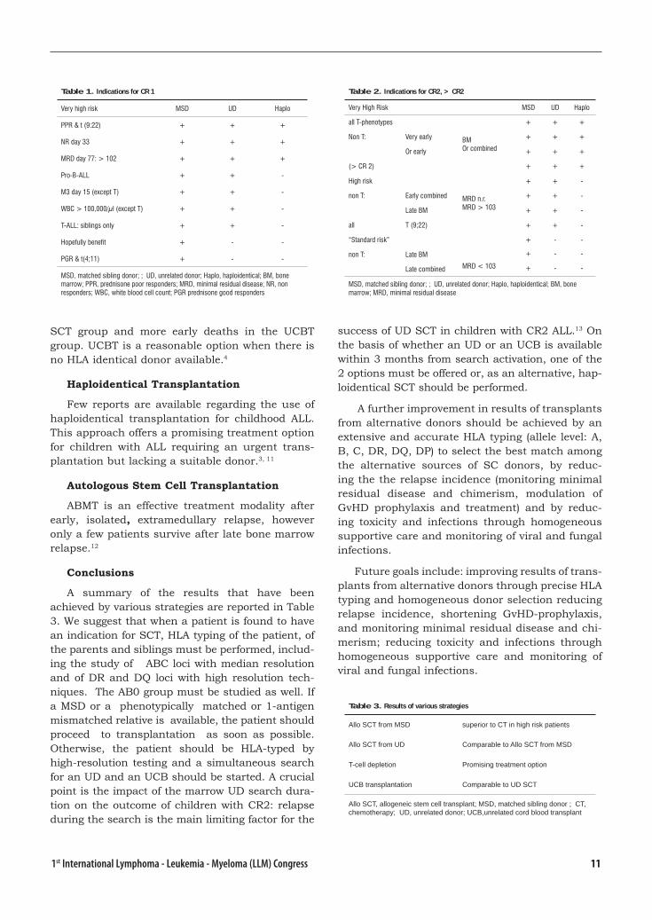
Children with VHR CR1 ALL, as defined in Table 1,5 benefit more from related donor SCT than from chemotherapy. The gap between the two strategies increases as the risk profile of the patient wors-ens6.
This is related to a higher relapse rate in chil-dren lacking a matched sibling donor (MSD), as compared to children with a MSD.
Stem Cell Transplantation (SCT) in Second Complete Remission (CR2) ALL
The I-BFM Study Group has defined Indications for allogeneic SCT in CR2 ALL, on the basis of the
Treatment of Pediatric ALL
Vincenza De Fazio, Edoardo Lanino, Giorgio Dini
Department of Paediatric Haematology and Oncology, Genova, Italy
site and timing of the relapse (Table 2)5 . DFS of patients given a MSD SCT following early relapse is significantly higher, as compared to chemotherapy. For those experiencing late relapse, the difference does not reach statistical significance.7, 8
Unrelated Donor Stem Cell Transplantation (UD-SCT)
Over the last 25 years, more than 18,000 UD-SCTs have been performed world-wide and facili-tated by a network that includes more than 11 million volunteer UDs enrolled in 89 registries. Currently, a suitable donor is located for 85% of the patients for whom a search is activated, and 70% of the donor phenotypes are found more than 4 times. The outcome of UD-SCT correlates with HLA matching: a single Class I or a single Class II mismatch is not relevant; multiple Class II mismatches are better than multiple Class I mis-matches, which are better than Class I plus Class II mismatches2 .Presently, the outcome of children with CR2 ALL given an UD SCT is comparable to that of SCT from MSDs. This improvement is mainly due to refinements in HLA typing, GvHD prophylaxis and supportive care.9, 10
Unrelated Umbilical Cord Blood Transplantation
Throughout the last 7 years, 373 Transplant Centres in 43 countries have performed more than 3,000 UCBTs by means of a network that includes more than 130,000 cord blood units in 37 banks. In children, the results of UD bone marrow or UCBT are similar; however, types of complications differ, with more GvHD being observed in the UD
111st International Lymphoma - Leukemia - Myeloma (LLM) Congress
SCT group and more early deaths in the UCBT group. UCBT is a reasonable option when there is no HLA identical donor available.4
Haploidentical Transplantation
Few reports are available regarding the use of haploidentical transplantation for childhood ALL. This approach offers a promising treatment option for children with ALL requiring an urgent trans-plantation but lacking a suitable donor.3, 11
Autologous Stem Cell Transplantation
ABMT is an effective treatment modality after early, isolated, extramedullary relapse, however only a few patients survive after late bone marrow relapse.12
Conclusions
A summary of the results that have been achieved by various strategies are reported in Table 3. We suggest that when a patient is found to have an indication for SCT, HLA typing of the patient, of the parents and siblings must be performed, includ-ing the study of ABC loci with median resolution and of DR and DQ loci with high resolution tech-niques. The AB0 group must be studied as well. If a MSD or a phenotypically matched or 1-antigen mismatched relative is available, the patient should proceed to transplantation as soon as possible. Otherwise, the patient should be HLA-typed by high-resolution testing and a simultaneous search for an UD and an UCB should be started. A crucial point is the impact of the marrow UD search dura-tion on the outcome of children with CR2: relapse during the search is the main limiting factor for the
success of UD SCT in children with CR2 ALL.13 On the basis of whether an UD or an UCB is available within 3 months from search activation, one of the 2 options must be offered or, as an alternative, hap-loidentical SCT should be performed.
A further improvement in results of transplants from alternative donors should be achieved by an extensive and accurate HLA typing (allele level: A, B, C, DR, DQ, DP) to select the best match among the alternative sources of SC donors, by reduc-ing the the relapse incidence (monitoring minimal residual disease and chimerism, modulation of GvHD prophylaxis and treatment) and by reduc-ing toxicity and infections through homogeneous supportive care and monitoring of viral and fungal infections.
Future goals include: improving results of trans-plants from alternative donors through precise HLA typing and homogeneous donor selection reducing relapse incidence, shortening GvHD-prophylaxis, and monitoring minimal residual disease and chi-merism; reducing toxicity and infections through homogeneous supportive care and monitoring of viral and fungal infections.
Table 1. Indications for CR 1
Very high risk MSD UD Haplo
PPR & t (9:22) + + +
NR day 33 + + +
MRD day 77: > 102 + + +
Pro-B-ALL + + -
M3 day 15 (except T) + + -
WBC > 100,000/μl (except T) + + -
T-ALL: siblings only + + -
Hopefully benefit + - -
PGR & t(4;11) + - -
MSD, matched sibling donor; ; UD, unrelated donor; Haplo, haploidentical; BM, bone marrow; PPR, prednisone poor responders; MRD, minimal residual disease; NR, non responders; WBC, white blood cell count; PGR prednisone good responders
Table 2. Indications for CR2, > CR2
Very High Risk MSD UD Haplo
all T-phenotypes + + +
Non T: Very early BMOr combined
+ + +
Or early + + +
(> CR 2) + + +
High risk + + -
non T: Early combined MRD n.r.MRD > 103
+ + -
Late BM + + -
all T (9;22) + + -
“Standard risk” + - -
non T: Late BM
MRD < 103
+ - -
+ - -Late combined
MSD, matched sibling donor; ; UD, unrelated donor; Haplo, haploidentical; BM, bone marrow; MRD, minimal residual disease
Table 3. Results of various strategies
Allo SCT from MSD superior to CT in high risk patients
Allo SCT from UD Comparable to Allo SCT from MSD
T-cell depletion Promising treatment option
UCB transplantation Comparable to UD SCT
Allo SCT, allogeneic stem cell transplant; MSD, matched sibling donor ; CT, chemotherapy; UD, unrelated donor; UCB,unrelated cord blood transplant
12 1 st International Lymphoma - Leukemia - Myeloma (LLM) Congress
References 1. Schrappe M, Reiter A, Zimmermann M, Harbott
J, Ludwig WD, Henze G, Gadner H, Odenwald E, Riehm H. Long-term results of four consecutive trials in childhood ALL performed by the ALL-BFM study group from 1981 to 1995. Berlin−Frankfurt−Munster. Leukemia 2000; 14: 2205−2222.
2. Petersdorf EW, Anasetti C, Martin PJ, Gooley T, Radich J, Malkki M, Woolfrey A, Smith A, Mickelson E, Hansen JA. Limits of HLA mismatching in unrela-ted hematopoietic cell transplantation. Blood. 2004 Nov 1;104(9):2976-80.
3. Handgretinger, T Klingebiel, P Lang, M Schumm, S Neu, A Geiselhart, P Bader, PG Schlegel, J Greil, D Stachel, RJ Herzog and D Niethammer Megadose transplantation of purified peripheral blood CD341 progenitor cells from HLA-mismatched parental donors in children. Bone Marrow Transplantation (2001) 27, 777–783
4. Rocha V, Cornish J, Sievers EL, Filipovich A, Loca-telli F, Peters C, Remberger M, Michel G, Arcese W, Dallorso S, Tiedemann K, Busca A, Chan KW, Kato S, Ortega J, Vowels M, Zander A, Souillet G, Oakill A, Woolfrey A, Pay AL, Green A, Garnier F, Ionescu I, Wernet P, Sirchia G, Rubinstein P, Chevret S, Gluckman E. Comparison of outcomes of unrelated bone marrow and umbilical cord blood transplants in children with acute leukemia. Blood 2001; 97: 2962-2971.
5. Peters C, Schrauder A, Schrappe M, von Stackel-berg A, Stary J, Yaniv I, Gadner H, Klingebiel T; BFM Study Group, the IBFM-Study Group and the Paediatric Disease Working Party of the EBMT. Allogeneic haematopoietic stem cell transplantation in children with acute lymphoblastic leukaemia: the BFM/IBFM/EBMT concepts. Bone Marrow Transp-lant. 2005 Mar;35 Suppl 1:S9-11.
6. Balduzzi A, Valsecchi MG, Uderzo C, De Lorenzo P, Klingebiel T, Peters C, Stary J, Felice MS, Mag-yarosy E, Conter V, Reiter A, Messina C, Gadner H, Schrappe M. Chemotherapy versus allogeneic transplantation for very-high-risk childhood acute lymphoblastic leukaemia in first complete remis-sion: comparison by genetic randomisation in an international prospective study. Lancet. 2005 Aug 20-26;366(9486):635-42.
7. Uderzo C, Valsecchi MG, Bacigalupo A, Meloni G, Messina C, Polchi P, Di Girolamo G, Dini G, Miniero R, Locatelli F, et al. Treatment of childhood acute lymphoblastic leukemia in second remission with allogeneic bone marrow transplantation and che-motherapy: ten-year experience of the Italian Bone Marrow Transplantation Group and the Italian Pediatric Hematology Oncology Association. J Clin Oncol. 1995 Feb;13(2):352-8.
8. Matsuzaki A, Nagatoshi Y, Inada H, Nakayama H, Yanai F, Ayukawa H, Kawakami K, Moritake H, Suminoe A, Okamura J. Prognostic factors for relapsed childhood acute lymphoblastic leukemia: impact of allogeneic stem cell transplantation--a report from the Kyushu-Yamaguchi Children’s Cancer Study Group. Pediatr Blood Cancer. 2005 Aug;45(2):111-20.
9. Locatelli F, Zecca M, Messina C, Rondelli R, Lanino E, Sacchi N, Uderzo C, Fagioli F, Conter V, Bonetti F, Favre C, Porta F, Giorgiani G, Pession A. Impro-vement over time in outcome for children with acute lymphoblastic leukemia in second remission given hematopoietic stem cell transplantation from unre-lated donors. Leukemia. 2002 Nov;16(11):2228-37.
10. Eapen M, Rubinstein P, Zhang MJ, Camitta BM, Stevens C, Cairo MS, Davies SM, Doyle JJ, Kurtz-berg J, Pulsipher MA, Ortega JJ, Scaradavou A, Horowitz MM, Wagner JE. Comparable long-term survival after unrelated and HLA-matched sibling donor hematopoietic stem cell transplantations for acute leukemia in children younger than 18 mont-hs. J Clin Oncol. 2006 Jan 1;24(1):145-51.
11. Klingebiel T, Handgretinger R, Lang P, Bader P, Niethammer D. Haploidentical transplantation for acute lymphoblastic leukemia in childhood. Blood Rev. 2004 Sep;18(3):181-92.
12. Messina C, Cesaro S, Rondelli R, Rossetti F, Locatel-li F, Pession A, Miniero R, Dini G, Uderzo C, Dallorso S, Meloni G, Vignetti M, Andolina M, Porta F, Amici A, Favre C, Basso G, Sotti G, Varotto S, Destro R, Gazzola MV, Pillon M, Petris MG, Rabusin M, Scar-zello G, et al. Autologous bone marrow transplanta-tion for childhood acute lymphoblastic leukaemia in Italy. AIEOP/FONOP-TMO Group. Italian Associati-on of Paediatric Haemato-Oncology. Bone Marrow Transplant. 1998 May;21(10):1015-21
13. Dini G, Valsecchi MG, Micalizzi C, Busca A, Bal-duzzi A, Arcese W, Cesaro S, Prete A, Rabusin M, Mazzolari E, Di Bartolomeo P, Sacchi N, Pession A, Georgiani G, Lanino E, Lamparelli T, Favre C, Bosi A, Manzitti C, Galimberti S, Locatelli F. Impact of marrow unrelated donor search duration on out-come of children with acute lymphoblastic leukemia in second remission. Bone Marrow Transplant 2003; 32: 325-31.
131st International Lymphoma - Leukemia - Myeloma (LLM) Congress
Several recent reports have indicated that not only a shift in the aetiology of infections and resistance patterns in patients with
febrile neutropenia, but also important differences between regions and countries. Viridans strepto-coccal bacteraemias are common among cancer patients being second only to the coagulase-nega-tive staphylococci. However, in certain centres in Europe Gram-negative bacilli have once again become the predominant infecting pathogens,. The problems associated with emerging resistance have been widely documented in the literature. In some institutions methicillin-resistance among coagulase-negative staphylococci has reached very high proportions, and in others the incidence of extended-spectrum beta-lactamase producing Gram-negative bacilli has risen markedly. These shifts in antimicrobial susceptibility are important in guiding the choice of agents for febrile neutro-penia. Antibiotic use and prophylaxis have both been associated with changes in susceptibility, and prescribing habits may influence emerging resistance. In this context, the choice of empiri-cal antibiotic therapy and the use of prophylaxis should be driven by a sound understanding of local circumstances.
Initiating empirical broad-spectrum antibacte-rial therapy has long been the standart practice for febrile neutropenic cancer patients. However, during the last decade it has become evident that patients with febrile neutropenia do not constitute a homogenous group. The risk factors for developing infection and other major complications vary widely in different subsets of patients with cancer. There-
Febrile Neutropenia in 2007
Murat Akova
Hacettepe University School of Medicine Section of Infectious Diseases Ankara, Turkey
fore, a valuable risk assessment of every febrile neutropenic patient is essential in order to define a tailored therapeutic approach. Those patients with hematological malignancies and severe and prolonged neutropenia will fall into the cathegory of “high-risk”, while others who were treated with less intensive chemotherapies and who were expected to have a short duration (e.g. less than 7-10 days) of neutropenia and fewer complications during the course of neutropenia will be categorized in the ‘low-risk’ group. Recently published “The Multina-tional Association for Supportive Care in Cancer (MASCC)” risk index has been shown to be a valu-able tool for identifiying low-risk patients among adult febrile neutropenic cancer patient population. Patients with solid tumors who were treated with conventional chemotherapy and with minimal or no comorbidities (such as mucositis, cellulitis, anorec-tal infection, pneumonia) will usually be placed into the cathegory of “low-risk”. On the other hand, more intensive chemotherapies have been increas-ingly used in solid tumor patients and some of them will also undergo an autologous hematopoietic stem cell transplantation (AHSCT). This approach will obviously increase the expected duration of neu-tropenia, the incidence of other comorbidities (e.g. mucositis), and may also affect the hemodynamic and clinical stability of the patient.
Once the patient is stratified in one of the risk groups, several options for empirical treatment exist. Nevertheless, several other factors need to be considered regarding to specific antimicrobial regimen. Among these are local epidemiological pattern of the infecting microorganisms and their
14 1 st International Lymphoma - Leukemia - Myeloma (LLM) Congress
antimicrobial resistance pattern. Recent published data indicate that low-risk patients who are able to swallow can successfully be treated with oral antibiotics. The most frequent used regimen for this indication is a combination of a quinolone derivative (e.g. ciprofloxacin) and amoxicillin/cla-vulanate. Newer quinolones with enhanced activity against gram-positive pathogens (e.g. moxifloxacin, gatifloxacin) have been currently under evaluation for a monotherapy option. This type of therapy is applicable for both inpatient and outpatient settings. Stringent criteria need to be applied for selecting patients who will be treated in an outpa-tient program which also requires a strong com-mittment from both patient and healthcare team’s side. Another option is to admit the patient to the hospital and treat with parenteral antibiotics until defervescence, and then swich to oral therapy. This provides a viable alternative for patients receiving more intensive chemotherapies for treating cancer with or without AHSCT. Upon switch to an oral regimen the patient could be discharged if his/her clinical condition is permissive. Comparative solid data for such a practice are lacking yet in the litera-ture, however several studies both in IATG/EORTC and in elsewhere are being undertaken on this issue. For the initial parenteral therapy, mono-therapy with various beta-lactam antibiotics has been extensively studied comparing with different beta-lactam plus aminoglycoside combinations. The data indicate that monotherapy with a broad-spec-trum cephalosporin (e .g. ceftazidime, cefepime) or beta-lactam/beta-lactamase inhibitor combination (e.g. piperacillin/tazobactam) or a carbapanem (i.e. imipenem or meropenem) is as effective as a beta-lactam plus aminoglycoside combination for initial empirical regimen. Specific concerns for ceftazidime use exist since this drug has been held responsi-ble for increased incidence of extended-spectrum beta-lactamase producing klebsiella infections in some institutions. Recently published metaanalyses caused concern about cefepime which was found to cause increased mortality in patients due to unexplained reasons. Parenteral quinolones has been less studied for this indication and the data are inconclusive. Therefore quinolones can not be recommended as the initial parenteral agent.
Glycopeptides should not be incorporated into the initial empirical regimen, until a document-
ed gram-positive bacterial infection is observed. Recent data indicate that empirical addition of these agents is also unnecessary in those patients without defervescence after 60-72 hours of empiri-cal broad-spectrum antibacterial therapy. Actual-ly, glycopeptide use should strongly be discouraged unless the patient has a documented gram-positive bacterial infection or has strong predisposing fac-tors to acquire such infections (e.g. clinically docu-mented vascular catheter infection, colonization with methicillin resistant staphylococci or penicil-lin resistant pneumococci).
In summary, a risk-based approach in patients with febrile neutropenia could be more cost-effec-tive. Various regimens with different antibiotics are available, but spesific regimens also need to be tailored to local epidemiological factors.
References 1. Akova M. Emerging problem pathogens: A review of
resistance patterns over time. Int J Infect Dis. 2006 Sep;10 Suppl 2:S3-8.
2. Cometta A, Kern WV, De Bock R, et al and Interna-tional Antimicrobial Therapy Group of the European Organization for Research Treatment of Cancer. Vancomycin versus placebo for treating persistent fever in patients with neutropenic cancer receiving piperacillin-tazobactam monotherapy. Clin Infect Dis. 2003;37:382-9.
3. Donowitz GR, Maki DG, Crnich CJ, Pappas PG, Rolston KVI. Infections in the Neutropenic Pati-ent–New Views of an Old Problem. Hematology (Am Soc Hematol Educ Program). 2001;:113-39.
4. Kern WV, Cometta A, De Bock R, et al. Oral versus intravenous empirical antimicrobial therapy for fever in patients with granulocytopenia who are receiving cancer chemotherapy. International Antimicrobial Therapy Cooperative Group of the European Organi-zation for Research and Treatment of Cancer. N Engl J Med. 1999;341:312-8.
5. Klastersky J, Paesmans M, Rubenstein EB, et al. The Multinational Association for Supportive Care in Cancer risk index: a multinational scoring system for identifying low-risk febrile neutropenic patients. J Clin Oncol 2000;18:3038-51.
6. Viscoli C, Castagnola E. Treatment of febrile neutropenia: what is new? Curr Opin Infect Dis. 2002;15:377-82.
7. Yahav D, Paul M, Fraser A, Sarid N, Leibovici L. Efficacy and safety of cefepime: a systematic revi-ew and meta-analysis. Lancet Infect Dis. 2007 May;7(5):338-48.

151st International Lymphoma - Leukemia - Myeloma (LLM) Congress
Follicular lymphoma (FL) is the most common type of low-grade B cell lymphoma seen in western countries. It is characterized by a
clinically indolent course. The cellular origin of the neoplastic cells are follicular center B lymphocytes [1]. The incidance of FL in eastern countries is low.
FL predominantly involves the lymph nodes, but spleen, bone marrow, peripheral blood, and Waldeyer’s ring involvement have also been report-ed. The gastrointestinal tract, soft tissue and skin are the most commonly involved extranodal sites.
Histologically, FL is composed of centrocytes and centroblasts, and usually has a follicular growth pattern. Neoplastic follicles are often ill-defined, and lack mantle zones. When the mantle zones are preserved there could be difficultiy on differentiation form reactive follicular hyperplasia. Interfollicular infiltration of the neoplastic cells is a helpful diagnostic criterion for the cases having this morphology. Diffuse pattern may be seen and it is thought to be of clinical significance. In the WHO classification, FL is graded as 1, 2, 3a, and 3b according to the number of centroblasts per high-power field [1]. Histological grade correlates with prognosis in FL, with grades 1 and 2 being indolent and grade 3 being more aggressive. In grade 3 FL, the presence of a diffuse component is commonly seen and some studies have demon-strated that this finding is correlated with a worse outcome [2]. Presence of residual reactive follicles within the involved lymph nodes could reflect the stage of the disease[3].
Follicular Lymphoma (FL) Pathology
Isinsu Kuzu
Ankara Faculty of Medicine, Department of Patology, Morphology, Ankara, Turkey
Cases of ‘in situ localization of FL’ have been reported in the literature [4]. It appears to repre-sent early microscopic involvement of FL within the lymph nodes. The clinical significance of these cases without other evidence of lymphoma is not known yet[4].
The tumor cells are positive for CD19, CD20, CD22, CD79a, surface Ig (IgM+/IgG-, IgG or rarely IgA), bcl-2, CD10, and bcl-6 and negative for CD5, CD43, CD23 and Cyclin D1 [1]. Immunohistoche-mistry is very useful for the diagnosis of FL, and several studies revealed the relation of expression of various proteins with clinical outcome. The pro-liferation index of the cells within the neoplastic follicles by MIB-1 (Ki-67) provides a measure of proliferative rate, and has been shown to correlate with FL grade but has limited prognostic significan-ce in some studies. Some recent data revealed that proliferation index may have prognostic value in FL [5]. High Ki-67 staining in the reactive lymphoid fol-licles is useful for the differentiation of reactive fol-licular hyperplasia and FL. The proliferation index of the neoplastic follicles in low grade FL (grades 1 and 2) is lower than in reactive follicular hyperpla-sia and grade 3 FL. But in a recent study of Wang et al., high proliferation index in low grade FL was determined in nearly 20% of their cases. The clini-cal behavior of these low grade FL cases showing high proliferation index was correlated with inferior disease-specific survival but higher five-year dis-ease free rate similar to grade 3 FL [6].
Although most patients with FL overexpress Bcl-2 protein, higher levels of expression have been corre-lated with worse outcome. In contrast, higher levels

16 1 st International Lymphoma - Leukemia - Myeloma (LLM) Congress
of expression of germinal center markers including CD-10, Bcl-6 and PU.1 have been correlated with a favorable outcome. [7.8]. The presence of more than 15 CD68+ macrophages per high power field has also been shown to predict for a poor outcome. [9].
The genetic hallmark of FL, t(14;18)(q32;q21), which juxtaposes the bcl-2 gene with the IgH gene, is seen in 80-90% of FLs [10]. It is not associated with the prognosis. Bcl-2 protein is expressed in the majority of the cases, and its expression reduc-es as histological grade increases. Although FL is rarely seen in pediatric patients, it should be noted that bcl-2 expression in pediatric FL is relatively infrequent in contrast to its adult counterpart [10,
11]. Primary cutaneous follicle center cell lymphoma is a accepted as a variant of FL and is often bcl-2-negative as well [12, 13, 14].
A number of cytogenetic abnormalities have been described in FL, including p53 mutations, loss of p16, upregulated MYC expression result-ing from translocation or other mechanisms, gains of chromosome arms 7p or 7q, Xp, 12q and 18q, as well as losses on 6q and possibly mutations of bcl-2 and/or bcl-6 genes. The presence of addi-tional genomic aberrations, in particular 17p and 6q deletions, is more frequent in grade 2 and 3 FL patients and correlated with shorter survival and a higher rate of transformation into diffuse large B cell lymphoma [10,11].
Approximately 25-35% of FL cases transform into diffuse large B cell lymphoma as well as Burkitt’s lymphoma, precursor B lymphoblastic lymphoma and classical type of Hodgkin’s lym-phoma [1, 15-17].
Molecular prognostic markers examinade for follicular lymphoma. [10,
11,15-17].
Prognostic Marker Effect on Outcome Mechanism
-Chromosomal gains Unfavorable Dominant oncogenes+7, +12q13-14, +18q-Chromosomal losses Unfavorable Loss tumor suppressorDel6q, –9p21, –17p13 gene-BCL-6 translocation Unfavorable Genomic instability-Bcl-2 expression Unfavorable Anti-apoptotic-Bcl-6 expression Favorable Germinal center phenotype-CD10 expression Favorable Germinal center phenotype-PU.1 Favorable Germinal center phenotype-Macrophage content Unfavorable Modulation by microenvironment-MDM2 expression Unfavorable Functional p53 loss-Bcl-XL Unfavorable Anti-apoptotic-Cyclin B1 Favorable Cell cycle progression-Immune response Variable Modulation by microenvironment(IR-1 versus IR-2) 81-gene predictor Variable Reflects tumor behavior
References 1. Jaffe ES, Harris NL, Stein H, Vardiman JW (Eds).
World Health Organization Classification of Tumours. Pathology and Genetics of Tumours of Haematopoie-tic and Lymphoid Tissues. Lyon: IARC Press, 2001
2. Anderson JR, Vose JM, Bierman PJ, Weisenburger DD, Sanger WG, Pierson J, Bast M, Armitage JO. Clinical features and prognosis of follicular large-cell lymphoma: a report from the Nebraska Lymphoma Study Group. J Clin Oncol 1993;11:218-224
3. Patrick Adam, MD, Tiemo Katzenberger, MD, Miriam Eifert, M. Michaela Ott, MD, Andreas Rosenwald, MD, Hans Konrad Mu¨ller-Hermelink, MD, and Ger-man Ott, MD. Presence of Preserved Reactive Ger-minal Centers in Follicular Lymphoma Is a Strong Histopathologic Indicator of Limited Disease Stage, Am J Surg Pathol 2005;29:1661–1664)
4. Tilly H, Rossi A, Stamatoullas A. Prognostic value of chromosomal abnormalities in follicular lymphoma. Blood 1994;84:1043-1049
Cong P, Raffeld M, Teruya-Feldstein J, Sorbara L, Pittaluga S, Jaffe ES. In situ localization of follicular lymphoma: description and analysis by laser captu-re microdissection. Blood 2002;99:3376-3382
5. Hall PA, Richards MA, Gregory WM. The prognostic value of Ki67 immunostaining in non-Hodgkin’s lymphoma. J Pathol 1988;154:223–235.
6. Wang SA, Wang L, Hochberg EP, Muzikansky A, Harris NL, Hasserjian RP. Low histologic grade follicular lymphoma with high proliferation index: morphologic and clinical features. Am J Surg Pathol 2005;29:1490–1496.
7. Bilalovic N, Blystad AK, Golouh R, et al. Expression of bcl-6 and CD10 protein is associated with longer overall survival and time to treatment failure in fol-licular lymphoma. Am J Clin Pathol. 2004;121:34-42.
8. Torlakovic E, Bilalovic N, Golouh R, Zidar A, Angel S. Prognostic significance of PU.1 in follicular lymp-homa. J Pathol. 2006;209:352-359.
9. Farinha P, Masoudi H, Skinnider BF, et al. Analysis of multiple biomarkers shows that lymphoma-asso-ciated macrophage (LAM) content is an independent predictor of survival in follicular lymphoma (FL). Blood. 2005;106:2169-2174.
10. Tilly H, Rossi A, Stamatoullas A. Prognostic value of chromosomal abnormalities in follicular lymphoma. Blood 1994;84:1043-1049.
11. Viardot A, Barth TF, Moller P, Dohner H, Bentz M. Cytogenetic evolution of follicular lymphoma. Semin Cancer Biol 2003;13:183–190.
12. Atra A, Meller ST, Stevens RS. Conservative mana-gement of follicular non-Hodgkin’s lymphoma in childhood. Br J Haematol 1998;103:220-223.
13. Finn LS, Viswanatha DS, Belasco JB. Primary folli-cular lymphoma of the testis in childhood. Cancer 1999;85:1626-1635.
171st International Lymphoma - Leukemia - Myeloma (LLM) Congress
14. Willemze R, Jaffe ES, Burg G, Cerroni L, Berti E, Swerdlow SH, Ralfkiaer E, Chimenti S, Diaz-Perez JL, Duncan LM, Grange F, Harris NL, Kempf W, Kerl H, Kurrer M, Knobler R, Pimpinelli N, Sander C, San-tucci M, Sterry W, Vermeer MH, Wechsler J, Whit-taker S, Meijer CJ. WHO-EORTC classification for cutaneous lymphomas. Blood 2005;105(10):3768-3785.
15. Li L, Gupta S, Bashir T, Koduru PR, Brody J, Allen SL. Serial cytogenetic alterations resulting in transformation of a low-grade follicular lymphoma to Burkitt lymphoma. Cancer Genet Cytogenet 2006;170(2):140-146.
16. Kobrin C, Cha SC, Qin H, Raffeld M, Fend F, Quin-tanilla-Martinez L, Grove S, Jaffe ES, Kwak LW. Molecular analysis of light-chain switch and acute lymphoblastic leukemia transformation in two folli-cular lymphomas: implications for lymphomagene-sis. Leuk Lymphoma 2006;47(8):1523-1534.
17. Marafioti T, Hummel M, Anagnostopoulos I, Foss HD, Huhn D, Stein H. Classical Hodgkin’s disease and follicular lymphoma originating from the same ger-minal center B cell. J Clin Oncol 1999;17(12):3804-3809.
18. Gascoyne RD. Hematopathology approaches to diag-nosis and prognosis of indolent B-cell lympho-mas. Hematology (Am Soc Hematol Educ Program). 2005:299-306.
19. Torlakovic E, Bilalovic N, Golouh R, Zidar A, Angel S. Prognostic significance of PU.1 in follicular lymp-homa. J Pathol. 2006;209:352-359.
Laurie H. Sehn Optimal Use of Prognostic Factors in Non-Hodgkin Lymphoma. (Am Soc Hematol Educ Program). 2006:295-302.
18 1 st International Lymphoma - Leukemia - Myeloma (LLM) Congress
Follicular lymphoma (FL) is a heterogenous disease, which is still considered as uncura-ble. New prognostic factors as the FLIPI (Fol-
licular Lymphoma International Prognostic Index) and histological grading are of help in deciding type of theurapeutic option. Also immune cells in the micro environment of the tumour have shown to be of clinical importance.
Improvement in therapy has been seen with the monoclonal anti-CD20 antibody rituximab, first used as single agent therapy in relapsed fol-licular lymphoma (the pivotal trial). Recently the combination of this antibody with chemotherapy (CVP , CHOP or fludarabine combinations) has been shown to extend both progression-free (PFS) and overall survival (OS), when used as first-and or second line therapy.
To improve the efficacy of rituximab and to delay the need of chemotherapy, different sched-ules of drug administration, as extended dosing, combination with interferon and maintenance, has been used.
R-CHOP (rituximab 375 mg/m2 at day 1 of each cycle of CHOP) was used in a phase III clinical trial
Treatment of Follicular Lymphoma
Eva Kimby
Karolinska University Hospital, Stockholm, Sweden
for patients with relapsed follicular lymphoma, the EORTC 20981 Intergroup Study. Patients with a CR or PR after six cycles of therapy under-went a second randomisation to no further treat-ment (observation) or maintenance treatment with rituximab (375 mg/m2 once every 3 months) until relapse or for a maximum of 2 years. Both treat-ment arms yielded similar PR rates but a highly significant higher CR rate with the combination. In patients randomised to maintenance an advantage was observed both in PFS and OS when compared with the observation arm.
The benefit of rituximab maintenance for patients with first-line immunochemotherapy will be determined in ongoing randomised studies.
The best schedule and length of therapy as well as longterm effects of rituximab treatment needs to be evaluated. New monoclonal antibodies are under development, but it is still not defined which subgroups of patients will benefit most of treat-ment with antibodies. Also other new drugs acting on the microenvironment of the lymphoma might show theurapeutic activity.
191st International Lymphoma - Leukemia - Myeloma (LLM) Congress
Gene expression profiling has defined major subtypes of aggressive non-Hodgkin’s lym-phomas (B-NHL) in recent years, in particu-
lar among diffuse large B-cell lymphomas (DLBCL). Specifically, a germinal center B-cell type (GCB DLBCL), an activated B-cell type (ABC DLBCL) and primary mediastinal B-cell lymphoma (PMBL) can be distinguished based on fundamental differences in underlying gene expression profiles. More recently, the Lymphoma and Leukemia Molecular Profiling Project (LLMPP)(Dave et al., NEJM 2006) as well as a major study conducted in Germany (Hummel et al., NEJM 2006) defined the highly aggressive entity of Burkitt lymphoma on a molecular level. Besides the molecular classification of B-NHL, gene expres-sion signatures can also be used to predict clinical outcome. For example, the gene expression-based measurement of proliferation (proliferation signa-ture) provides a powerful predictor of outcome in mantle cell lymphoma (MCL) that is superior to the immunohistochemical assessment of the Ki-67 index (Rosenwald et al., Cancer Cell 2003). How can these findings be translated into daily clinical practice?
Several attempts have been made to use a small set of immunohistochemical markers as surrogates for gene expression signatures. An example is the Hans-classifier (Hans et al., Blood 2004) that uses the immunohistochemical markers CD10, BCL6 and MUM1 to classify DLBCL into the GCB- and
Agressive Lymphoma – Pathology
Andreas Rosenwald
Institute of Pathology, University of Würzburg, Germany
non-GCB subtypes. While this approach appears to be easily applicable in the daily routine, there are also limitations, and a considerable variation in the staining process (between different institutions) as well as in the scoring process (between differ-ent pathologists) have to be overcome. In particu-lar, the germinal center-associated marker BCL6 appears to suffer from remarkable inter-laboratory variation (de Jong et al., JCO 2007).
Alternatively, quantitative RT-PCR (TaqMan) can be performed in routinely obtained formalin fixed and paraffin embedded tumor tissues. In mantle cell lymphoma (MCL), the quantitative mRNA expression measurement of 5 genes may be able to substitute for the proliferation signature determined by gene expression profiling (Hartmann et al., unpublished). Finally, the microarray plat-form itself may be used for diagnostic and prognos-tic purposes in clinical settings. Towards this goal, efforts are under way to achieve reliable and robust gene expression measurements across different laboratories. In one of these efforts, the Lymphoma and Leukemia Molecular Profiling Project (LLMPP) collaborates with Roche Diagnostics in the develop-ment of a diagnostic microarray. As a first step, a proficiency test between the 8 participating institu-tions was undertaken which showed highly con-sistent gene expression results of the same tumor tissues analyzed in a decentralized fashion.
20 1 st International Lymphoma - Leukemia - Myeloma (LLM) Congress
The aggressive non-Hodgkin’s lymphomas can be cured in more than half of the cases. For patients with localized aggressive non-Hodg-
kin’s lymphoma, heterogeneity in patients selection prevent us from defining a new standard of care. On the contrary, for patients with advanced stages of diffuse large B cell lymphoma a new standard of therapy now exist particularly for elderly and low risk young patients.
In this article I will separate the patients into 3 different groups: I) early stage NHL, II) advanced stage elderly and low risk young patients and III) young high risk patients.
I. Early (Limited) Stage Aggressive Lymphoma
There is extreme heterogeneity within the group of patients described as having early stage lym-phoma, making comparisons of outcome difficult.
SWOG compared 3 cycles of CHOP with RT to 8 cycles of CHOP in patients with localized aggressive NHL and found that CHOP(3) plus RT was superior to CHOP (8) through the first five years of follow-up (1) . Localized disease was defined as stage I and non-bulky stage II disease. Patients with bulky-stage II disease are known to have a prog-nosis similar to patients with advanced disease and should be excluded from limited-stage disease (2). Patients with bulky-stage II disease accrued to SWOG studies had a 5-year survival of 49%. Patients with stage III-IV disease had a 5-year sur-vival of 46%. Therefore bulky stage II is considered as “advanced” disease. If patients with bulky-stage II disease are included in trials and treated with a
DLBCL-L First Line Treatment
Burhan Ferhanoglu
Cerrahpasa Medical School, Department of Hematology, Istanbul, Turkey
short-course of chemotherapy plus RT they have an inferior outcome compared to similar patients treated with aggressive chemotherapy designed for advanced disease.
Reyes and colleagues from GELA (3) have reported the results of a randomized trial com-paring CHOP (3) plus RT to an aggressive combi-nation chemotherapy called ACVBP (LNH-93-1). ACVBP chemotherapy was originally designed for advanced disease. In the subgroup of bulky-stage II disease, treatment with ACVBP was superior to CHOP(3) plus RT with 5-year survival estimates of 82% and 50%, respectively (p=0.03)
By excluding patients with bulky-stage II disease one might presume that the remaining patients with limited disease comprise a homoge-neous group with regard to prognosis and choice of optimal therapy. But 10 year survival can vary from 90% to 10% within subgroup of limited stage patients (4). Predicting such variable outcome eas-ily accomplished using stage modified IPI. Most clinicians uses IPI and it includes five risk factors including age, stage, serum LDH, performance status(PS), and the number of extranodal sites of disease(5). Age, stage, serum LDH, and PS each predict significant outcome differences for patients with limited diseases. Patients with no adverse risk factors have a very good prognosis with 10 year survival estimates exceed 90% treated (Table 1 and 2). These very good results can be achieved regardless of the treatment strategy chosen; CHOP (3) +RT, CHOP (8), or ACVBP (1,4,6). This category is called ‘very limited disease’.

211st International Lymphoma - Leukemia - Myeloma (LLM) Congress
Another GELA study was published by Fillet et al. comparing 4 cycles of CHOP to CHOP plus 40 Gy involved-field RT in patients older than 60 years with no adverse risk factors according to the age-adjusted IPI (7). There was no advantage of the radiation and, a possible disadvantage in patients older than 70 years of age. Recent ECOG study suggested benefit of adjuvant radiation after 8 cycles of CHOP (8). But the main difference in these two trials was bulky disease rates. Although SWOG included only 6 (1.5%) bulky (stage I) patients, the rate of bulky disease in ECOG was 31%. So one third of the patients actually should be accepted as advanced disease in ECOG trial.
The MINT trial included some patients with early stage disease and compared a CHOP-like chemotherapy regimen to the same regimen with the addition of rituximab in young, good prognosis patients (9). For the most favorable patients (those without bulky disease) the results with a complete course of chemotherapy plus rituximab alone with-out radiation led to survival in excess of 90%. The Southwest Oncology Group in the United States reported a pilot study of an abbreviated course of CHOP plus rituximab followed by radiation show-ing progression free and overall survival in excess of 90% (10)
II. Treatment Of Advanced Stage Elderly And Low Risk Young Patients
After initial staging bulky stage II, stage III or stage IV aggressive lymphoma is documented in approximately 75% of all DLBCL patients. There-fore chemotherapy is main treatment modality for these patients.
The study that defined chemotherapy as stand-ard was an intergroup trial conducted by SWOG and ECOG. In this study previously untreated patients with stage II bulky, III, and IV disease with intermediate –or high-grade histology were rand-omized to one of four treatment arms: CHOP, m-BACOD,, ProMACE-CytaBOM or MACOP-B. There was no difference in any treatment arm. These results along with the fact that CHOP was cheaper and easier to administer than the other regi-mens, established CHOP as the standard therapy throughout the world. However, with a projected disease-free survival rate of 36%, it is obvious that it is far from an ideal therapy, and there is need for better treatment approaches.(11)
The International non-Hodgkin Lymphoma Prognostic Factors Index (IPI) utilized pretreat-ment prognostic factors in a sample of over 5000 patients to develop a predictive model of outcome for aggressive non-Hodgkin’s Lymphoma (12). Five pretreatment characteristics were found to be inde-pendent predictors of death: age (< 60 vs. >60), tumor stage II or II vs. III or IV, the number of involved extranodal sites (< 1 vs. >1), patient ECOG performance status (0,1;ambulatory vs. >2 ;non-ambulatory), and serum LDH level( normal or ele-vated). Each of the individual factors had compa-rable relative risks. The resulting model identified 4 risk groups with the following associated 5- year survival rates: low risk(0-1), 73%, low-intermedi-ate risk (2 risk factors), 51% ; high- intermediate risk ( 3 risk factors), 43%, and high risk (4-5 risk factors), 26%. However, the improvement in treat-ment response associated with the addition of the antibody rituximab to treatment regimens seems to have altered survival of prognostic groups using the International Prognostic Index (13). (Table 3)
Table 1. International Prognostic Index (12)
Full Index Age Adjusted
Prognostic FactorsAge> 60 yearsPerformance status > 2LDH> 1 x normalExtranodal sites >2Stage III or IV
Risk Category Factors• Low 0 or 1• Low-intermediate 2• High-intermediate 3• High 4 or 5
Prognostic Factors• Performance status > 1• LDH > 1 x normal• Stage III or IV
Risk Category Factors• Low 0• Low-intermediate 1 • High-intermediate 2• High 3
Table 2. Outcome for patients with diffuse aggressive lymphoma after anthracycline chemotherapy International Index ( 12)
RiskCategory
No RiskFactors
%Cases
CRrate
PFS of CRs5 yr
Survival5 yr
Low 0,1 35% 87% 70% 72%
Low-intermediate 2 27% 67% 50% 50%
High-intermediate 2 22% 55% 48% 43%
High 4,5 16% 44% 40% 26%
Table 3. Results with a revised IPI when R-CHOP is given for diffuse large B-cell lymphoma (13)
Group No Factors % Patients % 4-Year Overall Survival
Standard IPI 0,123
4,5
28272124
86815458
Revised IPI 01,2
3,4,5
104545
928258
Currently molecular profiling project has used complementary DNA (cDNA) microarray tech-niques to demonstrate two distinct subpopulations
22 1 st International Lymphoma - Leukemia - Myeloma (LLM) Congress
of DLBCL with different prognosis and different genetics. Patients with a germinal center B (GCB) cell like signature have a more favorable course than those with an activated B-cell (ABC) signature (14,15). At the present time gene expression profil-ing is not part of routine clinical practice (37). This may be due to technical difficulties in performing the c-DNA arrays. GCB versus ABC typing seems to be able to be reproduced by tissue microarray (16).
After the disappointing results with the third generation chemotherapy regimens, several new treatment approaches have been developed. GELA developed the ACVBP regimen (which involves very intensive chemotherapy for four courses followed by an intensive consolidation) which was shown to be superior to CHOP in subgroup of patients (17).
German high-grade lymphoma study group found that addition of etoposide to CHOP improved results in young patients (18), while CHOP admin-istered at 14 day instead of 21 day intervals seemed to improve the results in elderly patients (19).
An infusional chemotherapy regimen developed at the National Cancer Institute referred to as DA-EPOCH had very encouraging results (20).
The randomized study that changed practice throughout the world was performed by the GELA and compared CHOP versus Rituximab-CHOP (R-CHOP) in elderly patients (21,22). The GELA group randomized 399 previously untreated patients with DLBCL, 60 to 80 years old, to receive either 8 cycles of CHOP every 3 weeks or 8 cycles of CHOP plus rituximab given on day 1 of each cycle. Com-plete response rates of 76% and 63% (p=0.005) and 5- year overall survival rates of 58 % and 45 % (p< 0.007) were achieved by R-CHOP and CHOP, respectively. In patients with a low- risk age adjusted IPI (0 or 1 adverse prognostic fac-tors), 5-year event-free survival (EFS) was 63% and 34% for the R-COP and CHOP arms, respec-tively (p=0.0008). For those with high-risk disease (2, 3 adverse factors), 5 year EFS were 41% and 27% for the R-CHOP and CHOP arms, respectively (p=0.004). These results suggest that addition of rituximab therapy to standard CHOP may lead to significant prolongation of event-free and overall survival in elderly patients with both high-risk and low risk disease with no increase in toxicity. In an early analysis, R-CHOP appeared to be more effec-tive than CHOP in bcl-2 positive, but not in bcl-2 negative patients, suggesting that the benefit of
addition of rituximab might overcome bcl-2 associ-ated chemotherapy resistance (23,24).
In the United States a larger (N=632) intergroup study randomized elderly patients to receive initial therapy with either CHOP or R-CHOP. The rituxi-mab was given on a different schedule than the GELA study. Responding patients then were ran-domized to receive either rituximab maintenance therapy (4 doses q 6 months x 2 years) or no main-tenance. This study confirmed the GELA results with a significant advantage for receiving rituximab either in during induction or maintenance, but no advantage to getting both (25).
According to the IPI young good prognosis patients comprise the low and low-intermediate risk group (0 and 1 risk factor according to the age-adjusted IPI) and poor prognosis patients the high-intermediate and high risk group (2 risk factors).
Until recently there was no Phase III trial look-ing for the value of rituximab in younger patients with DLBCL. An International study called MInT Trial compared chemotherapy CHOP with or with-out rituximab in patients younger than age 60. Eligibility criteria included DLBCL, 18-60 years, IPI 0 or 1, stages II-IV or stage I with bulk. Patients received 6 cycles of any of several CHOP like regi-mens followed by radiation therapy to bulky dis-ease (9). The MInT trial demonstrated significant advantage in response, failure-free survival, and overall survival with the addition of rituximab.
The German High Grade Lymphoma Study Group (DSHNHL) studied the utility of six versus eight cycles of R-CHOP at 14 day intervals with or without rituximab in elderly patients with DLBCL. RICOVER 60 trial of DSHNHL demonstrated the importance of rituximab and in combination with rituximab 6 cycles of R-CHOP-14 are as good as 8 cycles.(26)
Investigators from the Cancer Institute from the British Columbia did a population based study of the impact of adding rituximab to CHOP. After approval of rituximab in British Columbia survival of DLBCL went up of about 20%.(27)
III. Treatment Of Young, High Risk Patients
According to the IPI, young poor prognosis patients comprise the high-intermediate and high risk group (aa IPI 2,3). For young poor prognosis patients, the five-year survival is around 50%,
231st International Lymphoma - Leukemia - Myeloma (LLM) Congress
and progress has not been demonstrated in these patients.
The role of high-dose therapy (HDT) followed by autologous stem cell transplantation (SCT) for primary treatment of aggressive non-Hodgkin lymphoma is still uncertain. Whereas some stud-ies demonstrated superiority of HDT over con-ventional treatment (28, 29,30), others failed to show significant differences (31,32,33) or reported inferior results (34). Besides differences in patient characteristics, the type HDT and its timing may have important implications for outcome. Recently published meta-analysis showed that there was no evidence that HDT improved OS and EFS in good risk NHL patients. The evidence for poor risk patients is inconclusive and high quality studies in poor risk patients are warranted (35).
Recently, German High Grade NHL study group published the result of Mega CHOEP chemo-therapy. The Mega CHOEP protocol consisted of 4 courses of Cyclophosphamide, doxorubicin, vinc-ristine, etoposide and prednisolone. MegaCHOEP was applied as high dose chemotherapy including autologous stem cell transplantation after courses 2, 3, 4 .The patients aged 18-60 years with pri-mary diagnosis of aggressive NHL and LDH levels above normal were included in the study. 70% of the patients achieved CR or CRu after a median follow-up of 55 months. OS was 75 % and 67.2 % after 2 and 5 years, respectively. Treatment related mortality was 4.5%. Risk of secondary MDS/AML was low with only one patient having MDS during follow up.
Comparison of the results of Mega CHOEP with R-CHOP chemotherapy is difficult. The only study published with R-CHOP chemotherapy was that of British Columbia’s and 4 year OS was 55% for this high risk patient group. But we have to keep in mind that the median age of this group was older (median age 61) than the patients included in the Mega CHOEP study (median age 43).
References 1. Miller TP, Dahlberg S, Cassady JR, et al.Chemotherapy
alone compared to chemotherapy plus radiotherapy for localised intermediate- and high-grade non-Hodgkin’s lymphoma. N Engl J Med. 1998; 339:21-26
2. Fisher RI, De Vita VT, Johnson BL, et al. Prognostic factors for advanced diffuse histiocytic lymphoma following treatment with combination chemothe-rapy.
3. Reyes F, Lepage, Ganem G. et al. ACVBP versus CHOP plus radiotherapy for localized aggressive lymphoma . N.Engl J Med2005; 352 (12): 1197-205
4. Miller TP, Le Blanc M, Spier C, et al.CHOP alone compare to CHOP plus radiotherapy for early stage aggressive non-Hodgkin’s lymphoma: update of the Southwest Oncology Group (SWOG) randomized trial (abstract ). Blood 2001; 98: 724 a
5. Shipp M for the International Non-Hodgkin’s Lymp-homa Prognostic Factors Project. A predictive model for aggressive non-Hodgkin’s lymphoma. N Engl J Med. 1993; 329: 987-994
6. Shenkier TN, Voss N, Fairey R, et al. Brief chemo-therapy and involved-region irradiation for limited-stage diffuse large-cell lymphoma: an 18-year expe-rience from the British Columbia Cancer Agency. J Clin Oncol.2001; 20:197-204
7. Fillet G, Bonnet C, Mounier N, et al.No role for che-moradiotherapy when compared with chemotherapy alone in elderly pateints with localized low risk aggressive lymphoma: final results of the LNH93-4 GELA study. Blood 2005; 106 (11):9a (abstract nb 15)
8. Horning SJ, Weller E, Kim K. Et al. Chemotherapy with or without radiotherapy in limited-stage dif-fuse aggressive non-Hodgkin’s lymphoma: Eastern Cooperative Oncology Group Study 1484. J Clin Oncol 2004; 22 (15): 3032-8
9. Pfreundschuh M, Trumper L, Osterborg A, et all. CHOP-like chemotherapy plus rituxiamb versus CHOP-like chemotherapy alone in young patients with good- prognosis diffuse large-B-cell lympho-ma: a randomised controlled trial by the Mab Thera International Trial (MInT) Group. Lancet Oncol 2006; 7(5):379-91
10. Miller TP, Spier CM, Rimsza L. Diffuse aggressive histologies of non-hodgkin lymphoma: treatment and biology of limited disease. Semin Hematol 2006; 43(4): 207-12
11. Fisher RI,Gaynor ER,Dahlberg S et all:Comparison of a standart regimen (CHOP) with three intensive chemotherapy regimens for advanced non-Hodgkin’s lymphoma.N.Eng J Med.1993;328:1002-1006
12. Shipp MA.,Harrington,DP.,Armitage JP.,Banadonna G et al.A predictive model for aggressive non-Hodgkin’s Lymphoma .The International Non-Hodgkin’s Lymphoma Prognostic Factors Project. N Engl J Med 1993;329:987-994
13. Sehn LH, Berry B, Chhanabhai M, Fitzgerald C, Gill K, Hoskins P, et al. Revised International Prognostic Index (R-IPI) is a better predictor of outcome than the standart IPI for patients with DLBCL treated with R-CHOP. Blood 2007;109: 1857-1861
14. Alizadeh AA,Eisen MB,Davis RE,et al:Distinc types of diffuse large B-cell lymphoma identified by gene expression profiling.Nature ,2000;403:503-511
15. Rosenwald A,Wright G,Chan WC,et al.The use of molecular profiling to predict survival after chemo-therapy for diffuse large B-cell lymhoma.N.Engl J Med. 2002; 346:1937-1947

24 1 st International Lymphoma - Leukemia - Myeloma (LLM) Congress
16. Christine P.Hans,Dennis D.Weisenburger,Timothy C.Greiner et all.Confirmation of the molecular clas-sification of diffuse large B-cell lymphoma by immu-nohistochemistry using a tissue microarray.Blood .2004;103:275-282
17. H.Tilly,E.Lepage,B.Coiffier et al.Intensive conventio-nal chemotherapy (ACVBP regimen) compared with standart CHOP for poor prognosis aggressive non- Hodgkin’s lymphoma.Blood.2003;102:4284-9
18. Pfreundschuh M, Trumper L, Kloess M, Schmits R, Feller AC, Rudolph C, et al. Two weekly or 3 weekly CHOP chemotherapy with or without etoposide for the treatment of young patients wşth good-prog-nosis( normal LDH) aggressive lymphomas: results of the NHL-B1 trial of the DSHNHL. Blood 2004; 104(3): 626-33
19. Pfreundschuh M, Trumper L, Kloess M, Schmits R, Feller AC, Rube C, et al. Two weekly or 3 weekly CHOP chemotherapy with or without etoposide for the treatment of elderly patients with aggressive lymphomas: results of the NHL-B2 trial of the DSH-NHL. Blood 2004; 104(3): 634-41
20. Wilson WH, Grossbard ML,Pittaluga S, Cole D Pearson D, Drbohlav N, et al.Dose-adjusted EPOCH cheotherapy for untreated large B-cell lymphomas:a pharmacodynamic approach with high efficacy. Blood 2002; 99 (8): 2685-93
21. Coiffier B,Lepage E,Briere J et all.CHOP chemothe-rapy plus Rituximab compared with CHOP alone in elderly patients with diffuse large-B cell lymphoma.N Engl J Med.2002;346:235-242.
22. Feugier P, Van Hoof A, Sebban C, Solal-Celigny P, Bouabdallah R, Ferme C, et al. Long-term results of the R-CHOP study in the treatment of elderly pati-ents with diffuse large B-cell lymphoma: a study by the Groupe d’Etude des Lymphomas de l’Adulte. J Clin Oncol 2005; 23 (18):4117-26
23. N.Mounier, J.Briere, C.Gisselbrecht et all. Ritui-mab plus CHOP (R-CHOP) overcomes bcl-2 asso-ciated resistance to chemotherapy in elderly pati-ents with diffuse large B –cell lymphoma .Blood 2003;101:4279-4284.
24. Wilson WH, Pittalugas S, Guttierrez M, Dunleavy K, Hegde U, Grant N, et al. Dose –adjusted EPOCH-R in untreated diffuse large B-cell lymphoma: benefit of rituximab appears restricted to tumours hardening anti-apoptotic mechanisms. Blood 2003;102:105 a (abstract 356)
25. Habermann TM, Weller EA, Morrison VA, Gascoyne RD, Cassileth PA, Cohn JB, et al. Rituximab- CHOP vesus CHOP alone or with maintenance rituximab in older patients with diffuse large B-cell lymphoma. J Clin Oncol 2006; 24(19): 3121-7
26. Pfreundschuh M, Kloess M, Schmits R, Zeynalova S, Lengfelder E, Franke A,et al. Six, not eight cycles of Bi-weekly CHOP with rituximab (R-CHOP 14) is the preferred treatment for elderly patients with diffuse large B-cell lymphoma (DLBCL): results of the RICI-VER-60 trial of the German high-grade non-Hodgkin lymphoma study group (DSHNHL). Blood 2005; 106(11):9a(abstract 13)
27. Sehn LH, Donaldson J, Chhanabhai M, Fitzgerald C, Gill K, Klasa R,et al. Introduction of combined CHOP plus rituximab therapy dramatically improved out-come of diffuse large B-cell lymphoma in British Columbia. J Clin Oncol 2005; 23 (22): 5027-33
28. Milpied N,Deconinck E, Gallard F,et al. Initial treat-ment of agressive lymphoma with high-dose chemo-therap and autologous stem-cell support. N Engl J Med. 2004; 350:1287-1295
29. C.Haioun,E.Lepage,C.Gisselbrecht et all.Survival benefit of high dose therapy in poor risk aggressi-ve Non-Hodgkin’s Lymphoma:Final analysis of the prospective LNH 87-2 protocol.A Groupe d’Etude des Lymphomes de l’Adulte study.J Clin Oncol 2000;18:3025-3030
30. A.M.Gianni,M.Bregni,S.Siena et all.High dose che-motherapy and autologous bone marrow transplan-tation compared with MACOP-B in aggressive B-cell lymphoma.N.Engl J Med 1997;336:1290-1297
31. Santini G,Salvagno L, Leoni P, et al. VACOP-B versus VACOP-B plus autologous bone-marrow transplantation for advanced diffuse non-Hodgkin’s lymphoma:result of a prospective randomized trial by the non-Hodgkin’s Lymphoma Cooperative Study Group. J Clin Oncol 1998; 16: 2796-2802
32. K.Nelemans,HC.Zagonel V,Anastasopoulou A et all.Standart chemotherapy with or without high-dose chemotherapy for aggressive non-Hodgkin’s lymp-homa: randomised phase III EORCT study.J Natl Cancer Inst 2001;93(1):22-30
33. Kaiser U,Uebelacker,Abel U et all.Randomised study to evaluate the use of high-dose therapy as part of primary treatment for ‘Aggressive’’Lymphoma.J Clin Oncol. 2002; 20:4413-4419
34. Gisselbrecht C,Lepage E,Molina T et all.Shorte-ned first line high dose chemotherapy for pati-ents poor prognosis aggressive lymphoma.J Clin Oncol.2002;15:20(10)2472-9
35. Greb A, Bohlius J, Trelle S, Schiefer D et al. High- dose chemotherapy with autologous stem cell support in first-line treatment of aggressive non-Hodgkin lymphoma-Results of a comprehensive meta-analysis. Cancer TreatmentReviews (2007) in press.
36. Glass B, Kloess M, Bentz M, Schlimok G, Berdel W.E et al. Dose escalated CHOP plus etoposide (Mega CHOEP) followed by repeated stem cell transplan-tation for primary treatment of aggressive high-risk non-Hodgkin lymphoma. Blood 2006, 107: 3058-3064
37. Armitage J.O. How I Treat Patients With Diffuse Large B-Cell Lymphoma.Blood, prepublished 2007.
251st International Lymphoma - Leukemia - Myeloma (LLM) Congress
Treatment of Relapsed and Refractory DLBCL
More than half of patients with aggressive lymphoma, initially entering remission with com-bination chemotherapy will relapse. The standard treatment approach for such patients is to deliver salvage chemotherapy followed by consolidative autologous stem cell transplantation in patients demonstrating chemosensitivity. Patients with chemorefractory disease and patients relapsing fol-lowing an autologous stem cell transplant have an overall poor prognosis and should be considered for allogeneic stem cell transplantation or for clini-cal trials with investigational agents.
The optimal salvage regimen is not known, and there are no phase III prospective randomized trials comparing various combinations. Most of the data is from phase II trials, and the choice of treatment is often influenced by both patient features and physician preferences. Some commonly used regi-mens include DHAP (dexamethasone, cytarabine, cisplatin), ESHAP (etoposide, methylprednisolone, cytarabine, cisplatin), ICE (ifosfamide, carbopla-tin, etoposide), IE (ifosfamide, etoposide), MINE (mesna, ifosfamide, mitoxantrone, etoposide) and EPOCH (etoposide, prednisone, vincristine, cyclo-phosphamide, doxorubicin), among others. The DHAP regimen is one of the first salvage regimens to be designed. In the Parma trial, patients with relapsed lymphomas receiving DHAP had an over-all response rate of 58%, but the 5 year event-free survival and overall survival of patients not sub-sequently transplanted were only 12% and 32%, respectively.
DLBCL-L Relapse, Resistant Cases
and Transplantation
Koen van Besien, Sonali M. Smith
The University of Chicago, Chicago, USA
Ifosfamide-based regimens are gaining in popu-larity, partly due to the ability to escalate the ifos-famide, and also because they are excellent stem cell mobilizing regimens. Overall response rates are over 60%, although the complete response rate is only 24%. The major advantage to improving salvage regimens is to demonstrate chemosensitiv-ity, since this is arguably the most crucial charac-teristic determining outcome following autologous stem cell transplantation in aggressive lympho-mas. Of the ifosfamide-based salvage regimens for aggressive lymphomas, extensive data has been published on the ICE (ifosfamide, carboplatin, etoposide) regimen developed at the Memorial Sloan Kettering Cancer Center (MSKCC). In an initial publication, investigators at MSKCC treated 163 consecutive transplant-eligible patients with relapsed or refractory aggressive NHL with 3 cycles of the ICE regimen. The overall response rate was 66%, allowing 89% of patients to proceed to a planned autologous stem cell transplant. There was minimal non-hematologic toxicity, although a third of patients had greater than grade 3 throm-bocytopenia. All patients received growth factor support during each cycle of treatment. There are several other high dose ifosfamide-based regimens that are in widespread use, and all appear to be effective at stem cell mobilization. However, despite high activity, none of these regimens are curative unless followed by a consolidative transplant pro-cedure.
The addition of rituximab to salvage regimens appears to substantially improve the response rate. For example, Kewalramani and colleagues
26 1 st International Lymphoma - Leukemia - Myeloma (LLM) Congress
show that the overall response rate and complete response rate increases to 81% and 55%, respec-tively, when adding rituximab to the ICE regimen. Although not specifically demonstrated for large cell lymphoma, rituximab also serves as an “in vivo purge” during stem cell collection [79,80]and is likely to be an important component of most pre-transplant salvage regimens for CD20-positive malignancies.
New Investigational Drugs:
There are a multitude of promising investiga-tional agents being pursued for the treatment of lymphomas. These include proteosome inhibitors (bortezomib or Velcade®), anti-Bcl-2 agents (oblim-ersen sodium or Genasense®), anti-angiogenic agents, liposomal formulations of standard chemo-therapeutic agents (liposomal vincristine, lipo-somal doxorubicin), newer monoclonal antibod-ies (epratuzamab), and radiolabelled monoclonal antibodies (ibritumomab tiuxetan or Zevalin®, tositumomab or Bexxar®). Phase II and III studies are ongoing, and several of the most active agents in preliminary studies are being incorporated into front-line regimens.
We have had a particular interest in the devel-opment of ixabepilone and of temsirolimus in the management of recurrent or refractory large cell lymphoma, both drugs have excellent efficacy in large cell lymphoma and have induced remission in otherwise refractory patients. Both have a favo-rable toxicity profile.
Transplantation in the management of recurrent lymphoma
Patients receiving autologous stem cell trans-plants for chemotherapy-sensitive relapsed non-Hodgkin lymphoma have significantly superior survival compared to those receiving conventional chemotherapy. For example, in one large trial 5-year survival was 46% in the transplant group vs. 12% in the group receiving chemotherapy. (The very rare patient with an International Prog-nostic Index (IPI) score of 0 at the time of relapse appears to do equally well with chemotherapy or transplant.) Most now accept autologous SCT as the best therapy for relapsed, chemotherapy-sen-
sitive lymphoma. The introduction of rituximab has improved the prognosis of aggressive B-cell lymphoma and rituximab may also have a role in the peri-transplant management of patients with aggressive lymphoma. When given before trans-plant it may have a purging effect. It is also given post-transplant in an effort to reduce recurrence. Interestingly, patients given rituximab before or after transplant are prone to severe but transient neutropenia, the mechanism of which is poorly understood.
Allogeneic transplantation has not been as extensively utilized for aggressive lymphoma. Although relapse rates are lower than after autolo-gous transplantation, the risk for TRM is greater after allogeneic SCT. The role of a GVL effect con-tinues to be investigated but the aggressive growth of the tumor may not allow for the full benefit of immunotherapy. Allogeneic transplantation may be preferable in some patient subsets such as those who fail to mobilize, have marrow involve-ment or have failed a previous autologous trans-plantation. Allogeneic SCT may also be preferable for patients with peripheral T-cell lymphoma, who have high recurrence rates after autologous trans-plantation.
Mantle cell lymphoma is a relentless illness with high recurrence rates after CHOP-rituximab therapy. Many centers now recommend consolida-tion with autologous transplantation in first remis-sion with extremely promising results. For patients with recurrent mantle cell lymphoma, allogeneic transplantation is preferred.
References 1. S. M. Smith and K. van Besien. Treatment appro-
ach to diffuse large B-cell lymphoma. In: Clinical malignant hematology, edited by M. A. Sekeres, M. Kalaycio, and B. J. Bollwell, New York:Mc Graw Hill, 2007, p. 531-542
2. H. C. Fung, M. Higman, and K. van Besien. Stem Cell Transplantation. In: ASH-SAP, edited by M. Kahn and S. A. Gregory,American Society of Hema-tology, 2007, p. 328-360.
3. S. M. Smith and K. van Besien. mTor inhibition in lymphoma: A rational and Promising Strategy. Let-ters in Design and Drug Discovery 4:224-231, 2007.
271st International Lymphoma - Leukemia - Myeloma (LLM) Congress
Introduction
Hodgkin Lymphoma (HL) is one of the best cur-able cancers of adulthood. In localized stages of the disease more than 95% of patients can be cured with modern treatment strategies. In advanced stages, comprising stages IIB with large medias-tinal tumors and all stages III and IV according to the Ann Arbor classification, more than 85% of patients experience long term tumor free survival and in the current cohorts of HL survivors after 15 years more individuals have died due to adverse sequelaes of treatment than of Hodgkin related causes of death. Therefore current intentions for improvement aim for preserving the high cure rates while reducing the acute and long term toxicities. The definition of advanced HL varies considerably
Hodgkin`s Diseases First Line Treatment
Volker Diehl
First Dept. of Internal Medicine, University Hospital Cologne, Cologne, Germany
between different study groups and makes inter- study comparisons difficult.
BEACOPP in advanced stage HL
Development and results of the three BEACOPP regimens: baseline, escalated and BEACOPP –14.
HL is a very chemo-radio-sensitive disease, the pathognomonic Reed- Sterberg cell is extremely fragile upon in vitro manipulation, the primary tumor lesion consists of less than 0,1-1,0% tumor cells, the rest is reactive tissue. Hence, biologi-cally this tumor should be responsive to a cyto-toxic regimen using an optimal drug combination in an optimal time schedule and drug dosage to yield the highest tumor cell kill. Having made this
AbstractThe German Hodgkin Study Group (GHSG) developed the BEACOPP regimen for further improving the outcome of patients with advanced Hodgkin’s Lymphoma (HL). Since 1992, BEACOPP has been introduced in 3 different prospective randomised clinical trials of the GHSG to find an equilibrium between maximal efficacy and least toxicity with the BEACOPP- principle for the treatment of advanced stage HL. The HD9 trial of the GHSG, compared the goldstandard at this time COP/ABVD (arm A) with the new BEACOPP principle in a standard setting (arm B) and in an escalated form (arm C).The recent 10 year update of this trial shows the following results: 1196 of 1201 eligible, randomized patients were evaluable (260, 469 and 466 in arms A, B and C, respectively). The median follow-up times were 122, 111 and 107 months in arms A, B and C respectively (29-32 months longer than in 2004). 10-year FFTF rates in arms A, B and C were 64%, 70% and 82% respectively (p<0.0001). FFTF was significantly better in the increased-dose arm than in the standard-dose arm (p<0.0001). 10-year overall survival rates were 75%, 80% and 86% respectively (p<0.001). Overall survival was also significantly better in the increased-dose arm than in the standard-dose arm (p=0.0053). 74 second malignancies were documented as follows: 1, 7 and 14 acute myeloid leukemias (AML); 7, 8 and 5 non-Hodgkin lymphomas; 7, 16 and 9 solid tumors/others in arms A, B and C respectively. Kaplan-Meier estimates of 10-year AML cumulative incidences were 0.5%, 2.6% and 3.4% in arms A, B and C respectively (p=0.032). These superior BEACOPP results are obtained inspite of a higher rate of secondary AML/MDS in the esc.BEACOPP arm. The number of toxic deaths during treatment, however, was lower for esc BEACOPP (1,6%) than for C/ABVD (1,8%) (1)
The majority of patients were treated in out- patient setting, in a multicenter study with more than 400 centers, including 120 private doctors, in Germany and 9 other European Countries. To reduce acute and longterm toxicity, the GHSG started in the consecutive studies HD12 and HD15 for advanced stage HL to de- escalate BEACOPP by reducing the number of escalated BEACOPP cycles and by applying the baseline- dose- BEACOPP, a time dense regimen, called BEACOPP-14 .The excellent results obtained with the BEACOPP principle challenge the seemingly global consensus that ABVD is the gold standard treatment strategy for advanced stage HL.
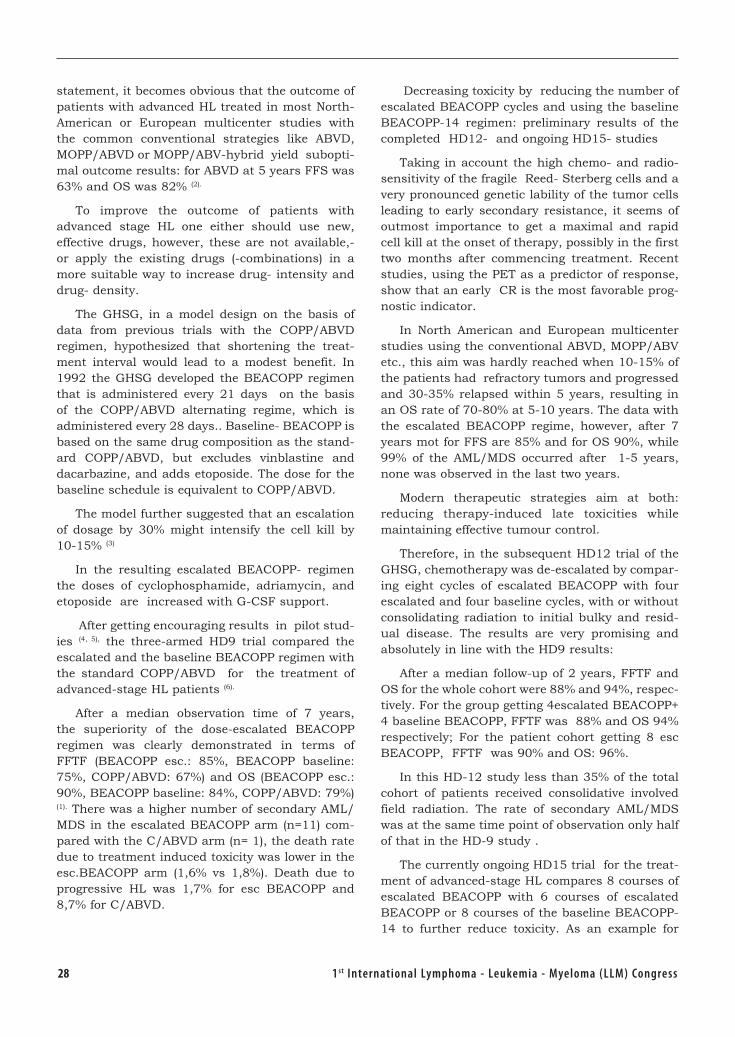
28 1 st International Lymphoma - Leukemia - Myeloma (LLM) Congress
statement, it becomes obvious that the outcome of patients with advanced HL treated in most North- American or European multicenter studies with the common conventional strategies like ABVD, MOPP/ABVD or MOPP/ABV-hybrid yield subopti-mal outcome results: for ABVD at 5 years FFS was 63% and OS was 82% (2).
To improve the outcome of patients with advanced stage HL one either should use new, effective drugs, however, these are not available,- or apply the existing drugs (-combinations) in a more suitable way to increase drug- intensity and drug- density.
The GHSG, in a model design on the basis of data from previous trials with the COPP/ABVD regimen, hypothesized that shortening the treat-ment interval would lead to a modest benefit. In 1992 the GHSG developed the BEACOPP regimen that is administered every 21 days on the basis of the COPP/ABVD alternating regime, which is administered every 28 days.. Baseline- BEACOPP is based on the same drug composition as the stand-ard COPP/ABVD, but excludes vinblastine and dacarbazine, and adds etoposide. The dose for the baseline schedule is equivalent to COPP/ABVD.
The model further suggested that an escalation of dosage by 30% might intensify the cell kill by 10-15% (3)
In the resulting escalated BEACOPP- regimen the doses of cyclophosphamide, adriamycin, and etoposide are increased with G-CSF support.
After getting encouraging results in pilot stud-ies (4, 5), the three-armed HD9 trial compared the escalated and the baseline BEACOPP regimen with the standard COPP/ABVD for the treatment of advanced-stage HL patients (6).
After a median observation time of 7 years, the superiority of the dose-escalated BEACOPP regimen was clearly demonstrated in terms of FFTF (BEACOPP esc.: 85%, BEACOPP baseline: 75%, COPP/ABVD: 67%) and OS (BEACOPP esc.: 90%, BEACOPP baseline: 84%, COPP/ABVD: 79%) (1). There was a higher number of secondary AML/MDS in the escalated BEACOPP arm (n=11) com-pared with the C/ABVD arm (n= 1), the death rate due to treatment induced toxicity was lower in the esc.BEACOPP arm (1,6% vs 1,8%). Death due to progressive HL was 1,7% for esc BEACOPP and 8,7% for C/ABVD.
Decreasing toxicity by reducing the number of escalated BEACOPP cycles and using the baseline BEACOPP-14 regimen: preliminary results of the completed HD12- and ongoing HD15- studies
Taking in account the high chemo- and radio- sensitivity of the fragile Reed- Sterberg cells and a very pronounced genetic lability of the tumor cells leading to early secondary resistance, it seems of outmost importance to get a maximal and rapid cell kill at the onset of therapy, possibly in the first two months after commencing treatment. Recent studies, using the PET as a predictor of response, show that an early CR is the most favorable prog-nostic indicator.
In North American and European multicenter studies using the conventional ABVD, MOPP/ABV etc., this aim was hardly reached when 10-15% of the patients had refractory tumors and progressed and 30-35% relapsed within 5 years, resulting in an OS rate of 70-80% at 5-10 years. The data with the escalated BEACOPP regime, however, after 7 years mot for FFS are 85% and for OS 90%, while 99% of the AML/MDS occurred after 1-5 years, none was observed in the last two years.
Modern therapeutic strategies aim at both: reducing therapy-induced late toxicities while maintaining effective tumour control.
Therefore, in the subsequent HD12 trial of the GHSG, chemotherapy was de-escalated by compar-ing eight cycles of escalated BEACOPP with four escalated and four baseline cycles, with or without consolidating radiation to initial bulky and resid-ual disease. The results are very promising and absolutely in line with the HD9 results:
After a median follow-up of 2 years, FFTF and OS for the whole cohort were 88% and 94%, respec-tively. For the group getting 4escalated BEACOPP+ 4 baseline BEACOPP, FFTF was 88% and OS 94% respectively; For the patient cohort getting 8 esc BEACOPP, FFTF was 90% and OS: 96%.
In this HD-12 study less than 35% of the total cohort of patients received consolidative involved field radiation. The rate of secondary AML/MDS was at the same time point of observation only half of that in the HD-9 study .
The currently ongoing HD15 trial for the treat-ment of advanced-stage HL compares 8 courses of escalated BEACOPP with 6 courses of escalated BEACOPP or 8 courses of the baseline BEACOPP-14 to further reduce toxicity. As an example for

291st International Lymphoma - Leukemia - Myeloma (LLM) Congress
a time dense/ intensified chemotherapy, the BEACOPP-14 schedule, representing a BEACOPP-baseline variant given in 14 day intervals, was developed .
Consolidative radiaton was given to 65% of patients. This regimen was tested in a multicenter pilot study with the final analysis showing an estimated FFTF of 90%, an OS of 95% at 5 years median observation time . The acute hematotoxic-ity was significantly reduced and ranged between that of escalated and baseline BEACOPP-21 (7). In this pilot study with 94 patients after 5 years mot there was no AML/MDS, no solid tumor, only 1 NHL as secondary neoplasia. There were two toxic deaths during treatment.
Conclusion and future aims
The question is relevantThe question is relevant
Is it justified to call ABVD the goldstand-ard treatment strategy for advanced stage HL patients?
The answer might be yes, if one takes the recent data of North American single center studies (Van-couver) in account, where 25-30% of patients with stages I-II with bulky tumors and B-symptoms are subsummized into the group of advanced stage HL patients. These patients, however, are treated in the GHSG in the early unfavorable (intermediate) cohort with just 4 cycles of ABVD+ 20 Gy IF-RT, resulting in a 5 year FFS of 90% and an OS rate of 97%!
The robust data from the North American multi-center studies (CALGB, ECOG, Intergroup) includ-ing only stages III-IV and possibly the IIB patients with large mediastinal masses in the advanced stage group showed rather disappointing results with a 10 year FFS of 50% (8).
The answer might be no, if one accepts that at a median observation time of 7 years the GHSG data with escalated BEACOPP (HD9- study) (6, 1) showed that 11 more young patients (median age 28 years) out of a group of 100 escalated BEACOPP treated patients are alive and possibly cured, compared with the group of C/ABVD treated patients, where 21 patients had already died at this time point.
The pivotal question isThe pivotal question is
How much long term toxicity do we expect for escalated BEACOPP treated patients after 10, 15 and 20 years follow up? There is certainly no
answer to this question at the moment. The 10 year data of the HD9 study, however, look prom-ising and inspite of a higher number of initially occuring AML/MDS there is a significant superior-ity for the escalated BEACOPP regimen compared to C/ABVD and 11 more young patients survive with this admittedly very toxic principle than with the ABVD regime in a multi center study with more than 500 participating centers and many patients treated in outpatient setting, as it is the case in the GHSG.
The difference for freedom from treatment fail-ure (FFTF) at 10 years is even higher in the two arms: 64% for C/ABVD and 82% for BEACOPP escalated. That means 18% of patients treated with BEACOPP escalated do not need a salvage treat-ment conferring a even higer risk of AML/MDS than BEACOPP escalated when high dose chemo-therapy with autologous stem cell support is given as salvage.
To further reduce the danger of late complica-tions, like secondary tumors, cardiac and pulmo-nary late toxicities, the GHSG aims at reducing the number of escalated BEACOPP cycles from a total of 8 to 6 (HD15) and in the HD12 trial even to 4 escalated BEACOPP + 4 baseline BEACOPP cycles. Parallel to this de-escalation of BEACOPP the amount of radiotherapy is reduced from 65% of patients receiving consolidating radiation in the HD9 trial to less than 5% in the HD15 trial, where involved field RT is only given to PET positive >2,5cm rest tumors. All PET negative rest tumors, irrespective of their size after 6 or 8 escalated BEACOPP- or 8 BEACOPP-14- cycles, do not get IF-RT anymore!
The future strategies for advanced HL are 2 fold:
1. risk adapted therapy allocating the patients according to their risk of being resistant or relapsing using the International Prognostic Score (Hasenclever/Diehl) and treating patients with the lower risk IPS 0-2 with ABVD and with the higher risk IPS > 3 with BEACOPP escalated and
2. response adapted therapy using FDG-PET as a response- and prognosis predictor. That means, after 2 courses of either ABVD or BEACOPP esc and a negative PET: continue with ABVD, if PET is positive, change to BEACOPP esc.
30 1 st International Lymphoma - Leukemia - Myeloma (LLM) Congress
The ultimate answer to the question whether escalated BEACOPP is superior to ABVD only can be answered in a randomized multi center, multi culture setting, testing both regimen with and without radiation and in the different strata of the risk groups of the International Prognostic Factor Project (9). A global study, lead by the EORTC is cur-rently being undertaken in North America, Canada, Australia and in Europe ( EORTC (#20012) (10).
References 1. Diehl V, Brillant C, Franklin J, et al. BEACOPP che-
motherapy for Advanced Hodgkin’s Disease: Results of Further Analyses of the HD9- and HD12- Trials of the German Hodgkin Study Group (GHSG). Blood 2004 Nov 16; 104(11)A: 307.
2. Duggan DB, Petroni GR, Johnson JL, et al. Rando-mized comparison of ABVD and MOPP/ABV hybrid for the treatment of advanced Hodgkin’s disease: report of an intergroup trial. J Clin Oncol. 2003 Feb 15;21(4):607-14.
3. Loeffler M, Hasenclever D, Diehl V Model based development of the BEACOPP regimen for advanced stage Hodgkin‘s disease. German Hodgkin‘s Lymp-homa Study Group. Ann Oncol. 1998;9 Suppl 5:S73-8.
4. Diehl V. Dose-escalation study for the treatment of Hodgkin’s disease. The German Hodgkin Study Group (GHSG). Ann Hematol 1993; 66: p139-140.
5. Diehl V, Sieber M, Ruffer U, et al. BEACOPP: an intensified chemotherapy regimen in advanced Hodgkin‘s disease. The German Hodgkin‘s Lympho-ma Study Group. Ann Oncol 1997; 8:143-148.
6. Diehl V, Franklin J, Pfreundschuh M, et al., Stan-dard and increased-dose BEACOPP chemotherapy compared with COPP-ABVD for advanced Hodgkin‘s disease. N Engl J Med 2003 Jun 12;348(24):2386-95.
7. Sieber M, Bredenfeld H, Josting A, et al. 14-day variant of the bleomycin, etoposide, doxorubicin, cyclophosphamide, vincristine, procarbazine, and prednisone regimen in advanced-stage Hodgkin‘s lymphoma: results of a pilot study of the German Hodgkin‘s Lymphoma Study Group. J Clin Oncol 2003 May 1;21(9):1734-9.
8. Canellos GP, Niedzwiecki D Long-term follow-up of Hodgkin‘s disease trial N Engl J Med. 2002 May 2;346(18):1417-8.
9. Hasenclever D, Diehl V. A prognostic score for advanced Hodgkin’s disease. International Prognos-tic Factors Project on Advanced Hodgkin’s Disease. N Engl J Med. 1998 Nov 19;339(21):1506-14.
10. Raemaekers J, Kluin-Nelemans H, Teodorovic I, et al. The achievements of the EORTC Lymphoma Group. European Organisation for Research and Treatment of Cancer. Eur J Cancer 38, Suppl 4:S107-S113, 2002.
311st International Lymphoma - Leukemia - Myeloma (LLM) Congress
From the first Department of Internal Medi-cine, University Hospital Cologne, Germany, and the German Hodgkin Lymphoma Study
Group (GHSG)
Introduction
Depending on stage and risk factor profile, up to 95% of patients with Hodgkin’s disease (HD) at first presentation reach complete remission (CR) after the initial standard treatment including radi-otherapy, combination chemotherapy, or combined modality therapy. Patients who relapse after a first CR can achieve a second CR with salvage treat-ment including radiotherapy for localized relapse in previously non-irradiated areas, conventional salvage chemotherapy, or high-dose chemotherapy (HDCT) with stem cell transplantation (SCT).1
Prognostic factors in relapsed and refractory Hodgkin`s disease
It was first noted in 1979 that the length of remission to first-line chemotherapy had a marked effect on the ability of patients to respond to sub-sequent salvage treatment.2 In 1992 the National Cancer Institute (NCI) updated their experience with the long-term follow up of patients who relapsed after polychemotherapy.3 Derived primari-ly from investigations involving failures after MOPP and MOPP variants, the conclusions are thought to be relevant to other chemotherapy programs as well. On this basis, chemotherapy failures can be divided into three subgroups:
• Primary progressive Hodgkin`s disease, i.e. patients who never achieve a complete remission
Relaps and Refractory Hodgkin`s Diseases
Andreas Josting
First Department of Internal Medicine, University Hospital Cologne, Cologne, Germany
• Early relapses within 12 months of CR
• Late relapses after CR lasting > 12 months
Using conventional chemotherapy for patients with primary progressive disease, virtually no patient survives more than eight years. In contrast, the projected 20-year survival for patients with early relapse or late relapse was 11% and 22%, respectively.3
Primary progressive Hodgkin`s diseasePrimary progressive Hodgkin`s disease
Patients with primary progressive disease, defined as progression during induction treatment or within 90 days after the end of treatment, have a particularly poor prognosis. Conventional sal-vage regimens have given disappointing results in the vast majority of patients: response to salvage treatment is low and the duration of response is often short. The 8-year OS ranges between 0% and 8%.3,4
The German Hodgkin`s disease Study Group (GHSG) retrospectively analysed 206 patients with PD to determine outcome after salvage therapy and identify prognostic factors.5 The five year freedom from second failure (FF2F) and OS for all patients was 17% and 26%. As reported from transplant centers, the five year FF2F and OS for patients treated with HDCT is 42% and 48%, respectively, but only 33% of all patients received HDCT. The low percentage of patients who received HDCT was due to rapidly fatal disease or life-threatening severe toxicity after salvage therapy. Other reasons not to proceed to HDCT were insufficient stem cell harvest, poor performance status and older age.
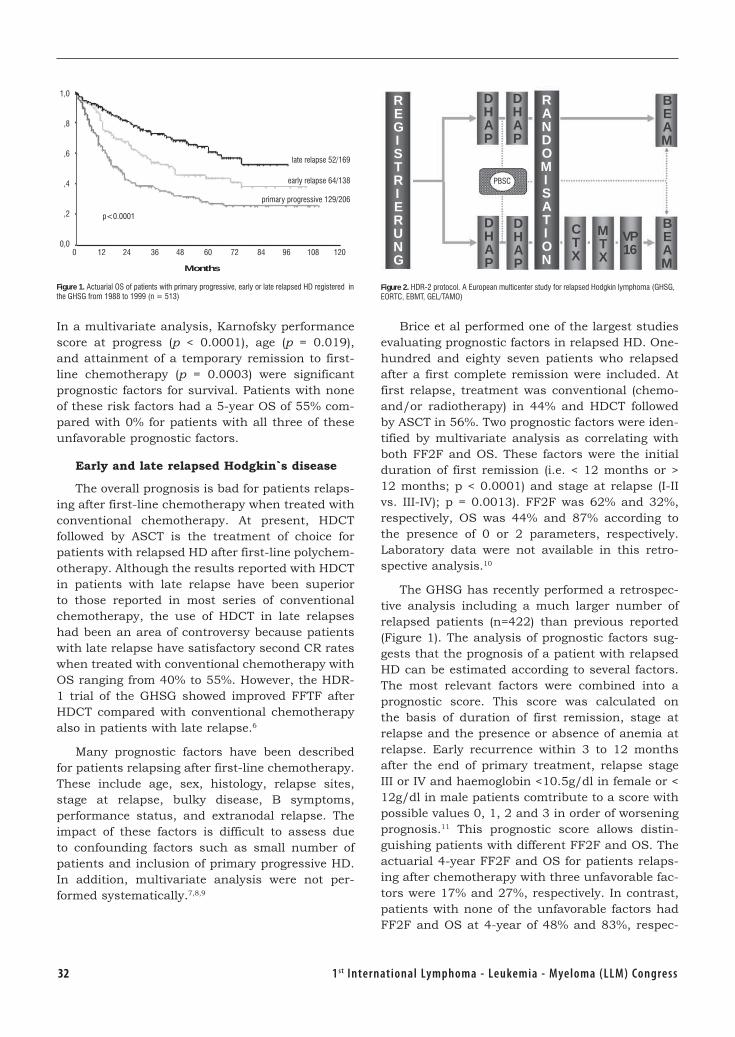
32 1 st International Lymphoma - Leukemia - Myeloma (LLM) Congress
In a multivariate analysis, Karnofsky performance score at progress (p < 0.0001), age (p = 0.019), and attainment of a temporary remission to first-line chemotherapy (p = 0.0003) were significant prognostic factors for survival. Patients with none of these risk factors had a 5-year OS of 55% com-pared with 0% for patients with all three of these unfavorable prognostic factors.
Early and late relapsed Hodgkin`s disease
The overall prognosis is bad for patients relaps-ing after first-line chemotherapy when treated with conventional chemotherapy. At present, HDCT followed by ASCT is the treatment of choice for patients with relapsed HD after first-line polychem-otherapy. Although the results reported with HDCT in patients with late relapse have been superior to those reported in most series of conventional chemotherapy, the use of HDCT in late relapses had been an area of controversy because patients with late relapse have satisfactory second CR rates when treated with conventional chemotherapy with OS ranging from 40% to 55%. However, the HDR-1 trial of the GHSG showed improved FFTF after HDCT compared with conventional chemotherapy also in patients with late relapse.6
Many prognostic factors have been described for patients relapsing after first-line chemotherapy. These include age, sex, histology, relapse sites, stage at relapse, bulky disease, B symptoms, performance status, and extranodal relapse. The impact of these factors is difficult to assess due to confounding factors such as small number of patients and inclusion of primary progressive HD. In addition, multivariate analysis were not per-formed systematically.7,8,9
Brice et al performed one of the largest studies evaluating prognostic factors in relapsed HD. One-hundred and eighty seven patients who relapsed after a first complete remission were included. At first relapse, treatment was conventional (chemo- and/or radiotherapy) in 44% and HDCT followed by ASCT in 56%. Two prognostic factors were iden-tified by multivariate analysis as correlating with both FF2F and OS. These factors were the initial duration of first remission (i.e. < 12 months or > 12 months; p < 0.0001) and stage at relapse (I-II vs. III-IV); p = 0.0013). FF2F was 62% and 32%, respectively, OS was 44% and 87% according to the presence of 0 or 2 parameters, respectively. Laboratory data were not available in this retro-spective analysis.10
The GHSG has recently performed a retrospec-tive analysis including a much larger number of relapsed patients (n=422) than previous reported (Figure 1). The analysis of prognostic factors sug-gests that the prognosis of a patient with relapsed HD can be estimated according to several factors. The most relevant factors were combined into a prognostic score. This score was calculated on the basis of duration of first remission, stage at relapse and the presence or absence of anemia at relapse. Early recurrence within 3 to 12 months after the end of primary treatment, relapse stage III or IV and haemoglobin <10.5g/dl in female or < 12g/dl in male patients comtribute to a score with possible values 0, 1, 2 and 3 in order of worsening prognosis.11 This prognostic score allows distin-guishing patients with different FF2F and OS. The actuarial 4-year FF2F and OS for patients relaps-ing after chemotherapy with three unfavorable fac-tors were 17% and 27%, respectively. In contrast, patients with none of the unfavorable factors had FF2F and OS at 4-year of 48% and 83%, respec-
Figure 1. Actuarial OS of patients with primary progressive, early or late relapsed HD registered in the GHSG from 1988 to 1999 (n = 513)
1,0
,8
,6
,4
,2
0,00 12 24 36 48 60 72 84 96 108 120
Months
p<0.0001
late relapse 52/169
early relapse 64/138
primary progressive 129/206
Figure 2. HDR-2 protocol. A European multicenter study for relapsed Hodgkin lymphoma (GHSG, EORTC, EBMT, GEL/TAMO)
R E G I S T R I E R U N G
D H A P
D H A P
D H A P
D H A P
C T X
M T X
VP 16
B E A M
B E A M
R A N D O M I S A T I O N
PBSC
331st International Lymphoma - Leukemia - Myeloma (LLM) Congress
tively. In addition, the prognostic score was also predictive for patients relapsing after radiotherapy, for patients relapsing after chemotherapy who were treated with conventional therapies or with HDCT followed by ASCT, and for patients under 60 years and a Karnofsky performance status ≥ 90% being the major candidate groups for dose intensi-fication. Our prognostic factor score uses clinical characteristics which can be easily collected at the time of relapse. It separates groups of patients with substantially different outcomes.
The prognostic factors identified may be use-ful to tailor the therapy for subgroups of patients, to define homogeneous cohorts for prospective randomized trials, and to identify more precisely patients with poor-risk relapse who should be treated with innovative approaches.
Treatment strategies
The survival of patients treated with convention-al chemotherapy after relapse of irradiated early-stage disease is at least equal to that of advanced-stage patients initially treated with chemotherapy. Overall survival (OS) and disease-free survival (DFS) range from 57% to 71%.12,13 Patients who relapse following radiation therapy alone for local-ized Hodgkin’s disease have satisfactory results with combination chemotherapy and are not con-sidered candidates for HDCT and ASCT.
HDCT followed by ASCT has been shown to produce 30%-65% long-term disease-free survival in selected patients with refractory and relapsed HD.14-17 In addition, the reduction of early trans-plant-related mortality from 10% - 25% reported in earlier studies to less than 5% in more recent studies has led to the widespread acceptance of HDCT and ASCT.
Although results of HDCT have generally been better than those observed after conventional-dose salvage therapy, the validity of these results has been questioned due to the lack of randomized tri-als. The most compelling evidence for the superior-ity of HDCT and ASCT in relapsed HD comes from two reports from the British National Lymphoma Investigation (BNLI) and the German Hodgkin’s Lymphoma Study Group (GHSG) together with the European Group for Blood and Marrow Transplan-tation (EBMT).
In the BNLI trial, patients with relapsed or refractory HD were treated with a combination of carmustine (BCNU), etoposide, cytarabine and mel-
phalan at a conventional-dose level (mini-BEAM) or a high-dose level (BEAM) with autologous bone-marrow transplantation.18 The actuarial 3-year event-free survival (EFS) was significantly better in patients who received high-dose chemotherapy (53% vs 10%).
The largest randomized, multicenter trial was performed by the GHSG/EBMT to determine the benefit of HDCT in relapsed HD. Patients with relapse after polychemotherapy were randomly assigned between four cycles of Dexa-BEAM (dex-amethasone, BCNU, etoposide, Ara-C and mel-phalan) and two cycles of Dexa-BEAM followed by HDCT (BEAM) and ABMT/PBSCT. The final analysis of 144 evaluable patients revealed that from 117 patients with PR or CR after two cycles of chemotherapy, FFTF in the HDCT group was 55% versus 34% for the patients receiving an additional two cycles of chemotherapy. OS was not signifi-cantly different.6
Sequential high-dose chemotherapy
In recent years, sequential high-dose chemo-therapy has increasingly been employed in the treatment of solid tumors, hematologic and lympho-proliferative disorders. Initial results from phase-I/II studies indicate that this kind of therapy offers safe and effective treatment.19-24 In accordance with the Norton-Simon hypothesis,25 following initial cytoreduction, few non-cross-resistant agents are given at short intervals. In general, the trans-plantation of PBSC and the use of growth factors allow the application of the most effective drugs at the highest possible doses at intervals of one to three weeks. Sequential high-dose chemotherapy thereby enables the highest possible dosing over a minimum period of time (dose intensification).
In 1997 a multicenter phase-II trial with a high-dose sequential chemotherapy program and a final myeloablative course was started to evaluate the feasibility and efficacy of this novel regimen in patients with relapsed HD.26 Eligibility crite-ria included age 18-60 yrs., histologically proven relapsed or primary progressive HD, second relapse with no prior HDCT and ECOG performance status 0-1.
The treatment program consists of two cycles of DHAP (dexamethasone, ara-C, cisplatin) in the first phase in order to reduce tumor burden before HDCT. Patients with partial remission (PR) or complete remission (CR) after two cycles of DHAP,
34 1 st International Lymphoma - Leukemia - Myeloma (LLM) Congress
receive sequential high-dose chemotherapy con-sisting of cyclophosphamide 4 g/m2 iv, methotrex-ate 8 g/m2 iv plus vincristine 1.4 mg/m2 iv; and etoposide 2 g/m2 iv. The final myeloblative course was BEAM followed by PBSCT with at least 2 x 10⁶ CD34+ cells/kg.
At the last interim analysis 102 patients were available for the final evaluation. State of remis-sion was multiple relapse in 10 patient, progressive disease in 16 patients, early relapse in 29 patients and late relapse in 44 patients. At 18 months of median follow-up (range 3-31 months) results are as follows: Response rate (RR) after DHAP 87% (23% CR, 64% PR) and RR at final evaluation 77% (68% CR, 9% PR). Toxicity was tolerable with no treatment related deaths. FFTF and OS for patients with early relapse were 64%/87% for early relapse; 68%/81% for late relapse; 30%/58% for patients with progressive disease and 55%/88% for patients with multiple relapse.26
In conclusion, sequential administration of high doses of cyclophosphamide, methotrexate and etoposide is feasible and did not affect the tolerability of final myeloablative BEAM. This new, three-phase treatment regimen is well tolerated and feasible in patients with relapsed and primary progressive HD. The preliminary data suggests a high efficacy in relapsed HD patients, warranting further randomized studies.
HDR-2 Protocol
In January 2001, the GHSG together with the EORTC, the GEL/TAMO and the EBMT started a prospective randomized study to compare the effectiveness of a standard HDCT (BEAM) with a sequential HDCT after initial cytoreduction with 2 cycles DHAP (HD-R2 protocol, Fig. 2).
Patients with histologically confirmed early or late relapsed HD, and patients in second relapse with no prior HDCT fulfilling the entry criteria receive two cycles of dexamethasone, high-dose cytarabine and cisplatin (DHAP) followed by G-CSF.
Patients achieving NC, PR or CR after DHAP are centrally randomized to receive either BEAM followed by PBSCT (arm A of the study) or HD cyclophosphamide + G-CSF, followed by HD-MTX + vincristine, followed by HD etoposide + G-CSF and a final myeloablative course with BEAM (arm B of the study).
Allogeneic transplantation after reduced conditioning in HD
Allogeneic transplantation (alloBMT) has clear advantages compared with autologous transplan-tation: Donor marrow cells uninvolved by malig-nancy are used avoiding the risk of infusing occult lymphoma cells, which may contribute to relapse in patients who undergo autologous transplanta-tion. In addition, donor lymphoid cells can poten-tially mediate a graft-versus-lymphoma effect.
Generally, donor availability and age constraints have limited a broader application of alloBMT in HD. Moreover, alloBMT is associated with a high treatment related mortality rate of up to 75% observed in patients with induction failure which casts doubt upon the feasibility of this approach in HD patients.27-30 In most cases, allogeneic transplantation from HLA-identical siblings is not recommended for patients with HD. The reduced relapse-rate associated with a potential graft-ver-sus-tumor effect is offset by lethal graft-versus-host toxicity.
Nevertheless, patients with induction failure and relapsed patients with additional risk factors have a poor prognosis also after HDCT and ASCT. Therefore, the role of alloBMT should be further evaluated in these patients taken advantage of new developments like non-meloablative conditioning regimens and alloPBSCT.
To circumvemt the problems inherent to the toxicity and treatment related mortality associated to allografting, the posibility to achieve engraftment of allogeneic stem cells after immunosuppressive therapy combined with myelosuppressive but non-myeloablative therapy has been assessed. Several groups have recently updated their experience with non-myeloablative conditioning regimens.31-33
The EBMT together with the GEL/TAMO, the EORTC and the GHSG activated a multicenter phase II study to evaluate the treatment-related mortality (TRM) of patients with primary progres-sive or relapsed HD (early relapse, multiple relapse and relapse after autologous SCT). Patients with an HLA compatible sibling donor or an HLA matched unrelated donor will be initially treated with 1-2 cycles of DHAP or other salvage protocols to reduce tumor burden before alloPBSCT. PBSC will be collected after G-CSF priming of the donor and reinfused after conditioning with fludarabine and melphalan.
351st International Lymphoma - Leukemia - Myeloma (LLM) Congress
Future directions
Alternative strategies have been developed to improve the outcome of relapsed and resistant HD. These approaches include the development of new cytostatic drugs and biological agents with proven efficacy in preclinical models.
One of the most promising new cytostatic drugs is the new vinca alkaloid vinorelbine, which has demonstrated activity in HD even in patients pretreated with vincristine or vinblastine.34 The use of vinorelbine in first and second-line therapy of HD in order to improve frequency and dura-tion of response is still under investigation. The pyrimidine analogue gemcitabine is the only drug currently under investigation that represents a new cytostatic mechanism of action. The ”self-potentiating” mechanism of action leads to an enhanced accumulation and prolonged retention of gemcitabine in the malignant cell. The results of gemcitabine in advanced relapsed HD are promis-ing, with an overall response rate of 53% in heavily pretreated patients.35
Although some clinical efficacy has been dem-onstrated in clinical trials with immunotoxins (IT) none of the current available IT seems to be suited for a clinical phase-III study.36-38 Bispecific mono-clonal antibodies (BiMoab) such as the recently reported CD30xCD64 BiMoab look more promis-ing with clinical development programs scheduled including phase III. The use of recombinant DNA technology for site-directed modifications of the IT and the development of humanized IT and BiMo-abs might optimize their efficacy.38 In the future, combining standard chemo-/radiotherapy with biological agents might result in the elimination of residual tumor cells and subsequently more relapse-free long term survivors.
References 1. Josting A, Wolf J, Diehl V. Hodgkin`s disease. Prog-
nostic factors and treatment startegies. Current Opinion in Oncology 2000, 12, 403-411
2. Fisher R, De VV, Hubbard S, et al: Prolonged dis-ease-free survival in Hodgkin‘s disease with MOPP reinduction after first relapse. Ann Intern Med, 1979; 90: 761-765
3. Longo D, Duffey P, Young R, et al: Conventional-dose salvage combination chemotherapy in patients relapsing with Hodgkin‘s disease after combination chemotherapy: the low probability for cure. J Clin Oncol, 1992; 10: 210-218
4. Bonfante V, Santoro A, Viviani S, et al: Outcome of patients with Hodgkin‘s disease failing after primary MOPP/ABVD. J Clin Oncol, 1997; 15: 528-534
5. Josting A, Rueffer U, Franklin J, et al: Procnostic factors and treatment outcome in primary progressi-ve Hodgkin`s lymphoma - A report from the German
Hodgkin`s Lymphoma Study Group (GHSG). Blood, 2000, Volume 96, No. 4, 1280-1286
6. Schmitz N, Pfistner B, Sextro M, et al: Aggressive conventional chemotherapy compared with high-dose chemotherapy with autologous haemopoietic stem-cell transplantation for relapsed chemosensi-tive Hodgkin‘s disease: a randomised trial. Lancet, 2002, 359 (9323): 2065-2071
7. Lohri A, Barnett M, Fairey RN, et al: Outcome of treatment of first relapse of Hodgkin‘s disease after primary chemotherapy: identification of risk factors from the British Columbia experience 1970 to 1988. Blood, 1991, 77(10), 2292-2298
8. Fermè C, Bastion Y, Lepage E, et al: The MINE regi-men as intensive salvage chemotherapy for relapsed and refractory Hodgkin‘s disease. Ann Oncol, 1995, 6(6), 543-549
9. Reece D, Barnett M, Shepherd J, et al: High-dose cyclophosphamide, carmustine (BCNU), and eto-poside (VP16-213) with or without cisplatin (CBV +/- P) and autologous transplantation for patients with Hodgkin‘s disease who fail to enter a complete remission after combination chemotherapy. Blood 1995, 86, 451-458
10. Brice P, Bastion Y, Divine M, et al: Analysis of prog-nostic factors after the first relapse of Hodgkin‘s disease in 187 patients. Cancer, 1996, 78 (6), 1293-1299
11. Josting A, Franklin J, May M, et al: A new prognos-tic score based on treatment outcome of patients with relapsed Hodgkin lymphoma registered in the database of the German Hodgkin Lymphoma Study Group (GHSG). J Clin Oncol, 2002, 20 (1), 221-230
12. Cannellos G, Young RC, De Vita VD: Combination chemotherapy for advanced Hodgkin`s disease in relapse following extensive radiotherapy. Clin Pharm Ther, 1972; 13: 750-758
13. Santoro A, Viviani S, Villarreal C, et al: Salvage che-motherapy in Hodgkin‘s disease irradiation failures: superiority of doxorubicin-containing regimens over MOPP. Cancer Treat Rep, 1986; 70: 343-351
14. Josting A, Katay I, Rueffer U, et al: Favorable outco-me of patients with relapsed or refractory Hodgkin‘s disease treated with high-dose chemotherapy and stem cell rescue at the time of maximal response to conventional salvage therapy (Dexa-BEAM). Ann Oncol, 1998; 9: 289-296
15. Biermann PJ, Bagin RG, Jagannath S, et al: High dose chemotherapy followed by autologous hema-topoietic rescue in Hodgkin`s disease: Long term follow-up in 128 patients. Ann Oncol, 1993, 4, 767-773
16. Reece DE, Connors JM, Spinelli JJ, et al: Intensive therapy with cyclophosphamide, carmustine, eto-poside +/- cisplatin, and autologous bone marrow transplantation for Hodgkin`s disease in first relap-se after combination chemotherapy. Blood, 1994, 83(5), 1193-1199
36 1 st International Lymphoma - Leukemia - Myeloma (LLM) Congress
17. Armitage JO, Biermann PJ, Vose JM, et al: Autolo-gous bone marrow transplantation for patients with relapsed Hodgkin`s disease. Am J Med, 1991, 91, 605-610
18. Linch D, Winfield D, Goldstone A, et al: Dose inten-sification with autologous bone marrow transplan-tation in relapsed and resistant Hodgkin‘s disease: results of a BNLI randomised trial. Lancet, 1993; 341: 1051-1054
19. Gianni AM, Siena S, Bregni M, Lombardi F, et al: High-dose sequential chemo-radiotherapy with peripheral blood progenitor cell support for relapsed or refractory Hodgkin`s disease - A 6-year update. Ann Oncol, 1993, 4, 889-891
20. Gianni AM, Bregni M, Siena S: 5-year update of the Milan Cancer Institute randomized trial of high-dose sequential (HDS) vs MACOP-B therapy for diffuse large-cell lymphomas. Proc ASCO , 1994, 13, 373 (A1263)
21. Patrone F, Ballestrero A, Ferrando F, Brema F, Moraglio L, Valbonesi M, Basta P, Ghio R, Gobbi M, Sessarego M: Four-step high-dose sequential che-motherapy with double hemato poetic progenitor-cell rescue for metastatic breast cancer. JCO, 1995, 13, 840-846
22. Shea T, Mason JR, Storniolo AM, et al: Sequen-tial cycles of high-dose carboplatin administered with recombinat human granulocyte-makrophage colony-stimulating factor and repeated infusions of autologous peripheral-blood progenitor cells: A novel and effective method for delivering multiple courses of dose-intensive therapy. JCO, 1992, 10, 464-473
23. Gianni AM, Taella C, Bregni M, et al: High-dose sequential chemo-radiotherapy, a widely applicable regimen, confers survival benefit to patients with high-risk multiple myeloma. JCO, 1994, 12, 503-509
24. Caracciolo D, Gavarotti P, Aglietta M, et al: High-dose sequential chemotherapy with blood and mar-row cell autograft as salvage treatment in very poor prognosis, relapsed Non-Hodgkin`s lymphoma. Bone Marrow Transplant, 1993, 12, 621-625
25. Norton L, Simon R: The Norton-Simon hypothesis revisited. Canc Treat Rep, 1986, 70, 163-169
26. Josting A, Rudolph C, Mapara M, et al. Cologne high-dose sequential chemotherapy in relapsed and refractory Hodgkin lymphoma - Results of a large multicenter study of the German Hodgkin Lympho-ma Study Group (GHSG). Blood, 2003, Vol. 102, 11, Suppl 1, 1461
27. Anderson JE, Litzow MR, Appelbaum FR, et al: Allo-geneic, syngeneic, and autologous marrow transp-lantation for Hodgkin`s disease: The 21-year Seattle experience. J Clin Oncol 11: 2342-2350, 1993
28. Phillips GL, Reece DE, Barnett MJ, et al: Allogeneic marrow transplantation for refractory Hodgkin`s disease. J Clin Oncol 7: 1039-1045, 1989
29. Jones RJ, Piantadosi S, Mann RB, et al: High-dose cytotoxic therapy and bone marrow transplantation for relapsed Hodgkin`s disease. J Clin Oncol 8: 527-537, 1990
30. Milpied N, Fielding AK, Pearce RM, et al: Allogeneic bone marrow transplantation is not better than autologous transplant for patients with relapsed Hodgkin`s disease. J Clin Oncol 14: 1291-1296, 1996
31. Carella AM, Cavaliere M, Beltrami G, et al: Immuno-suppressive nonmyeloablative allografting as salva-ge therapy in advanced Hodgkin‘s disease. Haema-tologica. 2001, 86 (11):1121-1123
32. Sureda A, Robinson S, De Elvira CR, et al: Alloge-neic stem cell tranplantation significantly reduces transplant related mortality in comparison with with conventional allogeneic transplantation in relapsed or refarctory Hodgkin`s disease: Results of the Euro-pean Group for Blood and Marrow Transplantation. Blood, 2003, Vol. 102, 11, Suppl 1, 1461
33. Schmitz N, Sureda A, Robinson S: Allogeneic transp-lantation of hematopoietic stem cells after nonm-yeloablative conditioning for Hodgkin‘s disease: indications and results. Semin Oncol, 2004, 31(1): 27-32
34. Devizzi L, Santaro A, Bonfante V, et al: Vinorelbine: An active drug for the management of patients with heavily pretreated hodgkin`s disease. Ann Oncol 5: 817-820, 1994
35. Santoro A, Bredenfeld H, Devizzi L, et al. Gemcitabi-ne in the treatment of refractory Hodgkin‘s disease: results of a multicenter phase II study. J Clin Oncol. 2000, 18: 2615-2619
36. Engert A, Diehl V, Schnell R, et al: A phase I study of an anti-CD25 ricin A chain immunotoxin (RFT5-SMPT-dgA) in patients with refractory Hodgkin`s lymphoma. Blood 15: 403-410, 1997
37. Schnell R, Stark O, Borchmann P, et al: A phase I study with an anti-CD30 ricin A-chain Immunotoxin (Ki-4.dgA) in patients with refractory CD30 positive Hodgkin`s and Non-Hodgkin`s lymphoma. Clin Canc Res, 2002, in press
38. Hartmann F, Renner C, Jung W, et al: Treatment of refractory Hodgkin`s disease with an anti-CD16/CD30 bispecific antibody. Blood 15: 2042-2047, 1997
39. Borchmann P, Morschhauser F, Parry A, et aL: The human anti-CD30 antibody 5F11 shows in vitro and in vivo activity against malignant lymphoma. Blood. 2003, 102 (10): 3737-3742
371st International Lymphoma - Leukemia - Myeloma (LLM) Congress
Introduction
In the 19th century chronic myeloid leuke-mia (CML) and polycythemia vera (PV) have been described as primary distinct disease entities1,2,3. In 1960 Nowell and Hungerford described the pres-ence of a minute chromosome in leukemic cells of patients with chronic myeloid leukemia (CML)4. This minute chromosome was called Philadelphia chromosome or Ph after the city of discovery4. Using banding techniques Janet Rowley (1973) dis-covered that the Ph originated from a translocation between the long arms of chromosomes 9 and 22, 9(9;:22)(q34;q11)5. Two groups working together in scientific friendship discovered that a hybrid gene is generated by the translocation consisting of the
Biology Diagnosis and Classification of MPD
Jan Jacques Michiels1,7, Hendrik De Raeve2, Konnie Hebeda3, King H. Lam4,
Freek Bot5, Zwi Berneman1, Wilfried Schroyens1
Departments of Hematology1 and Pathology2, University Hospital Antwerp, BelgiumDepartments of Pathology University Medical Center, Sint Radboud, Nijmegen3 and Erasmus University Medical Center, Rotterdam4,
University Medical Center Maastricht5, The Netherlands, and European Working Group on Myeloproliferative Disorders (EWG.MPD), Goodheart Institute7, Rotterdam, The Netherlands
BCR gene on chromosome 22 and the ABL onco-gene originating from chromosome 96. This results in a BCR/ABL fusion gene with high tyrokinase activity and CML-transformation capacity7,8. Nine-ty-five percent of all CML patients are Ph+; 90% are Ph+/BCR/ABL+, 5% are Ph-/BCR/ABL+, and 5% are Ph-/BCR/ABL- , the latter group usually diagnosed as atypical CML, juvenile CML, chron-ic neutrophilic leukemia or chronic myelomono-cytic leukemia9. According to strict morphological, biochemical, cytogenetic and molecular criteria including the Ph+ chromosome and bcr/abl fusion gene and protein, CML is a malignant disease with an obligate transition into acute leukemia, whereas essential thrombocythemia (ET), PV and agnogenic
AbstractAccording to strict morphological, biochemical, cytogenetic and molecular criteria including the Philadelphia (Ph) chromosome and bcr/abl fusion gene and protein, chronic myeloid leukemia is a malignant disease with an obligate transition into acute leukemia, whereas essential thrombocythemia (ET), polycythemia vera (PV) and agnogenic myeloid metaplasia (AMM) form the Ph-chromosome and brc/abl negative chronic myeloproliferative disorder (MPD) featured by a benign proliferation of the 3 hematopoietic cell lines. Increase and clustering of abnormal enlarged megakaryocytes together with no or various degrees of increased erythropoiesis and/or granulopoiesis in bone marrow biopsy specimens appears to be a pathognomonic clue to the diagnosis of prefibrotic MPD when WHO bone marrow features are applied. On top of established WHO bone marrow features in combination with JAK2V617F mutation screening, and laboratory features including endogenous elytroid colony (EEC) formation and serum erythropoietin (EPO) levels we propose updated European clinical, molecular and pathological (ECMP) criteria for the differential diagnosis of true ET, PV and chronic idiopathic myelofibrosis (CIMF) or AMM. The ECMP criteria reduced the platelet count of 600 x109/l to the upper limit of normal (>400 x109/l) as inclusion criterion for the diagnosis of thrombocythemia in various MPDs. When WHO and ECMP criteria are applied, PVSG-defined ET includes three distinct entities: true ET, early PV mimicking ET, and thrombocythemia associated with early CIMF-0 mimicking ET. Compared to characteristic bone marrow features pathognomonic for MPD, spontaneous EEC and low serum EPO levels are not sensitive enough as isolated markers for the diagnosis and differential diagnosis of prefibrotic ET, PV and CIMF-0. Bone marrow histology assessment remains the gold standard criterion for the diagnosis and staging of the MPDs. PVSG-defined ET and the presence of the JAK2V617F mutation, EEC, PRV-1 overexpression and low serum EPO levels is consistent with early PV (“forme fruste” PV) when ECMP criteria are applied. The combination of JAK2V617F mutation and increased hematocrit (>0.051 males and >0.48 females) is consistent with the diagnosis PV (specificity 100%, sensitivity 95%) without the need of red cell mass measurement. About half of the ET and CIMF patients are JAK2V617F positive (sensitivity 50%). The degree of JAK2V617F positivity of granulocytes is related to disease stage: heterozygous in true ET and early PV and mixed hetero/homozygous to homozygous in overt and advanced PV and CIMF-1 to 3. The proposed ECMP criteria for the differential diagnosis of ET, PV, CIMF in patients with JAK2V617F positive and JAK2 wild type MPD should be evaluated in prospective management studies in search for the most relevant prognostic factors of therapeutic significance. Key words: myeloproliferative disorders, essential thrombocythemia, polycythemia vera, chronic idiopathic myelofibrosis, erythropoietin, endogenous erythroid colony assay, JAK2 V617F mutation, bone marrow pathology
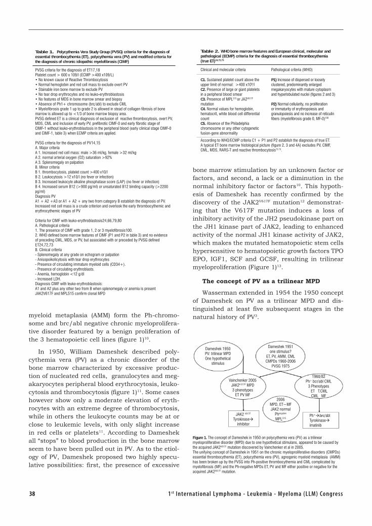
38 1 st International Lymphoma - Leukemia - Myeloma (LLM) Congress
myeloid metaplasia (AMM) form the Ph-chromo-some and brc/abl negative chronic myeloprolifera-tive disorder featured by a benign proliferation of the 3 hematopoietic cell lines (figure 1)10.
In 1950, William Dameshek described poly-cythemia vera (PV) as a chronic disorder of the bone marrow characterized by excessive produc-tion of nucleated red cells, granulocytes and meg-akaryocytes peripheral blood erythrocytosis, leuko-cytosis and thrombocytosis (figure 1)11. Some cases however show only a moderate elevation of eryth-rocytes with an extreme degree of thrombocytosis, while in others the leukocyte counts may be at or close to leukemic levels, with only slight increase in red cells or platelets11. According to Dameshek all “stops” to blood production in the bone marrow seem to have been pulled out in PV. As to the etiol-ogy of PV, Dameshek proposed two highly specu-lative possibilities: first, the presence of excessive
bone marrow stimulation by an unknown factor or factors, and second, a lack or a diminution in the normal inhibitory factor or factors10. This hypoth-esis of Dameshek has recently confirmed by the discovery of the JAK2V617F mutation12 demonstrat-ing that the V617F mutation induces a loss of inhibitory activity of the JH2 pseudokinase part on the JH1 kinase part of JAK2, leading to enhanced activity of the normal JH1 kinase activity of JAK2, which makes the mutated hematopoietic stem cells hypersensitive to hematopoietic growth factors TPO EPO, IGF1, SCF and GCSF, resulting in trilinear myeloproliferation (Figure 1)13.
The concept of PV as a trilinear MPD
Wasserman extended in 1954 the 1950 concept of Dameshek on PV as a trilinear MPD and dis-tinguished at least five subsequent stages in the natural history of PV3.
Table 1. Polycythemia Vera Study Group (PVSG) criteria for the diagnosis of essential thrombocythemia (ET), polycythemia vera (PV) and modified criteria for the diagnosis of chronic idiopathic myelofibrosis (CIMF)
PVSG criteria for the diagnosis of ET17,18Platelet count > 600 x 109/l (ECMP >400 x109/L)• No known cause of Reactive Thrombocytosis• Normal hemoglobin and red cell mass to exclude overt PV• Stainable iron bone marrow to exclude PV• No tear drop erythrocytes and no leuko-erythroblastosis• No features of MDS in bone marrow smear and biopsy• Absence of Ph1+ chromosome (brc/abl) to exclude CML• Myelofibrosis grade 1 up to grade 2 is allowed in stead of collagen fibrosis of bone marrow is allowed up to <1/3 of bone marrow biopsy area. PVSG defined ET is a clinical diagnosis of exclusion of reactive thrombocytosis, overt PV,MDS, CML and inclusion of early PV, prefibrotic CIMF-0 and early fibrotic stage ofCIMF-1 without leuko-erythroblastosis in the peripheral blood (early clinical stage CIMF-0 and CIMF-1, table 3) when ECMP criteria are applied.
PVSG criteria for the diagnosis of PV14,15A. Major criteriaA 1. Increased red cell mass: male >36 ml/kg, female >32 ml/kgA 2. normal arterial oxygen (O2) saturation >92%A 3. Splenomegaly on palpationB. Minor criteriaB 1. thrombocytosis, platelet count >400 x10/lB 2. Leukocytosis >12 x10/l (no fever or infection)B 3. Increased leukocyte alkaline phosphatase score (LAP) (no fever or infection)B 4. Increased serum B12 (>900 pg/ml) or unsaturated B12 binding capacity (>2200 pg/ml)Diagnosis PVA1 + A2 +A3 or A1 + A2 + any two from category B establish the diagnosis of PV. Increased red cell mass is a crude criterion and overlook the early thrombocythemic and erythrocythemic stages of PV
Criteria for CIMF with leuko-erythroblastosis24,66,79,80A. Pathological criteria 1. The presence of CIMF with grade 1, 2 or 3 myelofibrosis100.2. WHO defined bone marrow features of CIMF (P1 and P2 in table 3) and no evidence of preceding CML, MDS, or PV, but associated with or preceded by PVSG defined ET24,72,73 B. Clinical criteria - Splenomegaly at any grade on echogram or palpation- Anisopoikolicytosis with tear drop erythrocytes- Presence of circulating immature myeloid cells (CD34+).- Presence of circulating erythroblasts.- Anemia, hemoglobin <12 g/dl- Increased LDH.Diagnosis CIMF with leuko-erythroblastosis:A1 and A2 plus any other two from B when splenomegaly or anemia is present JAK2V617F and MPL515 confirm clonal MPD
Table 2. WHO bone marrow features and European clinical, molecular and pathological (ECMP) criteria for the diagnosis of essential thrombocythemia (true ET)24,72,73
Clinical and molecular criteria Pathological criteria (WHO)
C1. Sustained platelet count above the upper limit of normal: >400 x109/lC2. Presence of large or giant platelets in a peripheral blood smearC3. Presence of MPL515 or JA2V617F mutationC4. Normal values for hemoglobin, hematocrit, white blood cell differential countC5. Absence of the Philadelphia chromosome or any other cytogenetic fusion-gene abnormality
P1) Increase of dispersed or loosely clustered, predominantly enlarged megakaryocytes with mature cytoplasm and hyperlobulated nuclei (figures 2 and 3)
P2) Normal cellularity, no proliferation or immaturity of erythropoiesis and granulopoiesis and no increase of reticulin fibers (myelofibrosis grade 0, MF-0)100
According to WHO/ECMP criteria C1 + P1 and P2 establish the diagnosis of true ET. A typical ET bone marrow histological picture (figure 2, 3 and 4A) excludes PV, CIMF, CML, MDS, RARS-T and reactive thrombocytosis72.73.
Figure 1. The concept of Dameshek in 1950 on polycythemia vera (PV) as a trilinear myeloproliferative disorder (MPD) due to one hypothetical stimulans, appeared to be caused by the acquired JAK2V617F mutation discovered by Vainchenker et al in 2005.The unifying concept of Dameshek in 1951 on the chronic myeloproliferative disorders (CMPDs) essential thrombocythemia (ET), polycythemia vera (PV), agnogenic myeloid metaplasia (AMM) has been broken up by the PVSG into Ph-positive thrombocythemia and CML complicated by myelofibrosis (MF) and the Ph-negative MPDs ET, PV and MF either positive or negative for the acquired JAK2V617F mutation.
Dameshek 1950PV: trilinear MPDOne hypothetical
stimulus
Dameshek 1951one stimulus?
ET, PV, AMM, CMLCMPDs 1960-2006
PVSG 1975
Vainchenker 2005JAK2V 617F MPD 3 phenotypes
ET PV MF
1960/82Ph+ bcr/abl CML
3 Phenotypes ET T.CMLCML MF
2006MPD, ET~MFJAK2 normal
Phnegative
MPL515Ph+ brc/ablTyrokinase imatinib
JAK2 V617F
Tyrokinase inhibitor
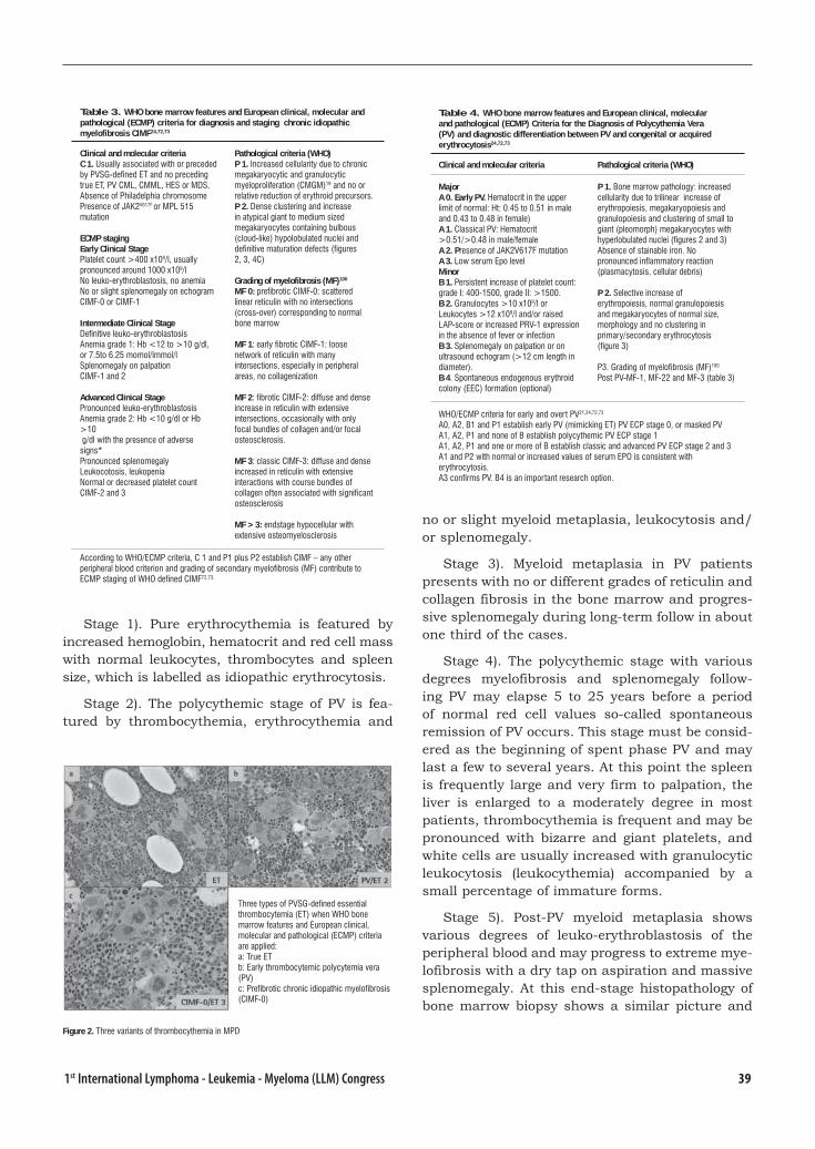
391st International Lymphoma - Leukemia - Myeloma (LLM) Congress
Stage 1). Pure erythrocythemia is featured by increased hemoglobin, hematocrit and red cell mass with normal leukocytes, thrombocytes and spleen size, which is labelled as idiopathic erythrocytosis.
Stage 2). The polycythemic stage of PV is fea-tured by thrombocythemia, erythrocythemia and
no or slight myeloid metaplasia, leukocytosis and/or splenomegaly.
Stage 3). Myeloid metaplasia in PV patients presents with no or different grades of reticulin and collagen fibrosis in the bone marrow and progres-sive splenomegaly during long-term follow in about one third of the cases.
Stage 4). The polycythemic stage with various degrees myelofibrosis and splenomegaly follow-ing PV may elapse 5 to 25 years before a period of normal red cell values so-called spontaneous remission of PV occurs. This stage must be consid-ered as the beginning of spent phase PV and may last a few to several years. At this point the spleen is frequently large and very firm to palpation, the liver is enlarged to a moderately degree in most patients, thrombocythemia is frequent and may be pronounced with bizarre and giant platelets, and white cells are usually increased with granulocytic leukocytosis (leukocythemia) accompanied by a small percentage of immature forms.
Stage 5). Post-PV myeloid metaplasia shows various degrees of leuko-erythroblastosis of the peripheral blood and may progress to extreme mye-lofibrosis with a dry tap on aspiration and massive splenomegaly. At this end-stage histopathology of bone marrow biopsy shows a similar picture and
Table 3. WHO bone marrow features and European clinical, molecular and pathological (ECMP) criteria for diagnosis and staging chronic idiopathic myelofibrosis CIMF24,72,73
Clinical and molecular criteria C 1. Usually associated with or preceded by PVSG-defined ET and no preceding true ET, PV CML, CMML, HES or MDS.Absence of Philadelphia chromosomePresence of JAK2V617F or MPL 515 mutation
ECMP stagingEarly Clinical Stage Platelet count >400 x109/l, usuallypronounced around 1000 x109/lNo leuko-erythroblastosis, no anemiaNo or slight splenomegaly on echogramCIMF-0 or CIMF-1
Intermediate Clinical StageDefinitive leuko-erythroblastosisAnemia grade 1: Hb <12 to >10 g/dl, or 7.5to 6.25 momol/lmmol/lSplenomegaly on palpationCIMF-1 and 2
Advanced Clinical StagePronounced leuko-erythroblastosisAnemia grade 2: Hb <10 g/dl or Hb >10 g/dl with the presence of adverse signs*Pronounced splenomegalyLeukocotosis, leukopeniaNormal or decreased platelet count CIMF-2 and 3
Pathological criteria (WHO)P 1. Increased cellularity due to chronic megakaryocytic and granulocytic myeloproliferation (CMGM)19 and no or relative reduction of erythroid precursors. P 2. Dense clustering and increase in atypical giant to medium sized megakaryocytes containing bulbous (cloud-like) hypolobulated nuclei and definitive maturation defects (figures 2, 3, 4C)
Grading of myelofibrosis (MF)100 MF 0: prefibrotic CIMF-0: scattered linear reticulin with no intersections (cross-over) corresponding to normal bone marrow
MF 1: early fibrotic CIMF-1: loose network of reticulin with many intersections, especially in peripheral areas, no collagenization
MF 2: fibrotic CIMF-2: diffuse and dense increase in reticulin with extensive intersections, occasionally with only focal bundles of collagen and/or focal osteosclerosis.
MF 3: classic CIMF-3: diffuse and dense increased in reticulin with extensive interactions with course bundles of collagen often associated with significant osteosclerosis
MF >3: endstage hypocellular with extensive osteomyelosclerosis
According to WHO/ECMP criteria, C 1 and P1 plus P2 establish CIMF – any other peripheral blood criterion and grading of secondary myelofibrosis (MF) contribute to ECMP staging of WHO defined CIMF72,73.
Table 4. WHO bone marrow features and European clinical, molecular and pathological (ECMP) Criteria for the Diagnosis of Polycythemia Vera (PV) and diagnostic differentiation between PV and congenital or acquired erythrocytosis24,72,73
Clinical and molecular criteria Pathological criteria (WHO)
MajorA 0. Early PV. Hematocrit in the upper limit of normal: Ht: 0.45 to 0.51 in male and 0.43 to 0.48 in female) A 1. Classical PV: Hematocrit >0.51/>0.48 in male/female A 2. Presence of JAK2V617F mutationA 3. Low serum Epo level MinorB 1. Persistent increase of platelet count: grade I: 400-1500, grade II: >1500.B 2. Granulocytes >10 x109/l or Leukocytes >12 x109/l and/or raised LAP-score or increased PRV-1 expression in the absence of fever or infectionB 3. Splenomegaly on palpation or on ultrasound echogram (>12 cm length in diameter).B 4. Spontaneous endogenous erythroid colony (EEC) formation (optional)
P 1. Bone marrow pathology: increased cellularity due to trilinear increase of erythropoiesis, megakaryopoiesis and granulopoiesis and clustering of small to giant (pleomorph) megakaryocytes with hyperlobulated nuclei (figures 2 and 3)Absence of stainable iron. No pronounced inflammatory reaction (plasmacytosis, cellular debris)
P 2. Selective increase of erythropoiesis, normal granulopoiesis and megakaryocytes of normal size, morphology and no clustering in primary/secondary erythrocytosis (figure 3)
P3. Grading of myelofibrosis (MF)100 Post PV-MF-1, MF-22 and MF-3 (table 3)
WHO/ECMP criteria for early and overt PV21,24,72,73
A0, A2, B1 and P1 establish early PV (mimicking ET) PV ECP stage 0, or masked PVA1, A2, P1 and none of B establish polycythemic PV ECP stage 1A1, A2, P1 and one or more of B establish classic and advanced PV ECP stage 2 and 3A1 and P2 with normal or increased values of serum EPO is consistent with erythrocytosis. A3 confirms PV. B4 is an important research option.
Figure 2. Three variants of thrombocythemia in MPD
Three types of PVSG-defined essential thrombocytemia (ET) when WHO bone marrow features and European clinical, molecular and pathological (ECMP) criteria are applied:a: True ETb: Early thrombocytemic polycytemia vera (PV)c: Prefibrotic chronic idiopathic myelofibrosis (CIMF-0)
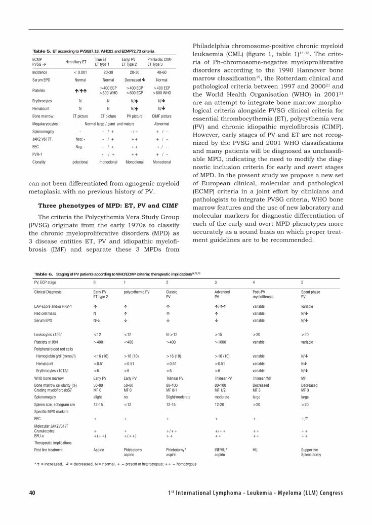
40 1 st International Lymphoma - Leukemia - Myeloma (LLM) Congress
can not been differentiated from agnogenic myeloid metaplasia with no previous history of PV.
Three phenotypes of MPD: ET, PV and CIMF
The criteria the Polycythemia Vera Study Group (PVSG) originate from the early 1970s to classify the chronic myeloproliferative disorders (MPD) as 3 disease entities ET, PV and idiopathic myelofi-brosis (IMF) and separate these 3 MPDs from
Philadelphia chromosome-positive chronic myeloid leukaemia (CML) (figure 1, table 1)14-18. The crite-ria of Ph-chromosome-negative myeloproliferative disorders according to the 1990 Hannover bone marrow classification19, the Rotterdam clinical and pathological criteria between 1997 and 200021 and the World Health Organisation (WHO) in 200121 are an attempt to integrate bone marrow morpho-logical criteria alongside PVSG clinical criteria for essential thrombocythemia (ET), polycythemia vera (PV) and chronic idiopathic myelofibrosis (CIMF). However, early stages of PV and ET are not recog-nized by the PVSG and 2001 WHO classifications and many patients will be diagnosed as unclassifi-able MPD, indicating the need to modify the diag-nostic inclusion criteria for early and overt stages of MPD. In the present study we propose a new set of European clinical, molecular and pathological (ECMP) criteria in a joint effort by clinicians and pathologists to integrate PVSG criteria, WHO bone marrow features and the use of new laboratory and molecular markers for diagnostic differentiation of each of the early and overt MPD phenotypes more accurately as a sound basis on which proper treat-ment guidelines are to be recommended.
Table 5. ET according to PVSG17,18, WHO21 and ECMP72,73 criteria
ECMPPVSG
Hereditary ETTrue ETET type 1
Earlyl PVET Type 2
Prefibrotic CIMF ET Type 3
Incidence < 0.001 20-30 20-30 40-60
Serum EPO Normal Normal Decreased Normal
Platelets />400 ECP>600 WHO
>400 ECP>600 ECP
>400 ECP>600 WHO
Erythrocytes N N N/ N/
Hematocrit N N N/ N/
Bone marrow: ET picture ET picture PV picture CIMF picture
Megakaryocytes Normal large / giant and mature Abnormal
Splenomegaly - - / + - / + + / -
JAK2 V617F Neg: - - / + ++ + / -
EEC Neg: - - / + ++ + / -
PVR-1 - / + ++ + / -
Clonality polyclonal monoclonal Monoclonal Monoclonal
Table 6. Staging of PV patients according to WHO/ECMP criteria: therapeutic implications24,72,73
PV, ECP stage 0 1 2 3 4 5
Clinical Diagnosis Early PV ET type 2
polycythemic PV Classic PV
Advanced PV
Post-PVmyelofibrosis
Spent phasePV
LAP-score and/or PRV-1 / variable variable
Red cell mass N variable N/
Serum EPO N/ variable N/
Leukocytes x109/l <12 <12 N->12 >15 >20 >20
Platelets x109/l >400 <400 >400 >1000 variable variable
Peripheral blood red cells
Hemoglobin g/dl (mmol/l) <16 (10) >16 (10) >16 (10) >16 (10) variable N/
Hematocrit <0.51 >0.51 >0.51 >0.51 variable N
Erythrocytes x1012/l <6 >6 >6 >6 variable N/
WHO bone marrow Early PV Early PV Trilinear PV Trilinear PV Trilinear /MF MF
Bone marrow cellularity (%)Grading myelofibrosis57
50-80MF 0
50-80MF 0
80-100MF 0/1
80-100MF 1/2
DecreasedMF 3
DecreasedMF 3
Splenomegaly slight no Slight/moderate moderate large large
Spleen size, echogram cm 12-15 <12 12-15 12-20 >20 >20
Specific MPD markers
EEC + + + + + +/?
Molecular JAK2V617FGranulocytes BFU-e
++(++)
++(++)
+/++++
+/++++
++++
++++
Therapeutic implications
First line treatment Aspirin Phlebotomyaspirin
Phlebotomy*aspirin
INF/HU*aspirin
HU SupportiveSplenectomy
* = increased, = decreased, N = normal, + = present or heterozygous; ++ = homozygous
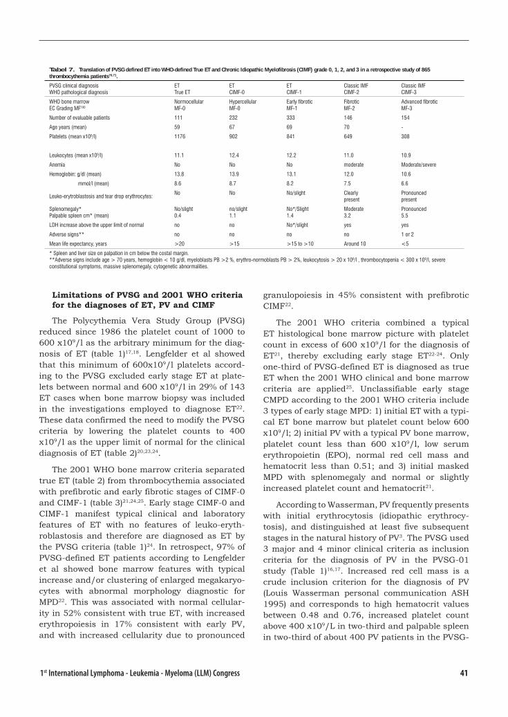
411st International Lymphoma - Leukemia - Myeloma (LLM) Congress
Limitations of PVSG and 2001 WHO criteria for the diagnoses of ET, PV and CIMF
The Polycythemia Vera Study Group (PVSG) reduced since 1986 the platelet count of 1000 to 600 x109/l as the arbitrary minimum for the diag-nosis of ET (table 1)17,18. Lengfelder et al showed that this minimum of 600x109/l platelets accord-ing to the PVSG excluded early stage ET at plate-lets between normal and 600 x109/l in 29% of 143 ET cases when bone marrow biopsy was included in the investigations employed to diagnose ET22. These data confirmed the need to modify the PVSG criteria by lowering the platelet counts to 400 x109/l as the upper limit of normal for the clinical diagnosis of ET (table 2)20,23,24.
The 2001 WHO bone marrow criteria separated true ET (table 2) from thrombocythemia associated with prefibrotic and early fibrotic stages of CIMF-0 and CIMF-1 (table 3)21,24,25. Early stage CIMF-0 and CIMF-1 manifest typical clinical and laboratory features of ET with no features of leuko-eryth-roblastosis and therefore are diagnosed as ET by the PVSG criteria (table 1)24. In retrospect, 97% of PVSG-defined ET patients according to Lengfelder et al showed bone marrow features with typical increase and/or clustering of enlarged megakaryo-cytes with abnormal morphology diagnostic for MPD22. This was associated with normal cellular-ity in 52% consistent with true ET, with increased erythropoiesis in 17% consistent with early PV, and with increased cellularity due to pronounced
granulopoiesis in 45% consistent with prefibrotic CIMF22.
The 2001 WHO criteria combined a typical ET histological bone marrow picture with platelet count in excess of 600 x109/l for the diagnosis of ET21, thereby excluding early stage ET22-24. Only one-third of PVSG-defined ET is diagnosed as true ET when the 2001 WHO clinical and bone marrow criteria are applied25. Unclassifiable early stage CMPD according to the 2001 WHO criteria include 3 types of early stage MPD: 1) initial ET with a typi-cal ET bone marrow but platelet count below 600 x109/l; 2) initial PV with a typical PV bone marrow, platelet count less than 600 x109/l, low serum erythropoietin (EPO), normal red cell mass and hematocrit less than 0.51; and 3) initial masked MPD with splenomegaly and normal or slightly increased platelet count and hematocrit21.
According to Wasserman, PV frequently presents with initial erythrocytosis (idiopathic erythrocy-tosis), and distinguished at least five subsequent stages in the natural history of PV3. The PVSG used 3 major and 4 minor clinical criteria as inclusion criteria for the diagnosis of PV in the PVSG-01 study (Table 1)16,17. Increased red cell mass is a crude inclusion criterion for the diagnosis of PV (Louis Wasserman personal communication ASH 1995) and corresponds to high hematocrit values between 0.48 and 0.76, increased platelet count above 400 x109/L in two-third and palpable spleen in two-third of about 400 PV patients in the PVSG-
Tabel 7. Translation of PVSG defined ET into WHO-defined True ET and Chronic Idiopathic Myelofibrosis (CIMF) grade 0, 1, 2, and 3 in a retrospective study of 865 thrombocythemia patients70,71.
PVSG clinical diagnosis WHO pathological diagnosis
ETTrue ET
ETCIMF-0
ETCIMF-1
Classic IMFCIMF-2
Classic IMFCIMF-3
WHO bone marrowEC Grading MF100
NormocellularMF-0
HypercellularMF-0
Early fibroticMF-1
FibroticMF-2
Advanced fibroticMF-3
Number of evaluable patients 111 232 333 146 154
Age years (mean) 59 67 69 70 -
Platelets (mean x109/l) 1176 902 841 649 308
Leukocytes (mean x109/l) 11.1 12.4 12.2 11.0 10.9
Anemia No No No moderate Moderate/severe
Hemoglobin: g/dl (mean) 13.8 13.9 13.1 12.0 10.6
mmol/l (mean) 8.6 8.7 8.2 7.5 6.6
Leuko-erytroblastosis and tear drop erythrocytes:No No No/slight Clearly
presentPronouncedpresent
Splenomegaly*Palpable spleen cm* (mean)
No/slight0.4
no/slight1.1
No*/Slight1.4
Moderate3.2
Pronounced5.5
LDH increase above the upper limit of normal no no No*/slight yes yes
Adverse signs** no no no no 1 or 2
Mean life expectancy, years >20 >15 >15 to >10 Around 10 <5
* Spleen and liver size on palpation in cm below the costal margin.**Adverse signs include age > 70 years, hemoglobin < 10 g/dl, myeloblasts PB >2 %, erythro-normoblasts PB > 2%, leukocytosis > 20 x 109/l , thrombocytopenia < 300 x 109/l, severe constitutional symptoms, massive splenomegaly, cytogenetic abnormalities.
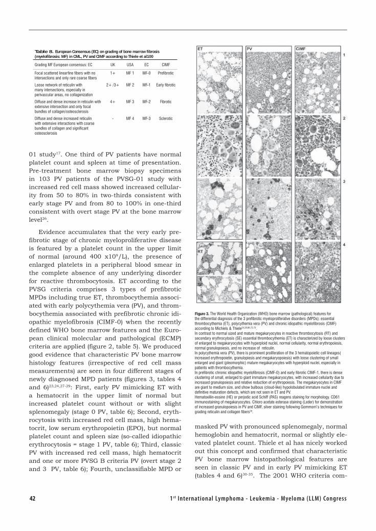
42 1 st International Lymphoma - Leukemia - Myeloma (LLM) Congress
01 study17. One third of PV patients have normal platelet count and spleen at time of presentation. Pre-treatment bone marrow biopsy specimens in 103 PV patients of the PVSG-01 study with increased red cell mass showed increased cellular-ity from 50 to 80% in two-thirds consistent with early stage PV and from 80 to 100% in one-third consistent with overt stage PV at the bone marrow level26.
Evidence accumulates that the very early pre-fibrotic stage of chronic myeloproliferative disease is featured by a platelet count in the upper limit of normal (around 400 x109/L), the presence of enlarged platelets in a peripheral blood smear in the complete absence of any underlying disorder for reactive thrombocytosis. ET according to the PVSG criteria comprises 3 types of prefibrotic MPDs including true ET, thrombocythemia associ-ated with early polycythemia vera (PV), and throm-bocythemia associated with prefibrotic chronic idi-opathic myelofibrosis (CIMF-0) when the recently defined WHO bone marrow features and the Euro-pean clinical molecular and pathological (ECMP) criteria are applied (figure 2, table 5). We produced good evidence that characteristic PV bone marrow histology features (irrespective of red cell mass measurements) are seen in four different stages of newly diagnosed MPD patients (figures 3, tables 4 and 6)23,24,27-29: First, early PV mimicking ET with a hematocrit in the upper limit of normal but increased platelet count without or with slight splenomegaly (stage 0 PV, table 6); Second, eryth-rocytosis with increased red cell mass, high hema-tocrit, low serum erythropoietin (EPO), but normal platelet count and spleen size (so-called idiopathic erythrocytosis = stage 1 PV, table 6); Third, classic PV with increased red cell mass, high hematocrit and one or more PVSG B criteria PV (overt stage 2 and 3 PV, table 6); Fourth, unclassifiable MPD or
masked PV with pronounced splenomegaly, normal hemoglobin and hematocrit, normal or slightly ele-vated platelet count. Thiele et al has nicely worked out this concept and confirmed that characteristic PV bone marrow histopathological features are seen in classic PV and in early PV mimicking ET (tables 4 and 6)30-35. The 2001 WHO criteria com-
Table 8. European Consensus (EC) on grading of bone marrow fibrosis (myelofibrosis: MF) in CML, PV and CIMF according to Thiele et al100
Grading MF European consensus: EC UK USA EC CIMF
Focal scattered linearfine fibers with nointersections and only rare coarse fibers
1+ MF 1 MF-0 Prefibrotic
Loose network of reticulin withmany intersections, especially in perivascular areas, no collagenization
2+ /3+ MF 2 MF-1 Early fibrotic
Diffuse and dense increase in reticulin withextensive intersection and only focalbundles of collagen/osteosclerosis
4+ MF 3 MF-2 Fibrotic
Diffuse and dense increased reticulinwith extensive interactions with coarsebundles of collagen and significant osteosclerosis
- MF 4 MF-3 Sclerotic
Figure 3. The World Health Organization (WHO) bone marrow (pathological) features for the differential diagnosis of the 3 prefibrotic myeloproliferative disorders (MPDs): essential thrombocythemia (ET), polycythemia vera (PV) and chronic idiopathic myelofibrosis (CIMF) according to Michiels & Thiele24,28,66,72,73.In contrast to normal sized and mature megakaryocytes in reactive thrombocytosis (RT) and secondary erythrocytosis (SE) essential thrombocythemia (ET) is characterized by loose clusters of enlarged to megakaryocytes with hyperploid nuclei, normal cellularity, normal erythropoiesis, normal granulopoiesis, and no increase of reticulin.In polycythemia vera (PV), there is prominent proliferation of the 3 hematopoietic cell lineages( increased erythropoietin, granulopiesis and megakaryopoiesis) with loose clustering of small enlarged and giant (pleomorphic) mature megakaryocytes with hyperploid nuclei, especially in patients with thrombocythemia.In prefibrotic chronic idiopathic myelofibrosis (CIMF-0) and early fibrotic CIMF-1, there is dense clustering of small, enlarged to giant immature megakaryocytes, with increased cellularity due to increased granulopoiesis and relative reduction of erythropoiesis. The megakaryocytes in CIMF are giant to medium size, and show bulbous (cloud-like) hypolobulated immature nuclei and definitive maturation defects, which are not seen in ET and PV.Hematoxillin-eosine (HE) or perjodic acid Schiff (PAS) reagens staining for morphology. CD61 immunostaining of megakaryocytes. Chloro acetate esterase staining (Leder) for demonstration of increased granulopoiesis in PV and CIMF, silver staining following Gommorri’s techniques for grading reticulin and collagen fibers28.
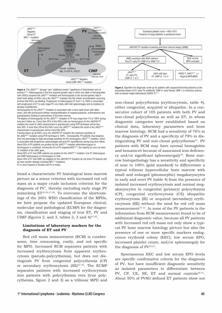
431st International Lymphoma - Leukemia - Myeloma (LLM) Congress
bined a characteristic PV histological bone marrow picture as a minor criterion with increased red cell mass as a major crude inclusion criterion for the diagnosis of PV7, thereby excluding early stage PV mimicking ET23,24,27-29. To overcome the shortcom-ings of the 2001 WHO classification of the MPDs, we here propose the updated European clinical, molecular and pathological (ECMP) for the diagno-sis, classification and staging of true ET, PV and CIMF (figures 2, and 3, tables 2, 3 and 4)24,28.
Limitations of laboratory markers for the diagnosis of ET and PV
Red cell mass measurement (RCM) is cumber-some, time consuming, costly, and not specific for MPD. Increased RCM separates patients with increased erythrocytosis from apparent erythro-cytosis (pseudo-polycythemia), but does not dis-tinguish PV from congenital polycythemia (CP) or secondary erythrocytosis (SE)27-29. The ECMP separates patients with increased erythrocytosis into patients with polycythemia vera (true poly-cythemia, figure 2 and 3) as a trilinear MPD and
non-clonal polycythemia (erythrocytosis, table 4), either congenital, acquired or idiopathic. In a con-secutive cohort of 105 patients with both PV and non-clonal polycythemia as well as ET, in whom diagnostic categories were established based on clinical data, laboratory parameters and bone marrow histology, RCM had a sensitivity of 76% in the diagnosis of PV and a specificity of 79% in dis-tinguishing PV and non-clonal polycythemia34. PV patients with RCM may have normal hemoglobin and hematocrit because of associated iron deficien-cy and/or significant splenomergaly35. Bone mar-row histopathology has a sensitivity and specificity of near to 100% (gold standard) to differentiate a typical trilinear hypercellular bone marrow with small and enlarged (pleomorphic) megakaryocytes in early and overt PV (table 4) from the presence of isolated increased erythrocytosis and normal meg-akaryocytes in congenital (primary) polycythemia (CP), congenital erythrocytosis (CE) idiopathic erythrocytosis (IE) or acquired (secondary) eryth-rocytosis (SE) without the need for red cell mass measurement27-33. In none of the PV patients is the information from RCM measurement found to be of additional diagnostic value, because all PV patients with increased red cell mass not only show a typi-cal PV bone marrow histology picture but also the presence of one or more specific markers endog-enous erythroid colony (EEC), low serum EPO, increased platelet count, and/or splenomegaly for the diagnosis of PV23,27.
Spontaneous EEC and low serum EPO levels are specific confirmative criteria for the diagnosis of PV, but have insufficient diagnostic sensitivity as isolated parameters to differentiate between PV, CP, CE, SE, ET and normal controls36-41. About 50% of PVSG defined ET patients show not
Figure 5. Algorithm for diagnostic work-up for patients with suspected thrombocythemia as the presenting feature of ET, early PV, prefibrotic CIMF-0, early fibrotic CIMF-1 or refractory anemia with increased ringed sideroblasts (RARS-T)72,73.
Figure 4. The JAK2V617F “dosage” and “additional events” hypothesis of Vainchenker and co-workers53-55. Heterozygosity is the first acquired genetic step in which one allele of hematopoietic cells (HSCs) acquires the Jak2V617F mutation and homozygosity is the second genetic step in which both alleles of HSCs carry the JAK2V617F mutation due the mitotic recombination occurring at times that HSCs are dividing. Progression of heterozygous ET from 1 to 100% is associated with progression of ET to early stage PV, very likely with mild splenomegaly and no tendency to develop myelofibrosis. Homozygosity for the JAK2V617F mutation is associated with a more rapid onset (still rather slow), and with pronounced trilinear myeloproliferation of megakaryopoiesis, erythropoiesis and granulopoiesis leading to panmyelosis of the bone marrow. The degree of homozygosity for the JAK2V617F mutation in PV may range from 1% to 100% during long-term follow-up. If less than 25% of the HSCs cells are homozygous for the JAK2V617F mutation the result of JAK2 measurement in granulocytes using PCR technique will be less than 50%. If more than 25%of the HSCs carry the JAK2V617F mutation the result of the JAK2V617F measurement in granulocytes will be more than 50%. If during follow-up all HSCs carry the JAK2V617F mutation the maximum positivity ofthe JAK2V617F mutation using PCR technique is 100%. Consequently, PV patients may progress from low percentage to high percentage positivity for the homozygous JAK2V617F mutation, which is associated with progressive splenomegaly and post-PV myelofibrosis during long-term follow.About 95% of PV patients are positive for the JAK2V617F mutation eitherheterozygous or homozygous or combined. Among the 5% of PV negativeJAK2V617F, the majority do carry an exon 12 mutation in the JAK2 gene.About 60% of ET and CIMF patients are positive for the JAK2V617F mutation: true ET heterozygous and CIMF heterozygous or homozygous or both.About 40% of ET and CIMF are negative for the JAK2V617F mutation do not show PV features and do have another etiology including MPL515 mutations.ET is more frequent in females and PV in males.
JAK2V617F NegativeClonal ET
MPL515 mutationAlternative
molecular event
HeterozygousJAK2V617F
ET
V617F low
Female
CIMF
PV
Genetic modifiers:
Phosphatases,SOCS,
Epo-R ?
JAK2V617F
Negative PVExon 12 JAK2 mutation
Alternative molecular event
MitoticRecombinationHomozygous
JAK2V671F
V617F high
Genetic modifiers: Mpl, G-CSF-R, SOCS?
Alternativemolecular event
JAK2V617F Negativeclonal CIMF, MPL515 mutation
male
Sustained platelet count >400 x109/l Presence of large platelets in peripheral blood
JAK2V617F mutation screening and bone marrow biopsy (BMB)
BMB consistent with WHO-definedTrue ET, early PV, CIMF-0 or CIMF-1/2
Both normalMPD excluded
JAK2V617F
ET, early PV,CIMF-0, 1 or 2
JAK2 wild typeET, CIMF-0, 1 or 2
RARS-T: JAK2V617F +/-Atypical MPD/MDS
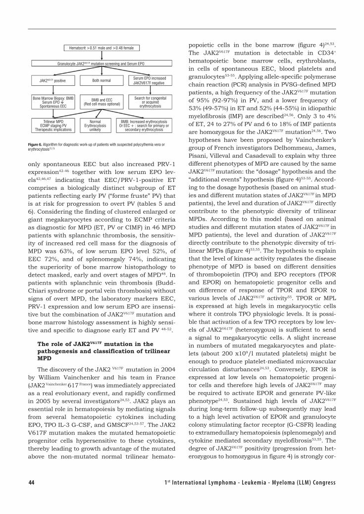
44 1 st International Lymphoma - Leukemia - Myeloma (LLM) Congress
only spontaneous EEC but also increased PRV-1 expression42-46 together with low serum EPO lev-els42,46,47 indicating that EEC/PRV-1-positive ET comprises a biologically distinct subgroup of ET patients reflecting early PV (“forme fruste” PV) that is at risk for progression to overt PV (tables 5 and 6). Considering the finding of clustered enlarged or giant megakaryocytes according to ECMP criteria as diagnostic for MPD (ET, PV or CIMF) in 46 MPD patients with splanchnic thrombosis, the sensitiv-ity of increased red cell mass for the diagnosis of MPD was 63%, of low serum EPO level 52%, of EEC 72%, and of splenomegaly 74%, indicating the superiority of bone marrow histopathology to detect masked, early and overt stages of MPD48. In patients with splanchnic vein thrombosis (Budd-Chiari syndrome or portal vein thrombosis) without signs of overt MPD, the laboratory markers EEC, PRV-1 expression and low serum EPO are insensi-tive but the combination of JAK2V617F mutation and bone marrow histology assessment is highly sensi-tive and specific to diagnose early ET and PV 48-52.
The role of JAK2V617F mutation in the pathogenesis and classification of trilinear MPD
The discovery of the JAK2 V617F mutation in 2004 by William Vainchenker and his team in France (JAK2 Vainchenker 617 France) was immediately appreciated as a real evolutionary event, and rapidly confirmed in 2005 by several investigators24,53. JAK2 plays an essential role in hematopoiesis by mediating signals from several hematopoietic cytokines including EPO, TPO IL-3 G-CSF, and GMSCF24,53-57. The JAK2 V617F mutation makes the mutated hematopoietic progenitor cells hypersensitive to these cytokines, thereby leading to growth advantage of the mutated above the non-mutated normal trilinear hemato-
popoietic cells in the bone marrow (figure 4)24,53. The JAK2V617F mutation is detectable in CD34+ hematopoietic bone marrow cells, erythroblasts, in cells of spontaneous EEC, blood platelets and granulocytes53-55. Applying allele-specific polymerase chain reaction (PCR) analysis in PVSG-defined MPD patients, a high frequency of the JAK2V617F mutation of 95% (92-97%) in PV, and a lower frequency of 53% (49-57%) in ET and 52% (44-55%) in idiopathic myelofibrosis (IMF) are described24,56. Only 3 to 4% of ET, 24 to 27% of PV and 6 to 18% of IMF patients are homozygous for the JAK2V617F mutation24,56. Two hypotheses have been proposed by Vainchenker’s group of French investigators Delhommeau, James, Pisani, Villeval and Casadevall to explain why three different phenotypes of MPD are caused by the same JAK2V617F mutation: the “dosage” hypothesis and the “additional events” hypothesis (figure 4)53-55. Accord-ing to the dosage hypothesis (based on animal stud-ies and different mutation states of JAK2V617F in MPD patients), the level and duration of JAK2V617F directly contribute to the phenotypic diversity of trilinear MPDs. According to this model (based on animal studies and different mutation states of JAK2V617F in MPD patients), the level and duration of JAK2V617F directly contribute to the phenotypic diversity of tri-linear MPDs (figure 4)53,55. The hypothesis to explain that the level of kinase activity regulates the disease phenotype of MPD is based on different densities of thrombopoietin (TPO) and EPO receptors (TPOR and EPOR) on hematopoietic progenitor cells and on difference of response of TPOR and EPOR to various levels of JAK2V617F activity55. TPOR or MPL is expressed at high levels in megakaryocytic cells where it controls TPO physiologic levels. It is possi-ble that activation of a few TPO receptors by low lev-els of JAK2V617F (heterozygous) is sufficient to send a signal to megakaryocytic cells. A slight increase in numbers of mutated megakaryocytes and plate-lets (about 200 x109/l mutated platelets) might be enough to produce platelet-mediated microvascular circulation disturbances24,53. Conversely, EPOR is expressed at low levels on hematopoietic progeni-tor cells and therefore high levels of JAK2V617F may be required to activate EPOR and generate PV-like phenotype24,53. Sustained high levels of JAK2V617F during long-term follow-up subsequently may lead to a high level activation of EPOR and granulocyte colony stimulating factor receptor (G-CSFR) leading to extramedullary hematopoiesis (splenomegaly) and cytokine mediated secondary myelofibrosis53,55. The degree of JAK2V617F positivity (progression from het-erozygous to homozygous in figure 4) is strongly cor-
Figure 6. Algorithm for diagnostic work-up of patients with suspected polycythemia vera or erythrocytosis72,73.
Hematocrit >0.51 male and >0.48 female
Granulocyte JAK2V617F mutation screening and Serum EPO
JAK2V617F positive Both normal Serum EPO increasedJAK2V617F negative
Bone Marrow Biopsy: BMBSerum EPO
Spontaneous EEC
BMB and EEC (Red cell mass optional)
Search for congenitalor acquired
erythrocytosis
Trilinear MPDECMP staging PV
Therapeutic implications
NormalErythrocytosis
unlikely
BMB: Increased erythrocytosisOr EEC + : search for primary or
secondary erythrocytosis

451st International Lymphoma - Leukemia - Myeloma (LLM) Congress
related with (polycythemia rubra vera-1 gene) PRV-1 over expression in granulocytes, with the ability to form spontaneous EEC and with progressive post-PV myelofibrosis45,55,57. Scott et al recently showed that BFU-e colonies are already homozygous for the JAK2V617F mutation in PV patients with a hetero-zygous pattern of JAK2V617F in their peripheral blood granulocytes58. In contrast, the BFU-E colonies from heterozygous patients with ET did not contain a subpopulation of JAK2V617F homozygous cells58. In a recent study, JAK2V617F was detected in 75% of ET (n=60) and in 97% of PV patients (n=62)59. Allelic ratios exceeding 50% JAK2V617F indicating the presence of granulocytes homozygous for JAK2V617F were found in 70% of PV at diagnosis but never in ET59. Passamonti et al produced good evidence that transition from heterozygosity to homozygosity for the JAK2V617F mutation represents a very important step in the progression from classic PV to post-PV myelofibrosis57.
JAK2V617F may be may be dependent not only on the amount of heterozygous and homozygous mutant protein, but also on the various pathway regulating JAK2 activity including MPL, JAK2, STAT-3 signalling pathway53. This has led to the recent discovery of the MPLW515L and MPLW515K muta-tions as the underlying etiology in some ET and CIMF patients60,61.Pikman et al identified a somatic activating mutation in the transmembrane domain of MPL, the thrombopoietin receptor (TPOR), in 4 of 45 (9%) of JAK2 wild type myelofibrosis patients60. In a mouse model transplant assay, expression of the MPLW515L, but not wild type MPL, resulted in a fully penetrant MPD characterized by marked thrombocytosis and leukocytosis with no evidence of polycythemia. Thrombocythemia with leukocytosis was caused by dual proliferation of megakaryopoiesis and granulopoiesis (myelopoi-esis) with increased numbers and clustering of atypical dysmorphic megakaryocytes in the bone marrow, myelofibrosis and marked splenomegaly due to extramedullary hemopoiesis consistent with true CIMF60. Pardanani et al screened 1182 PVSG-defined MPD patients (318 ET, 242 PV, and 290 IMF) and 64 controls for MPL515 mutations regard-less of JAK2V617F mutation46. MPL mutations either MPLW515L (n=17) or MPLW515K (n=5) was detected in 20 patients (de novo IMF in 12 = 4%, ET in 4 = 1%) and post-ET myelofibrosis in 1, but not in PV and controls61. Six cases carried the MPLW515L and JAK2V617F alleles indicating that these alleles have functional complementation in IMF. These experimental and clinical observations indicate
that MPL515 mutation related myelofibrosis may represent a distinct entity of JAK2 wild type IMF distinct from JAK2V617F trilinear MPD. According to the “additional events” hypothesis, alternative and/or additional molecular abnormalities modify, or precede a homozygous state deferred to by the JAK2V617 mutation alone, combinations carry-ing the JAKV617F and MPL515 mutations60,61 or other combinations of still unknown mutations (figure 4)53. Mechanisms other than mitotic recombina-tion such as duplication of the mutated allele is observed in a proportion of PV and CIMF patients displaying a gain of 9p, mostly due to trisomy 953. Therefore, there may be an overlap between “dos-age” and “additional molecular events” hypoth-eses. Long-term studies are warranted to delineate the chronology and impact of various putative additional molecular abnormalities especially in cases of progressive disease from JAK2V671F posi-tive true ET to PV and subsequent CIMF and in cases with combined JAK2V617F/MPL515 mutations or JAK2-wild-type/MPL515 positive ET and CIMF. Sex appears to be a powerful genetic background modifier in JAK2V617F-positive MPDs as ET is more common in females and PV in males.
WHO bone marrow features and ECMP criteria for the diagnosis of ET, PV and CIMF
In 1980s Georgii and Thiele independently defined the pathological features of ET, PV and idi-opathic myelofibrosis (IMF) or agnogenic myeloid metaplasia (AMM) as derived from histopatho-logical morphology of bone marrow biopsies19,62. ET is defined by persistent increase of platelets in excess of 400 x109/l without the Ph1+ chromosome together with monolinear proliferation of mature enlarged megakaryocytes in the bone marrow with normal cellularity, normal erythropoiesis and normal granulopoiesis (figure 2)19,62. PV is defined as a trilinear proliferation of megakaryopoiesis, erythropoiesis and granulopoiesis in which the erythropoiesis was most prominent together with variable degrees of increased platelets, erythro-cytes and granulocytes in the peripheral blood in the absence of the Ph1+ chromosome (figure 2)19,62. Georgii regarded myelofibrosis (MF) as a reactive feature secondary to progressive disease9,10 seen in CMGM, PV and CML19,63,64. Therefore, according to Georgii the terms agnogenic myloid metaplasia (AMM) or idiopathic myelofibrosis (IMF) lack accu-racy since they are applied to both the prefibrotic hypercellular and advanced fibrotic stages of AMM or IMF. Consequently, Georgii replaced the terms
46 1 st International Lymphoma - Leukemia - Myeloma (LLM) Congress
prefibrotic AMM and IMF by chronic megakaryo-cytic granulocytic myeloproliferation (CMGM) and proposed in 1990 the Hannover Classification to distinguish 3 distinct primary CMPDs ET, PV and CMGM19. The diagnosis of CMGM according to the Hannover classification19 or prefibrotic CIMF-0 according to the Cologne criteria proposed by Thiele66 (figure 3) is based on 3 main features. First, the presence of large megakaryocytes with immature cytoplasm and immature cloud-like nuclei not seen in ET and PV. Second, increased granulopoiesis but never disturbed in matura-tion. Third, erythropoiesis is usually relatively decreased19,62,63. The Hannover classification uses the term CMGM19,62,63, and the Cologne classifica-tion uses the term prefibrotic chronic IMF (CIMF-0) for the third entity of prefibtotic MPD65-71. The Hannover and Cologne classifications19,66 are based on specific bone marrow histology (pathological) features for the three prefibrotic CMPDs true ET, PV and CIMF-0 at the bone marrow level, and have been taken over in 2001 by the WHO21. Michiels and Thiele subsequently proposed the European clinical and pathological (ECP) criteria for the diagnosis of the Ph-negative MPDs to describe the full spectrum of clinical, laboratory, and WHO bone marrow features for true ET, early and overt PV and CIMF-0 (figure 3)24,28,72,73, In a recent joint effort by clinicians and pathologists, we added the molecular marker and updated the European clinical, molecular and pathological (ECMP) criteria to extend PVSG criteria by including WHO bone marrow features and the use of laboratory and molecular markers for the diagnosis and staging of the three prefibrotic MPDs true ET, PV and CIMF (tables 2, 3 and 4)24,72,73. These ECMP criteria allow a cross talk between clinicians and pathologists to translate PVSG clinical criteria (table 1) by includ-ing WHO bone marrow features and new biological and molecular markers (figures 2, and 3, tables 2, 3 and 4) to reach agreement on diagnosis, classifi-cation and staging of the MPDs72,73.
For the diagnosis of MPD bone marrow trephine biopsy specimens should be embedded in paraf-fin or plastic, both with its technical limitations. Paraffin requires decalcification with EDTA (prefer-able, allows reasonable DNA quality) or acid elec-trolysis28,72,73. The specimens should have at least 4 evaluable bone marrow spaces with hematopoi-esis. Recommended stains include: hematoxylin and eosin (H&E), Giemsa (3 μm sections), periodic acid-Schiff (PAS); Perls for estimation of hemosi-derin content; chloro-acetate esterase (Leder) for
identification of granulocytic differentiation; silver stain for reticulin; and trichrome-Masson for col-lagen staining. Immunostains of paraffin embed-ded specimens should include glycophorine C for erythropoiesis, myeloperoxydase for granulopoi-esis, CD42b, CD61 or FVIII- related antigen for megakaryocytes; CD34 for CD34-positive blasts and CD117 for myeloid differentiation. Regard-ing MPD, clinicians want to receive from their pathologist28,72,73 a detailed report according to the Cologne bone marrow evaluation form72,73. Accord-ing to WHO and ECMP criteria, increase and loose clustering of enlarged mature megakaryocytes with hyperlobulated nuclei in a normocellular bone marrow and platelet count >400 x109/l represent the hallmark of true ET (table 2, figures 2 and 3). In true ET there is no proliferation or immaturity of granulopoiesis or erythropoiesis. In congenital and acquired erythrocytosis and in reactive thrombo-cytosis the megakaryocytes are of normal size and morphology and there is no tendency to cluster. A typical histopathological ET picture of the bone marrow excludes RT and distinguishes true ET from early PV, CIMF-0, CIMF-1 (figure 2), and throm-bocythemia associated with atypical MPD, MDS, refractory anemia with increased ringed siderob-lasts (RARS-T) or Ph1–positive thrombocythemia in CML. The characteristic increase and clustering of small and enlarged pleomorphic megakaryocytes and increased erythropoiesis with increased granu-lopoiesis and increased cellularity (80-100%) are the diagnostic characteristics of classic PV with increased hematocrit >0.51, low serum EPO and/or JAK2V617F (tables 4 and 6) distinguishing it from congenital and secondary erythrocytosis. A typical histological PV picture with moderately increased bone marrow cellularity (according to age) is seen in patients with early PV mimicking ET or “forme fruste” PV featured by platelet count >400x109/l and hematocrit <0.51, low serum EPO and/or the presence of the JAK2V617F mutation (tables 4 and 6). A typical histological PV bone marrow picture is also seen in the early erythrocythemic stage 1 PV featured by increased hematocrit >0.51, nor-mal platelet count <400 x109/l, normal spleen, low serum EPO and/or the presence of the JAK2V617F mutation (tables 4 and 6).
According to Hannover19, Cologne66, WHO21 and ECMP criteria24,72,73, CIMF-0 is characterized by hypercellularity of the bone marrow (60-100%) due to increased granulopoiesis, relative decrease of erythropoiesis and the presence of dense clusters of immature megakaryocytes with maturation defects
471st International Lymphoma - Leukemia - Myeloma (LLM) Congress
of cytoplasm and nuclei with bulky nuclei show-ing clumpy lobuli and irregular roundish shaped forms (so-called cloud-like or clumsy nuclei), which are almost never seen in ET and PV (figures 2 and 3, table 3)28,64-73. The degree of dysmegakaryopoi-esis in CIMF-0 may range from slight maturation defect of megakaryocytes with no cloud-like nuclei to manifest maturation defects of megakaryocytes with typical cloud-like nuclei or a mixture of both. The risk of CIMF-0 to transform into early CIMF-1 and subsequent CIMF-2/3 with extramedullary hematopoiesis is clearly dependent on the degree of hypercellularity and on the degree of maturation defects of megakaryopoiesis70,71. High quality his-tological bone marrow preparations in the hands of experienced hematopathologists are required to distinguish CIMF-0 from true ET and PV in about 85 to 90% of the cases, which is only feasible in centers of excellence (figures 2 and 3)57-73. There are no studies that have examined the concordance between a number of pathologists who have used characteristic histological bone marrow features to assign cases to the prefibrotic stages of true ET, PV and CIMF-0 without knowledge of the clinical find-ings and biological MPD markers except age. Data on the very long-term natural history of patients with ECMP defined true ET, early PV and CIMF-0 as derived from large scale prospective studies are lacking. We don’t really know whether the early stages of PV patients are at no, low or high risk of progression to post-PV myelofibrosis or whether true ET never progress to CIMF as it is claimed. The bone marrow histology of CIMF-0 with slight dys-megakaryopoiesis can appear to overlap with early PV (hematocrit <0.51) presenting with a trilinear hypercellular bone marrow with relative increased granulopoiesis as compared to erythropoiesis and a similar degree of thrombocythemia, leukocytosis, increased LAP-score and slight splenomegaly. The histology of CIMF-0 slight dysmorphic megakary-opoiiesis may overlap with that of true ET in cases of very mild hyperplasia of granulopoiesis and/or a mixture of mild dysmorphic megakaryocytes and mature enlarged megakaryocytes with hyperploid nuclei. Diagnostic differentiation between true ET, early PV, CIMF-0 at the bone marrow level will be feasible in well equipped pathology laboratories by experienced trained pathologists in the majority of cases, but significant overlap (grey zones) between CIMF-0 with slight dysmegakaryopoiesis versus true ET or early PV due to a rather high inter-observer disagreement between hematopatholo-gists in routine daily practice is very likely.
Clinical relevance of ECMP criteria for diagnosis and staging of ET, PV and CIMF
The PVSG-defined ET patients (table 1) comprise three ECMP-defined phenotypes of thrombocythemia at the bone marrow level: true ET, early PV mimick-ing ET, and CIMF-0 or CIMF-1 without features of leuko-erytroblastosis in the peripheral blood25,70,71. These three ECMP defined ET phenotypes do not dif-fer significantly with regard to peripheral blood fea-tures, thrombocythemia related clinical presentation and laboratory findings during long-term follow-up (table 7)70,71. Therefore, patients with true ET and early PV and CIMF-0 mimicking ET are to be treated equally based on clinical risk stratification for thrombotic and bleeding complications irrespective of bone marrow features56. The relevance of recogni-tion of CIMF-0 and CIMF-1 lies in the increased risk of myelofibrotic transformation and decreased prog-nosis in terms of survival compared to true ET70,71. The prognostic importance of the WHO/ECMP crite-ria is demonstrated in a large retrospective study of 476 PVSG-defined ET patients (platelet count >600 x109/l), who were reclassified according to the WHO bone marrow criteria: true ET in 167, CIMF-0 in 174 and CIMF-1 in 13571. Mean age of true ET patients was 59 years, which is 8 to 10 years younger com-pared to CIMF-0 (67 years) and CIMF-1 (69 years) patients. The differences in relative 10 years survival rates: 99 ± 7.8% for true ET, 81 ± 11.7% for CIMF-0, and 67 ±17.8% for CIMF-1 patients, are signifi-cant due to an increased risk of myelofibrosis and splenomegaly during follow-up. In this retrospective “one-center-study” the majority of CIMF-0 patients have early stage disease without features of leuko-erythroblastosis (table 7), whereas CIMF-1 patients are a mixture of early and intermediated stage dis-ease without and with leuko-erythroblastosis71 (as defined in tables 1 and 3 when applying ECMP cri-teria). The ECMP criteria for diagnosis and staging of CIMF patients do clearly separate early stage CIMF-0 and CIMF-1 without leuko-erythroblastosis from intermediate stage CIMF-1 and CIMF-2 with leuko-erythroblastosis of the peripheral blood (table 3)72,73. Clinicians should realise that the overall survival curves of patients with early stage CIMF-0 or CIMF-1 and no leuko-erythroblastosis will be still as good as for newly diagnosed PV patients, and that the life expectancy of patients with CIMF-2 and CIMF-3 with leuko-erythroblastosis, splenomegaly and anemia will be significantly shortened (table 7).
A recent report of 116 PVSG-defined ET patients and reclassified based on WHO bone marrow cri-
48 1 st International Lymphoma - Leukemia - Myeloma (LLM) Congress
teria confirmed that such cohort of patients with the clinical picture of ET in fact comprises true ET in 19%, CIMF-0 in 21%, CIMF-1 in 37%, CIMF-2 in 12%, early PV in 8%, and unclassified MPD in 3%74. Median age of true ET and CIMF-0 patients was 54 and 52 years respectively, which is 7 to 14 years younger compared to CIMF-1 (59 years) and CIMF-2 (66 years), which points to the unexplored question whether true ET will progress to PV of CIMF after very long-term follow-up74. Thromboem-bolic events were equally frequent in ET, CIMF-0 and 1, but relevant life threatening events includ-ing acute myeloid leukemia, advanced CIMF and second malignancies were more frequent in CIMF-1 and 2 during long-term follow-up74
In ECMP-defined true ET, progression into mye-lofibrosis grade 1 or 2 was not seen five years after diagnosis in two recent studies, but data on very long-term follow-up of more than 10 to 15 years are lacking75,76. Kvasnicka and Thiele calculated that the estimated risk of transformation within 3 years into clinically defined IMF (with anemia, leuko-eryth-roblastosis and extramedullary hematopoiesis) was 2.2% in PVSG-defined ET and 2.8% in WHO-defined CIMF-0 and -1 (nearly 1% per year)71. In a large series of 195 PVSG-defined ET patients and a medi-an follow-up of 7.2 years, evolution into CIMF-2/3 (classic IMF with anemia, leuko-erythroblastosis and splenomegaly) occurred in 2.7% at 5 years, 8.3% at 10 years and 15% at 15 years (6.7% after a median of 8.3 years)77. In another retrospective study of 322 PVSG-defined ET, the cumulative risks of CIMF-2/3 and leukemia were 3.8% and 1.4% at 10 years, and 19.9% and 8.1% at 20 years respectively78. In these two studies of PVSG-defined ET patients, the overall survival was similar to that of the age-matched con-trol population in the first decade, but significantly worse beyond the first decade of the disease. Apply-ing ECMP criteria to PVSG-defined ET patients at time presentation will separate patients with true ET from early PV and CIMF-0 mimicking ET and there-fore surely will become of prognostic importance to distinguish true ET from prefibrotic CIMF-0 and early fibrotic CIMF-1 in the context of new prospec-tive clinical management studies.
Diagnostic work-up of patients with thrombocythemia in various MPD
Clinical features suspicious for thrombo-cythemia in various MPDs (true ET, early PV and CIMF-0) include a sustained increased platelet counts (>400 x109/l) in the absence of any cause
for reactive thrombocytosis20,23,24,72,73. The presence of giant platelets in a peripheral blood smear is indicative for MPD and precludes reactive thrombo-cytosis. Sustained increase of platelet counts (>400 x109/l) associated with slight splenomegaly on ech-ogram (>12cm), increased leukocytes (>12 x109/l) or LAP score with normal ESR is highly suspicious of myeloproliferative thrombocythemia. Clinical manifestations of thrombocythemia in various MPDs consist of microvascular circulation distur-bances including atypical and typical TIAs, ocular ischemic attacks, erythromelalgia, splanchnic or cerebral vein thrombosis41. Clinicians and patholo-gists should realise that PVSG-defined ET includes true ET, early PV mimicking ET, and thrombo-cythemia associated with CIMF-0 or CIMF-1 when ECMP criteria are applied (tables 1, 2, 3 and 4). The presence of numerous abnormal enlarged or giant mature megakaryocytes with hyperlobulated nuclei and preserved nuclear/cytoplasmic ratio or the presence of pleiomorphic small and enlarged or giant megakaryocytes with hypolobulated cloud-like nuclei, and/or the evidence of several clusters of enlarged megakaryocytes are the pathognomonic clues to the diagnosis of prefibrotic MPD (ET, PV or CIMF)3,19-33. The diagnostic work-up of patients with ET and thrombocythemia associated with CIMF according to ECMP criteria28,72,73 is based on positive criteria in peripheral blood and bone mar-row (figure 5). These include:
1. Thrombocythemia patients should fulfil the peripheral blood (clinical) criteria for the diagnosis of thrombocythemia irrespective of bone marrow features (tables 2 and 3).
2. The screening for JAK2V617F as a first inten-tion diagnostic test is very helpful in the diagnostic work-up of patients with suspected thrombo-cythemia in various MPDs, but only half of ET and CIMF patients carry this mutation.
3. Pretreatment bone marrow biopsy will allow clinicians and pathologists to diagnose the early stages of MPDs including JAK2V617F positive and JAK2 wild type thrombocythemia. The ECMP crite-ria classify the PVSG-defined ET (table 1) as: true ET (table 2); early PV mimicking ET (table 4); early stage CIMF-0 or CIMF-1 without features of leuko-erythrocytosis and extramedullary hematopoiesis (table 3); and intermediate CIMF-1, 2 and 3 with features of leucoerythroblastosis79,80 (table 7).
4. The ECMP criteria distinguish thrombo-cythemia in various MPDs from thrombocythemia
491st International Lymphoma - Leukemia - Myeloma (LLM) Congress
associated with Ph1-chromosome and bcr/abl posi-tive chronic myeloid leukemia (CML)81 or myelod-ysplastic syndromes (MDS) including the so-called 5q-syndrome, which clearly differs from refractory anemia with ringed sideroblasts and significant thrombocytosis (RARS-T) (figure 5)82-84. Among 9 RARS-T patients in a recent study, 6 showed the presence of JAK2V617F mutation84.
Comparing the laboratory features of JAK2V617F positive (in granulocytes) and JAK2 wild type PVSG-defined ET patients in the PT-1 study showed that JAK2V617F positive ET is characterized by higher val-ues for hemoglobin, hematocrit, neutrophil counts, LAP score, by lower values for serum EPO levels, serum ferritin and MCV, and by increased cellu-larity of the bone marrow in biopsy material85,86. This observation confirms the ECMP concept that JAK2V617F and EEC positive ET patients represent an early PV mimicking ET (“forme fruste” PV, stage 1 PV, table 5)24,56,72,73. As compared to JAK2V617F positive ET (early PV), JAK2 wild type ET patients had significantly higher platelet counts, normal serum EPO levels, a typical bone marrow picture of true ET, no features of early PV, and are at lower risk for the development of thrombotic complica-tions85,86. These data are in line with the hypoth-esis that JAK2V617 positive and JAK2 wild type ET patients at diagnosis represent two distinct entities with a related pathophysiology in the JAK-2/STAT signalling pathway but different molecular etiology similar to the gain of function mutation in the TPO or MPL genes causing hereditary ET87,88.
Diagnostic work-up of patients with polycythemia vera
Suspected polycythemia (PV) with characteristic PV features include increased hematocrit (>0.51), increased erythrocytes (>6 x10¹²/l), slight splenom-egaly, increased leukocytes (>12 x109/l) or LAP score with normal ESR, increased platelets (>400 x109/l). PV patients usually show the presence of large platelet in peripheral blood smear. PV patients frequently present with headache, TIAs, eryth-romelalgia, splanchnic or cerebral vein thrombosis and microcytosis of erythrocytes due to iron defi-ciency56. Patients with congenital or acquired eryth-rocytosis lack the clinical and laboratory features of MPD are usually asymptomatic. The presence of JAK2V617F has a sensitivity of about 95% and positive predictive value of 100% for the diagnosis of PV in the context of absolute erythrocytosis (hematocrit >0.51 in males and >0.48 in females) and excludes
congenital and secondary erythrocytosis (figure 6)89. Subsequent red cell mass measurement will distinguish apparent from absolute erythrocytosis but does not differentiate between PV and congeni-tal or secondary erythrocytosis. In contrast, bone marrow histology not only differentiates trilinear hypercellularity in PV (figures 2 and 3) from isolated increase of erythropoiesis (figure 3) in congenital polycythemia and secondary erythrocytosis, but also significantly contributes to phenotyping and staging of PV patients59,89.
The detection of JAK2V617F in granulocytes with sensitive PCR techniques as to play a key-role as a first intention diagnostic test for erythrocytosis (hematocrit >0.51), because it simplifies the diag-nostic work-up of PV (table 4, figure 6)89,90. The presence of the JAK2V617F mutation59,89 combined with increased hematocrit (>0.51), and EEC or low serum EPO91-93 is diagnostic for PV without the need of red cell mass measurement (table 4), but is not enough to define the broad spectrum of PV phe-notypes according to EMCP criteria (table 6). Since EEC is time consuming and difficult to establish in many (non-specialized) laboratories, clinicians and pathologists tend to replace it by the wide spread available bone marrow histology assessment as a gold standard criterion for the diagnosis of PV94,95 and its differentiation from the EMCP-defined true ET and prefibrotic or early fibrotic CIMF (tables 3 and 4, figures 2 and 3)19,24,28,72,73.
Comparing 45 JAK2V617F heterozygous and 13 homozygous PV patients showed that homozy-gote JAK2V617F PV patients displayed significantly higher hemoglobin at time of diagnosis, increased incidence of pruritus, higher PRV-1 transcripts in their blood granulocytes, and higher rate of fibrotic transformation96. These observations indicate that pretreatment and follow-up bone marrow histology examinations will be helpful for proper staging of PV patients for reasons of prognosis assessment and therapeutic implications (table 6)73. In the context of a prospective clinical study to discrimi-nate between early and advanced PV and to moni-tor disease progression to post-PV myelofibrosis, bone marrow histology, cytogenetic analysis and JAK2V617F mutation detection in granulocytes, and EEC should always be performed.
Grading of myelofibrosis in myeloproliferative disorders (MPD)
Myelofibrosis (MF) itself is not a disease because reticulin and collagen fibrosis are produced by pol-

50 1 st International Lymphoma - Leukemia - Myeloma (LLM) Congress
yclonal fibroblasts as the consequence of cytokines released from the clonal granulocytic and meg-akaryocytic proliferative cells in both PV and CIMF19,97. The Baumeister scoring system of MF was developed on aspirated bone marrow sam-ples, but proved to be not reliable for the proper grading of myelofibrosis in bone marrow biopsies by pathologist (table 8)98. The Manoharan system used silver stain according to Gordon and Sweet and scored the degree of reticulin in bone mar-row biopsy in a completely different (table 8)99. A scoring system based on morphometric analysis (point intersection with an ocular grid) and qual-ity of fibers (reticulin and collagen fibers) and the bone marrow fiber density (fine or course reticulin and some or course bundles of collagen) have been proposed by Georgii et al19,63,64 and Thiele et al65-68. All these different scoring systems for MF use dif-ferent criteria for grading of reticulin and collagen, are subjective and not comparable by lack of strict criteria. A panel of experienced European patholo-gists and a USA expert reached a consensus on how to grade bone fibrosis in bone marrow biopsies of patients with CIMF or PV (EC, tables 3 and 8)100 Grading of MF was simplified by using four easily reproducible categories that included differentia-tion between reticulin and collagen100. According to defined standardized semiquantitative grading of reticulin and collagen fibrosis in the bone mar-row, MF can reliably be graded at the pathological bone marrow level as 0 in prefibrotic, as 1 in early fibrotic, as 2 in classical fibrotic and as 3 in classi-cal sclerotic CIMF (table 3 and 8)100. Myelofibrosis is rare in ET and does occur in about one third of PV and in the majority of CIMF-0 patients during long-term follow-up63,64,69,70,76.
Conclusion: The diagnosis CIMF according to WHO bone marrow features does not distinguish between CIMF-0/CIMF-1 without leuko-erythrob-lastosis versus CIMF-1/CIMF-2 with leuko-eryth-roblastosis when ECMP criteria are applied (tables 3 and 7) indicating the need to use clinical score assessment on top of bone marrow features for prognosis prediction.
References 1. Heuck G. Zwei Fälle von Leukämie mit eigentümli-
chen Blut resp Knochenmark-befund. Virch Archiv 1879;78:475.
2. Vaquez MH. Sur une forme speciale de cyanose s’accompagnant d’hyperglobulie excessive et persi-tante. Compte Rendues des seances de la Societé de Biologie 1892 ;44 :384-388.
3. Wasserman LR. Polycythemia vera, its course and treatment: relation to myeloid metaplasia and leu-kemia. Bull NY Acad Med 1954;30:343-375.
4. Nowell PC, Hungerford DA. A minute chromosome in human chronic granulocytic leukemia. Science 1960;132:1497.
5. Rowley J. A new consistent chromosomal abnorma-lity in chronic myelogenous leukemia identified by quinacrine fluorescence Giemsa staining. Nature 1973;243:290-291.
6. De Klein A, Van Kessel AG, Grosveld G G et al. A cellular oncogene is translocated to the Philadelphia chromosome in chronic myelocytic leukemia. Nature 1982;300:765-767.
7. Lugo TG, Pendergast AM, Muller AJ, Witte ON. Tyro-sine kinase activity and transformation potency of bcr/abl oncogene products. Science 1990;247:1079-1082.
8. Kelliber MA, McLaughin J. , Witte ON, Rosenberg N. Induction of a chronic myelogenous-like syndrome in mice with v-abl and BCR/ABL. Proc Nat Sci USA 1990;87:6649-6653, with correction 1990;87:9072.
9. Shephard PCA, Ganesan TS, Galton DAG. Haema-tological classification of the chronic myeloid leuke-mias. Baillière’s Clin Haematol 1987 ;1:887-906.
10. Michiels JJ, Prins ME, Hagemeijer A, Brederoo P et al. Philadelphia chromosome-positive thrombo-cythemia and megakaryoblast leukemia. Am J Clin Pathol 1987;88:645-652.
11. Dameshek W. Physiopathology and course of poly-cythemia vera as related to therapy. J Am Med Ass 1950;142:790-797.
12. James C, Ugo V, Le Couedic PF, Staerk J, Del-hommeau F, Lacout C et al. A unique clonal JAK2 mutation leading to constitutive signalling causes polycythemia vera. Nature 2005;434:1144-1148.
13. James C, Ugo V, Casadevall N, Constantinescu SN, Vainchenker W. A JAK2 mutation in myeloprolife-rative disorders: pathogenesis and therapeutic and scientific prospects. Trends Mol Med 2005;11:546-554.
14. Wasserman LR. The management of polycythemia vera. Br J Haematol 1971;21:371-376.
15. Berlin NI. Diagnosis and classification of the polycy-themias. Sem Hematol 1975;12:339-351.
16. Laszlo J. Myeloproliferative disorders (MPD): myelo-fibrosis, myelosclerosis, extramedullary hematopoi-esis, undifferentiated MPD and hemorrhagic throm-bocythemia. Semin Hematol 1975;12:409-432.
17. Murphy S, Iland H, Rosenthal D, et al. Essenti-al thrombocythemia: An interim report from the Polycythemia Vera Study Group. Semin Hematol 1986;23:177–182.
18. Murphy S, Peterson P, Iland H, Laszlo J. Experience of the Polycythemia Vera Study Group with essential thrombocythemia: a final report on diagnostic crite-ria, survival, and leukemic transition by treatment. Sem Hematol 1997;34:29-39.
511st International Lymphoma - Leukemia - Myeloma (LLM) Congress
19. Georgii A, Vykoupil KF, Buhr Th, Choritz H Doehler U, Kaloutsi V, Werner M. Chronic myeloproliferative disorders in bone marrow biopsies. Path Res Pract 1990;186:3-27.
20. Michiels JJ. Diagnostic criteria of the myeloprolife-rative4 disorders (MPD): essential thrombocythemia, polycythemia vera, and chronic megakaryocytic gra-nulocytic metaplasia. Neth J Med 1997;51:57-64.
21. WHO classification of the chronic myeloproliferative diseases (CMPD) polycythemia vera, chronic idiopat-hic myelofibrosis, essential thrombocythemia and CMPD unclassifiable. In: Jaffe S, Harris NL, Stein H et al, editors. WHO Classification of Tumours. Tumours of Haematopoiesis and Lymphoid Tissues. Lyon. IARC 2001 pp 31-42.
22. Lengfelder E, Hochhaus A, Kronawitter U et al. Should a platelet count of 600 x109/l be used as a diagnostic criterion in essential thrombocythemia? An analysis of the natural course including early stages. Br J Haematol 1998;100:15-23.
23. Michiels JJ, Juvonen E. Proposal for revised diagnos-tic criteria of essential thrombocythemia and polycy-themia vera by the Thrombocythemia Vera Study Group. Semin Thromb Hemostas 1997;23:339-347.
24. Michiels JJ, De Raeve H, Berneman Z,, Van Bocks-taele D,, Hebeda K, Lam K, Schroyens W. The 2001 World Health Organization (WHO) and updated European clinical and pathological (ECP) criteria for the diagnosis, classification and staging of the Ph1-chromosome negative chronic myeloproli-ferative disorders (MPD). Sem Thromb Hemostas 2006;32:307-340.
25. Thiele J, Kvasnicka HM. Chronic myeloproliferative disorders with thrombocythemia: a comparative study of two classifications systems (PVSG-WHO) on 839 patients. Ann Hematol 2003;82:148-152.
26. Ellis JT, Silver RT, Coleman M, Geller SA. The bone marrow in polycythemia vera. Sem Hematol 1975;12:433-444.
27. Michiels JJ, Barbui T, Fruchtman SM, Kutti J, Rain JD, Silver RT, Tefferi A, Thiele J. Diagnosis and treatment of polycythemia vera and possible future study designs of the PVSG. Leukemia Lymphoma 2000;36:239-253.
28. Michiels JJ, Thiele J Clinical and pathological criteria for the diagnosis of essential thrombocyt-hemia, polycythemia vera and idiopathic myelofib-rosis (agnogenic myeloid metaplasia). Int J Hematol 2002;76:133-145.
29. Michiels JJ. Bone marrow histopathology and bio-logical markers as specific clues to the differential diagnosis of essential thrombocythemia, polycythe-mia vera and prefibrotic or fibrotic myeloid metapla-sia. Hematol J 2004;5:93-102.
30. Thiele J, Kvasnicka HM, Diehl V. Bone marrow features of diagnostic impact in erythrocytosis. Ann Haematol 2005;84:362-367.
31. Thiele J, Kvasnicka HM Diehl V. Initial (latent) polycythemia vera with thrombocytosis mimicking essential thrombocythemia. Acta Haematologica 2005;113:213-219.
32. Thiele J, Kvasnicka HM, Zankovich R, Diehl V. The value of bone marrow histopathology for the diffe-rentiation between early stage polycythemia vera and secondary (reactive) polycythemias. Haematolo-gica 2001;86:368-374.
33. Thiele J, Kvasnicka HM, Muehlhausen K, Walter S, Zankovich R, Diehl V. Polycythemia rubra vera ver-sus secondary polycythemias. A clinicopathological evaluation of distinctive features in 199 patients. Pathology Res Pract 2001;197:77-84.
34. Sirhan S, Fairbanks VG, Tefferi A. Red cell mass and plasma volume measurements in polycythemia. Cancer 2005;104:213-215.
35. Johansson PL, Safia-Kutti S, Kutti J. An elevated venous haemoglobin concentration cannot be used as a surrogate marker for absolute erythrocytosis: a study of patients with polycythemia vera and appa-rent polycythaemia. Br J Haematol 2005;129:701-705.
36. Westwood NB, Pearson TC. Diagnostic applications of haematopoietic progenitor culture techniques in polycythaemias and thrombocythaemias. Leukemia Lymphoma 1996;22(Suppl 1):95-103.
37. Juvonen E, Ikkala E, Oksanen K, et al. Megaka-ryocyte and erythroid colony formation in essential thrombocythaemia and reactive thrombocytosis: diagnostic value and correlation to complication. Br J Haematol 1993;83:192–197.
38. Shih LY, Lee CT. Identification of masked polycyt-hemia vera from patients with idiopathic thrombo-cytosis by endogenous elytroid colony assay. Blood 1994;83:744-748.
39. Liu E, Jelinek J, Pastore YD, Guan Y, Prchal JF. Discrimination of polycythemias and thrombocy-toses by novel simple, accurate clonality assays and comparison with PRV-1expression and BFU-e responses to erythropoietin. Blood 2003;101:3294-3301.
40. Dobo I, Donnard M, Giridon F, Mossuz P et al. Standardization and comparison of endogenous ery-throid colony assays performed with bone marrow or blood progenitors for the diagnosis of polycythemia vera. Hematol J 2004;5:161-167.
41. Mossuz P, Giridon F, Latger-Cannard V, Dobo I et al. Diagnostic value of serum erythropoietin level in patients with absolute erythrocytosis. Haematologi-ca 2004;1194-1198.
42. Johansson P, Andreason B, Safai-Kutti S, Wenns-trom L, Palmqvist L, Rickson A, Lindstedt G, Kutti J. The presence of a significant association between elevated PRV-1 mRNA expression and low plasma erythropoietin concentration in essential thrombo-cythemia. Eur J Haematol 2003;70:358-362.
43. Temerinac S, Klippel S, Strunck E, Röder S, Lüb-bert M, Lange M, Meinhardt G, Schaefer HE, Pahl HL. Cloning of PRV-1, a novel member of the uPAR receptor superfamily, which is over expressed in polycythemia rubra vera. Blood 2000;95:2569-2576.
44. Pahl HL. Polycythaemia vera: will new markers help us answer old questions? Acta Haematol 2002;108:120-131.

52 1 st International Lymphoma - Leukemia - Myeloma (LLM) Congress
45. Goertler PS, Steimle C, Maerz E, Johanson PL, Andreasson B, Griesshammer M, Gisslinger H, Heimpel H, Pahl HL. The JAK2 V617F mutation, PRV-1 over expression and EEC formation define a similar cohort of MPD patients. Blood 2005;106:2862-2864.
46. Griesshammer M, Klippel S, Strunk E, Temeric S, Mohr U, Heimpel H, Pahl HL. PRV-1 mRNA expression discrimina-tes two types of essential thrombocythemia. Ann Hematol 2004;83:364-370.
47. Messinezy M, Westwood NB, El-Hemaida I, Marsden JT, Sherwood RS, Pearson TC. Serum erythropoietin values in ery-throcytoses and in primary thrombocythaemia. Br J Haematol 2002;117:47-53.
48. Chait Y, Condat B, Cazals-Hatem D et al. Relevance of the cri-teria commonly used to diagnose myeloproliferative disorders in patients with splanchnic vein thrombosis. Br J Haematol 2005;129:553-560
49. Patel RK, Lea NC, Heneghan A Westwood N et al. Prevalence of the activating JAK2 troikas mutation V617F in the Budd-Chiari Syndrome. Gastroenterology 2006;130:2031-2038.
50. Boissinot M, Lippert E, Girodon F, Dobo I, Fouassier M, Mas-liah C, Praloran V, Hermpouet S. Latent myeloproliferative disorder revealed by the JAK2V617F mutation and endoge-nous megakaryocytic colonies in patients with splanchnic vein thrombosis. Blood 2006;108:3323-3324.
51. De Stefano V, Fiorini A, Rossi E, Farina G, Reddiconto G, Chiusolo P, Leone G. Prevalence of the JAK2V617F mutation in patients with splanchnic or cerebral vein thrombosis and without signs of overt chronic myeloproliferative disorder. Haematologica 2006;91(S1):351, Abstract 960.
52. Smalberg JH, Murad SD, Braakman E, Valk PJ, Janssen LA, Leebeek FWG. Myeloproliferative disease in the pathogene-sis and survival of Budd-Chiari syndrome. Haematologica 2006;91:1712—1713.
53. Delhommeau F, Pisani DF, James C, Casadevall N, Constati-nescu S, Vainchenker W. Oncogenic mechanism in myelopro-liferative disorders. Cell Mol Life Sci 2006 PubMed ahead of publication.
54. Vainchenker W, Constantinescu SN. A unique activating muta-tion in JAK2 (V617F) is at the origin of polycythemia vera and allows a new classification of myeloproliferative disease. Hematology (Am Soc Hematol Educ Program) 2005;195-200.
55. Villeval JL, James C, Pisani DF, Casadevall N, Vainchenker W. New insights into the pathogenesis of JAK2V617F-positive myeloproliferative disorders and consequences for the manage-ment of patients. Sem Thromb Hemostas 2006;32:341-351.
56. Michiels JJ, Berneman Z, Van Bockstaele D, Van Der Planken M, De Raeve H, Schroyens W. Clinical and laboratory features, pathobiology of platelet-mediated thrombosis and bleeding complications and the molecular etiology of essential thrombo-cythemia and polycythemia vera: therapeutic implications. Sem Thromb Hemostas 2006;32:174-207.
57. Passamonti F, Rumi E, Pietra D et al. Relation between JAK2 V617F mutation status, granulocyte activation, and constitutive mobilization of CD34+ cells into peripheral blood in myelopro-liferative disorders. Blood 2006;107:3676-3682.
58. Scott LM, Scott MA, Campbell PJ, Green AR. Progenitors homozygous for the V617F JAK2 mutation occur in most patients with polycythemia vera, but not essential thrombocyt-hemia. Blood 2006;108:2435-2437.
59. Lippert E, Boissinot M, Kralovics R, Girodon F, Dobo I, Praloran V, Boiret-Dupre, Skoda RC, Hermouet S. The JAK-V617F mutation is frequently present at diagnosis in pati-ents with essential thrombocythemia and polycythemia vera. Blood;108:1865-1867.
60. Pikman Y, Lee BH, Mercher Th et al. MPLW515L is a novel somatic activation mutation in myelofibrosis with myeloid metaplasia. PLOS Med 2006;3:July 2006.
61. Pardanani A, Levine RL, Lasho TL et al. MPL515 mutations in myeloproliferative and other myeloid disorders: a study of 1182 patients. Blood 2006;108:3472-3476.
62. Thiele J, Schneider G, hoeppner B, Wienhold S, Zankovich R, Fischer R. Histochemistry of bone marrow biosies in chronic myeloproliferative disorders associated with thrombocytosis – features of significance for the diagnosis of primary (essenti-al) thrombocythemia. Virch Archiv A Path Anat 1988;413:407-417.
63. Georgii A, Buhr T, Buesche G, Kreft A, Choritz H. Classifica-tion and staging of Ph-negative myeloproliferative disorders by histopathology from bone marrow biopsies. Leukemia and Lymphoma 1996;22(Suppl 1):15-29.
64. Georgii A, Buesche G, Kreft A. The histopathology of chro-nic myeloproliferative diseases. Bailière’s Clin Haematol 1998;11:721-749.
65. Thiele J, Kvasnicka HM, Werden C, Zankovich R, Diehl, Fischer R. Idiopathic primary osteomyelofibrosis: A clinico-pathological study on 208 patients wit special emphasis on evolution of disease features, differentiation from essential thrombocythemia and variables of prognostic impact. Leukemia and Lymphoma 1996;22:303-317.
66. Thiele J, Kvasnicka HM, Diehl V, Fischer R, Michiels JJ. Cli-nicopathological diagnosis and differential criteria of thrombo-cythemias in various myeloproliferative disorders by histopat-hology, histochemistry and immunostaining from bone marrow biopsies. Leukemia and Lymphoma 1999;33:207-218.
67. Thiele J. Kvasnicka HM, Fischer R. Histochemistry and morp-hometry on bone marrow biopsies in chronic myeloproliferative disorders: aids to diagnosis and classification Ann Hematol 1999;78:495-506.
68. Thiele J, Kvasnicka HM. Clinicopathological criteria for the differential diagnosis of thrombocythemia in various myelopro-liferative disorders. Sem Thromb Hemostas 2006;32:219-230.
69. Thiele J, Kvasnicka HM. A critical reappraisal of the WHO classification of the chronic myeloproliferative disorders. Leu-kemia and Lymphoma 2006;47:381-396.
70. Thiele J, Kvasnicka HM. Hematologic findings in chronic idio-pathic myelofibrosis. Sem Oncol 2005;32:380-304.
71. Kvasnicka HM, Thiele J. The impact of clinicopathological studies on staging and survival in ET, PV and IMF. Sem Thromb Hemostas 2006;32:362-371.
72. Michiels JJ, Schwarz J, Berneman Z, Schroyens W, Hebeda K, Bot F, Lam K, De Raeve H. Comparison of PVSG, WHO and European clinical, molecular and pathological (ECMP) diagnostic criteria for the classification of myeloproliferative disorders (MPD): a critical appraisa. Leukemia, in press
73. De Raeve H, Hebeda K, Lam KH, Berneman Z, Schroyen W, Schwarz J. WHO bone marrow features and European clinical molecular and pathlogical criteria for the diagnosis and clas-sification of myeloproliferative disorders. Leuk Res 2007, in press.
531st International Lymphoma - Leukemia - Myeloma (LLM) Congress
74. Gianelli U, Vener C, Ravielle PR, Moro A, Savi F, Annaloro C, somalvico F, Radaella F, franco V, Delliliers GL. Essential thrombocythemia or chronic mywlofibrosis? A single-center study based on hematopoietic bone marrow histology. Leuk Lymph 2006;47:1774-1781.
75. Kreft A, Buche G, Ghalibafian M, Buhr T, Fischer T, Kirkpatrick CJ. The incidence of myelofibrosis in essential thrombocythe-mia, polycythemia vera and chronic idiopathic myelofibrosis: a retrospective evaluation of sequential bone marrow biopsies. Acta Haematol 2005;113:137-143.
76. Thiele J, Kvasnicka HM, Schmitt-Graeff A, Zankovich R, Diehl V. Follow-up examinations including sequential bone marrow biopsies in essential thrombocythemia (ET): a retrospe-ctive clinicopathological study of 120 patients. Am J Hematol 2002;70:283-291.
77. Cervantes F, Alvarez-Larran A, Talarn C, Gomez M, Montserrat E. Myelofibrosis with myeloid metaplasia following essential thrombocythaemia: actuarial probability, presenting characte-ristics and evolution in a series of 195 patients. Br J Haematol 2002;118:786-790.
78. Wolansky A, Schwager SM, McClure RF, Larson DR, Tefferi A. Essential thrombocythemia beyond the first decade: life expe-ctancy, long-term complication rates, and prognostic factors Mayo Clin Proc 2006;81:159-166.
79. Barosi G, Ambrosetti A, finelli C et al. The Italian consensus on diagnostic criteria for myelofibrosis with myeloid metaplasia. Br J Haematol 999;104:730-737.
80. Barosi G, Myelofibrosis with myeloid metaplasia: diagnostic definition and prognostic classification for clinical studies and treatment guidelines. J Clin Oncol 199;17:2954-2970.
81. Michiels JJ, Berneman ZW, Schroyens W, Kutti J, Swolin B, Ridell B, Fernando P, Zanetto U. Phila-delphia (Ph) chromosome positive thrombocythemia without features of chronic myeloid leukemia in peripheral blood: natural history and diagnostic differentiation from Ph-negative essential thrombo-cythemia. Ann Hematol 2004;83:504-512.
82. Schmitt-Graeff A, Thiele J, Zuk I, Kvasnicka HM. Essential thrombocythemia with ringed siderob-lasts: a heterogenous spectrum of diseases, but not a distinct entity. Haematologica 2002;87:392-399.
83. Shaw GR. Ringed sideroblasts with thrombocytosis: an uncommon mixed myelodysplastic/myelopro-liferative disease of older adults. Br J Haematol 2005131:180-184.
84. Szpurka H, Tiu R, Murugesan G, Aboudola S, His ED, Theil KS, Skeres MA, Maciejewski J. Refractory anemia with ringed sideroblasts associated with matked thrombocytosis (RARS-T), another myelo-proliferative condition characterized by JAK2V617F mutation. Blood 2006;108:2173-2181.
85. Campbell P, Scott LM, Buck G et al. Definition of essential thrombocythemia and relation of essential thrombocythemia to polycythaemia vera based on JAK2 V617F mutation status: a prospective study. Lancet 2005;366: 1945-1953.
86. Campbell P, green AR. The myeloproliferative disor-ders. New Eng J Med 2006;355:2452-2466.
87. Wiestner A, Schlemper RJ, van der Maas APC, Skoda RC. An activating splice donor mutation in the thrombopoietin gene causes hereditary throm-bocythemia. Nat Genet 1998;18:49-52.
88. Ding J, Komatsu H, Wakita A, Kato-Uranishi M, Ito M et al. Familial essential thrombocythemia associ-ated with a dominant-positive activating mutation of the c-MPL gene, which encodes the receptor for thrombopoietin. Blood 2004;103:4198-4200.
89. James C, Delhommeau F, Marzac C et al. Detection of JAK2 V617F as a first intention diagnostic test for erythrocytosis. Leukemia 2006:20:350-353.
90. Tefferi A, Pardanani A. Mutation screening for JAK2V617F: when to order the test and how to interpret the results. Leukemia Res 2006;108:3472-3476.
91. Cotes PM, Dore CJ, Tin JA, Lewis SM, Messinezy M, Pearson TC, Reid C. Determination of serum immunoreactive erythropoietin in the investigation of erythrocytosis. N Eng J Med 1986;315:283-287.
92. Birgegard G, Wide L. Serum erythropoietin in the diagnosis of polycythemia and after phlebotomy treatment. Br J Haematol 1992;81:603-606.
93. Messinezy M, Westwood NB, Woodstock SP, Strong RM, Pearson TC. Low serum erythropoietin: a strong diagnostic criterion of primary polycythaemia even at normal haemoglobin levels. Clin Lab Haematol 1995;17:217-220.
94. Tefferi A. The diagnosis of polycythemia vera: new tests and old dictums. Best Practice & Research Clin Haematol 2006;19:455-469.
95. Andreasson B, Löfvenberg E, Westin J. Management of patients with polycythemia vera: results of a sur-vey among Swedish haematologists. Eur J Haematol 2005;74:489-495.
96. Tefferi A, Lasho TL, Schwager SM et al. The clinical phenotype of wild-type, heterozygous, and homozygous JAK2V617F in polycythemia. Cancer 2006;106:631-635.
97. Le Bousse-Kerdiles, MC, Martyré. Dual implication of fibrogenic cytokines in the pathogenesis of fibro-sis and myeloproliferation in myeloid metaplasia with myelofibrosis. Ann Hematol 1999;78:437-444.
98. Bauermeister DE. Quantification of bone marrow reticulin. Am J Clin Pathol 1971;56:24-31.
99. Manoharan A, Smart RC, Pitney WR. Prognostic fac-tors in myelofibrosis. Pathology 1982;14:445-461.
100. Thiele J, Kvasnicka HM, Facchetti F, Franco V, Van Der Walt J, Orazi A. European consensus for grad-ing of bone marrow fibrosis and assessment of cellu-larity in myeloproliferative disorders. Haematologica 2005;90:1128-1132.
54 1 st International Lymphoma - Leukemia - Myeloma (LLM) Congress
The most common myeloproliferative disor-ders (MPDs) include Polycythemia Vera (PV), Essential Thrombocythemia (ET) and Idio-
pathic Myelofibrosis (IMF).These are clonal diseases resulting from a transformation of a multipotent hematopoietic stem cell that leads to overactive hemopoiesis and bone marrow fibrosis. The clini-cal course of PV and ET is marked by significant thrombotic complications and a variable risk to evolve into acute myeloid leukemia. Polycythemia vera (PV) and Essential Thrombocythemia (ET) are Chronic myeloproliferative disorders (MPDs) in which morbidity and mortality are more frequently due to thrombohemorragic diathesis and a variable incidence of progression to myelofibrosis (MF) or acute leukemia.
Randomized clinical trials performed in USA and Europe have shown that cytoreductive treatment of blood hyperviscosity, chemotherapy and low-dose aspirin have dramatically reduced the number of thrombo-hemorrhagic episodes and substantially improved survival. However, there is a concern that certain myelosuppressive drugs accelerate the disease progression to acute leukemia. Thus, to minimize this risk, the best strategy in PV and ET is to limit the cytotoxic drugs to patients stratified on the basis of their risk for developing vascular events. The important risk factors are an age of 60 years or more and previous vascular events whereas hemorrhagic complications are paradoxi-cally associated with extreme thrombocytosis.
As compared to PV and ET, IMF has the worst prognosis with a median survival or 3-5 years. A prognostic score system was developed where the presence of leukocytosis, leukopenia or anemia was used to identify three groups of patients with differ-
The Targets of Therapy in Polycythemia
Vera and Thrombocythemia
Tiziano Barbui
Department of Hematology and Oncology,Ospedali Riuniti,Bergamo,Italy
ent survival, from 1 to 8 years. Conventional therapy in this disease is palliative and includes many drugs in addition to supportive therapy to improve anemia, thrombocytopenia and progressive splenomegaly. Recently, an experimental approach with hemopoi-etic stem cell transplantation after a reduced inten-sity conditioning regimen is offered in the younger patients and the results seem promising.
A new avenue for the treatment of MPDs was opned by the recent identification of an acquired mutation of the JAK2 gene (V617F) in 90% of patients with PV and in about half the patients with ET and IMF. The consequence of this mutation is a constitutive tyrosin kinase activity of JAK2 resulting in proliferative and survival advantage of hematopoi-etic progenitor cells. This finding is being explored in terms of diagnosis, prognosis and therapy. The mutational status and the allele burden in granu-locytes have been correlated with survival, specific disease symptoms and the evolution towards leuke-mia. The results on its prognostic value are so far inconclusive and do not provide new stratification for therapy. Novel JAK2 inhibitors are being devel-oped and promising results have been obtained with some of these compounds. In this context, it should be mentioned that JAK2V617F is not an essential pathogenetic component for MPDs other than PV and might not be the initial clonogenic event. There are data to admit that other mutations may cooper-ate to determine the phenotype. Thus, these drugs should be tested in all MPDs and not limited to those with JAK2 mutation. The inclusion criteria for phase II studies should consider high risk patients with poor survival and those with myelofibrosis (pri-mary or secondary to PV or ET) should be the first candidates for testing these experimental drugs.
551st International Lymphoma - Leukemia - Myeloma (LLM) Congress
Chronic myeloid leukaemia (CML) is a malig-nant haemopoietic stem cell disorder char-acterised by the t(9;22)(q34;q11) reciprocal
chromosomal translocation, the functional con-sequence of which is the Bcr-Abl oncoprotein. In 1998, imatinib, a tyrosine kinase inhibitor belong-ing to the 2-phenylaminopyrimidine group of phar-macological compounds, was introduced into the armamentarium of drugs for the treatment of CML, and has since revolutionised its management. Imatinib has a high affinity for the ATP-binding site of Abl, in addition to other kinases such as PDGFR, Kit and Arg, and clinical trials have validated the promise of this molecular targeted therapy. In the more advanced phases of CML, imatinib was able to induce a major (complete or partial) cytogenetic response in 16-60% of patients 1,2,3. In a phase III trial comparing imatinib with interferon-α plus cytosine arabinoside in newly diagnosed chronic phase CML, 85% of patients treated with imatinib attained a major cytogenetic response (MCyR) after a median follow-up of 19 months, compared to 22% in patients treated with the latter combination 4. A recent update has shown a further increase in MCyR of up to 92% of patients in the imatinib arm after a median follow-up of 54 months 5. In view of its high efficacy and low toxicity, imatinib has now replaced interferon-α as frontline treatment for CML patients who are not eligible for allogeneic stem cell transplantation6.
Clinical resistance to imatinib
The efficacy of imatinib in CML is remarkable, but the development of resistance and the persist-ence of minimal residual disease have dampened
CML: Case Closed?
Junia V. Melo
Imperial College London, Hammersmith Hospital, London, United Kingdom
the initial enthusiasm for this much heralded ‘magic bullet’. Resistance can be defined on the basis of its time of onset. Primary resistance is a failure to achieve a significant haematologi-cal or cytogenetic response, whereas secondary or acquired resistance is the progressive reap-pearance of the leukaemic clone after an initial response to the drug. Resistance is also defined on the basis of clinical and laboratory criteria used for detection of leukaemia, which includes haemato-logical, cytogenetic and molecular resistance. Hae-matological resistance is a lack of normalisation of peripheral blood counts and spleen size; cytoge-netic resistance is a failure to achieve a MCyR, i.e. less than 35% Philadelphia (Ph) chromosome positivity; and molecular resistance represents the failure to achieve or the loss of complete or major molecular response (MMR). MMR can be defined as a 3 or more log-reduction of BCR-ABL/control gene ratio from a laboratory standardised baseline or an international scale converted BCR-ABL/control gene ratio of < 0.1% 7. The attainment of a MCyR or MMR has an impact on progression-free survival. Early chronic phase patients who achieved a MCyR after 12 months of imatinib had a 96% rate of sur-vival without progression to accelerated phase or blast crisis at 54 months, compared to 81% who did not achieve a MCyR 5. Achieving a complete cytogenetic response (CCyR) and a MMR in this cohort of patients translated to a 100% rate of survival without progression to the more advanced phases, compared to 95% in those who achieved a CCyR but not a MMR, and 89% in those who do not achieve a CCyR 5. Recently, the European Leukaemia Net refined response criteria allowing

56 1 st International Lymphoma - Leukemia - Myeloma (LLM) Congress
resistance to be categorised into 2 groups, ‘sub-optimal response’ and ‘failure to respond’ (Table I). Continuing imatinib treatment is unlikely to be beneficial in those with failure to respond, while the suboptimal responders may still have a benefit in continuing, although with a less favourable long term outcome 8.
Molecular basis of resistance
Ever since the first reports of resistance were described in 2000, the mechanisms of resistance to imatinib have been extensively studied and three major mechanisms have been identified. The two most common affect the BCR-ABL gene itself, namely mutations in its tyrosine kinase domain and overexpression of the Bcr-Abl protein due to amplification of the BCR-ABL gene 9,10,11,12,13,14,15,16,17
,18,19,20,21,22,23,24,25,26. The third mechanism is less well characterised and understood, and is represented by phenomena which lead to resistance independ-ent of Bcr-Abl. These include upregulation of the drug efflux pumps 27,9,28,29,30, downregulation of drug influx transporters 31,32,33, binding of the α1-acid glycoprotein (AGP) 34, overexpression of Lyn, a Src kinase 35, and other Bcr-Abl- independent mechanisms 36.
Mutations in the Abl kinase domain
The emergence and selection of clones exhibit-ing point mutations in the Abl kinase domain is the most frequently identified mechanism of resist-ance in patients treated with imatinib and is more common in acquired than in primary resistance 37. These mutations are not induced by imatinib, but rather, just like antibiotic resistance in bacteria, arise through a process whereby the drug itself selects for rare pre-existing mutant clones, which gradually outgrow drug-sensitive cells 38.
Mutations can be categorised into 4 groups: (i) those which directly impair imatinib binding; (ii) those within the ATP binding site; (iii) those within the activation loop, preventing the kinase from achieving the conformation required for imat-inib binding; and (iv) those within the catalytic domain.
The substitution of the amino acid threonine with isoleucine at position 315 of the Abl protein was the first mutation to be detected in resist-ant patients 10. Based on the crystal structure of the catalytic domain of Abl complexed to a vari-ant of imatinib 39, this substitution was predicted to reduce the affinity for the drug in two ways.
Firstly, the oxygen atom provided by the side chain of threonine 315 is not present, and this prevents the formation of a hydrogen bond with the second-ary amino group of imatinib. Secondly, isoleucine contains an extra hydrocarbon group on its side chain and this sterically inhibits the binding of the inhibitor 10. Another amino acid that makes contact with imatinib is phenylalanine 317, and its mutation to leucine also leads to resistance.
Mutations can also cluster within the ATP-bind-ing loop (phosphate or P-loop). This domain is a highly conserved glycine-rich sequence that spans amino acids 248-256 and interacts with imatinib through hydrogen and van der Waals bonds 37. These mutations modify the flexibility of the P-loop and destabilise the conformation required for imat-inib binding 14. Apart from imatinib insensitivity, a feature of clinical relevance is that imatinib-treated patients who harbour P-loop mutations have been suggested to have a worse prognosis than those with non-P-loop mutations 19. However, this has not yet been confirmed in larger series.
The activation loop of the Abl kinase begins at amino acid 381 with a highly conserved motif of 3 amino acid residues (Aspartate-Phenylalanine-Gly-cine). This region of the kinase can adopt a closed (inactive) or an open (active) conformation. Imat-inib forces Abl into the inactive conformation and is incapable of binding to the active configuration 40. Mutations in the activation loop may disturb the energetic balance required to stabilise the closed conformation of the loop and thus favour the open, active conformation 14.
Finally, some amino acid substitutions cluster in the catalytic domain, a region that has a close topologic relation to the base of the activation loop. Mutations in this region can also influence the binding of imatinib 14.
At least 73 different point mutations leading to substitution of 50 amino acids in the Abl kinase domain have been isolated from CML patients resistant to imatinib so far, and this number is likely to increase with more sensitive methods of detection.
The detection of a Ph-positive clone harbouring an Abl kinase domain mutation is associated with resistance to imatinib and may be associated with progression to a more advanced phase disease 19,26. Using highly sensitive assays, mutations have also been detected in patients in complete cytogenetic response, and in imatinib-naïve advanced phase
571st International Lymphoma - Leukemia - Myeloma (LLM) Congress
but not chronic phase patients 24,25. However, detection of mutations in these patients did not always result in progressive disease while on imat-inib. It is likely that mutant clones in the presence of low leukaemic burden or low levels of mutant clones do not have a similar clinical impact as clones which are detected when disease burden is rising or high 25,41. Furthermore, the probability of detecting a clone is low when BCR-ABL transcript levels are stable or declining 21.
Bcr-Abl overexpression
Overexpression of the Bcr-Abl protein due to amplification of the BCR-ABL gene was first observed in vitro when resistant CML cell lines were generated by exposure to gradually increas-ing doses of imatinib 27,9,42,43. This phenomenon has been reported in a relatively small proportion of patients, with an overall percentage of 18% 17,10,37, but this may be an underestimate if its detection is only based on the cytogenetic findings of Ph chro-mosome duplication. In one study, 3 out of 11 CML patients in blast crisis who relapsed after initially responding to imatinib were shown to have multi-ple copies of the BCR-ABL gene by fluorescence in-situ hybridisation (FISH) 10. In another study, 7 out of 55 patients showed a more than 10-fold increase in BCR-ABL transcript levels and 2 out of the 32 patients evaluated were found to have genomic amplification of BCR-ABL by FISH 37. In the latter 2 patients, resistance was primary and not acquired. Overexpression of Bcr-Abl leads to resistance by increasing the amount of target protein needed to be inhibited by the therapeutic dose of the drug. It is also possible that a transient overexpression of Bcr-Abl may be an early phenomenon in the establishment of imatinib resistance, preceding the emergence of a dominant clone with a mutant kinase domain, as suggested by kinetic studies in cell lines 44
Drug efflux and influx transporters
Multidrug resistance (MDR) due to cross-resist-ance of mammalian cells to a number of anticancer agents following exposure to one such drug is a well described mechanism of resistance in cancer ther-apy. This is mediated by an increased expression at the cell surface of the MDR1 gene product, Pgp, an energy dependent efflux pump, which reduces intracellular drug concentrations and leads to inef-fective levels of the drug reaching its target 45,46. Imatinib and other tyrosine kinase inhibitors are substrates of Pgp, and the intracellular levels of
imatinib were shown to be significantly lower in Pgp-expressing cells 47,48,31,49,50. An imatinib-resist-ant CML cell line generated by gradual exposure to increasing doses of the drug was shown to exhibit Pgp overexpression, and MDR1 overexpression in CML cell lines also confers resistance to imatinib 9,28. Pgp overexpression has not been reported in patients who are resistant to imatinib. However, the addition of a Pgp pump inhibitor, PSC833, to cultures of imatinib-treated cells from drug-resist-ant CML patients produced a significant decrease in colony formation, thus suggesting that MDR1 overexpression may play a role in clinical imatinib resistance 28.
Recently, two other drug transporters, breast cancer resistance protein (BCRP)/ABCG2 and human organic cation transporter 1 (hOCT1), have been implicated as possible mechanisms for pro-moting imatinib resistance. Imatinib has been vari-ably reported to be a substrate and/or an inhibitor for the BCRP/ABCG2 drug efflux pump which is overexpressed in many human tumours and also found to be functionally expressed in CML stem cells 51,52,53,54,29,55,30. The drug transporter, hOCT1 mediates the active transport of imatinib into cells, and inhibition of hOCT1 decreases the intracel-lular concentration of imatinib, which may predict for a less favourable molecular response 31,33. The hOCT1 gene was also found to be expressed in sig-nificantly higher levels in patients who achieved a complete cytogenetic response to imatinib than in those who were more than 65% Ph chromosome positive after 10 months of treatment 32. This would suggest that patients with low baseline expression of hOCT1 may not achieve a complete cytogenetic response because of insufficient intracelluar levels of imatinib.
Bcr-Abl independent mechanismsBcr-Abl independent mechanisms
The Src family kinases, Lyn and Hck, are acti-vated in BCR-ABL-expressing cell lines. Lyn is overexpressed and activated in an imatinib-resist-ant CML cell line generated by incubation of the parental line in increasing concentrations of imat-inib and in samples from CML patients who were resistant to imatinib 35. Lyn suppression by a Src kinase inhibitor resulted in reduced proliferation and survival of the imatinib-resistant but not the sensitive cell line 35.
Microarray analysis have shown that tran-scripts from genes with anti-apoptotic or malignant transformation properties and with involvement in
58 1 st International Lymphoma - Leukemia - Myeloma (LLM) Congress
signal transduction/ transcriptional regulation are overexpressed in CML cells innately resistant to imatinib. This would suggest that pathways down-stream of Bcr-Abl and independent of its kinase activity may be important factors which confer resistance to imatinib 36. The phosphatidylinositol-3 (PI-3) kinase/Akt pathway is an important down-stream pathway activated by Bcr-Abl and is essen-tial for the proliferation of BCR-ABL-positive cells 56. The mammalian target of rapamycin (mTOR) is a serine-threonine kinase activated by the PI-3 kinase. Treatment with imatinib was shown to acti-vate the PI-3 kinase/Akt/mTOR pathway and this activation was important in mediating cell survival during the early development of imatinib resist-ance before overt resistance developed 57.
Overcoming imatinib resistance
Since the discovery of imatinib resistance it became clear that there is an urgency to discover and develop novel compounds or combinations capable of circumventing it. A number of potent Abl kinase inhibitors with in vitro and in vivo activ-ity in wild-type and Abl kinase mutant Bcr-Abl cell lines have been identified. Two of these compounds are now in clinical trials. Pre-clinical studies have also provided evidence that combination therapy may have an important role in preventing or com-bating imatinib-resistance.
Dual Src/Abl kinase inhibitors
The disruption of the proto-oncogene Src is associated with the pathogenesis of human can-cers 58. Synthetic small molecule inhibitors of Src-family kinases have been developed and these compounds, eg, PD180970, AP23464, SKI606, CGP76030, BMS-354825, also inhibit Bcr-Abl, Kit and PDGFβ receptors, and have in vitro anti-proliferative activity in imatinib-sensitive and -resistant CML cells59,60,61,62,63,64. Dasatinib (BMS-354825, Bristol Myers Squibbs) was found to be more potent than imatinib, and was capable of inhibiting the proliferation and kinase activity of wild type Bcr-Abl cell lines at picomolar concentra-tions 65. Similar to previously developed Src/Abl kinase inhibitors, dasatinib is also active against the imatinib-resistant active conformation of the kinase domain. In vitro assays revealed that dasat-inib inhibited the kinase activity and proliferation of 14 out of 15 clinically relevant Bcr-Abl mutant cell lines. However, the T315I mutant remained resistant, even at micromolar concentrations of the drug. In vivo studies in murine models have
confirmed the activity of dasatinib in inhibiting the leukaemic cell growth and prolonged the survival of mice harbouring the wild type Bcr-Abl and the M351T, but not the T315I mutant 63.
Clinical trials of dasatinib in imatinib-resistant and -intolerant CML and Ph chromosome-positive acute lymphoid leukaemia (Ph+ALL) are currently in progress, and the haematological and cytogenet-ic responses are summarised in Table II 66,67,68,69,70. Dasatinib is well tolerated but grade 3-4 myelo-suppression is common, especially in the advance phases. Non-haematological side effects include diarrhoea, nausea, headache, peripheral oedema and pleural effusion.
Second generation Abl kinase inhibitors
The N-methylpiperazine moiety was originally incorporated into imatinib to improve its solubil-ity and oral bioavailability. Substitution of this amide moiety with alternative binding groups, while maintaining H-bond interactions to Glu286 and Asp381, led to the discovery of a more potent compoud, nilotinib (AMN107, Novartis). Nilotinib also inhibits the activity of Arg, Kit, and PDGFα and β receptors, but not Src kinase. At submi-cromolar concentrations it is 10 to 50 times more potent than imatinib in inhibiting the proliferation and autophosphorylation of wild-type Bcr-Abl cell lines and of most of the clinically relevant Bcr-Abl mutants, except the T315I mutant. Nilotinib was also superior to imatinib in reducing leukaemic burden and prolonging the survival of mice trans-planted with marrow transduced with wild-type Bcr-Abl, the M351T and E255V mutants 71. Results from phase I clinical trials with nilotinib are sum-marised in Table III 72 .
Substrate-competitive inhibitorsSubstrate-competitive inhibitorsAdaphostin is a tyrphostin which alters the
binding of peptide substrates rather than the ATP-binding site. Imatinib-resistant cell lines were shown to remain sensitive to the inhibitory effects of adaphostin 73. Adaphostin does not target Bcr-Abl specifically but it selectively inhibited colony formation from primary CML cells 73. It has also been shown to induce cytotoxicity by generation of reactive oxygen species 74.
The resistance of the T315I mutant to the Src/Abl kinase inhibitors and nilotinib poses a thera-peutic challenge, and it is likely that this mutant will remain insensitive to other ATP-competitive inhibitors. A substrate-competitive inhibitor of
591st International Lymphoma - Leukemia - Myeloma (LLM) Congress
Bcr-Abl, ON012380, was recently reported to have potent in vitro inhibitory activity in cell lines expressing wild-type Bcr-Abl and all the Bcr-Abl mutants, including the T315I mutant. The activity against the T315I mutant was confirmed in vivo in mice expressing this form of Bcr-Abl protein where treatment with ON012380 caused a decrease in leukaemic cells 75.
Allosteric inhibitorsAllosteric inhibitorsA recent class of Bcr-Abl inhibitor compounds
was uncovered by differential cytoxicity screen in a 384-well format of approximately 50,000 com-binatorially derived kinase-directed heterocycles 76. This is a class of compounds which exert their activity through a newly described allosteric, non-ATP competitive mechanism, potentially involving binding to the myristate pocket in the C-loop of the Bcr-Abl kinase domain.
Other compounds with activity against Other compounds with activity against imatinib-resistanceimatinib-resistanceMany compounds have now been described to
exhibit in vitro activity against imatinib-resistant cells. These include the inhibitors of the Bcr-Abl chaperone heat shock protein 90, geldanamycin and 17-allylaminogeldanamycin (17-AAG)77; the MEK kinase inhibitor, PD18435278; the cyclin-depend-ent kinase inhibitor, flavopiridol79; the histone deacetylase inhibitors, LBH58980, suberoylanilide hydroxamic acid (SAHA) and sodium butyrate81; the proteasome inhibitor, bortezomib82; and the dual Abl/Lyn kinase inhibitor, NS-18783.
Other small molecule compounds have also been identified to have in vitro anti-prolifera-tive activity against the T315I mutant and these include a phosphoinositide-dependent kinase-1 inhibitor, OSU-03012 84; an Aurora kinase inhibi-tor, VX-680 85; a p38 inhibitor, BIRB-79685; and an Abl kinase inhibitor, SGX-7043086.
Combination therapy with inhibitors of effectors in Bcr-Abl downstream pathways
Targeting Bcr-Abl downstream pathways is another attractive strategy for overcoming and possibly preventing resistance. Farnesyl trans-ferase inhibitors (FTI) inhibit protein farnesylation and antagonise the oncogenecity of Ras, a protein that plays a central role in leukaemogenic trans-formation by Bcr-Abl. Lonafarnib (SCH66336, Schering-Plough) is an FTI which inhibits the proliferation of Bcr-Abl wild type, overexpressing and T315I mutant cell lines. While the drug itself
did not induce apoptosis, it enhanced imatinib-induced apoptosis in the first two cell lines but not the T315I mutant 87. Lonafarnib also enhances imatinib-induced cytotoxicity in primitive quies-cent CML cells, a population of cells known to persist in vitro in imatinib-treated primary CML cells 88. Nitrogen-containing bisphosphonates (e.g., zoledronic acid), via their inhibition of Ras prenyla-tion, have also been effective in inducing apoptosis and inhibiting proliferation of imatinib-sensitive and -resistant CML cells 89,90.
The PI-3 kinase/Akt/mTOR pathway effectors are also candidates for targeted molecular therapy. The combination of the mTOR inhibitor, rapamy-cin or its derivative, RAD001, with imatinib was effective in overcoming imatinib-resistance in cell lines which overexpressed Bcr-Abl or harboured mutants which retained a moderate sensitivity to imatinib. However this combination was not effective in mutants which were highly resist-ant to imatinib, for example, T315I and E255K 91,92. A novel phosphoinositide-dependent kinase-1 inhibitor, OSU-03012, acting via an Akt-dependent mechanism, was shown to synergise with imatinib in inducing apoptosis and inhibiting proliferation in both the T315I and E255K mutants 84.
The Jak-STAT pathway is the third major path-way downstream of Bcr-Abl. Mycophenolic acid (MPA), an inosine monophosphate dehydrogenase inhibitor that depletes intracellular guanine nucle-otides, reduced phosphorylation of STAT5 and S6 ribosomal protein, a substrate downstream of mTOR. When combined with imatinib, MPA produced synergistic antiproliferative and pro-apoptotic effects in imatinib-sensitive CML cells. However the effect on imatinib-resistant cells was not determined 93.
Conclusion
The introduction of imatinib has represented a major achievement for the treatment of CML but the development of resistance to this drug has become a potential therapeutic dilemma. It is clear, therefore, that monotherapy with imatinib may not be the best option in CML, or at least not for all patients. Therapeutic approaches to circumvent the problem of imatinib-resistance are now possi-ble with novel compounds and combinations being investigated pre-clinically and clinically. Dasatinib and nilotinib represent the first of the newer gen-eration tyrosine kinase inhibitors to have a good safety profile and efficacy in imatinib-resistant
60 1 st International Lymphoma - Leukemia - Myeloma (LLM) Congress
and -intolerant CML patients. It is not possible, currently, to determine which is more effective or less toxic. However, subclones with hitherto unseen Bcr-Abl mutants will probably develop in response to these new small-molecule inhibitors, leading again to resistance to these compounds. One strategy to delay or suppress the emergence of these mutants is, therefore, to inhibit Bcr-Abl downstream signalling pathways and, possibly, to achieve a synergistic combination with the newer Abl kinase inhibitors.
References 1. Sawyers, C.L. et al. Imatinib induces hematologic
and cytogenetic responses in patients with chro-nic myelogenous leukemia in myeloid blast crisis: results of a phase II study. Blood, 99: 3530-3539, 2002.
2. Talpaz, M. et al. Imatinib induces durable hema-tologic and cytogenetic responses in patients with accelerated phase chronic myeloid leukemia: results of a phase 2 study. Blood, 99: 1928-1937, 2002.
3. Kantarjian, H. et al. Hematologic and cytogenetic responses to imatinib mesylate in chronic myelo-genous leukemia. N. Engl. J. Med., 346: 645-652, 2002.
4. O’Brien, S.G. et al. Imatinib compared with inter-feron and low-dose cytarabine for newly diagnosed chronic-phase chronic myeloid leukemia. N. Engl. J. Med., 348: 994-1004, 2003.
5. Simonsson, B. & On Behalf of the IRIS (International Randomized IFN vs STI571) Study Group. Beneficial Effects of Cytogenetic and Molecular Response on Long-Term Outcome in Patients with Newly Diag-nosed Chronic Myeloid Leukemia in Chronic Phase (CML-CP) Treated with Imatinib (IM): Update from the IRIS Study. Blood, 106, (2005), 52a.
6. Druker, B.J. et al. Five-year follow-up of patients receiving imatinib for chronic myeloid leukemia. N. Engl. J. Med., 355: 2408-2417, 2006.
7. Hughes, T. et al. Monitoring CML patients respon-ding to treatment with tyrosine kinase inhibitors: review and recommendations for harmonizing cur-rent methodology for detecting BCR-ABL transcripts and kinase domain mutations and for expressing results. Blood, 108: 28-37, 2006.
8. Baccarani, M. et al. Evolving concepts in the mana-gement of chronic myeloid leukemia: recommendati-ons from an expert panel on behalf of the European LeukemiaNet. Blood, 108: 1809-1820, 2006.
9. Mahon, F.X. et al. Selection and characterization of BCR-ABL positive cell lines with differential sensiti-vity to the tyrosine kinase inhibitor STI571: diverse mechanisms of resistance. Blood, 96: 1070-1079, 2000.
10. Gorre, M.E. et al. Clinical resistance to STI-571 cancer therapy caused by BCR-ABL gene mutation or amplification. Science, 293: 876-880, 2001.
11. Branford, S. et al. High frequency of point mutations clustered within the adenosine triphosphate-binding region of BCR/ABL in patients with chronic myeloid leukemia or Ph-positive acute lymphoblastic leu-kemia who develop imatinib (STI571) resistance. Blood, 99: 3472-3475, 2002.
12. Roche-Lestienne, C. et al. Several types of mutati-ons of the Abl gene can be found in chronic myeloid leukemia patients resistant to STI571, and they can pre-exist to the onset of treatment. Blood, 100: 1014-1018, 2002.
13. Roumiantsev, S. et al. Clinical resistance to the kinase inhibitor STI-571 in chronic myeloid leu-kemia by mutation of Tyr-253 in the Abl kinase domain P-loop. Proc. Natl. Acad. Sci. U. S. A, 99: 10700-10705, 2002.
14. Shah, N.P. et al. Multiple BCR-ABL kinase domain mutations confer polyclonal resistance to the tyro-sine kinase inhibitor imatinib (STI571) in chronic phase and blast crisis chronic myeloid leukemia. Cancer Cell, 2: 117-125, 2002.
15. von Bubnoff, N. et al. BCR-ABL gene mutations in relation to clinical resistance of Philadelphia-chro-mosome-positive leukaemia to STI571: a prospective study. Lancet, 359: 487-491, 2002.
16. Corbin, A.S. et al. Several Bcr-Abl kinase domain mutants associated with imatinib mesylate resistan-ce remain sensitive to imatinib. Blood, 101: 4611-4614, 2003.
17. Gambacorti-Passerini, C.B. et al. Molecular mec-hanisms of resistance to imatinib in Philadelphia-chromosome-positive leukaemias. Lancet Oncol., 4: 75-85, 2003.
18. Azam, M. et al. Mechanisms of autoinhibition and STI-571/imatinib resistance revealed by mutagene-sis of BCR-ABL. Cell, 112: 831-843, 2003.
19. Branford, S. et al. Detection of BCR-ABL mutations in patients with CML treated with imatinib is vir-tually always accompanied by clinical resistance, and mutations in the ATP phosphate-binding loop (P-loop) are associated with a poor prognosis. Blood, 102: 276-283, 2003.
20. Shah, N.P. & Sawyers, C.L. Mechanisms of resistan-ce to STI571 in Philadelphia chromosome-associa-ted leukemias. Oncogene, 22: 7389-7395, 2003.
21. Branford, S. et al. Real-time quantitative PCR analy-sis can be used as a primary screen to identify ima-tinib-treated patients with CML who have BCR-ABL kinase domain mutations. Blood, 104: 2926-2932, 2004.
22. Al Ali, H.K. et al. High incidence of BCR-ABL kinase domain mutations and absence of mutations of the PDGFR and KIT activation loops in CML patients with secondary resistance to imatinib. Hematol. J., 5: 55-60, 2004.
23. Lahaye, T. et al. Response and resistance in 300 patients with BCR-ABL-positive leukemias treated with imatinib in a single center: a 4.5-year follow-up. Cancer, 103: 1659-1669, 2005.
611st International Lymphoma - Leukemia - Myeloma (LLM) Congress
24. Chu, S. et al. Detection of BCR-ABL kinase muta-tions in CD34+ cells from chronic myelogenous leukemia patients in complete cytogenetic remission on imatinib mesylate treatment. Blood, 105: 2093-2098, 2005.
25. Willis, S.G. et al. High-sensitivity detection of BCR-ABL kinase domain mutations in imatinib-naive patients: correlation with clonal cytogenetic evoluti-on but not response to therapy. Blood, 106: 2128-2137, 2005.
26. Soverini, S. et al. ABL mutations in late chronic phase chronic myeloid leukemia patients with up-front cytogenetic resistance to imatinib are associa-ted with a greater likelihood of progression to blast crisis and shorter survival: a study by the GIMEMA Working Party on Chronic Myeloid Leukemia. J. Clin. Oncol., 23: 4100-4109, 2005.
27. le Coutre, P. et al. Induction of resistance to the Abelson inhibitor STI571 in human leukemic cells through gene amplification. Blood, 95: 1758-1766, 2000.
28. Mahon, F.X. et al. MDR1 gene overexpression con-fers resistance to imatinib mesylate in leukemia cell line models. Blood, 101: 2368-2373, 2003.
29. Burger, H. et al. Chronic Imatinib Mesylate Exposu-re Leads to Reduced Intracellular Drug Accumulati-on by Induction of the ABCG2 (BCRP) and ABCB1 (MDR1) Drug Transport Pumps. Cancer Biol. Ther., 4: 747-752, 2005.
30. Jordanides, N.E. et al. Functional ABCG2 is ove-rexpressed on primary CML CD34+ cells and is inhi-bited by imatinib mesylate. Blood, 108: 1370-1373, 2006.
31. Thomas, J. et al. Active transport of imatinib into and out of cells: implications for drug resistance. Blood, 104: 3739-3745, 2004.
32. Crossman, L.C. et al. hOCT 1 and resistance to ima-tinib. Blood, 106: 1133-1134, 2005.
33. White, D.L. et al. OCT-1 mediated influx is a key determinant of the intracellular uptake of imatinib but not nilotinib (AMN107); reduced OCT-1 activity is the cause of low in vitro sensitivity to imatinib. Blood, 108: 697-704, 2006.
34. Gambacorti-Passerini, C. et al. Role of α1 acid gly-coprotein in the in vivo resistance of human BCR- ABL(+) leukemic cells to the Abl inhibitor STI571. J. Natl. Cancer Inst., 92: 1641-1650, 2000.
35. Donato, N.J. et al. BCR-ABL independence and LYN kinase overexpression in chronic myelogenous leu-kemia cells selected for resistance to STI571. Blood, 101: 690-698, 2003.
36. Tipping, A.J. et al. Comparative gene expression profile of chronic myeloid leukemia cells innately resistant to imatinib mesylate. Exp. Hematol., 31: 1073-1080, 2003.
37. Hochhaus, A. et al. Molecular and chromosomal mechanisms of resistance to imatinib (STI571) the-rapy. Leukemia, 16: 2190-2196, 2002.
38. Luzzatto, L. & Melo, J.V. Acquired resistance to imatinib mesylate: selection for pre-existing mutant cells. Blood, 100: 11052002.
39. Schindler, T. et al. Structural mechanism for STI-571 inhibition of Abelson tyrosine kinase. Science, 289: 1938-1942, 2000.
40. Nagar, B. et al. Crystal structures of the kinase domain of c-Abl in complex with the small molecule inhibitors PD173955 and imatinib (STI-571). Cancer Res., 62: 4236-4243, 2002.
41. Khorashad, J.S. et al. The presence of a BCR-ABL mutant allele in CML does not always explain clini-cal resistance to imatinib. Leukemia, 20: 658-663, 2006.
42. Weisberg, E. & Griffin, J.D. Mechanism of resistance to the ABL tyrosine kinase inhibitor STI571 in BCR/ABL-transformed hematopoietic cell lines. Blood, 95: 3498-3505, 2000.
43. Barnes, D.J. et al. Dose-dependent effects of Bcr-Abl in cell line models of different stages of chronic myeloid leukemia. Oncogene, 24: 6432-6440, 2005.
44. Barnes, D.J. et al. Bcr-Abl expression levels deter-mine the rate of development of resistance to ima-tinib mesylate in chronic myeloid leukemia. Cancer Res., 65: 8912-8919, 2005.
45. Arceci, R.J. Clinical significance of P-glycoprotein in multidrug resistance malignancies [editorial]. Blood, 81: 2215-2222, 1993.
46. Gottesman, M.M. et al. Multidrug resistance in can-cer: role of ATP-dependent transporters. Nat. Rev. Cancer, 2: 48-58, 2002.
47. Hegedus, T. et al. Interaction of tyrosine kinase inhibitors with the human multidrug transporter proteins, MDR1 and MRP1. Biochim. Biophys. Acta, 1587: 318-325, 2002.
48. Widmer, N. et al. Functional consequence of MDR1 expression on imatinib intracellular concentrations. Blood, 102: 11422003.
49. Illmer, T. et al. P-glycoprotein-mediated drug efflux is a resistance mechanism of chronic myelogenous leukemia cells to treatment with imatinib mesylate. Leukemia, 18: 401-408, 2004.
50. Rumpold, H. et al. RNAi-mediated knockdown of P-glycoprotein using a transposon-based vector system durably restores imatinib sensitivity in ima-tinib-resistant CML cell lines. Exp. Hematol., 33: 767-775, 2005.
51. Burger, H. et al. Imatinib mesylate (STI571) is a substrate for the breast cancer resistance protein (BCRP)/ABCG2 drug pump. Blood, 104: 2940-2942, 2004.
52. Burger, H. & Nooter, K. Pharmacokinetic resistance to imatinib mesylate: role of the ABC drug pumps ABCG2 (BCRP) and ABCB1 (MDR1) in the oral bio-availability of imatinib. Cell Cycle, 3: 1502-1505, 2004.
53. Houghton, P.J. et al. Imatinib mesylate is a potent inhibitor of the ABCG2 (BCRP) transporter and reverses resistance to topotecan and SN-38 in vitro. Cancer Res., 64: 2333-2337, 2004.
54. Ozvegy-Laczka, C. et al. High-affinity interaction of tyrosine kinase inhibitors with the ABCG2 multid-rug transporter. Mol. Pharmacol., 65: 1485-1495, 2004.

62 1 st International Lymphoma - Leukemia - Myeloma (LLM) Congress
55. Breedveld, P. et al. The effect of Bcrp1 (Abcg2) on the in vivo pharmacokinetics and brain penetration of imatinib mesylate (Gleevec): implications for the use of breast cancer resistance protein and P-gly-coprotein inhibitors to enable the brain penetration of imatinib in patients. Cancer Res., 65: 2577-2582, 2005.
56. Melo, J.V. & Deininger, M.W. Biology of chronic myelogenous leukemia--signaling pathways of ini-tiation and transformation. Hematol. Oncol. Clin. North Am., 18: 545-568, 2004.
57. Burchert, A. et al. Compensatory PI3-kinase/Akt/mTor activation regulates imatinib resistance deve-lopment. Leukemia, 19: 1774-1782, 2005.
58. Martin, G.S. The road to Src. Oncogene, 23: 7910-7917, 2004.
59. Dorsey, J.F. et al. The pyrido[2,3-d]pyrimidine derivative PD180970 inhibits p210Bcr-Abl tyrosine kinase and induces apoptosis of K562 leukemic cells. Cancer Res., 60: 3127-3131, 2000.
60. La Rosee, P. et al. Activity of the Bcr-Abl kinase inhi-bitor PD180970 against clinically relevant Bcr-Abl isoforms that cause resistance to imatinib mesylate (Gleevec, STI571). Cancer Res., 62: 7149-7153, 2002.
61. Golas, J.M. et al. SKI-606, a 4-anilino-3-quinoline-carbonitrile dual inhibitor of Src and Abl kinases, is a potent antiproliferative agent against chronic myelogenous leukemia cells in culture and causes regression of K562 xenografts in nude mice. Cancer Res., 63: 375-381, 2003.
62. Warmuth, M. et al. Dual-specific Src and Abl kinase inhibitors, PP1 and CGP76030, inhibit growth and survival of cells expressing imatinib mesylate-resis-tant Bcr-Abl kinases. Blood, 101: 664-672, 2003.
63. Shah, N.P. et al. Overriding imatinib resistance with a novel ABL kinase inhibitor. Science, 305: 399-401, 2004.
64. O’Hare, T. et al. Inhibition of wild-type and mutant Bcr-Abl by AP23464, a potent ATP-based oncoge-nic protein kinase inhibitor: implications for CML. Blood, 104: 2532-2539, 2004.
65. Lombardo, L.J. et al. Discovery of N-(2-chloro-6-methyl- phenyl)-2-(6-(4-(2-hydroxyethyl)- piperazin-1-yl)-2-methylpyrimidin-4- ylamino)thiazole-5-car-boxamide (BMS-354825), a dual Src/Abl kinase inhibitor with potent antitumor activity in preclini-cal assays. J. Med. Chem., 47: 6658-6661, 2004.
66. Talpaz, M. et al. Dasatinib in imatinib-resistant Phi-ladelphia chromosome-positive leukemias. N. Engl. J. Med., 354: 2531-2541, 2006.
67. Hochhaus, A. et al. Efficacy of Dasatinib in Patients with Chronic Phase Philadelphia Chromosome-Posi-tive CML Resistant or Intolerant to Imatinib: First Results of the CA180013 ‘START-C’ Phase II Study. Blood, 106, (2005), 17a-17a.
68. Guilhot, F. et al. A Phase II Study of Dasatinib in Patients with Accelerated Phase Chronic Myeloid Leukemia (CML) Who Are Resistant or Intolerant to Imatinib: First Results of the CA180005 ‘START-A’ Study. Blood, 106, (2005), 16a-16a.
69. Talpaz, M. et al. A Phase II Study of Dasatinib in Patients with Chronic Myeloid Leukemia (CML) in Myeloid Blast Crisis Who Are Resistant or Intolerant to Imatinib: First Results of the CA180006 ‘START-B’ Study. Blood, 106, (2005), 16a.
70. Ottmann, O.G. et al. A Phase II Study of Dasatinib in Patients with Chronic Myeloid Leukemia (CML) in Lymphoid Blast Crisis or Philadelphia-Chromo-some Positive Acute Lymphoblastic Leukemia (Ph+ ALL) Who Are Resistant or Intolerant to Imatinib: The ‘START-L’ CA180015 Study. Blood, 106, (2005), 17a.
71. Weisberg, E. et al. Characterization of AMN107, a selective inhibitor of native and mutant Bcr-Abl. Cancer Cell, 7: 129-141, 2005.
72. Kantarjian, H. et al. Nilotinib in imatinib-resistant CML and Philadelphia chromosome-positive ALL. N. Engl. J. Med., 354: 2542-2551, 2006.
73. Mow, B.M. et al. Effects of the Bcr/abl kinase inhi-bitors STI571 and adaphostin (NSC 680410) on chronic myelogenous leukemia cells in vitro. Blood, 99: 664-671, 2002.
74. Chandra, J. et al. Adaphostin-induced oxidative stress overcomes BCR/ABL mutation-dependent and -independent imatinib resistance. Blood, 107: 2501-2506, 2006.
75. Gumireddy, K. et al. A non-ATP-competitive inhibi-tor of BCR-ABL overrides imatinib resistance. Proc. Natl. Acad. Sci. U. S. A, 102: 1992-1997, 2005.
76. Adrian, F.J. et al. Allosteric inhibitors of Bcr-abl-dependent cell proliferation. Nat. Chem. Biol., 2: 95-102, 2006.
77. Gorre, M.E. et al. BCR-ABL point mutants isolated from patients with imatinib mesylate- resistant chronic myeloid leukemia remain sensitive to inhi-bitors of the BCR-ABL chaperone heat shock protein 90. Blood, 100: 3041-3044, 2002.
78. Yu, C. et al. Pharmacologic mitogen-activated pro-tein/extracellular signal-regulated kinase kinase/mitogen-activated protein kinase inhibitors interact synergistically with STI571 to induce apoptosis in Bcr/Abl-expressing human leukemia cells. Cancer Res., 62: 188-199, 2002.
79. Yu, C. et al. Flavopiridol potentiates STI571-induced mitochondrial damage and apoptosis in BCR-ABL-positive human leukemia cells. Clin. Cancer Res., 8: 2976-2984, 2002.
80. Fiskus, W. et al. Combined effects of novel tyrosine kinase inhibitor AMN107 and histone deacetyla-se inhibitor LBH589 against Bcr-Abl-expressing human leukemia cells. Blood, 108: 645-652, 2006.
81. Yu, C. et al. Histone deacetylase inhibitors promote STI571-mediated apoptosis in STI571-sensitive and -resistant Bcr/Abl+ human myeloid leukemia cells. Cancer Res., 63: 2118-2126, 2003.
82. Gatto, S. et al. The proteasome inhibitor PS-341 inhibits growth and induces apoptosis in Bcr/Abl-positive cell lines sensitive and resistant to imatinib mesylate. Haematologica, 88: 853-863, 2003.
631st International Lymphoma - Leukemia - Myeloma (LLM) Congress
83. Kimura, S. et al. NS-187, a potent and selective dual Bcr-Abl/Lyn tyrosine kinase inhibitor, is a novel agent for imatinib-resistant leukemia. Blood, 106: 3948-3954, 2005.
84. Tseng, P.H. et al. Synergistic interactions between imatinib mesylate and the novel phosphoinositide-dependent kinase-1 inhibitor OSU-03012 in over-coming imatinib mesylate resistance. Blood, 105: 4021-4027, 2005.
85. Carter, T.A. et al. Inhibition of drug-resistant mutants of ABL, KIT, and EGF receptor kinases. Proc. Natl. Acad. Sci. U. S. A, 102: 11011-11016, 2005.
86. Burley, S.K. Application of FASTTM Fragment-Based Lead Discovery and Structure-Guided Design to Discovery of Small Molecule Inhibitors of BCR-ABL Tyrosine Kinase Active Against the T315I Imatinib-Resistant Mutant. Blood, 106, (2005), 206a.
87. Hoover, R.R. et al. Overcoming STI571 resistance with the farnesyl transferase inhibitor SCH66336. Blood, 100: 1068-1071, 2002.
88. Jorgensen, H.G. et al. Lonafarnib reduces the resis-tance of primitive quiescent CML cells to imatinib mesylate in vitro. Leukemia, 19: 1184-1191, 2005.
89. Kuroda, J. et al. The third-generation bisphospho-nate zoledronate synergistically augments the anti-Ph+ leukemia activity of imatinib mesylate. Blood, 102: 2229-2235, 2003.
90. Chuah, C. et al. Zoledronate inhibits proliferation and induces apoptosis of imatinib-resistant chronic myeloid leukaemia cells. Leukemia, 19: 1896-1904, 2005.
91. Dengler, J. et al. Combination of imatinib with rapamycin or RAD001 acts synergistically only in Bcr-Abl-positive cells with moderate resistance to imatinib. Leukemia, 19: 1835-1838, 2005.
92. Ly, C. et al. Bcr-Abl kinase modulates the transla-tion regulators ribosomal protein S6 and 4E-BP1 in chronic myelogenous leukemia cells via the mam-malian target of rapamycin. Cancer Res., 63: 5716-5722, 2003.
93. Gu, J.J. et al. Synergy between imatinib and mycop-henolic acid in inducing apoptosis in cell lines exp-ressing Bcr-Abl. Blood, 105: 3270-3277, 2005.
64 1 st International Lymphoma - Leukemia - Myeloma (LLM) Congress
Introduction
Myelodysplastic syndrome (MDS) refers to a group of clonal disorders characterized by triline-age defects in hematopoiesis, including the eryth-rocytic, granulocytic, and megakaryocytic line-ages. Although clonal, it is sometimes considered a premalignant condition that often progresses to acute myeloid leukemia (AML), when additional genetic abnormalities are acquired.8-10 Over-all, MDS affects approximately 1 in 500 persons over 60 years of age, making it the most common hematologic malignancy in this age group;11 it may develop at any age, including childhood. As a complication associated with aggressive treatment of other cancers, MDS shows a high correlation with exposure to radiation, alkylating agents or topoisomerase II inhibitors.12-17 MDS often devel-ops following autologous bone marrow transplan-tation, affecting 20% of patients with non-Hodgkin lymphomas who received bone marrow trans-
plants.13,15,18,19 The prognosis for patients with primary or secondary MDS remains poor, especial-ly in the elderly. MDS usually requires allogeneic bone marrow transplantation for permanent cure, but unfortunately, older patients cannot generally tolerate this procedure, leaving them without effec-tive alternatives. In a study of adult patients with primary MDS, only 6% were alive and in remission 7 years after diagnosis.20 Children with MDS who undergo bone marrow transplantation have a 58% survival rate after 3 years, as compared with an average survival of only 0.9 years for those who do not receive a transplant.21 These bleak statistics underscore the importance of understanding MDS at the molecular level in order to expand the reper-toire of biologically based therapies. Improvement in the treatment of acute promyelocytic leuke-mia (M3 subtype), using all-trans-retinoic acid to induce differentiation of malignant promyeloblasts, illustrates the potential of such strategies.22-24
The Molecular Pathogenesis of MDS
A. Thomas Look
Department of Pediatric Oncology, Dana-Farber Cancer Institute, Children’s Hospital and Harvard Medical School, Boston, MA, USA
AbstractClonal disorders of hematopoiesis, such as myelodysplastic syndromes (MDS) and myeloproliferative diseases (MPD), affect both hematopoietic stem cells and progenitor cells within the erythroid, platelet and granulocytic lineages and can have devastating consequences in children and adults. The genetic features of these diseases often include clonal, nonrandom chromosomal deletions (e.g., 7q-, 5q-, 20q-, 6q-, 11q- and 13q-) that appear to inactivate tumor suppressor genes required for the normal development of myeloid cells (reviewed in Bench1 and Fenaux2). These putative tumor suppressors have proved to be much more difficult to identify than oncogenes activated by chromosomal translocations, the other major class of chromosomal lesions in MDS and MPD.3 Although MDS and MPD are almost certainly caused by mutations in stem/progenitor cells,4 the role of inactivated tumor suppressor genes in this process remains poorly understood. In a small portion of myeloid diseases, mutations have been identified in genes encoding factors known to be required for normal hematopoiesis, such as PU.1, RUNX1, and c/EBPα, and implicating these genes as tumor suppressors.5-7 In addition, we have recently shown that CTNNA1 (α-catenin) is deleted on one allele and epigenetically silenced on the other allele in MDS patients with (del)5q.7.5 Nonetheless, the identities of most deletion-associated tumor suppressors in these diseases remains elusive, despite complete sequencing of the human genome. The deleted regions detected by cytogenetic methods are generally very large, containing many hundreds of genes, thus making it hard to locate the critical affected gene or genes. It is also unclear whether dysfunctional myelopoiesis results from haploinsufficiency, associated with the deletion of one allele, or from homozygous inactivation due to additional point mutations or microdeletions of the retained wild-type allele. In general MDS have proved surprisingly resistant to conventional treatments. Targeted therapeutic advances in MDS will likely depend on a full comprehension of underlying molecular mechanisms, in particular the tumor suppressor genes lost through clonal, nonrandom chromosomal deletions, such as the 7q- and (del)5q.
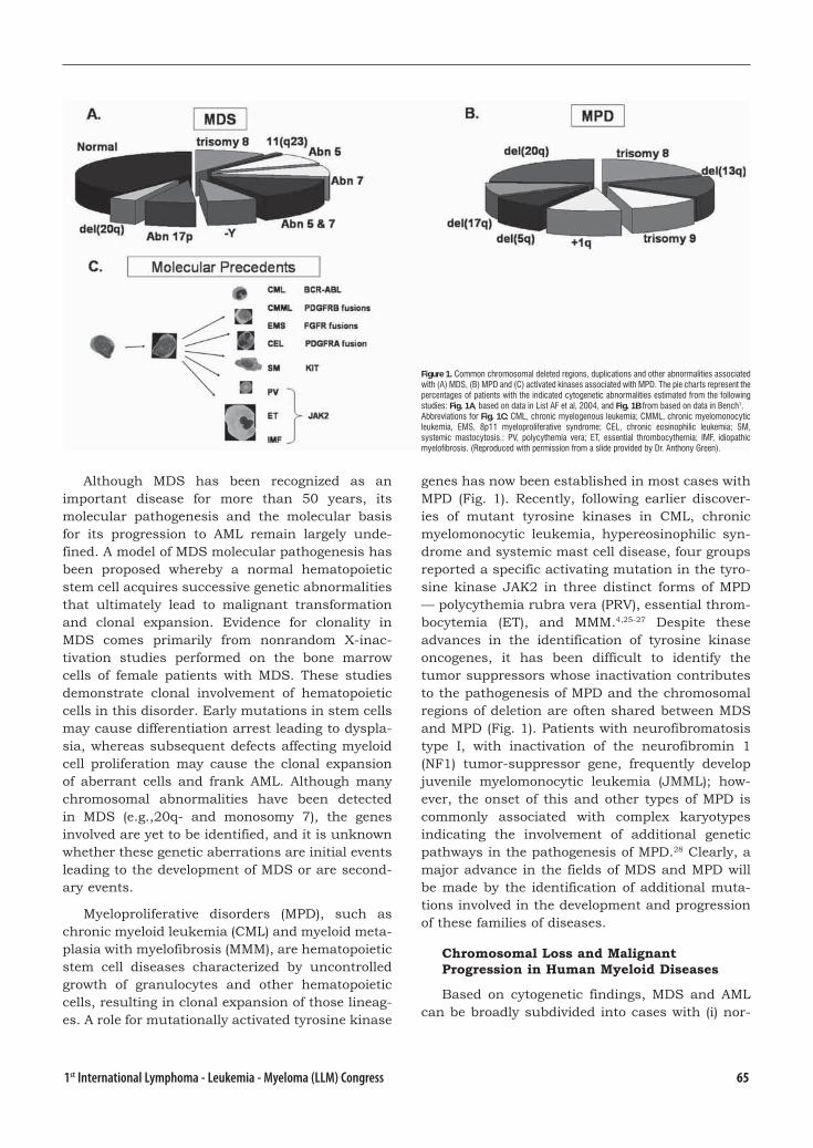
651st International Lymphoma - Leukemia - Myeloma (LLM) Congress
Although MDS has been recognized as an important disease for more than 50 years, its molecular pathogenesis and the molecular basis for its progression to AML remain largely unde-fined. A model of MDS molecular pathogenesis has been proposed whereby a normal hematopoietic stem cell acquires successive genetic abnormalities that ultimately lead to malignant transformation and clonal expansion. Evidence for clonality in MDS comes primarily from nonrandom X-inac-tivation studies performed on the bone marrow cells of female patients with MDS. These studies demonstrate clonal involvement of hematopoietic cells in this disorder. Early mutations in stem cells may cause differentiation arrest leading to dyspla-sia, whereas subsequent defects affecting myeloid cell proliferation may cause the clonal expansion of aberrant cells and frank AML. Although many chromosomal abnormalities have been detected in MDS (e.g.,20q- and monosomy 7), the genes involved are yet to be identified, and it is unknown whether these genetic aberrations are initial events leading to the development of MDS or are second-ary events.
Myeloproliferative disorders (MPD), such as chronic myeloid leukemia (CML) and myeloid meta-plasia with myelofibrosis (MMM), are hematopoietic stem cell diseases characterized by uncontrolled growth of granulocytes and other hematopoietic cells, resulting in clonal expansion of those lineag-es. A role for mutationally activated tyrosine kinase
genes has now been established in most cases with MPD (Fig. 1). Recently, following earlier discover-ies of mutant tyrosine kinases in CML, chronic myelomonocytic leukemia, hypereosinophilic syn-drome and systemic mast cell disease, four groups reported a specific activating mutation in the tyro-sine kinase JAK2 in three distinct forms of MPD — polycythemia rubra vera (PRV), essential throm-bocytemia (ET), and MMM.4,25-27 Despite these advances in the identification of tyrosine kinase oncogenes, it has been difficult to identify the tumor suppressors whose inactivation contributes to the pathogenesis of MPD and the chromosomal regions of deletion are often shared between MDS and MPD (Fig. 1). Patients with neurofibromatosis type I, with inactivation of the neurofibromin 1 (NF1) tumor-suppressor gene, frequently develop juvenile myelomonocytic leukemia (JMML); how-ever, the onset of this and other types of MPD is commonly associated with complex karyotypes indicating the involvement of additional genetic pathways in the pathogenesis of MPD.28 Clearly, a major advance in the fields of MDS and MPD will be made by the identification of additional muta-tions involved in the development and progression of these families of diseases.
Chromosomal Loss and Malignant Progression in Human Myeloid Diseases
Based on cytogenetic findings, MDS and AML can be broadly subdivided into cases with (i) nor-
Figure 1. Common chromosomal deleted regions, duplications and other abnormalities associated with (A) MDS, (B) MPD and (C) activated kinases associated with MPD. The pie charts represent the percentages of patients with the indicated cytogenetic abnormalities estimated from the following studies: Fig. 1A, based on data in List AF et al, 2004, and Fig. 1B from based on data in Bench1. Abbreviations for Fig. 1C: CML, chronic myelogenous leukemia; CMML, chronic myelomonocytic leukemia, EMS, 8p11 myeloproliferative syndrome; CEL, chronic eosinophilic leukemia; SM, systemic mastocytosis.: PV, polycythemia vera; ET, essential thrombocythemia; IMF, idiopathic myelofibrosis. (Reproduced with permission from a slide provided by Dr. Anthony Green).
66 1 st International Lymphoma - Leukemia - Myeloma (LLM) Congress
mal karyotypes, (ii) balanced chromosomal aberra-tions leading to the generation of fusion oncogenes and (iii) complex karyotypes (more than 3 chromo-somal aberrations). Complex chromosomal aberra-tions (CCAs) are associated with the most unfavo-rable prognosis among subtypes of MDS and AML, and MDS cases with a complex karyotype have a high propensity to evolve to AML. Despite intensive treatment including allogeneic stem cell transplan-tation, long-term remissions are achieved in less than 10% of patients with CCAs.29 The frequency of CCAs is remarkably high: 20% of de novo AMLs, 30% of de novo MDSs, 24% of secondary AMLs and up to 50% of therapy related AML and MDS cases.30,31 The lower frequency of CCAs in de novo AML reflects the higher prevalence of classic trans-location-generated oncogenes (e.g., AML1-ETO, PML-RARα and many others) in this disease com-pared with their paucity in MDS.3 Thus, myeloid leukemias and myelodysplastic syndromes with CCAs constitute important clinical entities in need of improved therapeutic strategies.
Cytogenetic studies in MDS have revealed pri-marily unbalanced chromosomal abnormalities and deletions, most commonly resulting in –5, 5q-, -7, 7q-, +8, 11q-, 13q- and 20q-,1,2 suggesting that genes within these regions have a role in MDS pathogenesis (Fig. 1). Finding the genes affected by such deletions poses a major investigative chal-lenge, but will be necessary to accelerate progress in research and treatment of these myeloid dis-eases.
Clonal chromosomal abnormalities are observed in bone marrow cells from 30% to 50% of de novo MDS cases and 80% of secondary MDS patients. The predominant abnormalities discovered in MDS are nonrandom chromosomal deletions, suggest-ing a pathogenic mechanism based on loss of tumor suppressor genes or haploinsufficiency of genes necessary for normal myelopoiesis. Common cytogenetic abnormalities in MDS include loss of chromosome 7 or partial deletions of chromosome arms 5q, 20q, 11q, or 7q. In addition, juvenile chronic myelomonocytic leukemia often involves monosomy 7, together with mutations of the NF1 gene.28 In a study of 1663 cases of MDS, 1098 (66%) had a single chromosomal abnormality, 237 (22%) were monosomic for chromosome 7, and 431 (39%) had a partial deletion of chromosome 5. Other abnormalities included chromosomes 6, 9, 11, 12, 13, and 17. Importantly, cytogenetic abnormalities correlate with prognosis. After inten-
sive chemotherapy, 60% of patients with an appar-ently normal karyotype entered complete remis-sion (average duration, 16 months), while patients with chromosome 5 or 7 deletions or complex chromosomal abnormalities had a 20% remission rate (average duration, 4 to 5 months). Similarly, secondary MDS usually displays monosomies of chromosome 5 or 7 or partial deletions involving 5q or 7q, with chromosome 7 defects associated with decreased survival time.13
Although the majority of putative tumor sup-pressors in MDS have not been cloned, many chro-mosomal translocation-mediated oncogenes (see Fig. 1, reviewed in Look3) and a few of the tumor suppressors have been identified. For example, genes inactivated in AML comprise a relatively small number of cases and include P53, RB, WT-1, NF1, AML1, C/EBPalpha, and nucleophosmin (NPM) (reviewed in Tenen6 and Side28), and besides p53, these genes are rarely mutated in MDS. In addition, the deleted region in the 5q- syndrome, which is commonly encountered in older individu-als with MDS but uncommon in pediatric patients, contains the alpha-catenin gene. As we have recently shown, this gene is both deleted on one allele and epigenetically suppressed in a subset of cases with 5q-, most likely contributing to dys-regulated and aberrantly increased stem cell self renewal within the clone.7.5
MLL Amplification in MDS
In contrast to AMLs harboring oncogenic tran-scription factor fusions, hardly any oncogene acti-vation has been assigned specifically to MDS and AML with CCAs. One exception is the 11q23 region and its resident MLL gene, which is amplified in a significant fraction of MDS and AML with karyotyp-ic complexity and an adverse prognosis.32-35 MLL has long been recognized as an important compo-nent of translocation-generated fusion proteins. In contrast to other oncogenic fusion proteins, MLL participates in translocations with more than 40 different partner chromosomal loci. Homodimeri-zation of the chimeric proteins appears to underlie the promiscuity of MLL in its ability to combine with many fusion partners, at least for a subset of its productive fusions.36
The new studies mentioned above suggested that amplification of MLL represents a new mecha-nism of oncogenic activation of this gene. A recent study confirmed the importance of MLL within the 11q23 amplicon by analysis of MLL target genes
671st International Lymphoma - Leukemia - Myeloma (LLM) Congress
like HOXA9 and MEIS1.37 It has long been recog-nized that HOXA9 is one of the important target genes of MLL and recent reports from several independent laboratories, including ours, provide a comprehensive view of other HOX genes that may be inappropriately activated in leukemias with MLL rearrangements.38,39 Very recently, Hoxa7 and Hoxa9 were shown to be essential for MLL-depend-ent leukemogenesis in vivo.40
Thus, aberrant MLL activation in MDS/AML and consequent dysregulation of its downstream targets are of clinical importance. Indeed, the remarkable synergy between MLL gene amplifica-tion and loss of 5q in MDS and AML are reported to result in an extremely poor overall survival rate of 30 days.35 Moreover, these data provide an important clue regarding the mechanism of disease evolution. It should be stressed that the impact of dysregulated MLL and HOX gene activation is like-ly to extend beyond the subgroup of patients with CCAs, as MLL and HOXA9 were also significantly upregulated in unselected MDS patient samples, including those with normal karyotypes.37
HOX Genes: Multiple Roles in Development, Hematopoiesis and Leukemogenesis
Homeobox genes were first recognized through the analysis of homeotic mutations of Drosophila, which alter the identity of various body segments. Homologous genes have been found in virtually every species, from yeast to humans. Class I home-obox genes are designated as HOX genes in humans (mouse:hox genes). HOX genes control morphogen-esis in early stages of embryonic development. The specific shape of discrete segments (pattern forma-tion) is decisively regulated by these genes. Beside their role as differentiation factors in embryonic development, the control of hematopoiesis by HOX genes is well established. Since the perturbation of hematopoietic stem cell development is a hallmark of leukemia, it is not surprising that the aberrant expression of HOX genes contributes decisively to leukemia pathogenesis.41
Early experimental evidence suggesting the oncogenic potential of the HOX gene family came from studies showing that the overexpression of Hoxb8 and IL-3 in murine bone marrow cells can induce aggressive, transplantable leukemia. A sim-ilar approach showed that Hoxa9 and Hoxa10 are able to induce AML in mice. Retroviral insertional mutagenesis has likewise implicated the Hox genes in leukemia induction. For example, Hoxa7 and
Hoxa9 were activated in the context of retrovirally induced AML in BXH-2 mice. The importance of Hoxa7, Hoxa9 and the cofactor Meis1 in the BXH-2 mouse leukemia model was impressively under-scored by a study based on large scale cloning of proviral integration sites.
Further compelling evidence of the oncogenic potential of HOX genes comes from their direct or indirect involvement in leukemia-associated translo-cations, such as the translocation t(7;11)(p15;p15), which generates the fusion protein NUP98-HOXA9 in AML patients. Additional translocations involv-ing HOX loci have been identified over the past years.42,43 As pointed out above, MLL translocations (6%-7% of all acute leukemia) and most likely also MLL amplifications lead likewise to a dysregula-tion of HOX gene expression. Very recently CDX4 was shown to regulate expression of HOXA7 and HOXA9.44 Thus, this homeobox transcription fac-tor and it’s relative CDX2 are attractive candidates for unidentified upstream regulators of HOX gene expression in leukemia. Taken together, published reports leave little doubt that specific HOX genes, particularly HOXA7, HOXA9 and HOXA10, are involved in the pathogenesis of AML and MDS, but virtually nothing is known about the downstream pathways through which these genes exert their oncogenic potential.
References 1. Bench, A.J., et al., Molecular genetics and cytoge-
netics of myeloproliferative disorders. Baillieres Clin Haematol. 1998;11:819-848.
2. Fenaux, P., P. Morel, and J.L. Lai, Cytogenetics of myelodysplastic syndromes. Semin Hematol. 1996;33(2):127-138.
3. Look, A.T., Oncogenic transcription fac-tors in the human acute leukemias. Science. 1997;278(5340):1059-1064.
4. Levine, R.L., et al., Activating mutation in the tyro-sine kinase JAK2 in polycythemia vera, essential thrombocythemia, and myeloid metaplasia with myelofibrosis. Cancer Cell. 2005;7(4):387-397.
5. Tenen, D.G., Abnormalities of the CEBP alpha transcription factor: a major target in acute myeloid leukemia. Leukemia. 2001;15(4):688-689.
6. Song, W.J., et al., Haploinsufficiency of CBFA2 cau-ses familial thrombocytopenia with propensity to develop acute myelogenous leukaemia. Nat Genet. 1999;23(2):166-175.
7. Mueller, B.U., et al., Heterozygous PU.1 mutations are associated with acute myeloid leukemia. Blood. 2003;101(5):2074.

68 1 st International Lymphoma - Leukemia - Myeloma (LLM) Congress
7.5 Liu TX, Becker MW, Jelinek J, et al. Chromosome 5q deletion and epigenetic suppression of the gene encoding alpha-catenin (CTNNA1) in myeloid cell transformation. Nature medicine 2007;13(1):78-83.
8. Sutton, L., et al., Factors influencing outcome in de novo myelodysplastic syndromes treated by allo-geneic bone marrow transplantation: a long-term study of 71 patients Societe Francaise de Greffe de Moelle. Blood. 1996;88(1):358-365.
9. Runde, V., et al., Bone marrow transplantation from HLA-identical siblings as first-line treatment in pati-ents with myelodysplastic syndromes: early transp-lantation is associated with improved outcome. Chronic Leukemia Working Party of the European Group for Blood and Marrow Transplantation. Bone Marrow Transplant. 1998;21(3):255-261.
10. Arnold, R., et al., Unrelated bone marrow transp-lantation in patients with myelodysplastic synd-romes and secondary acute myeloid leukemia: an EBMT survey. European Blood and Marrow Transplantation Group. Bone Marrow Transplant. 1998;21(12):1213-1216.
11. Hofmann, W.K., et al., Myelodysplastic syndromes: clinical features. Semin Hematol. 1996;33(3):177-185.
12. Krishnan, A., et al., Predictors of therapy-related leukemia and myelodysplasia following autologous transplantation for lymphoma: an assessment of risk factors. Blood. 2000;95(5):1588-1593.
13. Friedberg, J.W., et al., Outcome in patients with myelodysplastic syndrome after autologous bone marrow transplantation for non-Hodgkin’s lympho-ma. J Clin Oncol. 1999;17(10):3128-3135.
14. Kamada N,Uchino H. Preleukemic states in atomic bomb survivors in Japan. Nouv Rev Fr Hematol Blood Cells. 1976;17(1-2):57-65.
15. Miller, J.S., et al., Myelodysplastic syndrome after autologous bone marrow transplantation: an addi-tional late complication of curative cancer therapy. Blood, 1994. 83(12): p. 3780-6.
16. Kantarjian HM, Keating MJ. Therapy-related leuke-mia and myelodysplastic syndrome. Semin Oncol. 1987;14(4):435-443.
17. Tester, W.J., et al., Second malignant neoplasms complicating Hodgkin’s disease: the National Cancer Institute experience. J Clin Oncol. 1984;2(7):762-769.
18. Darrington, D.L., et al., Incidence and characte-rization of secondary myelodysplastic syndrome and acute myelogenous leukemia following high-dose chemoradiotherapy and autologous stem-cell transplantation for lymphoid malignancies. J Clin Oncol. 1994;12(12):2527-2534.
19. Anderson JR, Vose J, Kessinger A. Myelodysplastic syndrome after autologous transplant for lympho-ma. Blood. 1994;84(11):3988-3989.
20. Nevill, T.J., et al., Cytogenetic abnormalities in primary myelodysplastic syndrome are highly pre-dictive of outcome after allogeneic bone marrow transplantation. Blood. 1998;92(6):1910-1917.
21. Haas, O.A. and H. Gadner, Pathogenesis, biology, and management of myelodysplastic syndromes in children. Semin Hematol, 1996. 33(3): p. 225-35.
22. Huang, M.E., et al., Use of all-trans retinoic acid in the treatment of acute promyelocytic leukemia. Blood. 1988;72(2):567-572.
23. Castaigne, S., et al., All-trans retinoic acid as a diffe-rentiation therapy for acute promyelocytic leukemia. I. Clinical results. Blood. 1990;76(9):1704-1709.
24. Warrell, R.P., Jr., et al., Differentiation the-rapy of acute promyelocytic leukemia with tre-tinoin (all-trans-retinoic acid). N Engl J Med. 1991;324(20):1385-1393.
25. Steensma, D.P., et al., The JAK2 V617F activating tyrosine kinase mutation is an infrequent event in both “atypical” myeloproliferative disorders and the myelodysplastic syndrome. Blood, 2005.
26. Jones, A.V., et al., Widespread occurrence of the JAK2 V617F mutation in chronic myeloproliferative disorders. Blood, 2005.
27. Kralovics, R., et al., A gain-of-function mutation of JAK2 in myeloproliferative disorders. N Engl J Med. 2005;352(17):1779-1790.
28. Side, L., et al., Homozygous inactivation of the NF1 gene in bone marrow cells from children with neu-rofibromatosis type 1 and malignant myeloid disor-ders. N Engl J Med. 1997;336(24):1713-20.
29. Schoch C, Haferlach T, Haase D, et al. Patients with de novo acute myeloid leukaemia and complex kar-yotype aberrations show a poor prognosis despite intensive treatment: a study of 90 patients. Br J Haematol. 2001;112:118-126.
30. Mauritzson N, Johansson B, Albin M, et al. Survival time in a population-based consecutive series of adult acute myeloid leukemia--the prognostic impa-ct of karyotype during the time period 1976-1993. Leukemia. 2000;14:1039-1043.
31. Rossi G, Pelizzari AM, Bellotti D, Tonelli M, Barlati S. Cytogenetic analogy between myelodysplastic syndrome and acute myeloid leukemia of elderly patients. Leukemia. 2000;14:636-641.
32. Andersen MK, Christiansen DH, Kirchhoff M, Peder-sen-Bjergaard J. Duplication or amplification of chromosome band 11q23, including the unrearran-ged MLL gene, is a recurrent abnormality in the-rapy-related MDS and AML, and is closely related to mutation of the TP53 gene and to previous therapy with alkylating agents. Genes Chromosomes Can-cer. 2001;31:33-41.
33. Avet-Loiseau H, Godon C, Li JY, et al. Amplificati-on of the 11q23 region in acute myeloid leukemia. Genes Chromosomes Cancer. 1999;26:166-170.
34. Reddy KS, Parsons L, Mak L, et al. Segmental amp-lification of 11q23 region identified by fluorescence in situ hybridization in four patients with myelo-id disorders: a review. Cancer Genet Cytogenet. 2001;126:139-146.
35. Van Limbergen H, Poppe B, Michaux L, et al. Iden-tification of cytogenetic subclasses and recurring chromosomal aberrations in AML and MDS with complex karyotypes using M-FISH. Genes Chromo-somes Cancer. 2002;33:60-72.
691st International Lymphoma - Leukemia - Myeloma (LLM) Congress
36. So CW, Lin M, Ayton PM, Chen EH, Cleary ML. Dimerization contributes to oncogenic activation of MLL chimeras in acute leukemias. Cancer Cell. 2003;4:99-110.
37. Poppe B, Vandesompele J, Schoch C, et al. Expres-sion analyses identify MLL as a prominent target of 11q23 amplification and support an etiologic role for MLL gain of function in myeloid malignancies. Blood. 2004;103:229-235
38. Armstrong SA, Staunton JE, Silverman LB, et al. MLL translocations specify a distinct gene expressi-on profile that distinguishes a unique leukemia. Nat Genet. 2002;30:41-47.
39. Ferrando AA, Armstrong SA, Neuberg DS, et al. Gene expression signatures in MLL-rearranged T-lineage and B-precursor acute leukemias: dominan-ce of HOX dysregulation. Blood. 2003;102:262-268.
40. Ayton PM, Cleary ML. Transformation of myeloid progenitors by MLL oncoproteins is dependent on Hoxa7 and Hoxa9. Genes Dev. 2003;17:2298-2307.
41. Buske C, Humphries RK. Homeobox genes in leuke-mogenesis. Int J Hematol. 2000;71:301-308.
42. Taketani T, Taki T, Shibuya N, Kikuchi A, Hana-da R, Hayashi Y. Novel NUP98-HOXC11 fusion gene resulted from a chromosomal break within exon 1 of HOXC11 in acute myeloid leukemia with t(11;12)(p15;q13). Cancer Res. 2002;62:4571-4574.
43. Panagopoulos I, Isaksson M, Billstrom R, Strombeck B, Mitelman F, Johansson B. Fusion of the NUP98 gene and the homeobox gene HOXC13 in acute myeloid leukemia with t(11;12)(p15;q13). Genes Chromosomes Cancer. 2003;36:107-112.
44. Davidson AJ, Ernst P, Wang Y, et al. cdx4 mutants fail to specify blood progenitors and can be rescued by multiple hox genes. Nature. 2003;425:300-306.
45. List AF, Vardiman J, Issa JP, DeWitte TM. Mye-lodysplastic syndromes. Hematology. 2004;1:297-317.

70 1 st International Lymphoma - Leukemia - Myeloma (LLM) Congress
Allogeneic SCT (alloSCT) is the treatment of choice for patients with MDS who have a histocompatible donor, but the question is
up to which age? Patients with early stages of MDS may profit optimally from alloSCT, with long-term DFS in more than 50% of patients. Longer disease duration before transplantation is associated with an increased TRM after transplantation. A recent analysis confirmed that delayed transplantation may result in maximized overall survival for low risk MDS1. They hypothesized that the optimal timing of transplantation for this cohort is at the time of development of a new cytogenetic abnor-mality, the appearance of a clinically important cytopenia or an increase in the percentage of marrow blasts. Whether patients with advanced stages of MDS should receive remission-induction chemotherapy prior to the transplant condition-ing remains a point for discussion. Retrospective analyses showed conflicting data. Interpretation of the data is hampered by selection biases in the two treatment approaches. Therefore, the EBMT has launched a prospective study for this question (www.ebmt.org: Chronic leukemia; protocol: Allo-MDS 2x2). The results of allSCT with unrelated donors have improved markedly in recent years.2 The American National Marrow Donor Program (NMDP) reported an improved DFS in more recently performed transplantations in a cohort of 510 patients with MDS transplanted with unrelated donors. The relative risk for DFS was 1.43 (95% confidence interval: 1.01–2.01) for transplanta-tions performed between 1988 and 1993 versus more recent transplantations3.
The Role of Allogeneic Stem Cell
Transplantation in MDS-MPD
Theo de Witte, A. Van Biezen, R. Brand, S. Suciu, N. Kröger
University Medical Center St. Radboud Department of Hematology, Amsterdam, The Netherlands
The idea to reduce the intensity of the condition-ing regimen has been developed in view of the high TRM of conventional marrow ablative conditioning regimens. The principle of RIC regimens is based on intensive immune suppression during condi-tioning and/or after stem cell infusion to facilitate donor engraftment and to establish complete donor chimerism. It is difficult to reconcile the contribu-tion of RIC regimens to the improved outcome of alloSCT in view of the recently improved outcome of transplantation with marrow ablative regimens and the heterogeneity of the patient populations (age, co-morbidity, stage of disease).4 For this reason, the EBMT has launched a prospective randomized study comparing RIC regimens with standard conditioning regimens in patients with MDS older than 50 years.
The question remains whether autoSCT is a good alternative for alloSCT? In a recent study the 4-year DFS of the patients with a donor was 46%, higher than the DFS of 26% in patients without a donor. Subgroup analysis showed that the advan-tage of the presence of an HLA-identical sibling donor was only apparent in the patient group with intermediate and high risk cytogenetics.
General conclusion: allogeneic SCT may be considered the curative treatment option of choice. The upper age limit for alloSCT is mainly man-dated by co-morbidity and general condition of the patient (frailty index) rather age of 60 or 70 years. For patients lacking an suitable donor, treatment with autoSCT or chemotherapy may be a good alternative for MDS patients with good-risk cytoge-netic characteristics.
711st International Lymphoma - Leukemia - Myeloma (LLM) Congress
References 1. Cutler CS, Lee SJ, Greenberg P et al. A decision
analysis of allogeneic bone marrow transplantation for the myelodysplastic syndromes: delayed transp-lantation for low-risk myelodysplasia is associated with improved outcome. Blood 2004;104:579-585.
2. de Witte T, Hermans J, Vossen J et al. Haematopoie-tic stem cell transplantation for patients with myelo-dysplastic syndromes and secondary acute mye-loid leukaemias: a report on behalf of the Chronic Leukaemia Working Party of the European Group for Blood and Marrow Transplantation (EBMT). Br.J.Haematol. 2000;110:620-630.
3. Castro-Malaspina H, Harris RE, Gajewski J et al. Unrelated donor marrow transplantation for mye-lodysplastic syndromes: outcome analysis in 510 transplants facilitated by the National Marrow Donor Program. Blood 2002;99:1943-1951.
4. Martino R, Iacobelli S, Brand R et al. Retrospective comparison of reduced-intensity conditioning and conventional high-dose conditioning for allogeneic hematopoietic stem cell transplantation using HLA-identical sibling donors in myelodysplastic syndro-mes. Blood 2006;108:836-846.

72 1 st International Lymphoma - Leukemia - Myeloma (LLM) Congress
Multiple Myeloma (MM) is a chemoresist-ant malignancy and for decades, the only active drugs were alkylating agents
and high-dose corticosteroids. The introduction of novel agents (Thalidomide, Bortezomib and Lenalidomide) is changing the management of MM patients both for frontline therapy and at relapse. These agents have a different mode of action and can act not only on the myeloma cells but also on the microenvironment which is necessary for tumor cell survival and proliferation. In vitro experiments have shown possible synergy with dexamethasone or chemotherapeutic agents. They are active even in heavily pretreated patients and offer new pos-sibilities for relapsed/refractory MM.
Thalidomide
Thalidomide as a single agentThalidomide as a single agent
Thalidomide is administered orally. Due to its teratogenecity, it is prescribed according to risk-management programs to avoid exposure of women with child-bearing potential.
Thalidomide was introduced in the treatment of relapsed MM by the Arkansas group (1). Following the results of this pioneer work, a large number of Phase II studies were reported and were summa-rized in a comprehensive review (42 studies, 1674 patients) (2). From this experience, the following conclusions are possible (2-5).
- The response rate is approximatively 45% includ-ing 30% partial remissions (PR)
- Complete remission (CR) are rare in heavily pre-treated patients but possible
Recent Advances of Thalidomide in the
Treatment of Multiple Myeloma
Jean-Luc Harousseau
Centre Hospitalier Universitaire Hôtel-Dieu Place Alexis Ricordeau, Nantes Cedex, France
- The onset of response is rapid (less than 2 months) and maximal response is achieved with-in 6 months
- One-year event-free survival (EFS) is about 35% and median overall survival (OS) is 14 months
- Prognostic factors with thalidomide treatment are the same as with conventional chemotherapy and include tumor burden markers and cytogenetic abnormalities
- Thalidomide can be prescribed in patients with renal dysfunction
- Myelosuppression is very rare
- The incidence and severity of side effects is related to the daily dosage. With the doses initially used (400 mg/day) the most frequent toxicities were constipation, somnolence, fatigue and peripheral neuropathy.
Peripheral neuropathy causes numbness, par-esthesia and even pain in legs and arms. The over-all incidence of peripheral neuropathy is 30% with 10% grade ≥ 3, but is related to cumulated dose and is up to 75% in patients treated more than one year (6). Since there is no effective prophylaxis and treatment, the drug should be discontinued in case of peripheral neuropathy signs or symptoms (3-5).
Currently the daily doses of Thalidomide have been reduced to 100-200 mg/day.
Thalidomide in combination
The combination of Thalidomide plus Dexam-ethasone has been developed with the objectives of increasing the efficacy and of reducing the daily dose and the toxicity of Thalidomide (6-8). Although
731st International Lymphoma - Leukemia - Myeloma (LLM) Congress
no randomized study comparing Thalidomide and Thalidomide/Dexamethasone (TD) has been per-formed, TD is considered more effective than Thalidomide alone (approximatively 45% response rate) and superior to conventional chemotherapy as treatment of first relapse (9). Since Thalidomide is not myelotoxic, combinations with chemothera-py have also been evaluated in Phase II trials with response rates ranging from 36% to 73%.
While combinations of Thalidomide with Dex-amethasone or chemotherapy alone appear to be more active, they are more toxic with a higher inci-dence of infectious complications. Most importantly they induce an unexpected complication, deep vein thrombosis. While with Thalidomide alone the inci-dence of deep vein thrombosis is < 5% as with any treatment of MM, with TD the incidence is 10-15% and with chemotherapy (specially anthracyclines) it increases up to 30% (10, 11). This complication usually occurs during the first 3 months of treat-ment and is more frequent in newly diagnosed patients and in patients with a high tumor burden. The optimal prophylaxis is not yet known and oral anticoagulant, low molecular weigh heparin and low-dose aspirin are currently evaluated (3).
Bortezomib
Bortezomib is the first in class proteasome inhibitor. In MM it is active not only on the myelo-ma cell but also on the microenvironnement which is necessary for tumor cell proliferation and sur-vival. It is administered IV at a dose of 1.3 mg/m² on days 1, 4, 8, 11 in 21 days cycles. Two Phase II studies have shown that in heavily pretreated patients, response rate with Bortezomib as a single agent is 25-30% and can be increased by the addi-tion of Dexamethasone (12, 13). Following these studies, the drug has been approved in the US and Europe. The large randomised Phase III trial APEX has demonstrated that Bortezomib is supe-rior to Dexamethasone in relapsed MM, in terms of response rate (including CR), time to progres-sion and OS (14). The onset of response is rapid, usually within 2 cycles but the maximal response can be achieved after up to 8 cycles. A subgroup analysis on patients having received one line of treatment confirmed the superiority of Bortezomib compared to Dexamethasone as treatment of first relapse (15). Based on these Phase II-III studies the toxicity profile is well defined. The most frequent side effects are gastrointestinal symptoms (diarhea or constipation) and fatigue but they are usually
mild. The drug is not myelotoxic but can induce up to 60% decrease of the platelet count (16). This thrombocytopenia is rapidly reversible usually before the following cycle. Peripheral neuropathy is observed in 30-40% of cases (grade ≥ 3 in 10-15%). Signs and symptoms are reversible in 2/3 of cases after dose reduction or drug discontinuation (17). Bortezomib appears to be as effective in older patients and in patients with poor risk disease (18, 19). It can be prescribed safely in patients with renal failure, even in patients on dialysis (20, 21). Based on preclinical studies and on the drug toxicity profile, a number of Phase II studies have evaluated Bortezomib in combination with either Dexamethasone or with chemotherapy.
Lenalidomide
Lenalidomide is a Thalidomide analog which appears to be more potent in vitro. It is not tera-togenic in animal models but most importantly, after the Phase I study it became apparent that the toxicity profile of Lenalidomide was completely dif-ferent (22). Constipation, somnolence, fatigue and peripheral neuropathy that are frequent adverse events with Thalidomide, were not observed. The most frequent side effect was myelosuppression mostly after 28 days of treatment. In Phase II trials the drug was given orally for 21 consecutive days. The response rate in heavily pretreated patients was 25% and was increased by the addition of Dex-amethasone (23). Two large randomized trials were then conducted in the US and in Europe (24, 25). They both compared Lenalidomide (25 mg/day on 21 consecutive days) plus high-dose Dexametha-sone versus Dexamethasone. They both showed the superiority of the combination in terms of response rate (including CR), time to progression and OS. Based on these studies, the drug was approved by FDA in 2006 and the European approval is pend-ing. However, like with Thalidomide, the combina-tion of Lenalidomide with Dexamethasone induces deep vein thrombosis and justify prophylactic treatment at least with low-dose aspirin. The treat-ment should be used with caution in patients with renal failure due to a higher myelotoxicity (spe-cially thrombocytopenia) in patients with less than 50 ml/min creatinine clearance. Lenalidomide is currently tested upfront in combination with dex-amethasone or with chemotherapy.
Combination of novel agents
Since the toxicity profiled of these three agents is different, it appeared logical to combine them
74 1 st International Lymphoma - Leukemia - Myeloma (LLM) Congress
with the objective of increasing efficacy. The com-bination of Thalidomide or Lenalidomide with Bort-ezomib was the most attractive, due to a possible synergy of agents having different mode of actions. Thalidomide and Bortezomib have been combined with either Dexamethasone (VTD) or with Mel-phalan Prednisone (VMPT) with very encouraging results and an acceptable toxicity (26, 27). The combination Bortezomib-Lenalidomide is currently tested (28).
Role of Stem Cell Transplantation at first relapse
Autologous Stem Cell Transplantation (ASCT) is currently considered the standard of care for frontline therapy in patients up to 65 years of age. However ASCT is also a useful salvage treat-ment in chemosensitive or untreated relapses (29). When comparing early ASCT, and ASCT when conventional chemotherapy fails (late ASCT) there is no difference in OS although time to progres-sion is longer with early ASCT (30). If late ASCT is considered it should be useful to collect stem cells early (31) since the hematopoietic quality of grafts is often decreased by previous chemotherapy spe-cially alkylating agents (32). When ASCT has been performed upfront, a second ASCT is justified if the duration of first remission has been > 2 years.
Allogeneic SCT is probably the only treatment that can induce long term clinical and molecu-lar remissions. However standard myeloablative regimens prior to allogeneic SCT have been almost abandoned specially for relapsed MM, due to a high transplant-related mortality (up to 50%) (33).
With reduced-intensity conditioning allogeneic SCT, transplant-related mortality is reduced, even in relapsed MM (20-25% at 1 year) (34). However the risk of relapse is higher than with standard allogeneic SCT, specially in chemoresistant disease or when the tumor burden is high (35). Current strategy aims at reducing the tumor burden first, for instance with high-dose Melphalan followed by ASCT, and then to exploit the graft-versus myelo-ma effect of donor lymphoid cells. This strategy is mostly proposed for first line treatment in patients with an HLA-identical donor (36) but can also be offered at first relapse if CR or VGPR has been achieved with salvage therapy.
Therapeutic strategy
With the introduction of novel agents, CR achievement is possible in relapsed MM. In the
large randomized studies testing Bortezomib and Lenalidomide, the progression-free survival (PFS) and OS increase was associated to a CR rate increase, as compared to the control arms (12, 24, 25). In the APEX trial, PFS was 12 months for patients in CR versus 8 months for patients with only PR (37). Therefore, as for frontline therapy, the objective of first relapse treatment should be to achieve the best possible response.
With this objective, it is logical to use novel agents for first relapse treatment since they are effective even in heavily pretreated patients. Although no randomized study has compared Thalidomide, Bortezomib or Lenalidomide given as single agents to combinations, they are usu-ally administered in combination at least with dexamethasone (TD, VD or RD). Out of a clinical trial, the choice depends on a variety of parameters including age, previous treatment, prior toxicities, availability of novel agents (which is not the same all over the world). There are special situations in which the choice is easier.
For instance if Thalidomide has been used for frontline therapy it is better to use Bortezomib or Lenalidomide to avoid the risk of peripheral neuropathy related to cumulative doses of Tha-lidomide. In patients with renal failure, Bortezomib is the treatment of choice while in patients with peripheral neuropathy, Lenalidomide should be preferred. Fulminant relapses or poor risk cytoge-netics should be treted with combinations includ-ing Bortezomib or Lenalidomide.
The optimal duration of salvage treatment is unknown. In order to reduce the risk of toxicity and of resistant clones selection, treatment could be stopped 3 months after best result is achieved. In younger patients (< 65 years) ACSH can be con-sidered in sensitive relapses if stem cells are avail-able. Reduced intensity conditioning allogeneic SCT is possible in patients who achieve CR with salvage treatment.
References 1. Singhal S, Mehta J, Desikan R. Antitumor activity of
thalidomide in refractory multiple myeloma. N Engl J Med 1999;341:1565-1571
2. Glasmacher A, Hahn C, Hoffmann T et al. A sys-tematic review of phase II trials of thalidomide in monotherapy in patients with relapsed or refractory multiple myeloma. Br J Haematol 2005;132:584-593
3. Harousseau JL. Thalidomide in Multiple Myeloma: past, present and future. Future Oncol 2006; 2: 577-589
751st International Lymphoma - Leukemia - Myeloma (LLM) Congress
4. Dimopoulos MA, Anagnostopoulos A, Weber D. Treat-ment of plasma cell dyscrasia with Thalidomide and its derivatives. J Clin Oncol 2003;21:4444-4452
5. Cavenagh JD, Oakervee H. Thalidomide in multiple myeloma : current status and future prospects. Br J Haematol 2003;120:18-26
6. Weber DM, Gavino M, Delasalle K et al. Thalidomide alone or with dexamethasone for multiple myeloma. Blood 1999;94:604a
7. Anagnostopoulos A, Weber D, Rankin R et al. Tha-lidomide and Dexamethasone for resistant multiple myeloma. Br J Haematol 2003;121:768-771
8. Dimopoulos MA, Zervas K, Kouvatseas G et al. Thali-domide and Dexamethasone combination for refrac-tory multiple myeloma. Ann Oncol 2001;12:991-995
9. Palumbo A, Giaccone L, Bertola A et al. Low-dose Thalidomide plus Dexamethasone is an effective sal-vage therapy for advanced myeloma. Haematologica 2001;86:399-403
10. Bennet CL, Schumoki GT, Kwaan HC, Raisch DW. High incidence of Thalidomide associated deep vein thrombosis and pulmonary emboli when chemothe-rapy is also administered. Blood 2001;98:863a
11. Zangari M, Siegel E, Barlogie B et al. Thrombogenic activity of Doxorubicin in myeloma patients recei-ving Thalidomide : implications for therapy. Blood 2002;100:1168-1171
12. Richardson P, Barlogie B, Berenson J et al. A phase 2 study of bortezomib in relapsed, refractory myelo-ma. N Engl J Med 2003; 348: 2609-2617
13. Jagannath S, Barlogie B, Berenson J et al. A phase 2 study of two doses of bortezomib in relapsed or refra-ctory myeloma. Brit J Haemat 2004; 127: 165-172
14. Richardson PG, Sonneveld P, Schuster MW et al. Bor-tezomib or high-dose dexamethasone for relapsed mul-tiple myeloma. N Engl J Med 2005; 352: 2487-2498
15. Sonneveld P, Richardson PG, Schuster MW et al. Bortezomib at first relapse is superior to high-dose dexamethasone and more effective that when given later in relapsed multiple myeloma. Haematologica 2005; 90: 146-147
16. Lonial S, Waller EK, Richardson PG et al. Risk fac-tors and kinetics of thrombocytopenia associated with bortezomib for relapsed, refractory multiple myeloma. Blood 2005; 106: 3777-3784
17. Richardson PG, Briemberg H, Jagannath S et al. Frequency, characteristics and reversibility of perip-heral neuropathy during treatment of advanced multiple myeloma with bortezomib. JCO 2006; 19: 3113-3119
18. Richardson PG, Sonneveld P, Schuster MW et al. Safety and efficacy of bortezomib in high-risk elder-ly patients with relapsed multiple myeloma. Br J Hematol 2007; 19:
19. Jagannath S, Richardson PG, Sonnevel P et al. Bortezomib appears to overcome the poor prognosis conferred by chromosome 13 deletion in phase 2 and 3 trials. Leukemia 2007; 21: 151-157
20. Jagannath S, Barlogie B, Berenson JR et al. Borte-zomib in recurrent and/or refractory multiple mye-loma. Cancer 2005; 103: 1195-1200
21. Chanan-Khan AA, Kaufman JL, Mehta J et al. Acti-vity and safety of bortezomib in multiple myeloma patients with advanced renal failure: a multicenter retrospective study. Blood 2007; 109: 2604-2606
22. Richardson PG, Schlossman RL, Weller E et al. Immunomodulatory drug CC-5013 overcomes drug resistance and is well tolerated in patients with relapsed multiple myeloma. Blood 2002; 100: 3063-3067
23. Richardson PG, Blood E, Mitsiades CS et al. A ran-domized phase 2 study of lenalidomide therapy for patients with relapsed or relapsed and refractory multiple myeloma. Blood 2006; 108: 3458-3464
24. Weber DM, Chen C, Niesvizky R et al. A multicenter, randomized, parallel-group double-blind, placebo-controlled study of lenalidomide plus dexamethaso-ne versus dexamethasone alone in previously trea-ted subject with multiple myeloma. Haematologica 2005; 90 (S1), 155
25. Dimopoulos M, Spencer A, Attal M et al. Study of lenalidomide plus dexamethasone versus dexa-methasone alone in relapsed or refractory multiple myeloma. Blood 2005; 106: 6a (abstract)
26. Zangari M, Barlogie B, Burns MF. Velcade-Thalido-mide-Dexamethasone for advanced and refractory multiple myeloma: long-term follow-up of Phase I-II trial VARK 2001-37. Blood 2005; 106: 717 a (abstra-ct)
27. Palumbo A, Ambrosini MT, Benevolo G et al. Borte-zomib, melphalan, prednisone, and thalidomide for relapsed multiple myeloma. Blood 2007; 109: 2767-2772
28. 29. Harousseau JL, Attal M. The role of Autologous
hematopoietic stem cell transplantation in multiple myeloma. Semin Hematol 1997; 33 (suppl 1): 61-66
30. Fermand JP, Ravaud P, Chevret S et al. High-dose therapy and autologous peripheral blood stem cell transplantation in multiple myeloma: upfront or rescue treatment ? Blood 1998; 92: 3131-3136
31. Gertz MA, Lacy MQ, Inwards DJ. Early harvest and late transplantation as an effective therapeutic stra-tegy in multiple myeloma. Bone Marrow Transplant 1999; 23: 221-226
32. Harousseau JL. Optimizing peripheral blood pro-genitor cell autologous transplantation in multiple myeloma. Haematologica 1999; 84: 548-553
33. Bensinger WI, Buckner CD, Anasetti C et al. Alloge-neic marrow transplantation for multiple myeloma: an analysis of risk factors on outcome. Blood 1996; 88: 2787-2793
34. Crawley C, Iacobelli S, Björkstrand B et al. Redu-ced-intensity conditioning for myeloma: lower non-relapse mortality but higher relapse rates compared with myeloablative conditioning. Blood 2007; 109: 3588-3594
35. Crawley C, Lalancette M, Szydlo R et al. Outcomes of reduced-intensity allogeneic transplantation for multiple myeloma: an analysis of prognostic factors from the Chronic Leukemia Working Party of the EBMT. Blood 2005; 105: 4532-4539
36. Bruno B, Rotta M, Patriarca F et al. A comparison of allografting with autografting for newly diagnosed myeloma. N. Engl. J. Med 2007; 356: 1110-1120
37. Nievisky R, Richardson PG, Sonnevel P et al. Relati-onship between quality of response to Bortezomib in multiple myeloma in the APEX and SUMMIT studi-es. Blood 2006; 1008: 1007 a (abstract)
76 1 st International Lymphoma - Leukemia - Myeloma (LLM) Congress
Backgraund
Allogeneic hematopoietic stem cell transplanta-tion (SCT) is an effective potentially curative treat-ment of advanced or high-risk hematologic malig-nancies. High-dose chemo- radiotherapy with allo-geneic SCT is associated with significant morbidity and mortality because of the toxicity of the prepara-tive regimen, graft-versus-host disease (GVHD), and the immune deficiency state that accompanies the procedure. These risks are significantly increased with advanced age, concurrent medical problems, or extensive prior therapy, limiting standard SCT to younger patients in good medical condition. Hema-tologic malignancies are more common and have a worse prognosis in the elderly and disease and prior therapy may result in comorbidities preclud-ing further intensive therapy. Extensive research has been directed toward the development of safer and less toxic approaches to allogeneic SCT. The introduction of reduced-intensity conditioning (RIC)
Reduced Intensity Conditioning (RIC)
Allogeneic Stem Cell Transplantation for
LLM: Hype, Reality or Time for a Rethink
Avichi Shimoni, Arnon Nagler
Hematology Division and BMT, Chaim Sheba Medical Center, Tel Hashomer, Israel
regimens is a major step toward achieving this goal. Much experience has been gained with this approach over the past decade.
Rationale for RIC Stem Cell Transplantation
Stem cell transplantation was initially devel-oped as a means to deliver high-dose chemothera-py and radiation for elimination of the underlying disorder. Escalation of treatment doses results in better tumor kill, but it leads to irreversible myelo-suppression. SCT was viewed as a supportive-care modality to restore hematopoiesis after treatment. However, it has subsequently become apparent that high-dose chemoradiotherapy does not eradi-cate the disease in many patients and that much of the therapeutic benefit of SCT relates to an asso-ciated, immune-mediated, graft-versus-Ieukemia (GVL), or graft-versus-malignancy ( GVM ) effect, which led to a novel therapeutic approach . Low-dose relatively nontoxic and tolerable conditioning regimens have been designed, not to eradicate
AbstractReduced-intensity conditioning has been increasingly used before allogeneic SCT in a growing number of indications. It is well established that the first goal, allowing SCT for elderly and medically infirm patients has been achieved. RIC regimens result in consistent engraftment of allografts from related and unrelated donors. TRM rates have been markedly reduced, thus SCT can be administered relatively safely with no upper age limit, after prior autologous SCT, and in certain malignancies, such as lymphomas and myelomas, in which TRM rates were exceedingly high. However, toxicity may still be substantial with some regimens and in patients with a high comorbidity score. GVHD continues to be a major cause of morbidity and mortality after RIC and its incidence may not be lower than after myeloablative SCT. Invasive fungal infections are a second common cause of TRM, which are closely associated with GVHD, did not reduce in incidence. Novel approaches to further, reduce these two complications are required to further improve outcome. In-vivo T-cell depletion reduces initial rates of GVHD, but because DLI is required more often for increased risk of disease persistence and MC, the ultimate rate of GVHD remains unchanged. With gained experience, DLI is used more carefully after RIC, limiting is use to patients with persistent MRD after SCT or imminent graft rejection and delaying administration in other patients, may reduce the risk for GVHD. Methods to deliver cellular immune therapy without GVHD would be a major step forward. The development of tumor or minor histocompatibility antigen-restricted DLI and the combination with targeted therapy and tumor vaccines are promising. Despite initial data, there is no firm evidence for advantage of any of the regimens over the other regimens. Although TRM may reduce, with RIC, relapse rates may increase compared with ablative SCT, thus the net effects on disease-free survival are yet to be determined. Prospective comparative studies to determine the best NST regimen and randomized studies comparing RIC and ablative SCT are urgently required before RIC can be accepted as standard therapy and to better define its role.

771st International Lymphoma - Leukemia - Myeloma (LLM) Congress
the malignancy, but rather to provide sufficient immunosuppression to achieve donor cell engraft-ment and to allow induction of GVL as the primary treatment.
RIC stem cell transplantation does not eliminate all host hematopoiesis and commonly leads to a state of mixed chimerism (MC). MC describes per-sistence of donor cells with normal host hematopoi-etic cells and cells of the underlying malignancy. Stable long lived MC has been reported in animal models and in patients having RIC SCT for nonma-lignant disorders. However, in patients with malig-nancies, MC is most often transient and conversion to complete chimerism (CC), autologous recoRIC SCT itution, or relapse occurs spontaneously or after immune manipulations within the first few months after RIC SCT. The initial nonmyeloabla-tive treatment is expected to produce only transient suppression of the underlying malignancy, but it allows time for the immune GVM effect to develop. Patients with MC or with detected or minimal residual disease (MRD) after RIC SCT may require additional immune-therapeutic approaches. Immu-nosuppressive therapy administered after SCT for the prevention of GVHD can also suppress GVL. Early withdrawal of immunosuppressive therapy allows the occurrence of potent GVL effect that can potentially eliminate residual disease and host hematopoiesis producing complete remission and CC, respectively. If this does not occur, donor lym-phocyte infusions (DLI) may harness this effect and switch the balance toward CC/complete remission. The GVL and graft-versus-hematopoietic tissue effects are highly associated with GVHD, although they may also occur in its absence. Therefore, the initial RIC SCT serves as a platform for additional allogeneic cellular therapy.
RIC Stem Cell Transplantation Regimens
Nonmyeloablative stem cell transplantation reg-imens comprise of a spectrum of regimens with dif-ferent immunosuppressive and myelosuppressive properties. The kinetics of engraftment, chimerism, and eradication of residual disease differ accord-ingly. Conditioning regimens have been referred to as nonmyeloablative if they do not completely erad-icate host hematopoiesis and immunity. A few of these regimens have been administered as chemo-therapeutic regimens with no stem cell support and allow relatively prompt hematologic recovery. Autologous recoRIC SCT itution of hematopoiesis is expected if the allograft is rejected. These regi-
mens are only mildly myelosuppressive and com-monly result in induction of MC. CC and GVL may develop slowly, spontaneously, or after immune interventions. The Seattle regimen consisting of low-dose total body irradiation (TBI: 200 cGy) with (or initially without) fludarabine and intensive pre and post transplant immunosuppression is the prototype of these regimens.
More intensive regimens have also been devel-oped. These regimens have been referred to as reduced intensity conditioning regimens. They have not been administered without stem cell sup-port and autologous recovery may be slow, if at all. These regimens often combine immuno suppres-sive agents, such as fludarabine with or without sero-therapy (antithymocyte globulin [ATG] or ale-mtuzumab ), and agents with moderate myelosup-pressive effects, such as busulfan or melphalan. Although these regimens are more intensive than the nonmyeloablative regimens, dose intensity is still reduced compared to ablative regimens, allow-ing reduction of toxicity. Reduced intensity regi-mens, in similarity to myeloablative regimens, rap-idly induce CC and antitumor responses, but they are more toxic and are associated with a higher risk for GVHD. A third approach is using a double-step strategy. High- dose chemotherapy supported by autologous SCT is used for cytoreduction and also as an immunosuppressive platform for the second stage of allogeneic SCT with nonmyeloabla-tive or reduced-intensity conditioning administered 2 to 3 months later. The separation of high-dose chemotherapy and allogeneic effects results in reduced toxicity and better tolerability compared to when allogeneic SCT immediately follows high-dose chemotherapy. A novel approach is to combine RIC SCT with targeted therapy. Imatinib mesylate has been explored as adjuvant to RIC SCT before SCT, allowing reduction of conditioning intensity, and after SCT to eliminate MRD .Rituximab has been used in conjunction with RIC SCT in lymphoid malignancies and by us after SCT to target MRD. Radio-labeled immune conjugates, such as radio-labeled anti-CD20 monoclonal antibodies, may be used with SCT to target lymphoma cells, allowing the use of less intensive conditioning.
RIC Stem Cell Transplantation and Regimen-related Complications
Nonmyeloablative stem cell transplantation reg-imens were originally designed to enable treatment of older and medically infirm patients not eligible
78 1 st International Lymphoma - Leukemia - Myeloma (LLM) Congress
for ablative conditioning. This goal has largely been achieved. Standard ablative regimens are often limited to patients up to age 55 years. Age was not found to be an adverse factor for predic-tion of out- come after related and unrelated donor RIC SCT and is no longer a contraindication for SCT. Standard SCT in certain high-risk settings, such as in patients failing a prior autologous SCT, and in patients with certain diagnoses, such as multiple myeloma, Hodgkin’s, and non-Hodgkin’s lymphoma, was associated with unacceptably high treatment related mortality (TRM) rates as high as 50%. TRM in the range of 10% to 20% can be observed in these settings using RIC SCT regi-mens. In particular, RIC SCT was able to reduce TRM after unrelated donor SCT.
Reduction of TRM is largely attributed to reduc-tion in organ toxicity. The Seattle group has shown marked reduction in cardiovascular, gastrointes-tinal, hepatic, infectious, metabolic, neurologic, and pulmonary toxicity when comparing their low-dose TBI-based nonmyeloablative regimen to ablative regimens. Nonrelapse mortality within the first 100 days was 9% and 21 %, respectively. The major therapy-related organ dysfunction syn-dromes are reduced in incidence. In particular, idiopathic pneumonia syndrome is less frequent after RIC SCT (2.2% vs 8.4% in one study), despite treatment of older patients. Hepatic toxicity may still be substantial, especially after some reduced intensity regimens. However, not all syndromes are reduced. We have shown that thrombotic micro-angiopathy is a frequent devastating complication after RIC SCT, more common in second SCTs and in association with acute GVHD. Diffuse alveolar hemorrhage is also relatively common in this set-ting. We have hypothesized based on experimental data that fludarabine- related endothelial and pulmonary epithelial toxicity may be associated with this unexpected observation. Other hemo-lytic complications associated with donor-recipient ABO incompatibility may be more common after RIC SCT. Although direct toxicities of high-dose chemotherapy are reduced with RIC SCT, tox-icities involving immune mechanisms may not. Organ toxicities are largely associated with patient comorbidity score before SCT. Further research is required to define the relative organ toxicities in different regimens.
RIC stem cell transplantation is less myelosup-pressive compared to ablative conditioning: which results in a shorter duration of neutropenia and less
transfusion requirements. Some of the nonmyeloab-lative regimens result in minimal myelosuppression and can be safely administered in the outpatient setting. Reduced-intensity regimens often result in more profound cytopenias more similar to ablative conditioning. The reduced duration of neutrope-nia and the limitation of mucosal injury result in reduced risk for severe infections in the immedi-ate post-SCT period. However, the risk for invasive fungal infections is not reduced. These infections are often associated with GVHD and corticosteroid therapy and represent one of the major causes of TRM after NSI. In the Seattle study, invasive fungal infections occurred in 19% of RIC SCT recipients, relatively late in the course, and were the primary cause for 39% of non relapse-associated deaths.
RIC Stem Cell Transplantation and Graft-versus-host Disease
Graft-versus-host disease is one of the major causes of post-SCT morbidity and mortality. When RIC SCT was introduced, it was hoped that GVHD incidence would reduce. Acute GVHD results at least partially from tissue injury and cytokine release secondary to the toxicity of the preparative regimen, amplified by donor immune cells. The use of RIC SCT should theoretically limit tissue injury and cytokine release and reduce the incidence and severity of GVHD. MC that is more common after RIC SCT allows bilateral transplantation tolerance with some protection from GVHD. However, host-antigen presenting cells that have a major role in initiation of GVHD may persist after RIC SCT and contribute to GVHD. The duration of immunosup-pressive therapy is usually shorter after RIC SCT and immune manipulations are often incorporated into RIC SCT programs, increasing the likelihood of GVHD, although delayed immune manipulations, once the toxicity of conditioning and cytokine release are already resolved, are less likely to pro-duce severe GVHD. The net effect of these differ-ences between RIC and ablative SCT on GVHD is still not well established. The Seattle group report-ed that the incidence pf grade II/IV acute GVHD after RIC SCT was significantly lower than after ablative therapy, reaching 64% and 85%, respec-tively. However, the incidence of chronic GVHD was approximately 70% in both cohorts. Initiation of steroid therapy was delayed from an average of 1 to 3 months after SCT, corresponding to a new syndrome described as late-onset acute GVHD. This study suggests that GVHD is not reduced in incidence with RIC SCT, but it is only delayed. In
791st International Lymphoma - Leukemia - Myeloma (LLM) Congress
another study, The incidence of grade II/IV acute GVHD of 36% after myeloablative regimens, but only 12% after truly nonmyeloablative regimens. Chronic GVHD was also reduced. Further prospec-tive studies are needed to deter- mine the rela-tive incidence of GVHD after RIC SCT. However, because it is still a major cause of morbidity and mortality, several approaches have been explored to decrease the risk.
Initially, RIC SCT regimens called for only a short course of immune suppression and early administration of DLI for disease eradication and conversion to CC. These intervenetions markedly increase the risk of GVHD. More recently, more careful approaches were introduced including the extension of the duration of immune suppression, especially after unrelated donor transplantation, up to 6 months. With better understanding of chimer-ism and MRD kinetics, the indications for DLI have been restricted, trying to reserve it only for patients destined to relapse or reject their graft, thus reduc-ing the risk of GVHD in all other patients.
Another approach is the use of in-vivo T-cell depletion. Alemtuzumab is an effective agent in the prevention of GVHD. Alemtuzumab adminis-tered during conditioning depletes host T cells, thus reducing the risk of graft rejection reported with in-vitro T-cell depletion techniques. Alem-tuzumab also depletes host-antigen presenting cells involved in GVHD. It persists after SCT and partially depletes donor T cells, thus alemtuzumab is very effective in the prevention of GVHD. How-ever, patients administered alemtuzumab have a higher risk of opportunistic infections, in particu-lar cytomegalovirus. Furthermore, alemtuzumab recipients have a higher risk of MC and residual disease and require more DLI, thus after DLI, the ultimate net risk of GVHD is not reduced and there is no improvement in survival. ATG administered before SCT has similar effects, although it may be less effective in the prevention of GVHD. Studies are being conducted to determine the dose of alem-tuzumab or ATG that may result in net effects that would improve survival.
Immune-therapeutic Intervention After RIC Stem Cell Transplantation
In indolent malignancies, such as chronic mye-loid leukemia (CML), chronic lymphocytic leukemia (CLL), follicular lymphoma, and multiple myeloma, MRD is often detected after SCT following ablative conditioning and reduced-intensity conditioning.
MRD can be followed and no intervention is indi-cated unless progression or plateau in response is observed or quantitative MRD is rising. In aggres-sive malignancies, such as acute leukemia, timing is more crucial. There may not be sufficient time to follow MRD because relapse may occur within weeks, whereas effective DLI response may take 2 to 3 months. The sensitivity of the test is impor-tant. DLI may be administered early or when using very sensitive tests, such as quantitative polymer-ase chain reaction, when applicable. MRD can be followed very closely (eg, every 1-2 weeks). If MRD is declining, no intervention is needed.
The kinetics of MRD after RIC SCT is not well established as after ablative conditioning. The same level of MRD, may not necessarily have the same significance. MRD surviving high-dose chem-otherapy and, to a lesser extent, reduced-intensity conditioning represents highly resistant malignan-cy, whereas MRD is expected after RIC SCT. MRD remaining after T-cell depletion or the use of alem-tuzumab is also highly predictive of relapse.
In the future, tumor-specific lymphocytes or DLI generated against hematopoietic-specific minor histocompatibility antigens, such as HA-l and HA-2, may be used to harness anti-tumor responses without the risk of GVHD and follow SCT with T -cell depleted grafts.
Targeted therapy is another option for control of MRD. Imatinib mesylate may be effective in salvaging CML patients with relapse or persistent disease after SCT, frontline or after failure of DLI. Imatinib mesylate may be synergistic with DLI. We have shown that rituximab administered after SCT for aggressive lymphoma reduced relapse risk in very high- risk patients. Rituximab may have elimi-nated MRD and may have synergized with the donor immune system, providing effectors for antibody-dependant cytotoxicity. Similar effects of rituximab administered for residual CLL after RIC SCT. Future studies may identify other methods to target MRD, trying to reduce relapse risk after SCT.
References 1. Shimoni A, Kroger N, Zabelina T, Ayuk F, Hardan
I, Yeshurun M, Shem-Tov N, Avigdor A, Ben-Bassat I, Zander AR, Nagler A. Hematopoietic stem cell transplantation from unrelated donors in elderly patients (age >55 years) with hematologic malignan-cies; older age is no longer a contraindication when using reduced intensity conditioning. Leukemia; 17:7-12, 2005 (Commentary 19, 31-33)

80 1 st International Lymphoma - Leukemia - Myeloma (LLM) Congress
2. Shimoni A, Zwas T, Oksman Y, Hardan I, Shem-Tov N, Yerushalmi R, Avigdor A, Ben-Bassat I, Nagler A. Yttrium-90 - ibritumomab tiuxetan (zevalin) combined with high-dose BEAM chemotherapy and autologous stem-cell transplantation for chemo-ref-ractory aggressive non-Hodgkin’s lymphoma. Exp Hematol. 2007, 35:534-40.
3. Shimoni A, Kroger N, Zander AR, Rowe JM, Hardan I, Avigdor A, Yeshurun M, Ben-Bassat I, Nagler A. Imatinib mesylate (STI571) in preparation for allo-geneic hematopoietic stem cell transplantation and donor lymphocyte infusions in patients with Phila-delphia positive acute leukemias. Leukemia 17:290-297, 2002
4. Shimoni A, Hardan I, Shem-Tov N, Avigdor A, Ben-Basst I, Nagler A. Allogeneic hematopoietic stem cell transplantation in AML and MDS using myeloabla-tive versus reduced intensity conditioning: The role of dose intensity. Leukemia 20:322-328,2006
5. Shimoni A, Yeshurun M, Hardan I, Avigdor A, Ben-Bassat I, Nagler A. Thrombotic microangiopathy following allogeneic stem cell transplantaion in the era of reduced intensity conditioning: the incidence is not reduced. Biol. Blood & Marrow Transplant. 10:484-493, 2004.
6. Shimoni A, Bielorai B, Toren A, Hardan I, Avigdor A, Yeshurun M, Ben-Bassat I, Nagler A. Intravenous busulfan-based conditioning prior to allogeneic hematopoietic stem cell transplantation: Myeloab-lation with reduced toxicity. Exp. Hematol 31: 428-434,2003.
7. Kroger N, Shimoni A, Zagrivnaja M, Lioznov M, Schieder H, Ayuk F, Renges H, Fehse B, Zablenia T, Nagler A, Zander A. Low dose thalidomide followed by donor lymphocyte infusion as adoptive immuno-therapy after allogeneic stem cell transplantation in patients with multiple myeloma enhances anti-mye-loma effect with low incidence of graft versus host disease. Blood; 104:3361-3363, 2004.
8. Shimoni A, Hardan I, Avigdor A, Yeshurun M, Raa-nani P, Ben-Bassat I, Nagler A. Rituximab reduces relpase risk after allogeneic and autologous stem cell transplantation in patients with high-risk agg-ressive non-Hodgkin’s lymphoma. Br. J. Hematol. 122:457-464, 2003.

811st International Lymphoma - Leukemia - Myeloma (LLM) Congress
Hairy cell leukemia (HCL) is a B-cell chronic lymphoproliferative disorder characterized by splenomegaly, pancytopenia, circulat-
ing lymphocytes displaying prominent cytoplas-mic projections and reactive marrow fibrosis [1, 2]. HCL usually has an indolent disease course, and similarly to chronic lymphocytic leukemia , patients who are asymptomatic do not require therapy. However, treatment should be considered for symptomatic or cytopenic patients. Treatment is indicated for patients with significant neutro-penia, thrombocytopenia, symptomatic splenom-egaly, constitutional symptoms due to HCL or recurrent serious infections [3].
The first active agent for this disease was interfer-on-α (IFN-α) which can produce a complete response (CR) in approximately 10% of HCL patients and a partial response (PR) in the majority of remaining patients [4-7]. More recently, new purine nucleoside analogs (PNA), cladribine (2-chlorodeoxyadenosine, 2-CdA) and pentostatin (deoxycoformycin, DCF) have become a gold standard in the treatment of this disease [8-11]. ]. 2-CdA and DCF are the drugs of choice in the treatment of HCL. Both agents are equally active, and have impressive long-term effec-tiveness [5]. However, monoclonal antibody (MoAb) directed against CD209 and immunotoxins direct-ed against CD22 or CD25 have been introduced and are currently a promising novel approach to the treatment of resistant HCL [12-14].
Cladribine
Multiple studies demonstrated that 2-CdA induces durable and unmaintained CR in about 80% of patients after a single course of therapy [15-23]. 2-CdA is administered either as a con-tinuous i.v. infusion at a dose of 0.09 mg/kg
Current Management of Hairy Cell Leukemia
Tadeusz Robak
Department of Hematology, Medical University of Lodz , Lodz, Poland
over a 5-7 days period or as 2-hour i.v. infusion at a dose of 0.12 mg/kg for 5-7 days. However, similar results were achieved when the drug was given in subcutaneous injection [19,24].Patients in apparent clinical and hematological remission following a single course of 2-CdA administration may have residual disease detected with the use of flow cytometry or molecular assay .2-CdA is also highly effective in relapsed disease. Goodman et al. [20] has reported the results of re-treatment with this agent. The overall response rate of relapsed patients who were retreated with 2-CdA was 90% with 75% achieving a CR and 10 (17%) a PR. The median second response duration for all respond-ers was 35 months while the median time to first relapse was 42 months. The second relapse was observed in 20 patients and 10 of them received second re-treatment with 2-CdA. Overall response rate was 80% including 60% CR. The median third response duration was 20 months.
2-CdA is also an effective drug when adminis-tered at a dose of 0.15 mg/kg in 2-hour infusion once a week over 6 course .[22]. In our randomized study we compared weekly administration of 2-CdA (0.12 mg/kg in 2-h i.v. infusion once a week for 6 weeks) with daily administration (0.12 mg/kg in 2-h i.v. infusion for 5 consecutive days) [23]. The final results of this study indicate that both CR and overall response (OR) rates were similar in compared groups. There was no statistically significant difference in toxicity between groups. Moreover, HCL treatment with weekly 2-CdA infu-sion is not safer than standard 5-day 2-CdA.
Pentostatin
DCF is also a highly active agent in HCL[25-27]. The drug was usually used at a dose of 2-4
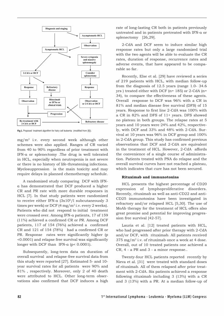
82 1 st International Lymphoma - Leukemia - Myeloma (LLM) Congress
mg/m2 i.v. every second week although other schemes were also applied. Ranges of CR varied from 40 to 90% regardless of prior treatment with IFN-α or splenectomy .The drug is well tolerated in HCL, especially when neutropenia is not severe or there is no history of life-threatening infections. Myelosuppression is the main toxicity and may require delays in planned chemotherapy schedule.
A randomized study comparing DCF with IFN-α has demonstrated that DCF produced a higher CR and PR rate with more durable responses in HCL [7]. In that study patients were randomized to receive either IFN-α (3x10⁶/l subcutaneously 3 times per week) or DCF (4 mg/m2 i.v. every 2 weeks). Patients who did not respond to initial treatment were crossed over. Among IFN-α patients, 17 of 159 (11%) achieved a confirmed CR or PR. Among DCF patients, 117 of 154 (76%) achieved a confirmed CR and 121 of 154 (78%) had a confirmed CR or PR. Response rates were significantly higher (p <0.0001) and relapse free survival was significantly longer with DCF than IFN-α (p< 0.0001).
Subsequently, long-term data on duration of overall survival and relapse-free survival data from this study were reported [27]. Estimated 5- and 10-year survival rates for all patients were 90% and 81% , respectively. Moreover, only 2 of 40 death were attributed to HCL. Other long-term obser-vations also confirmed that DCF induces a high
rate of long-lasting CR both in patients previously untreated and in patients pretreated with IFN-α or splenectomy [26,29].
2-CdA and DCF seem to induce similar high response rates but only a large randomized trial with the two agents will be able to evaluate the CR rates, duration of response, recurrence rates and adverse events, that have appeared to be compa-rable so far.
Recently, Else et al. [29] have reviewed a series of 219 patients with HCL, with median follow-up from the diagnosis of 12.5 years (range 1.0- 34.6 yrs ) treated either with DCF (n= 185) or 2-CdA (n= 34), to compare the effectiveness of these agents. Overall response to DCF was 96% with a CR in 81% and median disease free survival (DFS) of 15 years. Response to first line 2-CdA was 100% with a CR in 82% and DFS of 11+ years. DFS showed no plateau in both groups. The relapse rates at 5 years and 10 years were 24% and 42%, respective-ly, with DCF and 33% and 48% with 2-CdA. Sur-vival at 10 years was 96% in DCF group and 100% in 2-CdA group. This study has confirmed previous observations that DCF and 2-CdA are equivalent in the treatment of HCL. However, 2-CdA affords the convenience of a single course of administra-tion. Patients treated with PNA do relapse and the overall survival curves have not reached a plateau, which indicates that cure has not been secured.
Rituximab and immunotoxins
HCL presents the highest percentage of CD20 expression of lymphoproliferative disorders. Recently, rituximab as well as anti-CD22 and anti-CD25 immunotoxins have been investigated in refractory and/or relapsed HCL [5,30]. The use of MoAb therapy for the treatment of HCL offers also great promise and potential for improving progres-sion free survival [42-57].
Lauria et al. [12] treated patients with HCL, who had progressed after prior therapy with 2-CdA and/or DCF, with rituximab. All patients received 375 mg/m2 i.v. of rituximab once a week at 4 dose. Overall, out of 10 treated patients one achieved a CR, 4 - a PR and 3 - a minor response..
Twenty-four HCL patients reported recently by Nieva et al. [31] were treated with standard doses of rituximab. All of them relapsed after prior treat-ment with 2-CdA. Six patients achieved a response following rituximab including 3 (13%) with a CR and 3 (13%) with a PR. At a median follow-up of
Fig 1. Proposed treatment algorithm for hairy cell leukemia (modified from [8]).
Asymptomatic HCL
Diagnosis and staging
Symptomatic HCL
Severe cytopenia and massive
splenomegaly
2-CdA or DCF
Observation
Refractory disease
Response
Observation Rituximab or IFNα or
clinical trials
Consider splenectomy
Response
Observation or 2-CdA or DCF
Re-treatment with 2-CdA or DCF
Progression
Refractory disease
831st International Lymphoma - Leukemia - Myeloma (LLM) Congress
14.6 months, 2 responders relapsed. Even better result were presented by Thomas et al. [32]. Fifteen patients with relapsed or primary refractory HCL after 2-CdA or DCF received rituximab 375 mg/m2 weekly at a total of 8 planned doses. The overall response rate was 80% including 53% CR.
Rituximab seems to be an ideal drug for elimi-nation of MRD in HCL patients after treatment with PNA . Cervetti et al. [33] evaluated a role of this antibody in 10 patients previously treated with 2-CdA. Before starting rituximab, two patients were in CR, six in PR and two showed no signifi-cant response to 2-CdA. Rituximab was infused once a week at a dose of 375 mg/m2 for four doses. Two months after the end of rituximab therapy, all evaluated patients were in hematological CR. Moreover rituximab increased percentage of molec-ular remission up to 100% one year after the end of treatment. These results have been recently confirmed by Ravandi et al. [14]. They have treated 13 patients, 11 with newly diagnosed disease and two after failure of one prior therapy with 2-CdA at a dose of 5.6 mg/m2 daily for 5 days followed by rituximab at a dose of 375 mg/m2 weekly for 8 weeks. All patients achieved a CR, and MRD was eradicated in 12 (92%) of the patients. Promising results in patients with resistance to purine analogues have been obtained with two immunotoxins BL22 and LMB-2 targeting CD22 and CD25, respectively [13,34,35]. Recently, Kreitman et al. [13] have reported the results of their study testing the safety and efficacy of recom-binant immunotoxin containing anti-CD22 vari-able domain (Fv) fused to truncated Pseudomonas exotoxin (RF B4 (dsFv)-PE-38) named BL22 . Six-teen HCL patients who were resistant to 2-CdA were included into the study. All patients had cir-culating hairy cells that expressed CD22. BL22 at doses between 0.2-4.0 mg was administered as a 30 min intravenous infusion every other day to a total of 3 doses. Of 16 patients treated with BL22, 11 (69%) had a CR and 2 had a PR. Common toxic effect was transient hypoalbumina and the elevat-ed aminotransferase level. Two patients developed reversible hemolytic uremic syndrome.
Current indications for splenectomy and interferon-α
With the introduction of PNA, the indication for splenectomy is extremely limited. The removal of the spleen may be appropriate in patients with resistant massive symptomatic splenomegaly or
results in severe cytopenia following PNA therapy [60], and when progressive HCL developed dur-ing pregnancy [36,37]. Splenectomy may be also required for diagnostic reasons in patients with primary splenic HCL and in rare patients refractory to PNA, IFN-α and MoAb . Splenic rupture is also rare indication for splenectomy [58].
IFN-α may still have a place in HCL in preg-nancy [64] and in the patients presenting with very severe neutropenia to increase neutrophil count prior to PNA therapy [38,39]. There is no significant role for IFN-α in improving the propor-tion and the duration of CR in HCL patients previ-ously treated with DCF. In the study performed by Marotta et al [40] 135 patients who obtained CR or PR after treatment with DCF were randomized to receive IFN-α or not. Progression of disease was observed in 8 and 12 patients, with a median time of 27.8 and 26.9 months, respectively.
Conclusions
Treatment of progressive symptomatic HCL includes a variety of pharmacological approaches such as INF-α, DCF and 2-CdA, which have signifi-cantly improved the disease prognosis. 2-CdA and DCF seem to induce a similar high response rate and a long overall survival. They are also active in relapsed patients. About 80% of patients treated with 2-CdA or DCF survive for at least 10 years. In patients, who failed PNA therapy, anti-CD20 MoAb rituximab and anti-CD22 (BL22) immuno-toxin appear extremely effective. Future studies should be directed to optimizing the therapy for minimal residual disease as well as whether the introduction of new effective agents will translate into cure. A proposed flow-chart for therapeutic decision-making in the treatment of HCL is out-lined in Fig 1.
Acknowledgement. This work was supported in part by the grant from the Medical University of Lodz (No 503-1093-1 ) and by the Foundation for the Development of Diagnostics and Therapy, Warsaw, Poland.
References 1. Pettitt AR, Zuzel M, Cawley JC. Hairy cell leukemia:
biology and management. Br J Haematol. 1999; 106: 2-8.
2. Mey U, Strehl J, Gorschluter M, et al. Advances in the treatment of hairy cell leukaemia Lancet Oncol. 2003; 4: 86-94.
3. Goodman GR, Bethel KJ, Saven A. Hairy cell leu-kemia: an update. Curr Opin Hematol. 2003, 10: 258-66.
84 1 st International Lymphoma - Leukemia - Myeloma (LLM) Congress
4. Habermann TM. Splenectomy, interferon and treat-ments of historical interest in hairy cell leukemia. Hematol Oncol Clin North Am. 2006:20;1075-86.
5. Robak T. Current treatment options in hairy cell leukemia and hairy cell leukemia variant. Cancer Treat Rev. 2006; 32: 365-76.
6. Seymour JF, Estey EH, Keating MJ, et al. Response to interferon alpha in patients with hairy cell leuke-mia relapsing after treatment with 2-chlorodeoxya-denosine. Leukemia 1995; 9: 929-32.
7. Grever M, Kopecky K, Foucar MK, et al. Randomized comparison of pentostatin versus interferon alfa-2a in previously untreated patients with hairy cell leu-kemia: an Intergroup study. J Clin Oncol. 1995; 13: 974-82.
8. Belani R, Saven A. Cladribine in hairy cell leukemia. Hematol Oncol Clin North Am. 2006; 20: 1109-23.
9. Grever MR. Pentostatin: impact in outcome in hairy cell leukemia. Hematol Oncol Clin North Am. 2006; 20: 1099-108.
10. Juliusson G, Liliemark J. Purine analogues: ratio-nale for development, mechanisms of action, and pharmacokinetics in hairy cell leukemia. Hematol. Oncol Clin North Am. 2006; 20: 1087-97.
11. Robak T, Lech-Marańda E, Korycka A, Robak E. Purine nucleoside analogs as immunosuppressive and antineoplastic agents: mechanism of action and clinical activity. Curr Med Chem. 2006; 13: 3165-89.
12. Lauria F, Lenoci M, Annino L, et al. Efficacy of anti CD20 monoclonal antibodies (Mabthera) in patients with progressed hairy cell leukemia. Haematologica 2001; 86: 1046-50.
13. Kreitman RJ, Wilson WH, Bergeron K, et al. Efficacy of the anti CD22 recombinant immunotoxin BL22 in chemotherapy resistant hairy cel leukemia. N Engl J Med. 2001; 345: 241-7.
14. Ravandi F, Jorgensen JL, O’Brien SM et al. Eradica-tion of minimal residual disease (MRD) in hairy cell leukemia (HCL). Blood 2006; 107: 4658-4662.
15. Chadha P, Rademaker AW, Mendiratta P, et al. Treatment of hairy cell leukemia with 2-chlorode-oxyadenosine (2-CdA): long-term follow-up of the Northwestern University experience. Blood 2005; 106: 241-6.
16. Cheson BD, Sorensen JM, Vena DA. Treatment of hairy cell leukemia with 2-chlorodeoxyadenosine via the Group C Protocol mechanism of the Natio-nal Cancer Institute: a report of 979 ptients. J Clin Oncol. 1998; 16: 3007-15.
17. Juliusson G, Lenkei R, Liliemark J. Flow cytometry of blood and bone marrow cells from patients with hairy cell leukemia: phenotype of hairy cell and lymphocyte subsets after treatment with 2-chloro-deoxyadenosie. Blood 1994; 83: 3672-81.
18. Robak T, Błasińska-Morawiec M, Krykowski E, et al. 2-chlorodeoxyadenosine (2-CdA) in 2-hour ver-sus 24-hour intravenous infusion in the treatment of patients with hairy cell leukemia. Leuk Lympho-ma 1996; 22: 107-11.
19. von Rohr A, Schmitz SF, Tichelli A, et al. Treatment of hairy cell leukemia with cladribine (2-chlorode-oxyadenosine) by subcutaneous bolus injection: a phase II study. Ann. Oncol. 2002; 13: 1641-9.
20. Goodman GR, Burian C, Koziol JA, Saven A. Exten-ded follow-up of patients with hairy cell leukemia after treatment with cladribine J Clin Oncol. 2003; 21: 891-6.
21. Robak T, Błasińska-Morawiec M, Błoński J, et al. 2-chlorodeoxyadenosine: (cladribine) in the treatment of hairy cell leukemia and hairy cell leukemia vari-ant 7-year experience in Poland. Eur J Haematol. 1999; 62: 49-56.
22. Lauria F, Boachia M, Marotta G, Respadori D, Zinzani PL, Rondelli D. Weekly administration of 2-chlorodeoxyadenosine in patients with hairy cell leukemia is effective and reduces infectious compli-cations. Haematologica 1999; 84: 22-5.
23. Robak T, Jamroziak K, Góra-Tybor J, et al. Cladribi-ne weekly versus daily schedule for untreated hairy cell leukemia: final report from the Polish Adult Leu-kemia Group (PALG) of a prospective , randomized multicenter trial. Blood 2007; 109: Ehed of print.
24. Juliusson G, Heldal D, Hippe E, et al. Subcutaneous injections of 2-chlorodeoxya-denosine for symptomatic hairy cell leukemia. J Clin Oncol. 1995; 13: 989-95.
25. Spiers AS, Moore D, Cassileth PA, et al. Remissions in hairy cell leukemia with pentostatin (2’-deoxyco-formycin). N Engl J Med. 1987; 316: 825-30.
26. Dearden CR, Matutes E, Hilditch BT, Swansbury GJ, Catovsky D. Long term follow-up patients with hairy cell leukemia after treatment with pentostatin or cladribine. Br J Haematol. 1999; 106: 515-9.
27. Flinn IW, Kopecky KJ, Foucar MK, et al. Long-term follow-up of remission duration, mortality and second malignancies in hairy cell leukemia patients treated with pentostatin. Blood 2000; 96: 2981-6.
28. Maloisel F, Benboubker L, Gardembas M, et al. Long-term outcome with pentostatin treatment in hairy cell leukemia patients. A French retrospective study of 238 patients. Leukemia 2003; 17: 45-51.
29. Else M, Ruchlemer R, Osuji N, et al. Long remissions in hairy cell leukemia with purine analogs. A report of 219 patients with a median follow-up of 12.5 years. Cancer 2005; 104: 2442-2448.
30. Thomas DA, Revandi F, Kantarjian H. Monoclonal antibody therapy for hairy cell leukemia. Hematol Oncol Clin North Am. 2006; 20: 1125-36.
31. Nieva J, Bethel K, Saven A. Phase 2 study of rituxi-mab in the treatment of cladribine-failed patients with hairy cell leukemia. Blood 2003; 102: 810-3.
32. Thomas DA, O’Brien S, Bueso-Ramos C. et al. Rituximab in relapsed or refractory hairy cell leuke-mia. Blood 2003; 102: 3906-11.
33. Cervetti G, Galimberti S, Andreazzoli F. et al. Rituximab as treatment for minimal residual dise-ase in hairy cell leukaemia. Eur J Haematol. 2004; 73: 412-7.
851st International Lymphoma - Leukemia - Myeloma (LLM) Congress
34. Kreitman RJ, Wilson WH, White JD. et al. Phase I trial of recombinant immunotoxin anti-Tac (Fv) PE38 (LMB-2) in patients with hematologic malig-nancies. J Clin Oncol. 2000; 18: 1622-36.
35. Kreitman RJ, Pastan I. Immunotoxins in the tre-atment of refractory hairy cell leukemia. Hematol Oncol Clin North Am. 2006; 20: 1137-51.
36. Zakarija A, Peterson LC, Tallman MS. Splenektomy and treatment of historical interest. Best Pract Res Clin Haematol. 2003; 16: 57-68.
37. Stiles GM, Stanco LM, Saven A, Hoffmann KD. Sple-nectomy for hairy cell leukemia in pregnancy. J Perinatol 1998; 18: 200-1.
38. Ahmed S, Rai KR. Interferon in the treatment of hairy cell leukemia. Best Pract Res Clin. Haematol. 2003; 16: 69-81.
39. Haberman TM, Andersen JW, Cassileth PA, Ben-nett JM, Oken MM. Sequential administration of recombinant interferon-α and deoxycoformycin in the treatment of hairy cell leukemia. Br J Haematol. 1992; 80: 466-71.
40. Marotta G, Frassoldati A, Zinzani P, et al. Role of interferon-alpha administration after 2-deoxyco-formycin in the treatment of hairy cell leukemia patients. Eur J Haematol. 2006; 77: 109-13.
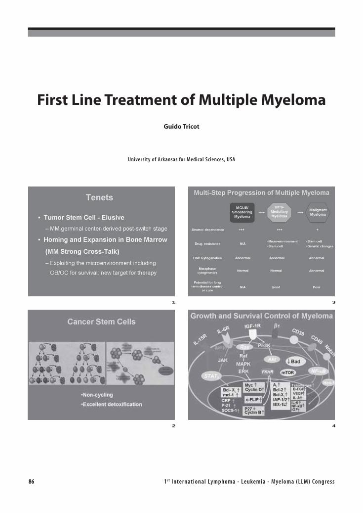
86 1 st International Lymphoma - Leukemia - Myeloma (LLM) Congress
First Line Treatment of Multiple Myeloma
Guido Tricot
University of Arkansas for Medical Sciences, USA
1
2
3
4
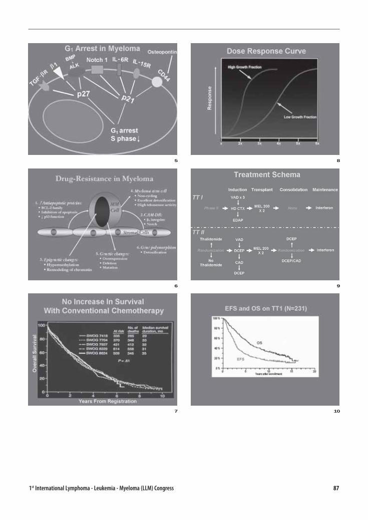
871st International Lymphoma - Leukemia - Myeloma (LLM) Congress
5
6
7
8
9
10
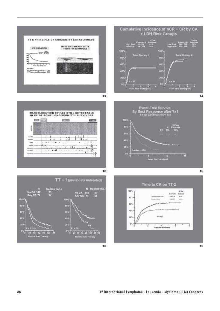
88 1 st International Lymphoma - Leukemia - Myeloma (LLM) Congress
11
12
13
14
15
16

891st International Lymphoma - Leukemia - Myeloma (LLM) Congress
17
18
19
20
21
22
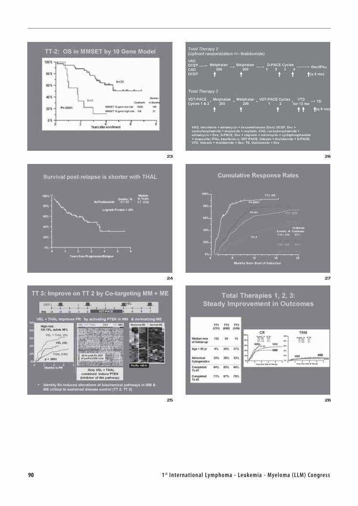
90 1 st International Lymphoma - Leukemia - Myeloma (LLM) Congress
23
24
25
26
27
28

911st International Lymphoma - Leukemia - Myeloma (LLM) Congress
29
30
31
32

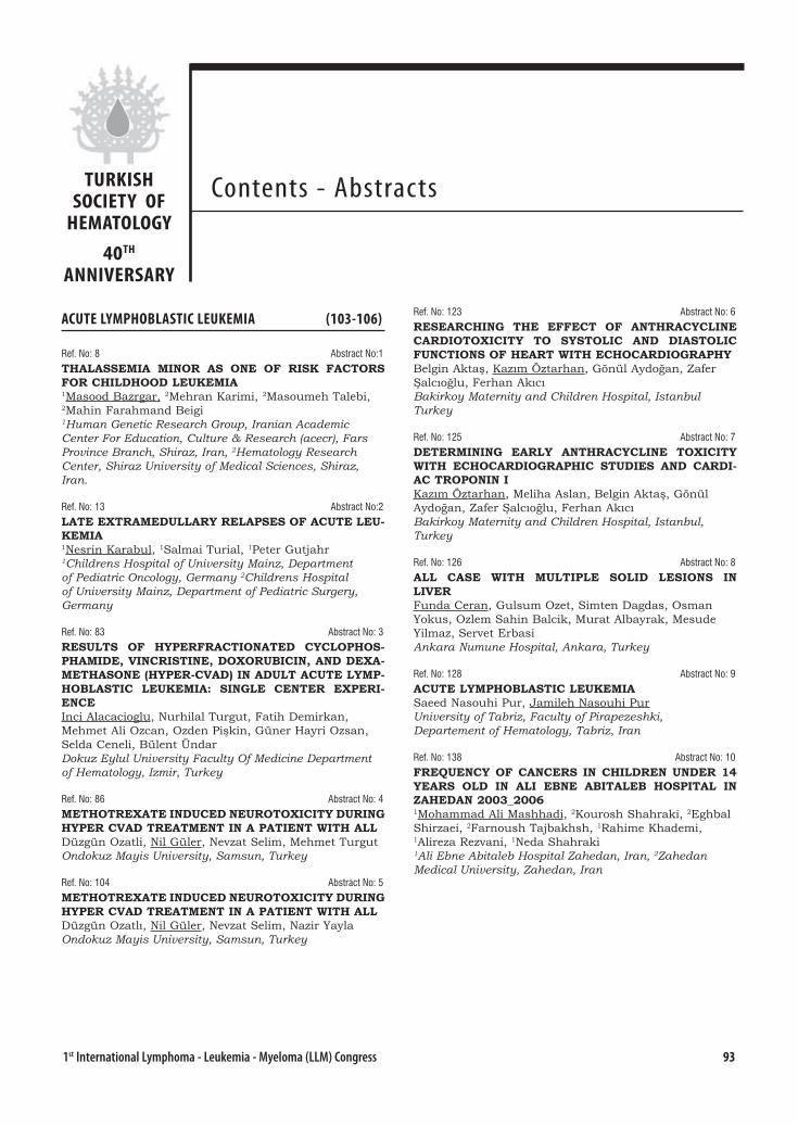
931st International Lymphoma - Leukemia - Myeloma (LLM) Congress
Contents - AbstractsTURKISH SOCIETY OF
HEMATOLOGY
40TH
ANNIVERSARY
ACUTE LYMPHOBLASTIC LEUKEMIA (103-106)
Ref. No: 8 Abstract No:1
THALASSEMIA MINOR AS ONE OF RISK FACTORS FOR CHILDHOOD LEUKEMIA1Masood Bazrgar, 2Mehran Karimi, 2Masoumeh Talebi, 2Mahin Farahmand Beigi1Human Genetic Research Group, Iranian Academic Center For Education, Culture & Research (acecr), Fars Province Branch, Shiraz, Iran, 2Hematology Research Center, Shiraz University of Medical Sciences, Shiraz, Iran.
Ref. No: 13 Abstract No:2
LATE EXTRAMEDULLARY RELAPSES OF ACUTE LEU-KEMIA1Nesrin Karabul, 1Salmai Turial, 1Peter Gutjahr1Childrens Hospital of University Mainz, Department of Pediatric Oncology, Germany 2Childrens Hospital of University Mainz, Department of Pediatric Surgery, Germany
Ref. No: 83 Abstract No: 3
RESULTS OF HYPERFRACTIONATED CYCLOPHOS-PHAMIDE, VINCRISTINE, DOXORUBICIN, AND DEXA-METHASONE (HYPER-CVAD) IN ADULT ACUTE LYMP-HOBLASTIC LEUKEMIA: SINGLE CENTER EXPERI-ENCEInci Alacacioglu, Nurhilal Turgut, Fatih Demirkan, Mehmet Ali Ozcan, Ozden Pişkin, Güner Hayri Ozsan, Selda Ceneli, Bülent ÜndarDokuz Eylul University Faculty Of Medicine Department of Hematology, Izmir, Turkey
Ref. No: 86 Abstract No: 4
METHOTREXATE INDUCED NEUROTOXICITY DURING HYPER CVAD TREATMENT IN A PATIENT WITH ALLDüzgün Ozatli, Nil Güler, Nevzat Selim, Mehmet TurgutOndokuz Mayis University, Samsun, Turkey
Ref. No: 104 Abstract No: 5
METHOTREXATE INDUCED NEUROTOXICITY DURING HYPER CVAD TREATMENT IN A PATIENT WITH ALLDüzgün Ozatlı, Nil Güler, Nevzat Selim, Nazir YaylaOndokuz Mayis University, Samsun, Turkey
Ref. No: 123 Abstract No: 6
RESEARCHING THE EFFECT OF ANTHRACYCLINE CARDIOTOXICITY TO SYSTOLIC AND DIASTOLIC FUNCTIONS OF HEART WITH ECHOCARDIOGRAPHYBelgin Aktaş, Kazım Öztarhan, Gönül Aydoğan, Zafer Şalcıoğlu, Ferhan AkıcıBakirkoy Maternity and Children Hospital, Istanbul Turkey
Ref. No: 125 Abstract No: 7
DETERMINING EARLY ANTHRACYCLINE TOXICITY WITH ECHOCARDIOGRAPHIC STUDIES AND CARDI-AC TROPONIN IKazım Öztarhan, Meliha Aslan, Belgin Aktaş, Gönül Aydoğan, Zafer Şalcıoğlu, Ferhan AkıcıBakirkoy Maternity and Children Hospital, Istanbul, Turkey
Ref. No: 126 Abstract No: 8
ALL CASE WITH MULTIPLE SOLID LESIONS IN LIVERFunda Ceran, Gulsum Ozet, Simten Dagdas, Osman Yokus, Ozlem Sahin Balcik, Murat Albayrak, Mesude Yilmaz, Servet ErbasiAnkara Numune Hospital, Ankara, Turkey
Ref. No: 128 Abstract No: 9
ACUTE LYMPHOBLASTIC LEUKEMIASaeed Nasouhi Pur, Jamileh Nasouhi PurUniversity of Tabriz, Faculty of Pirapezeshki, Departement of Hematology, Tabriz, Iran
Ref. No: 138 Abstract No: 10
FREQUENCY OF CANCERS IN CHILDREN UNDER 14 YEARS OLD IN ALI EBNE ABITALEB HOSPITAL IN ZAHEDAN 2003_20061Mohammad Ali Mashhadi, 2Kourosh Shahraki, 2Eghbal Shirzaei, 2Farnoush Tajbakhsh, 1Rahime Khademi, 1Alireza Rezvani, 1Neda Shahraki1Ali Ebne Abitaleb Hospital Zahedan, Iran, 2Zahedan Medical University, Zahedan, Iran
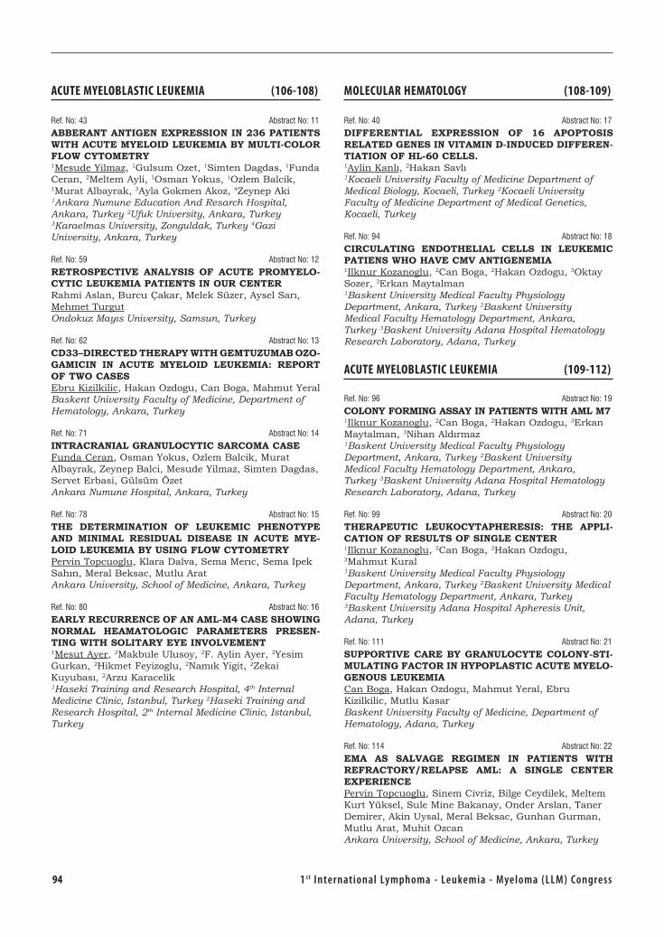
94 1 st International Lymphoma - Leukemia - Myeloma (LLM) Congress
ACUTE MYELOBLASTIC LEUKEMIA (106-108)
Ref. No: 43 Abstract No: 11
ABBERANT ANTIGEN EXPRESSION IN 236 PATIENTS WITH ACUTE MYELOID LEUKEMIA BY MULTI-COLOR FLOW CYTOMETRY1Mesude Yilmaz, 1Gulsum Ozet, 1Simten Dagdas, 1Funda Ceran, 2Meltem Ayli, 1Osman Yokus, 1Ozlem Balcik, 1Murat Albayrak, 3Ayla Gokmen Akoz, 4Zeynep Aki1Ankara Numune Education And Resarch Hospital, Ankara, Turkey 2Ufuk University, Ankara, Turkey 3Karaelmas University, Zonguldak, Turkey 4Gazi University, Ankara, Turkey
Ref. No: 59 Abstract No: 12
RETROSPECTIVE ANALYSIS OF ACUTE PROMYELO-CYTIC LEUKEMIA PATIENTS IN OUR CENTERRahmi Aslan, Burcu Çakar, Melek Süzer, Aysel Sarı, Mehmet TurgutOndokuz Mayıs University, Samsun, Turkey
Ref. No: 62 Abstract No: 13
CD33–DIRECTED THERAPY WITH GEMTUZUMAB OZO-GAMICIN IN ACUTE MYELOID LEUKEMIA: REPORT OF TWO CASESEbru Kizilkilic, Hakan Ozdogu, Can Boga, Mahmut YeralBaskent University Faculty of Medicine, Department of Hematology, Ankara, Turkey
Ref. No: 71 Abstract No: 14
INTRACRANIAL GRANULOCYTIC SARCOMA CASE Funda Ceran, Osman Yokus, Ozlem Balcik, Murat Albayrak, Zeynep Balci, Mesude Yilmaz, Simten Dagdas, Servet Erbasi, Gülsüm ÖzetAnkara Numune Hospital, Ankara, Turkey
Ref. No: 78 Abstract No: 15
THE DETERMINATION OF LEUKEMIC PHENOTYPE AND MINIMAL RESIDUAL DISEASE IN ACUTE MYE-LOID LEUKEMIA BY USING FLOW CYTOMETRY Pervin Topcuoglu, Klara Dalva, Sema Merıc, Sema Ipek Sahın, Meral Beksac, Mutlu AratAnkara University, School of Medicine, Ankara, Turkey
Ref. No: 80 Abstract No: 16
EARLY RECURRENCE OF AN AML-M4 CASE SHOWING NORMAL HEAMATOLOGIC PARAMETERS PRESEN-TING WITH SOLITARY EYE INVOLVEMENT 1Mesut Ayer, 2Makbule Ulusoy, 2F. Aylin Ayer, 2Yesim Gurkan, 2Hikmet Feyizoglu, 2Namık Yigit, 2Zekai Kuyubası, 2Arzu Karacelik1Haseki Training and Research Hospital, 4th Internal Medicine Clinic, Istanbul, Turkey 2Haseki Training and Research Hospital, 2th Internal Medicine Clinic, Istanbul, Turkey
MOLECULAR HEMATOLOGY (108-109)
Ref. No: 40 Abstract No: 17
DIFFERENTIAL EXPRESSION OF 16 APOPTOSIS RELATED GENES IN VITAMIN D-INDUCED DIFFEREN-TIATION OF HL-60 CELLS. 1Aylin Kanlı, 2Hakan Savlı1Kocaeli University Faculty of Medicine Department of Medical Biology, Kocaeli, Turkey 2Kocaeli University Faculty of Medicine Department of Medical Genetics, Kocaeli, Turkey
Ref. No: 94 Abstract No: 18
CIRCULATING ENDOTHELIAL CELLS IN LEUKEMIC PATIENS WHO HAVE CMV ANTIGENEMIA1Ilknur Kozanoglu, 2Can Boga, 2Hakan Ozdogu, 3Oktay Sozer, 3Erkan Maytalman1Baskent University Medical Faculty Physiology Department, Ankara, Turkey 2Baskent University Medical Faculty Hematology Department, Ankara, Turkey 3Baskent University Adana Hospital Hematology Research Laboratory, Adana, Turkey
ACUTE MYELOBLASTIC LEUKEMIA (109-112)
Ref. No: 96 Abstract No: 19
COLONY FORMING ASSAY IN PATIENTS WITH AML M7 1Ilknur Kozanoglu, 2Can Boga, 2Hakan Ozdogu, 3Erkan Maytalman, 3Nihan Aldırmaz1Baskent University Medical Faculty Physiology Department, Ankara, Turkey 2Baskent University Medical Faculty Hematology Department, Ankara, Turkey 3Baskent University Adana Hospital Hematology Research Laboratory, Adana, Turkey
Ref. No: 99 Abstract No: 20
THERAPEUTIC LEUKOCYTAPHERESIS: THE APPLI-CATION OF RESULTS OF SINGLE CENTER1Ilknur Kozanoglu, 2Can Boga, 2Hakan Ozdogu, 3Mahmut Kural1Baskent University Medical Faculty Physiology Department, Ankara, Turkey 2Baskent University Medical Faculty Hematology Department, Ankara, Turkey 3Baskent University Adana Hospital Apheresis Unit, Adana, Turkey
Ref. No: 111 Abstract No: 21
SUPPORTIVE CARE BY GRANULOCYTE COLONY-STI-MULATING FACTOR IN HYPOPLASTIC ACUTE MYELO-GENOUS LEUKEMIACan Boga, Hakan Ozdogu, Mahmut Yeral, Ebru Kizilkilic, Mutlu KasarBaskent University Faculty of Medicine, Department of Hematology, Adana, Turkey
Ref. No: 114 Abstract No: 22
EMA AS SALVAGE REGIMEN IN PATIENTS WITH REFRACTORY/RELAPSE AML: A SINGLE CENTER EXPERIENCEPervin Topcuoglu, Sinem Civriz, Bilge Ceydilek, Meltem Kurt Yüksel, Sule Mine Bakanay, Onder Arslan, Taner Demirer, Akin Uysal, Meral Beksac, Gunhan Gurman, Mutlu Arat, Muhit OzcanAnkara University, School of Medicine, Ankara, Turkey
951st International Lymphoma - Leukemia - Myeloma (LLM) Congress
Ref. No: 116 Abstract No: 23
ACUTE MYELOID LEUKEMIA PRESENTING AS ACUTE INFERIOR MYOCARDIAL INFARCTIONMahmut Yeral, Mutlu Kasar, Hakan Ozdogu, Can BogaBaskent University Faculty of Medicine, Department of Hematology, Adana, Turkey
Ref. No: 120 Abstract No: 24
EXPRESSION OF UROKINASE PLASMINOGEN ACTI-VATOR RECEPTOR IN ACUTE MYELOID LEUKEMIAFunda Ceran, Simten Dagdas, Gulsum Ozet, Mesude Yilmaz, Murat Albayrak, Osman Yokus, Ozlem Sahin BalcikAnkara Numune Hospital, Ankara, Turkey
Ref. No: 122 Abstract No: 25
STANDART AND DOSE MODIFIED ARA-C/IDARUBICIN REMISSION INDUCTION THERAPY IN ELDERLY DE-NOVO ACUTE MYELOID LEUKEMIA PATIENTS: A RET-ROSPECTIVE ANALYSIS FROM “DENIZLI LEUKEMIA-LYMPHOMA-MYELOMA STUDY GROUP” (DLLMSG)1Ali Keskin, 1Sibel Kabukcu Hacioglu, 1Ismail Sari, 2Burcu Yapar, 3Nilay Sen, 4Sami Karti1Pamukkale University, Faculty of Medicine, Department of Hematology, Denizli, Turkey 2Pamukkale University, Faculty of Medicine, Department of Internal Medicine, Denizli, Turkey 3Pamukkale University, Faculty of Medicine, Department of Pathology, Denizli, Turkey 4Denizli Education and Research Hospital, Hematology Unit, Denizli, Turkey
Ref. No: 140 Abstract No: 26
NEPHROTIC SYNDROME AS THE FIRST MANIFESTA-TION OF ACUTE MYELOGENOUS LEUKEMIA: CASE REPORT1Mohammad Ali Mashhadi, 2Kourosh Shahraki1Ali Ebne Abitaleb Hospital Zahedan, Iran, 2Zahedan Medical University, Zahedan, Iran
CHRONIC MYELOID LEUKEMIA (112-116)
Ref. No: 30 Abstract No: 27
ORAL AND CUTANEUS LICHENOID REACTION SECONDARY TO STANDARD DOSE IMATINIB: A CASE REPORTServet Erbasi, Muhterem Polat, Murat Albayrak, Ferda Artuz, Osman Yokus, Pınar Oztas, Funda CeranAnkara Numune Hastahanesi, Ankara, Turkey
Ref. No: 34 Abstract No: 28
CYTOGENETIC AND CLINICAL FINDINGS OF CHRO-NIC LYMHOCYTIC LEUKEMIA PATIENTS1Gül Ilhan, 1Neslihan Andıç, 1Sema Karakuş, 1Ebru Kızılkılıç, 1Hakan Özdoğu, 2Feride Şahin1Baskent University, Department of Hematology, Adana, Turkey 2Baskent Uiversity, Department of Medical Biology and Genetics, Ankara, Turkey
Ref. No: 48 Abstract No: 29
BOTH KAPPA AND LAMBDA POSITIVE B- CHRONIC LYMPHOCYTIC LEUKEMIA: A CASE REPORT1Servet Erbasi, 1Mesude Yılmaz, 1Gulsum Ozet, 1Osman Yokus, 1Ozlem Balcık, 1Funda Ceran, 1Aynur Albayrak1ANEAH Hematology, Ankara, Turkey 2ANEAH 2. Pathology, Ankara, Turkey
Ref. No: 93 Abstract No: 30
GOOD RESPONSE TO FLUDARABINE IN PATIENT WITH T-CELL LARGE GRANULAR LYMPHOCYTIC LEUKEMIA 1Mahmut Töbü, 2Burhanettin Uludag, 3Mine Hekimgil1Ege University Hospital, Department of Hematology, Izmir, Turkey 2Ege University Hospital, Department of Neurology, Izmir, Turkey 3Ege University Hospital, Department of Pathology, Izmir, Turkey
Ref. No: 101 Abstract No: 31
SEVERE NEUROTOXICITY OF CLADRIBINE IN A PATI-ENT WITH HAIRY CELL LEUKEMIA: A CASE REPORTGülten Sop, Zafer Gökgöz, Tuğba Gümüş, Şermin Çoban, Füsun ÖzdemirkıranIzmir Training and Research Hospital, Izmir, Turkey
Ref. No: 130 Abstract No: 32
POSSIBLE PNEUMOCYSTIS JIROVECI INFECTION IN AN CYTOMEGALOVIRUS POSITIVE CLL PATIENTMutlu Kasar, Hakan Ozdogu, Can Boga, Mahmut YeralBaskent University Faculty of Medicine, Department of Hematology, Adana, Turkey
Ref. No: 19 Abstract No: 33
PROMINENT PLEURAL AND PERICARDIAL EFFUSION DUE TO IMATINIB MESYLATE AFTER FIVE YEARS OF THERAPYNeslihan Andıç, Gül Ilhan, Sema KarakuşBaskent University, Faculty of Medicine, Department of Internal Medicine, Hematology Division, Ankara, Turkey
Ref. No: 45 Abstract No: 34
THE CLINICAL PRESENTATION AND DIFFERENT BCR-ABL TRANSCRIPTS IN CML1Erhan Alkan, 1Evren Kiriş, 1Seray Dizlek, 2Güçhan Alanoğlu, 1Nilay Uysalgil, 1Ayşen Timurağaoğlu1Akdeniz University, School of Medicine, Antalya, Turkey 2Süleyman Demirel University, School of Medicine, Isparta, Turkey
Ref. No: 112 Abstract No: 35
FLAG-IDA IN THE TREATMENT OF REFRACTORY/RELAPSED ACUTE MYELOID LEUKEMIA: SINGLE-CENTER EXPERENCE1Hakan Ozdogu, 1Can Boga, 1Ebru Kizilkilic, 2Ilknur Kozanoglu1Baskent University Faculty Medicine, Department of Hematology, Adana, Turkey 2Baskent University Faculty of Medicine, Hematology Research Laboratory, Adana, Turkey
Ref. No: 139 Abstract No: 36
CHRONIC MYELOGENOUS LEUKEMIA OCCURRING IN MOTHER AND SONCafer Adıgüzel, Işık Kaygusuz, Elif Birtaş Ateşoğlu, Figen Noyan, Mustafa Çetiner, Emel Demiralp, Tülin Fıratlı Tuğlular, Mahmut BayıkMarmara University School of Medicine,department of Medicine, Division of Hematology, Istanbul, Turkey
96 1 st International Lymphoma - Leukemia - Myeloma (LLM) Congress
HODGKIN’S LYMPHOMA (116-118)
Ref. No: 42 Abstract No: 37
COMPARISON OF BEACOPP AND EVA TREATMENT PROTOCOLS IN REFRACTORY OR RELAPSING HODG-KIN LYMPHOMA1Oktay Bilgir, 2Ferda Bilgir, 1Mehmet Çalan, 1Pınar Öner, 3Esin Kulaç, 1Murat Akyol, 1Elif Tuna1Izmir Education And Research Hospital 2nd Internal Diseases Clinic, Izmir, Turkey 2Buca State Hospital, Internal Diseases Clinic, Izmir, Turkey 3Kastamonu City Health Center, Kastamonu, Turkey
Ref. No: 76 Abstract No: 38
HODGKIN’S DISEASE: A RETROSPECTIVE ANALYSIS OF 103 PATIENTS FROM A SINGLE REFERRAL CEN-TRE1Hava Üsküdar Teke, 1O. Meltem Akay, 1Eren Gündüz, 2Ertuğrul Çolak, 1Zafer Gülbaş1Osmangazi University Faculty of Medicine, Haematology Department, Eskisehir, Turkey 2Osmangazi University Faculty of Medicine, Biostatistic Department, Eskisehir, Turkey
Ref. No: 77 Abstract No: 39
HODGKIN’S DISEASE IN CHILDREN: DEMOGRAPHIC DATA AND RESULTS OF OUR CLINIC 1Ferhan Akici, 1Gönül Aydogan, 1Zafer Salcioglu, 1Serdar Sander, 1Deniz Tugcu, 1Arzu Akcay, 1Hulya Sen, 1Aysel Kiyak, 1Huseyin Aldemir, 2Fulya Yaman1Bakirkoy Women and Children Diseases Education Hospital, Istanbul, Turkey 2Istanbul University Oncology Institute Division of Radiation Department, Istanbul, Turkey
Ref. No: 102 Abstract No: 40
HODGKIN’S LYMPHOMA: A RETROSPECTIVE ANALY-SIS OF 44 PATIENTS Gülten Sop, Füsun Özdemirkıran, Tuğba Gümüş, Şermin ÇobanIzmir Training and Research Hospital, Izmir, Turkey
Ref. No: 136 Abstract No: 41
PULMONARY INVOLVEMENT IN HODGKIN’S DISEASE 1Mustafa Yenerel, 1Serdar Sahinoglu, 2Oner Dogan, 1Reyhan Diz-kucukkaya, 1Meliha Nalcaci1Istanbul University, Istanbul Faculty of Medicine, Department Of Internal Medicine, Division of Hematology, Istanbul, Turkey, 2Istanbul University, Istanbul Faculty of Medicine, Department of Pathology, Istanbul, Turkey
NON-HODGKIN’S LYMPHOMA (118-124)
Ref. No: 15 Abstract No: 42
CASE REPORT: MANTLE CELL LYMPHOMA WITH PULMONARY INVOLVEMENT AT PRESENTATION1Serkan Ocakci, 1Nur Akad Soyer, 1Murat Tombuloglu, 2Nazan Ozhan1Ege University, Department of Internal Medicine, Division of Hematology, Izmir, Turkey 2Ege University, Department of Pathology, Izmir, Turkey
Ref. No: 28 Abstract No: 43
A NON HODGKIN’S LYMPHOMA CASE WITH OVARIAN INVOLVEMENTAbdullah Hacıhanefioğlu, Naile Gökkaya, Pınar Tarkun, Emel Gönüllü Kocaeli University, Hematology Department, Kocaeli, Turkey
Ref. No: 51 Abstract No: 44
A CASE OF PLASMABLASTIC LYMPHOMA TRANSFOR-MED FROM T CELL LYMPHOMA. Düzgün Özatlı, Nil Güler, Burcu Çakar, Güzin Gönüllü, Idris YücelOndokuz Mayis University, Samsun, Turkey
Ref. No: 56 Abstract No: 45
INCIDENTAL DIAGNOSIS OF THYROID PAPILLARY CANCER IN A ANGIOIMMUNOBLASTIC LYMPHOMA PATIENT BY FDG-PET1Mehmet Turgut, 1Müge Karaoğlanoğlu, 1Burcu Çakar, 1Recep Semiz, 1Ayşe Kevser Gökçe, 2Hakan Göker1Ondokuz Mayıs University, Samsun, Turkey 2Hacettepe University, Ankara, Turkey
Ref. No: 58 Abstract No: 46
NASAL NK/T CELL LYMPHOMA: CASE REPORTArzu Ergen, Barkın Sakallıoğlu, Nergiz Dağoğlu, Yavuz Dizdar, Fulya Yaman Ağaoğlu, Emin DarendelilerIstanbul University, Medicine Faculty of Istanbul, Department of Radiation Oncology, Istanbul, Turkey
Ref. No: 82 Abstract No: 47
PRIMARY MEDIASTINAL B-CELL NON-HODGKIN’S LYMPHOMA PRESENTED WITH CARDIAC INVOLVE-MENT1Inci Alacacioglu, 1Nurhilal Turgut, 1Guner Hayri Ozsan, 1Ozden Piskin, 1Mehmet Ali Ozcan, 1Fatih Demirkan, 2Bahri Akdeniz, 3Mustafa Secil, 2Omer Kozan, 1Bulent Ündar1Dokuz Eylül University Faculty of Medicine Department of Hematology, Izmir, Turkey 2Dokuz Eylül University Faculty of Medicine Department of Cardiology, Izmir, Turkey 3Dokuz Eylül University Faculty of Medicine Department of Radiology, Izmir, Turkey
Ref. No: 89 Abstract No: 48
THE ROLE OF RITUXIMAB ON AUTOLOGUS TRANSP-LANTATION FOR NON HODGKIN’S LYMPHOMASinem Civriz Bozdag, Pervin Topcuoglu, Ender Soydan, Mutlu Arat, Osman Ilhan, Haluk Koc, Meral Beksac, Akın Uysal, Hamdi Akan, Onder Aslan, Nahide Konuk, Muhit OzcanAnkara University Faculty of Medicine, Ankara, Turkey
Ref. No: 95 Abstract No: 49
FACS ANALYSIS OF PERIPHERAL T-CELL LYMPHO-MA 1Ilknur Kozanoglu, 2Can Boga, 2Hakan Ozdogu, 3Oktay Sozer1Baskent University Medical Faculty Physiology Department, Adana, Turkey 2Baskent University Medical Faculty Hematology Department, Adana, Turkey 3Baskent University Adana Hospital Hematology Research Laboratory, Adana, Turkey
971st International Lymphoma - Leukemia - Myeloma (LLM) Congress
Ref. No: 100 Abstract No: 50
ROLE OF HEPATITIS B VIRUS AND HEPATITIS C VIRUS INFECTIONS IN CLINICAL OUTCOMES OF NON-HODGKIN LYMPHOMA1Ramin Yaghobi, 2Mehdi Roshan Nia Jahromi, 2Mani Ramzi, 2Narges Rezaee, 2Vida Moaied1Shiraz Transplant Research Center, Shiraz, Iran 2Hematology-Oncology Research Center, Shiraz, Iran
Ref. No: 105 Abstract No: 51
LYMPHOMATOID PAPULOSIS: A CASE REPORT AND TREATMENT MODALITIES IN LOCAL THERAPY RESISTANT CASESOmer Doğru, Tiraje CelkanCerrahpasa Medical Faculty, Istanbul, Turkey
Ref. No: 119 Abstract No: 52
CLINICAL CHARACTERISTICS AND TREATMENT RESULTS OF PEDIATRIC NON-HODGKIN’S LYMPHO-MA1Ferhan Akıcı, 1Gönül Aydoğan, 1Zafer Şalcıoğlu, 1Serdar Sander, 1Deniz Tuğcu, 1Arzu Akçay, 1Hülya Şen, 1Aysel Kıyak, 1Hüseyin Aldemır, 2Öner Doğan1Ministry of Health Bakırköy Women And Children Diseases Education Hospital, Clinics of Pediatric Hematology-Oncology and Pediatric Surgery Istanbul, Turkey, 2Istanbul University, Division of Pathology, Istanbul, Turkey
Ref. No: 121 Abstract No: 53
CLINICOPATHOLOGIC FEATURES AND TREATMENT RESULTS OF NON-HODGKIN’S LYMPHOMAS IN ELDERLY PATIENTS: A RETROSPECTIVE ANALYSIS FROM “DENIZLI LEUKEMIA-LYMPHOMA-MYELOMA STUDY GROUP” (DLLMSG)1Sibel Kabukcu Hacioglu, 1Ismail Sari, 2Sami Karti, 3Sinemis Yuksel, 4Nilay Sen, 1Ali Keskin1Pamukkale University, Faculty of Medicine, Department of Hematology, Denizli, Turkey 2Denizli Education and Research Hospital, Hematology Unit, Denizli, Turkey 3Pamukkale University, Faculty of Medicine, Department of Internal Medicine, Denizli, Turkey 4Pamukkale University, Faculty of Medicine, Department of Pathology, Denizli, Turkey
Ref. No: 127 Abstract No: 54
ARG: A POTENTIAL BIOMARKER FOR DLBCL STA-GING1Mansoor Salehi, 1Zahra Kabiri, 2Mohammad Modaresi1Isfahan University of Medical Sciences, Isfahan, Iran 2Tehran University of Medical Sciences, Tehran, Iran
Ref. No: 131 Abstract No: 55
A CASE OF BURKITT’S LYMPHOMA WITH NECROTI-ZING GRANULOMATOUS REACTION1Evrim Kuş, 1Cengiz Erçin, 1Funda Çorapçıoğlu1Kocaeli University, School of Medicine, Department of Pathology, Kocaeli, Turkey 2Kocaeli University, School of Medicine, Department of Pediatric Hematology-Oncology, Kocaeli, Turkey
Ref. No: 133 Abstract No: 56
LYMPHOMA EXPERIENCE OF LAKES DISTRICT FROM SÜLEYMAN DEMIREL UNIVERSITY SCHOOL OF MEDI-CIINE1Güçhan Alanoğlu, 2Bülent Kara, 2Sema Sezgin Göksu, 1Nilgün Kapucuoğlu, 4Hasan Şenol Coşkun1Suleyman Demirel Universty School of Medicine Dept. of Hematology, Isparta, Turkey 2Suleyman Demirel University School of Medicine Department of Internal Medicine, Isparta, Turkey 3Suleyman Demirel University School of Medicine Department of Pathology, Isparta, Turkey 4Suleyman Demirel University School of Medicine Department of Medical Oncology, Isparta, Turkey
Ref. No: 135 Abstract No: 57
UTILITY OF PERIPHERAL BLOOD FLOW CYTOMETRY TO INVESTIGATE THE PERIPHERIZATION OF B-CELL MALIGNANT LYMPHOMASOlga Meltem Akay, Eren Gunduz, Hava U. Teke, Gulcihan Demirel, Zafer GulbasEskisehir Osmangazi University Medical School Hematology Department, Isparta, Turkey
Ref. No: 141 Abstract No: 58
EVALUATION THE RESPONSE RATE OF IEV REGIMEN AS SALVAGE THERAPY FOR RELAPSED / REFRAC-TORY NON-HODGKIN’S LYMPHOMA PATIENTS1Mohammad Ali Mashhadi, 2Kourosh Shahraki, 3Adineh Pour1Ali Ebne Abitaleb Hospital, Zahedan, Iran, 2Zahedan Medical University, Zahedan, Iran 3Resident of Internal Medicine in Zahedan Medical University, Zahedan, Iran
LYMHOPROLIFERATIVE DISORDERS (124-125)
Ref. No: 110 Abstract No: 59
OPPORTUNISTIC INFECTIONS IN CASES WITH HAIRY CELL LEUKEMIA Mahmut Yeral, Hakan Ozdogu, Can BogaBaskent University Faculty of Medicine, Department of Hematology, Ankara, Turkey
Ref. No: 137 Abstract No: 60
AUTOIMMUNE HEMOLYTIC ANEMIA AFTER CLADRI-BINE THERAPY FOR HAIRY CELL LEUKEMIAMustafa Yenerel, Esra Hatipoğlu, Abdullah Ozkök, Tanju AtamerIstanbul University, Istanbul Faculty of Medicine, Department of Internal Medicine, Division of Hematology, Istanbul, Turkey
CHRONIC LYMPHOCYTIC LEUKEMIA (125)
Ref. No: 129 Abstract No: 61
CD4- CD8+ T-CELL PROLYMPHOCYTIC LEUKEMIA: A REPORT OF TWO CASESCan Boga, Oktay Sozer, Suheyl Asma, Hakan OzdoguBaskent University Faculty of Medicine, Department of Hematology, Ankara, Turkey
98 1 st International Lymphoma - Leukemia - Myeloma (LLM) Congress
MULTIPLE MYELOMA (125-134)
Ref. No: 10 Abstract No: 62
A PROTEAZOM INHIBITOR IN THE TREATMENT OF MULTIPLE MYELOMA: BORTEZOMIB Ozlem Sahin Balcik, Simten Dagdas, Murat Albayrak, Osman Yokuş, Funda Ceran, Servet Erbasi, Gulsum OzetAnkara Numune Educational and Research Hospital Hematology Department, Ankara, Turkey
Ref. No: 11 Abstract No: 63
GENETIC ABNORMALITIES IN MULTIPLE MYELOMA, THEIR PREVELANCE AND RELATION WITH OTHER RISK FACTORSOzlem Sahin Balcik, Murat Albayrak, Simten Dagdas, Funda Ceran, Osman Yokuş, Gulsum OzetAnkara Numune Educational and Research Hospital Hematology Department, Ankara, Turkey
Ref. No: 17 Abstract No: 64
TC-99M MIBI OR F-18 FDG IMAGING?: A COMPA-RATIVE STUDY FOR EVALUATING PATIENTS WITH MULTIPLE MYELOMA1Ilknur Ak, 1Inci Uslu, 2Zafer Gülbaş1Eskisehir Osmangazi University Medical Faculty Department of Nuclear Medicine, Eskisehir, Turkey 2Eskisehir Osmangazi University Medical Faculty Department of Haematology, Eskisehir, Turkey
Ref. No: 24 Abstract No: 65
BORTEZOMIB AND DEXAMETHASONE INDUCED TUMOR LYSIS SYNDROME IN A CASE OF PLASMA CELL LEUKEMIA Gül Ilhan, Neslihan Andıç, Sema KarakuşBaskent University, Hematology Department, Ankara, Turkey
Ref. No: 38 Abstract No: 66
SUCCESSFUL TREATMENT OF EARLY RELAPSE OF OCULAR MYELOMA WITH BORTEZOMIB AND STEROID AFTER AUTOLOGOUS STEM CELL TRANSPLANTATION 1Irfan Yavasoglu, 2Tolga Kocaturk, 1Gurhan Kadikoylu, 2Volkan Dayanır, 3Yelda Dayanır, 1Zahit Bolaman1Adnan Menderes University, Medical Faculty, Hematology, Aydın, Turkey 2Adnan Menderes University, Medical Faculty, Opthalomology, Aydın, Turkey 3Adnan Menderes University, Medical Faculty, Radiology, Aydın, Turkey
Ref. No: 44 Abstract No: 67
COMBINED THERAPY WITH BORTEZOMIB AND DEXA-METHASONE IN PATIENTS WITH RELAPSING MUL-TIPLE MYELOMA1Oktay Bilgir, 2Ferda Bilgir, 1Mehmet Çalan, 1Pınar Öner, 1Murat Akyol, 1Elif Tuna1Izmir Educational and Research Hospital, Izmir, Turkey 2Buca State Hospital, Internal Diseases Clinic, Izmir, Turkey
Ref. No: 66 Abstract No: 68
BORTEZOMIB EFFICIENCY IN MULTIPLE MYELOMAEbru Kizilkilic, Can Boga, Hakan Ozdogu, Mahmut Yeral Baskent University Faculty of Medicine Department of Hematology, Ankara, Turkey
Ref. No: 67 Abstract No: 69
RETROSPECTIVE ANALYSIS FOR DEMOGRAPHIC FEATURES OF MULTIPLE MYELOMA PATIENTS, AKDENIZ UNIVERSITY EXPERIENCEMete Akın, Ilknur Nizam, Songul Akcan, Feyzi Bostan, Ihsan Karadogan, Aysen Timuragaoglu, Levent UndarAkdeniz University Dept. of Hematology, Antalya, Turkey
Ref. No: 68 Abstract No: 70
THE EFFECTS OF PLASMA EXCHANGE ON COAGULA-TION PARAMETERS, AND PLATELET FUNCTIONS IN PATIENTS WITH MULTIPLE MYELOMAAli Sahin, Ali Unal, Fatih Kurnaz, Leylagul Kaynar, Mehmet Oztekin, Musa Solmaz, Fevzi Altuntas, Bulent Eser, Mustafa CetinErciyes University, Medical Faculty, Hematology Department, Kayseri, Turkey
Ref. No: 69 Abstract No: 71
RETROSPECTIVE ANALYSIS FOR CLINICAL AND LABORATORY FINDINGS OF MULTIPLE MYELOMA PATIENTS, AKDENIZ UNIVERSITY EXPERIENCE Mete Akın, Ilknur Nizam, Songul Akcan, Feyzi Bostan, Ihsan Karadogan, Aysen Timuragaoglu, Levent UndarAkdeniz University Dept. of Hematology, Antalya, Turkey
Ref. No: 73 Abstract No: 72
MICROSATELLITE INSTABILITY IN PATIENTS WITH MULTIPLE MYELOMA1Aysen Timurağaoğlu, 1Evren Kiriş, 1Sema Demircin, 1Seray Dizlek, 2Güçhan Alanoğlu, 1Nilay Uysalgil1Akdeniz University, School Of Medicine, Antalya, Turkey 2Süleyman Demirel University, School Of Medicine, Isparta, Turkey
Ref. No: 75 Abstract No: 73
AN UNUSUAL PRESENTATION OF MULTIPLE MYELO-MA: PLASMACYTIC ASCITES COMPLICATED BY DUO-DENAL INVOLVEMENT1O. meltem Akay, 1Barış Cansu, 2F. Mustafa Açıkalın, 3Emre Entok, 1Zafer Gülbaş1 Osmangazi University Faculty of Medicine, Department of Haematology, Eskişehir, Turkey 2Osmangazi University Faculty of Medicine, Department of Pathology, Eskişehir, Turkey 3Osmangazi University Faculty of Medicine, Department of Nuclear Medicine, Eskişehir, Turkey
Ref. No: 79 Abstract No: 74
TIME INTERVALS PRECEEDING AUTOLOGOUS STEM CELL TRANSPLANTATION (SCT) IN MULTIPLE MYE-LOMA PATIENTS: A SINGLE CENTER INTENT TO TRANSPLANT ANALYSIS Mutlu Arat, Merih Kizil Cakar, Ender Soydan, Pervin Topcuoglu, Aynur Ugur Bilgin, Sule Mine Bakanay, Erol Ayyildiz, Onder Arslan, Muhit Ozcan, Gunhan Gurman, Meral Beksac, Osman IlhanAnkara University, School of Medicine, Department of Hematology, Ankara, Turkey
Ref. No: 81 Abstract No: 75
MULTIPLE MYELOMA WITH MASSIVE ASCITES: A CASE REPORTNurhilal Turgut, Inci Alacacioglu, Ozden Piskin, Selda Ceneli, Guner Hayri Ozsan, Fatih Demirkan, Mehmet Ali Ozcan, Bulent UndarDokuz Eylul University Faculty of Medicine Department of Hematology, Izmir, Turkey
991st International Lymphoma - Leukemia - Myeloma (LLM) Congress
Ref. No: 85 Abstract No: 76
TREATMENT OF RELAPSED/REFRACTORY MULTIP-LE MYELOMA WITH THALIDOMIDE: A RETROSPEC-TIVE EVALUATION IN A CENTER1Bahriye Payzin, 2Gülbin Seyman Çetinkaya1Izmir Atatürk Research Training Hospital, Department of Hematology, Izmir, Turkey 2Izmir Atatürk Research Training Hospital, Department of Internal Medicine, Izmir, Turkey
Ref. No: 103 Abstract No: 77
MULTIPLE MYELOMA: RETROSPECTIVE ANALYSIS OF 35 PATIENTSGülten Sop, Füsun Özdemirkıran, Tuğba Gümüş, Şermin ÇobanIzmir Training And Research Hospital, Izmir, Turkey
Ref. No: 113 Abstract No: 78
ORAL MELPHALAN AND PREDNISONE PLUS THALI-DOMIDE COMPARED WITH HIGH-DOSE THERAPY FOLLOWED BY AUTOLOGOUS PERIPHERAL-BLOOD STEM-CELL TRANSPLANTATION IN PATIENTS WITH MULTIPLE MYELOMAHakan Ozdogu, Can Boga, Ebru Kizilkilic, Mahmut YeralBaskent University Faculty of Medicine, Department of Hematology, Ankara, Turkey
Ref. No: 132 Abstract No: 79
CLINICAL AND BIOCHEMICAL FEATURES FOR MONI-TORING MULTIPLE MYELOMA: A RETROSPECTIVE ANALYSIS FROM “DENIZLI LEUKEMIA-LYMPHOMA-MYELOMA STUDY GROUP” (DLLMSG)1Sibel Kabukcu Hacioglu, 1Ismail Sari, 2Sami Karti, 3Nilay Sen, 4Belda Dursun, 1Ali Keskin1Pamukkale University, Faculty of Medicine, Department of Hematology, Denizli, Turkey 2Denizli Education and Research Hospital, Hematology Unit, Denizli, Turkey 3Pamukkale University, Faculty of Medicine, Department of Pathology, Denizli, Turkey 4Pamukkale University, Faculty of Medicine, Department of Nephrology, Denizli, Turkey
Ref. No: 98 Abstract No: 80
ENDOTHELIAL CELL KINETICS IN PLASMA CELL LEUKEMIA 1Ilknur Kozanoglu, 2Hakan Ozdogu, 2Can Boğa, 3Erkan Maytalman, 3Oktay Sözer1Baskent University Medical Faculty Physiology Department, Adana, Turkey 2Baskent University Medical Faculty Hematology Department, Adana, Turkey 3Baskent University Adana Hospital Hematology Research Laboratory, Adana, Turkey
Ref. No:143 Abstract No: 81
OSTEONECROSIS OF THE JAW IN PATIENTS WITH MULTIPLE MYELOMA TREATED WITH ZOLEDRONIC ACIDSedat Çetiner1, Gülsan Türköz Sucak2, Şahika Zeynep Akı2, Benay Kocakahyaoğlu1, Sevil Kahraman1, Mehmet Araç3, Mustafa Çetiner4, Ertan Delilbaşı1, Rauf Haznedar2
1Gazi University, Faculty of Dentistry, Department of Oral & Maxillofacial Surgery,Ankara, Turkey2Gazi University, Faculty of Medicine, Department of Hematology, Ankara, Turkey3Gazi University, Faculty of Medicine, Department of Radiology, Ankara, Turkey4Marmara University, Faculty of Medicine, Department of Hematology, Istanbul, Turkey
MYELODYSPLASTIC SYNDROMES (134-135)
Ref. No: 3 Abstract No: 82
DATA FROM THE REGISTRY OF THE PATIENTS WITH MYELODYSPLASTIC SYNDROME FROM CLINIC OF HEMATOLOGY, FUNDENI CLINICAL INSTITUTE, BUC-HAREST, ROMANIA. I. EPIDEMIOLOGICAL GENERAL DATAGologan Radu, Georgescu DanielaClinic of Hematology, Fundeni Clinical Institute, Bucharest, Romania
Ref. No: 108 Abstract No: 83
TREATMENT OF HIGH-RISK MYELODYSPLASTIC SYNDROME WITH DEMETHYLATING AGENTSBanu Diri, Can Boga, Hakan Ozdogu, Mutlu KasarBaskent University Faculty Of Medicine, Department Of Hematology, Adana, Turkey
Ref. No: 134 Abstract No: 84
FLOW CYTOMETRIC ANALYSIS OF PERIPHERAL BLOOD IN DIAGNOSIS OF MYELODYSPLASTIC SYND-ROMESEren Gündüz, Olga Meltem Akay, Hava Üsküdar Teke, Gülcihan Demirel, Zafer GülbaşOsmangazi University, Eskişehir, Turkey
MYELOPROLIFERATIVE DISORDERS (135)
Ref. No: 97 Abstract No: 85
CIRCULATING CD34 CELLS IN MYELOFIBROSIS1Ilknur Kozanoglu, 2Hakan Ozdogu, 2Can Boğa, 3Oktay Sözer1Baskent University Medical Faculty Physiology Department, Adana, Turkey 2Baskent University Medical Faculty Hematology Department, Adana, Turkey 3Baskent University Adana Hospital Hematology Research Laboratory, Adana, Turkey
Ref. No: 107 Abstract No: 86
NORMALIZATION OF PLATELET COUNT DURING PREGNANCY IN A PATIENT WITH ESSENTIAL THROM-BOCYTHEMIACan Boğa, Hakan ÖzdoğuBaskent University Faculty of Medicine, Department of Hematology, Ankara, Turkey
CHRONIC MYELOID LEUKEMIA (135-136)
Ref. No: 115 Abstract No: 87
DOES BRASSICA RAPE (A PLANT FROM FAMILY BRASSICACEAE) SOLUTION INDUCE FLUID RETEN-TION IN CML PATIENTS WHO RECEIVED IMATINIB?Suheyl Asma, Can Boga, Hakan OzdoguBaskent University Faculty of Medicine, Department of Hematology, Ankara, Turkey
Ref. No: 117 Abstract No: 88
TREATMENT ALTERNATIVES IN YOUNG-POOR RISK CML PATIENTSSuhayl Asma, Can Boga, Hakan Ozdogu, Ebru KizilkilicBaskent University Faculty of Medicine, Department of Hematology, Ankara, Turkey
100 1 st International Lymphoma - Leukemia - Myeloma (LLM) Congress
Ref. No: 118 Abstract No: 89
PERIPHERAL POLYNEUROPATHY ASSOCIATED WITH IMATINIB TREATMENTCan Boga, Hakan OzdoguBaskent University Faculty of Medicine, Department of Hematology, Ankara, Turkey
PALLIATIVE CARE – SUPPORTIVE THERAPY (136-137)
Ref. No: 47 Abstract No: 90
FEBRILE NEUTROPENIC EPISODES IN ACUTE LEUKE-MIA PATIENTS: EXPERIENCE OF BAŞKENT UNIVER-SITY HOSPITAL1Neslihan Andıç, 1Sema Karakuş, 1Gül Ilhan, 2Funda Timurkaynak, 2Hande Aslan1Baskent University, Faculty of Medicine, Department of Internal Medicine, Hematology Division, Adana, Turkey 2Baskent University, Faculty of Medicine, Department of Infectious Diseases, Ankara, Turkey
STEM CELL TRANSPLANTATION (137)
Ref. No: 7 Abstract No: 91
TESTS, HISTORICAL EFFORTS AND IMMUNE RECONS-TITUTION IN CORD BLOOD STEM CELLS TRANSP-LANTATION Shaban Alizadeh, Ali Abedi, Shahab BohlooliArums, Iran
Ref. No: 70 Abstract No: 92
AUTOLOGOUS HEMATOPOIETIC STEM CELL TRANSP-LANTATION FOR LYMPHOMA AND MYELOMA AT YEDITEPE UNIVERSITY HOSPITALSabiha Yuce, Gulcin Kalayci, Sema Aktas, Didem Aydin, Basak Oyan, Yener KocYeditepe University Stem Cell Transplant Unit, Istanbul, Turkey
MISCELLANOUS (138)
Ref. No: 142 Abstract No:93
RELATIONSHIP BETWEEN INSULIN RESISTANCE AND SOME COAGULATION AND FIBRINOLYTIC PARAME-TERS IN SUBJECTS WITH METABOLIC SYNDROME1Nashwa Abou Samra, 1Amany Ragab, 2Asmaa Higazy, 2Omayma Saleh1Departments Of Clinical Pathology Faculty Of Medicine, Mansoura University, Egypt 2Internal Medicine, Faculty Of Medicine, Mansoura University, Egypt

ABSTRACTS

1031st International Lymphoma - Leukemia - Myeloma (LLM) Congress
ACUTE LYMPHOBLASTIC LEUKEMIA
Ref. No: 8 Abstract No:1
THALASSEMIA MINOR AS ONE OF RISK FACTORS FOR CHILDHOOD LEUKEMIA1Masood Bazrgar, 2Mehran Karimi, 2Masoumeh Talebi, 2Mahin Farahmand Beigi1Human Genetic Research Group, Iranian Academic Center For Education, Culture & Research (acecr), Fars Province Branch, Shiraz, Iran, 2Hematology Research Center, Shiraz University of Medical Sciences, Shiraz, Iran.
Leukemia is a highly frequent cancer with unknown mechanism of pathogenesis in the most of the cases. This study was performed to determine the association of some probable life threatening factors with childhood leukemia. 153 bone marrow aspiration (BMA) confirmed leukemic children (age<18 years) and 153 age-sex mat-ched healthy controls from Southern Iran were asked about probable risk factors for leukemia. 88 of the patients who were in remission phase were studied for thalassemia trait according to complete blood count and hemoglobin electrophoresis. Significant differences were observed between patients and controls in birth weight (P<0. 003), maternal age during pregnancy (P<0. 001), life closed to powerful electrical stations (P<0. 002), Pregnant mother or child passive smoking (P<0. 001), father inju-ring with chemical weapons (P<0. 005) and family history of leukemia (P<0. 02) while encountering of mother and child with X-ray, history of other malignancies in child and history of abortion were not statistically different. Frequency of thalassemia minor in the patients was hig-her than normal population (23. 9% vs. 7. 5%, P<0. 001, odds ratio=3. 87, 1. 87<OR
Ref. No: 13 Abstract No:2
LATE EXTRAMEDULLARY RELAPSES OF ACUTE LEU-KEMIA1Nesrin Karabul, 1Salmai Turial, 1Peter Gutjahr1Childrens Hospital of University Mainz, Department of Pediatric Oncology, Germany 2Childrens Hospital of University Mainz, Department of Pediatric Surgery, Germany
Introduction: We report about two girls with isolated ovarial extramedullary relapse of acute lymphoblastic leukemia. We present the case histories and we will discuss the role of therapy (chemotherapy and radiatio) and of surgery in these cases. Case 1: This girl had the primary diagnosis of a Pre-B-All (acute lymphoblastic leu-kemia, immunologically typed as “low risk”). Treatment was done according to the CoALL82-protocol, which is one of the two usually used treatment protocols for ALL in Germany. At the age of 7 and 9 years bone marrow relapses occurred. They were retreated by even more aggressive chemotherapy till the age of 11 years. After a while she presented with a huge abdominal mass, the open biopsy showed lymphoblastic cells in the ovary, chemotherapy and low-dose radiatio. Salpingo-oophorec-tomy was done. 21 years after the first diagnosis of ALL, the young women is in complete continuous hematologic remission. Case 2: 14 years old girl, with Pre-B-All, typ “low risk” was treated according to the treatment proto-col CoALL06-97. She was in hematologic remission for
18 months. Then she had abdominal pain. Ultrasound and CT showed masses originating from both ovaries ( 5 and 8 cm). In the laparoscopic biopsies from one ovary and a lymph node we found ovarian relapse of ALL, in the bone marrow puncture, which was done during the operation, were no signs of malignacy. The treatment was following BFM protocol for ALL-recurrence. The tumor masses regressed and after 6 months control laparoscopy was done. At the age of 18 years she has ophthalmogical problems and now we diagnosed a isolated CNS-relapse of ALL. Discussion: In the second case additional radiatio as a potential treatment modality was avoided and fer-tility probably preserved. Laparoscopy is able to explore abdominal tumors, do staging procedures, and it can give a very good visualisation of intraabdominal tumors. In selected cases biopsies can be done; the indication can intraoperatively be discussed interdisciplinary. This way of exploration of abdominal gives safer results of histolo-gic specimen compared with percuataneous biopsy and it is less invasive compared with open biopsy.
Ref. No: 83 Abstract No: 3
RESULTS OF HYPERFRACTIONATED CYCLOPHOS-PHAMIDE, VINCRISTINE, DOXORUBICIN, AND DEXA-METHASONE (HYPER-CVAD) IN ADULT ACUTE LYMP-HOBLASTIC LEUKEMIA: SINGLE CENTER EXPERI-ENCEInci Alacacioglu, Nurhilal Turgut, Fatih Demirkan, Mehmet Ali Ozcan, Ozden Pişkin, Güner Hayri Ozsan, Selda Ceneli, Bülent ÜndarDokuz Eylul University Faculty Of Medicine Department of Hematology, Izmir, Turkey
Objective: The aim of the study was evaluation of the effect of hyperfractionated cyclophosphamide, vincristine, doxorubicin, and dexamethasone (Hyper-CVAD) regimen on remission-induction and overall survival (OS) in acute lymphoblastic leukemia (ALL) patients. Methods: Twenty patients with ALL who were treated with Hyper-CVAD between January 2001 and May 2006 in our clinic were evaluated retrospectively. The median age of the patients at diagnosis was 32±13. 1 with a 3/1 M/F ratio. 60% (12 patients) was diagnosed as precursor B cell, 5% (1 patient) as mature B cell, 35% (7 patients), as T-cell ALL. The overall incidence of Philadelphia chromosome (Ph) positive ALL was 15%. Results: The mean follow-up time of all patients was 14. 1 months (3-36 months). A complete response (CR) was achieved in 75% of the patients. Duration of CR was 12±7. 8 months in these patients. The induction mortality rate was 5% (1/20). The OS was 14±1. 8 months with a 20% 2-year survival rate. The patients with good performance status (ECOG 1-2) lived longer (21±5. 57months x 8±1. 5months, p=0. 03). Conclusion: Short term follow-up results with hyper-CVAD regimen does not seem favorable according to our experience.
Ref. No: 86 Abstract No: 4
METHOTREXATE INDUCED NEUROTOXICITY DURING HYPER CVAD TREATMENT IN A PATIENT WITH ALLDüzgün Ozatli, Nil Güler, Nevzat Selim, Mehmet TurgutOndokuz Mayis University, Samsun, Turkey
Methotrexate induced neurotoxcity during Hyper CVAD treatment in a patient with ALLDüzgün Özatlı, Nil Güler, Nevzat Selim, Mehmet TurgutOndokuz Mayis
104 1 st International Lymphoma - Leukemia - Myeloma (LLM) Congress
University, Medical School, Department of Hematology, Samsun, Turkey Methotrexate related neurotoxcitiy is well documented among children ALL patients. It is frequently presented as seizures. Most authors believe that neurotoxicity induced with methotrexate may be due to complex or multifactorial mechanisms. Here in, we will present young adult man complicated his treatment with neuropathy occurring during methotrexate infusion. Twenty years old man diagnosed with T cell acute lymp-hoblastic leukemia ( ALL ). Hyper CVAD A chemotherapy protocol treatment was given. His bone marrow aspira-tion was compatible with remission after chemotherapy protocol treatment. He was hospitalised again for Hyper CVAD B chemotherapy protocol treatment. The symp-toms that compatible with periferic facial paralysis was occurred during methotrexate loading dose infusion. Treatment was stopped. Ca,Mg,K levels were normal. Symptoms regressed after stopping the methotrexate treatment. After one hour, methotrexate infusion treat-ment was given the patient again. Same symptoms were occurred. The treatment was stopped again. A neurologi-cal consultation was made. His cranial CT and MR were normal. Neurological examination was compatible with periferic facial paralysis. It resolved with steroids within days. Our consideration is this situation was result of the methotrexate neurotoxicity. We found this case report as valuable. Because the methotrexate induced neurotoxcity is frequently presented as seizures.
Ref. No: 104 Abstract No: 5
METHOTREXATE INDUCED NEUROTOXICITY DURING HYPER CVAD TREATMENT IN A PATIENT WITH ALLDüzgün Ozatlı, Nil Güler, Nevzat Selim, Nazir YaylaOndokuz Mayis University, Samsun, Turkey
Methotrexate induced neurotoxcity during Hyper CVAD treatment in a patient with ALLDüzgün Özatlı, Nil Güler, Nevzat Selim, Nazir YaylaOndokuz Mayıs University, Medical School, Department of Hematology, Samsun, TurkeyMethotrexate related neurotoxcity is well documented among children acute lymphoblastic leuke-mia (ALL) patients. It is frequently presented as seizures. Most authors believe that neurotoxicity induced with methotrexate may be due to complex or multifactorial mechanisms. Here in, we will present young adult man complicated his treatment with peripheral facial paralysis occurring during methotrexate infusion. Twenty years old man diagnosed T cell ALL. Hyper CVAD chemotherapy treatment was given. After A arm of this protocol treat-ment his bone marrow aspiration was compatible with remission. During Hyper CVAD B arm chemotherapy tre-atment, the symptoms that compatible with periferic faci-al paralysis was occurred during methotrexate loading dose infusion. Treatment was stopped. All blood electrol-yte levels were normal limits. Symptoms regressed after stopping the methotrexate treatment. After one hour, methotrexate infusion treatment was given the patient again. Same symptoms were occurred. The treatment was stopped again. A neurological consultation was made. Neurological examination was compatible with periferic facial paralysis. His cranial CT and MR were normal. Steroid treatment was started and the symptoms were resolved within days. Our consideration is that the situ-ation was result of the methotrexate neurotoxicity. This is the first case presented with peripheral facial paralysis due to methotrexate neurotoxicity.
Ref. No: 123 Abstract No: 6
RESEARCHING THE EFFECT OF ANTHRACYCLINE CARDIOTOXICITY TO SYSTOLIC AND DIASTOLIC FUNCTIONS OF HEART WITH ECHOCARDIOGRAPHYBelgin Aktaş, Kazım Öztarhan, Gönül Aydoğan, Zafer Şalcıoğlu, Ferhan AkıcıBakirkoy Maternity and Children Hospital, Istanbul Turkey
Objective: The objective of this study was to examine the effects of anthracycline treatment to diastolic and systolic functions of heart by echocardiographic studies. Methods: Between March 2002 and February 2003, 25 children in the care of Bakirkoy Maternity and Children Hospital, receiving chemotherapy were examined. Studies were stratified acording to cumulative anthracycline dose into five groups. Diastolic and systolic functions of heart were determined for each patient before and during tre-atment by Doppler echocardiography. Results: The study included 25 patients. There were 13 girls and 12 boys. 15 children(7 boys and 8 girls) were taken to control group. Body surface area and ages of patient and control groups were similar. Blood pressure,left ventricular diastolic and systolic diameter of pretreatment group and control group were not similar. During treatment increase at heart rate, systolic blood pressure, increase a light dias-tolic filling velocity and decrease at E/A ratio of patient and control groups were not similar. Conclusions: The determination of the changes at the diastolic functions is important for the diagnosis early heart disease. Further research is needed to determine these datas.
Ref. No: 125 Abstract No: 7
DETERMINING EARLY ANTHRACYCLINE TOXICITY WITH ECHOCARDIOGRAPHIC STUDIES AND CARDI-AC TROPONIN IKazım Öztarhan, Meliha Aslan, Belgin Aktaş, Gönül Aydoğan, Zafer Şalcıoğlu, Ferhan AkıcıBakirkoy Maternity and Children Hospital, Istanbul, Turkey
Abstract: Childhood acute leukemia is the most common malignancy diagnosed in children (%32). In our country after leukemia the second common malignancy is lymphome(%25. 3) and the third is central nervous system tumors(%10. 6). By the last 30 years with the development in the diagnosis methods especially at lymp-hoblastic leukemia the long time survival percentage rea-ched up to %70-80. Anthracycline drugs are given as part of chemotherapy for a wide range of malignant diseases and have been particularly valuable for the successful treatment of many childhood malignancies. The most commonly used types are doxorubicin and daunorubicin. Because of the high antitumoral effect, doxorubicin is used at the treatment of either solid tumors or hemato-logic malignanies. Daunorubicin is used effectively at the treatment of acute lymphoblastic and myeloblastic leu-kemia for both children and adults. More than 20 years the anthracycline cardiotoxicity is associated with cumu-lative doses. This threats the patients cardiac functions and limits the use of these drugs. Clinically the cardio-toxicity related with dose can be seen weeks,months or years after treatment ends. To determine the cardiotoxic effects of these agents some of the recent used methods are: following cardiac enzymes, electrocardiography, echocardiography, radionücleid angiography and trans-venoz cardiac biopsy. Results: 158 echocardiographic and cardiac troponinI studies were performed. Studies

1051st International Lymphoma - Leukemia - Myeloma (LLM) Congress
were stratified according to cumulative anthracycline dose into nine groups. Diastolic and systolic functions of heart were determined for each patient before and during treatment by Doppler echocardiography and blood car-diac troponinI. The importance of echocardiography and cardiac troponinI by earlier diagnosing of cardiotoxicity is determined. Cardiac failure is occured at 8 patient during chemotherapy. Although antikongestive treatment 2 pati-ents were died. Both of them were ALL-L1 diagnosed girls. Conclusions: 1- Recent studies shows that before systolic functions diastolic functions are damaged. In our study we had the similar results to these studies. In our study peak atrial phase velocity(Av) was increased and E/A ratio was decreased. We determined the cut-off values of peak atrial phase velocity(Av) and E/A ratio. According to this Av>0. 6180m/sn and E/A<1 are the cut-off values of diastolic function damages. (Fig. 1-2) 2. Our study shows that cardiac troponinI doesn’t determines the myocard damage related with cumulative anthracycline dose but increases after the last term heart insufficiency begins. (Fig. 3) According to this, determining early cardiac damage related with anthracyclines cardiac troponinI doesn’t have a spesific role. 3. At early diagnosis of anth-racycline cardiotoxicity echocardiographic studies have the most important role. Further reserch is needed for the cardiac troponins.
Fig 1. Changes in E/A ratio with increasing cumula-tive anthracycline dose.
Ref. No: 126 Abstract No: 8
ALL CASE WITH MULTIPLE SOLID LESIONS IN LIVERFunda Ceran, Gulsum Ozet, Simten Dagdas, Osman Yokus, Ozlem Sahin Balcik, Murat Albayrak, Mesude Yilmaz, Servet ErbasiAnkara Numune Hospital, Ankara, Turkey
Introduction: Hepatomegaly is common in newly diagnosed and relapsed patients with acute lymphob-lastic leukemia(ALL). Usually, it is related to diffuse enlargement secondary to infiltration by leukemic lymp-hoblasts. Involvement as solid mass lesions is rare. Since multiple solid lesions in liver cases are not common, we have found this case suitable to declare. Case: 22 year old woman was diagnosed B-ALL a year ago. We pointed bone marrow relapse and planned to apply allogeneic hematopoietic stem cell transplantation. Multiple solid lesions in liver were detected by ultrasonograpy just befo-re treatment. Biopsy showed ALL infiltration. In addition heamatological relapse was observed. FLAG treatment protocol including fludarabine and ARA-C was given to
the patient. In bone marrow examination after treatment, we established remission. And there were no lesions in liver by ultrasonograpy.
Ref. No: 128 Abstract No: 9
ACUTE LYMPHOBLASTIC LEUKEMIASaeed Nasouhi Pur, Jamileh Nasouhi PurUniversity of Tabriz, Faculty of Pirapezeshki, Departement of Hematology, Tabriz, Iran
Abstract: we performed this study during 10 years, at the mentioned hospital From 1987 untile 1996. During this time studing performed on 76 leukemic Children at that Hospital. From 76 cases acute leukemias, 7 cases Were Acute Myeloid leukemias and 69 cases were ALL. From 69 cases Acute Lymphoblastic leukemias, 29 cases were ALL- L1, and 34 cases Were ALL-L2 and 6 cases were only ALL- L3. The blast cells in ALL-L3 Were large /uni-form, and nucleus shape was round to oval,homogenous, And had also one or more,vesicular, often prominent nucleoli and its Cytoplasm, abundant,deeply basophi-lic. Aims: Studing on ALL burkit Type and its causes. Methods: Examinations peripheral blood and B. M. and cytochimical methods, besides Cytochimical methods, immunologic procedures have proved very useful for Classifiying the acute leukemias and especially for the differentiation of ALL And its subgroups. Results: This type of ALL is rare and usually are B- ALL Conclusions: This type of ALL was seen in children who had 9 to 11 or 12 years old and it sounds that they was bor n from older mothers Or women, because, we observed that the their mothers had over 40 years old Email: hem1331@yahoo. com
Ref. No: 138 Abstract No: 10
FREQUENCY OF CANCERS IN CHILDREN UNDER 14 YEARS OLD IN ALI EBNE ABITALEB HOSPITAL IN ZAHEDAN 2003_20061Mohammad Ali Mashhadi, 2Kourosh Shahraki, 2Eghbal Shirzaei, 2Farnoush Tajbakhsh, 1Rahime Khademi, 1Alireza Rezvani, 1Neda Shahraki1Ali Ebne Abitaleb Hospital Zahedan, Iran, 2Zahedan Medical University, Zahedan, Iran
Background: The aim of this study is to present the frequency of cancers in children who were treated in Ali Ebne Abitaleb Hospital of the Zahedan in the period of 2003_2006. Patients & Methods: This retrospective sur-vey covered consecutively diagnosed and treated patients admitted to Ali Ebne Abitaleb Hospital in Zahedan. The research protocol was discussed extensively, so the data to be collected and the criteria for their evaluation were clearly pre-defined. We analyzed 714 patients diagnosed between 2003_2006 with various cancers. 147 cases under 14 years old was selected. From these analyses, the general and specific frequency by age and by sex were obtained for the different group of neoplasms. Also, the frequency of the stage of the disease that had been diag-nosed in cases of children with solid tumors was obtai-ned. Survival analyses were carried out using the SPSS and Kaplan-Meier Method, according to gender, age, vital status and stage. Results: A total 147 cases of children with cancer were diagnosed, with the male/female ratio at 1. 7/1. Leukemia had the highest frequency with 94 cases (63%) and, of theses, Acute Lymphoblastic Leukemia (ALL) was the most prevalent with 77 cases (82%). Thereafter, in descending order of frequency, were Lymphoma with 27 cases(18%); HD=9 & NHL=18, Bone
1,4
1,3
1,2
1,1
1,0
,9
,8
E/A
,00 30,00 60,00 90,00 120,00 150,00 180,00 210,00 240,00 Grup A Dozlar (mg/m2)
106 1 st International Lymphoma - Leukemia - Myeloma (LLM) Congress
tumors with 8 cases (5%), Tumors of the Central Nervous System (CNST) with 7 cases (4%), Neuroblastoma with 4 cases (2%). The highest frequency of cancer was found in the group of 4_8 years olds that had 53 cases (36%) and in the group of 0_4 years; N=47, 31%. In the all age groups, leukemia was the most frequent. Of those cases of solid tumors for which the stage of the disease had been determined 64% were diagnosed as being stage III or IV. Conclusions: The principals cancers in the children treated in Ali Ebne Abitaleb Hospital of Zahedan were Leukemia, Lymphoma, and CNST consistent with those reported by other place. In this population Leukemia had a very high incidence and that for Germinal Cell Tumor and Neuroblastoma is very low. This fact will need to be confirmed by a longer period of observation, but even now the total number of cases (particularly Leukemia) is high when compared with the data of other children Leukemia registries which give rates for longer period and for simi-lar or larger population.
ACUTE MYELOBLASTIC LEUKEMIA
Ref. No: 43 Abstract No: 11
ABBERANT ANTIGEN EXPRESSION IN 236 PATIENTS WITH ACUTE MYELOID LEUKEMIA BY MULTI-COLOR FLOW CYTOMETRY1Mesude Yilmaz, 1Gulsum Ozet, 1Simten Dagdas, 1Funda Ceran, 2Meltem Ayli, 1Osman Yokus, 1Ozlem Balcik, 1Murat Albayrak, 3Ayla Gokmen Akoz, 4Zeynep Aki1Ankara Numune Education And Resarch Hospital, Ankara, Turkey 2Ufuk University, Ankara, Turkey 3Karaelmas University, Zonguldak, Turkey 4Gazi University, Ankara, Turkey
Recently immunophenotyping is spreadely used in the diagnosis and classification of leukemia. To evaluate the immunophenotype and abberant antigen expression of acute myeloid leukemi (AML), multiparameter flow cyto-metry and CD45/SSC gating were used to analyse the surface and cytoplasmic antigen expression in 236 new diagnosed AML patients in Ankara Numune Education and Research Hospital between 2003-2005. The results were compared with FAB classification to help define the best use and role of multiparameter flow cytometry in the diagnosis and proper classification of AML. The distribu-tion of the patients according to FAB classification was as follows: 30 (13 %) patients were M0, 60 (26%) were M1, 80 (34%) were M2, 21 (9%) were M3, 27 (12%) were M4, 11 (5%) were M5 and 7 (3%) were M6. CD34,CD117, CD13, CD33, CD14, CD11b, CD15, CD64, cMPO, cTdT, Anti-HLADR, CD2, CD7, CD19, CD56 expressions were analysed in all of the patients’ marrow aspiration mate-rials or blood samples by flow cytometry. CD56 was the most commonly expressed abberant antigen (19 %), followed by CD7 (12%), CD7+CD56+ (8%), CD19 (6%), CD2 (4%), CD7+CD19+ (3%). Some immunophenotypes correlated with FAB type, including increased frequency of CD2 and CD56 in M3; increased frequency of CD19 in M2, and CD7+CD56 in M0.
Ref. No: 59 Abstract No: 12
RETROSPECTIVE ANALYSIS OF ACUTE PROMYELO-CYTIC LEUKEMIA PATIENTS IN OUR CENTERRahmi Aslan, Burcu Çakar, Melek Süzer, Aysel Sarı, Mehmet TurgutOndokuz Mayıs University, Samsun, Turkey
Acute promyelocytic leukemia comprises approxima-tely % 10-15 of acute myeloid leukemia. The disease is more often at younger age in contrast to the other acute myeloid leukemia subtypes and the most significant characteristic is that there is an increased tendency to hemorrhage. By the late 1980’s the disease had been treated like the other acute myeloid leukemia subtypes and complete remission was reported in about % 60-65 of the cases. In our study, we aimed to investigate the data of the patients with acute promyelocytic leukemia retrospectively between 2000-2006 and to determine our own findings and compare the compatibility of them with literature. Modify AIDA protocol was administered to all the patients. The median age of the group was 42 and it was younger than that of acute myeloid leukemia. There was no difference between the gender. The patients ave-rage application time to hospital was 11 days after the symptoms occured. The medium white cell count was 23. 180/mm3 which was 2000/mm3 much higher than the literature. %89 of patients at admission had symptoms and signs (ecchymosis, hematuria, epistaxis) of bleeding tendency. However, we demonstrated coagulopathy at %51 of patients by laboratory. The coagulopathy clinic and laboratory symptoms were concordant with the lite-rature. The complete remission rate of our patients was % 70. 8. Four patients (%17) died due to early hemorrhagic complications. The complete remission was lower and early death rate was higher when compared with the lite-rature. Retinoic acid syndrome rate was % 22. Retinoic acid syndrome rate is reported to be % 10-15 in literatu-re. Even if white blood cell count on admission to hospital is reported to be frequently high, there is no precise risk factor that has been demonstrated for the development of the retinoic acid syndrome. We also have failed to find a parameter leading to predisposition to the syndrome. Consequently, there was no difference between the pati-ent group and the literature in terms of the demographic findings such as age, gender. The development of retinoic acid syndrome and the loss of patients resulting from early death were higher, but complete remission rate was lower compared to literature. The reasons behind these were that the white blood cell count on admission to hospital were higher compared with the literature and the patients were referred to hospital late. The difference between literature and our findings might be caused by the fact that the number of our patients were lower than that of the multicenter study.
Ref. No: 62 Abstract No: 13
CD33–DIRECTED THERAPY WITH GEMTUZUMAB OZO-GAMICIN IN ACUTE MYELOID LEUKEMIA: REPORT OF TWO CASESEbru Kizilkilic, Hakan Ozdogu, Can Boga, Mahmut YeralBaskent University Faculty of Medicine, Department of Hematology, Ankara, Turkey
Acute myeloid leukemia is a heterogeneous group of haematopoetic clonal disorders. In most cases, a combi-nation of anthracylin and cytarabine is used during the induction phase. Unfortunately many patients relapse within a few months. CD33 is expressed on the malignant
1071st International Lymphoma - Leukemia - Myeloma (LLM) Congress
blast cells in most cases of acute myeloid leukemia (AML) but not on normal hematopoietic pluripotent stem cells. Antibody-based therapies have focused on the membra-ne antigen CD33. Gemtuzumab ozogamicin consists of a humanized IgG4 anti CD33 monoclonal antibody joined to N-acety-γ-calicheamicin dimethyl hydrazide. We report the use of single agent gemtuzumab ozogamicin (GO) in chemotherapy refractory relapse acute myeloid leuke-mia. A 17-years old boy was diagnosed with AML MO in May 2005. Cytogenetic analysis was normal. Induction treatment consisted of a combination of idarubicin and cytarabine. He was not response for induction therapy. He achieved partial response after fludarabine, cytara-bine and G-CSF (FLAG). He had identical related donor (his aunt) and peripheral stem cell transplantation was performed in March 2006. He relapsed on day +95. The patient received donor lymphocyte infusion and high dose cytarabine. This salvage therapy failed and a bone mar-row aspirate a month later showed massive infiltration of blast cells. The positive CD33 expression by the blast use of gemtuzumab ozogamicin was proposed. This treatment was given at conventional dose of 9 mg/m2, given 2h infusion on days 1 and 15. On day 35 after first GO dose, control bone marrow aspirate showed massive infiltrati-on. He died after 3 month due to sepsis. The other patient was 60 years old man. He was diagnosed with AML MO in April 2006. Cytogenetic revealed the presence of deletion 7q. He did not response induction therapy with cytara-bine and mitoxantrone. The patient was given 9 mg/m2 gemtuzumab ozogamicin. He died after one day with sep-sis. This observation supported the thesis that GO might be useful in patients with AML before deterioration.
Ref. No: 71 Abstract No: 14
INTRACRANIAL GRANULOCYTIC SARCOMA CASE Funda Ceran, Osman Yokus, Ozlem Balcik, Murat Albayrak, Zeynep Balci, Mesude Yilmaz, Simten Dagdas, Servet Erbasi, Gülsüm ÖzetAnkara Numune Hospital, Ankara, Turkey
Introduction: Granulacytic sarcoma (GS) is an extra-medullary tumor with immature myeloid cells. It can be seen as a complication during Acute Myeloid Leukemia (AML), Myelodisplastic Syndrome or Myeloproliferative Diseases. Many systems could be involved such as skin, lungs, genitourinary system and breasts. In central ner-vous system, cerebro spinal fluid invasion could be seen but paranchimal lesions are rare. Generally GS`s are diveded in 4 groups: 1) Primary GS 2) GS in AML 3) GS as isolated relapse 4) GS present at the diagnoses of AML. Intracerebral GS has a poor prognosis. Diagnosis is made by performing a biopsi on the mass detected by MR or CT. Systemic treatment that contains high dose ARA-C, local treatment (intrathecal) and radiotherapy could be helpful. As intracranial GS cases are rare we have found this case suitable to declare. CASE: 35 year old man, who was diagnosed AML 2 years ago, was in remision when he came up in february 2007 with headache, unable to walk and changes in conciousness. At the admission we pointed bone marrow relapse. With Cranial CT a tumor was detected in right posterior fossa which is 40x32 mm sized, surrounded with edema and compressing the fourth ventricule. Also MRI was performed. Steroids and anti-edematous treatment was given at first but there was no significant difference in the clinic of the patient. Patient couldn`t be operated or any biopsy couldn`t be taken from the lesion because of his general situation. We
initiated the high dose ARA-C therapy. In the second day of the treatment patients headaches started to heal and his general health showed improvement. In the following days the patient was able to walk again. In bone marrow examination after treatment, we established remission. Significant regression was seen in control CT and MRI. Under this circumstances we diagnosed patient as GS.
Ref. No: 78 Abstract No: 15
THE DETERMINATION OF LEUKEMIC PHENOTYPE AND MINIMAL RESIDUAL DISEASE IN ACUTE MYE-LOID LEUKEMIA BY USING FLOW CYTOMETRY Pervin Topcuoglu, Klara Dalva, Sema Merıc, Sema Ipek Sahın, Meral Beksac, Mutlu AratAnkara University, School of Medicine, Ankara, Turkey
The hematological remission in acute leukemia is defined as the achievement of complete hematological recovery in peripheral blood and the a decrease of leuke-mic blasts below 5 %. The persistency of malignant cells below 5% is called as minimal residual disease (MRD). A number of publications have reported that the presence of MRD after the treatment(s) might predict relapse of the underlying disease. One of the convenient methods used for the detection of MRD is immunophenotyping with FCM. In this study we aimed to evaluate both the leukemic cell immunophenotype(IP) at the diagnosis and the amount of MRD at the follow-up in AML patients (using FCM events). 166 patients diagnosed as AML between January 2004 and June 2006 in our center was analyzed retrospectively. Median age was 49 years. Male/Female was 104/62. Method: Immunophenotyping at the diagnosis was performed by using the monoclonal anti-bodies specific for CD45, HLA-DR, CD34, CD33, MPO, CD15(CD16), CD13, CD24, CD11b, CD117, CD64, CD10, CD2, CD19, TdT and (CD4)(CD7). The data were collec-ted in FC500 (Beckman Coulter, France) flow cytometer and were analyzed using the RX-P software program. We usually analyzed approximately 10. 000 cells at the diag-nostic samples and at least 500. 000 cells at the follow-up samples for the detection of MRD. The classification of MRD was evaluated in four groups according to Kern et al recommendations (Crit Rev Oncol/Hematol 2005). Results: The diagnostic samples in AML patients for FCM were obtained from bone marrow (n=133) or peripheral blood (n=33). The median value of immature cell ratio was 63 % (21%-98%). We detected leukemia associated IP(LAIP) in one and/or more than one categories at the diagnosis (Table): In summary: The 55 of the diagnostic samples, were positive for cross-lineage expression of lymphoid antigen, 148 show asynchronous expression of antigens, 155 present a lack of myeloid antigen expres-sion and 111 have had myeloid antigen overexpression. The most frequent LAIP in the diagnosis was the persis-tency of immature myeloid antigen during the maturation (33. 4%) or the lack of expression of myeloid antigen (34. 5%). Only 73 patients were able to be analysed for MRD on day of 14th to 54th following remission induction(RI) therapy. We could not evaluate 17 patients (23%) for MRD at the follow-up in which hematological remission was not achieved after a RI therapy. Eighty-nine percent of 56 patients in the remission had any LAIP permitting for the evaluation of MRD. The amount of MRD was 0. 1% to 10% in different follow-up(F/U) periods. We observed relapse within 6 months in 36% of 55 patients having MRD. Conclusion: We found less myeloid-antigen asso-ciated abnormal IP(7. 65%) in the diagnostic samples
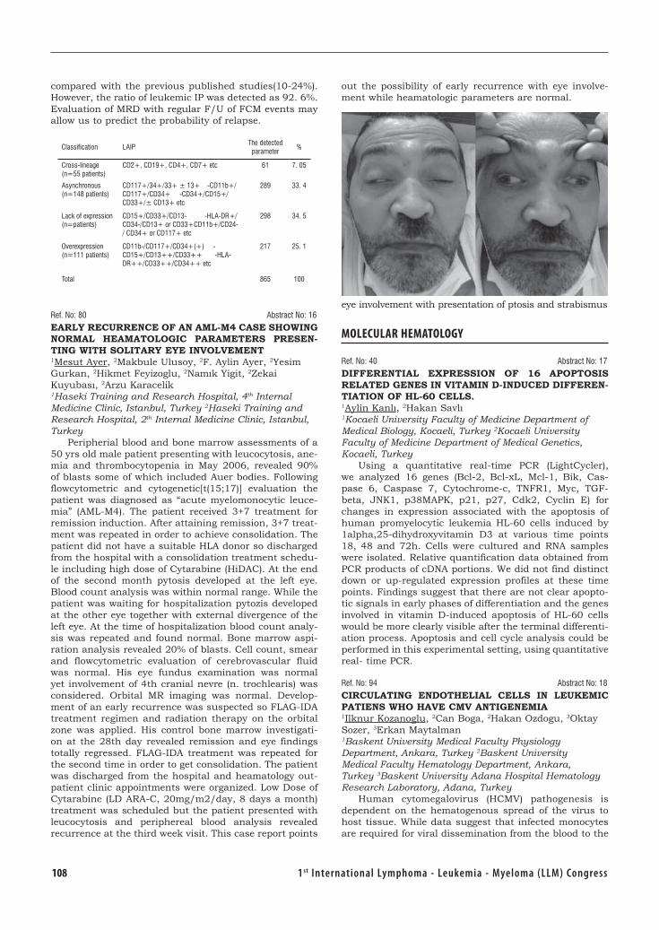
108 1 st International Lymphoma - Leukemia - Myeloma (LLM) Congress
compared with the previous published studies(10-24%). However, the ratio of leukemic IP was detected as 92. 6%. Evaluation of MRD with regular F/U of FCM events may allow us to predict the probability of relapse.
Classification LAIPThe detected
parameter%
Cross-lineage (n=55 patients)
CD2+, CD19+, CD4+, CD7+ etc 61 7. 05
Asynchronous (n=148 patients)
CD117+/34+/33+ ± 13+ -CD11b+/CD117+/CD34+ -CD34+/CD15+/CD33+/± CD13+ etc
289 33. 4
Lack of expression (n=patients)
CD15+/CD33+/CD13- -HLA-DR+/CD34-/CD13+ or CD33+CD11b+/CD24-/ CD34+ or CD117+ etc
298 34. 5
Overexpression (n=111 patients)
CD11b-/CD117+/CD34+(+) - CD15+/CD13++/CD33++ -HLA-DR++/CD33++/CD34++ etc
217 25. 1
Total 865 100
Ref. No: 80 Abstract No: 16
EARLY RECURRENCE OF AN AML-M4 CASE SHOWING NORMAL HEAMATOLOGIC PARAMETERS PRESEN-TING WITH SOLITARY EYE INVOLVEMENT 1Mesut Ayer, 2Makbule Ulusoy, 2F. Aylin Ayer, 2Yesim Gurkan, 2Hikmet Feyizoglu, 2Namık Yigit, 2Zekai Kuyubası, 2Arzu Karacelik1Haseki Training and Research Hospital, 4th Internal Medicine Clinic, Istanbul, Turkey 2Haseki Training and Research Hospital, 2th Internal Medicine Clinic, Istanbul, Turkey
Peripherial blood and bone marrow assessments of a 50 yrs old male patient presenting with leucocytosis, ane-mia and thrombocytopenia in May 2006, revealed 90% of blasts some of which included Auer bodies. Following flowcytometric and cytogenetic[t(15;17)] evaluation the patient was diagnosed as “acute myelomonocytic leuce-mia” (AML-M4). The patient received 3+7 treatment for remission induction. After attaining remission, 3+7 treat-ment was repeated in order to achieve consolidation. The patient did not have a suitable HLA donor so discharged from the hospital with a consolidation treatment schedu-le including high dose of Cytarabine (HiDAC). At the end of the second month pytosis developed at the left eye. Blood count analysis was within normal range. While the patient was waiting for hospitalization pytozis developed at the other eye together with external divergence of the left eye. At the time of hospitalization blood count analy-sis was repeated and found normal. Bone marrow aspi-ration analysis revealed 20% of blasts. Cell count, smear and flowcytometric evaluation of cerebrovascular fluid was normal. His eye fundus examination was normal yet involvement of 4th cranial nevre (n. trochlearis) was considered. Orbital MR imaging was normal. Develop-ment of an early recurrence was suspected so FLAG-IDA treatment regimen and radiation therapy on the orbital zone was applied. His control bone marrow investigati-on at the 28th day revealed remission and eye findings totally regressed. FLAG-IDA treatment was repeated for the second time in order to get consolidation. The patient was discharged from the hospital and heamatology out-patient clinic appointments were organized. Low Dose of Cytarabine (LD ARA-C, 20mg/m2/day, 8 days a month) treatment was scheduled but the patient presented with leucocytosis and periphereal blood analysis revealed recurrence at the third week visit. This case report points
out the possibility of early recurrence with eye involve-ment while heamatologic parameters are normal.
eye involvement with presentation of ptosis and strabismus
MOLECULAR HEMATOLOGY
Ref. No: 40 Abstract No: 17
DIFFERENTIAL EXPRESSION OF 16 APOPTOSIS RELATED GENES IN VITAMIN D-INDUCED DIFFEREN-TIATION OF HL-60 CELLS. 1Aylin Kanlı, 2Hakan Savlı1Kocaeli University Faculty of Medicine Department of Medical Biology, Kocaeli, Turkey 2Kocaeli University Faculty of Medicine Department of Medical Genetics, Kocaeli, Turkey
Using a quantitative real-time PCR (LightCycler), we analyzed 16 genes (Bcl-2, Bcl-xL, Mcl-1, Bik, Cas-pase 6, Caspase 7, Cytochrome-c, TNFR1, Myc, TGF-beta, JNK1, p38MAPK, p21, p27, Cdk2, Cyclin E) for changes in expression associated with the apoptosis of human promyelocytic leukemia HL-60 cells induced by 1alpha,25-dihydroxyvitamin D3 at various time points 18, 48 and 72h. Cells were cultured and RNA samples were isolated. Relative quantification data obtained from PCR products of cDNA portions. We did not find distinct down or up-regulated expression profiles at these time points. Findings suggest that there are not clear apopto-tic signals in early phases of differentiation and the genes involved in vitamin D-induced apoptosis of HL-60 cells would be more clearly visible after the terminal differenti-ation process. Apoptosis and cell cycle analysis could be performed in this experimental setting, using quantitative real- time PCR.
Ref. No: 94 Abstract No: 18
CIRCULATING ENDOTHELIAL CELLS IN LEUKEMIC PATIENS WHO HAVE CMV ANTIGENEMIA1Ilknur Kozanoglu, 2Can Boga, 2Hakan Ozdogu, 3Oktay Sozer, 3Erkan Maytalman1Baskent University Medical Faculty Physiology Department, Ankara, Turkey 2Baskent University Medical Faculty Hematology Department, Ankara, Turkey 3Baskent University Adana Hospital Hematology Research Laboratory, Adana, Turkey
Human cytomegalovirus (HCMV) pathogenesis is dependent on the hematogenous spread of the virus to host tissue. While data suggest that infected monocytes are required for viral dissemination from the blood to the
1091st International Lymphoma - Leukemia - Myeloma (LLM) Congress
host organs, infected endothelial cells are also thought to contribute to this key step in viral pathogenesis. Infe-ction of endothelial cells promoted the increased surface expression of cell adhesion molecules (intercellular cell adhesion molecule 1, vascular cell adhesion molecule 1, E-selectin, and platelet endothelial cell adhesion molecu-le 1), which were necessary for the recruitment of naive monocytes to the apical surface of the endothelium and for the migration of these monocytes through the endot-helial cell layer. We aimed to quantitated endotheial pro-genitor (EPCs) and circulating endothelial cells (CECs) by using flow cytometry in CMV pp65 positive leukemic pati-ents. Detection of CMV pp65 matrix protein in peripheral blood leukocytes by using immunofluorescence assay (Chemicon Internation, a Serologicals Company). All slides are preperated 200. 000 cells and investigated on fluorescence microscope (Eurostar, Euroımmun GmbH, Lübeck, Germany). At this time a panel of monoclonal antibodies, anti CD146 FITC, anti CD 144 PE, anti CD34 ECD, anti CD117 PC5 were used to enumarate CECs and EPCs in CMV pp65 positive patients. Flow cytometric measurement were performed with a FACS calibur flow cytometry (Coulter Epics XL- MLC, Beckman Coulter, Florida, USA) equipped with a 15 mW air-cooled 488-nm argon ion laser. Data were analyzed by using EXPO 32 ADC software. We analyzed CECs and EPCs by flow cyto-metry in CMV pp65 positive 3 patients (one AML M0, one chronic leukocytic leukemia and one hairy cell leukemia. CMV pp65 positive (40 cells) in AML patient, CECs 30. 540/mL and EPCs 14000/mL. One week later, after anti-viral therapy, 100 CMV pp65 positive cells detected while CECs number was 7860/mL and EPCs 4350/mL. On the subsequent week, 5 and 2 CMV positive cells detected on the fluorescence microscope and CECs and EPCs num-ber was decreased. Similar results was shown in CMV positive chronic leukocytic leukemia and one hairy cell leukemia patients (Table1). During the past decade an increasing population of immunosuppressed individuals has resulted in a resurgence of CMV as a major patho-gen. Induced immunosuppression has occurred more frequently via chemotherapy and transplant regimens. Detection of blood leukocytes is closely associated with the clinical manifestations of CMV disease and is useful in the diagnosis of CMV infection. We conclued that; CECs and EPCs identification method in peripheral blood of patients with CMV-associated clinical symptoms, as well as the quantification of CEC and EPCs in peripheral blood of patients with pathophysiological manifestations involving endothelial damage that are different from those caused by CMV infections, can be performed. Our obser-vation supported the idea that CECs and EPCs quantit
CMV pp65(+) Antiviral therapy CEC / ml EPC / ml
Patient 1 40 cells - 30500 14000
100 cells + 7860 4350
5 cells + 10260 3400
1 cell + 6570 4100
Patient 2 1 cell - 16800 11230
0 cell + 6040 5600
Patient 3 1 cell - 14900 10900
0 cell + 7160 6520
ACUTE MYELOBLASTIC LEUKEMIA
Ref. No: 96 Abstract No: 19
COLONY FORMING ASSAY IN PATIENTS WITH AML M7 1Ilknur Kozanoglu, 2Can Boga, 2Hakan Ozdogu, 3Erkan Maytalman, 3Nihan Aldırmaz1Baskent University Medical Faculty Physiology Department, Ankara, Turkey 2Baskent University Medical Faculty Hematology Department, Ankara, Turkey 3Baskent University Adana Hospital Hematology Research Laboratory, Adana, Turkey
The discovery of the colony-forming capacity of hema-topoietic precursor cells 40 years ago has revolutionized experimental, diagnostic and therapeutic hematology. In addition to quantitative abnormalities, cultures can reveal disease-spesific diagnostic growth patterns. They are help in the staging of some hematological disease. In untreated AML, cultures of bone marrow show either no growth, or growth of leukemic cells either in clusters or as single cells. Leukemic growty can be discrete or abundant ; single cells in leukemic clusters sometimes resemble either neutrophils, eosinophils, macrophages or immature erythroblasts according to their line of origin. The typical in vitro growth pattern in AML was observed soon after the discovery of the hematopoietic stem cell. Its association with an unfavorable prognosis, its disap-pearance in remission and its value in the prediction of relapse are well documented. On the other hand, absence of leukemic growth is an acknowledged good prognostic sing. The aim of our study were to evaulate endogenous colony formation in AML M7 patients. The bone marrow mononuclear cells of 2 patients with AML M7 were cul-tured by cytokine free methyl cellulose media (Methocult TM GF H4230 – StemCell Technologies, Canada) for endogenous colony evaluation. removed 100 μl blood, counted initial nucleated cell by adding 3% acetic acid and using hemacytometer or counted directly in auto-mated cell counter. Firstly, it was added 1/3 of ficoll and then added 2/3 of blood on the top of ficoll gently. It was santrifugated 400xg for 30 min. It was removed layer of mononuclear cells between sera and ficoll. It was added IMDM containing 2% FCS on the mononuclear cells and resuspended them and santrifugated 400xg for 10 min. It was decanted the supernatant repeat the step above. Decant the supernatant and then resuspended mono-nuclear cells in the way that cell count is 1-2,5 x 10⁶ by adding IMDM containing 2% FCS (in order to get cell count, perform the step mentioned on the top). It was thawed an alliquot overnight between 2-8 °C. Added 0,3 ml of resuspended cell prepared before. Last concentrati-on was 1-2,5 x 10⁵ cell for per ml. It was vortexed for 15 minutes and let stand for 15-30 min. to allow bubbles. It was dispensed 1. 1 ml of this mixture using syringe and 16-gauge blunt-end needle in to 35 mm culture dishes. Incubate the cells in a 37 °C and 5% CO2, 95% humi-dified incubator without disturbance during 14-16 day. We enumerated and characterized the colonies according to their morphology with an inverted microscope and a 100 mm culture dish marked with the scoring grid. We showed that, after 14 days of cultures, leukemic clusters and single cells resembling neutrophils, newly diagnosed AML M7 patients. After flowwing remission induction, the leukemic blast colony progenitors declined to undetectab-le levels and normal erythroid and myeloid colonogenic
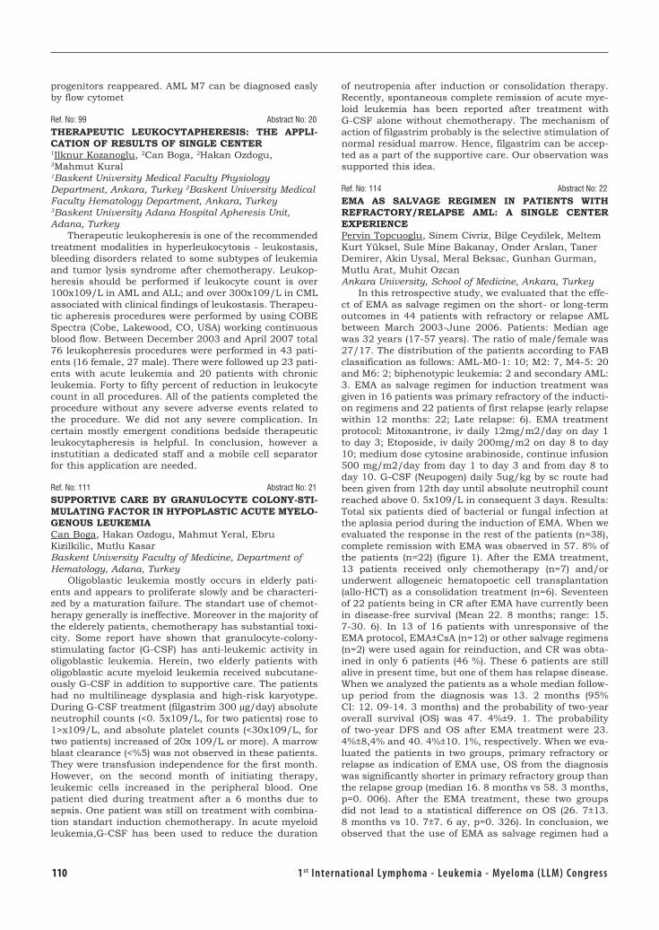
110 1 st International Lymphoma - Leukemia - Myeloma (LLM) Congress
progenitors reappeared. AML M7 can be diagnosed easly by flow cytomet
Ref. No: 99 Abstract No: 20
THERAPEUTIC LEUKOCYTAPHERESIS: THE APPLI-CATION OF RESULTS OF SINGLE CENTER1Ilknur Kozanoglu, 2Can Boga, 2Hakan Ozdogu, 3Mahmut Kural1Baskent University Medical Faculty Physiology Department, Ankara, Turkey 2Baskent University Medical Faculty Hematology Department, Ankara, Turkey 3Baskent University Adana Hospital Apheresis Unit, Adana, Turkey
Therapeutic leukopheresis is one of the recommended treatment modalities in hyperleukocytosis - leukostasis, bleeding disorders related to some subtypes of leukemia and tumor lysis syndrome after chemotherapy. Leukop-heresis should be performed if leukocyte count is over 100x109/L in AML and ALL; and over 300x109/L in CML associated with clinical findings of leukostasis. Therapeu-tic apheresis procedures were performed by using COBE Spectra (Cobe, Lakewood, CO, USA) working continuous blood flow. Between December 2003 and April 2007 total 76 leukopheresis procedures were performed in 43 pati-ents (16 female, 27 male). There were followed up 23 pati-ents with acute leukemia and 20 patients with chronic leukemia. Forty to fifty percent of reduction in leukocyte count in all procedures. All of the patients completed the procedure without any severe adverse events related to the procedure. We did not any severe complication. In certain mostly emergent conditions bedside therapeutic leukocytapheresis is helpful. In conclusion, however a instutitian a dedicated staff and a mobile cell separator for this application are needed.
Ref. No: 111 Abstract No: 21
SUPPORTIVE CARE BY GRANULOCYTE COLONY-STI-MULATING FACTOR IN HYPOPLASTIC ACUTE MYELO-GENOUS LEUKEMIACan Boga, Hakan Ozdogu, Mahmut Yeral, Ebru Kizilkilic, Mutlu KasarBaskent University Faculty of Medicine, Department of Hematology, Adana, Turkey
Oligoblastic leukemia mostly occurs in elderly pati-ents and appears to proliferate slowly and be characteri-zed by a maturation failure. The standart use of chemot-herapy generally is ineffective. Moreover in the majority of the elderely patients, chemotherapy has substantial toxi-city. Some report have shown that granulocyte-colony-stimulating factor (G-CSF) has anti-leukemic activity in oligoblastic leukemia. Herein, two elderly patients with oligoblastic acute myeloid leukemia received subcutane-ously G-CSF in addition to supportive care. The patients had no multilineage dysplasia and high-risk karyotype. During G-CSF treatment (filgastrim 300 μg/day) absolute neutrophil counts (<0. 5x10⁹/L, for two patients) rose to 1>x109/L, and absolute platelet counts (<30x109/L, for two patients) increased of 20x 109/L or more). A marrow blast clearance (<%5) was not observed in these patients. They were transfusion independence for the first month. However, on the second month of initiating therapy, leukemic cells increased in the peripheral blood. One patient died during treatment after a 6 months due to sepsis. One patient was still on treatment with combina-tion standart induction chemotherapy. In acute myeloid leukemia,G-CSF has been used to reduce the duration
of neutropenia after induction or consolidation therapy. Recently, spontaneous complete remission of acute mye-loid leukemia has been reported after treatment with G-CSF alone without chemotherapy. The mechanism of action of filgastrim probably is the selective stimulation of normal residual marrow. Hence, filgastrim can be accep-ted as a part of the supportive care. Our observation was supported this idea.
Ref. No: 114 Abstract No: 22
EMA AS SALVAGE REGIMEN IN PATIENTS WITH REFRACTORY/RELAPSE AML: A SINGLE CENTER EXPERIENCEPervin Topcuoglu, Sinem Civriz, Bilge Ceydilek, Meltem Kurt Yüksel, Sule Mine Bakanay, Onder Arslan, Taner Demirer, Akin Uysal, Meral Beksac, Gunhan Gurman, Mutlu Arat, Muhit OzcanAnkara University, School of Medicine, Ankara, Turkey
In this retrospective study, we evaluated that the effe-ct of EMA as salvage regimen on the short- or long-term outcomes in 44 patients with refractory or relapse AML between March 2003-June 2006. Patients: Median age was 32 years (17-57 years). The ratio of male/female was 27/17. The distribution of the patients according to FAB classification as follows: AML-M0-1: 10; M2: 7, M4-5: 20 and M6: 2; biphenotypic leukemia: 2 and secondary AML: 3. EMA as salvage regimen for induction treatment was given in 16 patients was primary refractory of the inducti-on regimens and 22 patients of first relapse (early relapse within 12 months: 22; Late relapse: 6). EMA treatment protocol: Mitoxantrone, iv daily 12mg/m2/day on day 1 to day 3; Etoposide, iv daily 200mg/m2 on day 8 to day 10; medium dose cytosine arabinoside, continue infusion 500 mg/m2/day from day 1 to day 3 and from day 8 to day 10. G-CSF (Neupogen) daily 5ug/kg by sc route had been given from 12th day until absolute neutrophil count reached above 0. 5x10⁹/L in consequent 3 days. Results: Total six patients died of bacterial or fungal infection at the aplasia period during the induction of EMA. When we evaluated the response in the rest of the patients (n=38), complete remission with EMA was observed in 57. 8% of the patients (n=22) (figure 1). After the EMA treatment, 13 patients received only chemotherapy (n=7) and/or underwent allogeneic hematopoetic cell transplantation (allo-HCT) as a consolidation treatment (n=6). Seventeen of 22 patients being in CR after EMA have currently been in disease-free survival (Mean 22. 8 months; range: 15. 7-30. 6). In 13 of 16 patients with unresponsive of the EMA protocol, EMA±CsA (n=12) or other salvage regimens (n=2) were used again for reinduction, and CR was obta-ined in only 6 patients (46 %). These 6 patients are still alive in present time, but one of them has relapse disease. When we analyzed the patients as a whole median follow-up period from the diagnosis was 13. 2 months (95% CI: 12. 09-14. 3 months) and the probability of two-year overall survival (OS) was 47. 4%±9. 1. The probability of two-year DFS and OS after EMA treatment were 23. 4%±8,4% and 40. 4%±10. 1%, respectively. When we eva-luated the patients in two groups, primary refractory or relapse as indication of EMA use, OS from the diagnosis was significantly shorter in primary refractory group than the relapse group (median 16. 8 months vs 58. 3 months, p=0. 006). After the EMA treatment, these two groups did not lead to a statistical difference on OS (26. 7±13. 8 months vs 10. 7±7. 6 ay, p=0. 326). In conclusion, we observed that the use of EMA as salvage regimen had a
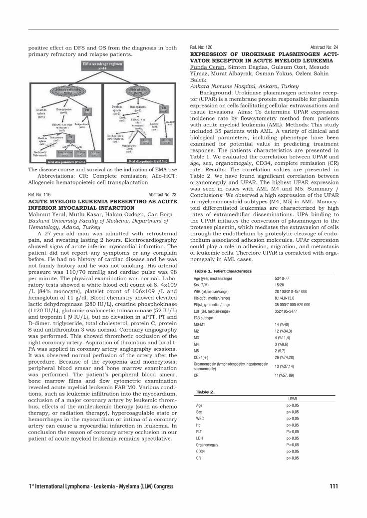
1111st International Lymphoma - Leukemia - Myeloma (LLM) Congress
positive effect on DFS and OS from the diagnosis in both primary refractory and relapse patients.
The disease course and survival as the indication of EMA useAbbreviations: CR: Complete remission; Allo-HCT:
Allogeneic hematopoieteic cell transplantation
Ref. No: 116 Abstract No: 23
ACUTE MYELOID LEUKEMIA PRESENTING AS ACUTE INFERIOR MYOCARDIAL INFARCTIONMahmut Yeral, Mutlu Kasar, Hakan Ozdogu, Can BogaBaskent University Faculty of Medicine, Department of Hematology, Adana, Turkey
A 27-year-old man was admitted with retrosternal pain, and sweating lasting 2 hours. Electrocardiography showed signs of acute inferior myocardial infarction. The patient did not report any symptoms or any complain before. He had no history of cardiac disease and he was not family history and he was not smoking. His arterial pressure was 110/70 mmHg and cardiac pulse was 98 per minute. The physical examination was normal. Labo-ratory tests showed a white blood cell count of 8. 4x10⁹ /L (84% monocyte), platelet count of 106x109 /L and hemoglobin of 11 g/dl. Blood chemistry showed elevated lactic dehydrogenase (280 IU/L), creatine phosphokinase (1120 IU/L), glutamic-oxaloacetic transaminase (52 IU/L) and troponin I (9 IU/L), but no elevation in aPTT, PT and D-dimer. triglyceride, total cholesterol, protein C, protein S and antithrombin 3 was normal. Coronary angiography was performed. This showed thrombotic occlusion of the right coronary artery. Aspiration of thrombus and local t-PA was applied in coronary artery angiography sessions. It was observed normal perfusion of the artery after the procedure. Because of the cytopenia and monocytosis; peripheral blood smear and bone marrow examination was performed. The patient’s peripheral blood smear, bone marrow films and flow cytometric examination revealed acute myeloid leukemia FAB M0. Various condi-tions, such as leukemic infiltration into the myocardium, occlusion of a major coronary artery by leukemic throm-bus, effects of the antileukemic therapy (such as chemo therapy, or radiation therapy), hypercoagulable state or hemorrhages in the myocardium or intima of a coronary artery can cause a myocardial infarction in leukemia. In conclusion the reason of coronary artery occlusion in our patient of acute myeloid leukemia remains speculative.
Ref. No: 120 Abstract No: 24
EXPRESSION OF UROKINASE PLASMINOGEN ACTI-VATOR RECEPTOR IN ACUTE MYELOID LEUKEMIAFunda Ceran, Simten Dagdas, Gulsum Ozet, Mesude Yilmaz, Murat Albayrak, Osman Yokus, Ozlem Sahin BalcikAnkara Numune Hospital, Ankara, Turkey
Background: Urokinase plasminogen activator recep-tor (UPAR) is a membrane protein responsible for plasmin expression on cells facilitating cellular extravasations and tissue invasions. Aims: To determine UPAR expression incidence rate by flowcytometry method from patients with acute myeloid leukemia (AML). Methods: This study included 35 patients with AML. A variety of clinical and biological parameters, including phenotype have been examined for potential value in predicting treatment response. The patients characteristics are presented in Table 1. We evaluated the correlation between UPAR and age, sex, organomegaly, CD34, complete remission (CR) rate. Results: The correlation values are presented in Table 2. We have found significant correlation between organomegaly and UPAR. The highest UPAR expression was seen in cases with AML M4 and M5. Summary / Conclusions: We observed a high expression of the UPAR in myelomonocytoid subtypes (M4, M5) in AML. Monocy-toid differentiated leukemias are characterized by high rates of extramedullar disseminations. UPA binding to the UPAR initiates the conversion of plasminogen to the protease plasmin, which mediates the extravasion of cells through the endothelium by proteolytic cleavage of endo-thelium associated adhesion molecules. UPAr expression could play a role in adhesion, migration, and metastasis of leukemic cells. Therefore UPAR is corraleted with orga-nomegaly in AML cases.
Table 1. Patient Characteristics
Age (year, median/range) 53/18-77
Sex (F/M) 15/20
WBC(μl,median/range) 28 100/310-457 000
Hb(gr/dl, median/range) 8,1/4,0-13,0
Plt(μl, (μl,median/range 35 000/7 000-520 000
LDH(U/l, median/range) 352/195-2477
FAB subtype
M0-M1 14 (%40)
M2 12 (%34,3)
M3 4 (%11,4)
M4 3 (%8,6)
M5 2 (5,7)
CD34(+) 26 (%74,28)
Organomegaly (lymphadenopathy, hepatomegaly, splenomegaly)
13 (%37,14)
CR 11(%57. 89)
Table 2.
UPAR
Age p>0,05
Sex p>0,05
WBC p>0,05
Hb p>0,05
PLT P>0,05
LDH p>0,05
Organomegaly P<0,05
CD34 p>0,05
CR p>0,05
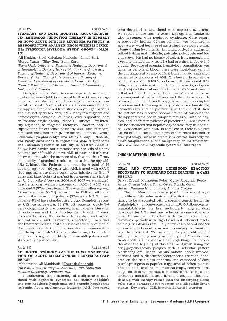
112 1 st International Lymphoma - Leukemia - Myeloma (LLM) Congress
Ref. No: 122 Abstract No: 25
STANDART AND DOSE MODIFIED ARA-C/IDARUBI-CIN REMISSION INDUCTION THERAPY IN ELDERLY DE-NOVO ACUTE MYELOID LEUKEMIA PATIENTS: A RETROSPECTIVE ANALYSIS FROM “DENIZLI LEUKE-MIA-LYMPHOMA-MYELOMA STUDY GROUP” (DLLM-SG)1Ali Keskin, 1Sibel Kabukcu Hacioglu, 1Ismail Sari, 2Burcu Yapar, 3Nilay Sen, 4Sami Karti1Pamukkale University, Faculty of Medicine, Department of Hematology, Denizli, Turkey 2Pamukkale University, Faculty of Medicine, Department of Internal Medicine, Denizli, Turkey 3Pamukkale University, Faculty of Medicine, Department of Pathology, Denizli, Turkey 4Denizli Education and Research Hospital, Hematology Unit, Denizli, Turkey
Background and Aim: Outcome of patients with acute myeloid leukemia (AML) who are older than 60 years of age remains unsatisfactory, with low remission rates and poor overall survival. Results of standart remission-induction therapy are often derived from studies in younger patients and may not apply to elderly AML. Many investigators and hematologists advocate, at times, only supportive care or frontline single agents, Phase I-II studies, low-inten-sity regimens, or ‘targeted’ therapies. However, baseline expectations for outcomes of elderly AML with ‘standard’ remission-induction therapy are not well defined. “Denizli Leukemia-Lymphoma-Myeloma Study Group” (DLLMSG) was nearly established to register the data of lymphoma and leukemia patients in our city in Western Anatolia. So, we have carried out a retrospective analysis of elderly patients (age>60) with de-novo AML followed at our hema-tology centers, with the purpose of evaluating the efficacy and toxicity of ‘standard’ remission-induction therapy with ARA-C/Idarubicin. Patients and methods: A total of 14 patients age > or = 60 years with AML treated with ARA-C (100 mg/m2 intravenous continuous infusion for 5 or 7 days) and idarubicin (12 mg/m2 intravenous short infusi-on for 2 or 3 days) between 2004 and 2007 were analyzed. Results: Among 14 elderly patients with AML, 6 (43%) were male and 8 (57%) were female. The overall median age was 68 years (range: 60-74). According to FAB classification system, the majority of patients (57%) were M2. Thirteen patients (93%) have standart risk group. Complete respon-se (CR) was achieved in 11 (78. 5%) patients. Grade 3-4 hematologic toxicity was observed in all patients. Duration of leukopenia and thrombocytopenia 14 and 17 days, respectively. Also, the median disease-free and overall survival were 6 and 10 months, respectively. There was one treatment-related death because of severe pneumonia. Conclusion: Standart and dose modified remission-induc-tion therapy with ARA-C and idarubicin might be effective and tolerable regimen in elderly de-novo AML patients with standart cytogenetic risk.
Ref. No: 140 Abstract No: 26
NEPHROTIC SYNDROME AS THE FIRST MANIFESTA-TION OF ACUTE MYELOGENOUS LEUKEMIA: CASE REPORT1Mohammad Ali Mashhadi, 2Kourosh Shahraki1Ali Ebne Abitaleb Hospital Zahedan, Iran, 2Zahedan Medical University, Zahedan, Iran
Introduction: The hematological malignancies asso-ciated with nephrotic syndrome are mainly hodgkin’s and non-hodgkin’s lymphomas and chronic lymphocytic leukemia. Acute myelogenous leukemia (AML) has rarely
been described in associated with nephritic syndrome. We report a rare case of Acute Myelogenous Leukemia who presented with nephrotic syndrome. Case report: A previously healthy 62-year-old man was admitted in nephrology ward because of generalized developing pitting edema during last month. Simultaneously, he had gene-ralized itching and urticaria, polyuria, polydypsia and low grade fever but had no history of weight loss, anorexia and sweating. In laboratory tests he had proteinuria above 3. 5 gr/day. Because of anemia, hematology consultation was done. In peripheral blood, there were myeloblast cells in the circulation at a ratio of 15%. Bone marrow aspiration confirmed a diagnosis of AML M, showing hypercellular bone marrow with 80-90% leukemic cells, increased M/E ratio, myeloblast(immature cell, fine chromatin, cytoplas-mic bleb) and these abnormal elements: >50% and mature cell about 10%. Unfortunately, we hadn’t renal biopsy as a consequent of patient illness and thromboastenia. He received induction chemotherapy, which led to a complete remission and decreasing urinary protein excretion during chemotherapy and no proteinuria at the end of it. Now the patient has received second course of consolidation therapy and remained in complete remission, with no phy-sical and laboratory evidence of proteinuria. Conclusion: It can be concluded that nephrotic syndrome may be additio-nally associated with AML. In some cases, there is a direct causal effect of the leukemic process on renal function or even pathology, while in others it is exerted indirectly via other complications of the malignancy or the treatment. KEY WORDS: AML, nephrotic syndrome, case report
CHRONIC MYELOID LEUKEMIA
Ref. No: 30 Abstract No: 27
ORAL AND CUTANEUS LICHENOID REACTION SECONDARY TO STANDARD DOSE IMATINIB: A CASE REPORTServet Erbasi, Muhterem Polat, Murat Albayrak, Ferda Artuz, Osman Yokus, Pınar Oztas, Funda CeranAnkara Numune Hastahanesi, Ankara, Turkey
Chronic Myeloid Leukemia (CML) is a clonal mye-lopro-liferatif disorder which is the first human malig-nancy to be associated with a specific genetic lesion,the Philadelphia chromosome,carryingBCR-ABLoncogene. Imatinib(Glivec)is the first molecularly targeted drug developed for CML and has achieved aremarkable suc-cess. Cutaneous side effect with this treatment are common(especially with High Doses)but lichenoid reacti-on drug eruption is rare. Only a few cases of oral and/or cutaneous lichenoid reaction secondary to imatinib have beenreported. We present a 42-years old woman with approximately one year history of CML. She was treated with standard dose Imatinib(400mg). Threemon-ths after the begining of this treatment,while using the drug,grey-violaceous plaques with a reticular pattern resembling oral lichen planus onboth cheek mucosal surfaces and a disseminatedcutaneous eruption appe-ared on the trunk,legs andarms and composed of dark purple,prurigenous papules suggestive of lichen planus. The cutaneousand the oral mucosal biopsy confirmed the diagnosis of lichen planus. It is believed that this patient developed imatinib-induced lichenoid eruption;this rela-tionship with therapy rather than the underlying diseas rules out a paraneoplastic reaction and idiopathic lichen planus. Key words: CML,Imatinib,lichenoid eruption
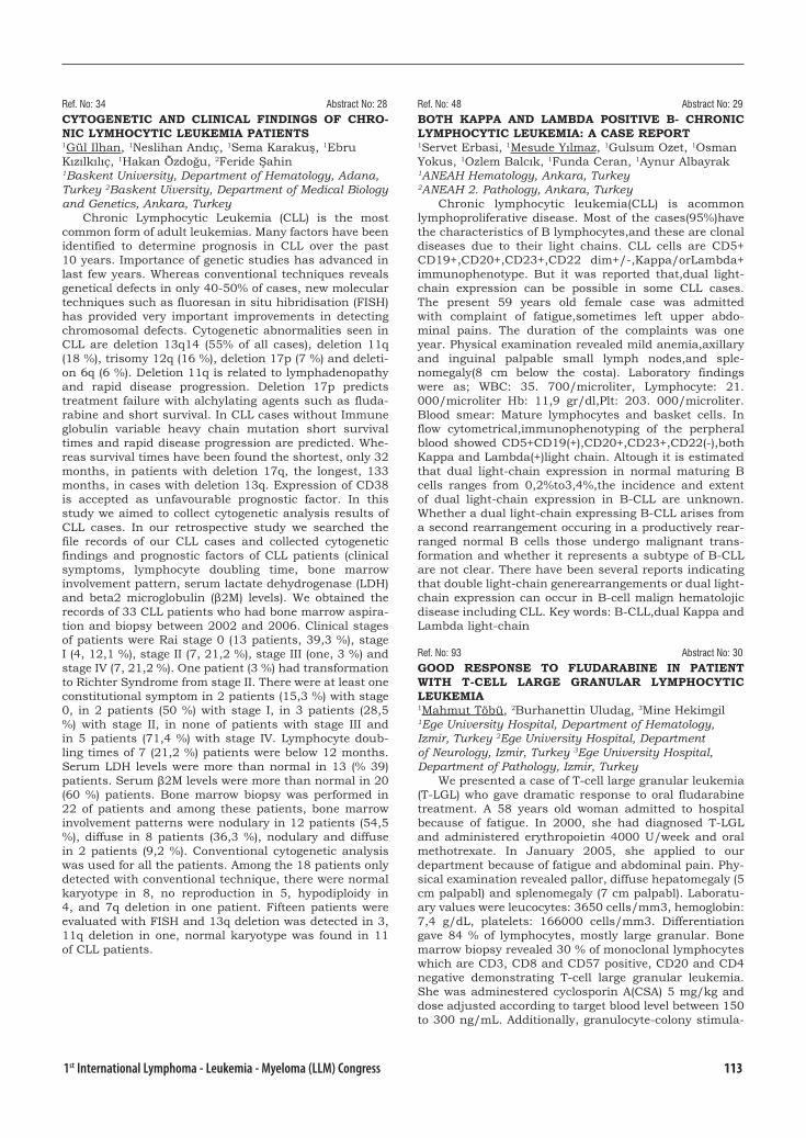
1131st International Lymphoma - Leukemia - Myeloma (LLM) Congress
Ref. No: 34 Abstract No: 28
CYTOGENETIC AND CLINICAL FINDINGS OF CHRO-NIC LYMHOCYTIC LEUKEMIA PATIENTS1Gül Ilhan, 1Neslihan Andıç, 1Sema Karakuş, 1Ebru Kızılkılıç, 1Hakan Özdoğu, 2Feride Şahin1Baskent University, Department of Hematology, Adana, Turkey 2Baskent Uiversity, Department of Medical Biology and Genetics, Ankara, Turkey
Chronic Lymphocytic Leukemia (CLL) is the most common form of adult leukemias. Many factors have been identified to determine prognosis in CLL over the past 10 years. Importance of genetic studies has advanced in last few years. Whereas conventional techniques reveals genetical defects in only 40-50% of cases, new molecular techniques such as fluoresan in situ hibridisation (FISH) has provided very important improvements in detecting chromosomal defects. Cytogenetic abnormalities seen in CLL are deletion 13q14 (55% of all cases), deletion 11q (18 %), trisomy 12q (16 %), deletion 17p (7 %) and deleti-on 6q (6 %). Deletion 11q is related to lymphadenopathy and rapid disease progression. Deletion 17p predicts treatment failure with alchylating agents such as fluda-rabine and short survival. In CLL cases without Immune globulin variable heavy chain mutation short survival times and rapid disease progression are predicted. Whe-reas survival times have been found the shortest, only 32 months, in patients with deletion 17q, the longest, 133 months, in cases with deletion 13q. Expression of CD38 is accepted as unfavourable prognostic factor. In this study we aimed to collect cytogenetic analysis results of CLL cases. In our retrospective study we searched the file records of our CLL cases and collected cytogenetic findings and prognostic factors of CLL patients (clinical symptoms, lymphocyte doubling time, bone marrow involvement pattern, serum lactate dehydrogenase (LDH) and beta2 microglobulin (β2M) levels). We obtained the records of 33 CLL patients who had bone marrow aspira-tion and biopsy between 2002 and 2006. Clinical stages of patients were Rai stage 0 (13 patients, 39,3 %), stage I (4, 12,1 %), stage II (7, 21,2 %), stage III (one, 3 %) and stage IV (7, 21,2 %). One patient (3 %) had transformation to Richter Syndrome from stage II. There were at least one constitutional symptom in 2 patients (15,3 %) with stage 0, in 2 patients (50 %) with stage I, in 3 patients (28,5 %) with stage II, in none of patients with stage III and in 5 patients (71,4 %) with stage IV. Lymphocyte doub-ling times of 7 (21,2 %) patients were below 12 months. Serum LDH levels were more than normal in 13 (% 39) patients. Serum β2M levels were more than normal in 20 (60 %) patients. Bone marrow biopsy was performed in 22 of patients and among these patients, bone marrow involvement patterns were nodulary in 12 patients (54,5 %), diffuse in 8 patients (36,3 %), nodulary and diffuse in 2 patients (9,2 %). Conventional cytogenetic analysis was used for all the patients. Among the 18 patients only detected with conventional technique, there were normal karyotype in 8, no reproduction in 5, hypodiploidy in 4, and 7q deletion in one patient. Fifteen patients were evaluated with FISH and 13q deletion was detected in 3, 11q deletion in one, normal karyotype was found in 11 of CLL patients.
Ref. No: 48 Abstract No: 29
BOTH KAPPA AND LAMBDA POSITIVE B- CHRONIC LYMPHOCYTIC LEUKEMIA: A CASE REPORT1Servet Erbasi, 1Mesude Yılmaz, 1Gulsum Ozet, 1Osman Yokus, 1Ozlem Balcık, 1Funda Ceran, 1Aynur Albayrak1ANEAH Hematology, Ankara, Turkey 2ANEAH 2. Pathology, Ankara, Turkey
Chronic lymphocytic leukemia(CLL) is acommon lymphoproliferative disease. Most of the cases(95%)have the characteristics of B lymphocytes,and these are clonal diseases due to their light chains. CLL cells are CD5+CD19+,CD20+,CD23+,CD22 dim+/-,Kappa/orLambda+ immunophenotype. But it was reported that,dual light-chain expression can be possible in some CLL cases. The present 59 years old female case was admitted with complaint of fatigue,sometimes left upper abdo-minal pains. The duration of the complaints was one year. Physical examination revealed mild anemia,axillary and inguinal palpable small lymph nodes,and sple-nomegaly(8 cm below the costa). Laboratory findings were as; WBC: 35. 700/microliter, Lymphocyte: 21. 000/microliter Hb: 11,9 gr/dl,Plt: 203. 000/microliter. Blood smear: Mature lymphocytes and basket cells. In flow cytometrical,immunophenotyping of the perpheral blood showed CD5+CD19(+),CD20+,CD23+,CD22(-),both Kappa and Lambda(+)light chain. Altough it is estimated that dual light-chain expression in normal maturing B cells ranges from 0,2%to3,4%,the incidence and extent of dual light-chain expression in B-CLL are unknown. Whether a dual light-chain expressing B-CLL arises from a second rearrangement occuring in a productively rear-ranged normal B cells those undergo malignant trans-formation and whether it represents a subtype of B-CLL are not clear. There have been several reports indicating that double light-chain generearrangements or dual light-chain expression can occur in B-cell malign hematolojic disease including CLL. Key words: B-CLL,dual Kappa and Lambda light-chain
Ref. No: 93 Abstract No: 30
GOOD RESPONSE TO FLUDARABINE IN PATIENT WITH T-CELL LARGE GRANULAR LYMPHOCYTIC LEUKEMIA 1Mahmut Töbü, 2Burhanettin Uludag, 3Mine Hekimgil1Ege University Hospital, Department of Hematology, Izmir, Turkey 2Ege University Hospital, Department of Neurology, Izmir, Turkey 3Ege University Hospital, Department of Pathology, Izmir, Turkey
We presented a case of T-cell large granular leukemia (T-LGL) who gave dramatic response to oral fludarabine treatment. A 58 years old woman admitted to hospital because of fatigue. In 2000, she had diagnosed T-LGL and administered erythropoietin 4000 U/week and oral methotrexate. In January 2005, she applied to our department because of fatigue and abdominal pain. Phy-sical examination revealed pallor, diffuse hepatomegaly (5 cm palpabl) and splenomegaly (7 cm palpabl). Laboratu-ary values were leucocytes: 3650 cells/mm3, hemoglobin: 7,4 g/dL, platelets: 166000 cells/mm3. Differentiation gave 84 % of lymphocytes, mostly large granular. Bone marrow biopsy revealed 30 % of monoclonal lymphocytes which are CD3, CD8 and CD57 positive, CD20 and CD4 negative demonstrating T-cell large granular leukemia. She was adminestered cyclosporin A(CSA) 5 mg/kg and dose adjusted according to target blood level between 150 to 300 ng/mL. Additionally, granulocyte-colony stimula-
114 1 st International Lymphoma - Leukemia - Myeloma (LLM) Congress
ting factor and erythropoietin were administered. After 2 months, her clinical findings did not improved, only neutrophils increased and splenic irradiation applied. Splenomegaly regressed (5 cm palpabl). CSA and growth factors treatment continued. After six months, splenome-galy (1 cm palpabl) and hepatomegaly (2 cm palpabl) and CBC values improved. Leucocytes: 13400 cells/mm3, hemoglobin: 10,6 g/L, platelets: 198000 cells/mm3. Differentiation gave 88 % of neutrophils, 10 % of lympho-cytes and 2 % of monocytes. CSA dose were tapered and discontinued. Because of her clinical picture reappeared, CSA treatment restarted but renal toxicity developed and CSA treatment stopped. She became transfusion depen-dent. She has adminestered first cycle of treatment of oral fludarabine 40 mg/m2 for 5 days. After four weeks of treatment, organomegaly regressed and CBC norma-lized. Leucocytes: 6260 cells/mm3, hemoglobin: 14. 1 g/dL, platelets: 122000 cells/mm3. Differantiation gave 82 % of neutrophils, 14 % of lymphocytes and 4 % of monocytes. Second cycle of fludarabine treatment admi-nistered. But she developed headache and multipl cranial neuropathy including n. opticus, n. oculomotorious, n. trigeminalis and n. vestibulocochlearis, and her cranial MRI was normal. This multipl cranial neuropathy may be depend on oral fludarabine treatment. It should keep in mind that fludarabine may cause severe multipl cranial neuropathy.
Ref. No: 101 Abstract No: 31
SEVERE NEUROTOXICITY OF CLADRIBINE IN A PATI-ENT WITH HAIRY CELL LEUKEMIA: A CASE REPORTGülten Sop, Zafer Gökgöz, Tuğba Gümüş, Şermin Çoban, Füsun ÖzdemirkıranIzmir Training and Research Hospital, Izmir, Turkey
Hairy cell leukemia (HCL) is an indolent chronic B cell lymphoproliferative disorder involving the bone marrow and spleen. Patients with HCL present with peripheral blood cytopenies, splenomegaly and circulating hairy cells. Past a few years cladribine purine analogue has an important role with %90-100 remission rates in HCL tre-atment. We presented a patient with hairy cell leukemia who developed severe neurotoxicity after cladribine infu-sion. A 52 years old male patient admitted to the hospital with fatigue, weakness and weight loss continuing for 2 months. On his physical examination; blood pressure was 100/60 mmHg, fever: 36’ C, heart rate: 72, cardio-vascular and respiratuary systems were normal. Spleen was palpable 10 cm below the costal magrine and hepato-megaly determined. Laboratory findings were as follows: complete blood count; hemoglobin: 11g/dl; hemotocrit: 32 %, wbc: 2500 /mm3, plt: 56000/mm3, erythrocyte sedimentation rate: 10 mm/h. In CT scans: 2 cm diame-ter bilateral axillar, 4 cm diameter chealiac, peripancre-atic, paraaortic and paracaval lymphadenomegalies were observed. We also determined spleen enlargement (long axis was 22 cm) and hepatomegaly before treatment. The diagnosis was established by examining bone marrow biopsy with imunnohistochemical stain. Bone marrow biopsy was examined in Ege University Pathology Depart-ment. Hairy cell leukemia infiltration was found, CD20 (+), TRAP (+), CD3 (-), CD 68 (-) and reticulin grade was 2 respectively. Our case received cladribine 0,1mg/kg/day, 7 day duration infusion. Cladribine therapy generally has minimal acute or subacute adverse events. Besides this, moderate bone marrow inhibition is the most common adverse event. One week after the treatment the patient
developed neutropenic fever, pulmoner infection, weak-ness in both legs, short term memory losts, and mislaid ability to walk. Then he received appropriate antibiotic and antifungal therapies. Cranial MRI and EEG was normal but EMG showed early stage axonal degeneration and segmental demyelinization which is significant in lower extremites. These neuropathic findings were inter-preted a neurotoxicity of cladribine by the neurologists. 6 weeks after the treatment his general condition beca-me well and laboratory findings regressed. Thorax and abdominal CT scans were all normal. Almost he regained his ability to walk and muscle strength. Our case is in remission hematologicaly but still going on neurology controls. When literatures have reviewed severe neu-rotoxicity was seen rarely. In National Cancer Institute group 932 cases were treated with cladribine and 26% of these had developed moderate neurotoxicity. Grade 3-4 neurotoxicity had developed only in 10 patients in this serie. We approved to present this case which developed opportunistic pulmoner infection, hemolytic anemia and serious neurotoxicity after stantard dose treatme
Ref. No: 130 Abstract No: 32
POSSIBLE PNEUMOCYSTIS JIROVECI INFECTION IN AN CYTOMEGALOVIRUS POSITIVE CLL PATIENTMutlu Kasar, Hakan Ozdogu, Can Boga, Mahmut YeralBaskent University Faculty of Medicine, Department of Hematology, Adana, Turkey
Chronic lymphocytic leukemia (CLL) is the most com-mon form of leukemia in Western countries and mainly affects elderly individuals. Infections are major cause of morbidity and mortality in patients with chronic lymp-hocytic leukemia. Predisposition to infection in CLL is mediated through various abnormalities including both the impairment of humoral and cellular immunity and further immunosuppression related to the therapy of CLL. Streptococcus pneumonia, staphylococcus aureus, streptococcus pyogenes and herpes zoster-varicella virus are most common infection agents. On the other hand, patients treated with purine analogues apparently have an increased incidence of infection with other opportu-nistic organisms such as cytomegalovirus, listeria mono-cytogenes and pneumocystis jiroveci. We are presenting a patient with CLL associated with CMV and pneumocystis jiroveci infection. A 63 year old man with 3 year history of CLL was treated with fludarabine and cyclophospha-mide. Because of development of autoimmune hemolytic anemia 2 weeks before admission to our hospital, steroid therapy of 70 mg prednisolone was started. Ten days after second course of fludarabine and cyclophospha-mide therapy, he presented with fever. The white count cell on admission was 2. 8 x 10⁹/L (% 30 lymphocytes). Physical examination and chest radiography was normal. Empirical meropenem therapy was started. The patient was unresponsive. Test for CMV pp65 antigenemia was found to be positive. Ganciclovir was started. Five days after ganciclovir therapy, progressive dyspnea occurred. Computed tomographic examination of the thorax revea-led consolidation and bilateral pleural effusion. The pati-ent was not tolerated bronchoscopy and empirically high dose co-trimoxazole was added to therapy, the clinical condition of the patient was improved. Atypical double opportunistic infections may see in CLL patients who are treated with purine analogs. Association of these two infections should keep in mind in these patients.
1151st International Lymphoma - Leukemia - Myeloma (LLM) Congress
Ref. No: 19 Abstract No: 33
PROMINENT PLEURAL AND PERICARDIAL EFFUSION DUE TO IMATINIB MESYLATE AFTER FIVE YEARS OF THERAPYNeslihan Andıç, Gül Ilhan, Sema KarakuşBaskent University, Faculty of Medicine, Department of Internal Medicine, Hematology Division, Ankara, Turkey
The signal transduction inhibitor imatinib mesylate, is the superior first line therapy in Philedelphia chromo-some positive chronic myeloid leukemia (CML) patients. Data obtained so far has shown that imatinib mesylate can induce hematologic and cytogenic remissions in CML patients in all stages of disease. The drug is generally very well tolerated. The most common side effects are mild nausea, myalgias, edema and diarrhea. The manifacturer suggests, especially patients over 60 and with higher doses of imatinib mesylate unexpected weight gain should be carefully monitorized. Severe fluid retantion was reported %1-2. The mechanism of this side effect is not clear. In the literature there are few cases reporting pleural-peri-cardial effusions after long term therapy. We describe this uncommon complication in a patient without progression of disease and at the dose of 400 mg imatinib mesylate. A 73 year of female patient admitted to our hospital with chest pain and dispnea. She was diagnosed as Ph-positi-ve CML 5 years ago and she was using imatinib mesylate since then. Before this admission there had been no signs of fluid gain ever. This time she had moderate pericardial effusion (no tamponade) and large pleural effusion. There are no symptoms of infection and no signs of infiltration and pulmonary embolism on the thoracal computarised tomography. Total amount of 1950 cc pleural fluid was drained on two occations. Both were transudative, ADA (adenosin deaminase) level was normal, microscophy showed lymphomononuclear cells and culture was nega-tive. Cytologic evaluation of the liquid did not show any myeloid or blastic cells. Ejection fraction and wall moti-ons of the hearth was normal. No immature myeloid cells were seen on peripheral blood smear and there were no blastic cells in bone marrow sample. Imatinib treatment was discontinued and furosemide was started. Five days after pericardial effusion dissolved. Resolution of pleural fluid lasted four weeks. After one month of clinic stability imatinib was restarted. Until then there is no sign of fluid retantion and the patient is in hematologic remission. In the present case we excluded progression of disease and extramedullary leukemic infiltration. There were no signs of infection and heart failure and other commonly known etiologies for effusions were excluded too. After cessation of imatinib mesylate pericardial effusion dissolved in five days but pleural effusion remained a problem for four weeks. There are case reports which steroids were used in the literature bur for this case furosemide was the only agent used for therapy. To our knowledge this the only case with 400 mg dose after 5 years of therapy. This case reminds that there can always be a risk for serious effu-sion in these patients even with standart dose. Imatinib can be started again after effusions resolved and may not cause the same clinic ever again. These patients must always be followed for recurrence tough.
Ref. No: 45 Abstract No: 34
THE CLINICAL PRESENTATION AND DIFFERENT BCR-ABL TRANSCRIPTS IN CML1Erhan Alkan, 1Evren Kiriş, 1Seray Dizlek, 2Güçhan Alanoğlu, 1Nilay Uysalgil, 1Ayşen Timurağaoğlu1Akdeniz University, School of Medicine, Antalya, Turkey 2Süleyman Demirel University, School of Medicine, Isparta, Turkey
The importance of different BCR/ABL transcripts in CML has not been explained exactly yet. It was reported that in almost all CML the breakpoints in the BCR gene are found within the M-bcr. It is also constituted two different transcripts named b3a2 and b2a2 both which encodes p210 protein. The breakpoints in the m-bcr regi-on are rare in CML. Clinically p190+ CML patients have a clinical findings between CML and CMML. The aim of this study was to evaluate the association of BCR/ABL transcripts with the spesific clinical features. Methods: We analysed the p210 and p190 BCR/ABL transcripts of 64 chronic phase CML patients and compared them aco-ording to their clinical (splenomegaly), laboratory findings [platelets (plt), hemoglobin (hb), white blood cell (WBC) counts] and Sokal score. Fifty one patients expressed p210 (79%), 10 patients had p190 (16%), 3 patients (5%) expressed both p210 and p190 transcripts. The three patients who had both p210 and p190 transcripts all had higher WBC counts and massive splenomegaly. We did the analysis after exclusion of these three patients. We couldn’t find significant difference between the study parameters and p210, p190 and both transcripts except WBC. Patients who expressed p210 transcript had signi-ficantly higher WBC count (p=0,028) than patients with p190. We then analysed according to b3a2 and b2a2 transcripts. There were 8 patients who expressed both transcripts. No significant difference was found between the p210 transcripts and the study parameters but all of the patients with both transcript had higher WBC count but not significantly. We had only 10 patients with p190 transcript (4 e1a2, 2 e1a3 and 4 both transcripts). WBC count was also tend to be higher in patients with e1a3 and platelets were slightly higher in e1a2 patients. No significant difference was found in Sokal score of pati-ents. As conclusion patients who express p210 transcript tend to have higher WBC count in CML but, the different p210 transcripts do not have an effect on WBC, plt, hb, SM and Sokal score in CML. The significance of p190 and both p210 and p190 transcripts should be evaluated with more patients.
Ref. No: 112 Abstract No: 35
FLAG-IDA IN THE TREATMENT OF REFRACTORY/RELAPSED ACUTE MYELOID LEUKEMIA: SINGLE-CENTER EXPERENCE1Hakan Ozdogu, 1Can Boga, 1Ebru Kizilkilic, 2Ilknur Kozanoglu1Baskent University Faculty Medicine, Department of Hematology, Adana, Turkey 2Baskent University Faculty of Medicine, Hematology Research Laboratory, Adana, Turkey
Background: We evaluated the efficacy and toxicity profiles of the combination of fludarabine, high-dose cytosine arabinoside (AraC), idarubicin, and granulocyte colony-stimulating factor (G-CSF) in refractory/relapsed acute myeloblastic leukemia (AML) patients. Methods: Between February 2003 and April 2007, 24 AML patients were treated with FLAG-IDA (fludarabine 30 mg/m(2),
116 1 st International Lymphoma - Leukemia - Myeloma (LLM) Congress
AraC 2 g/m(2) for 5 days, idarubicin 12 mg/m(2) for 3 days, and G-CSF 5 micro g/kg from day zero until neutrophil recovery). Results: Eighteen patients were in relapse after conventional chemotherapy including idaru-bicin 12 mg/m(2) for 3 days and cytarabine 200 mg/m(2) for 7 days, (3+7) protocols. Six patients had refractory disease (after 7 days of standard doses of cytarabine, 3 days of idarubicin) Recovery of neutrophils and platelets required a median of 18 and 21 days from the start of therapy. Complete remission (CR) was obtained in 16 of 24 patients (66. 7%), 8 cases (33. 3%) had resistant to this regimen (RD) and 3 of 24 (12. 5%) died during reinduction therapy: 2 due to sepsis cerebral and 1 due to cerebral hemorrhage. Fever >38. 5 degrees C was observed in 20 of 24 patients (83. 3%), 15 had fever of unknown origin (FUO) and 9 documented infections; 16 of 24 (66. 7%) developed mucositis and 6 of 24 (25%) had grade 2 WHO transient liver toxicity. After achieving CR, 4 patients received allogeneic stem cell transplantation, 4 were judged unable to receive any further therapy, and 3 refused other therapy. Eight patients are at present in continuous CR after a median follow-up of 11 months (range: 2-20). Conclusions: FLAG-IDA is a good choice in cases with refractory/relapsing acute leukemia for salvage chemotherapy. High efficacy and a low-toxicity profile are preferable properties of this regimen, and this regimen has been found to be useful for cytoreduction, especially in candidates for allo-SCT.
Ref. No: 139 Abstract No: 36
CHRONIC MYELOGENOUS LEUKEMIA OCCURRING IN MOTHER AND SONCafer Adıgüzel, Işık Kaygusuz, Elif Birtaş Ateşoğlu, Figen Noyan, Mustafa Çetiner, Emel Demiralp, Tülin Fıratlı Tuğlular, Mahmut BayıkMarmara University School of Medicine,department of Medicine, Division of Hematology, Istanbul, Turkey
Chronic myeloid leukemia (CML) is a clonal hema-topoietic stem cell disorder that result from an acquired chromosomal abnormality. Although it is very rare, there have been some case reports showing a family history of CML. Here we present a mother and her son diagnosed with CML one after the other. A 44- year-old woman was admitted to our hospital and diagnosed with chronic phase of CML in March 2005. The sokal score was 2. 02 at admission (High risk group). During her follow-up her son was admitted to our out-patient clinic with the complaints of malaise, 10 kg weigth loss in the last 2 months and abdominal pain on his left upper quadrant. He was also diagnosed with chronic phase CML in May 2005. His sokal score was 1. 02 (High risk group). The-rapy was started with Gleevec at a dose of 400 mg once daily. The HLA typing of the patients revealed that they both express HLA-A2, HLA-B37, HLA-Bw4, HLA-Cw6, HLA-DRB1 16, HLA-DQB1 05. There have been reported 16 patients in 7 families diagnosed with CML so far. But HLA typing did not performed to this patients so we don’t have knowledge about familial preponderance of CML. There are some studies of HLA types in CML patients. Although it varies in different populations, it has been shown that the frequency of some HLA antigens are inc-reased or decreased. In one study 169 CML patients were evaluated in Turkish population and HLA-B37 which is also expressed in our patients was, detected as a risk factor for CML development and familial transmission in Turkish population.
HODGKIN’S LYMPHOMA
Ref. No: 42 Abstract No: 37
COMPARISON OF BEACOPP AND EVA TREATMENT PROTOCOLS IN REFRACTORY OR RELAPSING HODG-KIN LYMPHOMA1Oktay Bilgir, 2Ferda Bilgir, 1Mehmet Çalan, 1Pınar Öner, 3Esin Kulaç, 1Murat Akyol, 1Elif Tuna1Izmir Education And Research Hospital 2nd Internal Diseases Clinic, Izmir, Turkey 2Buca State Hospital, Internal Diseases Clinic, Izmir, Turkey 3Kastamonu City Health Center, Kastamonu, Turkey
Objective: Comparison of the two treatment options in refractory and relapsing Hodgkin lymphoma, BEACOPP and EVA chemotherapy protocols, in terms of efficacy and frequency of febrile neutropenia. Methods: Patients refractory to treatment and patient not responding to treatment who have been treated for Hodgkin lympho-ma between 1999-2006 were studied retrospectively. Statistical analysis was performed using the EpiInfo version 6 Statcalc software. Chi-Square test was used. Results: A total of 42 patients were studied in the study. All of these patients were found to have advanced stage Hodgkin lymphoma (Stage 3 and stage 4 according to the Ann-Arbor staging system). All of the patients were prescribed 6-8 cycles of ABVD chemotherapy as the first line treatment. Data analysis revealed that the number of patients receiving BEACOPP chemotherapy was 24 (14 males, 10 females), mean age was 33. 2 (26-42), mean duration between the end of ABVD chemotherapy and occurrence of relapse was 17. 4 (6-29) months, and 6 patients were refractory of ABVD chemotherapy. On the other hand, number of patients receiving EVA chemothe-rapy was 18 (11 males, 7 females), mean age 31. 8 (24-43), mean duration from the end of ABVD chemotherapy and the occurrence of relapse was 19. 2 (7-27) months, and 4 patients were refractory to ABVD chemotherapy. In both treatment protocols, patients were evaluated after the administration of 3 cycles of chemotherapy. Results showed that complete remission was observed in 13 pati-ents (54. 1%) receiving BEACOPP, and partial remission was observed in 6 patients (25%) receiving BEACOPP. Five patients, however, did not respond to treatment (20. 8%). In the patient group receiving EVA chemotherapy protocol, however, complete remission was observed in 4 patients (22. 2%), and partial remission was observed in 4 patients (22. 2%). Ten patients (55. 5%) did not respond to treatment. In both groups patients not responding to treatment were referred to peripheral blood autologous bone marrow transplantation. The treatment protocols of patients with complete and partial remission were conti-nued for 3 more cycles. Patients were re-evaluated after a total of 6 cycles. Analysis of the results of the group receiving BEACOPP showed that complete remission was obtained in 15 patients (62. 5%), partial remission was obtained in 4 patients (16. 6%). In the patient group receiving EVA chemotherapy group, however, complete remission was observed in 5 patients (27. 7%) and partial remission was observed in 3 patients (16. 6%). Febrile neutropenia was established in 11 patients (45. 8%) rece-iving BEACOPP therapy, and 3 patients (16. 6%) receiving EVA therapy (Table: 1). Table: 1- Patient characteristics and response of chemotherapy protocols. Discussion: EVA and BEACOPP chemotherapies are treatment proto-cols used in refractory or relapsing Hodgkin lymphoma.
1171st International Lymphoma - Leukemia - Myeloma (LLM) Congress
Comparison of the response rates to the administration of these two treatment protocols showed that BEACOP
Patient Demographics EVA BEACOPP
Number 18 24
Female Patients 7 (% 38. 8) 10 (% 41. 6)
Male Patients 11(% 61. 1) 14 (% 58. 3)
Complete Remission 5 (% 27. 7) 15 (% 62. 5)
Partial Remission 3 (% 16. 6) 4 (% 16. 6)
Nodular Sclerozing Type 5 (% 27. 7) 6 ( % 25 )
Lymphocyte-rich Type 2 (% 11. 1) 4 (% 16. 6)
Mixed Type 6 (% 33. 3) 6 ( % 25 )
Lymphocyte Deficient Type 5 (% 27. 7) 8 (% 33. 3)
Stage 3 8 (% 44. 4) 10 (% 41. 6)
Stage 4 10 (% 55. 5) 14 (% 58. 3)
Number of patients with febrile neutropenia 3 (% 16. 6) 11 (% 45. 8)
Ref. No: 76 Abstract No: 38
HODGKIN’S DISEASE: A RETROSPECTIVE ANALYSIS OF 103 PATIENTS FROM A SINGLE REFERRAL CEN-TRE1Hava Üsküdar Teke, 1O. Meltem Akay, 1Eren Gündüz, 2Ertuğrul Çolak, 1Zafer Gülbaş1Osmangazi University Faculty of Medicine, Haematology Department, Eskisehir, Turkey 2Osmangazi University Faculty of Medicine, Biostatistic Department, Eskisehir, Turkey
A retrospective study was carried out in a group of 103 patients with Hodgkin’s disease (HD), 38 females and 65 males, ages 16-82 years (median 39. 5 ± 1. 54), who were treated and followed up in the period between 1986 and 2006 at Hematology Department of Osmangazi University Medical School. The clinical parameters used were sex, age at diagnosis, career, living place, type and number of nodal areas involved (peripheral, mediastinal or abdominal). Laboratory parameters considered inclu-de hemoglobin, absolute number of lymphocytes, ESR, serum levels of LDH, AST, ALT, GGT, ALP, fibrinogen and albumin levels, viral markers, blood type, immunoglobu-lin G, A, M and beta-2 microglobulin levels in addition to thorocal-abdominal computed tomography findings, histologic type, stage and follow-up time. Of patients; 25. 8% were housewives, 17. 2 % were workers, 16. 1% were students, 16. 1% were officers, 10. 8% were retired, 7. 5% were free-workers, and 6. 5% were farmers. 83% of the patients were living in cities while 17% were from countries. Peripheral lymph nodes (cervical, axillary and /or inguinal) were the most commonly involved location. At advanced-stage (III/IV) ESR, LDH, ALP, GGT levels and hypogammaglobulinemia were increased while levels of hemoglobin and albumin were decreased significantly. Patients with advanced-stage were also older than pati-ents with early-stage (I/II). In patients with advanced age sedimentation rate and beta-2 microglobulin were incre-ased and albumin was decreased significantly. Patients in whom abdominal involvement was documented; there was a significant increase in sedimentation rate, beta-2 microglobulin and levels of LDH and ALP while hemog-lobin, absolute number of lymphocytes and hemoglobin were found to be significantly decreased. Survival was poor in HD patients with anemia, lymphopenia, throm-bocytopenia, hypoalbuminemia and high levels of ALP.
2/71 patients were positive for hepatitis-B virus and direct-coombs positivity was detected in 4/34. Mixed cel-lular histology was the most common type and at initial presentation 53% of patients were diagnosed in advanced stage ( 35% stage III, 18% stage IV). In conclusion; mixed cellular histology and advanced-stage at diagnosis are features of Hodgkin’s disease in developing countries with a bimodal age distribution. Anemia, hypoalbuminemia, higher levels of sedimentation rate and LDH are markers of advanced-stage and abdominal involvement. Prognosis appears to correlate with levels of hemoglobin and albu-min in patients with Hodgkin’s disease.
Ref. No: 77 Abstract No: 39
HODGKIN’S DISEASE IN CHILDREN: DEMOGRAPHIC DATA AND RESULTS OF OUR CLINIC 1Ferhan Akici, 1Gönül Aydogan, 1Zafer Salcioglu, 1Serdar Sander, 1Deniz Tugcu, 1Arzu Akcay, 1Hulya Sen, 1Aysel Kiyak, 1Huseyin Aldemir, 2Fulya Yaman1Bakirkoy Women and Children Diseases Education Hospital, Istanbul, Turkey 2Istanbul University Oncology Institute Division of Radiation Department, Istanbul, Turkey
The epidemiologic pattern of Hodgkin’s Disease in developing countries is different when compared with developed countries. In this study conducted between September 1990 and February 2007, the demographic data and results of therapy of 60 patients under the age of 16 years with biopsy-proven Hodgkin’s Disease in our clinic are presented. The male female ratio was 2. 5: 1. The median age was 6. 5(3-16)years;73% were younger than 10 years of age. According to the Rye system, 35 cases (58%) were classified as mixed cellularity, 1 (2% ) as lymphocyte depleted, 23 (38%) as nodular sclerosis and 1(2%) as lymphocyte predominant type. Four patients (7%) was classified as stage 32(53%) as stage II, 15 (25%) as stage III, 9(15%) as stage IV. Twenty six (42%) patients had B symptoms. Twenty nine (49%) presented with bulky lymph nodes (> 6 cm), 20(33%) with bulky mediastinum. Staging procedures included selective exploratory laparo-tomy in 10 patients; in 3 of whom, there was a change in the stage. Treatment consisted of two cycles of ABVD chemotherapy for stages I and IIA, four cycles of ABVD for stages IIB and IIIA,six cycles of MOPP/ABV for stages IIIB and IV. All children received involved field radiothe-rapy of 15 Gy ≤5 years old, 20 Gy if 6-10 years old,25 Gy if ≥11 years old. An additional 5 Gy was given in patients presenting with bulky mediastinum,bulky lymph nodes and in patients in whom a complete response could not be attained following chemotherapy. Six patients relap-sed 8, 9, 11, 22, 36 and 98. months after cessation of therapy respectively and are alive after salvage therapy. Fourty-nine of the remaining 60 patients are alive with no evidence of disease, six are lost to follow up and five died due to sepsis or to progression. The 5 year overall survi-val was 100 % for stages I and II, 90% for stages III and 78% for stages IV respectively. In conclusion,there is a predominance of mixed cellularity subtype,male sex and younger age in our population. Results obtained with a combined modality therapy consisting of chemotherapy, modified according to stage, and low dose involved field radiotherapy are satisfactory.
118 1 st International Lymphoma - Leukemia - Myeloma (LLM) Congress
Ref. No: 102 Abstract No: 40
HODGKIN’S LYMPHOMA: A RETROSPECTIVE ANALY-SIS OF 44 PATIENTS Gülten Sop, Füsun Özdemirkıran, Tuğba Gümüş, Şermin ÇobanIzmir Training and Research Hospital, Izmir, Turkey
Hodgkin lymphoma,first described by Thomas Hodgkin,is a neoplasm of lymphoid tissue. Morphologic and immunophenotypic features can distinguish four subtype of classic hodgkin lymphoma. The incidence is 2-3/100000 per year and there is a bimodal distrubiti-on of hodgkin lymphoma in western countries with two peaks at 15-34 years and over 60 years. The noduler sclerosis subtype predominates in young adults whereas the mixed cellularity subtype is more common in the pediatric population and in older age population. In this study we evaluated retrospectively a total of 44 patients with hodgkin lymphoma were followed in our centre from 1995 to 2005. The mean age of patients was 44,2 years (range 24-72). Twenty-nine patients (66%) were male and fifteen patients (34%) were female. According to histopathologic subtype noduler sclerosis was the most common subtype(52%) and lymphocyte depleted subtype was only (7%). Ann-Arbor classification system was used for staging. Seventeen patients (38%) were stage II, fifte-en patients ( 34%) were stage III, seven patients ( 16%) stage I and five patients (12%) were stage IV. More than 50% of all patients had B symptoms at presentation. B symptoms were seen at thirtyfive patients ( 80%) with advanced stage and at nine patients (20%) with localized stage. Peripheral lymphadenomegaly was determined at the majority of patients and cervical involvement was 64%. Splenomegaly was found in 4 patients (9%). 11% of patients had mediastinal lymph node involvement at presentation and were seen the most common in noduler sclerosis subtype. When laboratory findings examined the mean erytrocyte sedimentation rate was 72,2 mm/h, the mean hemogloblin level was 11,1 g/dl and the mean LDH level was 393,5 U/L at presentation. At the same time there was leucopenia in 16% and leucocytosis in 18% of patients. As a first line therapy 80% of patients received ABVD and 14% received MOPP regimen. Three patients with localized stage received radiotherapy. According to histopathologic subtypes there were no differences betwe-en the patients in 5 years survival. Survival rates were 76% in patients with localized stage and 33% in patients with advanced stage. This was statistically significant (p <0,05). As a result most of our patients were in advan-ced stage at presentation. 52% of patients were noduler sclerosis hodgkin lymphoma as were seen most common in young adults.
Ref. No: 136 Abstract No: 41
PULMONARY INVOLVEMENT IN HODGKIN’S DISEASE 1Mustafa Yenerel, 1Serdar Sahinoglu, 2Oner Dogan, 1Reyhan Diz-kucukkaya, 1Meliha Nalcaci1Istanbul University, Istanbul Faculty of Medicine, Department Of Internal Medicine, Division of Hematology, Istanbul, Turkey, 2Istanbul University, Istanbul Faculty of Medicine, Department of Pathology, Istanbul, Turkey
We retrospectively reviewed 15 patients (9 female, 6 male) who were diagnosed with stage IV Hodgkin’s dise-ase (HD) and parenchymal pulmonary involvement on chest radiograph and computerized tomography scan. Seven patients had also mediastinal involvement and three of them had bone marrow involvement. Median
age at diagnosis was 29 years (range 18-64). All of the patients received 6-8 cycles of combined chemoterapy (7 ABVD, 6 MOPP, 2 C-MOPP). None of them received whole lung irradiation but two patients received irradiation to mediastinum and spleen respectively. All the patients except one had completely resolved after 3 cycles of che-motherapy. This particular patient didn’t respond to first three cycles of MOPP therapy. His therapy was continued with ABVD chemotherapy without any regression and accepted as refractory. One patient who had received MOPP chemotherapy relapsed after 3 years of remission and second remission was achieved with the same mode of therapy. At the time of analysis, the median follow-up of patients was 4,3 years (range 6 months-29 years). One patient died due to refractory disease. Three patients were alive but we lost follow-up. Eleven patients were in long term remission. The overall survival (OS) was 75% at 10 years from diagnosis. We conclude that the outcome for HD patients defined as stage IV, because of parenchymal lung involvement, is not discouraging and compares favourably with other extra lymphatic organ involvement. Combination chemotherapy is effective in achieving long-term remission and whole lung irradiation is unnecessary.
NON-HODGKIN’S LYMPHOMA
Ref. No: 15 Abstract No: 42
CASE REPORT: MANTLE CELL LYMPHOMA WITH PULMONARY INVOLVEMENT AT PRESENTATION1Serkan Ocakci, 1Nur Akad Soyer, 1Murat Tombuloglu, 2Nazan Ozhan1Ege University, Department of Internal Medicine, Division of Hematology, Izmir, Turkey 2Ege University, Department of Pathology, Izmir, Turkey
A 50-year-old man presented with 39 degrees celcius fever, dyspnea, fatigue, weight lose and night sweats was referred to our hospital because that he had mar-ked leucocytosis, 74000/mm3. His physical examination revealed multiple cervical lymphadenopathies, a right supraclavicular lymphadenopaty, massive splenomegaly, prolonged expiration, widespread rhonchi, and bilaterally basal pulmonary crackles. His hemoglobin level was 8. 4 g/dl and platelet count was 145000/mm3. Blood smear showed 80% atypical lymphomononuclear cells. A chest radiograph demonstrated a right pulmonary consolidati-on which was later confirmed as pulmonary nodules and consolidation suggestive of pulmonary lymphoma invol-vement by chest CT. Patient was diagnosed as mantle cell lymphoma by bone marrow aspiration biopsy and flow cytometry. No clinical or radiological response was seen after empiric antibiotherapy so a bronchoscopy was performed. Bronchoscopy did not show a mass lesion but a transbronchial biopsy showed mantle cell lymphoma involvement. Two cycles of Hyper-CVAD regimen were given. The patient could not tolerate Hyper-CVAD. Six cycles of Rituximab- CHOP regimen were given. Mantle cell lymphoma is an aggressive disease with a poor prog-nosis. Although systemic involvement frequently occurs in mantle cell lymphoma, primary pulmonary involve-ment has not been reported so far.
1191st International Lymphoma - Leukemia - Myeloma (LLM) Congress
Ref. No: 28 Abstract No: 43
A NON HODGKIN’S LYMPHOMA CASE WITH OVARIAN INVOLVEMENTAbdullah Hacıhanefioğlu, Naile Gökkaya, Pınar Tarkun, Emel Gönüllü Kocaeli University, Hematology Department, Kocaeli, Turkey
A 38 years old woman was admitted to our hospital with weakness, loss of appetite, abdominal distention and pain in June 2005. There were symptoms like weakness, loss of appetite, profuse sweating at night, fever, pruritis, abdominal pain and distension at her history. There was no peripheral lymphadenopathy and/or hepatosplenome-galy. In laboratory examination, whole blood count was normal except normocytic anemia ( Hb: 11,3 g/dl, HCT: %32 ). Erytrocyte sedimentation rate was 49 mm/h, C reactive protein was 5 mg/dl. In the biochemical values there were no abnormality except elevation of LDH ( LDH: 383 IU/L ). The viral markers were negative ( HBV, HCV, CMV, EBV ). The lesion that is 112x87 mm with hetero-gen echo and irregular border was found in right adnexial area at pelvic ultrasonography. The thoracal tomography was evaluated as normal. In the abdominal tomography a lesion the 12x10 cm diameter in the left pelvic area was found. There was also lymphadenopathy that is 4x3 cm in diameter with regular border and hypodens in paraaortic area. The patient was operated with diagnoses of over carsinoma by gynecologist and applied salpingo-opherectomy + right wedge resection + omentectomi. The pathological report of specimen revealed tight cytoplasm, coarse chromatine and angiosentritic arrangement in pla-ces at the tumoral cells. In immunohistochemical study CD 45 RO focal (+) ; CD 3 (-) ; CD 5 (-) ; CD 34 (-) ; HLA DR (-) ; MPO (-) and CD 10 (fokal (+) ) was found. These findings made us thinking of B lymphoblastic lympho-ma/ leukemia. In bone marrow examination there was not any infiltration of malignant cells and the marrow was normocellular. Hyper – CVAD chemotherapy regimen was begun with the diagnose of stage IE, B lymphocytic lymphoma. After the end of chemotherapy the patient had her first remission and discharged from the hospi-tal. She was periodically controlled but in June 2006, she addmitted to the emergency room with terrible bone pain. A thrombocytopenia (84900) was seen and the other parameters were normal ın complete blood count. Ery-throcyte sedimentation rate was 42 mm/ hour and LDH was1230 U/L. In bone marrow examination lymphob-lastic lymphoma/ leukemia infiltration was shown this time. Ida-FLAG chemotherapy regimen was given to the patient and at the end of this therapy bone marrow was normal. And she is already is being followed up in our policlinic. DISCUSSION: Primary ovarian lymphoma or secondary ovarian involvement as initial manifestation of lymphoma is rare (1). As in our patient, the most common presenting signs or symptoms of malignant lymphomas involving the ovaries are abdominal or pelvic pain or mass. (1) Malignant lymphoma affecting the ovary can be divided into two types ; primary and disseminated. And most patients with ovarian lymphomas are treated with surgery and chemotherapy. (3). Our case was secondary ovarian involvement and we used firstly the Hyper-CVAD and then FLAG-IDA chemoterapy regimens
Ref. No: 51 Abstract No: 44
A CASE OF PLASMABLASTIC LYMPHOMA TRANSFOR-MED FROM T CELL LYMPHOMA. Düzgün Özatlı, Nil Güler, Burcu Çakar, Güzin Gönüllü, Idris YücelOndokuz Mayis University, Samsun, Turkey
Plasmablastic lymphoma is a rare type of non-Hodg-kin Lymphoma and mostly associated with HIV infection. We are presenting a case with T cell lymphoma transfor-med to plasmablastic lymphoma during chemotherapies. Forty-eight years old man was admitted with neck swel-ling due to solid mass. Pathologic examination showed that normal lymphoid tissue in large areas with various sizes of pleomorfic atypical cells in groups painted posi-tive with T cell (CD 3), diagnosed as high grade T cell lymphoma. CHOP treatment was given and complete remission was achieved after 5th cure. Before 6th cure left submandibuler lymphadenopathies appeared again. Those rapidly proceeded to bilateral conglomerate lymp-hadenopathies. DHAP treatment was started. The lymp-hadenopathies regressed within days. Whereas, in a two weeks after the first cure of DHAP, the patient was admit-ted with dispnea, large mass at neck and high leukocyte counts as 23. 000/μ³. The mass was rapidly and diffusely growing, which was observed with naked eye within days. The bone marrow aspiration revealed 6 % blast and 22 % lymphoplasmositer cells. The situation was evaluated as blastic transformation. After one cure of Hyper CVAD treatment, the patient was referred to another center for bone marrow transplantation. The peripheral blood smear showed atypical lymphoid cells and rare plasma cells at that time. The new bone marrow biopsy revealed as atypical plasma cells viewed as diffuse plasmocytoid cells. According to these findings, the situation of patient was diagnosed as the plasmablastic lymphoma transfor-mation from T cell lymphoma. VAD treatment was star-ted. The patient’s status progressively worsened and died at 8 months after diagnosis. Lymphoma may transform to another form but it generally occurs between in the same type of lymphocyte class. Interesting point for this case the transformation was generating from T cell to B cell. According to literature search this is the first reported case with plasmablastic lymphoma transformed from T cell lymphoma.
Ref. No: 56 Abstract No: 45
INCIDENTAL DIAGNOSIS OF THYROID PAPILLARY CANCER IN A ANGIOIMMUNOBLASTIC LYMPHOMA PATIENT BY FDG-PET1Mehmet Turgut, 1Müge Karaoğlanoğlu, 1Burcu Çakar, 1Recep Semiz, 1Ayşe Kevser Gökçe, 2Hakan Göker1Ondokuz Mayıs University, Samsun, Turkey 2Hacettepe University, Ankara, Turkey
By using positron emission tomography (PET), an inc-reased number of incidental diseases discovered in recent years. Secondary malignancies started to be reported frequently in cancer patients. Here, we are reporting a patient with angioimmunoblastic lymphoma (AILD) who was diagnosed with thyroid papillary cancer during PET scanning for AILD. A 53 year old man who was admitted to our clinic with swelling at his right neck 2 years ago. On his physical examination, there was only subman-dibulary 2 cm painless, mobile lymphadenopathy. CT showed a 4 cm lymphadenopathy at the anterolateral part of the submandibulary gland, and additionally, a number of lymphadenopathies, the biggest of which was measu-

120 1 st International Lymphoma - Leukemia - Myeloma (LLM) Congress
ring 3 cm at the liver hilus, surrounding the portal vein and pancreatic head. The histopathological examination of excisional submandibulary lymphadenopathy revealed AILD. The stage was IIIA, and CHOP chemotherapy was administered soon. Control CT showed significant redu-ction in the sizes of the lymphadenopathies, hence AILD was in very good PR. A PET scan was performed in order to detect the possible lymphadenopathies in the abdomen and other regions. His PET scan demonstrated a focally increased FDG at the inferior pole of the left tyroid lobu-le. Thyroid USG performed, 2 cm and 1 cm hypoechoic view at isthmus and left lobe, respectively. Thyroid fine needle biopsy was performed and revealed suspicious malignancy, and hence a total thyroidectomy performed. The pathologic examination demonstrated papillary car-cinoma of the thyroid. There is no clear-cut data for the incidental findings of the secondary cancers. Most data are case reports. As increased number of PET scanning performed, secondary malignancies could be found more frequently. The frequency of thyroid cancer as incidental findings has been reported to be 1% in the published literature. As in our case, we do not have a pretreatment PET scan which prevented us to make a healthy judge-ment. The another important matter that if a patient PET scan show uptake in thyroid with other regions, it can be related to the primary disease, so secondary malignancies may be overlooked. It is highly probable that most cancer patients die without detecting their secondary malignan-cies. In conclusion, a positive PET finding in other solid organs in patients with AILD and lymphomas should not only be regarded as the metastasis of the primary malig-nancy, but also a possibility of a secondary malignancy should be undertaken.
Ref. No: 58 Abstract No: 46
NASAL NK/T CELL LYMPHOMA: CASE REPORTArzu Ergen, Barkın Sakallıoğlu, Nergiz Dağoğlu, Yavuz Dizdar, Fulya Yaman Ağaoğlu, Emin DarendelilerIstanbul University, Medicine Faculty of Istanbul, Department of Radiation Oncology, Istanbul, Turkey
Non-Hodgkin lymphomas originating from nasal cavity,paranasal sinuses and hard plate are a different subgroup of lymphomas. This rare group of lymphomas are characterised with progressive erosive lesion and destruction of bone, cartilage and soft tissue. Our male patient age 52 applied the hospital with sensation of burning in his nose and in his physical examination in ENT policlinic an acneiform lesion beginning to ulcerate and rhinnorea was found. He was diagnosed as maxillary sinusitis,had aspiration and was prescribed antibiotics but after two weeks of antibiotics treatment he did not recover. Later a biopsy was performed from the nasal cavity and he was diagnosed as extra nodal NK/T cell lymphoma (CD3 (+). CD56 (+), CD 45Ro(+), CD 16(+), CD 7(-), CD20(-) and was referred to oncology policlinic. In his CT scan a soft tissue mass was seen in right maxil-lary sinus extending in right orbit,skin and extra coanal area with concommitting submucosal soft tissue mas-ses in nasopharynx, oropharynx and hypopharynx. As being diagnosed Stage II NHL,CHOP chemotherapy was planned. In his control during the chemotherapy lesions were found to be progressing both on CT scan and clini-cal examination,he was referred to our department and was treated with 6 MeV photons with a dose of 180cGy daily fractions at total dose of 45 Gy RT in one anterior oblique field. No serious acute reactions were observed
during radiotherapy and after the treatment lesions were clinically stable. After a month in his control CT scans a new lesion was found in his tonsil, which was in his previous radiation field and because of this new lesion, second line chemotherapy was planned but at that time his condition deterioated and died before starting his first cycle of chemotherapy. Nasal NK/T cell lymphomas are morphologically,immunephenotypically and genotypically similar to non-nasal extra nodal NK/T cell lymphomas but have an aggressive clinical behaviour. In different clinical trials 5 year overall survival in nasal lymphomas is between %24 and %64. 5 year overall survival in nasal NK/T cell lymphomas is found to be %25 and in non-nasal NK/T cell lymphomas it is %10. Prognosis in NK/T cell lymphomas is poor and more aggressive and effective treatment modalities are needed.
Ref. No: 82 Abstract No: 47
PRIMARY MEDIASTINAL B-CELL NON-HODGKIN’S LYMPHOMA PRESENTED WITH CARDIAC INVOLVE-MENT1Inci Alacacioglu, 1Nurhilal Turgut, 1Guner Hayri Ozsan, 1Ozden Piskin, 1Mehmet Ali Ozcan, 1Fatih Demirkan, 2Bahri Akdeniz, 3Mustafa Secil, 2Omer Kozan, 1Bulent Ündar1Dokuz Eylül University Faculty of Medicine Department of Hematology, Izmir, Turkey 2Dokuz Eylül University Faculty of Medicine Department of Cardiology, Izmir, Turkey 3Dokuz Eylül University Faculty of Medicine Department of Radiology, Izmir, Turkey
Primary mediastinal large B-cell lymphoma (PMLBCL) represents a distinct clinical entity with unique clinico-pathologic and genetic features. It accounts for 2% of patients with non-Hodgkin’s lymphoma (NHL), is usually limited to the intrathoracic organs, but may spread to visceral organs such as liver, kidneys and the central nervous system. Lymphoma with cardiac involvement is very uncommon and often very difficult to detect while the patient is alive. 64-year-old female patient presented with angina and dyspnea on exertion to emergency room. She was diagnosed as unstable angina pectoris with electrocardiographic and clinical findings. The mass that extended from free wall of right atrium, passing through tricuspid valve, to free wall of right ventricle and constri-cted the pulmonary valve, another one at left atrium was seen at her transesophageal echocardiography (ECHO). Ejection fraction rate (EFR) was 50%. At her cardiac MRI, the huge mediastinal mass that filled whole medi-astinum, encircling main vascular structures, invading cardiac valve and wall, and right pleural effusion were seen. Surgical biopsy by mediastinoscopy revealed the histology of diffuse large B-cell lymphoma. Bone marrow biopsy and abdominal computerized tomography were normal. The patient was put on R-CHOP chemotherapy with near follow-up due to risk of sudden death following rapid tumor regression. Currently, after 2 cycles of che-motherapy all symptoms of the patient disappeared as shown by improved EFR from 50% to 65% on ECHO and re-evaluation by imaging procedures demonstrated good partial remission.
1211st International Lymphoma - Leukemia - Myeloma (LLM) Congress
Ref. No: 89 Abstract No: 48
THE ROLE OF RITUXIMAB ON AUTOLOGUS TRANSP-LANTATION FOR NON HODGKIN’S LYMPHOMASinem Civriz Bozdag, Pervin Topcuoglu, Ender Soydan, Mutlu Arat, Osman Ilhan, Haluk Koc, Meral Beksac, Akın Uysal, Hamdi Akan, Onder Aslan, Nahide Konuk, Muhit OzcanAnkara University Faculty of Medicine, Ankara, Turkey
High dose chemoterapy and autologus stem cell transplantation is the standart treatment regimen for relapsed non Hodgkin lymphoma(NHL) patients. We aimed to analyze the role of pretransplant rituximab therapy on graft function and outcome of autologus transplantation. After exclusion of T cell Lymphoma and lymphoblastic lymphoma patients,we studied on 68 NHL patients and analyzed pretranplant and posttransplant outcomes ret-rospectively. Median age was 42 years (17-64), 47 male and 21 female patients included to the study. Seventy percent of patients’ histopathology was diffuse large B cell lymphoma. Patients were grouped as those who never treated with rituximab before transplantation and those who had rituximab during salvage treatment or mobili-zation. Clinical features and transplantation outcomes are shown in the table. Rituximab treatment during pret-ransplantation period has no significant effect on remis-sion ratios. (p>0. 05). The outcome after transplantation also is also similar in both groups. The median follow up is 52 months for all patients. Also, no significant effect is observed on 2 years disease free and overall survival with rituximab. These outcomes has to be suported with larger prospective randomized trials.
Rituximab
(+) (n: 17)Rituximab (-) (n: 51)
p
Age (median) 47(20-64) 41(17-54)
Sex Female Male
98
3913
Pretransplant response Chemosensitive Chemorefractory
12 ( 70,5%)5 (29,5%)
30 (58,8%)17 ( 41,2%)
0,615
Transplant outcome: CR PR NR
13 (81,3%)1 ( 6,3%)2 (12,5%)
35 ( 71,4%)
5 ( 8,2%)10 ( 20,4%)
0,732
Relapse posttransplant Relapse(-) Relapse(+)
13 (76,5%)4 (23,5%)
26 (81,3%)6 ( 18,8%)
0,721
Trans related mortality 3/17 15/36 0,527
2 years disease free survival 51,9%±%6,6 46,9%±7,6 0,494
2 years overall survival 56,3%±%7,7 54,9%±9,3
Ref. No: 95 Abstract No: 49
FACS ANALYSIS OF PERIPHERAL T-CELL LYMPHO-MA 1Ilknur Kozanoglu, 2Can Boga, 2Hakan Ozdogu, 3Oktay Sozer1Baskent University Medical Faculty Physiology Department, Adana, Turkey 2Baskent University Medical Faculty Hematology Department, Adana, Turkey 3Baskent University Adana Hospital Hematology Research Laboratory, Adana, Turkey
Peripheral T-cell lymphoma consists of a diverse group of post-thymic tumors bearing a mature T-cell phenotype and, excluding mycosis fungoides, comprises approximately 10-20% of the non-Hodgkin’s lymphomas in the United States. This category of non-Hodgkin’s lymphomas exhibits considerable morphological, immu-
nological, and clinical diversity and is generally conside-red to be a high-grade malignancy. Flow cytometry (FC) has become the routine technique in the evaluation of hematopoietic neoplasms. We diagnosed a case of perip-heral T-cell lymphoma by using flow cytometry before paraffin-embedded biopsy specimens of lymph nodes. A 32-year-old male presented with fatigue, decreased appetite and multiple lymphadenopathy in his neck. Immunophenotypic analysis of the peripheral blood by flow cytometry demonstrated that the majority of cells were CD4-positive T-cells with a partial loss of CD3 but strongly expressed CD10. After a lenf node pulling and cells isolated, similar results was shown by using flow cytometry. Histopathology revealed a diffuse mixed-cell infiltrate of lymphocytes, histiocytes, and numerous eosinophils, which extended throughout the reticular dermis and into the subcutaneous adipose tissue. Scat-tered lymphocytes have enlarged, hyperchromatic nuclei. Lymphocytes extend into follicular and sebaceous epithe-lium but spare the overlying epidermis. Most peripheral T-cell lymphoma are aggressive malignant conditions with only cutaneous anaplastic large-cell lymphoma and mycosis fungoides typically displaying indolent clinical courses. This case demonstrates a rare presentation of peripheral T-cell lymphoma. FACS methodology has the advantage of rapid turn-around time as well as high sensitivity, enabling patients with lymphomas. In experi-enced hands, flow cytometry plays a valuable and comp-lementary role to histology and immunohistochemistry in diagnosing lymphomas.
Ref. No: 100 Abstract No: 50
ROLE OF HEPATITIS B VIRUS AND HEPATITIS C VIRUS INFECTIONS IN CLINICAL OUTCOMES OF NON-HODGKIN LYMPHOMA1Ramin Yaghobi, 2Mehdi Roshan Nia Jahromi, 2Mani Ramzi, 2Narges Rezaee, 2Vida Moaied1Shiraz Transplant Research Center, Shiraz, Iran 2Hematology-Oncology Research Center, Shiraz, Iran
Background: Non-Hohgkin lymphoma (NHL) is impor-tant types of lymphoproliferative disorders and multiple risk factors have different role in NHL presentation. Infe-ctions like, Hepatitis B virus (HBV) and hepatitis C virus (HCV) infections may have an effective role in NHL clinical out come. Objectives: In this research for determination the role of HBV and HCV infections in NHL patients, the molecular prevalence of these viruses were studied. Mate-rial and Methods: In this retrospective and cohort study, 70 and 100 EDTA treated blood samples were collected for 2 years from NHL patients and healthy control group, respectively and the prevalence of HBV and HCV viral genomes were analyzed. Results: HBV and HCV infec-tions were detected in 14% and 20% of NHL patients, respectively. HCV infection was detected in 7% of healthy control group, but HBV infection was not detected in nor-mal control group. HBV and HCV co-infection also was detected in 5. 7% of NHL patients. Conclusion: For high prevalence of HBV and HCV infections and co-infection in NHL patients, monitoring of these viral infections may have a role in therapeutic management of NHL patients are needed.
122 1 st International Lymphoma - Leukemia - Myeloma (LLM) Congress
Ref. No: 105 Abstract No: 51
LYMPHOMATOID PAPULOSIS: A CASE REPORT AND TREATMENT MODALITIES IN LOCAL THERAPY RESISTANT CASESOmer Doğru, Tiraje CelkanCerrahpasa Medical Faculty, Istanbul, Turkey
Lymphomatoid papulosis represents a benign, chro-nic, recurrent, self-healing, papulonodular, and necrotic skin eruption. It is a rare disease; the prevalence is esti-mated to be 1. 2-1. 9 cases per million population and 10-20% of patients may develop a lymphoid malignancy. We presented three years old boy had been followed up for 18 months at department of dermatology with compla-ints of erythematosis ulcerative, painful pruritic lesions. Skin punch biopsy revealed lymphomatoid papulosis type A and 60 cures of PUVA light therapy had been applied however no response was observed. The patient was referred to department of hematology and oncology for systemic chemotherapy. There is no curative treat-ment available for lymphomatoid papulosis and usually managed by observation, intralesional steroid injection, topical bexarotene, imiquimod (Aldara), ultraviolet light therapy, or low-dose methotrexate. Trials with SGN-30 (anti-CD30 mAb) are in progress. We presented this rare case to discuss treatment modalities for local therapy resistant cases.
Ref. No: 119 Abstract No: 52
CLINICAL CHARACTERISTICS AND TREATMENT RESULTS OF PEDIATRIC NON-HODGKIN’S LYMPHO-MA1Ferhan Akıcı, 1Gönül Aydoğan, 1Zafer Şalcıoğlu, 1Serdar Sander, 1Deniz Tuğcu, 1Arzu Akçay, 1Hülya Şen, 1Aysel Kıyak, 1Hüseyin Aldemır, 2Öner Doğan1Ministry of Health Bakırköy Women And Children Diseases Education Hospital, Clinics of Pediatric Hematology-Oncology and Pediatric Surgery Istanbul, Turkey, 2Istanbul University, Division of Pathology, Istanbul, Turkey
Purpose: The aim of this study is to evaluate the cli-nical characteristics of the non-Hodgkin Lymphoma (NHL) patients and treatment results of modified NHL-90 proto-col in our clinic. Methods: From January 1996 to February 2007, 54 newly diagnosed children with NHL wereenrolled. The Murphy classification was used for staging. The pati-ents were stratified into treegroups according to risk fac-tors (stage, LDH, CNS on bone marrow involvement) and treatedeither with a modified NHL-90 (Berlin-Frankfurt-Munster) protocol. The use of 1 gr/m2Methotrexate inste-ad of 5 gr/m2/24hr was the only important modification in BFM-90 protocol. Result: Fifty-four children (16 girls, 38 boys) with a median age of 7 years (range 2-15 years) were treated in our cilinic. Of these patients, 10(18,5%) had T-cell, 41(76%) had B-cell;3(5,5%) had anaplastic Large Cell Lymphoma. There were 3 patients in stage I, 8 in stage II,29 in stage III, 14 in stage IV. In 28 patients the primary tumor was in abdomen, in 9 at the head and neck region, in 9 at thorax, and remaining patients had disseminated disease. Complate remission occurred in 43 patients (80%), partial remission in 8 patients (15%) and progressive disease in 1 patient (2%). Only2 patient died of tumor lysis symptom at prephase. At a median follow up to 54 months (2-135 months) the 5 years overall survival (0S) for all patients was 66%, and event free survival (EFS) was 61%; factors associeted with lower EFS by univariate anlysis were risk groups, and LDH level (500 IU/L). But
there was no statistically significant difference (p=0,90) in EFS. The major toxicity were myelosuppression and muco-sitis, but these conditions were tolerated and manageable. Conclusion: Intensive, short chemotherapy regimen appe-ars to be superior regimen when compared to others regi-men. The treatment results in our clinic are comparable to those of BFM group This modified NHL-BFM 90 protocol is very effective for children and adolescent with Burkitt Lymphoma and Large Cell Lymphoma.
Ref. No: 121 Abstract No: 53
CLINICOPATHOLOGIC FEATURES AND TREATMENT RESULTS OF NON-HODGKIN’S LYMPHOMAS IN ELDERLY PATIENTS: A RETROSPECTIVE ANALYSIS FROM “DENIZLI LEUKEMIA-LYMPHOMA-MYELOMA STUDY GROUP” (DLLMSG)1Sibel Kabukcu Hacioglu, 1Ismail Sari, 2Sami Karti, 3Sinemis Yuksel, 4Nilay Sen, 1Ali Keskin1Pamukkale University, Faculty of Medicine, Department of Hematology, Denizli, Turkey 2Denizli Education and Research Hospital, Hematology Unit, Denizli, Turkey 3Pamukkale University, Faculty of Medicine, Department of Internal Medicine, Denizli, Turkey 4Pamukkale University, Faculty of Medicine, Department of Pathology, Denizli, Turkey
Background and Aim: Treatment of older patients with non-Hodgkin’s lymphoma (NHL) is difficult and conflic-ting. Lower responsiveness to therapy has been reported; however, the high risk of treatment morbidity, drug-dose reduction, and the occurrence of unrelated deaths might account for the poor outcome of NHL in the elderly. “Deniz-li Leukemia-Lymphoma-Myeloma Study Group” (DLLMSG) was nearly established to register the data of lymphoma and leukemia patients in our city in Western Anatolia. So, we have carried out a retrospective analysis of Non-Hodgkin lymphomas (NHL) in elderly patients (age>60) followed at our hematology centers, with the purpose of evaluating the clinicopathologic features and treatment results. Patients and methods: Thirty-one elderly lympho-ma patients were assessed with regard to their characte-ristics including age, gender, histologic distribution, stage, extranodal involvement, presenting symptoms, and also treatment responses. Results: Among 31 elderly patients with NHL, 16 (51%) were male and 15 (49%) were female. The overall median age was 68. 5 years (range: 61-87). Clinical presentation was characterized by superficial lymphadenopathy (70. 9%). According to the Ann Arbor staging system, the vast majority of patients (77. 4%) were advanced stage. The patients were classified according to the World Health Organization (WHO) system. The most commonly observed histopathologic type was Diffuse large B-cell lymphoma (DLBCL) were seen in 16 (51. 6%) pati-ents. International Prognostic Index (IPI) scores were high in 13 patients (42%). Extranodal involvement was found in 8 (25. 8%) patients. Majority of patients were treated with with 6-8 cycles of full dose R-CHOP regimen. Drug-dose reduction (25%) were done in 7 patients (22. 6%). Complete remission (CR) was obtained in 19 patients (61. 2%), 5 (16. 1%) of whom relapsed. Grade 3-4 hematologic toxicity was observed in only 16. 1% of cases and there was one treatment-related death because of septic shock. Conclusion: Clinicopathologic features of these patients resembles with younger patients. In addition, full dose R-CHOP regimen were effective and relatively tolerable as well as in younger adults.
1231st International Lymphoma - Leukemia - Myeloma (LLM) Congress
Ref. No: 127 Abstract No: 54
ARG: A POTENTIAL BIOMARKER FOR DLBCL STA-GING1Mansoor Salehi, 1Zahra Kabiri, 2Mohammad Modaresi1Isfahan University of Medical Sciences, Isfahan, Iran 2Tehran University of Medical Sciences, Tehran, Iran
ARG is a proto-oncogene and a member of a tyrosine kinase proteins family. It has great importance in many cancers, but there is no conclusive evidence on its role in DLBCL staging. The aim of this study was to evalu-ate importance of ARG, in this cancer’s staging. Sixteen DLBCL and 4 reactive lymph node (as control group) samples, after staging using Ann Arbor staging system, were used in this study. Formaldehyde fixed paraffin embedded blocks were prepared from the samples. After sectioning the samples were hybridized with fluorescently labeled probes against ARG; FITC labeled and GAPDH; Rodamine labeled (as control house keeping gene). After capturing the pictures using CCD camera, the intensity of green and red colures were measured and ratio betwe-en green/red, that demonstrate changes in ARG expres-sion, were calculated. The mean ratio of green/red (ARG expression) was significantly different between reactive lymph node, stage I, II and III of DLBCL. ARG expression was different between all groups except for between stage III and VI of DLBCL. The observed changes in ARG exp-ression is a potential biomarker for DLBCL staging. In addition specific inhibitors of ARG can be considered as new chemotherapy agents in DLBCL treatment.
Ref. No: 131 Abstract No: 55
A CASE OF BURKITT’S LYMPHOMA WITH NECROTI-ZING GRANULOMATOUS REACTION1Evrim Kuş, 1Cengiz Erçin, 1Funda Çorapçıoğlu1Kocaeli University, School of Medicine, Department of Pathology, Kocaeli, Turkey 2Kocaeli University, School of Medicine, Department of Pediatric Hematology-Oncology, Kocaeli, Turkey
Introduction: Burkitt lymphoma is a agressive, hete-rogenous B cell lymphoma. Burkitt lymphoma related to Ebstein Barr Virus and usually seen in children. Althou-gh well recognized in T-cell NHL and Hodgkin’s disease, Burkitt lymphoma with granulomatous reaction has been rarely reported in B cell lymphomas. Case Report: An 6-year-old boy presented with lymphadenopathy in head and neck region. The ultrasound and computed tomog-raphy procedures revealed a mediastinal mass. A surgical sample from the servical lymph node showed morphologic and immunophenotypic features of Burkitt’s lymphoma with large necrosis and granulomatous reaction. In the microscopical examination there was a diffuse infiltrate of atypical lymphoid cells with numerous mitoses and prominent starry-sky pattern because of the presence of multiple tingible body macrophages. Prominent infil-tration of epitelioid histiocytes, forming small clusters and granulomas of different size were remarkable. Multi-nucleated giant cells were identified within granulomas. Discussion: In this study, we present the a case of spo-radic Burkitt lymphoma with an extensive epithelioid cell granulomatous reaction. Burkitt’s lymphoma with granulomatous reaction has been rarely reported in B cell lymphomas. A granulomatous reaction may also be caused by a concominant infection by m. tuberculosis, yeast, fungi or other microorganisms.
Ref. No: 133 Abstract No: 56
LYMPHOMA EXPERIENCE OF LAKES DISTRICT FROM SÜLEYMAN DEMIREL UNIVERSITY SCHOOL OF MEDI-CIINE1Güçhan Alanoğlu, 2Bülent Kara, 2Sema Sezgin Göksu, 1Nilgün Kapucuoğlu, 4Hasan Şenol Coşkun1Suleyman Demirel Universty School of Medicine Dept. of Hematology, Isparta, Turkey 2Suleyman Demirel University School of Medicine Department of Internal Medicine, Isparta, Turkey 3Suleyman Demirel University School of Medicine Department of Pathology, Isparta, Turkey 4Suleyman Demirel University School of Medicine Department of Medical Oncology, Isparta, Turkey
Purpose: Lymphoma is one of the most common can-cers in adult patients. Especially Non-Hodgkin’s lympho-ma incidence is increasing. Neither national nor regional cancer statistic was not reliable in Turkey. Method: We analyzed principle epidemiologic data of patients with malignant lymphoma in Lakes District from Turkey. All patients were adopted from SDU cancer registry data. We analyzed 154 lymphoma patients. Of 154 patients 27 were diagnosed before 2002 when the department of hemato-logy and medical oncology was not established in SDU. Results: We analyzed 154 lymphoma patients of whom 44 were Hodgkin’s (HL), 110 were non-Hodgkin’s lymphoma (NHL) patients. Median age was 64. 5 (22-82) years for NHL. Sixty patients were male and fifty were female. We had 15 extranodal lymphoma patients. One had multipl extranodal (parotis, terminal ileum, pancreas), 7 gastric, 4 central nervous system, 2 skin, 2 parotid involvement. Sixty percent of the cases diagnosed as diffuse large B cell lymphoma. Most of the patients were at advanced stage when diagnosed, as stage III 29. 9% and stage IV 41%. Most of the cases were put on CHOP (38%) and R-CHOP (48%) protocols as up front treatment. Median age was 46 (18-89) years for HL. Thirty patients were male and 14 were female. Rate of early stage and advanced stage cases were as follows: Stage I+II: 48. 5%, stage III+IV: 51. 5%. Most of the patients were diagnosed as mixed cellularity HL 59. 4%, nodular sclerosis HL 31. 2%. Most of the pati-ents were treated with ABVD chemotherapy protocol (88. 2%). Conclusion: Any effort done to realize cancer status of Turkish cancer population would add more progress to understand the evaluation of cancer in Turkey.
Ref. No: 135 Abstract No: 57
UTILITY OF PERIPHERAL BLOOD FLOW CYTOMETRY TO INVESTIGATE THE PERIPHERIZATION OF B-CELL MALIGNANT LYMPHOMASOlga Meltem Akay, Eren Gunduz, Hava U. Teke, Gulcihan Demirel, Zafer GulbasEskisehir Osmangazi University Medical School Hematology Department, Isparta, Turkey
Aim: To determine the diagnostic value of investigating B-cell clonality by flow cytometric analysis of peripheral blood samples in B-cell lymphomas. Lymphomas are known to have a relatively high rate of peripheral blood involvement as well as lymph node areas, bone marrow and several organs. Methods and results: Blood flow immunophenotyping studies for B-cell clonality were performed in 43 cases who had persistent enlargement of lymph nodes or spleen and 11 healthy adults. The diagno-sis included B-cell NHL in 29 cases, T-cell NHL in1 case, Hodgkin’s disease in 2 cases and metastasis from solid cancers in two cases. The remaining 7 patients had a vari-ety of nonmalignant diseases. Flow cytometric immunop-
124 1 st International Lymphoma - Leukemia - Myeloma (LLM) Congress
henotyping of an EDTA, anticoagulated peripheral blood was performed in each case, with CD19- PerCp/kappa-flu-orescein isothiocynate (FITC)/lambda-phycoerythrin (PE) (Becton Dickinson, Franklin Lakes, NJ, USA). B-cell clo-nality was determined according to five different patterns: 1. Abnormal kappa/lambda ratio (>3) or lambda/kappa ratio (>2). 2. Abnormal B-lymphocyte subpopulation with different CD19 expression. 3. Abnormal B-lymphocyte subpopulation positive for kappa and lambda on the same cell population. 4. Abnormal B-lymphocyte subpopulati-on negative for both kappa and lambda on the same cell population. 5. Abnormal CD19 expression(increased or decreased) on B- lymphocytes. We found that 18/29(62%) of patients with B-cell NHL had abnormal findings consis-tent with B-cell clonality in peripheral blood: 16/29 ( 55%) had abnormal kappa/lambda ratio, the kappa light chain being dominant in 14; 15/29 (52%) had abnormal CD19 expression, increased CD19 expression being common in 12; 7/29 (24%) had abnormal B-lymphocyte subpopu-lation negative for both kappa and lambda on the same cell population; 1/29 (3%) had abnormal B-lymphocyte subpopulation positive for both kappa and lambda on the same cell population, and finally 2/29( 7%) had abnormal B-lymphocyte subpopulation with different CD19 expres-sion. Kappa/lambda ratio was also higher in patients with B-cell NHL than the patients with other causes of enlarged lymph nodes and spleen (p<0. 001). Interestingly, B-cell clonality was detected by peripheral blood flow cytometric immunophenotyping in three patients who presented with isolated splenomegaly and later diagnosed as B-cell NHL. Conclusions: We concluded that a peripheral blood flow cytometric immunophenotyping study could be used to investigate the peripherization of B-cell lymphomas and abnormal kappa/lambda ratio discriminates B-cell lymp-homas from other causes. Investigation of B-cell clonality by peripheral blood flow cytometry is also valuable for the differential diagnosis of splenomegaly in cases of suspec-ted B-cell lymphoma.
Ref. No: 141 Abstract No: 58
EVALUATION THE RESPONSE RATE OF IEV REGIMEN AS SALVAGE THERAPY FOR RELAPSED / REFRAC-TORY NON-HODGKIN’S LYMPHOMA PATIENTS1Mohammad Ali Mashhadi, 2Kourosh Shahraki, 3Adineh Pour1Ali Ebne Abitaleb Hospital, Zahedan, Iran, 2Zahedan Medical University, Zahedan, Iran 3Resident of Internal Medicine in Zahedan Medical University, Zahedan, Iran
Aims: Despite advances in the management of agg-ressive non-Hodgkin’s lymphoma, the treatment of relap-sed and primary refractory disease remains a major challenge Therapy for relapsed/refractory lymphomas should be based only on drugs not included in the front-line chemotherapy regimens. High-dose chemotherapy or radio-chemotherapy with and without autologus stem cell transplantation is a potentially curative treatment approach. Design and Methods:10 patients with relapsed or refractory non-Hodgkin’s lymphomas received Ifosfami-de & Mesna 2g/m2 daily for 3 days in combination with epirubicin 100 mg/m2. Day 1 and etoposide 150mg/m2 days 1-3. Of the 10 patients with non-Hodgkin’s lymp-homas in this study 2 had primarily refractory disease, 8 had developed relapse following primary treatment in less than 6 month. Results: The overall response rate was 90%; it was 50% complete response and 40% partial response. Two proceeded to autologous bone marrow transplantati-
on. Eight patients remain alive in continuous remission with a follow-up of 3-21 months. we treated 10 patients and observed NIH hematotoxicity grade 1 neutropenia (10%) and 4 (40%) grade 1 thrombocytopenia (20%), grade 2 anemia (40%) nausea (100%),fever in 80%,neutropenic fever in 20%,UTI in 30%,pneumonia in 20%,of patients, but improved over the three courses of treatment. There was no major toxicity. Further trials of this regimen in this clinical situation are indicated. Tolerance to the regimen was good. The probabilities of overall survival, and the disease-free survival at end of study were 80%, with dura-tion 13 & 5 months. Additional follow-up is necessary to determine if this improvement in the complete remission rate will confer an increase in the overall survival. Interp-retation and Conclusions: Our results indicate the efficacy of the IEV regimen in inducing a good remission rate. IEV is a predictable and highly effective in relapsed/refractory patients with aggressive NHL. Key words: IEV regimen, relapse/refractory NHL lymphoma, salvage therapy
LYMHOPROLIFERATIVE DISORDERS
Ref. No: 110 Abstract No: 59
OPPORTUNISTIC INFECTIONS IN CASES WITH HAIRY CELL LEUKEMIA Mahmut Yeral, Hakan Ozdogu, Can BogaBaskent University Faculty of Medicine, Department of Hematology, Ankara, Turkey
Hairy cell leukemia is a B-cell disease that the abnor-mal cell has prominent cytoplasmic projections and con-tains the tartrate-resistant isozyme 5 of acid phosphatase. This cell infiltrates bone marrow, liver and spleen, resul-ting in organomegaly and pancytopenia. A fewer may be due to the disease as well as infections. Hairy cell leukemia is associated with gram-positive and gram-negative bacte-rial, atypical mycobacterial and invasive fungal infections. Infection is the primary cause of morbidity and mortality in this disease. In this report we present 7 cases dealt with in the hematology department of Baskent University, from 2004 to 2007. Two of the patients were female and five were male. Average age was 45 years. Two patients were given the interferon therapy as an initial treatment, but they did not tolerate treatment well. One of the pati-ents was performed splenectomy. All patients were given cladribine 0. 1 mg/kg continual infusion for 7 days. In 6 patients, fever episodes were observed. In 3 patients pulmonary infection, in 1 patient tuberculous peritonitis and in 1 patient CMV infection was observed. Pulmonary infections that were seen in 3 patients developed before cladribine therapy. CMV infection was seen a month after and tuberculous peritonitis was seen two month after the therapy. There was a correlation between presence of pre-treatment infection and absolute neutrophil count at time of diagnosis. We observed pneumocystis carinii pneu-monia in the patient with the least absolute neutrophil count (120). Pseudomonas aeruginosa and staphylococcus aerius growth was seen on sputum cultures obtained in different times. No febrile episode was seen in the 3 cases with the highest absolute neutrophil count (500-1410) at the time of diagnosis. In 1 patient, pulmonary infection was seen but the case could be easily controlled. In two patients with high absolute leukocyte count at the time of diagnosis, we found CMV infection and tuberculous peri-tonitis after the treatment process. These opportunistic
1251st International Lymphoma - Leukemia - Myeloma (LLM) Congress
infections were thought to be related to lympholytic effect of cladribine. In hairy cell leukemia, infections can be categorized into 2 as bacterial infections and opportunistic infections that are related to impaired cellular immunity. Infections, especially pulmonary infections, are frequently seen in patients receiving cladribine therapy (totally 28%, severe infection 6%). A correlation between low granulocy-te count and bacterial infection incidence was reported in previous studies. However, no relation was found between infection and post treatment lymphopenia. Our results are compatible with the literature.
Ref. No: 137 Abstract No: 60
AUTOIMMUNE HEMOLYTIC ANEMIA AFTER CLADRI-BINE THERAPY FOR HAIRY CELL LEUKEMIAMustafa Yenerel, Esra Hatipoğlu, Abdullah Ozkök, Tanju AtamerIstanbul University, Istanbul Faculty of Medicine, Department of Internal Medicine, Division of Hematology, Istanbul, Turkey
Hairy cell leukemia (HCL) is a rare chronic B cell len-foprolipherative disease. Because of the clonal malignant B cell proliferation that infiltrates the reticuloendothelial system, particularly the bone marrow, pancytopenia is one of the main laboratory features. HCL is associated with other systemic immunologic disorders including scleroderma, polymyositis, polyarteritis nodosa, erythe-matous maculopapules, and pyoderma gangrenosum. The most common symptoms and presenting complaints are weakness and fatigue due to anemia. Anemia usu-ally is severe and normochromic-normocytic in chara-cter. But, because of the association with immunologic disorders autoimmune hemolytic anemia can also be seen infrequently. The first-line therapy for HCL is 2-chlorodeoxyadenosine (Cladribine). Cladribine is a potent purine nucleoside antimetabolite analogue, structurally related to fludarabine and pentostatin but has a different mechanism of action. Severe bone marrow suppression, including neutropenia, anemia and thrombocytopenia, has been commonly observed in patients treated with cladribine but AIHA is an extremely rare complication. We diagnosed hairy cell leukemia in a 45 year old lady by bone marrow biopsy and pheripheral blood immunop-henotyping. She succesfully treated with cladribine as a continuous infusion for 7 days and followed-up without any serious complication. She had to admit to our emer-gency department with a lumber back pain and hemoglo-binuria in 35th days of cladribine therapy and diagnosed acute warm type autoimmune hemolytic anemia. Early bone marrow examination by biopsy performed to see if hairy cell leukemia still persist and revealed normocellu-lar bone marrow with erythroid hyperplasia without any infiltration. We accepted cladribine as a causative agent for this complication. AIHA resolved completely with pre-dnisolon therapy (1mg/kg/day) in 40 days. AIHA induced by purine nucleoside analogues may be severe or even fatal at times and most of the reports are dealing with the fludarabine therapy. Cladribine, like fludarabine, is possibly able to produce this complication during or early after therapy. There are two case reports about the cladribine induced AIHA in the literature. But we have to be aware of the risk of this complication during the use of any purine nucleoside analogue in treating chronic lymphoproliferative disorders.
CHRONIC LYMPHOCYTIC LEUKEMIA
Ref. No: 129 Abstract No: 61
CD4- CD8+ T-CELL PROLYMPHOCYTIC LEUKEMIA: A REPORT OF TWO CASESCan Boga, Oktay Sozer, Suheyl Asma, Hakan OzdoguBaskent University Faculty of Medicine, Department of Hematology, Ankara, Turkey
T cell-prolymphocytic leukemia (T-PLL) is rare neop-lasm usually shows CD4+CD8- phenotype. A new variant of T-PLL showing CD-CD8+ phenotype, a lack of stop-lasmic azurophilic granules and NK antigens, nuclear polymorphism, and aggressive clinical course, and death within 20 months was reported for the first time in 1987. We reported here two cases with CD8+ T-PLL presenting with dizziness and weakness, respectevely. On the basis of results of morphologic examination and flow cytomet-ric analysis of the peripheral blood, a diagnosis of T-PLL was made (Table). One of the patients had a history of splenectomy 20 year earlier than admission. The patients followed up without therapy for three and six months, respectevely. The patients showed no progression of their disease while they were under close observation. Our patients were positive for CD7 and negative for HTLV-1 which was ruled out the possibility, adult T-cell leuke-mia/lymphoma, the lack of stoplasmic granules and NK markers excluded T-cell large granular leukemia, the absence of dermatological findings and positive markers for CD7 and CD8 excluded a diagnosis of Sezary syndro-me. Hairy cell leukemia ruled out by flow cytometry. We conclude that it may be difficult to distinguish T-PLL from other lymphoproliferative disease. They may not have always aggressive clinical course
Table. Cases summary
Initial leukocytes
(Lym%) CD2(
%)
CD3(
%)
CD4(
%)
CD7(
%)
CD8(
%)
CD10
(%)
CD16
(%)
CD56
(%)
TCRA/B(%)
TCR G/D(%)
Patient 1 109x109/L(79) 98 98 5 82. 6 93 - 0. 3 0. 5 99. 5 0. 5
Patient 2 72x109/L(82) 99 99 3 96 - - - 99. 7 -
MULTIPLE MYELOMA
Ref. No: 10 Abstract No: 62
A PROTEAZOM INHIBITOR IN THE TREATMENT OF MULTIPLE MYELOMA: BORTEZOMIB Ozlem Sahin Balcik, Simten Dagdas, Murat Albayrak, Osman Yokuş, Funda Ceran, Servet Erbasi, Gulsum OzetAnkara Numune Educational and Research Hospital Hematology Department, Ankara, Turkey
Multiple myeloma (MM) is a disease characterised by the proliferation of malignant plasma cells in bone mar-row. Although alkylating agents, corticosteroids, antra-cyclins, vinca alcaloids and thalidomide are used in its treatment, the disease recurs and becomes refractory to treatment. It has been seen that anormal NF-kB signali-sation is involved in the pathogenesis of MM and establis-hed that this may be inhibited by proteazome inhibitors. Bortezomib is new agent used in MM treatment with this aim. In our clinic, bortezomib was administered to 12 MM cases. The aim of this retrospective evaluation is to evalu-
126 1 st International Lymphoma - Leukemia - Myeloma (LLM) Congress
ate the side effects of bortezomib treatment and establish its efficacy. Mean age of 12 patients (8 male, 4 female) was 60(49-69). Bortezomib was administered as second line treatment in 5 cases, 3rd line treatment in 6 cases and 4th line treatment in 1 case. Bortezomib was administered as single agent to all cases. The dose schedule was planned as follows: maximum 6 courses every 28 days, in each course 1. 3mg/m2 was administered at 1. ,4. ,8. and 11th days. One case died with the presentation of respiratory failure after the second dose and another case died sud-denly with an unknown reason after the second course of treatment. Grade 1 neurotoxicity developed in one case, grade 2 in three cases and grade 4 in one case. In grade 1 neurotoxicity, treatment was maintained at the same dose, in grade 4 neurotoxicity treatment was discontinued. In grade 2 toxicity, dose was reduced to 1mg/m2. In one case, allergic reaction and subsequently rash developed in association with treatment and drug was discontinued. Hematological toxicity was observed in no patient. The responses of the cases to treatment were evaluated using the criteria suggested by SWOG and Blade et al. 1 case did not respond to treatment. Minor response was obtained in 2 cases, partial response in 1 case and clinic remisson in 2 cases and complete response in two cases. In conclusi-on, it has been established that bortezomib is an efficient agent in the treatment of refractory and relapsing multiple myeloma despite its neurological side effects. However, further studies are required with larger patient populati-ons in order to evaluate the efficacy and side effect profile of bortezomib in MM.
Ref. No: 11 Abstract No: 63
GENETIC ABNORMALITIES IN MULTIPLE MYELOMA, THEIR PREVELANCE AND RELATION WITH OTHER RISK FACTORSOzlem Sahin Balcik, Murat Albayrak, Simten Dagdas, Funda Ceran, Osman Yokuş, Gulsum OzetAnkara Numune Educational and Research Hospital Hematology Department, Ankara, Turkey
Multiple myeloma is disease characterised by the proliferation of malignant plasma cells in bone marrow. Although it always has a similar clinical presentation, prognosis may vary considerably. Upon the development of new treatment approaches in the management of MM, prognostic parameters used to determine treatment opti-ons have become inadequate and are influenced from other clinical pathologies accompanying MM. Therefore, it was thought that further prognostic markers are required such as cytogenetic characteristics. The aim of the present study is to determine the frequancy and types of cytogene-tic abnormalities in MM cases and their relation with other risk factors. 50 MM cases undergoing treatment in Ankara Numune Educational and Research hospital were included in the study. In addition to clinical and biochemical eva-luation, CRP and Beta 2-microglobuline values were mea-sured. Durie-Salmon and ISS stages were determined and cytogenetic evaluation made. In the conventional cytoge-netic evaluation of 42 cases, the most frequently observed anomaly was hypodiploidy 52. 4% (22/42), to be followed by in decreasing order of frequancy: near tetraploidy 7. 1% (3/42), hyperdiploidy 2. 4% (1/42), tetraploidy 2. 4% (1/42), del 13q 2. 4% (1/42) and complex karypotype 2. 4% (1/42). In 19% of cases (8/42),other chromosal abnor-malities were observed as well: Namely, chtb (7)(q10),-22, -Y, del (17)(p1?3), del (3)(q25). With FISH evaluation of 48 cases, the most frequently observed anomaly was del
13q 37,5% (18/48). In the 29 cases evaluated with FISH; t(11;14) was observed at the rate of 24. 1% (7/29),del 17p 10. 3% (3/29), t(4;14) 3. 4% (1/29), trisomia 11 3. 4% (1/29), trisomia 17 3. 4% (1/29) and CCND1 ampli-fication 6,9% (2/29). It was established that as DS stage increased,DS renal stage and ISS stage increased as well. Since the rate of cytogenetic abnormalities increased as Beta 2-microglobuline level increased, it should be used along with cytogenetic parameters in the determination of prognosis. No relation was found between cytogenetic cha-racteristics (conventional cytogenetics, with FISH del 13q, del 17p, t(4;14), t(11;14), DS stage,DS renal stage and ISS stage, which indicates that cytogenetic characteristics is an independent prognostic factor in MM. High expression of del 13q with FISH and complex caryotype anomaly was found to be associated with unfavorable prognosis. It was also seen that in FISH, 17p and t(11;14) rates increased together with del 13q. In order to evaluate the impact of established cytogenetic characteristics on prognosis, we aimed to follow the cases for at least five years.
Ref. No: 17 Abstract No: 64
TC-99M MIBI OR F-18 FDG IMAGING?: A COMPA-RATIVE STUDY FOR EVALUATING PATIENTS WITH MULTIPLE MYELOMA1Ilknur Ak, 1Inci Uslu, 2Zafer Gülbaş1Eskisehir Osmangazi University Medical Faculty Department of Nuclear Medicine, Eskisehir, Turkey 2Eskisehir Osmangazi University Medical Faculty Department of Haematology, Eskisehir, Turkey
Background: Both F-18 fluoro-deoxyglucose-PET (FDG-PET) and Technetium-99m sestamibi (MIBI) scans have been reported to identify sites of disease in patient with multiple myeloma (MM), however their relative utility has not been compared outside a few report. Therefore, the purpose of this study was to compare the diagnostic abili-ties of the MIBI scan and the FDG-PET scan in the evalu-ation of MM. Materials and methods: A total of 21 patients with MM (mean age: 61. 7±2. 4 years; 7 females, 14 males) were included in the study. Of the 21 patients, 15 were newly diagnosed with previously untreated MM and 6 had relapsed disease after therapy, which was determined by X-ray skeletal survey and hematological/biochemical para-meters including complete blood count, liver and kidney function test, erythrocyte sedimentation rate (ESR), serum immunoglobulins, urine light chain excretion, C-reactive protein, ß2-microglobulin, and bone marrow plasma cell infiltration. None of the patients with relapsed disease had undergone chemotherapy or radiotherapy during the 6 months preceding the study. F-18 FDG imaging was per-formed 1 h following administration of 370 MBq of F-18 FDG using a dual head coincidence mode gamma camera. Whole-body MIBI scans were obtained 20 min following iv. administration of 760 MBq of Tc-99m MIBI. Results: There was a positive correlation between the percentage marrow involvement and the number of sites detected on MIBI (P < 0. 001). No such correlation was seen with the number of sites detected on FDG imaging. In 11 of 21 cases (52%), F-18 FDG scan identified addition known active disease at other sites. Ten cases showed unexpected additional sites in patients thought to have limited/stable disease. In 14 of 21 cases (66%), Tc-99m MIBI identified additional sites of disease not seen on routine skeletal survey. Six of 14 cases had known active disease at other sites. Eight cases showed unexpected additional sites in patients thought to have limited/stable disease. Conclusion: F-18 FDG
1271st International Lymphoma - Leukemia - Myeloma (LLM) Congress
imaging and Tc-99m MIBI scintigraphy are useful addi-tional diagnostic tools for detecting otherwise occult sites of involvement by myeloma. However, MIBI imaging can detect more lesions than the FDG scan in patients with MM. The use of MIBI ± FDG imaging should particularly be considered in the evaluation of a patient with presumed limited disease, such as a solitary plasmacytoma, to exclu-de the presence of other disease sites.
Ref. No: 24 Abstract No: 65
BORTEZOMIB AND DEXAMETHASONE INDUCED TUMOR LYSIS SYNDROME IN A CASE OF PLASMA CELL LEUKEMIA Gül Ilhan, Neslihan Andıç, Sema KarakuşBaskent University, Hematology Department, Ankara, Turkey
Tumor lysis syndrome (TLS) is a treatment compli-cation which can be life threatining. This syndrome has been reported more commonly in bulky, hyperproliferati-ve malignancies than solid tumors. Because turnover rate of malignant B cells is low, TLS is seen rarely in plasma cell malignancies. Sensitivity to proteosome inhibitors has been demonstrated in a number of malignancies, particularly multiple myeloma. Bortezomib is the first proteasome inhibitor which has been used as second and third line therapy for patients with relapsed or refractory multiple myeloma. Additionally it has been reported as efficient agent for plasma cell leukemia (PCL). We descri-be the case of a patient with plasma cell leukemia treated with bortezomib and dexamethasone and developed TLS. A 60 years old man presented increase in lymphoplas-mocytoid cells in peripheral blood and bone marrow. He was diagnosed PCL and had been given 6 course of Hyper CVAD therapy. He was in remission after 3th cycle and completed 6 course of chemotherapy. After 3 months, increase of lymphoplasmocitary cells detected in perip-heral blood smear. Bone marrow aspiration and biopsy showed infiltration of lymphocyte, lymphoplasmocytoid cells and plasmoblasts. We decided to give him bortezo-mib 1. 3 mg/m2 i. v on days 1, 4, 8, 11 for three cycles. When starting the first cycle of bortezomib, his thrombo-cyte count was 55 000/mm3. He tolerated therapy well except for thrombocytopenia. Because of thrombocyto-penia we decreased the dose of bortezomib to 1 mg/m2. Dexamethasone 40 mg/day i. v was added on days 1, 2, 3, 4 to the second cycle, leading acute biochemical changes indicative of tumor lysis syndrome, acute renal failure and disseminated intravascular coagulation were seen. His leukocyte count decreased from 4000/mm3 to 1500/mm3 too. After 3 course of hemodialysis he reco-vered. He had partial responce to therapy. We gave only dexamethasone on 1, 2, 3, 4 days as the third cycle beca-use of severe thrombocytopenia and grade 4 neuropathy. TLS has been reported in cases who recieved talidomide, dexamethasone and bortezomib. Only one PCL case who recieved bortezomib has been described with TLS. Because PCL is more rapidly prolipherative disease than multiple myeloma, TLS may be seen in this patients with bortezomib and dexamethasone. Therefore this complica-tion should be looked for during treatment.
Ref. No: 38 Abstract No: 66
SUCCESSFUL TREATMENT OF EARLY RELAPSE OF OCULAR MYELOMA WITH BORTEZOMIB AND STE-ROID AFTER AUTOLOGOUS STEM CELL TRANSPLAN-TATION 1Irfan Yavasoglu, 2Tolga Kocaturk, 1Gurhan Kadikoylu, 2Volkan Dayanır, 3Yelda Dayanır, 1Zahit Bolaman1Adnan Menderes University, Medical Faculty, Hematology, Aydın, Turkey 2Adnan Menderes University, Medical Faculty, Opthalomology, Aydın, Turkey 3Adnan Menderes University, Medical Faculty, Radiology, Aydın, Turkey
Autologous stem cell transplantation (ASCT) can prolong remission duration, overall and progression free-survival in multiple myeloma (MM). Extramedullary plasmacytomas are rare plasma cell tumours originating mostly from the upper respiratory tract and oropharynx. Ocular relapse is rare in MM. Here we present a patient with only ocular relapsed and without evidence of bone marrow progression after ASCT. Ig A kappa myeloma, stage IIIA was diagnosed to the patient a 53-year-old man, according to Kyle-Greipp and Durie Salmon. He was treated with three courses of VAD (vincristine, adriamycin and dexamethasone) therapy. Then he rece-ived high dose melphalan (200 mg/m2), followed by the ASCT. After two months ASCT, he had bilateral blurry vision, pain, redness in both eyes and diplopia. We dete-cted 5 mm of right-sided proptosis by Hertel exophthal-mometry (base 110, 20 mm right eye, 15 mm left eye). Ocular motility of oculus dexter (OD) was restricted in up and lateral gaze. He has diplopia in up gaze. His color vision was 7 of 12 in the right eye and 10 of 12 in the left eye with Ishihara plates. Best corrected visual acuity was 6/10 in the right eye and 7/10 in the left eye. Intra-ocular pressures were 19 mmHg for OD and 18 mmHg for oculus sinister. Slit lamp biomicroscopy revealed subconjunctival hemorrhages superiorly and temporal-ly in the right eye and bilateral conjunctial hyperemia with chemosis. Fundus examination of both eyes were unremarkable. Computed tomography and magnetic resonance imaging of orbital revealed a right intraorbital extraconal soft tissue density mass that involved the lac-rimal gland and lateral rectus muscle. Prednisolon 1mg/kg/day and bortezomib 1. 3 mg/m2 (1, 4, 8, 11 days) were started to the patient. Eye findings were recovered after one month. Ocular relapse should be considered if there are ocular findings. Bortezomib and steroid may be useful for ocular extramedullary relapse.
Ref. No: 44 Abstract No: 67
COMBINED THERAPY WITH BORTEZOMIB AND DEXA-METHASONE IN PATIENTS WITH RELAPSING MUL-TIPLE MYELOMA1Oktay Bilgir, 2Ferda Bilgir, 1Mehmet Çalan, 1Pınar Öner, 1Murat Akyol, 1Elif Tuna1Izmir Educational and Research Hospital, Izmir, Turkey 2Buca State Hospital, Internal Diseases Clinic, Izmir, Turkey
Objective: To evaluate the efficacy of bortezomib + dexamethasone combined therapy in patients presen-ting with relapses after treatment of multiple myeloma. METHODS: Patients with relapsing multiple myeloma who were given bortezomib + dexamethasone betwe-en the years 2005-2007 were studied retrospectively. Results: A total of 7 patients were assessed in this study (3 males, 4 females). Mean age of the patients was 59.

128 1 st International Lymphoma - Leukemia - Myeloma (LLM) Congress
1 (54-65 years). All 7 patients included in the study were found to be of stage 3 according to the Salmon-Durie staging. Patients had previously been prescribed VAD chemotherapy and complete remission had been obtained, however relapses developed in these patients after a mean period of 6. 7 (3-10) months. Four cycles of i. v. bortezomib 1. 3 mg/m² (Day 1, Day 4, Day 8, and Day 11) plus p. o. dexamethasone 20 mg/day (Day 1-2, Day 4-5, Day 8-9, Day 11-12) was administered to patients with relapses. Patients were re-evaluated after the 4 cycles. Bone marrow aspiration, serum protein electrophoresis, levels of serum immune globulin, and fixation study in 24-hour-urine specimen were obtained from the patients. Examination results showed that complete remission was obtained in 4 subjects (57. 1%), and partial remission was obtained in 2 subjects (28. 5%). Another patient however (14. 2%), did not respond to treatment, and thus was switched to thalidomide + dexamethasone therapy. This patient went ex 2 months after the onset of thalidomide + dexamethasone therapy. Therapy was continued for 4 more cycles in patients with complete and partial remission and a total of 8 courses of bortezomib + dexamethasone was adminis-tered. Patients were re-evaluated after 8-cycles long therapy. The evaluation results showed that complete remission persisted in 4 patients, and that 2 patients with partial remission were still in partial remission after the administration of 8 cycles of therapy. Thalido-mide + dexamethasone treatment protocol was initiated in these patients with partial remission. Patients with complete remission have been followed up for a mean period of 12 (11-13) months without any problems ari-sing. Table. 1: Patient Demographics Table. 2: Levels of serum Ig G and gamma band in protein electrophoresis. Discussion: Combined treatment with bortezomib + dexamethasone is a treatment option in refractory or relapsing multiple myeloma patients. The results of our retrospective study revealed complete remission in 57. 1%, and partial remission in 28. 5%. Patients in comp-lete remission have been followed up since 12 months. These results show us that the combined treatment with bortezomib + dexamethasone is an effective treatment option in relapsing multiple myeloma. References: 1. Richardson PG, et al. A phase 2 study of bortezomib in relapsed, refractory myeloma. N Engl J Med. 2003;348: 2609-2617.
Table 1. Patient Demographics
Sex
Age
Dura
tion
of
rela
pse
afte
r VA
D (m
onth
s)
VAD
chem
othe
rapy
Com
plet
e re
mis
sion
Part
ial
rem
issi
on
Unre
spon
sive
to
ther
apy
1 Male 59 8 + +
2 Female 61 5 + +
3 Female 55 7 + +
4 Female 65 3 + +
5 Female 62 6 + +
6 Male 58 8 + +
7 Male 54 10 + +
Table 2. Levels of serum Ig G and gamma band in protein electrophoresis
1 2 3 4 5 6 7
Levels of serum IgG mg/dl
Before treatment 2150 3310 1980 1960 3430 2230 3200
After treatment with 4 courses of bortezomib
930 1650 870 2150 1730 960 850
After treatment with 8 courses of bortezomib
820 1610 830 ex 1720 940 820
Gamma band in serum protein electrophoresis%
Before treatment 28 36 25 24 35 23 33
After treatment with 4 courses of bortezomib
11 19 13 25 20 14 10
After treatment with 8 courses of bortezomib
8 19 11 ex 19 13 11
Ref. No: 66 Abstract No: 68
BORTEZOMIB EFFICIENCY IN MULTIPLE MYELOMAEbru Kizilkilic, Can Boga, Hakan Ozdogu, Mahmut Yeral Baskent University Faculty of Medicine Department of Hematology, Ankara, Turkey
Bortezomib is the first proteasome inhibitor to be used in clinical practice. It has shown significant activity in trials of patients with relapsed or refractory multiple myeloma. In the SUMMIT trial, response rate is 27% relapsed or refractory myeloma patients. The CREST trial showed similar response rate. With the addition of dexamethasone, the overall response rate is higher than monotherapy alone. Bortezomib at dose of 1. 3 mg/m2 was administered as an injection for eight 3-week cyc-les. Its adverse events including, thrombocytopenia and peripheral neuropathy. We used bortezomib alone or in combination with other agents. Nine multiple myeloma patients were treated. Bortezomib 1. 0 or 1. 3 mg/m2 was administered days 1, 4, 8 and 11 every 21 days for up to 8 cycles and dexamethasone (40mg orally) on days1 through 4 with thalidomide (200 mg orally every day). The patients had a median age of 63 years (range, 46-80). The median number of previous treatment was 3 (2-4). Regi-mens included bortezomib only in 2 patients, bortezomib plus a thalidomide and dexamethasone in 7 patients. Four patients stopped therapy because of adverse events (neuropathy 3; ürticarial skin lesions 1). The analysis of patient response to therapy revealed a complete response and or near complete response in 2 patients. We have seen partial response in 2 patients and none response in one patient. Our observation may support that treatment of multiple myeloma patients seems to be intolerant bor-tezomib used in combination with thalidomide.
Ref. No: 67 Abstract No: 69
RETROSPECTIVE ANALYSIS FOR DEMOGRAPHIC FEATURES OF MULTIPLE MYELOMA PATIENTS, AKDENIZ UNIVERSITY EXPERIENCEMete Akın, Ilknur Nizam, Songul Akcan, Feyzi Bostan, Ihsan Karadogan, Aysen Timuragaoglu, Levent UndarAkdeniz University Dept. of Hematology, Antalya, Turkey
Key words: Multipl myeloma, demographic feature-sIntroduction: The aim of this study is the retrospecti-ve determination of demographical data at the time of diagnosis, first referral clinical characteristics and risk factors for 134 Multiple Myeloma patients with avai-lable data records who had been diagnosed, followed and treated at Akdeniz University School of Medicine. Patients and Method: 134 Multiple myeloma patients with available data records, who had been diagnosed based on clinical and laboratory findings, bone marrow
1291st International Lymphoma - Leukemia - Myeloma (LLM) Congress
aspiration and biopsy, and radiological examinations of skeleton system, at Akdeniz University School of Medici-ne Department of Internal Medicine and Department of Haematology, between January 1994 and July 2006 were included into this study. Examinations were carried out retrospectively, based on formal patient files and followup files of Department of Haematology. Findings: Seventy-eight of the patients were male (58%) and 56 of them were female (%42). Male/female ratio was 1,4. Ages of the patients varied between 19 and 90, while the mean age was determined as 60. The highest percent of cases were at the 7th decade (40%). Considering the occupational distribution of the patients, most of them were housewi-ves, officers and farmers (36%, 19%, 16% respectively). Most of the patients referred from town centre (%49). According to body mass index, 53% of patients were over-weight (35%) or obese (18%), 47% of patients had normal weight. The risk factors were, greenhouses for 15%, agricultural disinfection for 13%, story of hair dye use for 9%, history of contact with products of petrol for 1%. The first referral clinics of the patients were, internal medicine for 61 (45%) patients, haematology for 23 (18%) patients, physical medicine and rehabilitation for 9 (7%) patients, orthopedics for 8 (6%) patients, nephrology for 8 (6%) patients, neurosurgery for 7 (5%) patients, chest clinics for 3 (2%) patients and thoracic surgery for 2 (1%) patients. Conclusion: This is a retrospective analysis of data from a single center. With data and findings from our study, we hope to gather data from other centers in order to form Turkiye data at near future.
Ref. No: 68 Abstract No: 70
THE EFFECTS OF PLASMA EXCHANGE ON COAGULA-TION PARAMETERS, AND PLATELET FUNCTIONS IN PATIENTS WITH MULTIPLE MYELOMAAli Sahin, Ali Unal, Fatih Kurnaz, Leylagul Kaynar, Mehmet Oztekin, Musa Solmaz, Fevzi Altuntas, Bulent Eser, Mustafa CetinErciyes University, Medical Faculty, Hematology Department, Kayseri, Turkey
Aim: The aim of this study is to investigate the effects of plasma exchange on coagulation parameters,and pla-telet functions in multiple myeloma patients. Materials (Patients) and Methods: Between February 2004 – July 2006, 10 multiple myeloma patients (male= 6, female= 4) who underwent plasma exchange were included in this study. Coagulation parameters; PT, aPTT, TT, Fibrinogen, AT III, D – Dimer, vWF, Lupus Anticoagulant, Protein C, Protein S, APC Resistance were studied. To determine the platelet functions; Collogen – ADP, Collogen – Epinephrin were performed. Replacement fluids were fresh frozen plasma (FFP), and albumin. Results: FFP was used in 6 patients (60 %), 20 seans (62%). Albumin was used in 4 patients (40%), 12 seans (38%). Significant decrease was detected in D – Dimer levels (p= 0,043) in the sera of the patients who underwent plasma exchange with FFP. After plasma exchange; vW Factor levels decreased significantly (pre-PE: 97,4±25,06/post-PE: 87,61±22,33, p< 0,001), but there was no changes in APC resistan-ce (pre-PE: 2,11±0,49/post-PE: 2,11±0,52, p= 0,236), and Lupus Anticoagulant (pre-PE: 1,04±0,12/post-PE: 0,99±0,13). There was a significant increase in bleeding time with Collogen – ADP (pre-PE: 174,7±72,77/post-PE: 218,5±76,39, p= 0,047), but there was not a significant change in bleeding time with Collogen – Ephinephrin (pre-PE: 199,5±74,81/post-PE: 207,7±60,7, p= 0,603).
During 32 seans plasma exchange procedures, urticerial symptoms(n= 12, 37. 5 %); itching, flushing, fever etc. were detected. Hypotension (n= 6, 18. 7%), different arr-hytmias (n= 1, 3 %), anaphylactic reactions (n= 2, 6 %), and other complications ( paresthesia, shivering, dyspnea etc. ) (n= 11, 34. 3%) were observed. There was no serious complication, and there was no plasma exchange related mortality. Conclusion: Plasma exchange lowers D – Dimer levels, vW Factor levels but there is no significant effects on other coagulation parameters. Significant increase in the bleeding time with Collogen – ADP was detected after plasma exchange. Plasmaexchange can be performed safely, and complications can be avoided when plasma components were used as replacement fluids in patients with multiple myeloma. Key Words: Multiple myeloma, plasma exchange, coagulation parameters, platelet fun-ctions.
Ref. No: 69 Abstract No: 71
RETROSPECTIVE ANALYSIS FOR CLINICAL AND LABORATORY FINDINGS OF MULTIPLE MYELOMA PATIENTS, AKDENIZ UNIVERSITY EXPERIENCE Mete Akın, Ilknur Nizam, Songul Akcan, Feyzi Bostan, Ihsan Karadogan, Aysen Timuragaoglu, Levent UndarAkdeniz University Dept. of Hematology, Antalya, Turkey
Key words: Multiple myeloma, clinical and laboratory findings. Introduction: The aim of this study is the ret-rospective determination of laboratory data at the time of diagnosis, types of immunoglobulins, first referral symptoms of the patients, stages of disease according to Durie-Salmon and International staging systems for 134 Multiple Myeloma patients with available data records, who had been diagnosed, followed and treated at Akdeniz University School of Medicine. Patients and Method: 134 Multiple Myeloma patients with available data records, who had been diagnosed based on clinical and laboratory findings, bone marrow aspiration and biopsy, and radiolo-gical examinations of skeleton system, at Akdeniz Univer-sity School of Medicine Department of Internal Medicine and Department of Haematology, between January 1994 and July 2006 were included into this study. Examina-tions were carried out retrospectively, based on formal patient files and followup files of Department of Haema-tology. Findings: First referral complaints of patients were mostly skeletal pain (waist, dorsum and extremity) and complaints concerning anemia (weakness, fatigue, palpi-tation, dyspnea) (55% and 31%, respectively). At the time of diagnosis osteoporosis and compression fractures were the most common findings. At the time of diagnosis, 48% of patients had common lytic bone lesions and %31 had no lytic bone lesions. The mean serum calcium, creatini-ne and haemoglobin levels of patients were 9,8 mg/dl, 2,3 mg/dl and 9 g/dl respectively. When types of immunog-lobulins for our cases were examined, it was determined that 59% had IgG, 22% had IgA, 15% had light chain (free kappa and lambda), 4% had nonsecretory, <1% had IgD. When distribution of stages for our patients according to Durie-Salmon staging system was evaluated, 7% were at stage 1, 23% were at stage 2, 70% were at stage 3. 67% of all cases were at subgroup A, 33% were at subgroup B. According to International Staging System, 27% of pati-ent were at stage 1, 31% were at stage 2 and 42% were at stage 3. Conclusion: This is a retrospective analysis of data from a single center. With data and findings from our study, we hope to gather data from other centers in order to form Turkiye data at near future.
130 1 st International Lymphoma - Leukemia - Myeloma (LLM) Congress
Ref. No: 73 Abstract No: 72
MICROSATELLITE INSTABILITY IN PATIENTS WITH MULTIPLE MYELOMA1Aysen Timurağaoğlu, 1Evren Kiriş, 1Sema Demircin, 1Seray Dizlek, 2Güçhan Alanoğlu, 1Nilay Uysalgil1Akdeniz University, School Of Medicine, Antalya, Turkey 2Süleyman Demirel University, School Of Medicine, Isparta, Turkey
Chromosome 14 abnormalities -mostly transloca-tions- are nearly seen 50 percent of multiple myeloma (MM) patients and these abnormalities are important in the pathogenesis of MM. Genomic instability is a charac-teristic of tumor cells. Microsatellites are short, tandemly repeated DNA sequences located in genomes. Microsatel-lite instability (MSI) is the other form to show alterations of DNA mismatch repair system which leads to replicati-on errors. In this report we examined the microsatellite instability in patients with MM in order to point to geno-mic instability in chromosome 14 and we also analysed 4 different STR locus which are located on different genes. We also compared them with clinical stage and Ig type of disease. We selected 5 different STR loci of chromosome 14 (14q32) and 4 different STR markers named CSF1PO (5q33. 3-34), TH01 (11p15. 5), TPOX (2p25. 1-pter), vWA (12p12-pter) [Promega Corporation] which are located on different chromosomes. Twenty-six patients were inc-luded into the study (10 female, 16 male, mean age 63 year). Seven patients diagnosed as stage one, 7 patients stage two, 12 patients were stage 3. According to Ig type 15 patients had IgG, 5 patients IgA, 5 patients had light chain disease and one had non secretory MM. DNA was extracted from the bone marrow plasma cells after the separation procedure with CD138 magnetic beads (Syn-decan-1, Miltenyi Biotec) from the residuel bone marrow cells and hair DNA was used as control. One of the each pair of PCR primers was fluorescent labeled. Amplified PCR products were run on automatic DNA sequencer (ABI 310, Applied Biosystems) and analyzed using the Genotyper software. MSI was detected in 54% of mul-tiple myeloma patients. Thirty two per cent of patients in D14S65 locus, 25% D14S272, 20% D14S292. Patient samples were also analyzed according to MSI scoring system and 19% had high instability, 35% had low insta-bility 46% of patients had stability at all the loci tested. Sixty per cent of IgG MM had MSI in at least one locus and 27%of all IgG MM patients had high instability, 33% had low instability. We could not find any significant effect of MSI on clinical stage of disease. Five patients with light chain myeloma did not display any abnorma-lity. We could not detect any abnormality on CSF1PO, TH01, TPOX, vWA genes which are located on different chromosomes. As conclusion in present study we showed that MSI is a common finding in MM patients who have heavy chain monoclonal protein especially in chromoso-me 14q32 region which we know that Ig Heavy chain is being encoded and according to our findings we can also suggest that the molecular defects on this chromosome may lead the malign transformation in MM.
Ref. No: 75 Abstract No: 73
AN UNUSUAL PRESENTATION OF MULTIPLE MYELO-MA: PLASMACYTIC ASCITES COMPLICATED BY DUO-DENAL INVOLVEMENT1O. meltem Akay, 1Barış Cansu, 2F. Mustafa Açıkalın, 3Emre Entok, 1Zafer Gülbaş1 Osmangazi University Faculty of Medicine, Department of Haematology, Eskişehir, Turkey 2Osmangazi University Faculty of Medicine, Department of Pathology, Eskişehir, Turkey 3Osmangazi University Faculty of Medicine, Department of Nuclear Medicine, Eskişehir, Turkey
Extramedullary plasmacytomas are rare, and mostly occur in the upper respiratory tract. Gastrointestinal involvement occurs in only 5% of patients with extrame-dullary involvement which includes the stomach most frequently, followed by the jejunum, ileum, colon, rec-tum, and rarely the duodenum. Peritoneal involvement in multiple myeloma is equally rare. We describe a case of 67-year-old man with Dune-Salmon stage IIIA immung-lobulin A-kappa multiple myeloma, which presented with tense ascites. Abdominal paracentesis revealed atypical plasma cells positive for CD38/CD138 which was confir-med by the presence of a monoclonal peak in the ascitic fluid by protein electrophoresis. The patient’s later cour-se was complicated by gastrointestinal bleeding from a large ulcerated mass localized in the first portion of the duodenum. Biopsy of the duodenal ulcer showed marked monoclonal plasma-cell infiltration by immunohistoc-hemistry. PET study was also peformed and high F-18 FDG uptake was noted in the tumor. The patient was successfully treated for bleeding with conservative mea-sures and later underwent VCMP( vincristine, cyclophosphamide,melphalan,prednisolone) chemotherapy protocol. Extramedulary spread of multiple myeloma occurs more frequently than is currently recognized. Gastrointestinal involvement may occur soon after the initial diagnosis of multiple myeloma and may be of serious clinical con-sequence. Failure to recognize myelomatous involvement of gastrointestinal tract may result in achieving inapp-ropriate treatment modalities of surgery, radiotherapy and/or chemotherapy
Ref. No: 79 Abstract No: 74
TIME INTERVALS PRECEEDING AUTOLOGOUS STEM CELL TRANSPLANTATION (SCT) IN MULTIPLE MYE-LOMA PATIENTS: A SINGLE CENTER INTENT TO TRANSPLANT ANALYSIS Mutlu Arat, Merih Kizil Cakar, Ender Soydan, Pervin Topcuoglu, Aynur Ugur Bilgin, Sule Mine Bakanay, Erol Ayyildiz, Onder Arslan, Muhit Ozcan, Gunhan Gurman, Meral Beksac, Osman IlhanAnkara University, School of Medicine, Department of Hematology, Ankara, Turkey
Introduction&Aim: Hematopoiteic SCT activity is less than expected in our country according to 2004 Euro-pean Activity Survey (1-50/10 million populations). In developing countries many referral centers are facing the problem of having extended waiting lists for transplant candidates. As a referral center with a performance of more than 100 transplants/year we aimed to analyze our kinetics within newly diagnosed myeloma patients under the age of 67, who are de novo transplant candi-dates. The transplant intervals are calculated in three periods; time from the diagnosis to the PBSC mobilization (tDxMob), time from mobilization to high dose therapy (HDT) (tMobTx) and time from diagnosis to HDT (tDxTx).
1311st International Lymphoma - Leukemia - Myeloma (LLM) Congress
Patients: Fifty four multiple myeloma patients under age of 67 admitted to our center between Jan 2004 to Apr 2006 were included into study in intent to transplant approach. The median age was 55 years (32-66 ys) with M/F ratio of 45/15. Results: Autologous SCT candidates (n=43) were followed up for median 18. 4 months (1. 5-31. 7 ms). Forty nine patients were treated with VAD regimen and 5 patients received thalidomide plus dexa-metasone as first line therapy. After the first line treat-ment, mobilization was performed in 46 patients (85%) and mobilization (>2x10⁶/kg CD34+ cells/patient weight) was successful in 43 patients (79. 6%). The median tDx-Mob was 7. 6 months (2. 8-27. 3 ms). Fourty patients (74%) received HDT supported by auto-PBSC rescue. We calculated intervals for tDxTx and tMobTx as median 11. 7 months (4. 6-29. 5 ms) and median 2. 9 months (0. 6-10. 2 ms), respectively. Two patients were still waiting for HDT. Unfortunately we have lost a patient, who had been successfully mobilized but died following pulmonary infection before HDT. Eight patients did not enter the mobilization program due to these reasons: Four of them died during primary treatment, two were lost to follow up, another patient is still on treatment and one patient is taken out of the transplant program. Conclusion: In this pilot single centre intent to transplant analysis, we were able to successfully mobilize 85% of the patients. Seventy-nine percent of the patients and 93% of success-fully mobilized patients have received HDT. The effects of the deviations of these intervals on the disease free and overall survival are in further evaluation. This analysis will help us on quality control processes and patient management.
Ref. No: 81 Abstract No: 75
MULTIPLE MYELOMA WITH MASSIVE ASCITES: A CASE REPORTNurhilal Turgut, Inci Alacacioglu, Ozden Piskin, Selda Ceneli, Guner Hayri Ozsan, Fatih Demirkan, Mehmet Ali Ozcan, Bulent UndarDokuz Eylul University Faculty of Medicine Department of Hematology, Izmir, Turkey
Ascites is a rare complication of muliple myeloma and may occur either at presentation or more often during the disease course. Most reported cases have been asso-ciated with Ig A type of myeloma. Herein, ascites as the presenting feature of multiple myeloma of the Ig G type was described. A 40-year-old female patient was admit-ted for the investigation of ascites and hip pain and was diagnosed as Ig G κ type multiple myeloma. Abdomi-nal paracentesis revealed transudative fluid containing myeloma cells and immunofixation analysis of fluid was positive for Ig G κ consistent with her diagnosis. Her treatment currently was changed to bortezomib therapy, because of the resistance of the disease to 4 cyles of VAD (vincristine, doxorubicin, dexamethasone) chemotherapy, although her ascites dissappeared. Involvement of serous
cavities in myeloma carries a poor prognosis, and there is no standard effective treatment.
Ref. No: 85 Abstract No: 76
TREATMENT OF RELAPSED/REFRACTORY MULTIP-LE MYELOMA WITH THALIDOMIDE: A RETROSPEC-TIVE EVALUATION IN A CENTER1Bahriye Payzin, 2Gülbin Seyman Çetinkaya1Izmir Atatürk Research Training Hospital, Department of Hematology, Izmir, Turkey 2Izmir Atatürk Research Training Hospital, Department of Internal Medicine, Izmir, Turkey
Between August 2004 and April 2007, 23 cases of multiple myeloma who were refractory to first line chemotherapy or had relapse of disease, and started to thalidomide treatment were evaluated retrospectively. There were 12 men (52,2%) and 11 women (47,8%), total 23 patients, median age was 65 (range 50-77). Eight patients had (34,7%) IgG kappa, 2 patients (8,6%) IgG lambda, 3 patients (13%) undetermined heavy chain kappa, 2 patients (8,6%) IgA lambda, 7 patients (30,8%) IgA kappa and 1 patient (4,3%) was kappa light chain gammapathy. There were 16 patients (69,5%) at stage IIIa, 7 patients (26%) at stage IIa and 1 patient (4,5%) at stage Ia. Median follow up duration was 9 months (3-32 months), median duration of thalidomide therapy was 7 months (2-32 months). Prior to thalidomide therapy 13 patients (56,5%) were treated with VAD chemotherapy, 8 patients (34,7%) with melphalan+ prednisolone therapy, 2 patients (8,8%) with dexamethasone therapy. Also radiotherapy were applied to 8 patients (34,7%) before chemotherapy and 5 patients (21,7%) also treated with oral cyclophosphamide therapy in addition to thalidomide therapy. Median thalidomide therapy dosage were 200 mg (100-300). Side effects of thalidomide were observed in 7 (30,7%) patients; 2 (8,6%) peripheral edema, 2 (8,6%) gastrointestinal side effects, 2 (8,6%) neurologic side effe-cts, 1 (2,3%) pulmonary side effects and 1 (2,3%) tinnitus. In 1 patient (2,3%) thalidomide therapy was stopped due to venous thrombotic event. The result of thalidomide therapy, 1 (4,3%) complete remission, 3 (13,4%) parti-al remission,, 4 (16,8%) minimal response, 11 (47,8%) stable phase disease, 3 (13,4%) refractory to treatment. Also 1 (4,3%) patient was not evaluated due to short term therapy (< 2 months). Total response rate was %35,5. In follow up period 4 patients (%17) were died and 4 (%17) patients were out of control. Although two patients who died were not refractory to thalidomide therapy, they had associated cardiovascular diseases that caused death. The estimated one-year OAS for all 23 patients was 65,6 %. Our study showed that thalidomide demonstrated appropriate efficancy with acceptable toxicity profile. Inf-luence on patients survival in multiple myeloma patients warrants further studies.
Ref. No: 103 Abstract No: 77
MULTIPLE MYELOMA: RETROSPECTIVE ANALYSIS OF 35 PATIENTSGülten Sop, Füsun Özdemirkıran, Tuğba Gümüş, Şermin ÇobanIzmir Training And Research Hospital, Izmir, Turkey
Multiple myeloma is a neoplastic monoclonal prolife-ration of bone marrow plasma cells, characterized by lytic bone lesions, plasma cell accumulation in the bone mar-row and the presence of monoclonal protein in the serum and urine. Multiple myeloma accounts for about one
132 1 st International Lymphoma - Leukemia - Myeloma (LLM) Congress
percent of all malignancies. The median age at diagnosis is 65 years and 15% of cases under age 60 years. In this study we evaluated a total of 35 patients with multiple myeloma which was diagnosed according to Kyle-Gre-ipp criteria and made a retrospective analysis regarding clinical characteristics at presentation, outcomes and survival. These patients were followed in our centre from 1999 to 2006. The mean age 64,5 years ( range 44-84). Twenty patients ( 57% ) were female and fifteen patients ( 43%) were male. (64%) of all patients had lomber pain, (70%) had fatigue and (12%) had disseminated bone pain in their whole body at presantation. Radiographic skeletal survey was made for all patients. There was no lytic lesi-on in four patients only but the rest of the patients had multiple lytic lesions. All patients were staged according to Durie – Salmon staging system. Two patients stage IA, five patients stage IIA and two patients were stage IIB. Twelve of the other twenty six patients were stage IIIA and fourteen patients were stage IIIB. Laboratory findings were as follows: The mean erythrocyte sedimantation rate was 114,5 mm/h (range 62-156 ) and hemoglobin level 8,7 g/dl ( range 4,1-12,5). The mean LDH level and albu-min level was 401 U/L (176-1810) and 3,2 g/dl ( 1,9-4,8) respectively. On the other hand the mean BUN level 89,2 mg/dl and creatinine level was 2,4 mg/dl at presentation. Immunfixation electrophoresis in serum and urine was made for all patients. Serum and urine immunelectrop-horesis confirmed IgG kappa monoclonal gammopathy in five patients, IgG kappa monoclonal gammopathy and kappa free light chain in fourteen patients IgA lambda monoclonal gammopathy in two patients, Ig A kappa monoclonal gammopathy in six patients. We have confir-med also kappa light chain in two and lambda light chain in six patients respectively. Twenty six patients received VAD ( Vincristine, Adriamycine and Dexamethasone) and nine patients received MP ( Melphelan and Prednisolon ) for first line therapy. Two patients received VAD after MP. Four patients treated with high dose melphelane chemo-threapy and autologous transplant after plateau phase. Refractory or relaps four patiens received thalidomide and three patients received bortezomib after VAD. Seven patients received paliative radiotherapy additionally. The mean follow -up duration of patients were 25,4 months. Five patients were out of follow up. Twenty patients were died. The other ten patients are still being follow up. Three years survival rates of stage II 50 % and 27 % for stage III. Since there were no enough cases we have not evaluated patients in stage I.
Ref. No: 113 Abstract No: 78
ORAL MELPHALAN AND PREDNISONE PLUS THALI-DOMIDE COMPARED WITH HIGH-DOSE THERAPY FOLLOWED BY AUTOLOGOUS PERIPHERAL-BLOOD STEM-CELL TRANSPLANTATION IN PATIENTS WITH MULTIPLE MYELOMAHakan Ozdogu, Can Boga, Ebru Kizilkilic, Mahmut YeralBaskent University Faculty of Medicine, Department of Hematology, Ankara, Turkey
Background: High dose chemotherapy followed by autologous stem cell transplant is currently used for the treatment of patients with advanced multiple myeloma. Oral melphalan and prednisone chemotherapy plus tha-lidomide (MPT) is also an effective treatment in patients who not eligible autologous stem cell transplantation. However, there are no comparative reports of the results of these treatment modalites. Methods: Efficacy of the
high dose chemotherapy followed by autologous stem cell transplantation and combination of melphalan, predniso-ne, and thalidomide have been appreciated in 8 (median age 57; Group I) and 6 (median age 61; Group II) newly diagnosed patients with multiple myeloma, respectively. Results: According to European Bone Marrow Transp-lantation/ International Bone Marrow Transplantation Registry (EBMT/IBMTR) criteria, 25% of patients achie-ved immunofixation-negative complete disease remission (CR), 13% achieved a very good partial response, and 62% achieved a partial response, with a 50-89% reduction in monoclonal paraprotein,in group I. Thirtythree percent of patients achieved immunofixation-negative complete disease remission (CR), no patient achieved a very good partial response, and 50% achieved a partial response, with a 50-89% reduction in monoclonal paraprotein in group II. . Seventeen percent showed progressive disease in patients who recived MPT treatment. The median time to maximum response was 3 months. It was roughly same in two groups. The major acute adverse events (National Cancer Institute Common Toxicity Criteria Grade III-IV) included thrombosis 0% and 16%, infections 12% and 8%, constipation 6% and 50%, and hematologic 50% and %33 and neurologic 12% and %33 toxicities res-pectively. Conclusions: These preliminary data suggested that MPT induced rapid and durable tumor responses with CR rates similar to those observed after autologous transplantation. MPT treatment may be suitable first line treatment in myeloma patients. MPT merits further inves-tigation in randomized clinical trials.
Ref. No: 132 Abstract No: 79
CLINICAL AND BIOCHEMICAL FEATURES FOR MONI-TORING MULTIPLE MYELOMA: A RETROSPECTIVE ANALYSIS FROM “DENIZLI LEUKEMIA-LYMPHOMA-MYELOMA STUDY GROUP” (DLLMSG)1Sibel Kabukcu Hacioglu, 1Ismail Sari, 2Sami Karti, 3Nilay Sen, 4Belda Dursun, 1Ali Keskin1Pamukkale University, Faculty of Medicine, Department of Hematology, Denizli, Turkey 2Denizli Education and Research Hospital, Hematology Unit, Denizli, Turkey 3Pamukkale University, Faculty of Medicine, Department of Pathology, Denizli, Turkey 4Pamukkale University, Faculty of Medicine, Department of Nephrology, Denizli, Turkey
Background and Aim: Multiple myeloma, a neop-lasm of plasma cells, accounts for approximately 15% of lymphatohematopoietic cancers (LHC) and 2% of all cancers. Incidence rates increase with age, particularly after age 40, and are higher in men than women. “Denizli Leukemia-Lymphoma-Myeloma Study Group” (DLLMSG) was nearly established to register the data of lymphoma, leukemia, and myeloma patients in our city in Western Anatolia. So, we have carried out a retrospective analy-sis of the clinical and biochemical features of the newly diagnosed multiple myeloma patients followed at our hematology centers. Patıents and Methods: Records of all patients in whom multiple myeloma was initially diagno-sed at the Departments of Hematology at the Pamukkale University and Denizli Education&Research Hospital from January 2004 to April 2007, were reviewed. Results: Of the 38 study patients, 2. 6% were younger than 40 years, and 25. 6% were 70 years or older. The median age was 65 years (range; 37-78 years). Among patiens with multipl myelom 20 (52 %) were female and 18 (48 %) were male. ECOG performance status were ≥ 2 in 22/38 (58%)
1331st International Lymphoma - Leukemia - Myeloma (LLM) Congress
patients. Twelve patients (32%) were in stage 3 according to Druie-Salmon staging system. Anemia was present initially in 84% of patients, hypercalcemia (calcium level > or = 11 mg/dL) in 12%, and a serum creatinine level of 2 mg/dL or more in 15%. The beta2-microglobulin level was increased in 75%. Serum protein electrophore-sis revealed a localized band in 28 (73%) of all patients, and immunoelectrophoresis or immunofixation showed a monoclonal protein in 84%. A monoclonal light chain was found in the urine in 66%. Nonsecretory myeloma was recognized in 5% of patients, whereas light-chain myeloma was present in 24%. Conventional radiographs showed lytic lesions in 69%. Extramedullary plasmacyto-ma were found in four (11%) patients. VAD (Vincristine, Adriablastina, Dexamethasone) were used in 26 patients, and the remaining 12 patients were treated with MP (Melphelane and prednisolone) at initial therapy. Overall response rates were 71%. Ten patients required salvage therapies. Two (5%) of all patients die within 60 days of diagnosis of MM. Infection with renal failure and blee-ding are the direct causes of early mortality. Multivariate analysis revealed that age, myeloma cell rate in bone marrow, low platelet count, erythrocyte sedimentation rate, serum lactate dehydrogenase level, serum albumin value, and creatinine value were the most important prognostic factors. The median duration of disease free survival and overall survival were 10 months and 32 months, respectively.
Ref. No: 98 Abstract No: 80
ENDOTHELIAL CELL KINETICS IN PLASMA CELL LEUKEMIA 1Ilknur Kozanoglu, 2Hakan Ozdogu, 2Can Boğa, 3Erkan Maytalman, 3Oktay Sözer1Baskent University Medical Faculty Physiology Department, Adana, Turkey 2Baskent University Medical Faculty Hematology Department, Adana, Turkey 3Baskent University Adana Hospital Hematology Research Laboratory, Adana, Turkey
Plasma cell leukemia (PCL) is a rare lymphoprolife-rative disorder characterized by a malignant proliferati-on of plasma cells in the bone marrow and peripheral blood. PCL is also characterized by a fulminant course and poor prognosis. . Despite chemotherapy and the use of novel therapeutic agents patients had short sur-vival. Diagnosis of PCL is established based on Kyle’s criteria which include an absolute plasma cell number comprising greater than 20% of peripheral blood cells. We described one case (53 year, male) of PCL patient. In this case, the bone marrow aspirate smears and biopsy specimens demonstrated a diffuse infiltrate of atypical plasma cells. Immunophenotypic studies showed that case was positive for plasma cell-associated antigens (cytoplasmic immunoglobin, CD38, or CD138) and nega-tive for CD20. We aim to enumarate circulating endothe-lial cells (CECs) and endothelial progenitor cells (EPCs) during the course of therapy in a patient with primary PCL. A panel of monoclonal antibodies, anti CD146 FITC, anti CD 144 PE, anti CD34 ECD, anti CD117 PC5 were used to enumarate CECs and EPCs CMV pp65 positive patients. Flow cytometric measurement were performed with a FACS calibur flow cytometry (Coulter Epics XL- MLC, Beckman Coulter, Florida, USA) equipped with a 15 mW air-cooled 488-nm argon ion laser. Data were analyzed by using EXPO 32 ADC software. CECs number was 33500/mL and EPCs number was 23000/mL before
therapy in our patient. After Bortezomib therapy CECs and EPCs numbers were decrease (CECs: 2400/mL and EPCs: 1840/mL). Changes in the number of circulating endothelial cells are becoming prognostic criteria for various clinical events. The quantification of CECs could indicated the presence of endothelial injury, and is simp-le method of evaluating endothelial-related physiologic and pathophysiologic states. If a correlation between the quantity of CECs and pathologic conditions could be established, circulating CECs could be useful in the diagnosis of the vascular disease, in the explanation of pathophysiologic factors, in the prognostic evaluation of the disease progressions and/or in the evaluation of the treatment efficicany. In one study serum level of VEGF in advanced state of multiple myeloma was elevated and correlated with clinical state. An elevated serum level of VEGF is tought to be associated with a poor prognosis. Plasma cell leukemia represents the most aggressive form of monoclonal gammopathy for which new treatment approaches are needed. Here we report the effect of Bor-tezomib on both plasma and endothelial cells from one patients with PCL. Bortezomib reduced both plasma cells and CECs numbers. Despite new therapeutic agents, PLC have poor prognosis and short survival. Our patient died 18 mounth later after diagnosis and we lost him by sepsis and chronic renal failure. These observations may help to improve new therapeutic tool for PCL.
Ref. No:143 Abstract No: 81
OSTEONECROSIS OF THE JAW IN PATIENTS WITH MULTIPLE MYELOMA TREATED WITH ZOLEDRONIC ACIDSedat Çetiner1, Gülsan Türköz Sucak2, Şahika Zeynep Akı2, Benay Kocakahyaoğlu1, Sevil Kahraman1, Mehmet Araç3, Mustafa Çetiner4, Ertan Delilbaşı1, Rauf Haznedar2
1Gazi University, Faculty of Dentistry, Department of Oral & Maxillofacial Surgery,Ankara, Turkey2Gazi University, Faculty of Medicine, Department of Hematology, Ankara, Turkey3Gazi University, Faculty of Medicine, Department of Radiology, Ankara, Turkey4Marmara University, Faculty of Medicine, Department of Hematology, Istanbul, Turkey
Intravenous bisphosphonates; the potent inhibitors of osteoclast mediated bone resorption are one of the most commonly prescribed drugs in the management of multiple myeloma (MM). Zoledronic acid (ZA) is a new generation potent intravenous bisphosphante, which has been approved for the treatment and prevention of bone lesions, and/or hypercalcemia associated with MM. Osteonecrosis of the jaw (ONJ) is an emerging serious side effect of the new generation bisphosphonates with a growing number of reports related to this pathologi-cal entity. ONJ usually appears following oral surgical and dental procedures, while sometimes spontaneously. These cases are mostly seen and treated by dentists and oral surgeons. The aim of this study was to assess the frequency, characteristics, risk factors and management of ZA induced ONJ in a homogenous group of patients with MM. Twenty six patients with MM who received ZA for a median period of 27 months (min: 5 months, max: 76 months) were evaluated. ONJ was detected in 4 patients and mean drug duration time was 35 months. The frequency was 15.4% and the patients were usually symptomatic. There was no significant difference in terms of the duration of ZA in patients with and without ONJ
134 1 st International Lymphoma - Leukemia - Myeloma (LLM) Congress
(p>0.05). Management of these established cases was performed with medical treatment, minor debridement and sequestrectomy. Our data indicates that ZA therapy has a major role in the development of ONJ a fact that should be considered by physicians treating MM pati-ents. Prevention rather than treatment is more effective in managing ZA induced ONJ; patients with myeloma should be referred to an experienced dentist before the initiation of ZA and should be followed up closely in terms of oral symptoms and signs during the course of their treatment. ZA should be deferred until dental and oral surgical treatments are complete. Keywords: Multiple myeloma, jaw osteonecrosis, bisphosphonates, zoledronic acid
MYELODYSPLASTIC SYNDROMES
Ref. No: 3 Abstract No: 82
DATA FROM THE REGISTRY OF THE PATIENTS WITH MYELODYSPLASTIC SYNDROME FROM CLINIC OF HEMATOLOGY, FUNDENI CLINICAL INSTITUTE, BUC-HAREST, ROMANIA. I. EPIDEMIOLOGICAL GENERAL DATAGologan Radu, Georgescu DanielaClinic of Hematology, Fundeni Clinical Institute, Bucharest, Romania
Background. Since the World Health Organization (WHO) recognized MDS as a disease entity only starting with 1997, epidemiological data on MDS cannot be obta-ined from official statistics on morbidity and mortality and have to be extracted from specialized registers. We present the first romanian study on the incidence and characteristics of MDS, based on the data existing in Fundeni Clinical Institute, Bucharest, the greatest hema-tological department in Romania. Method. The MDS files at diagnosis of the patients admitted during the period 1980-2005, recorded in the registration forms provided by the MDS Foundation (USA), represented the primary data-base. The hematological data of the MDS patients included in the registry were re-evaluated and classified according to French-American-British (FAB) criteria. The distribution by sex, age groups, subtypes and the annual number of new cases were analysed comparatively with other reference studies. Results. Four-hundred and three cases of MDS were identified. The distribution between sexes was relatively balanced with a slight global pre-ponderance of males ((M/F 1. 26), except for refractory anemia with excess of blasts (RAEB) 1. 94. The mean age at diagnosis was 62. 3 years (16-90). Most of the patients (60. 6 %) belonged to the group of age 61-80, where all the subtypes of MDS had the highest rates. A noticeable proportion (17%) had ages below 50 years, 25% of which in the range 16-30. On the other hand, few cases (4%) were above 81. Patients with refractory anemia (RA) and refractory anemia with ringed sideroblasts (RARS) accounted for 44. 5% of all cases (RA 29%, RARS 15. 5%), RAEB and RAEB in transformation 33%, chronic myelo-monocytic leukemia 5. 6% and unclassified 16. 7%. The annual number of new cases was constantly low during the period 1980-1989, but increased dramatically from 11 cases/year in 1990 to a maximum of 48 cases/year in 1999, showing a certain decrease afterwards. The subt-ypes with the most important increase in time were RA and RARS. Conclusions. This study indicates an actual
increase of the number of MDS cases in Romania over the investigated period of time. Particularly, a noticeab-le proportion of young patients and a low proportion of patients ≥ 81 years have been found, which make our findings in the middle between the Asian than to the Wes-tern MDS epidemiological results. e-mail: mds. fundeni@yahoo. com
Ref. No: 108 Abstract No: 83
TREATMENT OF HIGH-RISK MYELODYSPLASTIC SYNDROME WITH DEMETHYLATING AGENTSBanu Diri, Can Boga, Hakan Ozdogu, Mutlu KasarBaskent University Faculty Of Medicine, Department Of Hematology, Adana, Turkey
Myelodysplastic Syndrome (MDS) comprises a hete-rogeneous group of clonal hemopathies derived from an abnormality affecting a multipotent hematopoietic stem cell and characterized by maturation defects resulting in ineffective hematopoiesis. It most frequently occurs in elderly patients. Despite trials testing numerous agents in patients with MDS, no single drug has yet emerged as an accepted standard of treatment. The effect of available lineage-specific growth factors is limited to improvement of single lineages and has not resulted in the survival benefit. Observation and supportive care with blood products and antibiotics, when necessary, continue to be the mainstays of therapy. We administered 5-azacy-tidine, a cell-cycle specific ring analog of the pyrimidine nucleoside cytosine, as a continuous intravenous infusi-on, 75 mg/m2 per day for 7 days every 4 weeks to two MDS patients, one of whom is 48-year-old-female and the other is 77-year-old-male. The patients had refractory anemia with excess blasts (RAEB) and refractory anemia with excess blasts in transformation (RAEB-T). One pati-ent was received two cycle of 5-AZA and the other was received only one cycle. During the observation period after the treatment, a clear hematologic response, decre-ase in the need of transfusion and blasts clerearence did not occur. Hematological toxicity was mild and consisted of thrombocytopenia and leukopenia. Extramedullary toxicity consisted of arthralgia, diarrhea. But both of the patients died in thirty days due to sepsis. As the result of our observation we may suggest that after this treatment the control of the disease is hard and the possibility of infection is frequent.
Ref. No: 134 Abstract No: 84
FLOW CYTOMETRIC ANALYSIS OF PERIPHERAL BLOOD IN DIAGNOSIS OF MYELODYSPLASTIC SYND-ROMESEren Gündüz, Olga Meltem Akay, Hava Üsküdar Teke, Gülcihan Demirel, Zafer GülbaşOsmangazi University, Eskişehir, Turkey
Myelodysplastic syndromes (MDS) are a heterogenous group of diseases and more objective tests are needed for diagnosis. Flow cytometric analysis of peripheral blood has been suggested useful for diagnosis of MDS, recently. In this study we evaluated the diagnostic value of peripheral blood analysis by flow cytometry in MDS. 16 patients with MDS, 5 patients with cytopenias other than MDS and 10 healthy controls are included in the study. Peripheral blood samples are stained with CD45, CD33, CD7, CD13, HLA DR, CD117, CD34, CD10, CD11b, CD11c, CD71, CD16, CD15, TdT, MPO monoclo-nal antibodies and analysed at flow cytometry (BD-FACS Calibur). Neutrophils, lymphocytes and monocytes are
1351st International Lymphoma - Leukemia - Myeloma (LLM) Congress
identified by CD45/SSC. Presence of abnormal cell popu-lation, positive or negative antigenic expression or diffe-rent expressions, hypogranularity are evaluated. Abnor-mal cell population (7/16), hypogranularity (1/16), CD10 negativity (8/16), presence of HLA DR (3/16), presence of CD117 (3/16), lack of CD33 expression (2/16), lack of CD13 expression (2/16), presence of CD34 (2/16), lack of CD11c (2/16) on neutrophil gate; presence of CD33 (10/16), presence of CD13 (8/16), presence of CD117 (3/16), presence of CD34 (1/16) on lymphocyte gate were most frequent abnormalities in patients with MDS. In conclusion, flow cytometric analysis of peripheral blood is useful in MDS diagnosis. Most common abnormalities are presence of abnormal cell population on CD45/SSC, lack of CD 10 on neutrophils, presence of CD33 and CD13 in lymphocyte gate.
MYELOPROLIFERATIVE DISORDERS
Ref. No: 97 Abstract No: 85
CIRCULATING CD34 CELLS IN MYELOFIBROSIS1Ilknur Kozanoglu, 2Hakan Ozdogu, 2Can Boğa, 3Oktay Sözer1Baskent University Medical Faculty Physiology Department, Adana, Turkey 2Baskent University Medical Faculty Hematology Department, Adana, Turkey 3Baskent University Adana Hospital Hematology Research Laboratory, Adana, Turkey
Myelofibrosis with myeloid metaplasia was first desc-ribed in 1879, classified as a myeloproliferative disor-der in 1951, and characterized as a clonal stem cell disorder in 1978. It characterized by panmyelosis with intact maturation, progressive bone marrow fibrosis, and multiorgan extramedullary hematopoiesis. Idiopat-hic myelofibrosis characterize may lead to an increased number of CD34(+) cells in the peripheral blood. We aim to absolute number of circulating CD34 positive cells by flow cytometry in patients with idiopathic myelofibrosis. The diagnosis criteria of myelofibrosis were utilized for the Italian Consensus Conference criteria. We enrolled 11 patients (6 women and 5 men; age range, 45-74 years) with myelofibrosis. The patient group was a non-selected group. Peripheral blood samples were drawn into EDTA anticoagulated tubes. A panel of monoclonal antibodi-es, anti CD45FITC monoclonal antibody and anti CD 34 PE monoclonal antibody were used. Analyses were performed using a FACS calibur flow cytometry (Coulter Epics XL- MLC, Beckman Coulter, Florida, USA) and a single platform assay following the cell- gating guidelines recommended by the International Society of Hemothe-rapy and Graft Engineering (ISAGE). Data were analyzed by using EXPO 32 ADC software. Curcalating CD34 cells number were calculated in 11 patients by using ISAGE protocol by flow cytometry. Mean number of CD34 was 105. 2±2460. 9 (15-7936)/mL in this study group. CD 34 is a transmembran glycophosphoprotein expressed on early hematopoietic cells. Under normal conditions, CD 34 positive cells represent a small proportion (less than 1%) of bone marrow nucleated cells. There is little exchange of hematopoietic stem cells from the marrow through the blood and into the marrow under basal conditions and, in steady state, CD34 positive cells from less than %0. 1 of peripheral blood nucleated cells in humans. Using different approaches, increased numbers of circulating stem cells have been found in patients with
myelofibrosis. Our results were parallel with the previous reports, suggesting enumeration of CD34 positive cells in patients with myelofibrosis. Thus, enumeration of circu-lating CD34 positive cells may be useful in patients with myeloproliferative disorders.
Ref. No: 107 Abstract No: 86
NORMALIZATION OF PLATELET COUNT DURING PREGNANCY IN A PATIENT WITH ESSENTIAL THROM-BOCYTHEMIACan Boğa, Hakan ÖzdoğuBaskent University Faculty of Medicine, Department of Hematology, Ankara, Turkey
Essential thrombocythemia (ET) are myeloprolife-rative disease characterized by clonal proliferation of hematopoietic stem cells leading to increased production of mature circulating cells. There are no randomized or controlled studies about pregnancies in ET. In a reported cases, the obstetric complication rate of pregnancies was around 50% (mostly first trimester miscarriage). Mater-nal thrombotic episodes occur in about 5% of patients. A platelet count higher than 1500x109/L is considered to be the major risk factor for the vascular risk in ET patients. However, it was reported that the platelet count progressively declined by 15% to %20 during pregnant patient with ET. Herein, we report our experience of a successful pregnanciy in a women who have essential thrombocythemia. The patients had a significant fall in platelet counts from > 1500x109/L of non-pregnant state to normal values (< 400x109/L) during the course of the pregnancy. This patient had a history of regular use of hydroxyurea before pregnancy, which was interrupted in the pregnancy period. One month after delivery, the pla-telet count returned to non-pregnant state (>1000x109/L). An analysis of pooled outcome data from 461 preg-nancies in ET showed that the avarage platelet count was 1010x109/L in patients with succesful pregnancies. Our patient had normal platelet levels during her pregnancy. The mechanism responsible for such decline is unclear. This observation suggested that hematological effects of pregnancy may show individual discrepancies.
CHRONIC MYELOID LEUKEMIA
Ref. No: 115 Abstract No: 87
DOES BRASSICA RAPE (A PLANT FROM FAMILY BRASSICACEAE) SOLUTION INDUCE FLUID RETEN-TION IN CML PATIENTS WHO RECEIVED IMATINIB?Suheyl Asma, Can Boga, Hakan OzdoguBaskent University Faculty of Medicine, Department of Hematology, Ankara, Turkey
Imatinib has significantly improved the outcome of the patients with chronic myeloid leukemia. It was FDA approved because of its exceptional safet profile. Fluid retention is considered to be the most common adverse event associated with imatinib. The plant Brassica rape (BR) is belong the family of Brassicaceae. Brassica rape solution drinking is very common in the intriguing region of Meditteranean, which is located in southern Turkey. This solution is capable fluid retention because of high salt content. We aimed to observe the effects of regular use of BR that might contribute to fluid retention in patient with CML who received imatinib. Interview, was conducted with 15 (6 female and 9 male; age range, 23-72 years) participants with CML. The response rate was
136 1 st International Lymphoma - Leukemia - Myeloma (LLM) Congress
100%. Four of the patients stated that they drank BR solution roughly 200 mL/week. However, we found that our participants with CML had no severe symptoms or other clinical manifestations of fluid retension at the time of the study. This observation did not support the thesis that BR solution might induce fluid retention in patients with CML using imatinib.
Ref. No: 117 Abstract No: 88
TREATMENT ALTERNATIVES IN YOUNG-POOR RISK CML PATIENTSSuhayl Asma, Can Boga, Hakan Ozdogu, Ebru KizilkilicBaskent University Faculty of Medicine, Department of Hematology, Ankara, Turkey
Monitoring response at molecular level is suggested to provide a new reference measurement in treatment of Phi-ladelphia chromosome positive chronic myeloid leukemia (Ph+ CML) and plays an important role in optimizing tre-atment. The molecular response is described as removal or reducing of Bcr-Abl transcripts which produce abnormal proteins responsible for white blood cell proliferation in CML patients. In these patients who showed a major mole-cular response along with a complete cytogenetic response at 12th month, the survival ratios, without progression, were expressed as 100% at 24th month. With studies showing that Glivec (imatinib) provides a better molecular response than conventional combination therapies, the molecular response is expressed to be a new reference measurement that can reveal success of CML treatment. However, long term results of imatinibe in CML patients are not well known. In this report, we presented findings of 6 Ph+ CML cases (3 female, 3 male, all younger than 50 years of age) having received imatinib therapy between the years 2005 and 2007. For four of the patients, exactly matching donors were found but for one of the patients the search of a donor had not been completed yet. According to Sokal scoring 2 patients were located in low-good, 3 patients in intermediate and 1 patient was in high-poor risk group. The hematological response was observed at 12th day in the low risk group and approximately at 45th day in the other groups. On the other hand, cytogenetic response was seen in 3rd month in low risk patients and approximately in 9th month in intermediate and high risk groups. Hematological and molecular responses were achieved in all patients in one year. In 24th month, Philadelphia chromosome became positive in PCR in one patient from poor risk group. Generally imatinib therapy was well tolerated. However, since we do not know the long term outcomes, there is not sufficient data about the question “Which patients should receive PSCT first?” Today, we know that the curative therapy can only be achi-eved with PSCT. Particularly in patients from early chronic phase and good risk group, long term disease free and total survival was observed with PSCT. However, factors affecting transplantation related deaths must be described well because of the long term mortality and morbidity that are associated with stem cell transplantation. But we still think that PSCT will be a good treatment alternative in cases with HLA matched sibling donor and at early chronic phase with molecular and hematological response, parti-cularly in young-poor risk patients (<45 years).
Ref. No: 118 Abstract No: 89
PERIPHERAL POLYNEUROPATHY ASSOCIATED WITH IMATINIB TREATMENTCan Boga, Hakan OzdoguBaskent University Faculty of Medicine, Department of Hematology, Ankara, Turkey
It has been reported that neurological side effects and complications peripheral may occur during the course of chronic myeloproliferative diseases. Among these comp-lications, peripheral polyneuropathy has been shown to be associated with the development of cryoglobulinemia, paraproteinemia and/or interferon therapy. Imatinib (a tyrosine kinase inhibitor) was FDA approved because of its exceptional safe profile. However, polyneuropathy associa-ted with imatinib has not been reported yet. A 58-year-old man was hospitalized because of leukocytosis. A diagnosis of chronic phase chronic myelocytic leukemia was made. The patient did not complained of dysesthesia or pain in his feet during the initial therapy of hydroxyurea lasting after four weeks. Administration of imatinib were started. Two months later, the patients achieved hematologic res-ponse. After 8 weeks of imatinib treatment, the patient complained of worsening dysesthesia an severe pain in his feet, particularly in the right. An mixed form of peripheral neuropathy was diagnosed by electrophysiological exami-nation. The patient had no history of diabetes mellitus, hypertension, hyperlipidemia, and alcohol consumption. Biochemical and immunological studies revealed no parap-rotein and cryoglobulin. There was no sign of vasculitis. His neoropathic complaints persisted for two years Therefore, the clinical course suggested that mixed cryoglobulinemia was associated with chronic myelocytic leukemia.
PALLIATIVE CARE – SUPPORTIVE THERAPY
Ref. No: 47 Abstract No: 90
FEBRILE NEUTROPENIC EPISODES IN ACUTE LEUKE-MIA PATIENTS: EXPERIENCE OF BAŞKENT UNIVER-SITY HOSPITAL1Neslihan Andıç, 1Sema Karakuş, 1Gül Ilhan, 2Funda Timurkaynak, 2Hande Aslan1Baskent University, Faculty of Medicine, Department of Internal Medicine, Hematology Division, Adana, Turkey 2Baskent University, Faculty of Medicine, Department of Infectious Diseases, Ankara, Turkey
Febrile neutropenia is a major cause of morbidity and mortality in acute leukemia patients. Besides infections caused by resistant bacterial microorganisms, fungal and viral infections are serious clinical problems in these pati-ents. Although there are guidelines for management of the febrile neutropenia, every hosipital has its own mic-roorganism spectrum and every center has to know its own patient profile. Social securities, costs, clinical expe-riences and patient’s own clinical caracteristics effect the antimicrobial choice. With this study we deside to reveal our data so that we can make more accurate decisions in future febrile neutropenia episodes and can compare our results with other centers and we can also let them to do so. We researched the medical files of our adult acute leukemias admitted to our hospital since january 2002. 42 febrile neutropenic episodes of 22 patients were taken under consideration. Mean age was 48 (SD±19) years. There were four acute lymphoblastic leukemias (ALL), two secondary leukemias, 16 acute myeloid leukemias (AML). Fever developed minimum at day 0 of absolute

1371st International Lymphoma - Leukemia - Myeloma (LLM) Congress
neutropenia and maximum at day 15 (mean 3. 19 days). Most of the febrile episodes were on day 0 (16 of 42) and most of these first day fevers were newly diagnosed pati-ents (68. 8%). At diagnosis 69% of patients were started treatment without any signs of infection other than fever, 14. 3% of patients had clinical sign of infection, at 11. 9% of patients radiology effected the treatment, only 2 episodes (4. 8%) there had been culture positivity guiding the initial therapy. On follow up, radiologic tests showed infection’s source in 31% of episodes and at 38. 1% of the episodes, the agent was isolated. 11 episodes (24%), had proven fungal infection. At 15 (47%) of 32 febrile episode with no proven fungal infection, some kind of antifungal agent were used and in 11 (73%) of them liposomal amp-hoterisin B was the choice. On 27 episode (64. 3%) GCSF was used. In patients receiving standart or high dose tre-atment, GCSF was used in 67. 7% of the episodes, with reduced dose regimens the percentage was 71%. Among all febrile episodes 71. 4% ended with resolution, 14. 3% ended with death, 6 patient (14. 3% of episodes) were discharged because of the resistancy of their primary illness. In our study in 31% of patients we couldn’t show the infectious source of fever in the follow up. Empirical antifungal therapy for suspected infections is standart of care in neutropenic cancer patients with persistent fever despite broad-spectrum antibiotics. In our center in about half of the episodes, antifungal treatment is added to the therapy. GCSF use in our patient group is parallel with the literature. Sample of cases are insufficient to give complicated statistics but it revealed our patients data and will be used in our future desicions. On this very important subject the we need more data and future studies will be planned.
STEM CELL TRANSPLANTATION
Ref. No: 7 Abstract No: 91
TESTS, HISTORICAL EFFORTS AND IMMUNE RECONS-TITUTION IN CORD BLOOD STEM CELLS TRANSP-LANTATION Shaban Alizadeh, Ali Abedi, Shahab BohlooliArums, Iran
Allogeneic stem cell transplantation is an accepted treatment modality for selected malignant and non-malig-nant diseases. However, the ability to identify suitably matched related or unrelated donors can be difficult in some patients. Alternative sources of stem cells such as cord blood provide a readily available graft for such pati-ents. Since the cell numbers of hematopoietic progenitors in cord blood is limited and the collection can occur only in a single occasion, its use in adult patients can be more problematic. The patient outcomes should be review and analyze for various factors such as cell dose, HLA typing, and patient selection that could have contribute to the final outcome of these adult patients. Discussion of the various benefits and risks should be present. Descripti-on of the historical efforts associated with expansion of hematopoietic stem cells, specifically with cord blood cells expand cord blood cells continue with novel methods. Moreover, a better understanding of stem cell biology and signaling is critical if we are to be able to effectively expand these cells for clinical use. Describe the immune reconstitution or lack thereof following cord blood transp-lantation appears be very important, one of the hallmarks of successful hematopoietic stem cell transplantation is
the ability to fully reconstitute the immune system of the recipient. Thus, the relationship between stem cell source and the development of T lymphocyte functions required for protection of the recipient from infection will be desc-ribed, and cord blood recipients will be compared with those receiving other sources of stem cells. Key words: stem cell, cord blood, transplantation
Ref. No: 70 Abstract No: 92
AUTOLOGOUS HEMATOPOIETIC STEM CELL TRANSP-LANTATION FOR LYMPHOMA AND MYELOMA AT YEDITEPE UNIVERSITY HOSPITALSabiha Yuce, Gulcin Kalayci, Sema Aktas, Didem Aydin, Basak Oyan, Yener KocYeditepe University Stem Cell Transplant Unit, Istanbul, Turkey
Stem cell transplantation(SCT) is an effective treat-ment modality in hematological malignancies. SCT Unit at Yeditepe University Hospital(CIC-919) was activated in October 2005 and accredited for unrelated SCT by EBMT in April 2007. Until April 2007, a total of 26 autologous transplantation was performed for 18 pati-ents with lymphoma (9 NHL, 9 HL) and 8 patients with myeloma. Patients with lymphoma received high-dose sequential chemotherapy consisted of HDVP16 followed by HD-MTZ+HD-MEL preparative regimen after IIVP or HIDAC+HDMTX salvage regimens. Patients with myelo-ma received HD-MEL at 200 mg/m2 if they are less than 70 years of age, and 140 mg/m2 over 70 years of age. Median age was 43,5 (13-71) years and mean time from diagnosis to transplant was 2,1 years. Lymphoma pati-ents received a mean number of 8 (2-18) salvage regimens prior to transplantation. All patients engrafted and medi-an engraftment period was 13 (9-24) days. Median follow-up period was 7 (1-18) months. During follow-up of 18 patients with lymphoma, 4 patients (22%) relapsed and none died during the follow-up period. 2 of these patients are induced into CR, one following allogeneic transplan-tation and the other following radiotherapy combined with R-EPOCH regimen. DFS for lymphoma patients at 18 months is 73. 3% at 18 months. Of 8 patients with myeloma, 2 patients (%25) relapsed, and one patient was found to be refractory to HD-MEL preparative regimen. The patient with refractory disease is currently in CR fol-lowing an unrelated allogeneic stem cell transplantation. Of 2 patients with progressive disease, 1 is currently in CR following 2 cycles of EPOCH regimen and the other died secondary to progressive disease. Post-transplant complications were DVT, transient hyperbilirubinemia, sleep apnea related cardiac arrest which responded to resuscitation (n=1 each). A patient with myeloma expe-rienced transient congestive heart failure in and HBV hepatitis following a dental procedure. All patients were alive following auto-SCT at day +100 (TRM=%0), only one patient with myeloma died due to relapse at 9 months. At present, of 25 surviving patients, 20 are assessed for response and 18 of these patients are in CR. The high survival rate (96%) achieved following autologous SCT, low TRM (0%) and the high remission rate (90%) may be related to team approach, 24-hour patient follow-up, and effective management of complications encountered during early transplant period. Patients with lymphoma or myeloma relapsing following autologous transplanta-tion can be successfully salvaged by a second allogeneic transplantation procedure.

138 1 st International Lymphoma - Leukemia - Myeloma (LLM) Congress
MISCELLANOUS
Ref. No: 142 Abstract No:93
RELATIONSHIP BETWEEN INSULIN RESISTANCE AND SOME COAGULATION AND FIBRINOLYTIC PARAME-TERS IN SUBJECTS WITH METABOLIC SYNDROME1Nashwa Abou Samra, 1Amany Ragab, 2Asmaa Higazy, 2Omayma Saleh1Departments Of Clinical Pathology Faculty Of Medicine, Mansoura University, Egypt 2Internal Medicine, Faculty Of Medicine, Mansoura University, Egypt
Background: Insulin resistance syndrome has been shown to be associated with many coagulation and fib-rinolytic proteins and these associations suggest that some coagulation and fibrinolytic proteins have a role in atherothrombotic disorders. Aim: This study was condu-cted to determine the levels of some of the haemostatic parameters in subjects having metabolic syndrome and to correlate these values with the anthropometric and meta-bolic variables associated with this syndrome. Subjects and methods: The study included 46 obese non diabetic subjects of whom 28 subjects(group1) fulfilled the ATP III criteria of the metabolic syndrome and 18 subjects (group2) did not have metabolic syndrome as well as 14 lean subjects (group 3) of matched age and sex as a control group. Clinical and laboratory evaluation of the study groups stressed on anthropometric measurements
(weight, height, body mass index, waist circumference, and sagittal abdominal diameter), blood pressure, and laboratory measurements of fasting plasma glucose, fas-ting insulin, serum lipids, tissue plasminogen activator (t-PA), antithrombin III activity (ATIII), protein C and von Willebrand factor (vWf) antigen. Results revealed: Sig-nificant increase in the concentrations of t-PA and vWf antigens in subjects having metabolic syndrome (group 1) in comparison to the other groups while there were non-significant changes in the levels of protein C antigen and AT III activity. Both t-PA and vWf showed significant correlation with HOMA-IR as a measure of insulin sensi-tivity. The t-PA showed also significant correlation with most of the variables of metabolic syndrome including waist circumference, BMI, systolic blood pressure, fasting plasma glucose, fasting insulin, and HDL cholesterol. On the other hand, vWf showed significant correlations with fasting plasma glucose, fasting insulin and sagital abdo-minal diameter, with non-significant correlations with the other variables. Conclusion: Haemostatic and fibrinolytic parameters should be included in the features and cha-racterization of the insulin resistance syndrome. t-PA and vWf antigens concentrations were increased in subjects with metabolic syndrome and correlated with the HOMA-IR measure of insulin sensitivity. Taking into considerati-on that both t-PA and vWf are mainly released from vas-cular endothelium, these findings could be an indicator of endothelial dysfunction in that group of subjects.


TU
RK
ISH
JOU
RN
AL
OF
HE
MA
TO
LO
GY
• VO
L. 2
4 • N
O:1
• 20
07
• SU
PP
LE
ME
NT
1 T
SH
1st In
tern
atio
na
l LL
M C
on
gre
ss • Ma
y 2
4 - 2
7, 2
00
7
TURKISH JOURNALOF HEMATOLOGYThe Offi cial Journal of the Turkish Society of Hematology
Vol. 24 • No: 1 • 2007
Supplement 1
ISSN 1300-7777
Published Quarterly • Üç Ayda Bir Yayımlanır
Turkish Society of Hematology1st International Lymphoma - Leukemia - Myeloma (LLM) Congress
Proceedings and Abstract Book
May 24 - 27, 2007Lykia World - Ölüdeniz, Fethiye / Turkey