truncated fragments of alpha-synuclein in lewy body disease
TRANSCRIPT
Printed by Jouve, 75001 PARIS (FR)
(19)E
P2
272
539
A2
��&�� � ���� �(11) EP 2 272 539 A2
(12) EUROPEAN PATENT APPLICATION
(43) Date of publication: 12.01.2011 Bulletin 2011/02
(21) Application number: 10006457.5
(22) Date of filing: 19.10.2005
(51) Int Cl.:A61K 49/00 (2006.01)
(84) Designated Contracting States: AT BE BG CH CY CZ DE DK EE ES FI FR GB GR HU IE IS IT LI LT LU LV MC NL PL PT RO SE SI SK TRDesignated Extension States: AL BA HR MK YU
(30) Priority: 19.10.2004 US 96933529.07.2005 US 194115
(62) Document number(s) of the earlier application(s) in accordance with Art. 76 EPC: 05814041.9 / 1 809 341
(71) Applicant: ELAN PHARMACEUTICALS, INC.South San Francisco, CA 94080 (US)
(72) Inventors: • Chilcote, Tamie J.
San Francisco, CA 94116 (US)
• Goldstein, JasonAtlanta, CA 30329-3639 (US)
• Anderson, John P.San Francisco, CA 94115 (US)
• Walker, DonaldPleasant Hill, CA 94523 (US)
(74) Representative: Carpmaels & RansfordOne Southampton RowLondonWC1B 5HA (GB)
Remarks: This application was filed on 22-06-2010 as a divisional application to the application mentioned under INID code 62.
(54) Truncated fragments of alpha-synuclein in lewy body disease
(57) The application identifies novel fragments of al-pha-synuclein in patients with Lewy Body Disease (LBD)and transgenic animal models thereof. These diseasesare characterized by aggregations of alpha-synuclein.The fragments have a truncated C-terminus relative tofull-length alpha-synuclein. Some fragments are charac-terized by a molecular weight of about 12 kDa as deter-mined by SDS gel electrophoresis in tricine buffer and a
truncation of at least ten contiguous amino acids fromthe C-terminus of natural alpha-synuclein. The site ofcleavage preferably occurs after residue 117 and beforeresidue 126 of natural alpha-synuclein. The identificationof these novel fragments of alpha-synuclein has anumber of application in for example, drug discovery, di-agnostics, therapeutics, and transgenic animals.
EP 2 272 539 A2
2
5
10
15
20
25
30
35
40
45
50
55
Description
CROSS-REFERENCE TO RELATED APPLICATIONS
[0001] The present application is a continuation of USSN 11/194,115 filed July 29, 2005, which is a continuation-in-part of USSN 10/969,335, filed October 19, 2004, which is a continuation-in-part of USSN 10/850,570 filed May 19,2004, which claims the benefit under 35 USC §119(e) of USSN 60/471,929, filed May 19, 2003, each of which areincorporated by reference in its entirety for all purposes. USSN 10/969,335, filed October 19, 2004, is also a continuation-in-part of PCT/US0401/15836 filed May 19, 2004, published as WO 05/013889, which claims the benefit under 35 USC§119(e) of USSN 60/471,929, filed May 19, 2003, each incorporated by reference in its entirety for all purposes.
BACKGROUND
[0002] Lewy body diseases (LBDs) are characterized by degeneration of the dopaminergic system, motor alterations,cognitive impairment, and formation of Lewy bodies (LBs). (McKeith et al., Clinical and pathological diagnosis of dementiawith Lewy bodies (DLB): Report of the CDLB International Workshop, Neurology (1996) 47:1113-24). LBDs includeParkinson’s disease, Diffuse Lewy body disease (DLBD), Lewy body variant of Alzheimer’s disease (LBV), and combinedPD and Alzheimer’s disease (AD) and the syndromes identified as multiple system atrophy (MSA). Dementia with Lewybodies (DLB) is a term coined to reconcile differences in the terminology of LBDs. Disorders with LBs continue to be acommon cause for movement disorders and cognitive deterioration in the aging population (Galasko et al., Clinical-neuropathological correlations in Alzheimer’s disease and related dementias. Arch. Neurol. (1994) 51:888-95). Althoughtheir incidence continues to increase creating a serious public health problem, to date these disorders lack approvedtreatments (Tanner et al., Epidemiology of Parkinson’s disease and akinetic syndromes, Curr. Opin. Neurol. (2000) 13:427-30). The cause for LBD’s is controversial and multiple factors have been proposed to play a role, including variousneurotoxins and genetic susceptibility factors.[0003] AD, PD, and DLBD are the most commonly found neurodegenerative disorders in the elderly. Recent epide-miological studies have demonstrated a close clinical relationship between AD and PD, as about 30% of Alzheimer’spatients also have PD. Compared to the rest of the aging population, patients with AD are thus more likely to developconcomitant PD. Furthermore, PD patients that become demented usually have developed classical AD. Although eachneurodegenerative disease appears to have a predilection for specific brain regions and cell populations, resulting indistinct pathological features, PD, AD, and DLBD also share common pathological hallmarks. Patients with familial AD,Down syndrome, or sporadic AD develop LBs on the amygdala, which are the classical neuropathological hallmarks ofPD. Additionally, each disease is associated with the degeneration of neurons, interneuronal synaptic connections andeventually cell death, the depletion of neurotransmitters, and abnormal accumulation of misfolded proteins, the precursorsof which participate in normal central nervous system function. Biochemical studies have confirmed a link between AD,PD and DLB.[0004] In recent years, new hope for understanding the pathogenesis of LBD has emerged. Specifically, several studieshave shown that the synaptic protein alpha-synuclein plays a central role in PD pathogenesis since: (1) this proteinaccumulates in LBs (Spillantini et al., Nature (1997) 388:839-40; Takeda et al., J. Pathol. (1998) 152:367-72; Wakabayashiet al., Neurosci. Lett. (1997) 239:45-8), (2) mutations in the alpha-synuclein gene co-segregate with rare familial formsof parkinsonism (Kruger et al., Nature Gen. (1998) 18:106-8; Polymeropoulos, et al., Science (1997) 276:2045-7) and,(3) its overexpression in transgenic mice (Masliah et al., Science (2000) 287:1265-9) and Drosophila (Feany et al.,Nature (2000) 404:394-8) mimics several pathological aspects of PD. Thus, the fact that accumulation of alpha-synucleinin the brain is associated with similar morphological and neurological alterations in species as diverse as humans, mice,and flies suggests that this molecule contributes to the development of PD.[0005] The neuritic plaques that are the classic pathological hallmark of AD consist essentially of amyloid beta (Aβ)peptide, an amino acid proteolytic product of the amyloid precursor protein (APP), and NAC, a 35 amino acid proteolyticfragment of alpha-synuclein. Both Aβ and NAC were first identified in amyloid plaques as proteolytic fragments of theirrespective full-length proteins, for which the full-length cDNAs were identified and cloned. (Iwai A., Biochim. Biophys.Acta (2000) 1502:95-109); Masliah et al., AM. J. Pathol (1996) 148:201-10; Ueda et al., Proc. Natl. Acad. Sci. USA(1993) 90:11282-6).[0006] Alpha-synuclein is part of a large family of proteins including beta- and gammasynuclein and synoretin. Alpha-synuclein is expressed in the normal state associated with synapses and is believed to play a role in neural plasticity,learning and memory. Mutations in human (h) alpha-synuclein that enhance the aggregation of alpha-synuclein havebeen identified (Ala30Pro and Ala53Thr) and are associated with rare forms of autosomal dominant forms of PD. Themechanism by which these mutations increase the propensity of alpha-synuclein to aggregate are unknown.
EP 2 272 539 A2
3
5
10
15
20
25
30
35
40
45
50
55
SUMMARY OF THE CLAIMED INVENTION
[0007] The invention provides methods of screening for an agent having a pharmacological activity useful for treatinga Lewy Body Disease (LBD). The method involves contacting the agent with a fragment of alpha-synuclein, wherein thefragment is characterized by presence of at least 100 contiguous amino acids of intact (i.e., full-length) alpha-synucleinand a deletion of 1-25 contiguous amino acids from the C-terminus of intact alpha-synuclein; and determining the rateor extent of aggregation of the fragment of alpha-synulein, wherein a reduction in the rate or extent of aggregation relativeto a control lacking the agent indicates the agent has the pharmacological activity.[0008] Optionally, the fragment has a C-terminus at a residue within residues 115 and 125 of intact alpha-synucleinwith residues numbered according to SEQ ID NO:1. Preferred fragments include alpha-synuclein SN1-115, SN1-116,SN1-117, SN1-118, SN1-119, SN1-120, SN1-121, SN1-122, SN1-123, SN1-124, and SN1-125. SN1-115, SN1-119,SN1-122, SN1-133 and SN1-135 are particularly preferred. Optionally, the fragment of alpha-synuclein is 1-X, whereinX is 130-139. Optionally, the fragment of alpha-synuclein bears a mutation associated with a hereditary LBD, such asan A53T mutation. Optionally, the method involves an additional step of conducting a trial in a human having a LBD oran animal model of LBD to determine whether the agent treats or inhibits a symptom of the LBD.[0009] The invention further provides methods of screening for an agent having a pharmacological activity useful fortreating a Lewy Body Disease (LBD). The methods involve contacting the agent with phosphorylated alpha-synucleinor a phosphorylated fragment thereof, wherein the fragment is characterized by presence of at least 100 contiguousamino acids of intact alpha-synuclein and a deletion of 1-11 contiguous amino acids from the C-terminus of intact alpha-synuclein; and determining the rate or extent of aggregation of the alpha synuclein or fragment of alpha-synuclein,wherein a reduction in the rate or extent of aggregation relative to a control lacking the agent indicates the agent hasthe pharmacological activity. Optionally, the agent is contacted with intact alpha synuclein or SN1-133 or SN1-135numbered according to SEQ ID NO:1. Optionally, intact alpha synuclein or the fragment of alpha-synuclein bears amutation associated with a hereditary LBD. Optionally, the mutation is an A53T mutation. Optionally, the method furthercomprises conducting a trial in a human having a LBD or an animal model of LBD to determine whether the agent treatsor inhibits a symptom of the LBD.[0010] The invention further provides methods of screening an agent for a pharmacological activity useful in treatinga LBD (e.g., Parkinson’s disease or DLBD). These methods comprise contacting a cell expressing alpha-synuclein andprocessing the alpha-synuclein into a fragment with an agent. The fragment is characterized by presence of at least 100contiguous amino acids of intact alpha-synuclein and a deletion of 1-25 contiguous amino acids from the C-terminus ofintact alpha-synuclein. One then determines a level of the fragment in the cell relative to a baseline level in the samecell type in the absence of the agent, a reduction in the level of the fragment relative to the baseline indicating the agenthas the pharmacological activity useful in treating a LBD. Optionally, the fragment of alpha-synuclein has a C-terminusat a residue between 115 and 125 of intact alpha-synuclein. Preferred fragments are SN1-115, SN1-116, SN1-117,SN1-118, SN1-119, SN1-120, SN1-121, SN1-122, SN1-123, SN1-124, and SN1-125 of alpha synuclein. SN1-115,SN1-119, SN1-122, SN1-133 and SN1-135 are particularly preferred. Optionally, the fragment of alpha-synuclein is 1-X, wherein X is 130-139. Optionally, the fragment of alpha-synuclein bears a mutation associated with a hereditary LBD,such as an A53T mutation. The cell can be a human cell, a neuronal cell, a dopaminergic cell or a nondopaminergiccell. Optionally, the cell is a PC12 or Sy5Y cell. Optionally, the method involves a step of conducting a trial in a humanhaving a LBD or an animal model of LBD to determine whether the agent treats or inhibits a symptom of the LBD.[0011] The invention further provides methods of screening for an agent having a pharmacological activity useful fortreating a LBD (e.g., Parkinson’s disease or DLBD). The methods involve contacting a transgenic animal expressing afragment of alpha-synuclein, wherein the fragment is characterized by presence of at least 100 contiguous amino acidsof intact alpha-synuclein and a deletion of 1-25 contiguous amino acids from the C-terminus of intact alpha-synuclein;and determining a level of aggregated forms of the fragment in the brain of the transgenic animal relative to a baselinelevel of aggregated forms of the fragment in a comparable transgenic animal in the absence of the agent, a reductionin the level of the aggregated forms fragment relative to the baseline indicating the agent has a pharmacological activityuseful in treating a LBD. Optionally, the fragment of alpha-synuclein has a C-terminus at a residue between 115 and125 of intact alpha-synuclein. Preferred fragments include SN1-115, SN1-116, SN1-117, SN1-118, SN1-119, SN1-120,SN1-121, SN1-122, SN1-123, SN1-124, and SN1-125 of alpha synuclein. SN1-115, SN1-119, SN1-122, SN133 andSN135 are particularly preferred. Optionally, the fragment of alpha-synuclein is 1-X, wherein X is 119-139. Optionally,the fragment of alpha-synuclein bears a mutation associated with a hereditary LBD, such as an A53T mutation. Optionally,the transgenic animal is a rodent. The transgenic animal can also be a Drosophila. Optionally, the method involvesconducting a trial in a human having a LBD or an animal model of LBD to determine whether the agent treats or inhibitsa symptom of the LBD.[0012] The invention further provides methods of screening an agent for a pharmacological activity useful for treatinga LBD (e.g., Parkinson’s disease or DLBD). The methods involve contacting a transgenic animal expressing alpha-synuclein and processing the alpha-synuclein into a fragment with an agent, wherein the fragment is characterized by
EP 2 272 539 A2
4
5
10
15
20
25
30
35
40
45
50
55
presence of at least 100 contiguous amino acids of intact alpha-synuclein and a deletion of 1-25 contiguous amino acidsfrom the C-terminus of intact alpha-synuclein; and determining a level of the fragment in a neuronal cell relative to abaseline level in the absence of the agent, a reduction in the level of the fragments relative to the baseline indicatingthe agent has the pharmacological activity useful for treating the LBD. Optionally, the fragment of alpha-synuclein hasa C-terminus at a residue between 115 and 125 of intact alpha-synuclein. Preferred fragments include SN1-115, SN1-116,SN1-117, SN1-118, SN1-119, SN1-120, SN1-121, SN1-122, SN1-123, SN1-124, and SN1-125 of alpha synuclein.SN1-115, SN1-119, SN1-122, SN133 and SN135 are particularly preferred. Optionally, the fragment of alpha-synucleinis 1-X, wherein X is 130-139. Optionally, the fragment of alpha-synuclein bears a mutation associated with a hereditaryLBD, such as an A53T mutation. Optionally, the transgenic animal is a rodent, mouse or Drosophila. Optionally, themethod involves a step of conducting a trial in a human having a LBD or an animal model of LBD to determine whetherthe agent treats or inhibits a symptom of the LBD.[0013] The invention further provides a transgenic animal having a genome comprising a transgene comprising apromoter operably linked to a nucleic acid segment encoding a fragment of alpha-synuclein wherein the fragment ischaracterized by presence of at least 100 contiguous amino acids of intact alpha-synuclein and a deletion of 1-25contiguous amino acids from the C-terminus of intact alpha-synuclein; wherein expression of the fragment in the trans-genic animal disposes the animal to develop at least one characteristic of a LBD. Optionally, the fragment of alpha-synuclein is selected from the group consisting of SN1-115, SN1-116, SN1-117, SN1-118, SN1-119, SN1-120, SN1-121,SN1-122, SN1-123, SN1-124, and SN1-125. SN1-115, SN1-119, SN1-122, SN133 and SN135 are particularly preferred.Optionally, the fragment of alpha-synuclein is 1-X, wherein X is 130-139. Optionally, the promoter is a PDGF promoter.Optionally, at least one characteristic is an impairment of motor function. Optionally, at least one characteristic of thetransgenic animal is an impairment of cognitive function. Optionally, the transgenic animal is a rodent, mouse or Dro-sophila.[0014] The invention further provides methods of detecting presence or susceptibility to an LBD in a patient. Themethods involve detecting a level of a fragment of alpha-synuclein in cerebrospinal fluid, wherein the fragment is char-acterized by presence of at least 100 contiguous amino acids of intact alpha-synuclein and a deletion of 1-25 contiguousamino acids from the C-terminus of intact alpha-synuclein. A change in level, usually an increase, relative to the baselinelevel in undiseased individuals indicating presence or susceptibility to LBD.[0015] The invention further provides an antibody that specifically binds to a fragment of alpha-synuclein, wherein thefragment is characterized by presence of at least 100 contiguous amino acids of intact alpha-synuclein and a deletionof 1-25 contiguous amino acids from the C-terminus of intact alpha-synuclein; without specifically binding to full-lengthalpha synuclein. Preferred fragments include SN1-115, SN1-116, SN1-117, SN1-118, SN1-119, SN1-120, SN1-121,SN1-122, SN1-123, SN1-124, and SN1-125 of alpha synuclein. SN1-115, SN1-119, SN1-122, SN133 and SN135 areparticularly preferred. Optionally, the fragment is 1-X, wherein X is 130-139. Optionally, the antibody is a human, hu-manized, chimeric antibody. Optionally, the antibody is monoclonal. Optionally, the antibody has human isotype IgG1.[0016] The invention further provides methods of diagnosing presence or susceptibility to LBD. The methods involveadministering to a patient an antibody that specifically binds to a fragment of alpha-synuclein having a free C-terminusat residues 115-135 without specifically binding to full length synuclein; and determining a level of binding of the antibodyin the patients, wherein a higher level of binding relative to a base line level in undiseased individuals indicates presenceor susceptibility to the LBD. Preferably the antibody specifically binds to the free C-terminus of a fragment selected fromthe group consisting of SN1-115, SN1-119, SN1-122, SN133 and SN135.[0017] The invention further provides methods of effecting treatment or prophylaxis of a LBD, comprising administeringto a patient suffering from or at risk of a LBD, an effective regime of a fragment of alpha-synuclein, wherein the fragmentis characterized by presence of at least 100 contiguous amino acids of intact alpha-sytruclein and a deletion of 1-25contiguous amino acids from the C-terminus of intact alpha-synuclein, and thereby effecting treatment or prophylaxis ofthe LBD. Optionally, the fragment of alpha-synuclein is SN1-115, SN1-116, SN1-117, SN1-118, SN1-119, SN1-120,SN1-121, SN1-122, SN1-123, SN1-124, and SN1-125 of alpha synuclein. SN1-115, SN1-11 9, SN1-122, SN133 andSN135 are particularly preferred. Optionally, the fragment is 1-X, wherein X is 130-139. Optionally, the method furthercomprises administering an adjuvant that augments an immune response comprising antibodies to the fragment. Op-tionally, the fragment is linked to a carrier forming a fusion protein, wherein the carrier augments an immune responsecomprising antibodies to the fragment.[0018] The invention further provides methods of effecting treatment or prophylaxis of a LBD. The method involvesadministering to a patient suffering from or at risk of a LBD an effective regime of an antibody that specifically binds toa fragment of alpha-synuclein, wherein the fragment is selected from the group consisting of SN1-115, SN1-116, SN1-117,SN1-118, SN1-119, SN1-120, SN1-121, SN1-122, SN1-123, SN1-124, and SN1-125 and 1-X, wherein X is 130-139,without binding to intact alpha-synuclein, whereby the antibody effects prophylaxis or treatment of the disease. SN1-115,SN1-119, SN1-122, SN133 and SN135 are particularly preferred.[0019] The invention further provides methods of effecting treatment or prophylaxis of a LBD. The methods compriseadministering to a patient suffering from or at risk of a LBD, an effective regime of phosphorylated alpha synuclein or a
EP 2 272 539 A2
5
5
10
15
20
25
30
35
40
45
50
55
phosphorylated fragment of alpha-synuclein, wherein the fragment is characterized by presence of at least 100 contiguousamino acids of intact alpha-synuclein and a deletion of 1-10 contiguous amino acids from the C-terminus of intact alpha-synuclein; and thereby effecting treatment or prophylaxis of the LBD. Optionally, the fragment is SN1-133 or SN1-135.Optionally, the method further comprises administering an adjuvant that augments an immune response comprisingantibodies to the fragment. Optionally, the fragment is linked to a carrier forming a fusion protein, wherein the carrieraugments an immune response comprising antibodies to the fragment.[0020] The invention further provides a method of isolating a protease that cleave s intact alpha-synuclein to form afragment, wherein the fragment is characterized by presence of at least 100 contiguous amino acids of intact alpha-synuclein and a deletion of 1-25 contiguous amino acids from the C-terminus of intact alpha-synuclein. SN1-115, SN1-119,SN1-122, SN133 and SN135 are particularly preferred. The method involves identifying an inhibitor of the protease;contacting the inhibitor with a cellular or tissue extract containing the protease, whereby the protease binds to the inhibitor;and releasing the protease from the inhibitor. Optionally, the inhibitor is a peptide of alpha-synuclein comprising acontiguous segment of at least 5 residues and up to 20 residues of intact alpha-synuclein between positions 111 and130. Alternatively the peptide is an inhibitor comprising least 5 residues of intact alpha-synuclein between positions 129and 139. Optionally, the peptide comprises a contiguous segment of at least 5 residues between positions 118 and 122.Optionally, the peptide comprises a contiguous segment of at least four residues between positions 114 and 117.Optionally, at least one of the residues is a transition state analog.[0021] The invention further provides a monoclonal antibody that specifically binds to an epitope within residues109-120 of alpha synuclein. Optionally, the monoclonal antibody is chimeric, humanized or human.[0022] The invention further provides a monoclonal antibody that specifically binds to an epitope within residues115-123 of alpha synuclein.[0023] The invention further provides a monoclonal antibody that specifically binds to a discontinuous epitope withinresidues 43-51 and 58-65 of alpha synuclein. Optionally, the antibody is chimeric, humanized or human.[0024] The invention further provides an end-specific monoclonal antibody that specifically binds to isolated full-lengthalpha-synuclein having a free C-terminus without specifically binding to a fusion protein comprising alpha synucleinhaving a C-terminus linked to a second polypeptide. Optionally, the antibody is chimeric, humanized or human.[0025] The invention further provides methods of detecting presence or susceptibility to a Lewy body disease in apatient. The methods involves determining a level of alpha-synuclein phosphorylated at position 129 or phosphorylatedor nitrated at position 125 of alpha-synuclein in a sample from a brain of the patient, an elevated level relative to themean level in a population of undiseased individuals indicating the patient has or is susceptible to a Lewy body disease.The invention provides other methods of detecting presence or susceptibility to a Lewy body disease in a patient bydetermining a level of ubiquitinated alpha synuclein in a sample from a brain of the patient, an elevated level relative tothe mean level in a population of undiseased individuals indicating the patient has or is susceptible to a Lewy bodydisease. Optionally, levels of both phosphorylated alpha-synuclein and ubiquitinated alpha-synuclein can be detected.
BRIEF DESCRIPTION OF THE FIGURES
[0026] Figs. 1A and B show a Western blot of various extracts from the cortex and hippocampus of a transgenic mouse(B) and a matched control (A) with a polyclonal antibody that binds to an epitope within SN115-122.[0027] Fig. 2 shows a Western blot with the same antibody as Figs. 1A and B to compare the level of the truncatedform of alpha-synuclein in Triton-X100 extractions of the cortex and hippocampus mice of 3 months and 12 months in age.[0028] Figs. 3A and B shows a Western blot with a different antibody termed 12C1 (a monoclonal binding to epitopeat amino acids 43-51 and 58-65) of a Triton extracts from the brain of a transgenic mouse three months old (B) comparedwith an aged matched control (A).[0029] Fig. 4 shows a further Western blot using the same antibody as Fig. 3 on a Triton extract from the brain oftransgenic mice of three and twelve months of age.[0030] Figs. 5 A, B, C, D, E show Western blots with four different antibodies (B, C, D, E)and an epitope map (A) ofthe binding sites (SEQ ID NOS:5, 6, 7, and 8) of the antibodies to various extracts from the brains of transgenic mice.[0031] Figs. 6 A, B, C shows Tris extracts of the brain of a patient with Lewy body disease probed with three differentantibodies (A, B, C), subject to 2-D gel electrophoresis and subjected to Western blotting. All 2D gels in this documentare shown with acidic proteins on the left, more basic proteins on the right.[0032] Fig. 7A, B, C, D shows additional blots of Tris extracts of the brain of a patient with Lewy body disease withfour antibodies (A, B, C, D) of additional specificities.[0033] Fig. 8 summarizes the sites of cleavage relative to the epitopes (SEQ ID NO:9) bound by antibodies used inthe Western blotting.[0034] Figs. 9A, B compares the Tris soluble proteins (A) with proteins extracted from Lewy bodies (B) by 2D elec-trophoresis and Western blotting.[0035] Figs. 10A, B, C, D show the immunoblots of proteins from Lewy bodies reprobed with various C-terminal
EP 2 272 539 A2
6
5
10
15
20
25
30
35
40
45
50
55
antibodies.[0036] Figs. 11A, B show Western blots of various extracts of an undiseased and Contursi patient probed with anantibody recognizing either total alpha synuclein (A) or specific for phospho-129 alpha synuclein (B).[0037] Fig. 12 Extracted ion chromatogram of C-terminal peptide of SN1-122.[0038] Fig. 13: Extracted ion chromatogram of C-terminal peptide of SN1-119.[0039] Figs. 14 A, B, C, D, E and F: 2D immunoblot with antibody recognizing total synuclein. Dashes mark positionsof four rows of C-truncated synuclein species. Figs. 14A, B, C, E, and F show different preparations from different patientsand Fig. 14D is a control.[0040] Figs. 15 A, B: 2D immunoblots comparing ELADW101 (B), which is end specific for SN1-119 with an antibodyto total alpha synuclein (A). Asterisks indicate spots which react with both antibodies. These spots are identified asSN1-119.[0041] Figs. 16A and B respectively show labeling of Lewy bodies and neuritis with the SN1-119 end-specific polyclonalantibody ELADW-101.[0042] Figs. 17A and B are controls from a normal individual stained with ELADW-101.[0043] Figs. 18A and B are brain sections from a DLBD patient stained with SN1-119 end-specific monoclonal antibody12C6.
DEFINITIONS
[0044] The term "agent" is used to describe a compound that has or may have a pharmacological activity. Agentsinclude compounds that are known drugs, compounds for which pharmacological activity has been identified but whichare undergoing further therapeutic evaluation, and compounds that are members of collections and libraries that are tobe screened for a pharmacological activity.[0045] A "pharmacological" activity means that an agent exhibits an activity in a screening system that indicates thatthe agent is or may be useful in the prophylaxis or treatment of a disease. The screening system can be in vitro, cellular,animal or human. Agents can be described as having pharmacological activity notwithstanding that further testing maybe required to establish actual prophylactic or therapeutic utility in treatment of a disease.[0046] In the context of molecular weight determinations based on gel electrophoresis, the term "about" indicates thestandard deviation of molecular weight expected due to experimental error in repetitions of the method under the sameconditions. The molecular weight determination of 12 kDa for certain fragments of alpha-synuclein applies to determi-nations using a tricine buffer.[0047] The phrases "specifically binds" refers to a binding reaction which is determinative of the presence of the proteinin the presence of a heterogeneous population of proteins and other biologics. Thus, under designated conditions, aspecified ligand binds preferentially to a particular protein and does not bind in a significant amount to other proteinspresent in the sample. A molecule such as antibody that specifically binds to a protein often has an association constantof at least 106M-1 or 107 M-1, preferably 108 M-1 to 109 M-1, and more preferably, about 1010 M-1 to 1011 M-1 or higher.A variety of immunoassay formats may be used to select antibodies specifically immunoreactive with a particular protein.For example, solid-phase ELISA immunoassays are routinely used to select monoclonal antibodies specifically immu-noreactive with a protein. See, e.g., Harlow and Lane (1988) Antibodies, A Laboratory Manual, Cold Spring HarborPublications, New York, for a description of immunoassay formats and conditions that can be used to determine specificimmunoreactivity.[0048] For sequence comparison, typically one sequence acts as a reference sequence, to which test sequences arecompared. When using a sequence comparison algorithm, test and reference sequences are input into a computer,subsequence coordinates are designated, if necessary, and sequence algorithm program parameters are designated.The sequence comparison algorithm then calculates the percent sequence identity for the test sequence(s) relative tothe reference sequence, based on the designated program parameters.[0049] Optimal alignment of sequences for comparison can be conducted, e.g., by the local homology algorithm ofSmith & Waterman, Adv. Appl. Math. 2:482 (1981), by the homology alignment algorithm of Needleman & Wunsch, J.Mol. Biol. 48:443 (1970), by the search for similarity method of Pearson & Lipman, Proc. Nat’l. Acad. Sci. USA 85:2444(1988), by computerized implementations of these algorithms (GAP, BESTFIT, FASTA, and TFASTA in the WisconsinGenetics Software Package, Genetics Computer Group, 575 Science Dr., Madison, WI), or by visual inspection (seegenerally Ausubel et al., supra).[0050] Another example of algorithm that is suitable for determining percent sequence identity and sequence similarityis the BLAST algorithm, which is described in Altschul et al., J. Mol. Biol. 215:403-410 (1990). Software for performingBLAST analyses is publicly available through the National Center for Biotechnology Information (http://www.nc-bi.nlm.nih.gov/). This algorithm involves first identifying high scoring sequence pairs (HSPs) by identifying short wordsof length W in the query sequence, which either match or satisfy some positive-valued threshold score T when alignedwith a word of the same length in a database sequence. T is referred to as the neighborhood word score threshold
EP 2 272 539 A2
7
5
10
15
20
25
30
35
40
45
50
55
(Altschul et al., supra.). These initial neighborhood word hits act as seeds for initiating searches to find longer HSPscontaining them. The word hits are then extended in both directions along each sequence for as far as the cumulativealignment score can be increased. Cumulative scores are calculated using, for nucleotide sequences, the parametersM (reward score for a pair of matching residues; always > 0) and N (penalty score for mismatching residues; always <0). For amino acid sequences, a scoring matrix is used to calculate the cumulative score. Extension of the word hits ineach direction are halted when: the cumulative alignment score falls off by the quantity X from its maximum achievedvalue; the cumulative score goes to zero or below, due to the accumulation of one or more negativescoring residuealignments; or the end of either sequence is reached. For identifying whether a nucleic acid or polypeptide is within thescope of the invention, the default parameters of the BLAST programs are suitable. The BLASTN program (for nucleotidesequences) uses as defaults a word length (W) of 11, an expectation (E) of 10, M=5, N=-4, and a comparison of bothstrands. For amino acid sequences, the BLASTP program uses as defaults a word length (W) of 3, an expectation (E)of 10, and the BLOSUM62 scoring matrix. The TBLATN program (using protein sequence for nucleotide sequence) usesas defaults a word length (W) of 3, an expectation (E) of 10, and a BLOSUM 62 scoring matrix. (see Henikoff & Henikoff,Proc. Natl. Acad. Sci. USA 89:10915 (1989)).[0051] In addition to calculating percent sequence identity, the BLAST algorithm also performs a statistical analysisof the similarity between two sequences (see, e.g., Karlin & Altschul, Proc. Nat’l. Acad. Sci. USA 90:5873-5787 (1993)).One measure of similarity provided by the BLAST algorithm is the smallest sum probability (P(N)), which provides anindication of the probability by which a match between two nucleotide or amino acid sequences would occur by chance.For example, a nucleic acid is considered similar to a reference sequence if the smallest sum probability in a comparisonof the test nucleic acid to the reference nucleic acid is less than about 0.1, more preferably less than about 0.01, andmost preferably less than about 0.001.[0052] For purposes of classifying amino acids substitutions as conservative or non-conservative, amino acids aregrouped as follows: Group I (hydrophobic side chains): norleucine, met, ala, val, leu, ile; Group II (neutral hydrophilicside chains): cys, ser, thr; Group III (acidic side chains): asp, glu; Group IV (basic side chains): asn, gln his, lys, arg;Group V (residues influencing chain orientation): gly, pro; and Group VI (aromatic side chains): trp, tyr, phe. Conservativesubstitutions involve substitutions between amino acids in the same class. Non-conservative substitutions constituteexchanging a member of one of these classes for a member of another.[0053] Therapeutic agents of the invention are typically substantially pure from undesired contaminant. This meansthat an agent is typically at least about 50% w/w (weight/weight) purity, as well as being substantially free from interferingproteins and contaminants. Sometimes the agents are at least about 80% w/w and, more preferably at least 90 or about95% w/w purity. However, using conventional protein purification techniques, homogeneous peptides of at least 99%w/w can be obtained.[0054] The term "antibody" or "immunoglobulin" is used to include intact antibodies and binding fragments thereof.Typically, fragments compete with the intact antibody from which they were derived for specific binding to an antigenfragment including separate heavy chains, light chains Fab, Fab’ F(ab’)2, Fabc, and Fv. Fragments are produced byrecombinant DNA techniques, or by enzymatic or chemical separation of intact immunoglobulins. The term "antibody"also includes one or more immunoglobulin chains that are chemically conjugated to, or expressed as, fusion proteinswith other proteins. The term "antibody" also includes bispecific antibody. A bispecific or bifunctional antibody is anartificial hybrid antibody having two different heavy/light chain pairs and two different binding sites. Bispecific antibodiescan be produced by a variety of methods including fusion of hybridomas or linking of Fab’ fragments. See, e.g., Songsivilai& Lachmann, Clin. Exp. Immunol. 79:315-321 (1990); Kostelny et al., J. Immunol. 148, 1547-1553 (1992).[0055] The term "adjuvant" refers to a compound that when administered in conjunction with an antigen augmentsthe immune response to the antigen, but when administered alone does not generate an immune response to the antigen.Adjuvants can augment an immune response by several mechanisms including lymphocyte recruitment, stimulation ofB and/or T cells, and stimulation of macrophages.[0056] The term "patient" includes human and other mammalian subjects that receive either prophylactic or therapeutictreatment.[0057] Competition between antibodies is determined by an assay in which the immunoglobulin under test inhibitsspecific binding of a reference antibody to a common antigen, such as alpha-synuclein. Numerous types of competitivebinding assays are known, for example: solid phase direct or indirect radioimmunoassay (RIA), solid phase direct orindirect enzyme immunoassay (EIA), sandwich competition assay (see Stahli et al., Methods in Enzymology 9:242-253(1983)); solid phase direct biotin-avidin EIA (see Kirkland et al., J. Immunol. 137:3614-3619 (1986)); solid phase directlabeled assay, solid phase direct labeled sandwich assay (see Harlow and Lane, Antibodies, A Laboratory Manual, ColdSpring Harbor Press (1988)); solid phase direct label RIA using I-125 label (see Morel et al., Molec. Immunol. 25(1):7-15 (1988)); solid phase direct biotin-avidin EIA (Cheung et al., Virology 176:546-552 (1990)); and direct labeled RIA(Moldenhauer et al., Scand. J. Immunol. 32:77-82 (1990)). Typically, such an assay involves the use of purified antigenbound to a solid surface or cells bearing either of these, an unlabelled test immunoglobulin and a labeled referenceimmunoglobulin. Competitive inhibition is measured by determining the amount of label bound to the solid surface or
EP 2 272 539 A2
8
5
10
15
20
25
30
35
40
45
50
55
cells in the presence of the test immunoglobulin. Usually the test immunoglobulin is present in excess. Antibodiesidentified by competition assay (competing antibodies) include antibodies binding to the same epitope as the referenceantibody and antibodies binding to an adjacent epitope sufficiently proximal to the epitope bound by the referenceantibody for steric hindrance to occur. Usually, when a competing antibody is present in excess, it will inhibit specificbinding of a reference antibody to a common antigen by at least 50 or 75%.[0058] Epitope co-ordinates are approximate (�2 amino acids). Not every amino acid within an epitope is necessarilyrequired for binding.[0059] Compositions or methods "comprising" one or more recited elements may include other elements not specificallyrecited. For example, a composition that comprises alpha-synuclein peptide encompasses both an isolated alpha-synuclein peptide and alpha-synuclein peptide as a component of a larger polypeptide sequence.[0060] Unless otherwise apparent from the context, each embodiment, element, step or feature of the invention canbe used in combination with any other.
DETAILED DESCRIPTION OF THE INVENTION
I. General
[0061] The invention is premised in part on the identification of novel fragments of alpha-synuclein in patients withLewy Body Disease (LBD) and transgenic animal models thereof. These diseases are characterized by aggregationsof alpha-synuclein. The fragments have a truncated C-terminus relative to full-length alpha-synuclein. Some fragmentsare characterized by a molecular weight of about 12 kDa (corresponding to SN1-119), 12.5 kDa (corresponding toSN1-122), 13.5 kDa (which fragment is found only in patients with LBD) and 15 kDa (probably corresponding to SN1-133or SN1-135) as determined by SDS gel electrophoresis in tricine buffer and a truncation of at least ten contiguous aminoacids from the C-terminus of natural alpha-synuclein. The site of cleavage preferably occurs after residue 115 and beforeresidue 136 of natural human alpha-synuclein. Particularly preferred sites of cleavage are between residues 115 and116, 119 and 120, between residues 122 and 123, between residues 132 and 133 and between residues 135 and 136.The identification of these novel fragments of alpha-synuclein has a number of application in for example, drug discovery,diagnostics, therapeutics, and transgenic animals.[0062] The invention is further premised in part on the result that phosphorylation synuclein partitions more to theparticulate (Lewy body enriched fraction) relative to the soluble cytosolic fraction in patients with synucleinopathic diseaserelative to controls. Phosphorylation occurs at position 129 of alpha synuclein. Although an understanding of mechanismis not required for practice of the invention, it is proposed that phosphorylation of alpha synuclein drives subsequentprocessing to truncated forms (i.e., cleavages between residues between residues 132 and 133 and between residues135 and 136) and aggregation of alpha synuclein. The invention further shows that small amounts of alpha synuclein ininsoluble fractions from patients with synucleinopathic disease are ubiquitinated at lysine residues 6, 10, 12, 21, 23, 32and 34. Ubiquitination is known to have a role in degradation of proteins (see, e.g., Cierchanover, EMBO J. 17, 7151-7160(1998)). Ubiquitination also renders alpha synuclein prone to aggregation and changes its intracellular path from prote-osomes to lysosomes. Thus, ubiquitination can both increase degradation of alpha synuclein and promote its aggregation.Modulation of ubiquitination can therefore be useful in treating synucleinopathic disease.[0063] The invention provides several methods for screening agents for activity useful in treating LBDs. Some methodsidentify agents that inhibit the cleavage reaction that generates the novel fragments of the invention. Other methodidentify agents that inhibit aggregation of the products of the cleavage reaction. Such inhibitors are useful for treatmentof LBD’s. Inhibitors of the cleavage reaction are also useful for affinity purification of the protease responsible for thecleavage reaction.[0064] The invention also provides transgenic animal models and cells expressing fragments of alpha-synuclein asdescribed above. The transgenic animal models and cells are disposed to develop characteristics of Lewy body disease,including Lewy bodies containing aggregations of the fragments. The animal models and cells can be used in thescreening methods described above.[0065] The invention further provides end-specific antibodies that specifically bind to fragments of alpha-synucleinwithout specifically binding to intact alpha-synuclein per se. These antibodies are useful for in vivo imaging of alpha-synuclein aggregations and also in methods of treatment. The novel alpha-synuclein fragments can also be used inmethods of treatment, optionally, in combination with an adjuvant.
II. Alpha-synuclein fragments
[0066] Human alpha-synuclein is a peptide of 140 amino acids having the following amino acid sequence:
EP 2 272 539 A2
9
5
10
15
20
25
30
35
40
45
50
55
(Ueda et al., Proc. Natl. Acad. Sci. USA (1993) 90:11282-6).; GenBank accession number: P37840). The protein hasthree recognized domains, a KTKE repeat domain covering amino acids 1-61, a NAC (Non-amyloid component) domainrunning from about amino acids 60-95, and a C-terminal acidic domain running from about amino acid 98 to 140.[0067] Some novel fragments of the invention have C-terminal truncations of at least ten contiguous amino acids,preferably at least 15 contiguous amino acids, and optionally at up to 20, 22, 23 or 25 amino acids. The fragments includeall or substantially all (i.e., at least 100 contiguous residues from alpha-synuclein other than the deletion). Some fragmentsalso have relatively short truncations at the N-terminus of up to 20 amino acids, such as deletions of residues 1-4, 1-6,1-10 and 1-12. Some fragments have N-terminal deletions of residues 1-23, 1-38 or 1-45. Preferred fragments areSN1-115, SN1-116, 1-117, SN1-118, SN1-119, SN1-120, SN1-121, SN1-122, SN1-123, SN1-124, SN1-125, SN1-126,SN1-127, SN1-128, SN 1-129 and SN1-130. Particularly preferred fragments are SN1-115, SN1-119, SN1-120, SN1-121,SN1-122, SN1-123, SN 1-124 and SN 1-125. Especially preferred fragments are SN1-115, SN1-119, SN1-122 SN1-133and SN1-135. The cleavage reaction preferably occurs at a peptide bond between amino acid residues 115 and 116,or 118 and 136, e.g., particularly between residue 119 and 120 or residues 122 and 123 or residues 133-134 or residues135-136.[0068] The C-terminal fragments resulting from cleavage are also included in the invention and can be used in themethods described below. These fragments include SN116-140, SN117-140, SN118-140, SN119-140, SN119-140,SN120-140, SN121-140, SN122-140, SN123-140, SN124-140, SN125-140, SN126-140, SN1-127-140, SN128-140,SN129-140, SN 130-140 and SN131-140. Preferred fragments are SN116-140, SN120-140, SN123-140, SN 134-140and SN136-140.[0069] Other fragments of the invention include N-terminal fragments of alpha-synuclein of about 6 to 7 kDa (asdetermined by SDS electrophoresis) or 50-80 amino acids. Other fragments of the invention include N-terminal fragmentsof alpha-synuclein that are free of 1-10 amino acids from the C-terminus of intact alpha-synuclein, i.e., SN 1-X, whereinX is 130-139. Some fragments are characterized by specific binding to antibodies ELADW43 (free N- terminus) and 5C12(111-118) and lack of specific binding to 8A5 (free C-terminus), LB509 (115-123) and ELADW47 (115-122). Somefragments are characterized by specific binding to ELADW43 (intact N-terminus) and 5C12 (111-118), LB509 (115-123)and ELADW47 (115-122) and lack of specific binding to 8A5 (free C-terminus). Some fragments are characterized byspecific binding to ELADW43 (free N-terminus) and 5C12 (111-118), LB509 (115-122) and ELADW47 (118-123) and8A5 (free C-terminus) and lack of specific binding to ELADW43 (free N-terminus).[0070] Some fragments or full-length alpha synuclein are phosphorylated at position 125 or 129 or nitrated at thetyrosine residue occupying position 125 of alpha synuclein. Fragments retaining amino acid serine 125 or full-lengthalpha synuclein can also be phosphorylated at this position. Detection of enhanced phosphorylation or nitration at position125 or phosphorylation at position 129 in a patient relative to the mean in a population of undiseased individuals is anindication of a Lewy body disease. Detection can be performed using an antibody specific for alpha-synuclein phospho-rylated or nitrated at position 125. A level is considered enhanced if greater than the mean plus one standard deviationin a population of undiseased individuals.[0071] The invention also provides isolated peptides of up to five or ten contiguous residues of alpha synuclein con-taining at least one of the above-mentioned ubiquitination sites. These peptides can be used to compete with sites inthe full-length alpha-synuclein for ubiquitination or as immunogens to generate antibodies that block ubiquitination offull-length alpha-synuclein.[0072] The fragments of the invention are distinct from the non-Aβ component of Alzheimer’s disease amyloid (NAC)previously reported. This fragment consisting of at least 28 amino acids residues (residues 60-87) and optionally 35amino acid residues (residues 61-95). See Iwai, et al., Biochemistry, 34:10139-10145); Jensen et al., Biochem. J. 310(Pt 1): 91-94 (1995); GenBank accession number S56746.[0073] Unless otherwise apparent from the context, reference to alpha-synuclein or its fragments includes the naturalhuman amino acid sequence indicated above, or fragments thereof, as well as analogs including allelic, species andinduced variants (e.g., E83Q, A90V, A76T). Amino acids of analogs are assigned the same numbers as correspondingamino acids in the natural human sequence when the analog and human sequence are maximally aligned. Analogstypically differ from naturally occurring peptides at one, two or a few positions, often by virtue of conservative substitutions.Some natural allelic variants are genetically associated with hereditary LBD. The term "allelic variant" is used to refer
EP 2 272 539 A2
10
5
10
15
20
25
30
35
40
45
50
55
to variations between genes of different individuals in the same species and corresponding variations in proteins encodedby the genes. Allelic variants include E46K, A30P and A53T (the first letter indicates the amino acid in SEQ ID NO:1,the number is the codon position in SEQ ID NO:1, and the second letter is the amino acid in the allelic variant). Analogscan include any combination of allelic variants. The A53T variation is associated with enhanced levels of phosphorylationat position 129 of alpha synuclein in an individual having the mutation relative to the norm of phosphorylation in undiseasedindividuals who lack the mutation. Analogs exhibit at least 80 or 90% sequence identity with natural peptides. Someanalogs also include unnatural amino acids or modifications ofN or C terminal amino acids at one, two or a few positions.For example, the natural glutamic acid residue can be replaced with iso-aspartic acid. Examples of unnatural aminoacids are D, alpha, alpha-disubstituted amino acids, N-alkyl amino acids, lactic acid, 4-hydroxyproline, gamma-carbox-yglutamate, epsilon-N,N,N-trimethyllysine, epsilon-N-acetyllysine, O-phosphoserine, N-acetyl serine, N-formylmethio-nine, 3-methylhistidine, 5-hydroxylysine, omega-N-methylarginine, β-alanine, ornithine, norleucine, norvaline, hydrox-proline, thyroxine, gamma-amino butyric acid, homoserine, citrulline, and isoaspartic acid. Analogs typically specificallybind to a polyclonal antibody population generated against natural human alpha-synuclein, and each end of an analogof a specific fragment of a natural human alpha synuclein also specifically bind to a monoclonal antibody that is endspecific for the respective end of the natural fragment. The invention also provides D-peptides, in which D-amino acidscan be substituted for corresponding natural L-amino acids of alpha-synuclein at most or all positions. A fragmentdesignated in the form SNx-y means a fragment of alpha synuclein that begins at amino acid X and ends at amino acidY, and contains each amino acid between X and Y. Such a fragment can (but need not) be linked to a heterologouspolypeptide but not to other amino acids of human alpha synuclein such that the fragment begins before X or ends afterY. Residues in a fragment are numbered according to SEQ ID NO:1 when the fragment is maximally aligned with SEQID NO:1 as described above using default parameters.[0074] Alpha-synuclein, its fragments, and analogs can be synthesized by solid phase peptide synthesis or recombinantexpression, or can be obtained from natural sources. Automatic peptide synthesizers are commercially available fromnumerous suppliers, such as Applied Biosystems, Foster City, California. Recombinant expression can be in bacteria,such as E. coli, yeast, insect cells or mammalian cells. Procedures for recombinant expression are described by Sambrooket al., Molecular Cloning: A Laboratory Manual (C.S.H.P. Press, NY 2d ed., 1989).
III. Lewy Body Diseases
[0075] Lewy Body Disease (LBD) is characterized by degeneration of the dopaminergic system, motor alterations,cognitive impairment, and formation of Lewy bodies (LBs). (McKeith et al., Clinical and pathological diagnosis of dementiawith Lewy bodies (DLB): Report of the CDLB International Workshop, Neurology (1996) 47:1113-24). Lewy Bodies arespherical protein deposits found in nerve cells. Their presence in the brain disrupts the brain’s normal function interruptingthe action of chemical messengers including acetylcholine and dopamine. Lewy Body diseases include Parkinson’sdisease (including idiopathic Parkinson’s disease(PD)), Diffuse Lewy Body Disease (DLBD) also known as Dementiawith Lewy Bodies (DLB), Combined Alzheimer’s and Parkinson disease and multiple system atrophy (MSA). DLBDshares symptoms of both Alzheimer’s and Parkinson’s disease. DLBD differs from Parkinson’s disease mainly in thelocation of Lewy Bodies. In DLBD Lewy Bodies form mainly in the cortex. In Parkinson’s disease, they form mainly inthe substantia nigra. Other Lewy Body diseases include Pure Autonomic Failure, Lewy body dysphagia, Incidental LBD,Inherited LBD (e.g., mutations of the alpha-synuclein gene, PARK3 and PARK4) and Multiple System Atrophy (e.g.,Olivopontocerebellar Atrophy, Striatonigral Degeneration and Shy-Drager Syndrome).
IV. Transgenic Animals and Cells
[0076] The invention provides transgenic animals having a genome comprising a transgene comprising a nucleic acidsegment encoding a C-terminal truncated form of alpha-synuclein as described above. Preferred truncated forms areSN1-115, SN1-119, SN1-122, SN1-133 and SN1-135. The transgene is preferably present in all or substantially of thesomatic and germline cells of the transgenic animal. The nucleic acid segment encoding the C-terminal truncated formof alpha-synuclein is operably linked to one or more regulatory segments that allow the truncated form of alpha-synucleinto be expressed in neuronal cells of the animal. Promoters such as the rat neuron specific enolase promoter, humanbeta-actin gene promoter, human platelet derived growth factor B (PDGF-B) chain gene promoter, rat sodium channelgene promoter, mouse myelin basic protein gene promoter, human copper-zinc superoxide dismutase gene promoter,and mammalian POU-domain regulatory gene promoter can be used. The PDGF promoter is particularly suitable.Optionally, an inducible promoter is used. The mouse metallothionine promoter, which can be regulated by addition ofheavy metals such as zinc to the mouse’s water or diet, is suitable. Such transgenic animals can be produced by thesame general approaches described by (Masliah et al., Am. J. Pathol. (1996) 148:201-10 and Feany et al., Nature (2000)404:394-8)) for transgenic animals with full-length alpha-synuclein or US 5,811,633 (for transgenic animals with a mutantform of APP). Optionally, transgenic animals bearing a transgene expressing a truncated alpha-synuclein protein can
EP 2 272 539 A2
11
5
10
15
20
25
30
35
40
45
50
55
be crossed with other transgenic models of neurogenic disease, such as models of Alzheimer’s disease. For example,transgenic animals bearing a transgene expressing a truncated alpha-synuclein protein can be crossed with transgenicanimals bearing a transgene expressed APP with a FAD mutation as described by e.g., Games et al., Nature 373, 523(1995) McConlogue et al., US 5,612,486, Hsiao et al., Science 274, 99 (1996); Staufenbiel et al., Proc. Natl. Acad. Sci.USA 94, 13287-13292 (1997); Sturchler-Pierrat et al., Proc. Natl. Acad. Sci. USA 94, 13287-13292 (1997); Borchelt etal., Neuron 19, 939-945 (1997)). The procedure for performing such a cross is described by e.g., Masliah et al., PNASUSA 98:12245-12250 (2001), which reports a cross between transgenic mice expressing a full length alpha-synucleinwith PDAPP mice as described by Games et al Transgenic animals of the invention are preferably rodents, such asmice or rats, or insects, such as Drosophila. Transgenic animals can be produced by introduction of a transgene at thegermline stage in which case all or substantially all (except for rare loss through somatic mutation) of the cells of thetransgenic animal include the transgene integrated into the genome. Transgenes can be introduced by microinjection,nuclear transfer or viral infection into cells or animals. Lentiviruses are particularly suitable for the latter. Alternatively,transgenes can be introduced by viral infection into the brain of the animal. Such transgenes are not part of the gennlineof recipient animals but can be targeted to regions of the brain responsible for disease (e.g., the substantia nigra). Suchanimal models incorporate an alpha synuclein into the genome of brain cells and are disposed to develop at least onecharacteristic of synucleinopathic disease. Lentiviruses provide a suitable vehicle for so introducing an alpha synucleintransgene into the brain (see Brain Pathology 13, 364-372 (2003); Bjorklund, Trends Neurosci. 26, 386-92 (2003),Lotharius et al., J. Biol. Chem. 277, 38884-94 (2002), Zhou et al., Brain Research 866, 33-43 (2000)).[0077] The expression of truncated forms of alpha-synuclein in animal models gives rise to animals disposed to developat least one characteristic of a Lewy Body disease. Such characteristics include increased levels of intracellular depositsof alpha-synuclein, increased formation of Lewy bodies, and impaired cognitive and motor functions relative to normalnontransgenic animals of the same species. Such transgenic animals are useful for screening agents for pharmacologicalactivity in treating Lewy Body disease.[0078] The invention also provides cells transformed with truncated alpha -synuclein which form inclusion bodiescontaining aggregated truncated alpha-synuclein. The transformed cells are preferably neuronal cells, such as GT1-7neuronal cells (Hsue et al. Am. J. Patho1. 157:401-410 (2000)), PC12 cells or SY5Y neuroblastoma cells. PEAK cellscan also be used. The cells are preferably human cells. A vector comprising a segment encoding a truncated form ofalpha-synuclein operably linked to one or more regulatory sequences that ensure expression of the truncated expressionis transfected into the cells. Transfected cells can be used to screen agents for activity in clearing alpha-synucleininclusions.
V. Screening Methods
[0079] The invention provide several screening methods to identify agents having a pharmacological activity usefulin treating a LBD. The methods include screens that can be performed in vitro, in cells or transgenic animals, and whichtest a variety of parameters as an indication of activity. Agents determined to have an activity in these screens can beretested in secondary screens of animal models of LBD or in clinical trials to determine activity against behavioral orother symptoms of these diseases.
1. In vitro
[0080] In vitro assays are performed to test the capacity of an agent to inhibit aggregation of truncated forms of alpha-synuclein, particularly SN1-115, SN1-119, SN1-122, SN1-133 and SN1-135. The basis format for analyzing in vitroaggregation of alpha-synuclein, albeit in the context of full-length alpha-synuclein, is described by (Wood, J. Biol. Chem.274, 19509-19512 (1999)). Truncated fragments can be phosphorylated for performing the assay. The assay can alsobe performed with full-length phosphorylated alpha synuclein. Phosphorylation is preferably at position 129. Synucleincan be phosphorylated in vitro using a serine kinase. In the present methods, the assay is performed in the presenceof an agent being tested. The rate or extent of aggregation of alpha-synuclein in the presence of an agent is determinedand compared with the rate or extent of aggregation of alpha-synuclein in a contemporaneous or historical control inwhich the agent was omitted. A reduction in the rate or extent of aggregation in the presence of the agent relative to thecontrol indicates that the agent has activity in inhibiting aggregation of truncated forms of alpha-synuclein. This activityis potentially useful in treating or preventing Lewy Body diseases.
2. Cellular Assays
[0081] Some cellular assays are performed on cells transfected with nucleic acids encoding truncated forms of alpha-synuclein as described above (particularly SN1-115, SN1-119, SN1-122, SN1-133 and SN1-135), optionally with ahereditary variation, such as Ala30Pro or Ala53Thr. Cells can also bear mutations in other genes associated with Par-
EP 2 272 539 A2
12
5
10
15
20
25
30
35
40
45
50
55
kinson’s disease, such as leucine rich repeat kinase PARK8. Such cells are contacted with an agent under test, and therate of extent of aggregation of the truncated alpha-synuclein is measured. The rate of extent of aggregation of alpha-synuclein is then compared to that of similarly transfected control cells in the absence of the agent. Aggregation can bemonitored by immunohistochemical analysis, light microscopy, sedimentation, or by gel analysis. Gel analysis can detectformation of dimmers, trimers or higher oligomers as well as inability of synuclein to enter gels due to a high level ofoligomerization. A reduction in the rate or extent of aggregation in the presence of the test agent relative to the controlindicates the agent has activity has a pharmacological activity in inhibiting aggregation of truncated forms of alpha-synuclein. This activity is potentially useful in treating or preventing Lewy Body diseases.[0082] Other cellular assays are performed on cells transfected with nucleic acids encoding full-length alpha-synuclein,optionally with a hereditary variation, such as Ala30Pro or Ala53Thr. Cells can also have mutations in other genesassociated with Parkinson’s disease such as leucine rich repeat kinase, PARK8. Similar assays can also be performedon cells naturally expressing alpha synuclein. Such cells are contacted with an agent under test and the rate or extentof formation of truncated forms of alpha-synuclein (particularly SN1-115, SN1-119, SN1-122, SN1-133 and SN 1-135)and/or phosphorylated or nitrated forms of synuclein is/are measured. The presence of these forms can be detected byWestern blotting using one or more antibodies to alpha-synuclein. End specific antibodies (i.e., antibodies that bind toa truncated form without binding to full length alpha-synuclein) are particularly useful for this analysis. Collections ofantibodies having different epitope specificities can also be used. For example, presence of truncated forms of alpha-synuclein can be shown by presence of bands when blotted with antibodies recognizing an epitope N-terminal of anamino acid segment defined approximately by amino acids 115-125 or 118-135 (particularly SN1-115, SN1-119, SN1-122,SN1-133, and SN1-135) of intact alpha-synuclein, and, and lack of bands when blotted with an antibody recognizing anepitope C-terminal of this region. The rate or extent of formation of truncated forms of alpha-synuclein and/or phospho-rylated or nitrated forms in the presence of agent is compared with that of comparable control cells in the absence ofagent. A reduction in the rate or extent of formation of truncated forms of alpha-synuclein in the presence of the testagent relative to the control indicates that the agent has a pharmacological activity that inhibits processing of alpha-synuclein to its truncated forms. This activity is useful for treating or preventing LBD.
3. Transgenic Animal Assays
[0083] Transgenic animals have a transgene expressing a truncated form of alpha-synuclein as described above(particularly SN1-115, SN1-119, SN1-122, SN1-133 or SN1-135), optionally with a hereditary variation, such as Ala30Proor Ala53Thr. Transgenic animals can also bear mutations in other genes associated with Parkinson’s disease such asleucine rich repeat kinase, PARK8. Such an animal is contacted with an agent under test, and the rate of extent ofaggregation of the truncated form of alpha-synuclein is measured compared with that in a contemporaneous or historicalcontrol. The control is usually a similar transgenic animal of the same species that has not been exposed to the agent.Aggregation of alpha-synuclein in a transgenic animal can be monitored by Western blotting or immunohistochemistryas described in the examples. Alternatively or additional, activity of the agent in such transgenic animals can be determinedfrom behavioral characteristics such as motor or cognitive characteristics, as described in the Examples. In such assays,pharmacological activity of the agent is shown by improved motor or cognitive characteristics (i.e., decrease impairmentof such characteristics) relative to a comparable control transgenic animal not exposed to the agent.[0084] Other assays are performed on transgenic animals having a transgene expressing a full-length form of alpha-synuclein, optionally with a hereditary variation, such as Ala30Pro or Ala53, or mutations in other genes associated withParkinson’s disease such as leucine rich repeat kinase, PARK8. Similar assays can be performed on nontransgenicanimals expressing endogenous alpha synuclein. Such animals are contacted with an agent under test, and the rate orextent of appearance of truncated forms of alpha-synuclein (particularly SN1-115, SN1-119, SN1-122, SN1-133 orSN1-135) is detected, optionally with a hereditary variation, such as Ala30Pro or Ala53Th. Such forms can be detectedusing Western blotting or immunohistochemical analysis using appropriate anti-alpha-synuclein antibodies (as describedfor the cellular assays). The rate of extent of appearance of truncated forms of alpha-synuclein and/or phosphorylatedor nitrated forms is compared with the rate or extent of appearance of such forms in a contemporaneous or historicalcontrol constituting a comparable transgenic animal that has not been exposed to the agent. A reduction in the rate orextent of appearance of the truncated forms of alpha-synuclein in the animal exposed to the test agent relative to thecontrol indicates that agent has activity in inhibiting processing of full-length alpha-synuclein to truncated forms.
4. Agents to be Screened
[0085] Agents to be screened include antibodies to alpha-synuclein, peptides of alpha-synuclein, drugs known orsuspected to have activity in treating a LBD, natural products, and combinatorial libraries. Preferred peptides of alpha-synuclein are relatively short peptides of 30, 25, 20 10, 5 or fewer amino acid including amino acids 114-117, 117-126,118-125, 117-120, 120-124, 130-136, 132-138, 131-135, 132-134, 133-137, 134-136 of alpha-synuclein. Optionally, an
EP 2 272 539 A2
13
5
10
15
20
25
30
35
40
45
50
55
amino acid immediately on the N-terminal side of the cleavage site that generates C-terminal truncated forms of alpha-synuclein is replaced with a transition state analog amino acid that forms a nonhydrolizable bond between the two aminoacids flanking the cleavage site, e.g., between residues 115-116, 119-120, 122-123, 133-134 and 135-136 of alphasynuclein. Examples of analogs are transition state analogs are statine, hydroxyethelene, hydroxyethelamine, AHPPA,ACHPA, and derivatives thereof. One or more amino acids of a natural alpha-synuclein sequence can also be substitutedwith other natural amino acids.[0086] Natural products to be screened can also be obtained from the National Cancer Institute’s Natural ProductRepository, Bethesda, MD. Random libraries of peptides or other compounds can also be screened for suitability.Combinatorial libraries can be produced for many types of compounds that can be synthesized in a step-by-step fashion.Such compounds include polypeptides, beta-turn mimetics, polysaccharides, phospholipids, hormones, prostaglandins,steroids, aromatic compounds, heterocyclic compounds, benzodiazepines, oligomeric N-substituted glycines and oligo-carbamates. Large combinatorial libraries of the compounds can be constructed by the encoded synthetic libraries (ESL)method described in Affymax, WO 95/12608, Affymax, WO 93/06121, Columbia University, WO 94/08051, Pharmaco-peia, WO 95/35503 and Scripps, WO 95/30642 (each of which is incorporated herein by reference for all purposes).Peptide libraries can also be generated by phage display methods. See, e.g., Devlin, W0 91/18980. Combinatoriallibraries and other compounds can initially be screened for suitability by determining their capacity to bind to alpha-synuclein.
VI. Toxicity Assays
[0087] Analogous strategies to those described in the screening assays can be used to determine whether existingdrugs, foods, environmental toxins, and other compounds exert toxic effects via promotion of alpha-synuclein processing,phosphorylation or aggregation. Such assays are performed in the same manner as the screening assays. Toxic activityis indicated by the opposite result to pharmacological activity in the screening assays.
VII. Isolation of Protease
[0088] Processing of full-length alpha-synuclein to the truncated forms of the invention is effected by a protease. Theprotease can be purified using an inhibitor identified by the screening methods discussed above. A preferred inhibitoris a peptide of alpha-synuclein of e.g., up to 20 contiguous amino acids from SEQ ID NO:1 including residues 114-117,111-126,113-126, 113-119, 117-121 or 120-125, or 130-136, 132-138, 131-135, 133-134, 133-137, or 135-136, in whicha residue N-terminal to the cleavage site (e.g., between residues 115-116, 119-120, 122-123, 133-134 and 135-136)has been replaced by a transition state analog. Such an inhibitor is used as an affinity purification reagent to purify theprotease from extracts of brain cells. Such cells can be obtained from cadaver of a normal individual or one who hassuffered from a LBD disease. Levels of protease may be elevated in the latter. The protease can be assayed by presentingit with an alpha-synuclein substrate and monitoring formation of cleavage products. End-specific antibodies describedbelow are useful for detection of cleavage products. The substrate can be, for example, the natural human form of alpha-synuclein described above, a fragment thereof containing residues flanking both sides of the cleavage site, or a mutantform thereof in which the mutation is associated with a hereditary form of LBD. Optionally, the C-terminus of the substratecan be immobilized to the solid phase, and the N-terminus to a label. Cleavage of the substrate releases the label tothe liquid phase. The liquid phase can readily be separated from the solid phase, and the amount of label quantified asa measure of proteolytic activity.
VIII. End-specific antibodies
[0089] The invention provides end-specific antibodies. Such antibodies specifically bind to a truncated form of alpha-synuclein (at the C-terminus), preferably a form selected of the group consisting of SN1-115, SN1-116, SN1-117,SN1-118, SN1-119, 1-120, 1-121, 1-122, 1-123, 1-124, 1-125, 1-126 without specifically binding to full-length alpha-synuclein. Preferred antibodies are end-specific for SN1-115, SN1-119, SN1-122, SN1-133 and SN1-135. Such anti-bodies are useful for in vivo imaging of alpha-synuclein deposits, as therapeutic agents (see below), and for detectingcleavage products resulting from proteolytic cleavage of alpha-synuclein in the screening methods described above.End-specific antibodies are also provided to corresponding C-terminal fragments, e.g., 116-140, 117-140, 118-140,119-140, 120-140, 121-140, 122-140, 123-140, 124-140, 125-140, 126-140, 134-140 and 136-140. Preferred fragmentsare 116-140, 120-140, 123-140, 134-140 and 136-140. The end-specific antibodies recognize the N-terminus of thesefragments such that they specifically bind to the fragment without specifically binding to full-length alpha synuclein.[0090] Preferred end specific antibodies are ELADW-101 (polyclonal) and 12C6 (monoclonal) specific for the C-terminus of SN1-119, and ELADW-105 (polyclonal) and 7G8 (monoclonal) specific for the C-terminus of SN1-122. Themonoclonals are mouse monoclonals expressed by hybridomas produced by conventional methods.
EP 2 272 539 A2
14
5
10
15
20
25
30
35
40
45
50
55
[0091] Such antibodies can be generated by immunizing a laboratory animal with alpha-synuclein or a fragment thereofto induce antibodies, and screening the resulting antibodies to identify those having the desired binding specificity.Optionally, immunization can be performed with relatively short peptides of less than 20 amino acids, usually 7 or 8amino acids that include the C-terminus of the truncated fragments of the invention (e.g., SN 99-118, SN106-115, SN110-119, SN-113-122, SN126-133, SN128-135. Optionally, such short peptides are linked to a carrier that helps elicitan immune response. For example, the peptide CGGDMPVD (SEQ ID NO:10) which corresponds to amino acids SN115-119 with a CGG linker is useful for generating antibodies such as ELADW-101 and 12C6, and the peptide CG-GVDPDN (SEQ ID NO:10) which corresponds to amino acids 118-122 with a CGG linker is useful for generating antibodiesELADW-105 and 7G8.[0092] Optionally, specific binding to a labeled or immobilized truncated fragment can be performed in competitionwith unlabelled full-length alpha-synuclein. Optionally, large libraries of antibodies can be screened simultaneously usingthe phage display technique.[0093] The production of non-human monoclonal antibodies, e.g., murine, guinea pig, primate, rabbit or rat, can beperformed as described by Harlow & Lane, Antibodies, A Laboratory Manual (CSHP NY, 1988) (incorporated by referencefor all purposes). Complete Freund’s adjuvant followed by incomplete adjuvant is preferred for immunization of laboratoryanimals. Rabbits or guinea pigs are typically used for making polyclonal antibodies. Mice are typically used for makingmonoclonal antibodies- Binding can be assessed, for example, by Western blot or ELISA. The smallest fragment toshow specific binding to the antibody defines the epitope of the antibody. Alternatively, epitope specificity can be deter-mined by a competition assay is which a test and reference antibody compete for binding to alpha-synuclein. If the testand reference antibodies compete, then they bind to the same epitope or epitopes sufficiently proximal that binding ofone antibody interferes with binding of the other.[0094] Chimeric and humanized antibodies have the same or similar binding specificity and affinity as a mouse orother nonhuman antibody that provides the starting material for construction of a chimeric or humanized antibody.Chimeric antibodies are antibodies whose light and heavy chain genes have been constructed, typically by geneticengineering, from immunoglobulin gene segments belonging to different species. For example, the variable (V) segmentsof the genes from a mouse monoclonal antibody may be joined to human constant (C) segments, such as IgG1 andIgG4. Human isotype IgG1 is preferred. In some methods, the isotype of the antibody is human IgG1. IgM antibodiescan also be used in some methods. A typical chimeric antibody is thus a hybrid protein consisting of the V or antigenbindingdomain from a mouse antibody and the C or effector domain from a human antibody.[0095] Humanized antibodies have variable region framework residues substantially from a human antibody (termedan acceptor antibody) and complementarity determining regions substantially from a mouse-antibody, (referred to asthe donor immunoglobulin). See, Queen et al., Proc. Natl. Acad. Sci. USA 86:10029-10033 (1989), WO 90/07861, US5,693,762, US 5,693,761, US 5,585,089, US 5,530,101, and Winter, US 5,225,539 (each of which is incorporated byreference in its entirety for all purposes). The constant region(s), if present, are also substantially or entirely from ahuman immunoglobulin. The human variable domains are usually chosen from human antibodies whose frameworksequences exhibit a high degree of sequence identity with the murine variable region domains from which the CDRswere derived. The heavy and light chain variable region framework residues can be derived from the same or differenthuman antibody sequences. The human antibody sequences can be the sequences of naturally occurring human anti-bodies or can be consensus sequences of several human antibodies. See Carter et al., WO 92/22653. Certain aminoacids from the human variable region framework residues are selected for substitution based on their possible influenceon CDR conformation and/or binding to antigen. Investigation of such possible influences is by modeling, examinationof the characteristics of the amino acids at particular locations, or empirical observation of the effects of substitution ormutagenesis of particular amino acids.[0096] Human antibodies against alpha-synuclein are provided by a variety of techniques described below. Somehuman antibodies are selected by competitive binding experiments, or otherwise, to have the same epitope specificityas a particular mouse antibody. Techniques for producing human antibodies include the trioma methodology of Oestberget al., Hybridoma 2:361-367 (1983); Oestberg, US Patent No. 4,634,664; and Engleman et al., US Patent 4,634,666(each of which is incorporated by reference in its entirety for all purposes), use of non-human transgenic mammalshaving transgenes encoding at least a segment of the human immunoglobulin locus as described by, e.g., Lonberg etal., WO93/1222, US 5,877,397, US 5,874,299, US 5,814,318, US 5,789,650, US 5,770,429, US 5,661,016, US 5,633,425,US 5,625,126, US 5,569,825, US 5,545,806, Nature 148, 1547-1553 (1994), Nature Biotechnology 14, 826 (1996),Kucherlapati, WO 91/10741(each of which is incorporated by reference in its entirety for all purposes) and phage displaymethods see, e.g., Dower et al., WO 91/17271 and McCafferty et al., WO 92/01047, US 5,877,218, US 5,871,907, US5,858,657, US 5,837,242, US 5,733,743 and US 5,565,332 (each of which is incorporated by reference in its entiretyfor all purposes).[0097] The heavy and light chain variable regions of chimeric, humanized, or human antibodies can be linked to atleast a portion of a human constant region. The choice of constant region depends, in part, whether antibody-dependentcomplement and/or cellular mediated toxicity is desired. For example, isotopes IgG1 and IgG3 have complement activity
EP 2 272 539 A2
15
5
10
15
20
25
30
35
40
45
50
55
and isotypes IgG2 and IgG4 do not. Choice of isotype can also affect passage of antibody into the brain. Human isotypeIgG1 is preferred. Light chain constant regions can be lambda or kappa. Antibodies can be expressed as tetramerscontaining two light and two heavy chains, as separate heavy chains, light chains, as Fab, Fab’ F(ab’)2, and Fv, or assingle chain antibodies in which heavy and light chain variable domains are linked through a spacer.[0098] In another embodiment, monoclonal antibodies specifically binding to an epitope within residues 109-120, or115-122, of alpha synuclein, or a discontinuous epitope within residues 43-51 and 58-65, or end-specific to the C-terminus of alpha-synuclein are also provided, including humanized, chimeric and human forms thereof. An end-specificantibody to the C-terminus of alpha-synuclein can be recognized by capacity to specifically bind to alpha-synuclein asa free protein without specifically binding to alpha synuclein as a component of a fusion protein when the C-terminus ofalpha-synuclein is linked to a second peptide. These antibodies can be screened for therapeutic activity, and if positiveresults are obtained, can be used in therapeutic methods. The antibodies can also be used in detecting fragments ofalpha-synuclein as described above.
IX. Diagnostics
[0099] The invention provides methods of in vivo imaging LBs in a patient. Such methods are useful to diagnose orconfirm diagnosis of a Lewy Body disease of PD or susceptibility thereto. For example, the methods can be used on apatient presenting with symptoms of dementia. If the patient has LBs, then the patient is likely suffering from a LewyBody disease. The methods can also be used on asymptomatic patients. Presence of abnormal deposits of amyloidindicates susceptibility to future symptomatic disease. The methods are also useful for monitoring disease progressionand/or response to treatment in patients who have been previously diagnosed with a Lewy Body disease.[0100] The methods work by administering an end-specific antibody as described above that binds to alpha-synucleinin the patient and then detecting the antibody after it has bound. If desired, a clearing response can be avoided by usingantibody fragments lacking a full length constant region, such as Fabs. In some methods, the same antibody can serveas both a treatment and diagnostic reagent.[0101] Diagnostic imaging can also be performed using an antibody specific for phosphorylated alpha synuclein, suchas the 11A5 (phosphor-specific) or 5C12 (binds to phosphorylated and nonphosphorylated forms of alpha synuclein)monoclonal described in copending application 10/984,192. Presence of phosphorylated alpha synuclein associatedwith deposits of alpha synuclein is an indication of synucleinopathic disease or susceptibility thereto. Presence of ubiq-uitinated alpha-synuclein is also a marker of disease. Such can be detected using a two step assay in which alpha-synuclein is precipitated with a first antibody to alpha-synuclein, and the amount of ubiquitinated alpha-synuclein isdetected using an antibody to ubiquitin.[0102] Diagnostic reagents can be administered by intravenous injection into the body of the patient, or directly intothe brain by intracranial injection or by drilling a hole through the skull. The dosage of reagent should be within the sameranges as for treatment methods. Typically, the reagent is labeled, although in some methods, the primary reagent withaffinity for alpha-synuclein is unlabelled and a secondary labeling agent is used to bind to the primary reagent. Thechoice of label depends on the means of detection. For example, a fluorescent label is suitable for optical detection.Use of paramagnetic labels is suitable for tomographic detection without surgical intervention. Radioactive labels canalso be detected using PET or SPECT.[0103] Diagnosis is performed by comparing the number, size and/or intensity of labeled loci to corresponding baseline values. The base line values can represent the mean levels in a population of undiseased individuals. Base linevalues can also represent previous levels determined in the same patient. For example, base line values can be deter-mined in a patient before beginning treatment and measured values thereafter compared with the base line values. Adecrease in values relative to base line signals a positive response to treatment.[0104] End-specific antibodies are also useful to determine whether truncated forms of alpha-synuclein are presentin cerebrospinal fluid or other body tissues or fluids. Presence of such forms at significantly changed, usually greater,levels (i.e., greater or less than mean plus one standard deviation) in a patient relative to the normal level in a populationof undiseased individuals is indicative of presence or susceptibility to a LBD.
X. Methods of Treatment
[0105] The invention provides several methods of preventing or treating Lewy Body disease in patients suffering fromor at risk of such disease. Therapeutic agents include the truncated forms of alpha-synuclein described above, particularlySN1-115, SN-1-119, SN1-122, SN1-133 and SN1-135 and fragments thereof effective to induce antibodies, end-specificantibodies as described above, and inhibitors of aggregation of truncated fragments of alpha-synuclein or proteolyticprocessing of alpha-synuclein as described above. Optionally, the fragments are phosphorylated particularly at position129. Other agents include full-length phosphorylated alpha synuclein, preferably at position 129, agents that inhibitphosphorylation of alpha synuclein, or which promote removal of phosphorylated alpha synuclein by ubiquitination or
EP 2 272 539 A2
16
5
10
15
20
25
30
35
40
45
50
55
otherwise, or which promote or inhibit ubiquitination. General approaches for administering agents to patients sufferingor at risk of LBD are described in copending application USSN 60/423,012 filed November 1, 2002, and PCT US00/15239filed June 1, 2000, and PCT/US03/34527, filed October 31, 2003, each of which are incorporated by reference in theirentirety for all purposes, including all references cited therein.[0106] Patients amenable to treatment include individuals at risk of disease of a LBD but not showing symptoms, aswell as patients presently showing symptoms. Therefore, the present methods can be administered prophylactically toindividuals who have a known genetic risk of a LBD. Such individuals include those having relatives who have experiencedthis disease, and those whose risk is determined by analysis of genetic or biochemical markers. Genetic markers of risktoward PD include mutations in the alpha-synuclein or Parkin, UCHLI, and CYP2D6 genes; particularly mutations atpositions 30 and 53 of the alpha-synuclein gene. Individuals presently suffering from Parkinson’s disease can be rec-ognized from its clinical manifestations including resting tremor, muscular rigidity, bradykinesia and postural instability.[0107] In some methods, the patient is free of clinical symptoms or risk factors any amyloidogenic disease other thanone characterized by Lewy bodies. In some methods, the patient is free of clinical symptoms or risk factors of any diseasecharacterized by extracellular amyloid deposits. In some methods, the patient is free of diseases characterized by amyloiddeposits of Aβ peptide. In some methods, the patient is free of clinical symptoms and risk factors of Alzheimer’s disease.In some methods, the patient has concurrent Alzheimer’s disease and a disease characterized by Lewy bodies. In somemethods, the patient has concurrent Alzheimer’s and Parkinson’s disease.[0108] In asymptomatic patients, treatment can begin at any age (e.g., 10, 20, 30). Usually, however, it is not necessaryto begin treatment until a patient reaches 40, 50, 60 or 70. Treatment typically entails multiple dosages over a period oftime. Treatment can be monitored by assaying antibody, or activated T-cell or B-cell responses to a therapeutic agent(e.g., a truncated form of alpha-synuclein peptide) over time. If the response falls, a booster dosage is indicated.[0109] In prophylactic applications, pharmaceutical compositions or medicaments are administered to a patient sus-ceptible to, or otherwise at risk of a LBD in regime comprising an amount and frequency of administration of the compositionor medicament sufficient to eliminate or reduce the risk, lessen the severity, or delay the outset of the disease, includingphysiological, biochemical, histologic and/or behavioral symptoms of the disease, its complications and intermediatepathological phenotypes presenting during development of the disease. In therapeutic applications, compositions ormedicates are administered to a patient suspected of, or already suffering from such a disease in a regime comprisingan amount and frequency of administration of the composition sufficient to cure, or at least partially arrest, the symptomsof the disease (physiological, biochemical, histologic and/or behavioral), including its complications and intermediatepathological phenotypes in development of the disease. An amount adequate to accomplish therapeutic or prophylactictreatment is defined as a therapeutically- or prophylactically-effective dose. A combination of amount and dosage fre-quency adequate to accomplish therapeutic or prophylactic treatment is defined as a therapeutically or prophylatically-effective regime. In both prophylactic and therapeutic regimes, agents are usually administered in several dosages untila sufficient immune response has been achieved. Typically, the immune response is monitored and repeated dosagesare given if the immune response starts to wane.[0110] In some methods, administration of an agent results in reduction of intracellular levels of aggregated alpha-synuclein. In some methods, administration of the agent results in a reduction in levels of C-terminal truncated forms ofalpha-synculein. In some methods, administration of an agent results in improvement in a clinical symptom of a LBD,such as motor or cognitive function in the case of Parkinson’s disease. In some methods, reduction in intracellular levelsof aggregated alpha-synuclein or improvement in a clinical symptom of disease is monitored at intervals after adminis-tration of an agent.[0111] Effective doses of the compositions of the present invention, for the treatment of the above described conditionsvary depending upon many different factors, including means of administration, target site, physiological state of thepatient, whether the patient is human or an animal, other medications administered, and whether treatment is prophylacticor therapeutic. Usually, the patient is a human but nonhuman mammals including transgenic mammals can also betreated. Treatment dosages need to be titrated to optimize safety and efficacy.[0112] In some methods, the agent is a truncated fragment of alpha-synuclein or a fragment thereof capable of inducingantibodies to alpha-synuclein. The amount of such a fragment depends on whether adjuvant is also administered, withhigher dosages being required in the absence of adjuvant. The amount of a fragment for administration sometimesvaries from 1-500 Pg per patient and more usually from 5-500 Pg per injection for human administration. Occasionally,a higher dose of 1-2 mg per injection is used. Typically about 10, 20, 50 or 100 Pg is used for each human injection.The mass of fragment also depends on the mass ratio of immunogenic epitope within the fragment to the mass offragment as a whole. Typically, 10-3 to 10-5 micromoles of immunogenic epitope are used for microgram of fragment.The timing of injections can vary significantly from once a day, to once a year, to once a decade. On any given day thata dosage of immunogen is given, the dosage is greater than 1 Pg/patient and usually greater than 10 Pg/ patient ifadjuvant is also administered, and greater than 10 Pg/patient and usually greater than 100 Pg/patient in the absenceof adjuvant. A typical regimen consists of an immunization followed by booster injections at time intervals, such as 6week intervals. Another regimen consists of an immunization followed by booster injections 1, 2 and 12 months later.
EP 2 272 539 A2
17
5
10
15
20
25
30
35
40
45
50
55
Another regimen entails an injection every two months for life. Alternatively, booster injections can be on an irregularbasis as indicated by monitoring of immune response.[0113] Truncated fragments of alpha-synuclein can also be administered in the form of nucleic acids encoding thefragments operably linked to one or more regulatory elements to ensure expression of the truncated fragment of alpha-synuclein. Doses for nucleic acids encoding immunogens range from about 10 ng to 1 g, 100 ng to 100 mg, 1 Pg to 10mg, or 30-300 Pg DNA per patient. Doses for infectious viral vectors vary from 10-100, or more, virions per dose.[0114] Some methods involve passive immunization with an end-specific antibody. In such methods, the dosageranges from about 0.0001 to 100 mg/kg, and more usually 0.01 to 5 mg/kg, of the host body weight. For example dosagescan be 1 mg/kg body weight or 10 mg/kg body weight or within the range of 1-10 mg/kg or, in other words, 70 mg or 700mg or within the range of 70-700 mg, respectively, for a 70 kg patient. An exemplary treatment regime entails administrationonce per every two weeks or once a month or once every 3 to 6 months. In some methods, two or more monoclonalantibodies with different binding specificities are administered simultaneously, in which case the dosage of each antibodyadministered falls within the ranges indicated. Antibody is usually administered on multiple occasions. Intervals betweensingle dosages can be weekly, monthly or yearly. Intervals can also be irregular as indicated by measuring blood levelsof antibody to alpha-synuclein in the patient. In some methods, dosage is adjusted to achieve a plasma antibody con-centration of 1-1000 Pg/ml and in some methods 25-300 Pg/ml. Alternatively, antibody can be administered as a sustainedrelease formulation, in which case less frequent administration is required. Dosage and frequency vary depending onthe half-life of the antibody in the patient. In general, human antibodies show the longest half life, followed by humanizedantibodies, chimeric antibodies, and nonhuman antibodies. The dosage and frequency of administration can vary de-pending on whether the treatment is prophylactic or therapeutic. In prophylactic applications, a relatively low dosage isadministered at relatively infrequent intervals over a long period of time. Some patients continue to receive treatmentfor the rest of their lives. In therapeutic applications, a relatively high dosage at relatively short intervals is sometimesrequired until progression of the disease is reduced or terminated, and preferably until the patient shows partial orcomplete amelioration of symptoms of disease. Thereafter, the patent can be administered a prophylactic regime.[0115] Therapeutic agents can be administered by parenteral, topical, intravenous, oral, subcutaneous, intraarterial,intracranial, intrathecal, intraperitoneal, intranasal or intramuscular means for prophylactic and/or therapeutic treatment.The most typical route of administration of an immunogenic agent is subcutaneous although other routes can be equallyeffective. The next most common route is intramuscular injection. This type of injection is most typically performed inthe arm or leg muscles. In some methods, agents are injected directly into a particular tissue where deposits haveaccumulated, for example intracranial injection. Intramuscular injection or intravenous infusion is preferred for adminis-tration of antibody. In some methods, particular therapeutic antibodies are injected directly into the cranium. In somemethods, antibodies are administered as a sustained release composition or device, such as a Medipad™ device. Smallmolecules that act by inhibiting protease processing of alpha-synuclein can be administered intravenously if the smallmolecules pass through the blood brain barrier sufficiently for therapeutic or prophylactic efficacy or directly into thecranium otherwise.[0116] Agents of the invention can optionally be administered in combination with other agents that are at least partlyeffective in treatment of LBD. Agents of the invention can also be administered in conjunction with other agents thatincrease passage of the agents of the invention across the blood-brain barrier.[0117] Immunogenic agents are sometimes administered in combination with an adjuvant. A variety of adjuvants canbe used in combination with a peptide, such as alpha-synuclein, to elicit an immune response. Preferred adjuvantsaugment the intrinsic response to an immunogen without causing conformational changes in the immunogen that affectthe qualitative form of the response. Preferred adjuvants include aluminum hydroxide and aluminum phosphate, 3 De-O-acylated monophosphoryl lipid A (MPL™) (see GB 2220211 (RIBI ImmunoChem Research Inc., Hamilton, Montana,now part of Corixa). Stimulon™ QS-21 is a triterpene glycoside or saponin isolated from the bark of the Quillaja SaponariaMolina tree found in South America (see Kensil et al., in Vaccine Design: The Subunit and Adjuvant Approach (eds.Powell & Newman, Plenum Press, NY, 1995); US Patent No. 5,057,540), (Aquila BioPharmaceuticals, Framingham,MA). Other adjuvants are oil in water emulsions (such as squalene or peanut oil), optionally in combination with immunestimulants, such as monophosphoryl lipid A (see Stoute et al., N. Engl. J. Med. 336, 86-91 (1997)), pluronic polymers,and killed mycobacteria. Another adjuvant is CpG (WO 98/40100). Alternatively, alpha-synuclein can be coupled to anadjuvant. However, such coupling should not substantially change the conformation of alpha-synuclein so as to affectthe nature of the immune response thereto. Adjuvants can be administered as a component of a therapeutic compositionwith an active agent or can be administered separately, before, concurrently with, or after administration of the therapeuticagent.[0118] A preferred class of adjuvants is aluminum salts (alum), such as alum hydroxide, alum phosphate, alum sulfate.Such adjuvants can be used with or without other specific immunostimulating agents such as MPL or 3-DMP, QS-21,polymeric or monomeric amino acids such as polyglutamic acid or polylysine. Another class of adjuvants is oil-in-wateremulsion formulations. Such adjuvants can be used with or without other specific immunostimulating agents such asmuramyl peptides (e.g., N-acetylmuramyl-L-threonyl-D-isoglutamine (thr-MDP), N-acetyl-normuramyl-L-alanyl-D-iso-
EP 2 272 539 A2
18
5
10
15
20
25
30
35
40
45
50
55
glutamine (nor-MDP), N-acetylmuramyl-L-alanyl-D-isoglutaminyl-L-alanine-2-(1’-2’dipalmitoyl-sn-glycero-3-hydroxy-phosphoryloxy)-ethylamine (MTP-PE), N-acetylglucsaminyl-N-acetylmuramyl-L-Al-D-isoglu-L-Ala-dipalmitoxy propyla-mide (DTP-DPP) theramideTM), or other bacterial cell wall components. Oil-in-water emulsions include (a) MF59 (WO90/14837), containing 5% Squalene, 0.5% Tween 80, and 0.5% Span 85 (optionally containing various amounts of MTP-PE) formulated into submicron particles using a microfluidizer such as Model 110Y microfluidizer (Microfluidics, NewtonMA), (b) SAF, containing 10% Squalene, 0.4% Tween 80, 5% pluronic-blocked polymer L121, and thr-MDP, eithermicrofluidized into a submicron emulsion or vortexed to generate a larger particle size emulsion, and (c) Ribi™ adjuvantsystem (RAS), (Ribi ImmunoChem, Hamilton, MT) containing 2% squalene, 0.2% Tween 80, and one or more bacterialcell wall components from the group consisting of monophosphoryllipid A (MPL), trehalose dimycolate (TDM), and cellwall skeleton (CWS), preferably MPL + CWS (Detox™).[0119] Another class of preferred adjuvants is saponin adjuvants, such as Stimulon™ (QS-21, Aquila, Framingham,MA) or particles generated there from such as ISCOMs (immunostimulating complexes) and ISCOMATRIX. Otheradjuvants include RC-529, GM-CSF and Complete Freund’s Adjuvant (CFA) and Incomplete Freund’s Adjuvant (IFA).Other adjuvants include cytokines, such as interleukins (e.g., IL-1, IL-2, IL-4, IL-6, IL-12, IL13, and IL-15), macrophagecolony stimulating factor (M-CSF), granulocyte-macrophage colony stimulating factor (GM-CSF), and tumor necro sisfactor (TNF). Another class of adjuvants is glycolipid analogues including N-glycosylamides, N-glycosylureas and N-glycosylcarbamates, each of which is substituted in the sugar residue by an amino acid, as immuno-modulators oradjuvants (see US Pat. No. 4,8 55,283). Heat shock proteins, e.g., HSP70 and HSP90, may also be used as adjuvants.[0120] An adjuvant can be administered with an alpha-synuclein fragment as a single composition, or can be admin-istered before, concurrent with or after administration of the alpha-synuclein fragment. The alpha-synuclein fragmentand adjuvant can be packaged and supplied in the same vial or can be packaged in separate vials and mixed beforeuse. The alpha-synuclein fragment and adjuvant are typically packaged with a label indicating the intended therapeuticapplication. If the alpha-synuclein fragment and adjuvant are packaged separately, the packaging typically includesinstructions for mixing before use. The choice of an adjuvant and/or carrier depends on the stability of the immunogenicformulation containing the adjuvant, the route of administration, the dosing schedule, the efficacy of the adjuvant for thespecies being vaccinated, and, in humans, a pharmaceutically acceptable adjuvant is one that has been approved or isapprovable for human administration by pertinent regulatory bodies. For example, Complete Freund’s adjuvant is notsuitable for human administration. Alum, MPL and QS-21 are preferred. Optionally, two or more different adjuvants canbe used simultaneously. Preferred combinations include alum with MPL, alum with QS-21, MPL with QS-21, MPL orRC-529 with GM-CSF, and alum, QS-21 and MPL together. Also, Incomplete Freund’s adjuvant can be used (Changet al., Advanced Drug Delivery Reviews 32, 173-186 (1998)), optionally in combination with any of alum, QS-21, andMPL and all combinations thereof.[0121] Agents of the invention are often administered as pharmaceutical compositions comprising an active therapeuticagent, i.e., and a variety of other pharmaceutically acceptable components. See Remington’s Pharmaceutical Science(15th ed., Mack Publishing Company, Easton, Pennsylvania, 1980). The preferred form depends on the intended modeof administration and therapeutic application. The compositions can also include, depending on the formulation desired,pharmaceutically-acceptable, non-toxic carriers or diluents, which are defined as vehicles commonly used to formulatepharmaceutical compositions for animal or human administration. The diluent is selected so as not to affect the biologicalactivity of the combination. Examples of such diluents are distilled water, physiological phosphatebuffered saline, Ringer’ssolutions, dextrose solution, and Hank’s solution. In addition, the pharmaceutical composition or formulation may alsoinclude other carriers, adjuvants, or nontoxic, nontherapeutic, nonimmunogenic stabilizers and the like.[0122] Pharmaceutical compositions can also include large, slowly metabolized macromolecules such as proteins,polysaccharides such as chitosan, polylactic acids, polyglycolic acids and copolymers (such as latex functionalizedSepharose(TM), agarose, cellulose, and the like), polymeric amino acids, amino acid copolymers, and lipid aggregates(such as oil droplets or liposomes). Additionally, these carriers can function as immunostimulating agents (i.e., adjuvants).[0123] For parenteral administration, agents of the invention can be administered as injectable dosages of a solutionor suspension of the substance in a physiologically acceptable diluent with a pharmaceutical carrier that can be a sterileliquid such as water oils, saline, glycerol, or ethanol. Additionally, auxiliary substances, such as wetting or emulsifyingagents, surfactants, pH buffering substances and the like can be present in compositions. Other components of phar-maceutical compositions are those of petroleum, animal, vegetable, or synthetic origin, for example, peanut oil, soybeanoil, and mineral oil. In general, glycols such as propylene glycol or polyethylene glycol are preferred liquid carriers,particularly for injectable solutions. Antibodies can be administered in the form of a depot injection or implant preparationwhich can be formulated in such a manner as to permit a sustained release of the active ingredient. An exemplarycomposition comprises monoclonal antibody at 5 mg/mL, formulated in aqueous buffer consisting of 50 mM L-histidine,150 mM NaCl, adjusted to pH 6.0 with HCl. Compositions for parenteral administration are typically substantially sterile,substantially isotonic and manufactured under GMP conditions of the FDA or similar body.[0124] Typically, compositions are prepared as injectables, either as liquid solutions or suspensions; solid formssuitable for solution in, or suspension in, liquid vehicles prior to injection can also be prepared. The preparation also can
EP 2 272 539 A2
19
5
10
15
20
25
30
35
40
45
50
55
be emulsified or encapsulated in liposomes or micro particles such as polylactide, polyglycolide, or copolymer for en-hanced adjuvant effect, as discussed above (see Langer, Science 249, 1527 (1990) and Hanes, Advanced Drug DeliveryReviews 28, 97-119 (1997). The agents of this invention can be administered in the form of a depot injection or implantpreparation which can be formulated in such a manner as to permit a sustained or pulsatile release of the active ingredient.[0125] Additional formulations suitable for other modes of administration include oral, intranasal, and pulmonary for-mulations, suppositories, and transdermal applications. For suppositories, binders and carriers include, for example,polyalkylene glycols or triglycerides; such suppositories can be formed from mixtures containing the active ingredientin the range of 0.5% to 10%, preferably 1%-2%. Oral formulations include excipients, such as pharmaceutical gradesof mannitol, lactose, starch, magnesium stearate, sodium saccharine, cellulose, and magnesium carbonate. Thesecompositions take the form of solutions, suspensions, tablets, pills, capsules, sustained release formulations or powdersand contain 10%-95% of active ingredient, preferably 25%-70%.[0126] Topical application can result in transdermal or intradermal delivery. Topical administration can be facilitatedby co-administration of the agent with cholera toxin or detoxified derivatives or subunits thereof or other similar bacterialtoxins (See Glenn et al., Nature 391, 851 (1998)). Co-administration can be achieved by using the components as amixture or as linked molecules obtained by chemical crosslinking or expression as a fusion protein. Alternatively, transder-mal delivery can be achieved using a skin path or using transferosomes (Paul et al., Eur. J. Immunol. 25, 3521-24 (1995);Cevc et al., Biochem. Biophys. Acta 1368, 201-15 (1998)).
Examples
1. Detecting truncated forms of alpha-synuclein in a transgenic animal
[0127] Transgenic mice having a nucleic acid encoding intact alpha-synuclein operably linked to a PDFG promoterwere analyzed at 6 weeks, 3 months and 12 months old. The animals were euthanized and the cortex and hippocampustissue from four mice (2 male/2 female) was pooled. The tissue was homogenized in TBS (250 mM NaCl), and spun at150,000 x g for 15 minutes. The pellet was then extracted with 1% Triton-X 100 for 30 min at 4 degrees and spun asbefore. The resulting pellet was then extracted with 1% SDS for 30 min at 25 degrees and spun as before. Finally, thepellet was extracted with 8 M Urea/ 1% SDS. This procedure resulted in four extracts which will be referred to as Tris,Triton, SDS, and Urea extracts in the description that follows.[0128] Figs. 1A and B show a Western blot of extracts from a transgenic mouse and a matched control with antibodyELADW-47. This antibody is a polyclonal that binds to an epitope within SN115-122 (but does not necessarily requireeach amino acid for some binding to occur). The antibody preferentially binds the human form of alpha-synuclein butalso binds the mouse form to a lesser extent. Figs. 1A and B shows an alpha-synuclein band at 14 kDa for both thecontrol mouse and the transgenic mouse. The band is stronger for the transgenic mouse than the control. For the differentextracts, the band is most intense in the Triton extract. This extract solubilizes membrane bound alpha-synuclein andpossibly Lewy body-like inclusions. The Tris and particularly the Triton extractions of the transgenic mouse (but not thecontrol) show a band at about 12 kDa in a tricine buffer. This is a truncated form of alpha-synuclein. The molecularweight of the band corresponds to a length of about 115-120 amino acids.[0129] Fig. 2 shows a Western blot with the same antibody as Figs. 1A and B to compare the level of the truncatedform of alpha-synuclein in mice of 3 months and 12 months in age. The Figure shows that the truncated form appearsmore strongly in the 3 month old mice. Again, the truncated band does not appear in the control mice. The more intenseappearance of the truncated form of alpha-synuclein early in development of the transgenic mice indicates that thetruncated form of alpha-synuclein has a role early in the pathogenesis of Lewy Body disease.[0130] Figs. 3A and B show a Western blot with a different antibody termed 12C1 (binds epitope at amino acids 43-51and 58-65, monoclonal, IgG1 k). This antibody binds equally to mouse and human forms of alpha-synuclein at an epitopeincluding amino acids 43-51 and 58-65. Fig. 3 shows the truncated band of 12 kDa in the Triton extract of the transgenicmice. The same band appears much more faintly in the Triton extract of the control mice. Thus, processing of alpha-synuclein to a truncated form occurs in both normal mice and transgenic mice, but more strongly in the latter. The greaterextent of processing in the transgenic mice may be due to processing of the human alpha-synuclein directly, or may bedue to the presence of human alpha-synuclein driving mouse alpha-synuclein down a path that is used to a lesser extentin nontransgenic mice.[0131] Fig. 4 shows a further Western blot using the same antibody as Fig. 3. This gel shows two additional bands ofmolecular weights approximately 6 or 7 kDa. The 7 kDa band appears more strongly in the transgenic mice than controlmice. The 6 kDa band appears only in the transgenic mouse, and then only in the 3 mo sample. The 6 or 7 kDa bandsare indicative of shorter N-terminal fragments of alpha-synuclein of length about 50-80 amino acids.[0132] Figs. 5A, B, C, D, E shows Western blots with four different antibodies and epitope maps of the binding sitesof the antibodies. ELADW-44 is a polyclonal that binds only to the human form of alpha-synuclein (i.e., not the mouseform) It binds to at epitope at amino acids 103-105. ELADW-47 is a polyclonal that binds preferentially to the human
EP 2 272 539 A2
20
5
10
15
20
25
30
35
40
45
50
55
form but also binds the mouse form. It binds to an epitope at amino acids 115-122. ELADW-48 is a polyclonal that bindsthe human and mouse forms equally. It binds to an epitope between amino acids 131 and 140. 8A5 is a monoclonalthat binds to the human and mouse forms equally. It binds to the C-terminus of alpha-synuclein. Figs. 5A-E shows thatof these four antibodies, only ELADW-47 generated a 12 kDa band indicative of a truncated form of alpha-synuclein.The result that ELADW48 did not give rise to this band is of assistance in mapping the site of cleavage. Because ELADW-47 did bind and ELADW-48 did not, the site of cleavage is bordered by the N-terminal end of the ELADW-47 epitopeand the C-terminal amino acid of the ELADW48 epitope. Further, because some amino acids from the ELADW-47epitope must be present to allow binding and some of the ELADW-48 epitope must be absent to prevent binding, thecleavage site is further confined to a region approximately within amino acids 118-135. When this data is consideredwith the size of the truncated fragment (about 115-120 amino acids) then the probable site of cleavage is around aminoacids 118-121. The lack of binding by the C-terminal antibody 8A5 is consistent with this cleavage site. The lack ofbinding by the antibody ELADW-44, however, requires further comment. The lack of cleavage can be explained if atruncated form of human alpha-synuclein resulting from cleavage adapts a different conformation to intact alpha-synucleinpreventing binding of ELADW-44. Alternatively, the truncated form of alpha-synuclein present in transgenic mice to agreater extent than in normal mice represents a form of mouse alpha-synuclein. In this case, the greater amount of thetruncated form in the transgenic mouse would be due to the presence of the human alpha-synuclein driving more of themouse alpha-synuclein down a processing path that leads to truncated alpha-synuclein relative to the situation in acontrol mouse.
2. Detecting Truncated Forms of Alpha-Synuclein in the Brain of a Patient with DLBD
[0133] This example compares alpha-synuclein species in LBs to those in the remaining soluble and particulate proteinfractions of a DLBD brain. LBs and soluble protein were prepared from the cortex of a single DLBD patient (see Jensenet al., J. Biol. Chem. 275 21500-21507 (2000)). Tissue was homogenized in Tris/sucrose (0.32 mM)/EDTA (5mM) andprotease inhibitors buffer. The homogenate was spun at 1000 x g. The supernatant was subjected to a further spin at150,000 x g. The supernatant from this spin was used to prepare a Tris soluble fraction of proteins. The pellet from the1000 x g was resuspended and used to prepare a Lewy body fraction. Lewy bodies were purified by immunoprecipitationon magnetic beads bearing anti-synuclein antibodies. The precipitate was then extracted with 7 M Urea/2 M Thiourea/4% CHAPS. The unextracted material was reextracted with Urea/Thiourea/CHAPS. The extracts from this step and theprevious extraction were then pooled and analyzed by 2D PAGE and immunoblot. The unextracted residue was subjectto further extraction with 90% formic acid. This extract was stored diluted to 9% formic acid. The extract was thenanalyzed by SDS PAGE and RP-HPLC. Little or no synuclein was found in this last extract, and what was presentresembled the material extracted by Urea/Thiourea/CHAPS, indicating that the Urea/Thiourea/CHAPS gave a compre-hensive extraction.[0134] Synuclein species were resolved on 2-D gels and detected on Western blots. All 2D Western blots are shownwith more acidic proteins on the left, more basic on the right. Multiple alpha-synuclein species, including phosphorylatedand truncated species, were present in both LBs and the soluble brain fraction. The predominant truncations were inthe C-terminal region of alpha-synuclein at approximately amino acids 118-125. An additional larger fragment cleavedclose to the C-terminus was also observed. No beta or gammasynuclein was detected in the LBs despite being foundin the soluble protein fraction. The alpha-synuclein in the LB preparation differed from that in the soluble fraction in thatit had additional C-terminal cleavages, and that overall the truncated alpha-synuclein species were enriched in the LBsrelative to the soluble protein fraction. In addition, multiple alpha-synuclein species of higher molecular mass, approxi-mately 25-35 kDa, were detected only in the LB preparation. These include ubiquitinated species, as identified by usand by others (Tofaris et al. J. Biol. Chem. 278(45): 444405-44411, 2003). The C-terminally truncated fragments are ofthe same size as those observed in the transgenic mouse model of Example 1 indicating a role in disease pathogenesis.[0135] Figs. 6A, B, C shows Tris extracts probed with different antibodies, subject to 2-D gel electrophoresis andsubjected to Western blotting. The dark spots present toward the left of the charts represent full-length alpha-synuclein.The most notable feature is four spots in the Syn-1 chart that are absent in the 8A5 chart. These four spots representtruncated forms of alpha-synuclein that are unable to bind the 8A5 antibody because of the lack of a C-terminal aminoacid. These truncations correspond approximately to forms of SN between 1-118 and 1-125. Several additional spotsare seen underneath and adjacent to the full length alpha-synuclein spots. The spots underneath the full length spotsprobably represent minor truncations from the C-terminus (i.e., synuclein 1-X, wherein X is 130-139) , since they reactwith antibodies to phosphorylated S129 but not with 8A5. The spot adjacent the full-length spots but to the right representa minor deletion from the N-terminus (due to lack of this spot in the blot with ELADW43).[0136] Figs. 7A, B, C, D shows blots with additional antibodies. The four spots are present with 5C12 (111-118). Twoof the spots are present with ELADW47 (115-122) and the spots are absent with LB509 (115-123). The spots may differfrom each other both in molecular weight and in the presence or absence of posttranslational modification, such asnitration or phosphorylation. The shift of these fragments towards a basic pH, relative to the full length synuclein, is
EP 2 272 539 A2
21
5
10
15
20
25
30
35
40
45
50
55
consistent with removal of a portion of the acidic C-terminal sequence. These results fix the sites of cleavage to withinabout amino acids 120-125 of alpha-synuclein. Also notable are several spots running slight below (lower molecularweight) or to the left (lower pH) than the unmodified synuclein spots. These likely may represent forms of synuclein thathave undergone a small extent ofN-terminal truncation and/or different posttranslational modification relative to the mainspots. Note that some spots visualized by ELADW43 and 8A5 in the blots of soluble protein are beta synuclein, particularlythe prominent spot to the left and slightly above the full length alpha.[0137] Fig. 8 summarizes the sites of cleavage relative to the epitopes bound by antibodies used in the Western blotting.[0138] Figs. 9A, B compare the Tris soluble proteins with proteins extracted from Lewy bodies by 2D electrophoresisand Western blotting. The Tris blot on the left shows four spots at lower molecular weight representing truncated formsof alpha-synuclein (probably in the 1-120 to 1-125 amino acid range). These are of relatively low intensity compared tothe spots representative of full length alpha-synuclein. The blot of proteins from Lewy bodies shows more spots repre-sentative of truncated forms of alpha synuclein in the 1-120 to 1-125 range. However, these are of greater intensityrelative to the spots representative of full length alpha synuclein. Also, apparent are two spots migrating faster than full-length alpha synuclein but slower than the collection of spots at the bottom of the blot. These spots probably representtruncations in the range 1-X wherein X is 130-139 amino acids. As above, these spots react with antibodies to phos-phorylated S129 but not with 8A5.[0139] Figs. 10A, B, C, D show the immunoblots of proteins from Lewy bodies reprobed with various C-terminalantibodies. All spots appear with Syn-1 (91-96) and 5C12 (111-118). With ELADW47, the spot running at the fastestspeed and most basic position in the Syn-1 and 5C12 blots is missing. In the LB509 blot, the 12 kD spots correspondingto those in the Tris soluble samples are missing or faint, although the row of spots just above them ("tier 3") still react.The absence or reduced intensity of certain spots in the ELADW47 and LB509 blots indicates that these spots representtruncated forms of alpha-synuclein and are consistent with cleavage occurring approximately between amino acids 120and 125.
3. Detecting Aggregated Alpha-Synuclein in a Transgenic Animal
[0140] Transgenic animals are euthanized and brains are removed for neurochemical and neuropathological analysisBriefly, the right hemibrain is frozen and homogenized for determinations of aggregated and non-aggregated humanalpha-synuclein immunoreactivity by Western blot (Masliah et al., Science (2000) 287:1265). The left hemibrain is fixedin 4% paraformaldehyde, serially sectioned in the vibratome for immunocytochemistry and ultrastructural analysis.[0141] Brain sections are immunostained with a rabbit polyclonal antibody against human alpha-synuclein (1:500).After an overnight incubation at 4°C, sections are incubated with biotinylated anti-rabbit secondary antibody followed byAvidin D-Horseradish peroxidase (HRP) complex (1:200, ABC Elite, Vector). The reaction is visualized with 0.1% 3,3,-di-aminobenzidine tetrahydrochloride (DAB) in 50mM Tris-HCl (pH 7.4) with 0.001% H2O2 and sections are then mountedon slides under Entellan. Levels of immunoreactivity are semi quantitatively assessed by optical densitometry using theQuantimet 570C. These sections are also studied by image analysis to determine the numbers of alpha-synucleinimmunoreactive inclusions and this reliable measure of alpha-synuclein aggregation acts as a valuable index of the anti-aggregation effects (Masliah et al. Science (2000) 287:1265).[0142] Analysis of patterns of neurodegeneration is achieved by analyzing synaptic and dendritic densities in thehippocampus, frontal cortex, temporal cortex and basal ganglia utilizing vibratome sections double-immunolabeled forsynaptophysin and microtubule-associated protein 2 (MAP2) and visualized with LSCM. Additional analysis of neuro-degeneration is achieved by determining tyrosine hydroxylase (TH) immunoreactivity in the caudoputamen and substantianigra (SN) as previously described (Masliah, et al. (2000)). Sections will be imaged with the LSCM and each individualimage is interactively thresholded such that the TH-immunoreactive terminals displaying pixel intensity within a linearrange are included. A scale is set to determine the pixel to Pm ratio. Then, this information is used to calculate the %area of the neuropil covered by TH-immunoreactive terminals. These same sections are also utilized to evaluate thenumbers of TH neurons in the SN.
4. Analysis of Alpha-Synuclein in LBD patients
[0143] To determine which species of α-synuclein are enriched in or unique to disease tissue, we have examinedbrain samples from patients with multiple system atrophy (MSA) and a familial Parkinson’s disease mutation (A53T;Contursi kindred). Particulate fractions of MSA and Contursi brain were prepared by homogenizing brain tissue in 50mM Tris, 140 mM NaCl and 1%Triton respectively Age matched, control patients ("normals") were prepared identicallyto the disease brain. Samples were analyzed on western blots of 1-D gels and by ELISA as described below, and alsoon 2-D gels. Part of the particulate fraction was analyzed. The rest was spun. The supernatant was also analyzed. Thepellet was extracted in 7 M urea. The supernatant from this extraction was analyzed. The pellet was further extractedin 7 M urea/1% SDS. The supernatant was analyzed. Western blots using an antibody to detect total alpha-synuclein
EP 2 272 539 A2
22
5
10
15
20
25
30
35
40
45
50
55
or to specifically alpha-synuclein phosphorylated at position 129 are shown in Figs. 11 A and B.[0144] The synuclein fractionated differently from the particulate fraction of the Contursi brain versus control brain.Most of the synuclein in the particulate fraction of the normal brain was soluble after homogenization in tris bufferedsucrose but almost all of the synuclein in the Contursi brain required urea plus SDS for solubilization suggesting amassive amount of Lewy bodies in this patient. The synuclein in the Contursi patient was strikingly different from that inthe control patient in the amount of ser 129 phosphorylation. Only a small amount of phosphorylated α-synuclein wasdetected in the control patient (left tracks), whereas the Contursi patient (right tracks) had an extremely large amountof phospho-synuclein by comparison on western blots. Thus, the insolubility of synuclein in the Contursi brain wasassociated with a large increase in synuclein phosphorylation at ser 129. The α-synuclein in the Contursi patient alsodiffered from that in normal brain in the distribution of C-terminal truncations. C-terminally truncated α-synuclein wereobserved in both control and Contursi particulate brain fraction However, all detectable truncations were highly insoluble(urea/SDS extract) in the Contursi patient, whereas those in the control brain were soluble (tris buffered sucrose extract).The enrichment of the C-terminally truncated synuclein in a LB-enriched fraction of a Contursi patient is in agreementwith our finding of C-terminally truncated synuclein enrichment in DLBD LBs. The MSA brain was also enriched inphospho (ser 129)-α-synuclein revealed C-terminal truncation and an abundance of phosphorylation and other acidicmodifications also seen in LBs. High levels of phospho (ser 129) were also seen in the brain of a DLBD patient relativeto an undiseased control.
5. Identification of Truncated Synuclein Species from DLBD Brain by LC-MS/MS
[0145] To generate an enriched pool of α-synuclein, a Urea/Thiourea/CHAPS solubilized particulate fraction of DLBDbrain was first purified by anion exchange chromatography. The resulting fractions were analyzed by Western Blot andseparated into crude pools enriched in truncated, full length, or phospho-synuclein. The truncated pool was furtherpurified by affinity chromatography (5C12 antibody conjugated to Sepharose). Next, individual fractions were individuallyconcentrated and separated by capillary HPLC.[0146] The three major peaks from one fraction (designated C6) were subsequently digested with trypsin and analyzedby LC-MS/MS to determine the identity and composition of the protein present in the sample. Peak 1 was analyzed andα-synuclein was identified with sequence coverage spanning amino acids 1-97. Since the sequence coverage ends atthe C-terminus with a lysine residue, which is a trypsin cleavage site, additional downstream peptides may be present.[0147] Peak 2 from fraction C6 was analyzed and found to contain α-synuclein sequence from positions 11-97. Again,since tryptic fragments on both the N and C termini are absent; the exact composition of the species is not able to bedetermined. Also, the majority of the protein present in this fraction is not synuclein, but a form of myelin basic proteinthat copurified with this synuclein pool.[0148] Analysis of Peak 3 identified two different synuclein species, both beginning at amino acid 1, with one terminatingat position 119D and the other at position 122N. Both of these truncated forms were identified using a database searchdesigned to detect truncated C-terminal tryptic peptides. The database search results listing the truncated C-terminalpeptide fragments are shown below (Table 1) along with their respective extracted ion chromatograms (Figures 12 and13) which illustrate the intensity of the precursor (unfragmented) ion signals for these peptides when compared to baselinelevel.
[0149] The search identified the 122N truncation twice, finding two different tryptic sequences for the 122N species,with one peptide being the result of a missed tryptic cleavage of the N-terminal lysine residue. Also, the Xcorr, or crosscorrelation score, for the first listed sequence of the 122N variant is very high. Three additional truncated forms SN1-115,SN1-133 and SN1-35 have also been found in DLBD brains. Truncations were identified by matching MS/MS peptidefragmentation spectra from the truncated C-terminal a-synuclein peptides against a theoretical spectrum generated byTurboSequest Mass spectral analysis software (licensed from ThermoElectron, Inc). Both forms were phosphorylatedat Ser129. The greater the Xcorr value, the higher the confidence in the match, which lends more support to the data.[0150] Figs. 14 A-F show immunoblots of 2D gels of extracts of Lewy body preparations from DLB patients P48 andP52 and MSA patient P2 (strictly speaking, the MSA preparation contains glial cortical inclusions) prepared as above
Table 1: Database search results for Fraction-C6 Peak 3Reference Scan(s) Sequence MH+ Charge XCorr
#2 alpha_synuclein(C122) 2690 - 2694 (SEQ. ID NO: 2)
DQLGKNEEGAPQEGILEDMPVDPDN. 2710.22 2 4.53
2344 - 2679 (SEQ. ID NO: 3) KDQLGKNEEGAPQEGILEDMPVDPDN. 2838.31 3 2.38#3 alpha_synuclein(C119) 2675
(SEQ. ID NO: 4)KDQLGKNEEGAPQEGILEDMPVD. 2512.19 2 1.94
EP 2 272 539 A2
23
5
10
15
20
25
30
35
40
45
50
55
compared with a control. The probe is antibody to total alpha synuclein. The identifying numbers (A/1, U/1, etc.) referto different Lewy body preparations, done on different days and using different cortical regions. Different preparationsfrom diseased patients are shown in Figs. 14 A, B, C, E and F. The control is shown in Fig. 14D. No synuclein modificationswere consistently present or absent in the MSA preparation compared to those from DLB, so the two pathologies willbe discussed together. Most preparations have the same groups of adducts migrating above full-length synuclein mon-omer although the relative amount of each group varies. The C-truncated synuclein between 16 and 12 kD can begrouped into four tiers, ranging from full-length in the first tier to the 12 kD fragment in the fourth. The relative amountsof the different truncations vary. P52U/2 and P28U/1 have none of the DLB-specific tier 3 detectable. P48 has very littleC-truncated material; its tier 3 spots are barely visible (although present), and the tier 4 is not much more intense, if atall, than the soluble proteins. Fig. 15B shows the results of reprobing a 2D immunoblot of the P52U/2 Lewy bodypreparation with ELADW101. A previous probe of the same blot with the monoclonal Syn-1 is shown to the left forcomparison (Fig. 15A). Three asterisks mark spots that overlay each other on the two probes. The P28U/1 blot was alsoreprobed with identical results. The polyclonal antibody ELADW101, directed against termination at Asp119, specificallyreacts with the 3 most prominent spots in the 12 kD fourth row of truncated species The multiple spots suggest differentforms of SN1-119 differing by charge modifications, e.g. phosphorylation, of the remaining sequence.[0151] Free-floating, cortical brain sections from a normal and DLBD patient were probed with the neo-epitope specificantibody ELADW-101. The results are shown in Figs. 16A and B (diseased brain) and Figs. 17A and B (controls). Theboxed regions labeled A and B are shown magnified in the right panels. A typical LB and LN are labeled with arrows.Only faint synaptic staining is seen in the normal brain. The same experiment was performed with neo-epitope specificantibody 12C6. The results are shown in Figs. 18A and B. The boxed regions labeled A and B are shown magnified inthe right panels. Typical LB and LNs are labeled with arrows. In panel B, an arrow indicates granular cytoplasmic stainingby 12C6. This staining is not seen in normal brain. These results show that the truncated alpha synuclein fragmentSN1-119 is enriched in patients with Lewy Body disease.
5. Behavioral Analysis in a Transgenic Animal
[0152] For locomotor activity, mice are analyzed for 2 days in the rotarod (San Diego) Instruments, San Diego, CA),as previously described (Masliah, et al. (2000)). On the first day mice are trained for 5 trials: the first one at 10rpm, thesecond at 20rpm and the third to fifth at 40rpm. On the second day, mice are tested for 7 trials at 40rpm each. Mice areplaced individually on the cylinder and the speed of rotation is increased from 0 to 40 rpm over a period of 240 sec. Thelength of time mice remain on the rod (fall Latency) is recorded and used as a measure of motor function.[0153] Mice are tested for cognitive ability in the Morris Water maze (Morris, Leam Motivat. 12;239-260 (1981)). Inthis procedure, the animal is placed in a circular pool filled with water, with an escape platform submerged just belowthe surface of the water. A visible marker is placed on the platform so that the animal can find it by navigating toward aproximal visual cue. Alternatively, a more complex form of the test in which there are no formal cues to mark the platform’slocation will be given to the animals. In this form, the animal must learn the platform’s location relative to distal visualcues. The length of time the animal remains in the water is inversely related to its cognitive ability.
6. Analysis of Aggregated Alpha-Synuclein Fragments in a Cell Line
[0154] GT1-7 neuronal cells (Hsue et al. Am. J. Pathol. 157:401-410 (2000)) are transfected with a pCR3.1-T expressionvector (Invitrogen, Carlsbad, CA) expressing a truncated fragment of alpha-synuclein as described above murine alpha-synuclein and compared with cells transfected with expression vector alone Cells transfected with vector alone have afibroblastic appearance while cells transfected with alpha-synuclein are rounded, with inclusion bodies at the cell surfacevisible via both light and confocal scanning microscopy. Transfected GT1-7 cells can be used to screen agents for activityin clearing synuclein inclusions.[0155] The above examples are illustrative only and do not define the invention; other variants will be readily apparentto those of ordinary skill in the art. The scope of the invention is encompassed by the claims of any patent(s) issuingherefrom. The scope of the invention should, therefore, be determined not with reference to the above description, butinstead should be determined with reference to the issued claims along with their full scope of equivalents. All publications,references, and patent documents cited in this application are incorporated by reference in their entirety for all purposesto the same extent as if each individual publication or patent document were so individually denoted.[0156] The invention comprises at least the following numbered embodiments:
1. A method of screening for an agent having a pharmacological activity useful for treating a Lewy Body Disease(LBD) comprising:
contacting the agent with phosphorylated alpha-synuclein or a phosphorylated fragment thereof, wherein the
EP 2 272 539 A2
24
5
10
15
20
25
30
35
40
45
50
55
fragment is characterized by presence of at least 100 contiguous amino acids of intact alpha-synuclein and adeletion of 1-11 contiguous amino acids from the C-terminus of intact alpha-synuclein;determining the rate or extent of aggregation of the alpha synuclein or fragment of alpha-synuclein, wherein areduction in the rate or extent of aggregation relative to a control lacking the agent indicates the agent has thepharmacological activity.
2. The method of embodiment 1, wherein the agent is contacted with intact alpha synuclein or SN1-133 or SN1-135numbered according to SEQ ID NO:1.
3. The method of embodiment 1, wherein the fragment of alpha-synuclein bears a mutation associated with ahereditary LBD.
4. The method of embodiment 3, wherein the mutation is an A53T mutation.
5. The method of embodiment 1, further comprising conducting a trial in a human having a LBD or an animal modelof LBD to determine whether the agent treats or inhibits a symptom of the LBD.
6. A method of screening for an agent having a pharmacological activity useful for treating a Lewy Body Disease(LBD) comprising:
contacting the agent with a fragment of alpha synuclein, which is SN1-115, SN1-133 or SN 1-135, with residuesnumbered according to SEQ ID NO:1;determining the rate or extent of aggregation of the alpha synuclein or fragment of alpha-synuclein, wherein areduction in the rate or extent of aggregation relative to a control lacking the agent indicates the agent has thepharmacological activity.
7. The method of embodiment 6, wherein the fragment of alpha-synuclein bears a mutation associated with ahereditary LBD.
8. The method of embodiment 37 wherein the mutation is an A53T mutation.
9. The method of embodiment 6, further comprising conducting a trial in a human having a LBD or an animal modelof LBD to determine whether the agent treats or inhibits a symptom of the LBD.
10. A method of screening an agent for a pharmacological activity useful in treating a LBD, comprisingcontacting a cell expressing alpha-synuclein and processing the alpha-synuclein into a fragment with an agent,wherein the fragment is SN1-115, SN1-133 or SN1-135 numbered according to SEQ ID NO:1;determining a level of the fragment in the cell relative to a baseline level in the same cell type in the absence of theagent, a reduction in the level of the fragment relative to the baseline indicating the agent has the pharmacologicalactivity useful in treating a LBD.
11. The method of embodiment 10, wherein the fragment of alpha-synuclein bears a mutation associated with ahereditary LBD.
12. The method of embodiment 11, wherein the mutation is an A53T mutation.
13. The method of embodiment 10, further comprising conducting a trial in a human having a LBD or an animalmodel of LBD to determine whether the agent treats or inhibits a symptom of the LBD.
14. The method of embodiment 13, wherein the Lewy Body disease is Parkinson’s disease or Diffuse Lewy BodyDisease (DLBD).
15. A method of screening for an agent having a pharmacological activity useful for treating a LBD, comprising:
contacting a transgenic animal having a transgene expressing a fragment of alpha-synuclein, wherein thefragment is SN1-115, SN1-133 or SN1-135 numbered according to SEQ ID NO:1;determining a level of aggregated forms of the fragment in the brain of the transgenic animal relative to a baselinelevel of aggregated forms of the fragment in a comparable transgenic animal in the absence of the agent, a
EP 2 272 539 A2
25
5
10
15
20
25
30
35
40
45
50
55
reduction in the level of the aggregated forms fragment relative to the baseline indicating the agent has apharmacological activity useful in treating a LBD.
16. The method of embodiment 15, wherein the fragment of alpha-synuclein bears a mutation associated with ahereditary LBD.
17. The method of embodiment 16, wherein the mutation is an A53T mutation.
18. The method of embodiment 15, wherein the transgenic animal is a mouse.
19. The method of embodiment 15, wherein the transgenic animal is a Drosophila.
20. The method of embodiment 15, further comprising conducting a trial in a human having a LBD or an animalmodel of LBD to determine whether the agent treats or inhibits a symptom of the LBD.
21. The method of embodiment 20, wherein the LBD is Parkinson’s disease or DLBD.
22. A method of screening an agent for a pharmacological activity useful for treating a LBD, comprising:
contacting a transgenic animal having a transgene expressing alpha-synuclein and processing the alpha-sy-nuclein into a fragment with an agent, wherein the fragment is SN1-115, SN1-133 or SN1-135 numberedaccording to SEQ ID NO:1;determining a level of the fragment in a neuronal cell relative to a baseline level in the absence of the agent, areduction in the level of the fragments relative to the baseline indicating the agent has the pharmacologicalactivity useful for treating the LBD.
23. The method of embodiment 22, wherein the fragment of alpha-synuclein bears a mutation associated with ahereditary LBD.
24. The method of embodiment 23, wherein the mutation is an A53T mutation.
25. The method of embodiment 22, wherein the transgenic animal is a mouse.
26. The method of embodiment 22, wherein the transgenic animal is a Drosophila.
27. The method of embodiment 22, further comprising conducting a trial in a human having a LBD or an animalmodel of LBD to determine whether the agent treats or inhibits a symptom of the LBD.
28. The method of embodiment 27, wherein the LBD is Parkinson’s disease or DLBD.
29. A transgenic animal having a genome comprising a transgene comprising a promoter operably linked to a nucleicacid segment encoding a fragment of alpha-synuclein wherein the fragment is SN1-115, SN1-133 or SN1-135numbered according to SEQ ID NO:1;wherein expression of the fragment in the transgenic animal disposes the animal to develop at least one characteristicof a LBD.Screening any animal or cell
30. The transgenic animal of embodiment 29, wherein the promoter is a PDGF promoter.
31. The transgenic animal of embodiment 29, wherein the at least one characteristic is an impairment of motorfunction.
32. The transgenic animal of embodiment 29, wherein the at least one characteristic is an impairment of cognitivefunction.
33. The transgenic animal of embodiment 29 that is a mouse.
34. The transgenic animal of embodiment 29 that is a Drosophila.
EP 2 272 539 A2
26
5
10
15
20
25
30
35
40
45
50
55
35. A method of detecting presence or susceptibility to an LBD in a patient, comprisingdetecting a level of a fragment of alpha-synuclein in body fluid, wherein the fragment is SN1-115, SN1-133 orSN1-135 numbered according to SEQ ID NO:1;a level greater than a baseline level in undiseased individuals indicating presence or susceptibility to LBD.
36. An antibody that specifically binds to a fragment of alpha-synuclein, wherein the fragment is SN1-115, SN116-140,SN1-133, SN1-135, SN134-140 or SN136-140 numbered according to SEQ ID NO:1; without specifically bindingto full-length alpha synuclein.
37. The antibody of embodiment 36 that is a human antibody.
38. The antibody of embodiment 36 that is a humanized antibody.
39. The antibody of embodiment 36 that is a monoclonal antibody.
40. The antibody of embodiment 36 having human isotype IgG1.
41. The antibody of embodiment 36 designated 12C6 or 7G8.
42. A method of diagnosing presence of or susceptibility to LBD, comprisingadministering to a patient an antibody that specifically binds to SN1-115, SN1-133 or SN1-135 numbered accordingto SEQ ID NO:1 without specifically binding to full length alpha-synuclein;determining a level of binding of the antibody in the patients, wherein a higher level of binding relative to a base linelevel in undiseased individuals indicates presence or susceptibility to the LBD.
43. A method of effecting treatment or prophylaxis of a LBD, comprising administering to a patient suffering from orat risk of a LBD, an effective regime of phosphorylated alpha synuclein or a phosphorylated fragment of alpha-synuclein, wherein the fragment is characterized by presence of at least 100 contiguous amino acids of intact alpha-synuclein and a deletion of 1-11 contiguous amino acids from the C-terminus of intact alpha-synuclein; , and therebyeffecting treatment or prophylaxis of the LBD.
44. The method of embodiment 43, wherein the fragment is SN1-133 or SN1-135.
45. The method of embodiment 43, further comprises administering an adjuvant that augments an immune responsecomprising antibodies to the fragment.
46. The method of embodiment 43, wherein the fragment is linked to a carrier forming a fusion protein, wherein thecarrier augments an immune response comprising antibodies to the fragment.
47. A method of effecting treatment or prophylaxis of a LBD, comprising administering to a patient suffering from orat risk of a LBD, an effective regime of a fragment of alpha-synuclein, wherein the fragment is SN1-115, SN1-133or SN1-135 numbered according to SEQ ID NO:1; or a subfragment of any of these including at least five continuousresidues starting from the C-terminus of one of the fragments, and thereby effecting treatment or prophylaxis of theLBD.
48. A method of effecting treatment or prophylaxis of a LBD comprising administering to a patient suffering from orat risk of a LBD an effective regime of an antibody that specifically binds to a fragment of alpha-synuclein, whereinthe fragment is SN1-115, SN1-133 or SN1-135, with residues numbered according to SEQ ID NO:1 without bindingto intact alpha-synuclein, whereby the antibody effects prophylaxis or treatment of the disease.
49. A method of purifying a protease that cleaves intact alpha-synuclein to form a fragment, wherein the fragmentis SN1-115, SN1-133 or SN1-135 with residues numbered according to SEQ ID NO:1; comprisingidentifying an inhibitor of the protease;contacting the inhibitors with a cellular our tissue extract containing the protease, whereby the protease binds tothe inhibitor; andreleasing the protease from the inhibitor.
50. The method of embodiment 49, wherein the inhibitor is a peptide of alpha-synuclein comprising a contiguous

EP 2 272 539 A2
27
5
10
15
20
25
30
35
40
45
50
55
segment of at least 5 residues of intact alpha-synuclein between positions 129-139, or 111-119.
51. The method of embodiment 49, wherein at least one of the residues is a transition state analog.
52. A monoclonal antibody designated 12C6 or 7G8 or a chimeric or humanized form thereof.
EP 2 272 539 A2
28
5
10
15
20
25
30
35
40
45
50
55
EP 2 272 539 A2
29
5
10
15
20
25
30
35
40
45
50
55
EP 2 272 539 A2
30
5
10
15
20
25
30
35
40
45
50
55
Claims
1. A fragment of alpha-synuclein, which is alpha-synuclein SN1-133 or SN1-135, with residues numbered accordingto SEQ ID NO: 1.
2. The fragment of claim 1, wherein the fragment of alpha-synuclein bears a mutation associated with a hereditaryLewy Body Disease (LBD), for example, an A53T mutation.
3. A fragment of alpha-synuclein, which is alpha-synuclein SN134-140 or SN136-140, with residues numbered ac-cording to SEQ ID NO: 1.
4. A method of screening an agent having a pharmacological activity useful for treating a Lewy Body Disease (LBD)comprising:
contacting the agent with a fragment of alpha synuclein, as defined in claim 1 or 2;
EP 2 272 539 A2
31
5
10
15
20
25
30
35
40
45
50
55
determining the rate or extent of aggregation of the alpha synuclein or fragment of alpha-synuclein, wherein areduction in the rate or extent of aggregation relative to a control lacking the agent indicates the agent has thepharmacological activity.
5. A method according to claim 4, wherein the fragment of alpha synuclein is phosphorylated.
6. A method of screening an agent for a pharmacological activity useful in treating a LBD, comprising:
contacting a cell expressing alpha-synuclein and processing the alpha-synuclein into a fragment with an agent,wherein the fragment is as defined in claim 1 or 2;determining a level of the fragment in the cell relative to a baseline level in the same cell type in the absenceof the agent, a reduction in the level of the fragment relative to the baseline indicating the agent has thepharmacological activity useful in treating a LBD.
7. A method of screening for an agent having a pharmacological activity useful for treating a LBD, comprising:
contacting a transgenic animal having a transgene expressing a fragment of alpha-synuclein, as defined inclaim 1 or 2, with an agent;determining a level of aggregated forms of the fragment in the brain of the transgenic animal relative to a baselinelevel of aggregated forms of the fragment in a comparable transgenic animal in the absence of the agent, areduction in the level of the aggregated forms fragment relative to the baseline indicating the agent has apharmacological activity useful in treating a LBD, wherein the transgenic animal is preferably a mouse or aDrosphila.
8. A method of screening an agent for a pharmacological activity useful for treating a LBD, comprising:
contacting a transgenic animal having a transgene expressing alpha-synuclein and processing alpha-synucleininto a fragment as defined in claim 1 or 2 with an agent;determining a level of the fragment in a neuronal cell relative to a baseline level in the absence of the agent, areduction in the level of the fragments relative to the baseline indicating the agent has the pharmacologicalactivity useful for treating the LBD, wherein the transgenic animal is preferably a mouse or Drosphila.
9. The method of claims 4-8, further comprising conducting a trial in a human having a LBD or an animal of LBD todetermine whether the agent treats or inhibits a symptom of the LBD, wherein the LBD is preferably Parkinson’sdisease or DLBD.
10. A transgenic animal, preferably a mouse or Drosphila, having a genome comprising a transgene comprising apromoter operably linked to a nucleic acid segment encoding a fragment of alpha-synuclein as defined in claim 1;wherein expression of the fragment in the transgenic animal disposes the animal to develop at least one characteristicof a LBD, wherein the promoter is preferably a PDGF promoter, or wherein the at least one characteristic is preferablyan impairment of motor function or an impairment of cognitive function.
11. A method of detecting presence of or susceptibility to an LBD in a patient, comprising:
detecting a level of a fragment of alpha-synuclein as defined in claim 1 in body fluid;a level greater than a baseline level in undiseased individuals indicating presence or susceptibility to LBD.
12. An antibody that specifically binds to a fragment of alpha-synuclein, as defined in claim 1 or 3, without specificallybinding to a full-length alpha synuclein, wherein the antibody is preferably a human or humanized antibody, morepreferably of human insotype IgG1.
13. A method of diagnosing presence of or susceptibility to LBD, comprising:
determining a level of binding of an antibody according to claim 12 in a patient, wherein a higher level of bindingrelative to a baseline level in undiseased individuals indicates presence or susceptibility to the LBD.
14. A fragment of alpha-synuclein, wherein the fragment is as defined in claim 1 or 2, or a subfragment including atleast five continuous residues staring from the C-terminus of one of the fragments, or an antibody to alpha synuclein
EP 2 272 539 A2
32
5
10
15
20
25
30
35
40
45
50
55
as defined in claim 12, for use in a method of treatment or prophylaxis of a Lewy Body Disease.
15. A method of purifying a protease that cleaves intact alpha-synuclein to form a fragment, as defined in claim 1,comprising:
identifying an inhibitor of the protease;contacting the inhibitor with a cellular or tissue extract containing the protease, whereby the protease binds tothe inhibitor; and;releasing the protease from the inhibitor, optionally wherein the inhibitor is a peptide of alpha-synuclein com-prising a contiguous segment of at least 5 residues of intact alpha-synuclein between positions 129-139, or111-119, preferably wherein at least one of the residues is a transition state analog.
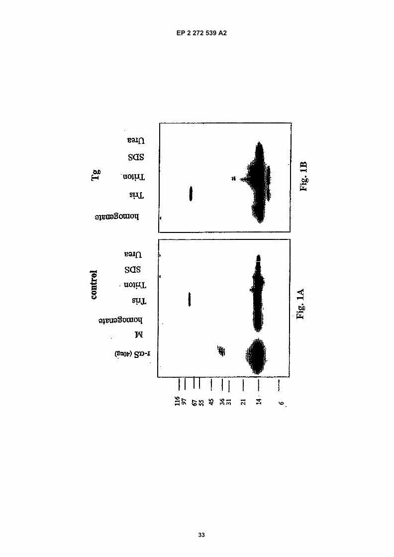
EP 2 272 539 A2
33
EP 2 272 539 A2
34
EP 2 272 539 A2
35
EP 2 272 539 A2
36
EP 2 272 539 A2
37
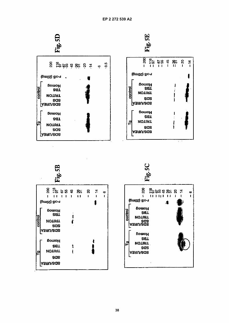
EP 2 272 539 A2
38
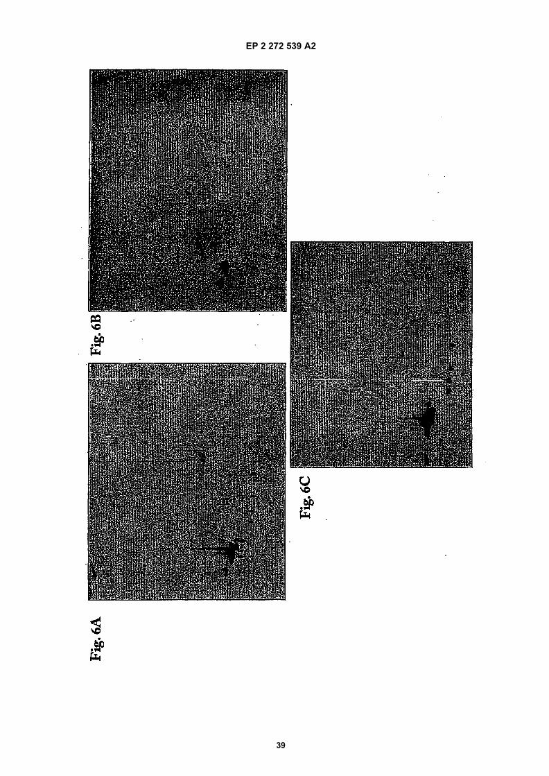
EP 2 272 539 A2
39
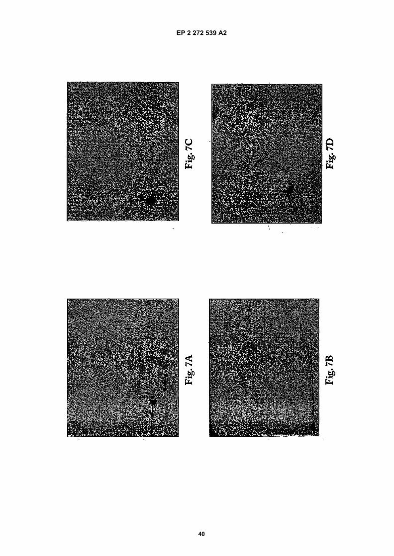
EP 2 272 539 A2
40
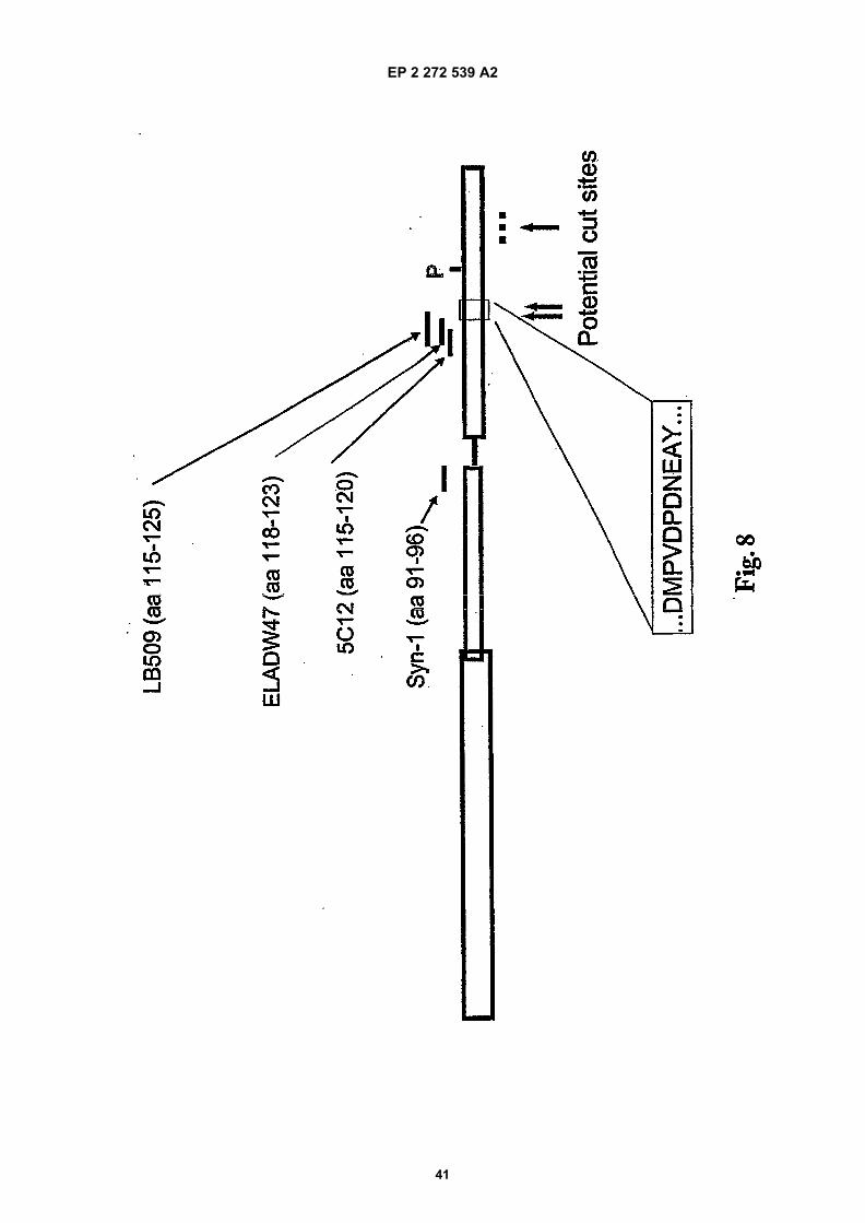
EP 2 272 539 A2
41
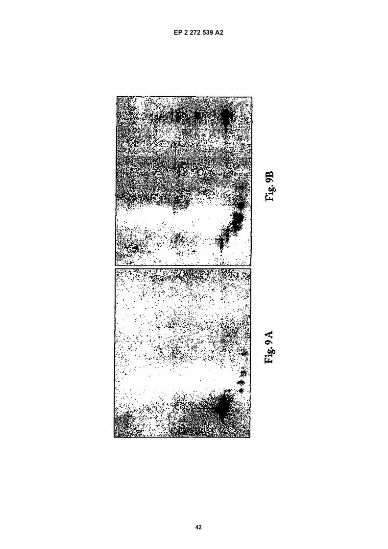
EP 2 272 539 A2
42
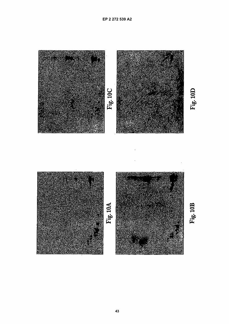
EP 2 272 539 A2
43
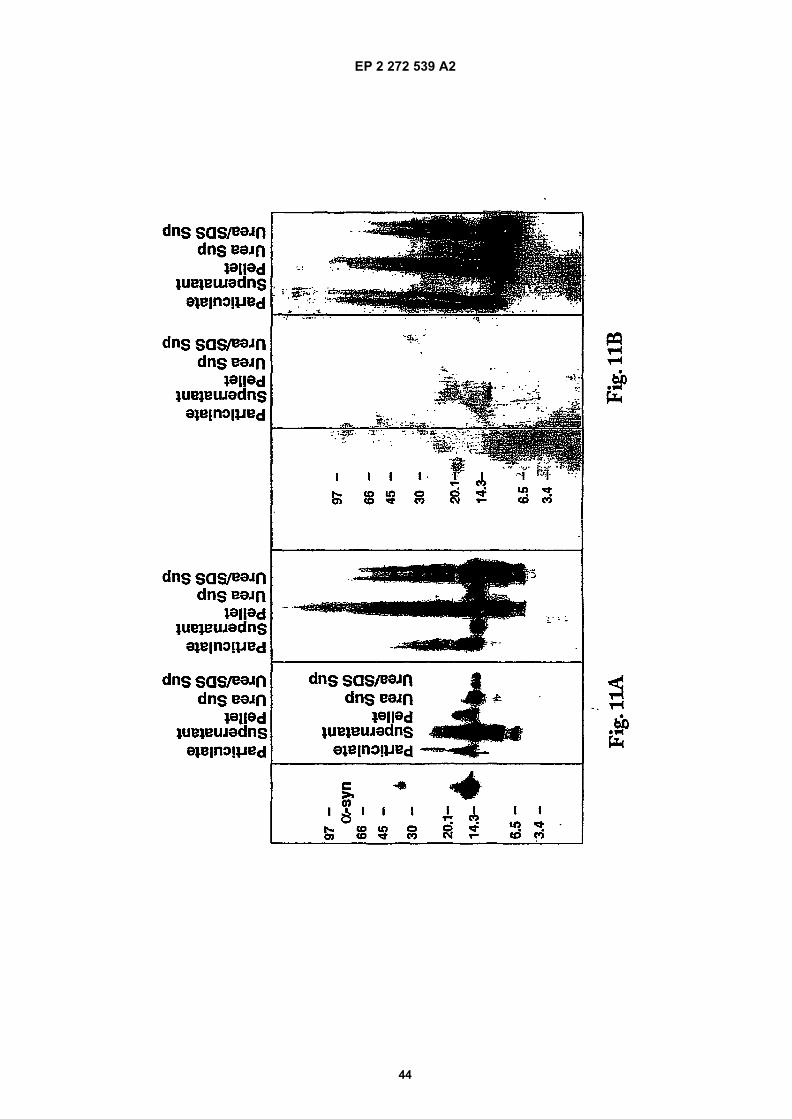
EP 2 272 539 A2
44
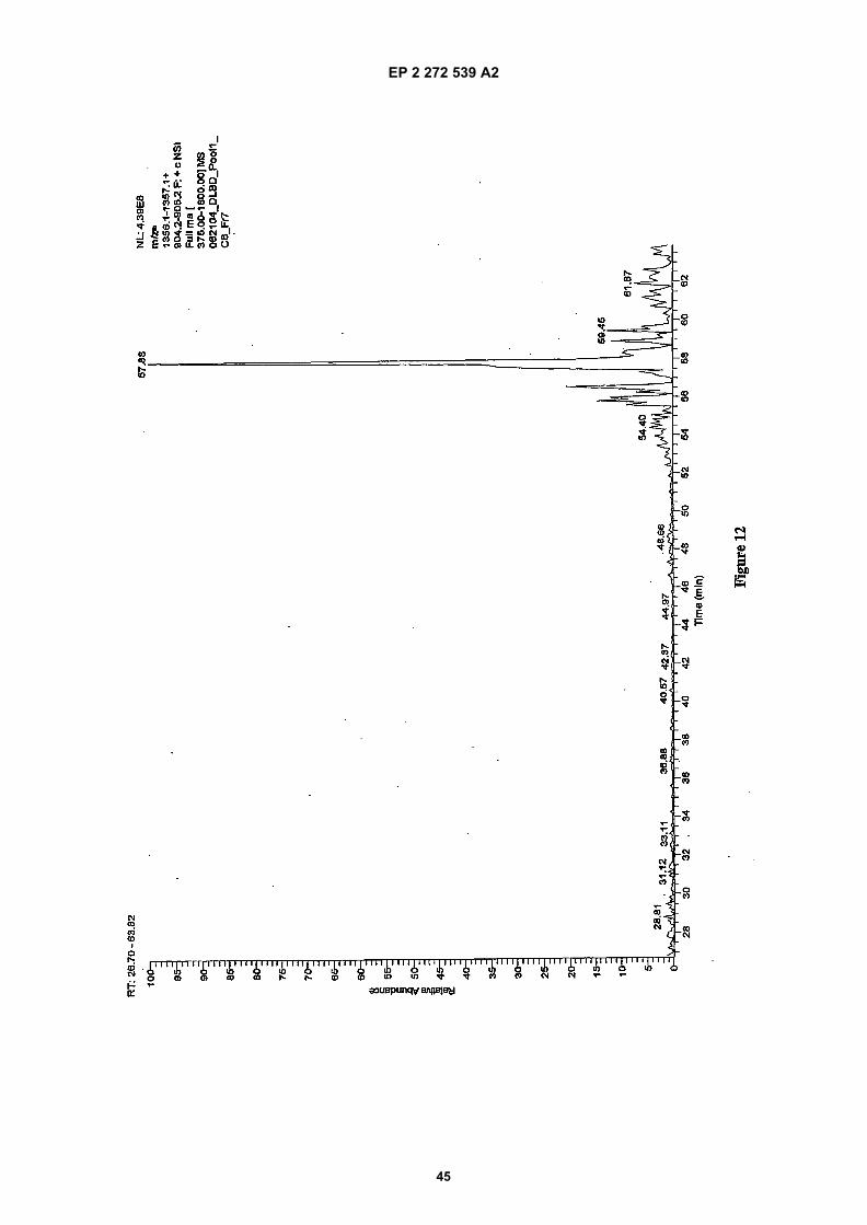
EP 2 272 539 A2
45
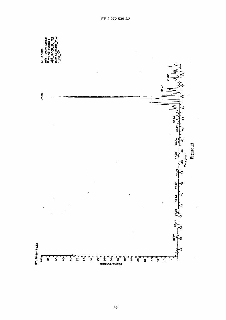
EP 2 272 539 A2
46
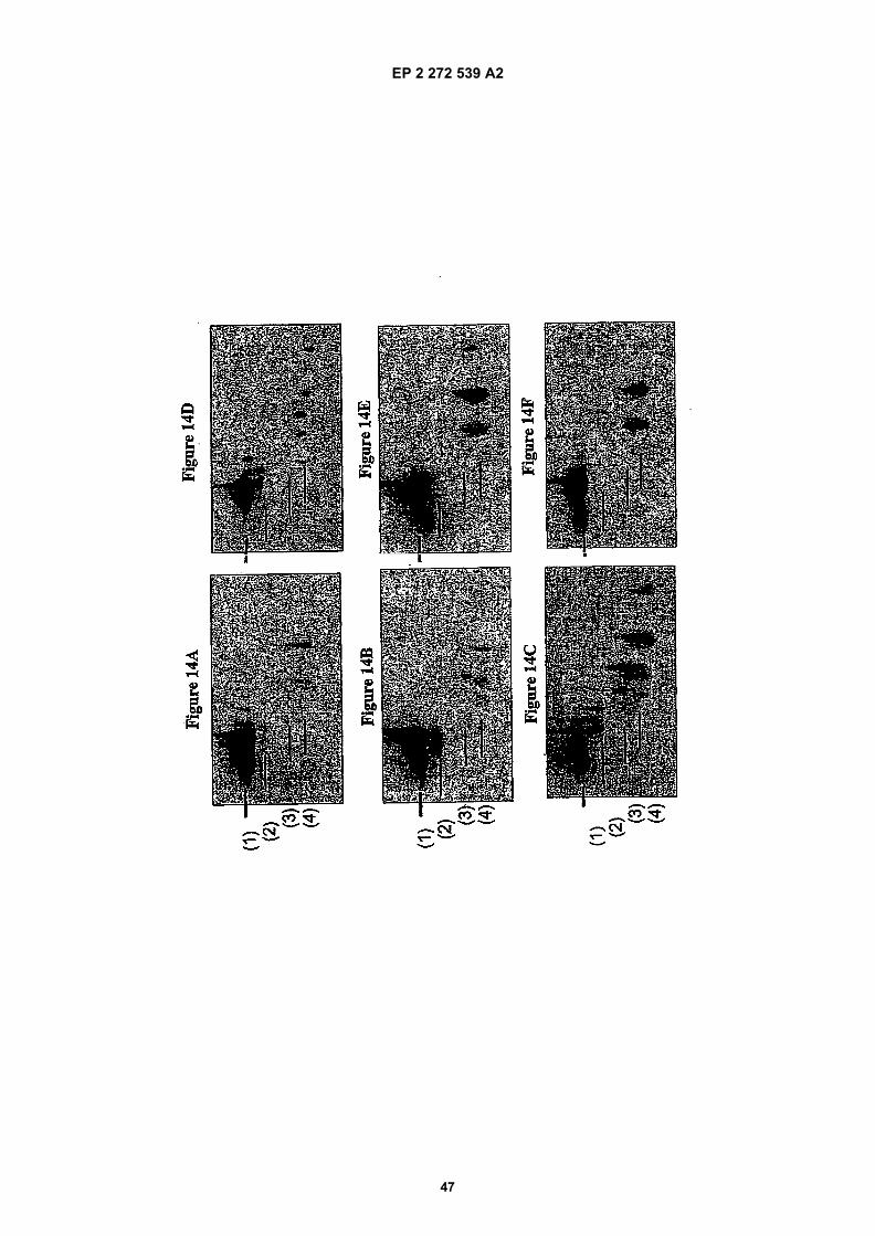
EP 2 272 539 A2
47
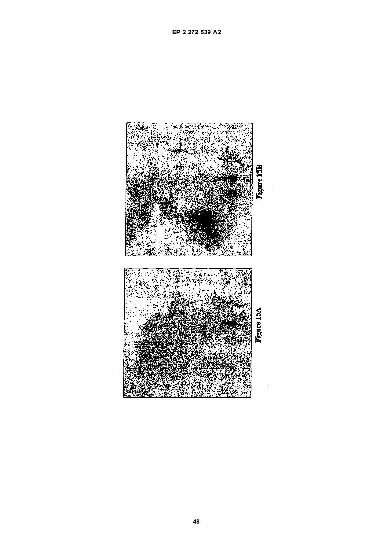
EP 2 272 539 A2
48
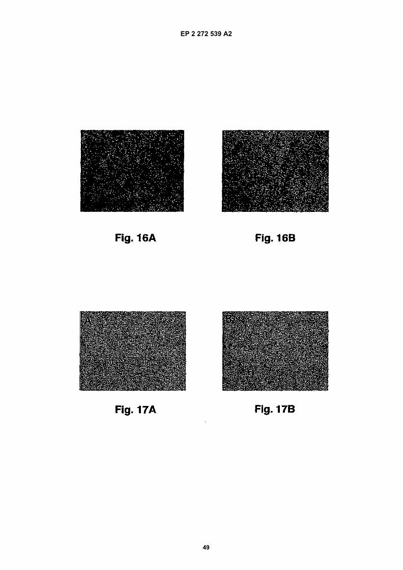
EP 2 272 539 A2
49
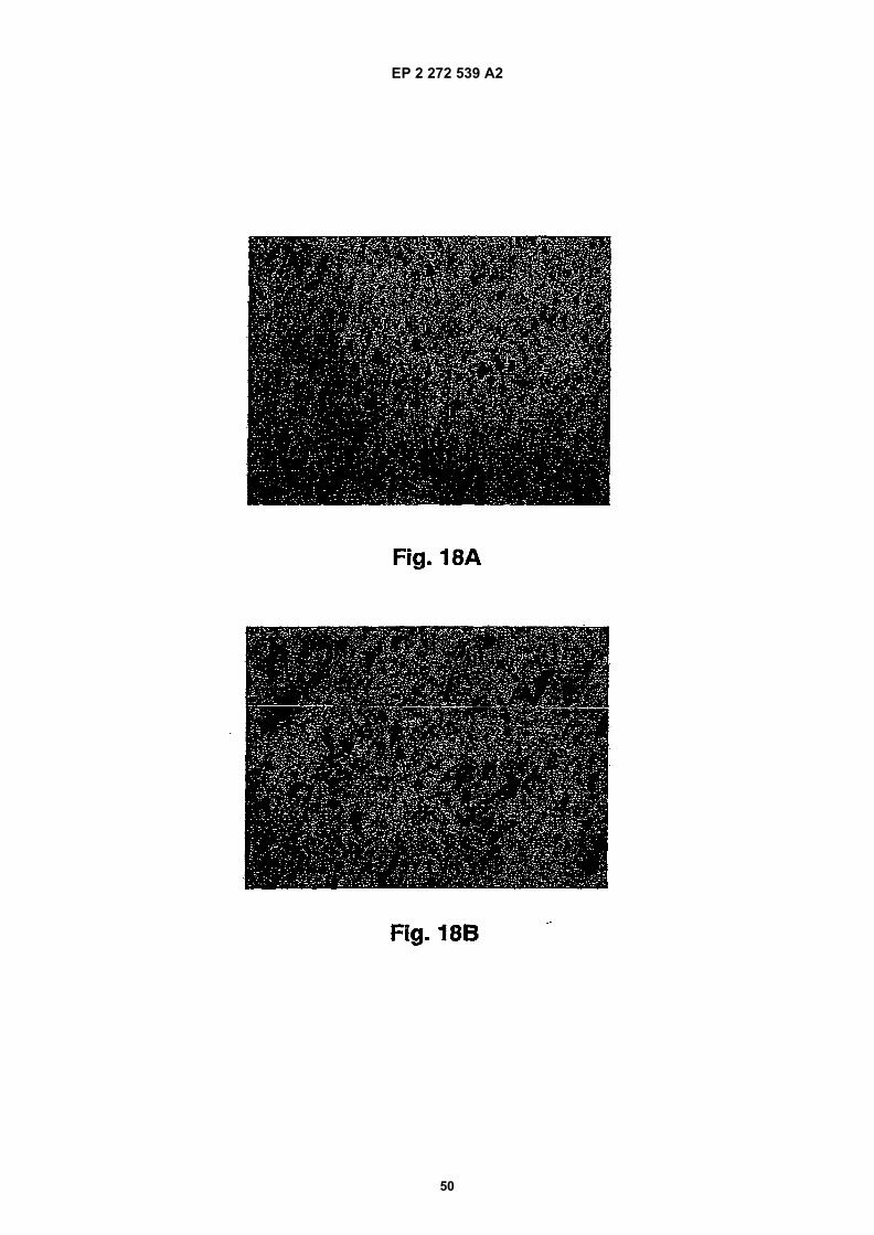
EP 2 272 539 A2
50
EP 2 272 539 A2
51
REFERENCES CITED IN THE DESCRIPTION
This list of references cited by the applicant is for the reader’s convenience only. It does not form part of the Europeanpatent document. Even though great care has been taken in compiling the references, errors or omissions cannot beexcluded and the EPO disclaims all liability in this regard.
Patent documents cited in the description
• US SN11194115 A [0001]• US SN10969335 A [0001]• US SN10850570 A [0001]• US SN60471929 P [0001]• US 040115836 W [0001]• WO 05013889 A [0001]• US 5811633 A [0076]• US 5612486 A, McConlogue [0076]• WO 9512608 A [0086]• WO 9306121 A [0086]• WO 9408051 A [0086]• WO 9535503 A [0086]• WO 9530642 A [0086]• WO 9118980 A [0086]• WO 9007861 A [0095]• US 5693762 A [0095]• US 5693761 A [0095]• US 5585089 A [0095]• US 5530101 A [0095]• US 5225539 A, Winter [0095]• WO 9222653 A, Carter [0095]• US 4634664 A, Oestberg [0096]• US 4634666 A, Engleman [0096]• WO 931222 A, Lonberg [0096]• US 5877397 A [0096]• US 5874299 A [0096]
• US 5814318 A [0096]• US 5789650 A [0096]• US 5770429 A [0096]• US 5661016 A [0096]• US 5633425 A [0096]• US 5625126 A [0096]• US 5569825 A [0096]• US 5545806 A [0096]• WO 9110741 A, Kucherlapati [0096]• WO 9117271 A, Dower [0096]• WO 9201047 A, McCafferty [0096]• US 5877218 A [0096]• US 5871907 A [0096]• US 5858657 A [0096]• US 5837242 A [0096]• US 5733743 A [0096]• US 5565332 A [0096]• US SN60423012 P [0105]• US 0015239 W [0105]• US 0334527 W [0105]• GB 2220211 A [0117]• US 5057540 A [0117]• WO 9840100 A [0117]• WO 9014837 A [0118]• US 4855283 A [0119]
Non-patent literature cited in the description
• McKeith et al. Clinical and pathological diagnosis ofdementia with Lewy bodies (DLB): Report of theCDLB International Workshop. Neurology, 1996, vol.47, 1113-24 [0002] [0075]
• Galasko et al. Clinical-neuropathological correla-tions in Alzheimer’s disease and related dementias.Arch. Neurol., 1994, vol. 51, 888-95 [0002]
• Tanner et al. Epidemiology of Parkinson’s diseaseand akinetic syndromes. Curr. Opin. Neurol., 2000,vol. 13, 427-30 [0002]
• Spillantini et al. Nature, 1997, vol. 388, 839-40[0004]
• Takeda et al. J. Pathol., 1998, vol. 152, 367-72[0004]
• Wakabayashi et al. Neurosci. Lett., 1997, vol. 239,45-8 [0004]
• Kruger et al. Nature Gen., 1998, vol. 18, 106-8[0004]
• Polymeropoulos et al. Science, 1997, vol. 276,2045-7 [0004]
• Masliah et al. Science, 2000, vol. 287, 1265-9 [0004]• Feany et al. Nature, 2000, vol. 404, 394-8 [0004]
[0076]• Iwai A. Biochim. Biophys. Acta, 2000, vol. 1502,
95-109 [0005]• Masliah et al. AM. J. Pathol, 1996, vol. 148, 201-10
[0005]• Ueda et al. Proc. Natl. Acad. Sci. USA, 1993, vol.
90, 11282-6 [0005] [0066]• Harlow ; Lane. Antibodies, A Laboratory Manual.
Cold Spring Harbor Publications, 1988 [0047]• Smith ; Waterman. Adv. Appl. Math., 1981, vol. 2,
482 [0049]• Wunsch. J. Mol. Biol., 1970, vol. 48, 443 [0049]• Pearson ; Lipman. Proc. Nat’l. Acad. Sci. USA,
1988, vol. 85, 2444 [0049]
EP 2 272 539 A2
52
• Altschul et al. J. Mol. Biol., 1990, vol. 215, 403-410[0050]
• Henikoff ; Henikoff. Proc. Natl. Acad. Sci. USA,1989, vol. 89, 10915 [0050]
• Karlin ; Altschul. Proc. Nat’l. Acad. Sci. USA, 1993,vol. 90, 5873-5787 [0051]
• Songsivilai ; Lachmann. Clin. Exp. Immunol.,1990, vol. 79, 315-321 [0054]
• Kostelny et al. J. Immunol., 1992, vol. 148,1547-1553 [0054]
• Stahli et al. Methods in Enzymology, 1983, vol. 9,242-253 [0057]
• Kirkland et al. J. Immunol., 1986, vol. 137,3614-3619 [0057]
• Harlow ; Lane. Antibodies, A Laboratory Manual.Cold Spring Harbor Press, 1988 [0057]
• Morel et al. Molec. Immunol., 1988, vol. 25 (1), 7-15[0057]
• Cheung et al. Virology, 1990, vol. 176, 546-552[0057]
• Moldenhauer et al. Scand. J. Immunol., 1990, vol.32, 77-82 [0057]
• Cierchanover. EMBO J., 1998, vol. 17, 7151-7160[0062]
• Iwai et al. Biochemistry, vol. 34, 10139-10145 [0072]• Jensen et al. Biochem. J., 1995, vol. 310, 91-94
[0072]• Sambrook et al. Molecular Cloning: A Laboratory
Manual. C.S.H.P. Press, 1989 [0074]• Masliah et al. Am. J. Pathol., 1996, vol. 148, 201-10
[0076]• Games et al. Nature, 1995, vol. 373, 523 [0076]• Hsiao et al. Science, 1996, vol. 274, 99 [0076]• Staufenbiel et al. Proc. Natl. Acad. Sci. USA, 1997,
vol. 94, 13287-13292 [0076]• Sturchler-Pierrat et al. Proc. Natl. Acad. Sci. USA,
1997, vol. 94, 13287-13292 [0076]• Borchelt et al. Neuron, 1997, vol. 19, 939-945
[0076]• Masliah et al. PNAS USA, 2001, vol. 98,
12245-12250 [0076]• Brain Pathology, 2003, vol. 13, 364-372 [0076]
• Bjorklund. Trends Neurosci., 2003, vol. 26, 386-92[0076]
• Lotharius et al. J. Biol. Chem., 2002, vol. 277,38884-94 [0076]
• Zhou et al. Brain Research, 2000, vol. 866, 33-43[0076]
• Hsue et al. Am. J. Patho1, 2000, vol. 157, 401-410[0078]
• Wood. J. Biol. Chem., 1999, vol. 274, 19509-19512[0080]
• Harlow ; Lane. Antibodies, A Laboratory Manual.CSHP, 1988 [0093]
• Queen et al. Proc. Natl. Acad. Sci. USA, 1989, vol.86, 10029-10033 [0095]
• Oestberg et al. Hybridoma, 1983, vol. 2, 361-367[0096]
• Nature, 1994, vol. 148, 1547-1553 [0096]• Nature Biotechnology, 1996, vol. 14, 826 [0096]• Kensil et al. Vaccine Design: The Subunit and Ad-
juvant Approach. Plenum Press, 1995 [0117]• Stoute et al. N. Engl. J. Med., 1997, vol. 336, 86-91
[0117]• Chang et al. Advanced Drug Delivery Reviews,
1998, vol. 32, 173-186 [0120]• Remington’s Pharmaceutical Science. Mack Publish-
ing Company, 1980 [0121]• Langer. Science, 1990, vol. 249, 1527 [0124]• Hanes. Advanced Drug Delivery Reviews, 1997, vol.
28, 97-119 [0124]• Glenn et al. Nature, 1998, vol. 391, 851 [0126]• Paul et al. Eur. J. Immunol., 1995, vol. 25, 3521-24
[0126]• Cevc et al. Biochem. Biophys. Acta, 1998, vol. 1368,
201-15 [0126]• Jensen et al. J. Biol. Chem., 2000, vol. 275,
21500-21507 [0133]• Tofaris et al. J. Biol. Chem., 2003, vol. 278 (45),
444405-44411 [0134]• Masliah et al. Science, 2000, vol. 287, 1265 [0140]
[0141]• Morris. Leam Motivat., 1981, vol. 12, 239-260 [0153]• Hsue et al. Am. J. Pathol., 2000, vol. 157, 401-410
[0154]