trkb modulates fear learning and amygdalar synaptic plasticity by specific docking sites
TRANSCRIPT
Cellular/Molecular
TrkB Modulates Fear Learning and Amygdalar SynapticPlasticity by Specific Docking Sites
Gabriele Musumeci,1* Carla Sciarretta,1* Antonio Rodríguez-Moreno,2* Mumna Al Banchaabouchi,1
Vicente Negrete-Díaz,2 Marco Costanzi,3 Valeria Berno,1 Alexei V. Egorov,4 Oliver von Bohlen und Halbach,4
Vincenzo Cestari,3,5 Jose M. Delgado-García,2 and Liliana Minichiello1,6
1Mouse Biology Unit, European Molecular Biology Laboratory, 00015 Monterotondo, Italy, 2Division de Neurociencias, Universidad Pablo de Olavide,41013 Sevilla, Spain, 3Istituto di Neuroscienze, Consiglio Nazionale delle Ricerche, 00143 Rome, Italy, 4Interdisciplinary Center for Neurosciences,Department of Neuroanatomy, University of Heidelberg, D-69120 Heidelberg, Germany, 5Facolta di Scienze della Formazione, Universita Lumsa,00193 Rome, Italy, and 6Centre for Neuroregeneration, University of Edinburgh, EH16 4SB Edinburgh, United Kingdom
Understanding the modulation of the neural circuitry of fear is clearly one of the most important aims in neurobiology. Protein phos-phorylation in response to external stimuli is considered a major mechanism underlying dynamic changes in neural circuitry. TrkB(Ntrk2) neurotrophin receptor tyrosine kinase potently modulates synaptic plasticity and activates signal transduction pathways mainlythrough two phosphorylation sites [Y515/Shc site; Y816/PLC� (phospholipase C�) site]. To identify the molecular pathways required forfear learning and amygdalar synaptic plasticity downstream of TrkB, we used highly defined genetic mouse models carrying single pointmutations at one of these two sites (Y515F or Y816F) to examine the physiological relevance of pathways activated through these sites forpavlovian fear conditioning (FC), as well as for synaptic plasticity as measured by field recordings obtained from neurons of differentamygdala nuclei. We show that a Y816F point mutation impairs acquisition of FC, amygdalar synaptic plasticity, and CaMKII signaling atsynapses. In contrast, a Y515F point mutation affects consolidation but not acquisition of FC to tone, and also alters AKT signaling. Thus,TrkB receptors modulate specific phases of fear learning and amygdalar synaptic plasticity through two main phosphorylation dockingsites.
IntroductionEmotional fear-related disorders are among the most commonhuman psychiatric disorders. Determining the molecular mech-anisms underlying the development and manifestation of suchdisorders is essential. Pavlovian fear conditioning (FC), a simpleform of associative learning, involves learning that certain envi-ronmental stimuli predict aversive events and is considered to bea model system to examine the neurobiological basis of learningand memory in the mammalian brain. The amygdala is centralfor the acquisition, storage, and expression of conditioned fearmemory (Lavond et al., 1993; LeDoux, 1996; Davis, 1997; Fendtand Fanselow, 1999); it consists of different nuclei, including thelateral (LA), basolateral (BL), and basal medial (BM) nuclei,which together form the basolateral complex (BLA), and the cen-
tral nucleus (CeA) (Swanson and Petrovich, 1998). The BLA re-ceives synaptic inputs from different sensory structures, and le-sions of these structures impair pavlovian FC (Gale et al., 2004;Koo et al., 2004). Synaptic inputs to the LA from the thalamicmedial geniculate nucleus and from the auditory cortex are es-sential for conditioning with an auditory conditioned stimulus(CS) (LeDoux et al., 1990; Romanski and LeDoux, 1992; Cam-peau and Davis, 1995), whereas projections from the hippocam-pus to the BLA support conditioning with contextual CSs (Kimand Fanselow, 1992; Phillips and LeDoux, 1992; Maren et al.,1997). FC also induces behavioral long-term potentiation (LTP)in the amygdala, and synaptic responses measured in the amyg-dala of a behaving rat (Rogan et al., 1997) and in amygdalar slicesobtained from previously conditioned rats (McKernan andShinnick-Gallagher, 1997) are similar. Interestingly, behavioralLTP exhibits properties common to tetanic LTP in both prepa-rations. TrkB has been genetically shown to play an importantrole in hippocampal synaptic plasticity and learning (Minichielloet al., 1999; Xu et al., 2000). BDNF/NT4 binding to TrkB activatesthree main intracellular signaling cascades. The Ras/mitogen-activated protein kinase (MAPK) pathway and the phosphoino-sitide 3-kinase (PI3K) pathway are activated primarily throughShc/FRS-2 binding to Y515, whereas the calcium/calmodulin ki-nase pathway is activated through phospholipase C� (PLC�) re-cruitment at Y816. We have shown that the Ras/MAPK andPLC�/CaMK/CREB (cAMP response element-binding protein)pathways act in parallel downstream of TrkB and that the PLC�
Received April 8, 2009; revised May 15, 2009; accepted July 6, 2009.This work was supported in part by grants from the European Union (EU) [EU Sixth Framework Programme (FP6)
Project Memories Grant 037831; EU FP6 StemStroke Grant 037526] to L.M., Grants BFU2005-01024 and BFU2006-14155 from the Spanish Ministry of Education and Science to J.M.D.-G. and A.R.-M., respectively, and DeutscheForschungsgemeinschaft Grant SFB636/A5 to O.v.B.
*G.M., C.S., and A.R.-M. contributed equally to this work.Correspondence should be addressed to Dr. Liliana Minichiello, EMBL Mouse Biology Unit, Via Ramarini, 32,
00015 Monterotondo, Italy. E-mail: [email protected]. Sciarretta’s present address: Actelion Pharmaceuticals Ltd., Gewerbestrasse 16, CH-4123 Allschwil,
Switzerland.O. von Bohlen und Halbach’s present address: Institute of Anatomy and Cell Biology, Ernst Moritz Arndt Univer-
sity, D-17487 Greifswald, Germany.DOI:10.1523/JNEUROSCI.1707-09.2009
Copyright © 2009 Society for Neuroscience 0270-6474/09/2910131-13$15.00/0
The Journal of Neuroscience, August 12, 2009 • 29(32):10131–10143 • 10131

docking site is necessary for TrkB-mediated hippocampal synap-tic plasticity (Minichiello et al., 2002). Moreover, a TrkB Y816Fbut not Y515F point mutation impairs the acquisition of an as-sociative learning task and in vivo hippocampal LTP (Gruart etal., 2007). Experiments based on temporal changes in BDNF geneexpression and the intra-amygdala infusion of a lentiviral vectorexpressing a dominant-negative TrkB isoform have establishedan essential role for TrkB in pavlovian FC acquisition (Rattiner etal., 2004). However, the molecular pathways downstream ofTrkB contributing to fear learning and amygdalar synaptic plas-ticity were not identified. The use of genetic mouse models car-rying a point mutation at either the Shc or the PLC� docking siteof TrkB (trkBSHC or trkBPLC, respectively) allows examination ofthe physiological relevance of pathways activated through thesesites during pavlovian FC. We found that in vivo, pathways acti-vated through the TrkB/PLC� site modulate the acquisition ofconditioned fear responses to contextual and auditory stimuli, aswell as, synaptic plasticity at the lateral– basolateral (LA–BL) andthe thalamic–lateral (thalamic–LA) amygdala synapses. In con-trast, the TrkB/Shc site through its downstream effector(s)mainly modulates the consolidation but not acquisition of fearlearning to tone and the amygdalar synaptic plasticity at the tha-lamic–LA synapses but not at the LA–BL synapses, indicating thatTrkB Y816 and Y515 contribute to specific phases of fear learningand amygdalar synaptic plasticity.
Materials and MethodsEthics statementAll animal procedures were conducted in accordance with the rules per-taining to animal well-being and were performed in agreement withnational and international laws and policies [European EconomicCommunity Council Directive 86/609, OJ L 358, 1, December 12,1987; National Institutes of Health (NIH) Guide for the Care and Use ofLaboratory Animals, NIH Publication No. 85-23, 1985, revised in 1995).All initiatives to minimize animal experimentation and to alleviate ani-mal pain, suffering, and distress were taken into account.
Mouse strainsThe mouse strains used in this study have been previously described(Minichiello et al., 1998, 2002); however, a summary of the strategiesused and validation of the point mutations are reported in the sup-plemental Methods, available at www.jneurosci.org as supplementalmaterial.
Behavioral proceduresFear conditioningAdult mice (2– 4 months of age) were trained and tested in an operantchamber (18.5 � 18 � 21.5 cm) possessing aluminum sidewalls andPlexiglas rear and front walls (Coulbourn Instruments). The auditorycue (tone) emanated from a loudspeaker located in the sidewall, 15 cmfrom the floor. The activity of animals was recorded directly on a com-puter through a single camera located at the top of the chamber. Thepresentation of tone and shock stimuli in all training and testing sessionswas controlled by GraphicState software (Coulbourn Instruments). Thechamber was cleaned with ethanol and dried between mice after eachtraining or testing session.
Conditioning. Mice were allowed to acclimate to the training chamberfor 2 min, and then a tone (CS) of 2800 Hz frequency and 85 dB intensitywas presented for 30 s and coterminated in the last 2 s with a mildfootshock (0.5 mA) [unconditioned stimulus (US)]. Two minutes later,another CS–US pairing was presented, and the mice were returned totheir home cages 30 s later. To facilitate the acquisition of conditionedfear in trkBPLC/� mutants, five CS–US pairings were used instead of twoCS–US pairings.
Behavioral testing. To test for a conditioned fear response to context,the mice were placed in the same chamber used for the conditioning trial
24 h after conditioning and allowed to explore for 4 min without presen-tation of the auditory CS or footshock. Freezing behavior, defined ascomplete absence of voluntary movements except for respiratory move-ments, was scored every 1 s. Twenty-four hours after the contextualconditioning test, the mice were tested for cue memory by returningthem to the same chamber, which was modified by the addition of vari-ous shapes and designs on the walls and a vanilla scent. Freezing behaviorwas scored for 2 min before delivery of the tone and then for 2 min in thepresence of a continuous auditory cue. Freezing behavior was measuredwith an automated system whereby continuous video data were analyzedwith the aid of Freeze-frame software (Coulbourn Instruments). Thesoftware measures the variance in pixel intensity across successive videoframes (taken at 1 Hz) and computes its SD. A threshold is then appliedto the data to yield a percentage freezing score. Two-way ANOVA tests(with genotype as a between-subjects factor and context or tone tests as awithin-subject factor) were used for statistical analysis with p � 0.05 setas the criterion for statistical significance. Post hoc group comparisonswere performed with Fisher’s LSD test.
Radial mazeThe radial maze test was performed essentially as described previously(Minichiello et al., 1999). Briefly, adult male mice (3– 6 months of age)were singly housed with water provided ad libitum and gradually reducedto 85% of their free-feeding body weight, which was maintained for theduration of the experiment by providing the mice with a premeasuredamount of chow each day. The maze was constructed from gray plasticand was comprised of eight identical arms radiating 37 cm from a centralstarting platform (perimeter was 7 � 8 cm). At the end of each arm 20 mgof food pellets was placed in a cup. All arms were baited only once and atthe beginning of each daily session. Animals received one trial per day for10 consecutive training days. Each daily trial terminated when 8 correctchoices were made (maximum 15 choices) or 15 min elapsed. An armchoice was defined as placement of all paws on a maze arm. An error wasscored when the mouse entered a previously explored arm. The percent-age of errors made over 15 trials, the number of correct arm choices in thefirst 8 trials, and the occurrence of first error were considered. For statis-tical analysis, mean values were compared using a two-way ANOVA withthe factors genotype (2 levels � mutants and controls) and learning (10levels � training days), in which the numbers of correct arm choices outof the first 8 trials, the percentages of errors out of 15 trials, or theoccurrences of the first error on each training day were compared. Indi-vidual between-group comparisons, when appropriate, were performedusing post hoc tests (Duncan multiple-range test).
ElectrophysiologyCoronal slices (400 �m thick) containing the amygdala were preparedand maintained using standard procedures [artificial CSF (ACSF) me-dium: 124 mM NaCl, 3 mM KCl, 1.25 mM KH2PO4, 2 mM MgSO4, 26 mM
NaHCO3, 1.8 mM CaCl2, and 10 mM glucose; temperature, 28°C; sub-merged recording). To evoke field EPSPs (fEPSPs), electric pulses wereapplied by a bipolar electrode placed either in the LA nucleus near the BLnucleus, in which recording glass electrodes were located, or in the ven-tral striatum, just medial to the LA, to activate fibers originating, at leastin part, in the auditory thalamus. The recording glass electrodes werelocated in the LA nucleus (Weisskopf et al., 1999). Synaptic field poten-tials were elicited at a frequency of 0.1 Hz. The slope of the recorded fieldwas calculated and used as a measurement of synaptic strength. LTP wasinduced with a theta-burst stimulation (TBS) of 10 bursts of four pulseseach (100 Hz, 100 ms duration, 200 ms interburst interval) with the samestrength as the test stimulus. This TBS was repeated three times at inter-vals of 10 s. Alternatively, LTP was induced with a tetanus of 3 � 30 pulses(100 Hz, 100 ms duration, at intervals of 10 s) with the same strength asthe test stimulus. Strong tetanic stimulation consisted of three series of100 pulses (100 Hz) with pulse lengths of 100 ms and delivered at inter-vals of 5 min. Poststimulation recordings were continued for 60 min forearly LTP (E-LTP) and for 180 min for late LTP (L-LTP). E-LTP orL-LTP was considered successfully induced when the average of thefEPSP slope size (measured at 55– 60 min or 175–180 min, respectively,
10132 • J. Neurosci., August 12, 2009 • 29(32):10131–10143 Musumeci et al. • TrkB (Ntrk2) Differentially Modulates Fear Learning
after TBS or tetanus) showed an increase of at least 20% above baseline(100%) values. Routinely, paired-pulse facilitation (PPF) was tested atintervals of 10, 30, 40, 50, 100, and 200 ms. All measurements wereperformed and analyzed in a strictly blind manner. The genotypes of theanimals were revealed only after the electrophysiological experimentsand their evaluation were complete. To analyze the functionality ofthe NMDA and AMPA/kainate receptors, fEPSPs were recorded ini-tially for 20 min (baseline recording) and then after the addition of6-cyano-7-nitroquinoxaline-2,3-dione (CNQX) in low-Mg2� (0.5 mM)ACSF applied to the bath solution. After 15 min, DL-2-amino-5-phosphonovalerate (AP-5) (50 mM) together with CNQX was applied for30 min to the same slice in low-Mg 2� ACSF. Afterward, normal ACSFwas used for washout. The results were analyzed for statistical signifi-cance using two-tailed, unpaired Student’s t tests.
Anatomical/biochemical experimentsConditioningAdult mice (2– 4 months) were trained in a polymodal operant chamber(see above). Before training, they were allowed to acclimate for 2 min,and then two or five conditioning trials were applied consisting of a 30 s,2800 Hz, and 85 dB tone that coterminated with a 2 s, 0.5 mA footshock;intertrial intervals were 120 s. Mice were returned to their home cagesand 45 min later they were either immediately killed (when analyzed forpAKT and pMAPKs) or first tested for both context (3 min) and cue (2min tone) and then killed immediately thereafter (when analyzed forpCaMKII). Brains were taken for histological and biochemical analysis.Control animals were not exposed to tones or shocks.
Brain sample collectionFor biochemical analysis, mice were killed by cervical dislocation, and thebrains were removed from the skull and (without olfactory bulbs) posi-tioned in a cold matrix with which 3 mm coronal slices were collected,followed by the isolation of amygdala nuclei from the basolateral por-tions of the slice. Samples were frozen in liquid nitrogen and stored at�80°C until use.
BiochemistryBrain samples were homogenized using mi-cropestles (Eppendorf) and lysed on ice inNP-40 complete lysis buffer [10% glycerol,50 mM HEPES/NaOH (pH 8.0), 100 mM KCl,1% NP-40, 2 mM EDTA, 2 mM DTT, 10 mM
NaF, 1 mM Na3VO4, 1 mM NaPP, 5 mM ben-zamidine, 10 �g/ml leupeptine, 14 �g/mlaprotinin]. Cellular debris were removed bycentrifugation at 16,000 rpm for 20 min at4°C. Cortical neurons were washed twicewith cold PBS and then lysed on ice for 15min with 0.6 ml of 1% Triton X-100 lysisbuffer [50 mM Tris (pH 7.5), 120 mM NaCl,10 mM NaF, 1 mM Na3VO4, and protease in-hibitors as described above]. Insoluble mate-rial was removed from the protein extractsby centrifugation at 13,000 rpm for 15 min at4°C. The protein content in supernatant fractionswas quantified (Bio-Rad DC protein assay kit)and aliquots were frozen in liquid nitrogen andstored at �80°C until use. Protein levels were an-alyzed by Western blot as previously described(Minichiello et al., 1998, and supplementalMethods, available at www.jneurosci.org as sup-plemental material). The antibodies (Abs) usedwere anti-pAKT [Cell Signaling, catalog no.(Cat.) 9271; 1:1000], anti-AKT (Cell Signaling,Cat. 9272; 1:1000), anti-pMAPK (New EnglandBiolabs, clone E10; 1:2000), and anti-ERK1 (ex-tracellular signal-regulated kinase 1) (Zymed 13-8600; 1:3000). The relative levels of proteins werequantified as described in the supplementalMethods, available at www.jneurosci.org as sup-plemental material.
Synaptosome preparationBrain samples were processed according to the synaptosomal prepa-ration described by Nagy and Delgado-Escueta (1984). Briefly, theamygdalae of each animal were homogenized in 2 ml of buffer [0.32 M
sucrose, 1 mM EDTA, 1 mg/ml BSA, 5 mM HEPES (pH 7.4, reached using1 M NaOH)] by use of a Dounce homogenizer (10 strokes and 520 rpm).Thirty microliters of crude homogenate was removed and served as thewhole-tissue lysate sample and the remaining homogenate was centri-fuged for 10 min at 3000 � g. The supernatants were subsequently cen-trifuged for 12 min at 14,000 � g and resultant pellets were resuspendedin 220 �l of Krebs–Ringer buffer [140 mM NaCl, 5 mM KCl, 10 mM
HEPES, 1 mM EDTA, 5 mM glucose (pH 7.4, reached using 1 M NaOH)]and 180 �l of Percoll (Sigma-Aldrich, P7828). Samples were then centri-fuged for 2 min at 14,000 � g. The synaptosome fraction of the gradientwas removed, washed twice with 500 �l of Krebs–Ringer buffer, andcentrifuged for 30 s at 14,000 � g. The final pellets were resuspended in 40�l of Krebs–Ringer buffer. All steps of the procedure were performed at 4°Cusing ice-cold buffers. Samples were separated by SDS-PAGE using 10%acrylamide and transferred onto nitrocellulose membranes. The blots wereprobed using anti-pCaMKII (ProMega, V1111; 1:1000), anti-CaMKII(Santa Cruz, H-300 s.c.-13082; 1:1000), anti-synapsin I/II (Synaptic Sys-tems, 106 002; 1:5000), and anti-PSD95 (Upstate Biotechnology,cloneK28/43 05-494; 1:200,000). The secondary antibodies used were agoat anti-rabbit HRP-conjugated (Jackson ImmunoResearch, 115-035-003; 1:5000) and a goat anti-mouse HRP-conjugated (Jackson Immu-noResearch 115-035-146; 1:5000) IgG.
HistologyMice were anesthetized with avertin (20 mg/kg, i.p.) and transcardi-ally perfused with ice-cold PBS followed by 3% paraformaldehyde(PFA) in 0.1 M phosphate buffer (PB). The perfusion was performedusing a peristaltic pump and a flow rate of 1 ml/min. The brains wereremoved from the skull and postfixed in 3% PFA–PB overnight at
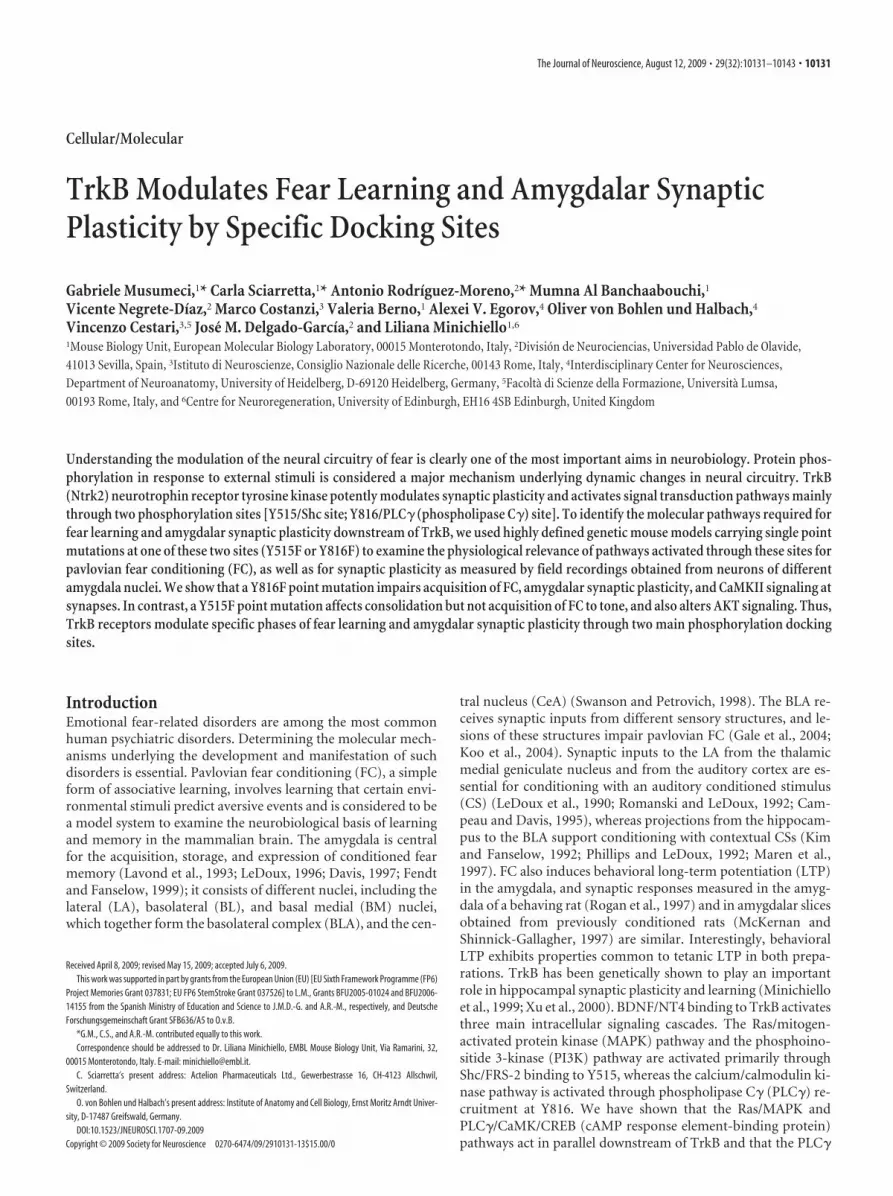
Figure 1. Associative CS–US pairing is impaired in trkBPLC/� mutants but normal in trkBSHC/� mutants. A, The percent-age of freezing of control (trkBWT/�) and mutant (trkBPLC/�) mice from a typical training trial. Whereas the control mice(trkBWT/�) showed normal acquisition during the training phase, the trkBPLC/� mutants showed impaired or highlyreduced CS–US association (ANOVA; p � 0.001). B, C, The control (trkBWT/�) and the mutant (trkBPLC/�) group of micewere tested 24 h later for contextual fear conditioning (B) followed by cued fear conditioning (C). D, Freezing of trkBSHC/�
mutants during the acquisition phase was similar to that of control mice (trkB�/�). E, F, Twenty-four hours after condi-tioning, both groups of mice were tested first for contextual fear conditioning (E) and then for cued fear conditioning (F ).n � number of mice per group. Bars represent mean � SEM. **Statistically significant (see Results), p � 0.001; ns, notsignificant; ITI, intertrial interval; Hb, habituation.
Musumeci et al. • TrkB (Ntrk2) Differentially Modulates Fear Learning J. Neurosci., August 12, 2009 • 29(32):10131–10143 • 10133

4°C, cryoprotected in 30% sucrose–PB at 4°C for 48 h, and thenfrozen in OCT Tissue Tek compound (Bayer) by immersion in liquidnitrogen-cooled isopentane. Immunohistochemistry was performed as de-scribed by Minichiello et al. (1999). Briefly, 30-�m-thick coronal free-floating sections through the amygdala were obtained using a cryostat.Selected sections were immunostained using an anti-pMAPK Ab (CellSignaling, 9106; 1:4000). More details of the tissue stainings are re-ported in the supplemental Methods, available at www.jneurosci.orgas supplemental material.
Neuronal culturesCerebral cortices were dissected from embryonic day 15.5 mouse em-bryos generated from breedings of control mice or trkB point mutants.Cortical neurons were dissociated and plated onto poly-L-lysine hy-drobromide (Sigma) in the presence of 10% heat-inactivated horseserum for 3 h. Cultures were maintained for 16 h before ligand stim-ulation in N2–MEM at 37°C in 5% pCO2. Cells were stimulated with5 or 50 ng/ml of purified recombinant BDNF (Genentech and Regen-eron Pharmaceuticals) as described previously (Minichiello et al.,2002).
ImagingImmunofluorescenceSections were imaged on a Leica TCS SP5 confocal microscope using aLeica 40�/1.4 NA Plan Apochromat oil objective.
ImmunohistochemistryImaging of stained sections was performed using a Leica bright-fieldDMRA microscope.
ResultsMouse modelsTo determine which site-specific activated pathway(s) down-stream of the TrkB receptor contributes to fear-related synapticplasticity in the amygdala, we used genetic mouse models carry-ing a point mutation at the Shc or the PLC� docking site (trkBSHC
or trkBPLC) of the TrkB receptor [these two mouse strains werepreviously described by Minichiello et al. (1998, 2002); also seesupplemental Methods, available at www.jneurosci.org as sup-plemental material]. To avoid concerns about developmental ab-normalities, we focused mainly on heterozygous point mutantsof both strains, as they do not display relevant developmentalphenotypes (supplemental Methods and Figs. S1, S2, available at
www.jneurosci.org as supplemental material) and thus allow forbehavioral analysis.
Differential regulation of fear-related learning by the PLC�and Shc site in TrkBAnalysis of behavior in the open-field test and elevated plus mazerevealed no significant difference in terms of locomotor, nonlo-comotor, grooming and object exploration activity, andanxiety-like behavior between all groups of mice analyzed[trkB�/�, trkBSHC/�, trkB wild-type knock-in (trkBWT/�), andtrkBPLC/� mice] (supplemental Fig. S3, available at www.jneurosci.org as supplemental material). We then used an asso-ciative learning paradigm (classical fear conditioning) to assessaversive learning and memory. On the conditioning day, micewere given two pairings of a tone (CS) and a footshock (US)(Fig. 1 A, D). The trkBPLC/� mice showed impaired associativelearning of the CS–US pairing during the training phase com-pared with the control mice (trkBWT/�) (Fig. 1A), as evidencedby a similar amount of freezing to the second CS presentation(CS2) as to the first CS presentation (CS1) (CS2 vs CS1; F(4.83) �2.5; p � 0.29). In contrast, the control mice (trkBWT/�) showednormal acquisition during the training phase (CS2 vs CS1; F(6.83) �19.1; p � 0.001) and an overall significantly higher number offreezing responses compared with the trkBPLC/� mice (F(2.648) �9.398; p � 0.001). At 24 h, control (trkBWT/�) and mutant (trk-BPLC/�) mice were tested for contextual fear conditioning. Asexpected, the trkBPLC/� mice were unable to recognize the contextand displayed almost no freezing responses compared with theircontrols (trkBWT/� mice) (F(7.827) � 26.336; p � 0.001) (Fig. 1B).Twenty-four hours after the test of contextual conditioning, themice were placed in a novel chamber for 2 min before the tonewas delivered for 2 min. Whereas the control mice showed asignificantly higher number of freezing responses at the tone on-set (CS onset vs pre-CS; F(10.40) � 33.54; p � 0.001), the trkBPLC/�
mutants elicited a very low number of freezing responses (CSonset vs pre-CS; F(7.359) � 8.89; p � 0.02) (Fig. 1C) and signifi-cantly less compared with control mice (F(6.373) � 20.31; p �0.001). However, similar amounts of freezing were observedbetween the trkBPLC/� mutants and control mice before CS onset(trkBPLC/� pre-CS vs trkBWT/� pre-CS; F(9.013) � 7.98; p � 0.08).
Figure 2. Synaptic plasticity at the LA–BL synapses is impaired in trkBPLC/� but not in trkBSHC/� mutants. A, Schematic representation of stimulating and recording electrodes. B, C, Symbolsrepresent average responses plotted every 2 min. After 20 min of control recordings, an HFS train was presented, marked by the arrow. Data are represented as the mean � SEM. SLA, Stimulating(lateral amygdala); RBL, recording (basolateral amygdala). B, Normal E-LTP in trkBSHC/� point mutant mice compared with control trkB�/� mice ( p � 0.1). The number of slices analyzed were fivefor control mice (trkB�/�) and eight for trkBSHC/� mice. C, E-LTP is abolished in trkBPLC/� mice. The difference between trkBPLC/� mutants and control mice (trkBWT/�) is statistically significant( p � 0.01). Traces show EPSP before (1) and 60 min after (2) LTP protocol was applied. Calibration: 5 ms, 0.5 mV. The number of slices analyzed were 6 for control mice (trkBWT/�) and 10 fortrkBPLC/� mice.
10134 • J. Neurosci., August 12, 2009 • 29(32):10131–10143 Musumeci et al. • TrkB (Ntrk2) Differentially Modulates Fear Learning
In contrast, the trkBSHC/� mutants showed normal acquisitionduring the training phase when compared with the control mice(trkB�/�), and no significant difference was observed betweenthe two groups (F(3.085) � 0.369; p � 0.081) (Fig. 1D). Twenty-four hours after conditioning the trkBSHC/� mutants togetherwith the control mice were tested first for contextual fear condi-tioning. Both groups of mice were able to recognize the contextand displayed comparable levels of freezing ( p � 0.05) (Fig. 1E).However, when later tested for auditory fear conditioning,whereas significantly increased levels of freezing elicited by theauditory conditioned stimulus were observed in the controlgroup (CS onset vs pre-CS; F(11.28) � 27.99; p � 0.001), thetrkBSHC/� mutants showed a significantly reduced number of
freezing responses compared with thecontrol mice (F(11.28) � 16.80; p � 0.005)(Fig. 1F). Similar amounts of freezingwere observed for the trkBSHC/� mu-tants and control mice before CS onset(trkBSHC/� pre-CS vs trkB�/� pre-CS;F(11.28) � 5.79; p � 0.3). To verify whetherthe deficits observed at 48 h after condi-tioning in the trkBSHC/� point mutantmice were specifically related to consol-idation processes, new groups of mice(trkBSHC/� and control) were tested for cuedfear conditioning 30 min after training. Inthis test, the trkBSHC/� mutants showedfreezing responses (44.5 � 3%; n � 5) tothe cue similar to those of the controlgroup (39 � 10%; n � 4) ( p � 0.5; t test),and similar levels of freezing were dis-played by the two groups before the on-set of the CS ( p � 0.3) (supplementalFig. S4 D, E, available at www.jneurosci.org as supplemental material), suggestingthat the deficit observed 48 h after training iscaused by a failure in the trkBSHC/� mutantsto consolidate the auditory fear-relatedmemory. A similar test was performed withthe trkBPLC/� and control mice. As shown insupplemental Figure S4F–H, available atwww.jneurosci.org as supplemental mate-rial, trkBPLC/� mice exhibited a deficit infreezing responses to both context and cuecompared with control mice 30 min aftertraining ( p � 0.05 and p � 0.0001, respec-tively; t test), suggesting that the reducedlevel of conditioned fear responses seen at 24and 48 h is caused mainly by an impairedacquisition. To directly assess memory con-solidation in the trkBPLC/� mice, we at-tempted to facilitate the acquisition phaseby increasing the number of CS–US pair-ings to five during training. We foundthat with this protocol trkBPLC/� miceshowed improved acquisition, which wascomparable with that of control mice( p � 0.5) (supplemental Fig. S5A, avail-able at www.jneurosci.org as supplementalmaterial), as well as improved memory con-solidation when tested 24 h later for con-textual fear conditioning and 48 h laterfor cued fear conditioning ( p � 0.3 and
p � 0.2 for CTX1 and CTX2, respectively, compared with con-trols) (supplemental Fig. S5B, available at www.jneurosci.org assupplemental material) and CS onset ( p � 0.4 compared withcontrols) (supplemental Fig. S5C, available at www.jneurosci.org as supplemental material).
Pain sensitivity is normal in both trkBPLC/�and trkBSHC/�
point mutantsTo rule out the possibility that differences in freezing behaviorwere attributable to altered pain sensitivity in the trkB pointmutant mice, we measured the current threshold of flinch move-ments, vocalization, and jump movements caused by the noci-ceptive shock. We also checked for fecal boli and micturition
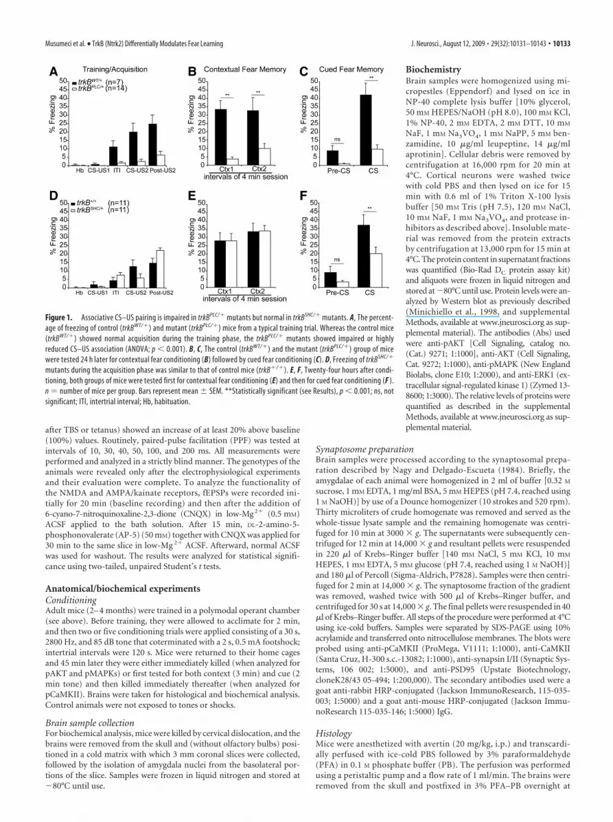
Figure 3. Normal basal synaptic transmission at BL–LA synapses of trkBPLC/� mice. A, PPF was measured to determine whetherthis aspect of synaptic transmission was normal in trkBPLC/� mice compared with controls. The percentages denote the ratio of thesecond fEPSP slope to the first fEPSP slope. PPF was tested at 10, 30, 40, 50, 100, and 200 ms ISIs. There was no significant differencebetween genotypes. Error bars correspond to the SEM. B, AMPA/kainate receptor (CNQX) and NMDA receptor (AP-5) antagonistswere used to analyze whether NMDA and AMPA/kainate receptors are functional in trkBPLC/� mice. Shown are single traces ofextracellular recordings for trkBWT/�, trkBPLC/�, and trkBSHC/� genotypes. Arrows indicate baseline EPSP 15 min after CNQXapplication or 15 min after AP-5-plus-CNQX application. C, Summary data for all pharmacological experiments. Plotted is the ratioof the EPSP slope 15 min after CNQX application in comparison with baseline recordings. There was no statistically significantdifference between genotypes ( p � 0.1; Student’s t test). D, E, Synaptic fatigue during high-frequency stimulation. Single tracescollected during theta-burst stimulation from trkBWT/� control animal (D) and from trkBPLC/� mutant mouse (E). The response tothe fourth stimulus (indicated by 4) was compared with the response to the first one (indicated by 1). F, Data corresponding tocontrols and to trkBSHC/� and trkBPLC/� mutants during high-frequency stimulation. Plotted is the ratio of the fourth to the firstresponse in a burst of four stimuli during theta-burst stimulation. For LTP induction, three theta bursts were applied, and data forall three are shown. Synaptic fatigue can be observed in all genotypes, and there is no statistically significant difference betweencontrol, trkBSHC/�, and trkBPLC/� mice ( p � 0.1; Student’s t test).
Musumeci et al. • TrkB (Ntrk2) Differentially Modulates Fear Learning J. Neurosci., August 12, 2009 • 29(32):10131–10143 • 10135

(supplemental Table S1, available at www.jneurosci.org as sup-plemental material). Using Fisher’s exact probability test (twotailed), no differences in pain sensitivity were found betweengenotypes (trkBWT/� vs trkBPLC/�, p � 0.8; trkB�/� vs trkBSHC/�,p � 0.9). Moreover, no differences were found between the con-trol groups (trkBWT/� vs trkB�/�, p � 0.54) or between the twopoint mutants (trkBPLC/� vs trkBSHC/�, p � 0.54).
trkBPLC but not trkBSHC phosphorylation mutants presentedimpaired early LTP at the BL amygdalar synapsesTo characterize the molecular basis for the impaired fear condi-tioning learning to both context and an auditory cue that wasobserved in the trkBPLC/� mutants, and of impaired consolida-tion for cued fear conditioning in the trkBSHC/� mutants, weobtained field potential recordings from neurons of the BL nu-cleus of the amygdala by stimulating neurons in the LA nucleus inbrain slices of adult mice. To prevent an underestimation of therequirements of TrkB signaling in fear learning, both homozy-gous and heterozygous point mutant mice were used for the invitro electrophysiology experiments. Both types of stimulation,TBS and tetanus, produced LTP. As shown in supplemental Fig-ure S6A, available at www.jneurosci.org as supplemental mate-rial, C57BL/6 or trkB�/� control mice showed LTP in 91.7% ofthe cases (24 slices from nine mice) or 80% of the cases (5 slicesfrom three mice), respectively; trkBWT/� or trkBWT/WT controlmice showed LTP in 83.3% of the cases (6 slices from three mice)or 100% of the cases (4 slices from three mice), respectively;trkBSHC/� or trkBSHC/SHC mutant mice showed LTP in 87.5% ofthe cases (8 slices from three mice) or 100% of the cases (6 slicesfrom three mice), respectively; whereas trkBPLC/� or trkBPLC/PLC
mutant mice showed LTP in 10% of the cases (10 slices from threemice) or 20% of the cases (10 slices from three mice), respec-tively. The magnitude of the potentiation, expressed as themean percentage of fEPSP slope with respect to baseline (set at100%), quantified at 55– 60 min after stimulation, was 167 �12% of baseline (n � 5) for the trkB�/� control mice and162 � 16% of baseline (n � 8) for the trkBSHC/� mutants (Fig.2 B). Similar results were observed in trkBSHC/SHC mutantslices (164 � 9% of baseline; n � 6) (supplemental Fig. S6 B,available at www.jneurosci.org as supplemental material),
suggesting that ablation of signaling from the Shc site of TrkBdoes not interfere with this form of synaptic activity. In con-trast, E-LTP was abolished in trkBPLC/� mice (102 � 3% ofbaseline; n � 10) compared with control mice (trkBW/�, 160 �8.5% of baseline; n � 6) (Fig. 2C). The difference betweentrkBPLC/� mutants and control mice (trkBWT/�) was statisti-cally significant ( p � 0.01). Similar results were observed intrkBPLC/PLC mutant slices (109 � 7% of baseline; n � 10)(supplemental Fig. S6 B, available at www.jneurosci.org assupplemental material).
Synaptic transmission in trkBPLC mice is indistinguishablefrom that of control miceA number of control experiments were performed to rule outthe possibility that the failure to induce LTP in the trkBPLC/�
mutants was attributable to impaired synaptic transmission atthe BL synapses. First, the presynaptic fiber volley (PSFV), whichis proportional to the number of presynaptic neurons recruitedby stimulation, was compared with the slope of the field poten-tial, and this comparison revealed that basal synaptic transmis-sion was normal in trkBPLC/� mutants. The EPSP slope/PSFVratio was 1.6 � 0.21 for controls, 1.6 � 0.3 for trkBPLC/� mice,and 1.5 � 04 for trkBPLC/PLC. Second, using an interstimulusinterval (ISI) from 10 to 200 ms, the PPF was found to be normalin all genotypes analyzed (Fig. 3A), including the homozygouspoint mutants for both strains (trkBPLC/PLC, 10 ms: 108 � 6; 30ms: 160 � 8; 40 ms: 168 � 8; 100 ms: 128 � 8; 200 ms: 105 � 5.trkBSHC/SHC, 10 ms: 110 � 8; 30 ms: 164 � 9; 40 ms: 164 � 9; 100ms: 126 � 7; 200 ms: 106 � 4). Although the induction of LTPfailed in trkBPLC/� mutants, all slices showed post-tetanic poten-tiation when the TBS or tetanus was applied (data not shown).We next tested glutamate receptor-mediated transmission bymeasuring the NMDA receptor component of the fEPSP underlow-Mg 2� conditions (Fig. 3B). In the presence of the AMPA/kainate receptor antagonist CNQX, amygdalar slices from allgenotypes showed a comparable NMDA receptor componentof the EPSP. The fEPSP slope was reduced to 24.2 � 3.9%(baseline � 100%) in trkBPLC/� mice, compared with 23.5 �3.1% in trkBWT/� control mice and 24 � 2% in trkBSHC/� mutants(Fig. 3B,C). Thus, there was no statistically significant difference
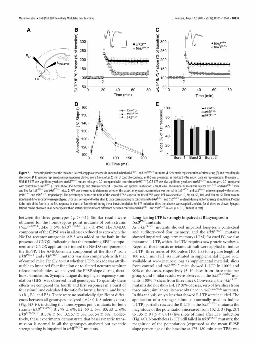
Figure 4. Long-lasting LTP is strongly impaired in BL–LA synapses of trkBPLC/� mutants. A, Schematic representation of stimulating and recording electrodes. SLA, Stimulating (lateral amygdala);RBL, recording (basolateral amygdala). B, C, Slope size of fEPSP recordings before and after the TBS stimulus was plotted. Symbols represent average responses plotted every 4 min. After 20 min ofcontrol recordings, HFSs were presented at the time marked by the arrow. Data are represented as the mean � SEM. B, L-LTP is not significantly different in trkBSHC/� mutants compared withcontrols (trkB�/�) ( p � 0.1). The number of slices was 5 for control mice (trkB�/�) and 10 for trkBSHC/� mice. C, L-LTP is abolished in trkBPLC/� mice. The difference between trkBPLC/� mutantsand control mice (trkBWT/�) was statistically significant ( p � 0.01). Traces show EPSP before (1) and 180 min after (2) LTP protocol was applied. Calibration: 5 ms, 0.5 mV. The number of slices wasfour for control mice (trkBWT/�) and five for trkBPLC/� mice.
10136 • J. Neurosci., August 12, 2009 • 29(32):10131–10143 Musumeci et al. • TrkB (Ntrk2) Differentially Modulates Fear Learning
between the three genotypes ( p � 0.1). Similar results wereobtained for the homozygous point mutants of both strains(trkBPLC/PLC, 24.6 � 5%; trkBSHC/SHC, 23.9 � 4%). The NMDAcomponent of the fEPSP was in all cases reduced to zero when theNMDA receptor antagonist AP-5 was added to the bath in thepresence of CNQX, indicating that the remaining EPSP compo-nent after CNQX application is indeed the NMDA component ofthe fEPSP. The AMPA/kainate component of the fEPSP fromtrkBPLC/� and trkBSHC/� mutants was also comparable with thatof control mice. Finally, to test whether LTP blockade was attrib-utable to impaired fiber function or to altered neurotransmitterrelease probabilities, we analyzed the EPSP slope during theta-burst stimulation. Synaptic fatigue during high-frequency stim-ulation (HFS) was observed in all genotypes. To quantify theseeffects we compared the fourth and first responses in a burst offour stimuli and calculated the ratio for burst 1, burst 2, and burst3 (B1, B2, and B3). There were no statistically significant differ-ences between all genotypes analyzed ( p � 0.1; Student’s t test)(Fig. 3D–F), including the homozygous point mutants for bothstrains (trkBPLC/PLC, B1: 78 � 6%, B2: 60 � 5%, B3: 53 � 6%;trkBSHC/SHC, B1: 76 � 6%, B2: 57 � 5%, B3: 54 � 6%). Collec-tively, these experiments demonstrate that basal synaptic trans-mission is normal in all the genotypes analyzed but synapticstrengthening is impaired in trkBPLC/� mutants.
Long-lasting LTP is strongly impaired at BL synapses intrkBPLC mutantsAs trkBPLC/� mutants showed impaired long-term contextualand auditory-cued fear memory, and the trkBSHC/� mutantsshowed impaired long-term memory (LTM) for cued FC, we alsomeasured L-LTP, which like LTM requires new protein synthesis.Repeated theta bursts or tetanic stimuli were applied to induceL-LTP [three series of 100 pulses (100 Hz) for a pulse length of100 �s, 5 min ISI]. As illustrated in supplemental Figure S6C,available at www.jneurosci.org as supplemental material, slicesfrom control and trkBSHC/� mice showed L-LTP in 100% and90% of the cases, respectively (5–10 slices from three mice pergroup), and similar results were observed in the trkBSHC/SHC mu-tants (100%, 7 slices from three mice). Conversely, the trkBPLC/�
mutants did not show L-LTP (0% of cases, zero of five slices fromthree mice; similar results were obtained in trkBPLC/PLC mutants).In this analysis, only slices that showed E-LTP were included. Theapplication of a stronger stimulus (normally used to induceL-LTP) partially rescued the E-LTP in the trkBPLC/� mutants; themagnitude of the potentiation increased from 102 � 3 (Fig. 2C)to 135 � 9 ( p � 0.01) (five slices of nine) after LTP induction(Fig. 4C). Nonetheless L-LTP still failed in trkBPLC/� mutants; themagnitude of the potentiation (expressed as the mean fEPSPslope percentage of the baseline at 175–180 min after TBS) was
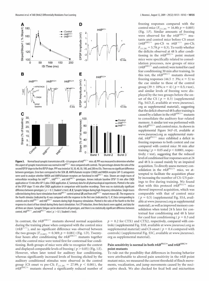
Figure 5. Synaptic plasticity at the thalamic–lateral amygdala synapses is impaired in both trkBPLC/� and trkBSHC/� mutants. A, Schematic representation of stimulating (S) and recording (R)electrodes. B, C, Symbols represent average responses plotted every 2 min. After 20 min of control recordings, an HFS was presented, as marked by the arrow. Data are represented as the mean �SEM. B, E-LTP was significantly reduced in trkBSHC/� mutant mice. p �0.01 compared with control mice (trkB�/�). C, E-LTP was also significantly reduced in trkBPLC/� mutants. p �0.01 comparedwith control mice (trkBWT/�). Traces show EPSP before (1) and 60 min after (2) LTP protocol was applied. Calibration: 5 ms, 0.5 mV. The number of slices was four for trkB�/� and trkBSHC/� miceand five for trkBWT/� and trkBPLC/� mice. D, PPF was measured to determine whether this aspect of synaptic transmission was normal in trkBSHC/� and trkBPLC/� mice compared with controls(trkB�/� and trkBWT/�, respectively). The percentages denote the ratio of the second fEPSP slope to the first fEPSP slope. PPF was tested at 10, 30, 40, 50, 100, and 200 ms ISI. There was nosignificant difference between genotypes. Error bars correspond to the SEM. E, Data corresponding to controls and to trkBSHC/� and trkBPLC/� mutants during high-frequency stimulation. Plottedis the ratio of the fourth to the first response in a burst of four stimuli during theta-burst stimulation. For LTP induction, three theta bursts were applied, and data for all three are shown. Synapticfatigue can be observed in all genotypes with no statistically significant difference between controls and trkBSHC/� and trkBPLC/� mice ( p � 0.1; Student’s t test).
Musumeci et al. • TrkB (Ntrk2) Differentially Modulates Fear Learning J. Neurosci., August 12, 2009 • 29(32):10131–10143 • 10137

167 � 8% for the trkB�/� control mice, 171 � 12% for thetrkBSHC/� mutants, 161 � 9% for the trkBWT/� controls, and105 � 6% for the trkBPLC/� mutant mice (Fig. 4B,C). Differencesbetween the controls and the trkBPLC/� mutant were statisticallysignificant ( p � 0.05). These results indicate that the signalingactivated through the PLC� site of the TrkB receptor is necessaryfor the acquisition of the pavlovian FC paradigm as well as forLTP generated at synapses in the BL nucleus of the amygdala. Incontrast, the TrkB/Shc site-activated signaling is dispensable forplasticity at BL synapses.
E-LTP is impaired at the LA synapses in both trkBPLC andtrkBSHC phosphorylation mutantsThe dorsal subdivision of the LA is the primary target of thalamicinputs (Rodrigues et al., 2004a). Thus, field recordings were ob-tained in the neurons of the dorsal LA nucleus in brain slices ofadult mice by stimulating fibers emerging from the internal cap-sule in the ventral striatum, which has been shown to carry, inpart, efferents from the medial geniculate nucleus to the LA(Weisskopf et al., 1999). This form of LTP is essential for auditoryFC in mice (LeDoux, 2000). We used TBS as well as tetanusstimulation. Both protocols evoked LTP: C57BL/6 or trkB�/�
control mice showed LTP in 100% of the cases (16 slices or 4 slicesfrom eight or three mice each group, respectively), trkBWT/� con-trol mice showed LTP in 80% of the cases (5 slices from threemice), trkBSHC/� mice showed LTP in 25% of the cases (4 slicesfrom three mice), and trkBPLC/� mice showed LTP in 40% ofthe cases (5 slices from three mice) (supplemental Fig. S7A, avail-able at www.jneurosci.org as supplemental material). The mag-nitude of the potentiation, expressed as the mean percentage offEPSP slope with respect to baseline (set at 100%) was quantifiedat 55– 60 min after stimulation as follows: trkB�/� control mice,190 � 16%, trkBSHC/� mice, 121 � 7%, trkBWT/� controls, 172 �10%, and trkBPLC/� mice, 119 � 5% (Fig. 5B,C). The differencesbetween controls and trkBSHC/� mutants, as well as between con-trols and trkBPLC/� mice, were significant ( p � 0.01) (Student’s ttest, two tailed). Similar results were obtained for the trkBSHC/SHC
and the trkBPLC/PLC homozygous point mutants (supplementalFig. S7B, available at www.jneurosci.org as supplemental mate-rial). Thus, both trkBSHC/� and trkBPLC/� mice showed reducedE-LTP in the auditory input pathways to the LA. Control ex-
periments suggested that the reduced E-LTP observed in bothtrkBSHC/� and trkBPLC/� mutants was not caused by impairedsynaptic transmission, as it was indistinguishable from that ofcontrol mice at LA synapses. PPF was normal in all genotypesanalyzed (Fig. 5D). To test whether poor LTP induction at LAsynapses in trkBSHC/� and trkBPLC/� mice was attributable toimpaired fiber function or to altered neurotransmitter releaseprobabilities, we analyzed the EPSP slope during theta-burststimulation. Synaptic fatigue during high-frequency stimulationwas observed in all genotypes. Comparing the fourth and firstresponses in a burst of four stimuli, and calculating this ratio forthe three theta bursts given, revealed no statistically significantdifferences between all genotypes analyzed ( p � 0.1; Student’s ttest) (Fig. 5E). These experiments indicated that basal synaptictransmission is normal in all the genotypes analyzed, but synapticstrengthening is reduced at the LA synapses in trkBSHC/� andtrkBPLC/� point mutants. Of note is that short-term memory(STM) in trkBSHC/� mutants is intact 30 min after training,whereas LTP in amygdala slices is decayed to baseline within 30min. This suggests that the electrical pattern of stimulation nec-essary to induce LTP in vitro could be quantitatively differentfrom the natural pattern of activation in the LA of behaving miceduring CS–US pairings. In addition, the preparation of the brainslices for the in vitro experiments could disrupt modulatory in-puts that are normally present in vivo (Schafe et al., 2001).
L-LTP is reduced at the LA synapses in trkBSHC and trkBPLC
mutantsWe then looked at expression of LTP at the thalamic–LA synapsesof both trkBSHC/� and trkBPLC/� mutants by using the L-LTPparadigm induced with a stronger stimulus protocol (Fig. 6B). Asillustrated in supplemental Figure S7C, available at www.jneurosci.org as supplemental material, slices from control mice(trkB�/� or trkBWT/�) showed L-LTP in 100% of cases (four orfive slices, respectively, from three mice), whereas trkBSHC/�
showed L-LTP in 33.3% of cases (six slices from three mice), andsimilar results were observed in trkBSHC/SHC mice (42.8%) (sevenslices from three mice). The trkBPLC/� mutants showed L-LTP in40% of cases (five slices from three mice), and similar results wereobtained in trkBPLC/PLC mutants. All of the slices analyzed showedE-LTP. In particular, with the stronger stimulus protocol the
Figure 6. Long-lasting LTP is also reduced in trkBSHC/� and trkBPLC/� mutants at thalamic–lateral amygdala synapses. A, Schematic representation of stimulating (S) and recording (R)electrodes. B, C, Slope size of fEPSP recordings before and after the TBS stimulus was plotted. Symbols represent average responses plotted every 4 min. After 20 min of control recordings, HFSs werepresented at the time marked by the arrow. Data are represented as the mean � SEM. B, L-LTP was significantly reduced in trkBSHC/� mutants compared with control mice (trkB �/�); p � 0.01.C, L-LTP was significantly reduced also in trkBPLC/� mice compared with control mice (trkBWT/�); p � 0.01. Traces show EPSP before (1) and 180 min after (2) LTP protocol was applied. Calibration:5 ms, 0.5 mV. Four slices were analyzed for trkB�/� mice, six for trkBSHC/� mice, and five for trkBWT/� and trkBPLC/� mice.
10138 • J. Neurosci., August 12, 2009 • 29(32):10131–10143 Musumeci et al. • TrkB (Ntrk2) Differentially Modulates Fear Learning
EPSP slope size increased from 121 � 7 to 152 � 14 ( p � 0.01)(six slices of nine) in trkBSHC/� mutants and from 119 � 5 to148 � 12 ( p � 0.01) (five slices of eight) in trkBPLC/� mutants.However, slices from both the trkBSHC/� and the trkBPLC/�
mutants presented reduced L-LTP; the magnitude of the poten-tiation (expressed as the mean fEPSP slope percentage of thebaseline at 175–180 min after TBS) was as follows: for the trkB�/�
control mice, 175 � 10%; for the trkBSHC/� mice, 122 � 6%; forthe trkBWT/� mice, 169 � 12%; and for the trkBPLC/� mice, 119 �10% (Fig. 6B,C). The differences between controls and trkBSHC/�
mutants as well as controls and trkBPLC/� mice were significant( p � 0.01) (Student’s t test, two tailed). Similar results wereobtained for the trkBSHC/SHC and the trkBPLC/PLC homozygouspoint mutants (supplemental Fig. S7D, available at www.jneurosci.org as supplemental material).
Point mutation at the Shc site in TrkB impairs specificamygdala function but not hippocampal functionIt has been suggested that the amygdala is essential for the condi-tioning of fear responses to both a cue and a context, whereas thehippocampus is involved in contextual but not auditory-cuedfear conditioning (Phillips and LeDoux, 1992). The differentialinvolvement of the Shc and the PLC� phosphorylation sites ofTrkB in fear learning and amygdalar synaptic plasticity indicates
that the hippocampal formation, con-trary to what is seen for the trkBPLC/�
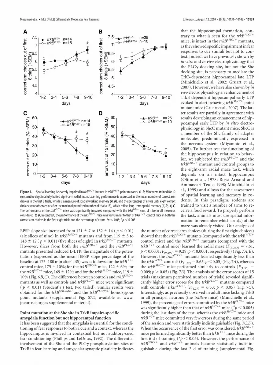
mice, is intact in the trkBSHC/� mutants,as they showed specific impairment in fearresponses to cue stimuli but not to con-text. Indeed, we have previously shown byin vitro and in vivo electrophysiology thatthe PLC� docking site, but not the Shcdocking site, is necessary to mediate theTrkB-dependent hippocampal late LTP(Minichiello et al., 2002; Gruart et al.,2007). However, we have also shown by invivo electrophysiology an enhancement ofTrkB-dependent hippocampal early LTPevoked in alert behaving trkBSHC/� pointmutant mice (Gruart et al., 2007). The lat-ter results are partially in agreement withresults describing an enhancement of hip-pocampal early LTP by in vitro electro-physiology in ShcC mutant mice; ShcC isa member of the Shc family of adaptermolecules, predominantly expressed inthe nervous system (Miyamoto et al.,2005). To further test the functioning ofthe hippocampus in relation to behav-ior, we subjected the trkBPLC/� and thetrkBSHC/� mutant and control groups tothe eight-arm radial maze task, whichdepends on an intact hippocampus(Olton et al., 1978; Rossi-Arnaud andAmmassari-Teule, 1998; Minichiello etal., 1999) and allows for the assessmentof spatial learning and memory in ro-dents. In this paradigm, rodents aretrained to visit a number of arms to re-ceive a food reward. To properly performthe task, animals must use spatial infor-mation to remember which arm(s) of themaze was already visited. Our analysis of
the number of correct arm choices (during the first eight choices)showed that the trkBPLC/� mutants (compared with the trkBWT/�
control mice) and the trkBSHC/� mutants (compared with thetrkB�/� control mice) learned the radial maze (F(9.243) � 7.61;p � 0.0001; F(9.297) � 9.29; p � 0.0001, respectively) (Fig. 7A,B).However, the trkBPLC/� mutants learned significantly less thanthe trkBWT/� controls (F(1.27) � 5.65; p � 0.05) (Fig. 7A), whereasthe trkBSHC/� mice performed similarly to controls (F(1.33) �0.009; p � 0.05) (Fig. 7B). The analysis of the error scores of 15trials (maximum permitted number of trials) revealed signifi-cantly higher error scores for the trkBPLC/� mutants comparedwith controls (trkBWT/�) (F(1.27) � 6.53; p � 0.05) (Fig. 7C).Interestingly, as previously observed in adult mice lacking TrkBin all principal neurons (the trkBcre mice) (Minichiello et al.,1999), the percentage of errors committed by the trkBPLC/� micewas significantly higher than that of trkBWT/� mice ( #p � 0.005)during the last days of the test, whereas the trkBSHC/� mice andtrkB�/� mice committed very few errors during the same periodof the session and were statistically indistinguishable (Fig. 7C,D).When the occurrence of the first error was considered, trkBSHC/�
mice performed significantly better than trkB�/� mice during thefirst 6 d of training (*p � 0.05). However, the performance oftrkBSHC/� and trkB�/� animals became statistically indistin-guishable during the last 2 d of training (supplemental Fig.
Figure 7. Spatial learning is severely impaired in trkBPLC/� but not in trkBSHC/� point mutants. A–D, Mice were trained for 10consecutive days in a fully baited eight-arm radial maze. Learning performance is expressed as the mean number of correct armchoices in the first 8 trials, which is a measure of spatial working memory (A, B), and the percentage of errors until eight correctchoices were observed or after the maximal permitted number of trials (15), which reflect long-term spatial memory (C, D). A, C,The performance of the trkBPLC/� mice was significantly impaired compared with the trkBWT/� control mice in all measuresconsidered. B, D, In contrast, the performance of the trkBSHC/� mice was very similar to that of trkB�/� control mice in both thecorrect arm choices in the first eight trials and the percentage of errors. *p � 0.05; #p � 0.005.
Musumeci et al. • TrkB (Ntrk2) Differentially Modulates Fear Learning J. Neurosci., August 12, 2009 • 29(32):10131–10143 • 10139

S8A,B, available at www.jneurosci.org as supplemental mate-rial). Together, these results show a clear impairment in bothshort- and long-term spatial memory in mice carrying a pointmutation at the PLC� site of the TrkB receptor but not at the Shcsite.
CaMKII phosphorylation is weak in amygdala synapses oftrkBPLC point mutantsIt has been suggested that the phosphorylation at Thr 286 ofCaMKII� is critically involved in synaptic plasticity and the ac-quisition and/or initial formation of short-term memory duringfear conditioning in the LA. CaMKII� is found to be distributedthroughout the LA and to be postsynaptic to thalamic inputs.In addition, inhibition of CaMKII in the amygdala impairsthe acquisition of auditory and contextual fear conditioning(Rodrigues et al., 2004b). BDNF has been shown to induce thephosphorylation of this kinase in neurons (Finkbeiner et al.,1997), and we have previously shown that CaMKII phosphor-ylation is impaired in trkBPLC/PLC but not trkBSHC/SHC mutantsynaptosomal preparations isolated from adult mouse cerebralcortices and stimulated with BDNF (Minichiello et al., 2002).Here, we found that point mutation at the PLC� site of TrkBresults in impaired fear acquisition and impaired amygdalar syn-aptic plasticity at the LA and the BL synapses. To further examinethe mechanism by which the PLC� site of TrkB regulates con-ditioned fear responses and LTP at amygdalar synapses, wemeasured the activation (p-Thr 286) of CaMKII in untrained(control) versus fear-conditioned mice. Since it has been re-ported that fear conditioning increases CaMKII phosphorylationat the lateral amygdala spines (Rodrigues et al., 2004b), and wefound that impairments in the acquisition of conditioned fearresponses by trkBPLC/� mutant mice are revealed with a milder(two CS–US pairings), but not an intense (five CS–US pairings),conditioning trial, we asked whether the degree of CaMKII phos-
phorylation at amygdalar synapses correlated with the acquisi-tion of conditioned fear in the trkBPLC/� mutants. No differencesin pCaMKII levels were found in synaptosomes isolated fromtrkBPLC/� mice that were either unconditioned or trained usingtwo CS–US pairings (ANOVA, two CS–US versus control, p �0.16); however, a significant increase in pCaMKII levels was seenin amygdalar synaptosomes from trkBPLC/� mice trained withfive CS–US pairings (ANOVA; five CS–US versus control, p �0.01; five CS–US versus two CS–US, p � 0.03) (Fig. 8B). ThepCaMKII levels in synaptosomes from trkBPLC/PLC mice eitheruntrained (control) or conditioned with two CS–US or fiveCS–US pairings were similarly affected (ANOVA; two CS–USversus control, p � 0.09; five CS–US versus control, p � 0.0007;five CS–US versus two CS–US, p � 0.0002) (Fig. 8C). Conversely,amygdalar synaptosomes isolated from trkB�/� mice trainedwith either two CS–US or five CS–US pairings possessed similarlevels of pCaMKII that were significantly greater than the levelseen in untrained trkB�/� mice (ANOVA; two CS–US versuscontrol, p � 0.047; five CS–US versus control, p � 0.0059; twoCS–US versus five CS–US, p � 0.3) (Fig. 8A). Collectively, theseresults suggest that impaired fear acquisition and amygdalar syn-aptic plasticity downstream of the TrkB/PLC� site correlate withweak pCaMKII at the amygdalar synapses.
AKT but not MAPK phosphorylation is affected in amygdalasynapses of trkBSHC point mutantsPreviously, we showed normal MAPK activation and nucleartranslocation after BDNF stimulation in dissociated cortical neu-rons from trkBPLC/PLC mice (Minichiello et al., 2002). In contrast,in cortical neurons from trkBSHC/SHC mutants the activation ofMAPK by BDNF is transient and reduced in amplitude(Minichiello et al., 1998). Thus, we analyzed MAPK phosphory-lation in response to BDNF in cultured cortical neurons fromtrkBSHC/� mutants and found similar activation kinetics compared
Figure 8. Weak pCaMKII at amygdala synapses in trkBPLC point mutants correlates with impaired fear acquisition. A–C, Immunoblots showing pCaMKII (Thr 286) levels in synaptosomalpreparations from amygdala tissues of trkB�/� untrained (control, n � 4) versus trained (2 CS–US, n � 5; or 5 CS–US, n � 5), trkBPLC/� (control, n � 3; 2 CS–US, n � 3; or 5 CS–US, n � 3) andtrkBPLC/PLC (control, n � 3; 2 CS–US, n � 3; or 5 CS–US, n � 3) mice. Blots were reprobed with CaMKII� Abs to visualize CaMKII� levels. Top, Quantification of pCaMKII levels A, **, ANOVA, fiveCS–US versus control, p � 0.0059; *, two CS–US versus control, p � 0.047. B, *, ANOVA, five CS–US versus control, p � 0.01; five CS–US versus two CS–US, p � 0.03. C, ***, ANOVA, five CS–USversus control, p � 0.0007; five CS–US versus two CS–US, p � 0.0002. D–F, Immunoblots of PSD95 and synapsin I/II confirmed that the synaptosomal preparation from amygdala tissues containedboth post- and presynaptic fractions, respectively.
10140 • J. Neurosci., August 12, 2009 • 29(32):10131–10143 Musumeci et al. • TrkB (Ntrk2) Differentially Modulates Fear Learning
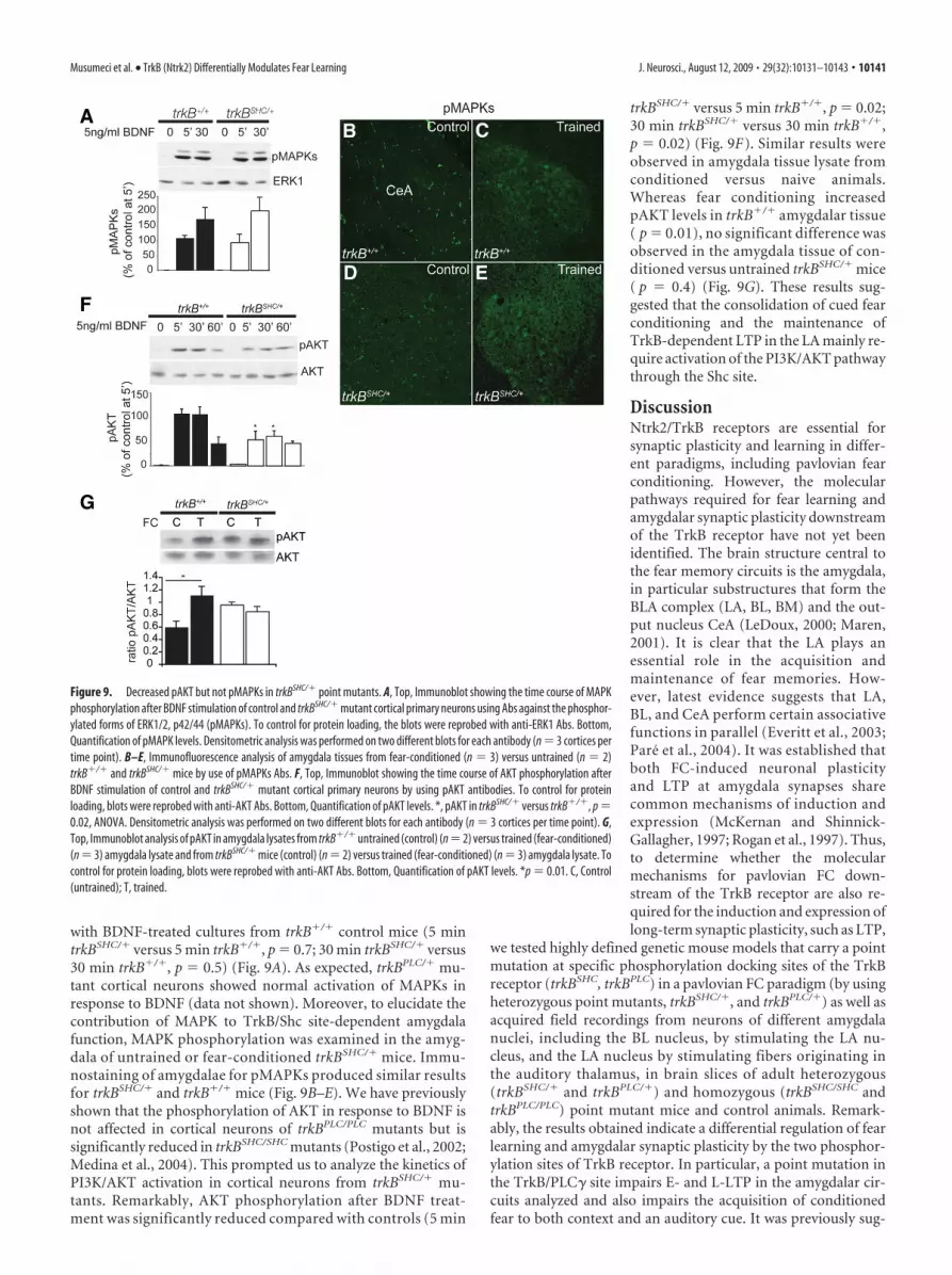
with BDNF-treated cultures from trkB�/� control mice (5 mintrkBSHC/� versus 5 min trkB�/�, p � 0.7; 30 min trkBSHC/� versus30 min trkB�/�, p � 0.5) (Fig. 9A). As expected, trkBPLC/� mu-tant cortical neurons showed normal activation of MAPKs inresponse to BDNF (data not shown). Moreover, to elucidate thecontribution of MAPK to TrkB/Shc site-dependent amygdalafunction, MAPK phosphorylation was examined in the amyg-dala of untrained or fear-conditioned trkBSHC/� mice. Immu-nostaining of amygdalae for pMAPKs produced similar resultsfor trkBSHC/� and trkB�/� mice (Fig. 9B–E). We have previouslyshown that the phosphorylation of AKT in response to BDNF isnot affected in cortical neurons of trkBPLC/PLC mutants but issignificantly reduced in trkBSHC/SHC mutants (Postigo et al., 2002;Medina et al., 2004). This prompted us to analyze the kinetics ofPI3K/AKT activation in cortical neurons from trkBSHC/� mu-tants. Remarkably, AKT phosphorylation after BDNF treat-ment was significantly reduced compared with controls (5 min
trkBSHC/� versus 5 min trkB�/�, p � 0.02;30 min trkBSHC/� versus 30 min trkB�/�,p � 0.02) (Fig. 9F). Similar results wereobserved in amygdala tissue lysate fromconditioned versus naive animals.Whereas fear conditioning increasedpAKT levels in trkB�/� amygdalar tissue( p � 0.01), no significant difference wasobserved in the amygdala tissue of con-ditioned versus untrained trkBSHC/� mice( p � 0.4) (Fig. 9G). These results sug-gested that the consolidation of cued fearconditioning and the maintenance ofTrkB-dependent LTP in the LA mainly re-quire activation of the PI3K/AKT pathwaythrough the Shc site.
DiscussionNtrk2/TrkB receptors are essential forsynaptic plasticity and learning in differ-ent paradigms, including pavlovian fearconditioning. However, the molecularpathways required for fear learning andamygdalar synaptic plasticity downstreamof the TrkB receptor have not yet beenidentified. The brain structure central tothe fear memory circuits is the amygdala,in particular substructures that form theBLA complex (LA, BL, BM) and the out-put nucleus CeA (LeDoux, 2000; Maren,2001). It is clear that the LA plays anessential role in the acquisition andmaintenance of fear memories. How-ever, latest evidence suggests that LA,BL, and CeA perform certain associativefunctions in parallel (Everitt et al., 2003;Pare et al., 2004). It was established thatboth FC-induced neuronal plasticityand LTP at amygdala synapses sharecommon mechanisms of induction andexpression (McKernan and Shinnick-Gallagher, 1997; Rogan et al., 1997). Thus,to determine whether the molecularmechanisms for pavlovian FC down-stream of the TrkB receptor are also re-quired for the induction and expression oflong-term synaptic plasticity, such as LTP,
we tested highly defined genetic mouse models that carry a pointmutation at specific phosphorylation docking sites of the TrkBreceptor (trkBSHC, trkBPLC) in a pavlovian FC paradigm (by usingheterozygous point mutants, trkBSHC/�, and trkBPLC/�) as well asacquired field recordings from neurons of different amygdalanuclei, including the BL nucleus, by stimulating the LA nu-cleus, and the LA nucleus by stimulating fibers originating inthe auditory thalamus, in brain slices of adult heterozygous(trkBSHC/� and trkBPLC/�) and homozygous (trkBSHC/SHC andtrkBPLC/PLC) point mutant mice and control animals. Remark-ably, the results obtained indicate a differential regulation of fearlearning and amygdalar synaptic plasticity by the two phosphor-ylation sites of TrkB receptor. In particular, a point mutation inthe TrkB/PLC� site impairs E- and L-LTP in the amygdalar cir-cuits analyzed and also impairs the acquisition of conditionedfear to both context and an auditory cue. It was previously sug-
Figure 9. Decreased pAKT but not pMAPKs in trkBSHC/� point mutants. A, Top, Immunoblot showing the time course of MAPKphosphorylation after BDNF stimulation of control and trkBSHC/� mutant cortical primary neurons using Abs against the phosphor-ylated forms of ERK1/2, p42/44 (pMAPKs). To control for protein loading, the blots were reprobed with anti-ERK1 Abs. Bottom,Quantification of pMAPK levels. Densitometric analysis was performed on two different blots for each antibody (n � 3 cortices pertime point). B–E, Immunofluorescence analysis of amygdala tissues from fear-conditioned (n � 3) versus untrained (n � 2)trkB�/� and trkBSHC/� mice by use of pMAPKs Abs. F, Top, Immunoblot showing the time course of AKT phosphorylation afterBDNF stimulation of control and trkBSHC/� mutant cortical primary neurons by using pAKT antibodies. To control for proteinloading, blots were reprobed with anti-AKT Abs. Bottom, Quantification of pAKT levels. *, pAKT in trkBSHC/� versus trkB�/�, p �0.02, ANOVA. Densitometric analysis was performed on two different blots for each antibody (n � 3 cortices per time point). G,Top, Immunoblot analysis of pAKT in amygdala lysates from trkB�/� untrained (control) (n�2) versus trained (fear-conditioned)(n � 3) amygdala lysate and from trkBSHC/� mice (control) (n � 2) versus trained (fear-conditioned) (n � 3) amygdala lysate. Tocontrol for protein loading, blots were reprobed with anti-AKT Abs. Bottom, Quantification of pAKT levels. *p � 0.01. C, Control(untrained); T, trained.
Musumeci et al. • TrkB (Ntrk2) Differentially Modulates Fear Learning J. Neurosci., August 12, 2009 • 29(32):10131–10143 • 10141
gested that the amygdala and the hippocampus differentially con-tribute to cue and contextual FC. Lesion experiments in ratsrevealed that whereas the amygdala is involved in the condition-ing of fear-related responses to a context and cue, the hip-pocampus instead modulates conditioning to context but notto a cue (Phillips and LeDoux, 1992). We previously showed thatthe PLC� docking site, but not the Shc site, of TrkB is necessaryfor hippocampal synaptic plasticity, as well as for the acquisitionof associative learning (classical conditioning of eyelid re-sponses), in vivo LTP at CA3-CA1 hippocampal synapses(Minichiello et al., 2002; Gruart et al., 2007), and spatial learning(this study). Thus, impaired hippocampal plasticity in trkBPLC
mutants likely contributes to the impaired fear conditioning tocontext. In contrast, the trkBSHC mutants show normal acquisi-tion of contextual and cued FC but impaired consolidation of theconditioned fear response to an auditory cue, as well as a specificimpairment in amygdalar synaptic plasticity at the thalamic–LAsynapses (observed in both the trkBSHC/� and the trkBSHC/SHC
mutants). We also obtained recordings within the LA (the ven-tral LA) by stimulation of the dorsal lateral nucleus and foundimpaired plasticity in the trkBSHC mutants (data not shown),suggesting a specific impairment in plasticity in the LA of thetrkBSHC mutants. This is supported by the fact that hippocampalfunctions are mainly spared in these mutants. Of note is that wehave reported by in vivo electrophysiology an enhancement ofTrkB-dependent hippocampal early LTP evoked in alert behav-ing trkBSHC/� mice (Gruart et al., 2007). Consistent with theseresults, we now also show a better performance of the trkBSHC
mutants in a specific phase of a spatial learning task (namely, theradial maze). Although these results are similar to some of theresults reported for the ShcC mutant mice, the mechanisms bywhich ShcC regulates hippocampal synaptic plasticity seem to beindependent of the TrkB/Shc site and apparently via modulationof the NMDA receptor function (Miyamoto et al., 2005). In ad-dition, transgenic mice overexpressing the full-length TrkBreceptor show selective activation of the PLC� site as well asincreased spatial learning and improved contextual fear con-ditioning, whereas signaling via the Shc site remains unaltered(Koponen et al., 2004).
Regarding the biochemical mechanisms underlying pavlovianFC, Rodrigues et al. (2004b) suggested that the phosphorylationof Thr 286 of CaMKII� is critically involved in synaptic plasticityand the acquisition and/or initial formation of STM during fearconditioning in the LA. Here, we show that a point mutation atthe PLC� site of TrkB (trkBPLC/�) results in impaired fear acqui-sition and impaired amygdalar synaptic plasticity at the LA andthe BL synapses, likely attributable to the reduced phosphoryla-tion of CaMKII at Thr 286, as seen in amygdalar synapses of FCtrkBPLC mice (both trkBPLC/� and trkBPLC/PLC mutants) trainedwith two CS–US pairings, indicating that TrkB receptors throughthe Y816 typically modulate CaMKII phosphorylation and acqui-sition of FC, as well as amygdalar LTP. However, an intense train-ing protocol (five CS–US pairings) induces normal CaMKIIphosphorylation in trkBPLC amygdala synapses (observed in boththe trkBPLC/� and trkBPLC/PLC mutants). These results were con-sistent with the fact that training trials using five CS–US pairingsrescued the memory acquisition deficit in the trkBPLC/� mice andthat application of a stronger stimulus partially rescued theE-LTP in these mutants. Under conditions of stronger stimula-tion, classical pathways still activated downstream of the mutantTrkB receptor may compensate for the signaling lost from thePLC� site. Alternatively, recent studies have shown that neuro-trophin activation of TrkB can rapidly mediate the gating of so-
dium channels, resulting in an increase in intracellular calciumconcentration and cellular responses independent of PLC� re-cruitment to TrkB (Blum and Konnerth, 2005). It is also possiblethat the effects observed in the trkBPLC/� mutants under moreintense inputs involve the activation of undefined signalingmechanisms directly or indirectly implicated in fear condition-ing. The normal phosphorylation of CaMKII seen in the amyg-dalae of trkBSHC/� point mutants (data not shown) correlateswith their normal acquisition of fear conditioning. Moreover,mice heterozygous for a point mutation at Y515 or Y816 of TrkBpossess normal brain levels of BDNF (supplemental Fig. S8, avail-able at www.jneurosci.org as supplemental material), supportingthe conclusion that the fear conditioning and the synaptic activityin amygdalar nuclei of these mutants are not affected by changesin the levels of this TrkB ligand.
Gean and colleagues have reported that the PI3K/AKT path-way contributes to amygdalar LTP and the consolidation ofamygdala-dependent cued fear conditioning in rats (Lin et al.,2001). Pharmacological studies by the same laboratory revealedthat BDNF is required for the acquisition of fear learning viarecruitment of the Shc adapter protein to TrkB and the activationof MAPK and PI3K (Ou and Gean, 2006). As discussed above, byusing very precise mouse genetic models we have determined inthis study that it is primarily the PLC� site of TrkB that medi-ates the TrkB-dependent acquisition of FC and amygdalarsynaptic plasticity at the LA and the BL synapses through mod-ulation of CaMKII at synapses, whereas the Shc site contrib-utes mainly to consolidation of amygdala-dependent cued fearconditioning and LTP at the LA synapses predominantlythrough AKT activation.
A concern could be raised that the mice used in this studypossess the mutated form of the receptor in all TrkB-expressingcells and this precludes conclusions regarding the role of TrkB ina specific brain region such as the amygdala. However, we haveextensively analyzed the PNS and CNS development of the het-erozygous point mutants of both strains [shown in previous work(Minichiello et al., 1998, 2002; Medina et al., 2004; Gruart et al.,2007)] (supplemental Figs. S1, S2, available at www.jneurosci.orgas supplemental material) and have not identified any relevantabnormalities. This, combined with the fact that the animals dis-play normal basic behavior (supplemental Fig. S3, available atwww.jneurosci.org as supplemental material), suggests that thephenotypes observed in fear conditioning are not caused by de-velopmental defects in the amygdala. Experiments based on theintra-amygdala infusion of a lentiviral vector expressing adominant-negative TrkB isoform (TrkB.T1) in adult rats havealso established that the TrkB neurotrophin receptor plays anessential role in the acquisition of pavlovian fear conditioningtriggered in the amygdala nuclei (Rattiner et al., 2004). More-over, we find that basal synaptic activities are similar betweenthe different genotypes analyzed (Fig. 3). Animals possessingdevelopmental defects would show changes especially in basalsynaptic transmission. The electrophysiological recordings pro-duced identical results for heterozygous and homozygous pointmutants (supplemental Figs. S7–10, available at www.jneurosci.org as supplemental material), suggesting that one copy of a wild-type TrkB allele is sufficient to prevent the developmental PNSphenotypes observed in the homozygous point mutants but is notsufficient to support TrkB-dependent learning in the adult brain(Minichiello et al., 1999, 2002). Similar results were obtained bybiochemical analysis of synaptosomal preparations from amyg-dala tissues of untrained versus trained trkBPLC/� and trkBPLC/PLC
point mutants (Fig. 8). Together, these findings uphold the no-
10142 • J. Neurosci., August 12, 2009 • 29(32):10131–10143 Musumeci et al. • TrkB (Ntrk2) Differentially Modulates Fear Learning

tion that the genetic tools of the present study are valid and sup-port our main conclusions. However, future work focused on acell-type-specific ablation of TrkB will be necessary to under-stand the contribution of TrkB to fear learning and amygdalarLTP in the different neuronal populations.
ReferencesBlum R, Konnerth A (2005) Neurotrophin-mediated rapid signaling in the
central nervous system: mechanisms and functions. Physiology 20:70 –78.Campeau S, Davis M (1995) Involvement of subcortical and cortical affer-
ents to the lateral nucleus of the amygdala in fear conditioning measuredwith fear-potentiated startle in rats trained concurrently with auditoryand visual conditioned stimuli. J Neurosci 15:2312–2327.
Davis M (1997) Neurobiology of fear responses: the role of the amygdala.J Neuropsychiatry Clin Neurosci 9:382– 402.
Everitt BJ, Cardinal RN, Parkinson JA, Robbins TW (2003) Appetitive be-havior: impact of amygdala-dependent mechanisms of emotional learn-ing. Ann N Y Acad Sci 985:233–250.
Fendt M, Fanselow MS (1999) The neuroanatomical and neurochemicalbasis of conditioned fear. Neurosci Biobehav Rev 23:743–760.
Finkbeiner S, Tavazoie SF, Maloratsky A, Jacobs KM, Harris KM, GreenbergME (1997) CREB: a major mediator of neuronal neurotrophin re-sponses. Neuron 19:1031–1047.
Gale GD, Anagnostaras SG, Godsil BP, Mitchell S, Nozawa T, Sage JR,Wiltgen B, Fanselow MS (2004) Role of the basolateral amygdala in thestorage of fear memories across the adult lifetime of rats. J Neurosci24:3810 –3815.
Gruart A, Sciarretta C, Valenzuela-Harrington M, Delgado-García JM,Minichiello L (2007) Mutation at the TrkB PLC{gamma}-docking siteaffects hippocampal LTP and associative learning in conscious mice.Learn Mem 14:54 – 62.
Kim JJ, Fanselow MS (1992) Modality-specific retrograde amnesia of fear.Science 256:675– 677.
Koo JW, Han JS, Kim JJ (2004) Selective neurotoxic lesions of basolateraland central nuclei of the amygdale produce differential effects on fearconditioning. J Neurosci 24:7654 –7662.
Koponen E, Voikar V, Riekki R, Saarelainen T, Rauramaa T, Rauvala H, TairaT, Castren E (2004) Transgenic mice overexpressing the full-length neu-rotrophin receptor trkB exhibit increased activation of the trkB-PLCgamma pathway, reduced anxiety, and facilitated learning. Mol CellNeurosci 26:166 –181.
Lavond DG, Kim JJ, Thompson RF (1993) Mammalian brain substrates ofaversive classical conditioning. Annu Rev Psychol 44:317–342.
LeDoux JE (1996) The emotional brain: the mysterious underpinnings ofemotional life. New York: Simon and Schuster.
LeDoux JE (2000) Emotion circuits in the brain. Annu Rev Neurosci23:155–184.
LeDoux JE, Cicchetti P, Xagoraris A, Romanski LM (1990) The lateralamygdaloid nucleus: sensory interface of the amygdala in fear condition-ing. J Neurosci 10:1062–1069.
Lin CH, Yeh SH, Lin CH, Lu KT, Leu TH, Chang WC, Gean PW (2001) Arole for the PI-3 kinase signaling pathway in fear conditioning and syn-aptic plasticity in the amygdala. Neuron 31:841– 851.
Maren S (2001) Neurobiology of pavlovian fear conditioning. Annu RevNeurosci 24:897–931.
Maren S, Aharonov G, Fanselow MS (1997) Neurotoxic lesions of the dorsalhippocampus and pavlovian fear conditioning in rats. Behav Brain Res88:261–274.
McKernan MG, Shinnick-Gallagher P (1997) Fear conditioning induces alasting potentiation of synaptic currents in vitro. Nature 390:607– 611.
Medina DL, Sciarretta C, Calella AM, Von Bohlen Und Halbach O, UnsickerK, Minichiello L (2004) TrkB regulates neocortex formation throughthe Shc/PLCgamma-mediated control of neuronal migration. EMBO J23:3803–3814.
Minichiello L, Casagranda F, Tatche RS, Stucky CL, Postigo A, Lewin GR,Davies AM, Klein R (1998) Point mutation in trkB causes loss of NT4-dependent neurons without major effects on diverse BDNF responses.Neuron 21:335–345.
Minichiello L, Korte M, Wolfer D, Kuhn R, Unsicker K, Cestari V, Rossi-Arnaud C, Lipp HP, Bonhoeffer T, Klein R (1999) Essential role forTrkB receptors in hippocampus-mediated learning. Neuron 24:401– 414.
Minichiello L, Calella AM, Medina DL, Bonhoeffer T, Klein R, Korte M(2002) Mechanism of TrkB-mediated hippocampal long-term potentia-tion. Neuron 36:121–137.
Miyamoto Y, Chen L, Sato M, Sokabe M, Nabeshima T, Pawson T, Sakai R,Mori N (2005) Hippocampal synaptic modulation by the phosphoty-rosine adapter protein ShcC/N-Shc via interaction with the NMDA re-ceptor. J Neurosci 25:1826 –1835.
Nagy A, Delgado-Escueta AV (1984) Rapid preparation of synaptosomesfrom mammalian brain using nontoxic isoosmotic gradient material(Percoll). J Neurochem 43:1114 –1123.
Olton DS, Walker JA, Gage FH (1978) Hippocampal connections and spa-tial discrimination. Brain Res 139:295–308.
Ou LC, Gean PW (2006) Regulation of amygdala-dependent learning bybrain-derived neurotrophic factor is mediated by extracellular signal-regulated kinase and phosphatidylinositol-3-kinase. Neuropsychophar-macology 31:287–296.
Pare D, Quirk GJ, Ledoux JE (2004) New vistas on amygdala networks inconditioned fear. J Neurophysiol 92:1–9.
Phillips RG, LeDoux JE (1992) Differential contribution of amygdala andhippocampus to cued and contextual fear conditioning. Behav Neurosci106:274 –285.
Postigo A, Calella AM, Fritzsch B, Knipper M, Katz D, Eilers A, Schimmang T,Lewin GR, Klein R, Minichiello L (2002) Distinct requirements for TrkBand TrkC signaling in target innervation by sensory neurons. Genes Dev16:633– 645.
Rattiner LM, Davis M, French CT, Ressler KJ (2004) Brain-derived neuro-trophic factor and tyrosine kinase receptor B involvement in amygdala-dependent fear conditioning. J Neurosci 24:4796 – 4806.
Rodrigues SM, Schafe GE, LeDoux JE (2004a) Molecular mechanisms un-derlying emotional learning and memory in the lateral amygdala. Neuron44:75–91.
Rodrigues SM, Farb CR, Bauer EP, LeDoux JE, Schafe GE (2004b) Pavlovianfear conditioning regulates Thr286 autophosphorylation of Ca 2�/calmodulin-dependent protein kinase II at lateral amygdala synapses.J Neurosci 24:3281–3288.
Rogan MT, Staubli UV, LeDoux JE (1997) Fear conditioning induces asso-ciative long-term potentiation in the amygdala. Nature 390:604 – 607.
Romanski LM, LeDoux JE (1992) Equipotentiality of thalamo-amygdalaand thalamo-cortico-amygdala circuits in auditory fear conditioning.J Neurosci 12:4501– 4509.
Rossi-Arnaud C, Ammassari-Teule M (1998) What do comparative studiesof inbread mice add to current investigations on the neural basis of spatialbehaviors? Exp Brain Res 123:36 – 44.
Schafe GE, Nader K, Blair HT, LeDoux JE (2001) Memory consolidation ofpavlovian fear conditioning: a cellular and molecular perspective. TrendsNeurosci 24:540 –546.
Swanson LW, Petrovich GD (1998) What is the amygdala? Trends Neurosci21:323–331.
Weisskopf MG, Bauer EP, LeDoux JE (1999) L-type voltage-gated calciumchannels mediate NMDA-independent associative long-term potentiation atthalamic input synapses to the amygdala. J Neurosci 19:10512–10519.
Xu B, Gottschalk W, Chow A, Wilson RI, Schnell E, Zang K, Wang D, NicollRA, Lu B, Reichardt LF (2000) The role of brain-derived neurotrophicfactor receptors in the mature hippocampus: modulation of long-term po-tentiation through a presynaptic mechanism involving TrkB. J Neurosci 20:6888–6897.
Musumeci et al. • TrkB (Ntrk2) Differentially Modulates Fear Learning J. Neurosci., August 12, 2009 • 29(32):10131–10143 • 10143