transition to a novel advanced integrated vitrectomy platform: comparison of the surgical impact of...
TRANSCRIPT

© 2013 Murray et al, publisher and licensee Dove Medical Press Ltd. This is an Open Access article which permits unrestricted noncommercial use, provided the original work is properly cited.
Clinical Ophthalmology 2013:7 367–377
Clinical Ophthalmology
Transition to a novel advanced integrated vitrectomy platform: comparison of the surgical impact of moving from the Accurus vitrectomy platform to the Constellation Vision System for microincisional vitrectomy surgery
Timothy G Murray1,2
Andrew J Layton3
Kuo B Tong3
Michael Gittelman2
Azeema Latiff1,2
Daniel Gologorsky 2
Michael M Vigoda2
1Murray Ocular Oncology and Retina, Miami, FL, USA; 2Bascom Palmer Eye Institute, Anne Bates Leach Eye Hospital, Departments of Ophthalmology, Anesthesiology and Radiation Oncology, University of Miami Miller School of Medicine, Miami, FL, USA; 3Quorum Consulting, San Francisco, CA, USA
Correspondence: Timothy G Murray Murray Ocular Oncology and Retina, 6705 Red Road, Suite 412, Miami, FL 33143, USA Tel +1 305 487 7470 Fax +1 786 567 4380 Email [email protected]
Background: Microincisional vitrectomy surgery (MIVS) is the current standard surgical
approach for pars plana vitrectomy. Historically, the most common surgical platform for vitrec-
tomy surgery, since its introduction in 1997, has been the Accurus vitrectomy system. Recent
introduction of the next generation of vitrectomy platforms has generated concerns associated
with transitioning to new technology in the operating room environment. This study compared,
in a matched fashion, surgical use of the Accurus vitrectomy system and the next generation
Constellation Vision System to evaluate surgical efficiencies, complications, and user percep-
tions of this transition.
Methods: Electronic health records were abstracted as a hospital quality assurance activity and
included all vitreoretinal surgical procedures at the Bascom Palmer Eye Institute, Anne Bates
Leach Eye Hospital, during two discrete 12-month time periods. These two periods reflected
dedicated usage of the Accurus (June 2008–May 2009) and Constellation Vision (July 2009–
June 2010) systems. Data were limited to a single surgeon and evaluated for operating room (OR)
total time usage/day, OR case time/case, and OR surgical time/case. Further analysis evaluated all
patients undergoing combined MIVS and clear cornea phacoemulsification/intraocular lens (IOL)
implantation during each individual time period to determine the impact of the instrumentation
on these parameters. All records were evaluated for intraoperative complications.
Results: Five hundred and fourteen eligible patients underwent MIVS during the 2-year study
windows, with 281 patients undergoing surgery with the Accurus system and 233 patients under-
going surgery with the Constellation system. Combined MIVS and phacoemulsification with
IOL implantation was performed 141 times during this period with the Accurus and 158 times
during the second study period with the Constellation. Total number of patients operated per
day increased from 7.55 with Accurus to 8.53 with Constellation. Surgical room time decreased
from 56 minutes with Accurus to 52 minutes with Constellation, and procedure time decreased
from 35 minutes with Accurus to 31 minutes with Constellation (P , 0.004). Combined MIVS/
phacoemulsification surgery saw similar declines in surgical room time and procedure time
(P , 0.001). Subset analysis of procedures limited by case number per day (eg, four cases/day,
five cases/day, six cases/day, and seven or more cases/day) showed similar outcomes with a
decrease in surgical room time and procedure time. No increases in surgery-related complica-
tions were noted by quality assurance review during these time periods.
Discussion: Transitioning to advanced surgical technology is a complex issue for the surgeon,
the hospital team, and the hospital administration. This study documents improvement in three
Dovepress
submit your manuscript | www.dovepress.com
Dovepress 367
O R I G I n A L R E S E A R C H
open access to scientific and medical research
Open Access Full Text Article
http://dx.doi.org/10.2147/OPTH.S35603
Clinical Ophthalmology 2013:7
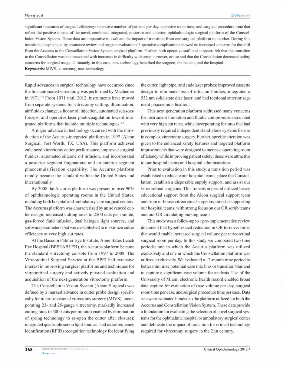
significant measures of surgical efficiency: operative number of patients per day, operative room time, and surgical procedure time that
reflect the positive impact of the novel, combined, integrated, posterior and anterior, ophthalmologic surgical platform of the Constel-
lation Vision System. These data are imperative to evaluate the impact of transition from one surgical platform to another. During this
transition, hospital quality assurance review and surgeon evaluation of operative complications showed no increased concerns for the shift
from the Accurus to the Constellation Vision System surgical platform. Further, both operative staff and surgeons felt that the transition
to the Constellation was not associated with increases in difficulty with setup, turnover, or use and that the Constellation decreased safety
concerns for surgical usage. Ultimately, in this case, new technology benefited the surgeon, the patient, and the hospital.
Keywords: MIVS, vitrectomy, new technology
Rapid advances in surgical technology have occurred since
the first automated vitrectomy was performed by Machemer
in 1971.1,2 From 1971 until 2012, instruments have moved
from separate systems for vitrectomy cutting, illumination,
air/fluid exchange, silicone oil injection, automated scissors/
forceps, and operative laser photocoagulation toward inte-
grated platforms that include multiple technologies.3–5
A major advance in technology occurred with the intro-
duction of the Accurus-integrated platform in 1997 (Alcon
Surgical, Fort Worth, TX, USA). This platform achieved
enhanced vitrectomy cutter performance, improved surgical
fluidics, automated silicone oil infusion, and incorporated
a posterior segment fragmatome and an anterior segment
phacoemulsification capability. The Accurus platform
rapidly became the standard within the United States and
internationally.
By 2008 the Accurus platform was present in over 90%
of ophthalmologic operating rooms in the United States,
including both hospital and ambulatory care surgical centers.
The Accurus platform was characterized by an advanced cut-
ter design, increased cutting rates to 2500 cuts per minute,
gas-forced fluid infusion, dual halogen light sources, and
software parameters that were established to maximize cutter
efficiency at very high cut rates.
At the Bascom Palmer Eye Institute, Anne Bates Leach
Eye Hospital (BPEI/ABLEH), the Accurus platform became
the standard vitrectomy console from 1997 to 2008. The
Vitreoretinal Surgical Service at the BPEI had extensive
interest in improving surgical platforms and techniques for
vitreoretinal surgery and actively pursued evaluation and
acquisition of the next generation vitrectomy platform.
The Constellation Vision System (Alcon Surgical) was
defined by a marked advance in cutter probe design specifi-
cally for micro-incisional vitrectomy surgery (MIVS), incor-
porating 23- and 25-gauge vitrectomy, markedly increased
cutting rates to 5000 cuts per minute (enabled by elimination
of spring technology to re-open the cutter after closure);
integrated quadruple xenon light sources; had radiofrequency
identification (RFID) recognition technology for identifying
the cutter, light pipe, and endolaser probes; improved cassette
design to eliminate loss of infusion fluidics; integrated a
532 nm solid state disc laser; and had torsional anterior seg-
ment phacoemulsification.
This next generation platform addressed many concerns
for instrument limitation and fluidic compromise associated
with very high cut rates, while incorporating features that had
previously required independent stand-alone systems for use
in complex vitrectomy surgery. Further, specific attention was
given to the enhanced safety features and targeted platform
improvements that were designed to increase operating room
efficiency while improving patient safety; these were attractive
to our hospital teams and hospital administration.
Prior to evaluation in this study, a transition period was
established to educate our hospital teams, place the Constel-
lation, establish a disposable supply support, and assist our
vitreoretinal surgeons. This transition period utilized heavy
educational support from the Alcon surgical support team
and from in-house vitreoretinal surgeons aimed at supporting
our hospital teams, with strong focus on our OR scrub teams
and our OR circulating nursing teams.
This study was a follow-up to a pre-implementation review
document that hypothesized reduction in OR turnover times
that would enable increased surgical volume per vitreoretinal
surgical room per day. In this study we compared two time
periods: one in which the Accurus platform was utilized
exclusively and one in which the Constellation platform was
utilized exclusively. We evaluated a 12-month time period to
better minimize potential case mix bias or transition bias and
to capture a significant case volume for analysis. Use of the
University of Miami electronic health record enabled broad
data capture for evaluation of case volume per day, surgical
room time per case, and surgical procedure time per case. Data
sets were evaluated blinded to the platform utilized for both the
Accurus and Constellation Vision System. These data provide
a foundation for evaluating the selection of novel surgical sys-
tems for the ophthalmic hospital or ambulatory surgical center
and delineate the impact of transition for critical technology
required for vitrectomy surgery in the 21st century.
submit your manuscript | www.dovepress.com
Dovepress
Dovepress
368
Murray et al

Clinical Ophthalmology 2013:7
MethodsA data extract from the BPEI/ABLEH electronic health
record system, satisfying internal review board requirements,
was obtained for all surgeries performed by a single surgeon
(TGM) during two time periods established through the hos-
pital quality assurance program. The first time period, repre-
senting usage of the Accurus platform, was from June 2008
through May 2009. The second time period, representing
usage of the Constellation platform, was from July 2009
through June 2010.
The patient’s electronic health records were then matched
to BPEI’s billing system to extract the current procedural
terminology (CPT) codes for each patient encounter. The
two databases were then combined to form one de-identified
analytic dataset. The dataset included the following variables:
claim number, date of service, CPT codes, attending physi-
cian, operating room number, unique patient identifier, patient
operating room in-time, surgery start time, surgery end time,
patient operating room out-time.
The operative time data was manually entered into the
patient electronic health record by the nursing staff on the
service date as part of their standard operating procedures.
Total room time was calculated by subtracting the in-room
time from the out-room time. Total surgery time was calcu-
lated by subtracting the surgery start time from the surgery
end time. Total surgical day time was calculated from the
first time in the OR to the last time in the OR for the entire
surgical day.
We eliminated procedures that could not be performed
on the two platforms, such as primary scleral buckle, enucle-
ation, or examination under anesthesia. To enhance the evalu-
ation, we evaluated all surgical dates and then surgical dates
with four, five, six, or greater then/equal to seven cases per
day. Finally, we eliminated cases that did not include MIVS
surgery on the surgical day evaluated, such as primary scleral
buckle, enucleation, or examination under anesthesia.
To evaluate the changes in efficiency between the two
platforms, we analyzed three metrics: case volume by analyz-
ing patient volume per day, patient throughput by analyzing
the operating room time, and intra-operative time by analyz-
ing procedure time.
A subset analysis was performed on combination MIVS
and phacoemulsification surgeries. Combination surgeries
were defined as surgeries with both an anterior segment and
a posterior segment procedure coded on the same claim.
We evaluated risk management reporting to detect any
increase in operative complications, instrument concerns, or
reported surgical delays. We did not measure the profitability
or profit margin between the two time periods. Based on
the staffing model at BPEI, it was determined that staffing
levels and staff hours remained consistent between the two
time periods and did not affect any change in throughput
efficiency.
Statistical analysis utilized a paired t-test (Student’s
paired t-test, SAS v9.3; SAS, Cary, NC, USA). Statistical
significance was established as a P-value less than 0.05 for
the comparative analysis.
ResultsA total of 514 eligible patients identified by evaluation of
the surgical electronic health records were included in this
analysis. In the first 12-month study period (Accurus surgi-
cal system), 281 patients underwent vitrectomy surgery with
141 patients undergoing combined pars plana vitrectomy
and phacoemulsification/IOL implantation. In the second
12-month study period (Constellation Vision System),
233 patients underwent vitrectomy surgery with 158 patients
undergoing combined pars plana vitrectomy and torsional
phacoemulsification/IOL implantation (Table 1).
Evaluation of surgical efficiencies documented an increase
from an average of 7.5 cases per day during the Accurus time
period to 8.5 cases per day during the Constellation Vision
System time period. During these study windows, overall
surgical time per day decreased during the Constellation
Vision System time period (Table 1).
Operating room case times averaged 58 minutes during
the Accurus time period and decreased to 52 minutes during
the Constellation Vision System time period. Operating room
Table 1 Overall efficiency review comparing the Accurus to the Constellation surgical platform
Accurus platform
Constellation platform
P-value
MIVS cases 281 233 n/aMIVS/phaco 141 158 n/aSurgical patients (per day) 7.55 8.53 P , 0.04MIVS surgical room time (per case, minutes)
56 52 P , 0.01
MIVS surgical case time (per case, minutes)
35 31 P , 0.004
MIVS/phaco surgical case time (per case, minutes)
43 37 P , 0.001
Notes: Statistically significant increase in number of cases per day (surgical patients), decrease in surgical room time, and decrease in surgical procedure time (surgical case time). Combined MIVS and phacoemulsification with IOL implantation showed greatest improvement in time reduction for Constellation compared with Accurus across the entire 2-year cohorts. note sample size of 514 MIVS cases and 299 combined MIVS/phacoemulsification cases accrued over two 1-year windows.Abbreviations: MIVS, microincisional vitrectomy surgery; IOL, intraocular lens; Phaco, phacoemulsification.
submit your manuscript | www.dovepress.com
Dovepress
Dovepress
369
Transition to a novel advanced integrated vitrectomy platform

Clinical Ophthalmology 2013:7
Tab
le 2
Com
pari
son
of A
ccur
us a
nd C
onst
ella
tion
surg
ical
pla
tform
s by
pro
cedu
re t
ime
and
room
tim
e (in
min
utes
) fo
r al
l cas
es p
erfo
rmed
with
at
leas
t se
ven
case
s pe
r da
y
Surg
ical
vol
ume:
sev
en o
r m
ore
pati
ents
per
day
O
R 5
and
6, a
t le
ast
seve
n pa
tien
ts/d
ay, ±
2 st
anda
rd d
evia
tion
from
mea
n ro
om a
nd p
roce
dure
tim
e
Min
imum
15
min
ute
proc
edur
e ti
me
Equ
ipm
ent
OR
num
N c
laim
sN
pat
ient
sN
day
sC
laim
s pe
r da
yP
atie
nts
per
day
Cal
c ro
om t
ime
(H:M
M)
Cal
c pr
oced
ure
tim
e (H
:MM
)
Min
Med
ian
Max
Mea
nM
inM
edia
nM
axM
ean
Acc
urus
512
311
029
4.24
3.79
0:27
0:56
1:37
0:57
0:15
0:36
1:18
0:36
612
612
029
4.34
4.14
0:30
0:56
1:40
0:58
0:15
0:31
1:14
0:36
All
249
219
298.
597.
550:
270:
561:
400:
580:
150:
351:
180:
36C
onst
ella
tion
587
8419
4.58
4.42
0:28
0:48
1:17
0:51
0:15
0:30
0:57
0:31
686
8519
4.53
4.47
0:31
0:53
1:31
0:54
0:15
0:33
1:03
0:34
All
173
162
199.
118.
530:
280:
521:
310:
520:
150:
311:
030:
32
Not
es: D
ata
pres
ente
d in
clud
e nu
mbe
r of
pat
ient
s, p
atie
nts
per
day,
roo
m ti
me,
and
pro
cedu
re ti
me.
Tim
es a
re r
epor
ted
as m
inim
um, m
edia
n, m
axim
um, a
nd m
ean
time
for
oper
atin
g ro
oms
5 an
d 6
and
as c
ombi
ned
(all)
. The
com
pari
son
by c
ase
num
ber
per
day
was
to
eval
uate
the
impa
ct o
f hig
h su
rgic
al n
umbe
rs p
er d
ay o
n ov
eral
l effi
cien
cy in
the
ope
ratin
g ro
om.
Abb
revi
atio
n: O
R, o
pera
ting
room
.
surgical times averaged 36 minutes during the Accurus time
period and decreased to 31 minutes during the Constellation
Vision System time period (P , 0.004) (Tables 1–5).
Finally, operating room surgical times for combined pars
plana vitrectomy and phacoemulsification with IOL implan-
tation averaged 43 minutes during the Accurus time period
and decreased to 37 minutes during the Constellation Vision
System time period (P , 0.001) (Tables 1, 6–9).
Ongoing surgical documentation of intra-operative and
postoperative complications noted stable complication pro-
files as previously reported.
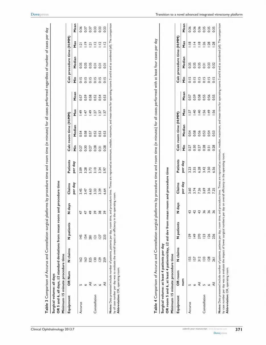
To determine the potential impact of case volume on
efficiency evaluation, we correlated all cases and then inde-
pendently evaluated datasets with cutoffs of at least four,
five, six, or greater than/equal to seven cases per day. This
analysis showed no statistically significant increased effi-
ciency but clearly suggested a trend to increased efficiency
with increasing case volume. Clear positive impacts were
seen for each case volume noting a benefit even for surgical
volumes as low as four cases per day (unreported analysis
of a second data set documented improved efficiencies with
case volumes averaging approximately two cases per day)
(Tables 2–5).
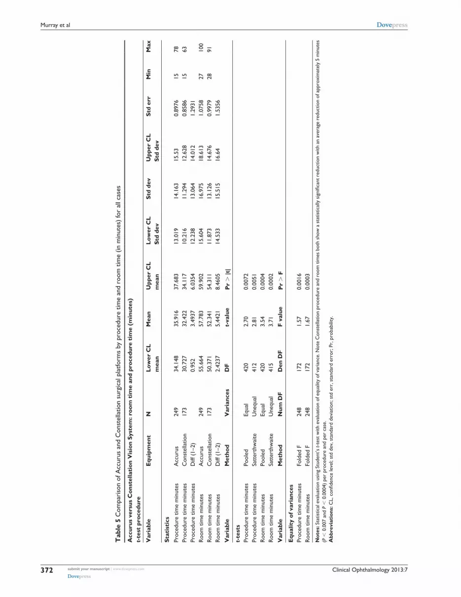
Statistical analysis noted statistically significant improve-
ments in efficiencies for operative time with both decreased
procedure and room time associated with transition to the
Constellation Vision System (P , 0.0004). Additionally,
for combined MIVS pars plana vitrectomy and torsional
phacoemulsification/IOL implantation, a marked decrease
in both procedure and room time were documented
(P , 0.0001) (Tables 6–9).
DiscussionVitreoretinal surgical advances have been rapid since the first
automated vitrectomy surgical units were developed four
decades ago.6–12 During this period, marked improvements
in instrument design contributed to significant increases in
patient safety and improved surgical outcomes. Initially,
Machemer and others focused on multifunction single-port
instrumentation, but instrument design continuously evolved
toward smaller instrument sizes and integrated multiport pars
plana vitrectomy surgery.1,13–15
Further modifications, from Machemer’s initial vit-
rectomy system1 continued to decrease instrument size,
ultimately achieving a standard instrument approach with
20-gauge instruments placed through the sclera in a three-
port pars plana vitrectomy approach whereby one port was
utilized for infusion, one port for illumination, and one port
submit your manuscript | www.dovepress.com
Dovepress
Dovepress
370
Murray et al
Clinical Ophthalmology 2013:7
Tab
le 3
Com
pari
son
of A
ccur
us a
nd C
onst
ella
tion
surg
ical
pla
tform
s by
pro
cedu
re t
ime
and
room
tim
e (in
min
utes
) fo
r al
l cas
es p
erfo
rmed
reg
ardl
ess
of n
umbe
r of
cas
es p
er d
ay
Surg
ical
vol
ume:
all
days
O
R 5
and
6, a
ll da
ys, ±
2 st
anda
rd d
evia
tion
s fr
om m
ean
room
and
pro
cedu
re t
ime
M
inim
um 1
5 m
inut
e pr
oced
ure
tim
e
Equ
ipm
ent
OR
Num
N c
laim
sN
pat
ient
sN
day
sC
laim
s pe
r da
yP
atie
nts
per
day
Cal
c ro
om t
ime
(H:M
M)
Cal
c pr
oced
ure
tim
e (H
:MM
)
Min
Med
ian
Max
Mea
nM
inM
edia
nM
axM
ean
Acc
urus
516
214
547
3.45
3.09
0:27
0:54
1:49
0:57
0:15
0:35
1:21
0:36
616
315
447
3.47
3.28
0:30
0:58
1:47
0:59
0:15
0:35
1:19
0:37
All
325
281
496.
635.
730:
270:
571:
490:
580:
150:
351:
210:
37C
onst
ella
tion
513
012
139
3.33
3.10
0:28
0:52
1:27
0:52
0:15
0:31
1:12
0:33
612
912
738
3.39
3.34
0:31
0:53
1:37
0:54
0:15
0:31
1:12
0:33
All
259
233
396.
645.
970:
280:
531:
370:
530:
150:
311:
120:
33
Not
es: D
ata
pres
ente
d in
clud
e nu
mbe
r of
pat
ient
s, p
atie
nts
per
day,
roo
m ti
me,
and
pro
cedu
re ti
me.
Tim
es a
re r
epor
ted
as m
inim
um, m
edia
n, m
axim
um, a
nd m
ean
time
for
oper
atin
g ro
oms
5 an
d 6
and
as c
ombi
ned
(all)
. The
com
pari
son
by a
ll ca
se n
umbe
r pe
r da
y w
as t
o ev
alua
te t
he o
vera
ll im
pact
on
effic
ienc
y in
the
ope
ratin
g ro
om.
Abb
revi
atio
n: O
R, o
pera
ting
room
.
Tab
le 4
Com
pari
son
of A
ccur
us a
nd C
onst
ella
tion
surg
ical
pla
tform
s by
pro
cedu
re t
ime
and
room
tim
e (in
min
utes
) fo
r al
l cas
es p
erfo
rmed
with
at
leas
t fo
ur c
ases
per
day
Surg
ical
vol
ume:
at
leas
t 4
pati
ents
per
day
O
R r
oom
5 a
nd 6
, at
leas
t 4
pati
ents
/day
, ±2
std
dev
from
mea
n ro
om a
nd p
roce
dure
tim
e
Min
imum
15
min
ute
proc
edur
e ti
me
Equ
ipm
ent
OR
roo
m
num
N c
laim
sN
pat
ient
sN
day
sC
laim
s
per
day
Pat
ient
s
per
day
Cal
c ro
om t
ime
(H:M
M)
Cal
c pr
oced
ure
tim
e (H
:MM
)
Min
Med
ian
Max
Mea
nM
inM
edia
nM
axM
ean
Acc
urus
515
513
943
3.60
3.23
0:27
0:54
1:37
0:57
0:15
0:35
1:18
0:36
615
714
942
3.74
3.55
0:30
0:58
1:40
0:59
0:15
0:35
1:14
0:36
All
312
270
437.
266.
280:
270:
561:
400:
580:
150:
351:
180:
36C
onst
ella
tion
513
312
336
3.69
3.42
0:28
0:53
1:56
0:55
0:15
0:31
1:26
0:35
612
812
636
3.56
3.50
0:31
0:54
1:49
0:56
0:15
0:33
1:28
0:35
All
261
236
367.
256.
560:
280:
531:
560:
550:
150:
321:
280:
35
Not
es: D
ata
pres
ente
d in
clud
e nu
mbe
r of
pat
ient
s, p
atie
nts
per
day,
roo
m ti
me,
and
pro
cedu
re ti
me.
Tim
es a
re r
epor
ted
as m
inim
um, m
edia
n, m
axim
um, a
nd m
ean
time
for
oper
atin
g ro
oms
5 an
d 6
and
as c
ombi
ned
(all)
. The
com
pari
son
by c
ase
num
ber
per
day
was
to
eval
uate
the
impa
ct o
f low
er s
urgi
cal n
umbe
rs p
er d
ay o
n ov
eral
l effi
cien
cy in
the
ope
ratin
g ro
om.
Abb
revi
atio
n: O
R, o
pera
ting
room
.
submit your manuscript | www.dovepress.com
Dovepress
Dovepress
371
Transition to a novel advanced integrated vitrectomy platform
Clinical Ophthalmology 2013:7
Tab
le 5
Com
pari
son
of A
ccur
us a
nd C
onst
ella
tion
surg
ical
pla
tform
s by
pro
cedu
re t
ime
and
room
tim
e (in
min
utes
) fo
r al
l cas
es
Acc
urus
ver
sus
Con
stel
lati
on V
isio
n Sy
stem
: roo
m t
ime
and
proc
edur
e ti
me
(min
utes
) t-
test
pro
cedu
re
Var
iabl
eE
quip
men
tN
Low
er C
L
mea
nM
ean
Upp
er C
L
mea
nLo
wer
CL
St
d de
vSt
d de
vU
pper
CL
St
d de
vSt
d er
rM
inM
ax
Stat
isti
csPr
oced
ure
time
min
utes
Acc
urus
249
34.1
4835
.916
37.6
8313
.019
14.1
6315
.53
0.89
7615
78Pr
oced
ure
time
min
utes
Con
stel
latio
n17
330
.727
32.4
2234
.117
10.2
1611
.294
12.6
280.
8586
1563
Proc
edur
e tim
e m
inut
esD
iff (
1–2)
0.
952
3.49
376.
0354
12.2
3813
.064
14.0
121.
2931
Roo
m t
ime
min
utes
Acc
urus
249
55.6
6457
.783
59.9
0215
.604
16.9
7518
.613
1.07
5827
100
Roo
m t
ime
min
utes
Con
stel
latio
n17
350
.371
52.3
4154
.311
11.8
7313
.126
14.6
760.
9979
2891
Roo
m t
ime
min
utes
Diff
(1–
2)2.
4237
5.44
218.
4605
14.5
3315
.515
16.6
41.
5356
Var
iabl
eM
etho
dV
aria
nces
DF
t-va
lue
Pr
. |t
|
t-te
sts
Proc
edur
e tim
e m
inut
esPo
oled
Equa
l42
02.
700.
0072
Proc
edur
e tim
e m
inut
esSa
tter
thw
aite
Une
qual
412
2.81
0.00
51R
oom
tim
e m
inut
esPo
oled
Equa
l42
03.
540.
0004
Roo
m t
ime
min
utes
Satt
erth
wai
teU
nequ
al41
53.
710.
0002
Var
iabl
eM
etho
dN
um D
FD
en D
FF
valu
eP
r .
F
Equ
alit
y of
var
ianc
esPr
oced
ure
time
min
utes
Fold
ed F
248
172
1.57
0.00
16R
oom
tim
e m
inut
esFo
lded
F24
817
21.
670.
0003
Not
es: S
tatis
tical
eva
luat
ion
usin
g St
uden
t’s t-
test
with
eva
luat
ion
of e
qual
ity o
f var
ianc
e. N
ote
Con
stel
latio
n pr
oced
ure
and
room
tim
es b
oth
show
a s
tatis
tical
ly s
igni
fican
t red
uctio
n w
ith a
n av
erag
e re
duct
ion
of a
ppro
xim
atel
y 5
min
utes
(P
, 0
.007
and
P ,
0.0
004)
per
pro
cedu
re a
nd p
er c
ase.
Abb
revi
atio
ns: C
L, c
onfid
ence
leve
l; st
d de
v, s
tand
ard
devi
atio
n; s
td e
rr, s
tand
ard
erro
r; P
r, p
roba
bilit
y.
submit your manuscript | www.dovepress.com
Dovepress
Dovepress
372
Murray et al

Clinical Ophthalmology 2013:7
Tab
le 6
Com
pari
son
of A
ccur
us a
nd C
onst
ella
tion
surg
ical
pla
tform
s fo
r co
mbi
ned
mic
roin
cisi
onal
vitr
ecto
my
surg
ery
with
pha
coem
ulsi
ficat
ion
and
IOL
impl
anta
tion
for
all c
ases
pe
rfor
med
with
at
leas
t se
ven
case
s pe
r da
y
Mur
ray
com
bine
d su
rger
y: s
ubse
t of
at
leas
t 7
pati
ents
per
day
M
urra
y co
mbi
ned
surg
ery
sum
mar
y
Ant
erio
r: 6
6820
–669
86 P
oste
rior
: 670
15–6
7113
, not
670
28 a
nd 6
7107
O
R r
oom
5 a
nd 6
, at
leas
t 7
pati
ents
/day
, ±2
std
dev
from
mea
n ro
om a
nd p
roce
dure
tim
e M
inim
um 1
5 m
inut
e pr
oced
ure
tim
e
Equ
ipm
ent
OR
roo
m
num
N c
laim
sN
pat
ient
sN
day
sC
laim
s pe
r da
yP
atie
nts
per
day
Cal
c ro
om t
ime
(H:M
M)
Cal
c pr
oced
ure
tim
e (H
:MM
)
Min
Med
ian
Max
Mea
nM
inM
edia
nM
axM
ean
Acc
urus
560
5929
2.07
2.03
0:47
1:06
1:32
1:06
0:24
0:42
1:01
0:43
652
5128
1.86
1.82
0:37
1:09
1:40
1:09
0:26
0:43
1:14
0:44
All
112
108
293.
863.
720:
371:
071:
401:
070:
240:
431:
140:
43C
onst
ella
tion
557
5618
3.17
3.11
0:36
0:56
1:17
0:56
0:23
0:35
0:57
0:36
662
6219
3.26
3.26
0:37
0:56
1:31
0:58
0:20
0:38
1:00
0:37
All
119
114
196.
266.
000:
360:
561:
310:
570:
200:
351:
000:
37
Not
es: D
ata
pres
ente
d in
clud
e nu
mbe
r of
pat
ient
s, p
atie
nts
per
day,
roo
m ti
me,
and
pro
cedu
re ti
me.
Tim
es a
re r
epor
ted
as m
inim
um, m
edia
n, m
axim
um, a
nd m
ean
time
for
oper
atin
g ro
oms
5 an
d 6
and
as c
ombi
ned
(all)
. The
com
pari
son
by c
ase
num
ber
per
day
was
to
eval
uate
the
impa
ct o
f hig
her
surg
ical
num
bers
per
day
on
over
all e
ffici
ency
in t
he o
pera
ting
room
.A
bbre
viat
ions
: OR
, Ope
ratin
g ro
om; I
OL,
intr
aocu
lar
lens
.
Tab
le 7
Com
pari
son
of A
ccur
us a
nd C
onst
ella
tion
surg
ical
pla
tform
s fo
r co
mbi
ned
mic
roin
cisi
onal
vitr
ecto
my
surg
ery
with
pha
coem
ulsi
ficat
ion
and
IOL
impl
anta
tion
for
all c
ases
pe
rfor
med
Com
bine
d su
rger
y su
mm
ary:
all
days
O
R r
oom
5 a
nd 6
, all
pati
ents
/day
, ±2
std
dev
from
mea
n ro
om a
nd p
roce
dure
tim
e M
inim
um 1
5 m
inut
e pr
oced
ure
tim
e
Equ
ipm
ent
OR
roo
m
num
N c
laim
sN
pat
ient
sN
day
sC
laim
s pe
r da
yP
atie
nts
per
day
Cal
c ro
om t
ime
(H:M
M)
Cal
c pr
oced
ure
tim
e (H
:MM
)
Min
Med
ian
Max
Mea
nM
inM
edia
nM
axM
ean
Acc
urus
574
7340
1.85
1.83
0:46
1:06
1:49
1:06
0:24
0:42
1:15
0:43
671
7040
1.78
1.75
0:37
1:09
1:40
1:09
0:26
0:45
1:14
0:45
All
145
141
443.
303.
200:
371:
071:
491:
070:
240:
441:
150:
44C
onst
ella
tion
580
7834
2.35
2.29
0:36
0:58
1:23
0:58
0:20
0:35
1:02
0:37
686
8634
2.53
2.53
0:37
0:56
1:32
0:58
0:20
0:36
1:00
0:37
All
166
158
374.
494.
270:
360:
571:
320:
580:
200:
351:
020:
37
Not
es: D
ata
pres
ente
d in
clud
e nu
mbe
r of
pat
ient
s, p
atie
nts
per
day,
roo
m ti
me,
and
pro
cedu
re ti
me.
Tim
es a
re r
epor
ted
as m
inim
um, m
edia
n, m
axim
um, a
nd m
ean
time
for
oper
atin
g ro
oms
5 an
d 6
and
as c
ombi
ned
(all)
. The
com
pari
son
by a
ll ca
ses
per
day
was
to
elim
inat
e se
lect
ion
bias
on
eval
uatio
n of
ope
ratin
g ro
om e
ffici
ency
.A
bbre
viat
ions
: OR
, Ope
ratin
g ro
om; I
OL,
intr
aocu
lar
lens
.
submit your manuscript | www.dovepress.com
Dovepress
Dovepress
373
Transition to a novel advanced integrated vitrectomy platform

Clinical Ophthalmology 2013:7
Tab
le 8
Com
pari
son
of A
ccur
us a
nd C
onst
ella
tion
surg
ical
pla
tform
s fo
r co
mbi
ned
mic
roin
cisi
onal
vitr
ecto
my
surg
ery
with
pha
coem
ulsi
ficat
ion
and
IOL
impl
anta
tion
for
all c
ases
pe
rfor
med
with
at
leas
t fo
ur c
ases
per
day
Com
bine
d su
rger
y su
mm
ary:
at
leas
t 4
pati
ents
per
day
O
R r
oom
5 a
nd 6
, at
leas
t 4
pati
ents
/day
, ±2
std
dev
from
mea
n ro
om a
nd p
roce
dure
tim
e
Min
imum
15
min
ute
proc
edur
e ti
me
Equ
ipm
ent
OR
roo
m
num
N c
laim
sN
pat
ient
sN
day
sC
laim
s pe
r da
yP
atie
nts
per
day
Cal
c ro
om t
ime
(H:M
M)
Cal
c pr
oced
ure
tim
e (H
:MM
)
Min
Med
ian
Max
Mea
nM
inM
edia
nM
axM
ean
Acc
urus
571
7038
1.87
1.84
0:46
1:06
1:32
1:05
0:24
0:42
1:01
0:43
669
6838
1.82
1.79
0:37
1:09
1:40
1:08
0:26
0:45
1:14
0:45
All
140
136
413.
413.
320:
371:
071:
401:
070:
240:
441:
140:
44C
onst
ella
tion
580
7834
2.35
2.29
0:36
0:58
1:56
0:58
0:20
0:35
1:19
0:37
686
8634
2.53
2.53
0:37
0:56
1:45
0:59
0:20
0:36
1:00
0:37
All
166
157
364.
614.
360:
360:
571:
560:
580:
200:
351:
190:
37
Not
es: D
ata
pres
ente
d in
clud
e nu
mbe
r of
pat
ient
s, p
atie
nts
per
day,
roo
m ti
me,
and
pro
cedu
re ti
me.
Tim
es a
re r
epor
ted
as m
inim
um, m
edia
n, m
axim
um, a
nd m
ean
time
for
oper
atin
g ro
oms
5 an
d 6
and
as c
ombi
ned
(all)
. The
com
pari
son
by c
ase
num
ber
per
day
was
to
eval
uate
the
impa
ct o
f low
er s
urgi
cal n
umbe
rs p
er d
ay o
n ov
eral
l effi
cien
cy in
the
ope
ratin
g ro
om.
Abb
revi
atio
ns: O
R, O
pera
ting
room
; IO
L, in
trao
cula
r le
ns.
for the cutter/forceps/scissors. Seeking smaller wounds, more
rapid wound healing, and elimination of transscleral repeti-
tive instrument passage led to smaller gauge instruments that
incorporated transconjunctival/transscleral trocars focused on
23-gauge and 25-gauge surgical instruments.16–20 This MIVS
approach has rapidly become the current standard with a
transition from 20-gauge sutured sclerotomies to 23-gauge
and 25-gauge trocared instrument approaches not requiring
suture closure. Currently, 27-gauge (and smaller) instruments
are available and in design.
A major impetus for the development of a novel, advanced,
integrated platform design has been the surgical requirements
of increased cutting rates, improved intraocular fluidics,
enhanced lighting, and deliverable endolaser. This shift to
small gauge surgery, along with the interest in an integrated
platform, necessitated the design and development of a novel
next-generation surgical platform. This ideal platform would
incorporate very high speed cutting, stable real-time evalua-
tion of intraocular fluidics, markedly improved illumination
sources, capacity for delivery of high centistoke liquids,
microvolume deliveries, and integrated laser technology.21–37
These characteristics define the minimal surgical requirements
for an integrated platform designed for the 21st century.
Additionally, this study took place during the transition
from 20-gauge pars plana vitrectomy to 23/25-gauge MIVS.
This surgical platform transition recognized the initial
concerns for increased risk of endophthalmitis, choroidal
detachment, iatrogenic retinal tear and/or detachment, or
postoperative hypotony associated with microincisional
vitrectomy.38–42 Focused investigation and training on wound
construction and surgical technique were instrumental in the
use of transconjunctival, trocared, nonsutured pars plana
vitrectomy in the surgical care of our patients. Fortunately,
these concerns have been alleviated by clinical reviews that
have not noted increased complication profiles with micro-
incisional vitrectomy.43
In this study, we evaluated the “real world” surgical per-
formance of the platform that has been the “gold standard”
in vitreoretinal surgical systems, the Accurus platform, and
contrasted that performance with the next generation vit-
reoretinal platform, the Constellation Vision System.44–48 This
comparison utilized standard metrics incorporated within the
BPEI/ABLEH surgical electronic medical record to determine
case volume per room per day, operating room time per case,
and surgical time per case along with total operating time per
room per day. These metrics allow a standardized comparison
of technologies but require evaluation after a transition window
when each technology has achieved a steady implementation
submit your manuscript | www.dovepress.com
Dovepress
Dovepress
374
Murray et al

Clinical Ophthalmology 2013:7
Tab
le 9
Com
pari
son
of A
ccur
us a
nd C
onst
ella
tion
surg
ical
pla
tform
s by
pro
cedu
re ti
me
and
room
tim
e (in
min
utes
) for
com
bine
d m
icro
inci
sion
al v
itrec
tom
y an
d ph
acoe
mul
sific
atio
n w
ith IO
L im
plan
tatio
n
Acc
urus
ver
sus
Con
stel
lati
on V
isio
n Sy
stem
C
ombi
ned
MIV
S an
d ph
acoe
mul
sific
atio
n
Pro
cedu
re t
ime
and
room
tim
e (m
inut
es)
t-te
st p
roce
dure
Var
iabl
eE
quip
men
tN
Low
er C
L
Mea
nM
ean
Upp
er C
L
Mea
nLo
wer
CL
st
d de
vSt
d de
vU
pper
CL
st
d de
vSt
d er
rM
inM
ax
Stat
isti
csPr
oced
ure
time
min
utes
Acc
urus
112
41.4
7143
.339
45.2
088.
8203
9.97
811
.488
0.94
2824
74Pr
oced
ure
time
min
utes
Con
stel
latio
n11
935
.041
36.7
7338
.505
8.46
329.
5407
10.9
350.
8746
2060
Proc
edur
e tim
e m
inut
esD
iff (
1–2)
4.
0357
6.56
629.
0967
8.93
769.
7551
10.7
381.
2843
Roo
m t
ime
min
utes
Acc
urus
112
64.7
4667
.205
69.6
6511
.611
13.1
3515
.123
1.24
1137
100
Roo
m t
ime
min
utes
Con
stel
latio
n11
955
.09
57.1
2659
.162
9.94
9711
.216
12.8
551.
0282
3691
Roo
m t
ime
min
utes
Diff
(1–
2)
6.91
8710
.079
13.2
411
.163
12.1
8413
.412
1.60
4
Var
iabl
eM
etho
dV
aria
nces
DF
t-va
lue
Pr
. |t
|
t-te
stPr
oced
ure
time
min
utes
Pool
edEq
ual
229
5.11
,0.
0001
Proc
edur
e tim
e m
inut
esSa
tter
thw
aite
Une
qual
226
5.11
,0.
0001
Roo
m t
ime
min
utes
Pool
edEq
ual
229
6.28
,0.
0001
Roo
m t
ime
min
utes
Satt
erth
wai
teU
nequ
al21
96.
25,
0.00
01
Var
iabl
eM
etho
dN
um D
FD
en D
FF-
valu
eP
r .
F
Equ
alit
y of
var
ianc
esPr
oced
ure
time
min
utes
Fold
ed F
111
118
1.09
0.63
10R
oom
tim
e m
inut
esFo
lded
F11
111
81.
370.
0917
Not
es: S
tatis
tical
eva
luat
ion
usin
g St
uden
t’s t-
test
with
eva
luat
ion
of e
qual
ity o
f var
ianc
e. N
ote
Con
stel
latio
n pr
oced
ure
and
room
tim
es b
oth
show
a s
tatis
tical
ly s
igni
fican
t red
uctio
n w
ith a
n av
erag
e re
duct
ion
of a
ppro
xim
atel
y 7
min
utes
(P
, 0
.000
1) a
nd 1
0 m
inut
es (
P ,
0.0
001)
per
pro
cedu
re a
nd p
er c
ase.
Abb
revi
atio
ns: I
OL,
intr
aocu
lar
lens
; MIV
S, m
icro
inci
sion
al v
itrec
tom
y su
rger
y; C
L, c
onfid
ence
leve
l; st
d de
v, s
tand
ard
devi
atio
n; s
td e
rr, s
tand
ard
erro
r.
submit your manuscript | www.dovepress.com
Dovepress
Dovepress
375
Transition to a novel advanced integrated vitrectomy platform

Clinical Ophthalmology 2013:7
state of usage. Further, evaluation of a large time frame coupled
with high surgical numbers, as in this study, eliminates many
potential biases to evaluation of the utility of new technology,
such as the Constellation Vision System.
This study documents the increased efficiency of the
Constellation platform relative to the prior standard Accu-
rus system. The Constellation achieved increased patient
surgical cases per day by decreasing both operative case
time and room time. This increase is related to an integrated
design that facilitates case turnover, vitrectomy instrument
and cassette setup (and particularly combined vitrectomy/
phacoemulsification instrument setup), and enhancements
with integrated instrument recognition technology, prepopu-
lated user settings, surgeon-controlled endolaser parameters,
and rapid priming associated with improved fluidics. These
platform design changes clearly target improvements for the
surgeon, the OR team, and the patient.
Ultimately, the decision to transition from existing tech-
nology to new technology should focus first on enhanced
patient care, including patient safety, improved anatomic
outcomes, improved visual outcomes, and translation to a
better quality of life.
DisclosureThe authors report no relevant financial conflicts of interest
in this work. Dr Murray consults for Thrombogenics and
Alcon.
References1. Machemer R, Buettner H, Norton EW, Parel JM. Vitrectomy:
a pars plana approach. Trans Am Acad Ophthalmol Otolaryngol. 1971;75(4):813–820.
2. Norton EW, Machemer R. New approach to the treatment of selected retinal detachments secondary to vitreous loss at cataract surgery. Am J Ophthalmol. 1971;72(4):705–707.
3. Chang S. Intraocular gases. In: Ryan S, Glaser BM, editors. Retina. 2nd ed. St Louis: Mosby; 1994.
4. Abrams GW, Azen SP, McCuen BW II, et al. Vitrectomy with silicone oil or long-acting gas in eyes with severe proliferative vitreoretinopathy: results of additional and long term follow-up. Silicone Study Report #11. Arch Ophthalmol. 1997;115:335–344.
5. Chang S. Vitrectomy. In: Duker J, Yanoff MA, editors. Ophthalmology. London: Mosby; 2008.
6. Koenig SB, Mieler WF, Han DP, et al. Combined phacoemulsification, pars plana vitrectomy, and posterior chamber intraocular lens insertion. Arch Ophthalmol. 1992;110:1101–1104.
7. Thompson JT, de Bustros S, Michels RG, et al. Results and prognostic factors in vitrectomy for diabetic vitreous hemorrhage. Arch Ophthalmol 1987;105:191–195.
8. Chang S, Lincoff H, Zimmerman NJ, et al. Giant retinal tears: surgical techniques and results using perfluorocarbon liquids. Arch Ophthalmol. 1989;107:761–766.
9. Gardner T, Blankenship GW. Proliferative diabetic retinopathy: principles and techniques of surgical treatment. In: Ryan S, Glaser BM, editors. Retina. 3rd ed. St Louis: Mosby; 2012.
10. Coll GE, Chang S, Sun J, et al. Perfluorocarbon liquid in the management of retinal detachment with proliferative vitreoretinopathy. Ophthalmol-ogy. 1994;102:630–638.
11. Melberg NS, Thomas MA, Dickinson JD, et al. Surgical removal of subfovealchoroidal neovascularization: ingrowth site as a predictor of visual outcome. Retina. 1996;16:190–195.
12. Sjaarda RN, Michels RG. Macular Pucker. In: Ryan S, Glaser BM, editors. Retina. 3rd ed. St Louis: Mosby; 2012.
13. Freeman W, Macular Hole Study Group. Vitrectomy for the treatment of full-thickness stage 3 or stage 4 macular holes: results of a multicentered randomized clinical trial. Arch Ophthalmol. 1997;115:11–21.
14. Borne MJ, Tasman W, Regillo C, et al. Outcomes of vitrectomy for retained lens fragments. Ophthalmology. 1996;103:971–976.
15. Scott IU, Flynn HW, Lai M, Chang S, Azen SP: First operation anatomic success and other predictors of postoperative vision after complex retinal detachment repair with vitrectomy and silicone oil tamponade. Am J Ophthalmol. 2000;130:745–750.
16. Ibarra MS, Hermel M, Prenner JL, Hassan TS. Longer-term outcomes of transconjunctival sutureless 25-gauge vitrectomy. Am J Ophthalmol. 2005;139:831–836.
17. Lakhanpal RR, Humayun MS, de Juan E Jr, et al. Outcomes of 140 consecutive cases of 25-gauge transconjunctival surgery for posterior segment disease. Ophthalmology. 2005;112:817–824.
18. Oshima Y, Ohji M, Tano Y. Surgical outcomes of 25-gauge transcon-junctivalvitrectomy combined with cataract surgery for vitreoretinal diseases. Ann Acad Med Singapore. 2006;35:175–180.
19. Kadonosono K, Yamakawa T, Uchino E, et al. Comparison of visual function after epiretinal membrane removal by 20- gauge and 25-gauge vitrectomy. Am J Ophthalmol. 2006;142:513–515.
20. Fine HF, Iranmanesh R, Iturralde D, Spaide RF. Outcomes of 77 consecutive cases of 23-gauge transconjunctival vitrectomy surgery for posterior segment disease. Ophthalmology. 2007;114: 1197–1200.
21. Shimada H, Nakashizuka H, Hattori T, Mori R, et al. Incidence of endophthalmitis after 20- and 25-gauge vitrectomy: Causes and prevention. Ophthalmology. 2008;15:2215–2220.
22. Shinoda H, Shinoda K, Satofuka S, et al. Visual recovery after vitrectomy for macular hole using 25-gauge instruments. Acta Ophthalmol. 2008;86:151–155.
23. Bamonte G, Mura M, Tan HS. Hypotony after 25-gauge vitrectomy. Am J Ophthalmol. 2010;151:(1):156–160.
24. Fujii GY, de Juan E Jr, Humayun MS, et al. Initial experience using the transconjunctival sutureless vitrectomy system for vitreoretinal surgery. Ophthalmology. 2002;109:1814–1820.
25. Fujii GY, De Juan E Jr, Humayun MS, et al. A new 25-gauge instru-ment system for transconjunctival sutureless vitrectomy surgery. Ophthalmology. 2002;109:1807–1812.
26. Nam Y, Chung H, Lee JY, Kim JG, Yoon YH. Comparison of 25- and 23-gauge sutureless microincision vitrectomy surgery in the treatment of various vitreoretinal diseases. Eye. 2010;24:191.
27. Eckardt C. Transconjunctival sutureless 23-gauge vitrectomy. Retina. 2005;25:208–211.
28. Okamoto F, Okamoto C, Sakata N, et al. Changes in corneal topog-raphy after 25-gauge transconjunctival sutureless vitrectomy versus after 20-gauge standard vitrectomy. Ophthalmology. 2007;114: 2138–2141.
29. Kadonosono K, Yamakawa T, Uchino E, et al. Comparison of visual function after epiretinal membrane removal by 20- gauge and 25-gauge vitrectomy. Am J Ophthalmol. 2006;142:513–515.
30. Shinoda H, Shinoda K, Satofuka S, et al. Visual recovery after vitrec-tomy for macular hole using 25-gauge instruments. Acta Ophthalmol. 2008;86:151–155.
31. Chang SE, Kim KH, Kang SW. Retinal breaks associated with the induction of posterior vitreous detachment. Am J Ophthalmol. 2009;147(6):1012–1016.
32. Tan HS, Mura M, de Smet MD. Iatrogenic retinal breaks in 25-gauge macular surgery. Am J Ophthalmol. 2009;148(3):427–430.
submit your manuscript | www.dovepress.com
Dovepress
Dovepress
376
Murray et al

Clinical Ophthalmology
Publish your work in this journal
Submit your manuscript here: http://www.dovepress.com/clinical-ophthalmology-journal
Clinical Ophthalmology is an international, peer-reviewed journal covering all subspecialties within ophthalmology. Key topics include: Optometry; Visual science; Pharmacology and drug therapy in eye diseases; Basic Sciences; Primary and Secondary eye care; Patient Safety and Quality of Care Improvements. This journal is indexed on
PubMed Central and CAS, and is the official journal of The Society of Clinical Ophthalmology (SCO). The manuscript management system is completely online and includes a very quick and fair peer-review system, which is all easy to use. Visit http://www.dovepress.com/ testimonials.php to read real quotes from published authors.
Clinical Ophthalmology 2013:7
33. Oshima Y, Shima C, Wakabayashi T, et al. Microincision vitrectomy surgery and intravitreal bevacizumab as a surgical adjunct to treat diabetic traction retinal detachment. Ophthalmology. 2009;116(5): 927–938.
34. Rizzo S. Comparative study between standard 25-gauge vitrectomy systems and new ultra-high speed 25-gauge system with duty cycle control in the treatment of various vitreoretinal diseases. Paper presented at: Vail Vitrectomy 2010; March 13–17, 2010; Vail, CO.
35. Rizzo S, Genovesi–Ebert F, Murri S, et al. 25-gauge, sutureless vit-rectomy and standard 20-gauge pars plana vitrectomy in idiopathic epiretinal membrane surgery: a comparative pilot study. Graefes Arch Clin Exp Ophthalmol. 2006;244(4):472–479.
36. Chen E. 25-Gauge transconjunctival sutureless vitrectomy. Curr Opin Ophthalmol. 2007;18(3):188–193.
37. Kellner L, Wimpissinger B, Stolba U, et al. 25-gauge vs 20-gauge system for pars plana vitrectomy: a prospective randomized clinical trial. Br J Ophthalmol. 2007;91(7):945–948.
38. Kunimoto DY, Kaiser RS. Incidence of endophthalmitis after 20- and 25-gauge vitrectomy. Ophthalmology. 2007;114(12):2133–2137.
39. Scott IU, Flynn HW Jr, Dev S, et al. Endophthalmitis after 25-gauge and 20-gauge pars plana vitrectomy: incidence and outcomes. Retina. 2008;28(1):138–142.
40. Taban M, Sharma S, Ventura AA, Kaiser PK. Evaluation of wound closure in oblique 23- gauge sutureless sclerotomies with Visante optical coherence tomography. Am J Ophthalmol. 2009;147(1):101–107.
41. Taban M, Ventura AA, Sharma S, Kaiser PK. Dynamic evaluation of sutureless vitrectomy wounds: an optical coherence tomography and histopathology study. Ophthalmology. 2008;115(12):2221–2228.
42. Martidis A, Chang TS. Sutureless 25-gauge vitrectomy: risky or rewarding? Ophthalmology. 2007;114(12):2131–2132.
43. Kaiser RS, Prenner J, Scott IU, et al. The Microsurgical Safety Task Force: evolving guidelines for minimizing the risk of endophthalmitis associated with microincisional vitrectomy surgery. Retina. 2010;30(4): 692–699.
44. Parke DW 3rd, Sisk RA, Houston SK, Murray TG. Ocular hypertension after intravitreal triamcinolone with vitrectomy and phacoemulsification. Clin Ophthalmol. 2012;6:925–631.
45. Murray TG, Tornambe P, Dugel P, Tong KB. Evaluation of economic efficiencies in clinical retina practice: activity-based cost analysis and modeling to determine impacts of changes in patient management. Clin Ophthalmol. 2011;5:913–925. Epub July 12, 2011.
46. Sisk RA, Berrocal AM, Feuer WJ, Murray TG. Visual and anatomic outcomes with or without surgery in persistent fetal vasculature. Ophthalmology. 2010;117(11):2178–2183.
47. Sisk RA, Murray TG. Combined phacoemulsification and sutureless 23-gauge pars plana vitrectomy for complex vitreoretinal diseases. Br J Ophthalmol. 2010;94(8):1028–1032.
48. Hartley KL, Smiddy WE, Flynn HW Jr, Murray TG. Pars plana vit-rectomy with internal limiting membrane peeling for diabetic macular edema. Retina. 2008;28(3):410–419.
submit your manuscript | www.dovepress.com
Dovepress
Dovepress
Dovepress
377
Transition to a novel advanced integrated vitrectomy platform