transcriptional inhibition of the human insulin receptor gene by aldosterone
TRANSCRIPT

Journal of Steroid Biochemistry & Molecular Biology 84 (2003) 543–553
Transcriptional inhibition of the human insulinreceptor gene by aldosterone
Consuelo Callea,∗, Javier Campióna, Moisés Garcıa-Arencibiaa,Begoña Maestroa, Norma Dávilab
a Department of Biochemistry and Molecular Biology, School of Medicine, Complutense University, Madrid 28040, Spainb Biochemistry Unit, Puerta de Hierro Hospital, Madrid 28040, Spain
Received 20 May 2002; accepted 17 January 2003
Abstract
In earlier studies, we reported reduced human insulin receptor (hIR) mRNA levels, insulin binding and insulin responsiveness in U-937human promonocytic cells treated with aldosterone. The mechanism for this inhibition could be diminished IR gene transcription, sincealdosterone did not affect hIR mRNA stability. All the effects were mediated by a downregulation of the mineralocorticoid receptor (MR,NR3C2) expressed at both the RNA and protein levels, suggesting that MR could act as a transcription factor that binds to hormoneresponse elements in the hIR gene promoter. Indeed, MR has been shown to bind glucocorticoid response elements (GREs) in targetgenes. Given that five GREs have been characterized in the hIR promoter, we decided to test whether these elements could mediate thealdosterone-elicited inhibition of hIR expression detected by us in U-937 cells. In the present report, we demonstrate that aldosteroneinhibits the activity of the hIR wild-type promoter by 23%, and causes 23 and 31% reductions in the activity of progressive deletions ofthis promoter comprised of fragments up to−1473 and−876 bp, respectively. This indicates that the−876 to−271 bp region of the hIRpromoter may be sufficient for this transcriptional inhibition by aldosterone. We also provide evidence for direct MR interaction with someof the GREs of this promoter region, specifically with the cGRE1 and cGRE3, presumably as MR–MR homodimers, and with pGRE asa MR–GR heterodimer. This heterodimer may play the most relevant role and participate in the cross-talk between mineralocorticoids,glucocorticoids and insulin signalling in U-937 cells.© 2003 Elsevier Science Ltd. All rights reserved.
Keywords: Aldosterone; Mineralocorticoid receptor; Dexamethasone; Glucocorticoid receptor; Glucocorticoid response element; Heterodimerization; Humaninsulin receptor gene promoter; Transcriptional regulation; U-937 human promonocytic cells
1. Introduction
It is know that the genomic effects of mineralocorticoidsand glucocorticoids are mediated by interaction with theirreceptors[1,2]. Both types of hormone passively diffusethrough the cell membrane and bind the mineralocorticoidreceptor (MR, NR3C2) and the glucocorticoid receptor (GR,NR3C1) in the cytoplasm/nucleus of the cell. The affinity ofMR for glucocorticoids is almost identical to that for miner-alocorticoids[3]. After binding, the activated receptors actas transcription factors, which bind to hormone response el-ements in the promoter region of responsive genes[4]. Sinceno selective mineralocorticoid response elements have beenidentified, MR and GR appear to promiscuously bind andtransactivate glucocorticoid response elements (GREs) inthe promoters of target genes[4,5]. Moreover, MR and GR
∗ Corresponding author. Tel.:+34-913941451; fax:+34-913941691.E-mail address: [email protected] (C. Calle).
seem to act as dimers, either homodimers or heterodimers[6–9], enhancing [7] or lowering [8] transcriptionalactivity.
Results from our laboratory have previously indicatedin vivo and in vitro modulation of insulin receptor (IR)gene expression by glucocorticoids. Thus, we were ableto demonstrate that IR mRNA levels were modulated in atissue-specific manner in patients with Cushing’s syndrome[10]. We also observed tissue-specific changes in IR mRNAconcentrations in dexamethasone-treated rats[11]. In addi-tion, we reported dose- and time-dependent dexamethasonestimulation of the two major species of the human insulinreceptor (hIR) mRNA (11 and 8.5 kb in size) occurring inU-937 human promonocytic cells[12,13]. In these cells,dexamethasone failed to affect both hIR mRNA half-lifeand protein turnover, suggesting an effect on transcription[13].
We also reported the in vivo and in vitro modulation ofIR gene and protein expression by mineralocorticoids. We
0960-0760/03/$ – see front matter © 2003 Elsevier Science Ltd. All rights reserved.doi:10.1016/S0960-0760(03)00072-4

544 C. Calle et al. / Journal of Steroid Biochemistry & Molecular Biology 84 (2003) 543–553
noted a reduction in the number and affinity of hIRs inadipocytes isolated from the subcutaneous adipose tissue ofa patient with primary hyperaldosteronism[14]. We also ob-served tissue-specific modulation of IR gene expression ina model of mineralocorticoid excess in the rat[15]. Further,we recently demonstrated dose- and time-dependent aldos-terone inhibition of the two major species of hIR mRNAin U-937 cells [16]. RNA stability and protein turnoverwere both unaltered, suggesting an effect on transcription.Aldosterone also decreased the hIR number but not theaffinity of this receptor, suggesting possible failure in theinsulin responsiveness of the cells. When this possibilitywas explored, the maximal cellular response to insulin,both in terms of glucose transport and DNA synthesis,was decreased after aldosterone treatment[17]. The pres-ence of MR in U-937 cells was also observed[16,17], andblockade of this receptor by the mineralocorticoid antag-onist spironolactone indicated the involvement of this MRin the aldosterone-elicited inhibition of hIR mRNA levels[16].
The promoter of the hIR gene has been the subject ofintense research. This promoter contains binding sites formany transcription factors including five GREs. TheseGREs were well characterized by Lee and co-workers[18,19] through transfection experiments and footprintingassays. Although these GREs did not match the canonicalGRE sequence, they were able to confer a capacity for
Fig. 1. Transcriptional effect of aldosterone and dexamethasone on the promoter activity of the human insulin receptor (hIR) gene in U-937 cells. Thecells were transiently transfected with the phIR(−1819)-GL2 plasmid, which contains the−1819 to−271 promoter fragment of the hIR gene, consideredas the wild-type promoter, and with phIR(−1473)-GL2 or phIR(−876)-GL2 plasmids, containing progressive 5′ deletions (up to−1473 and−876 bp,respectively) of the wild-type promoter. The transfected cells were left untreated (C), or were treated either with 5× 10⁻⁶ M dexamethasone for 15 h(G) or with 10−9 M aldosterone for 24 h (M). Luciferase activity is given in arbitrary units relative to the value of 100 assigned to that of the wildpromoter in untreated cells after correcting for transfection efficiency. Values are means± S.E.M. of at least five experiments.∗P < 0.05, ∗∗P < 0.01vs. respective control.
glucocorticoid-dependent transcriptional induction upon thehIR gene. Given the intimate relationship between MR andGR, these GREs could mediate the aldosterone-elicited in-hibition of hIR gene expression and insulin action detectedby us in U-937 cells.
On this setting, the present study was designed to extendour previous studies and investigate the possibility of a directtranscriptional effect of aldosterone on the promoter activityof the hIR gene in U-937 cells. In addition, we explored theinteraction of the activated MR with each of the functionalGREs previously described by Lee and co-workers[18,19]inthe hIR promoter. In parallel, we examine the transcriptionaleffect of dexamethasone and binding of the activated GRwith each of the GREs of this promoter.
2. Materials and methods
2.1. Cell culture and treatments
U-937 human promonocytic cells (mycoplasma-free)were grown in RPMI-1640 medium, supplemented with10% (v/v) heat-inactivated foetal calf serum and antibioticsat 37◦C in a humidified 5% CO2 atmosphere as describedpreviously [12,20]. Besides the MR[16,17], these cellsare naturally endowed with other receptors, including IR[12,21], and GR[22].

C. Calle et al. / Journal of Steroid Biochemistry & Molecular Biology 84 (2003) 543–553 545
2.2. Plasmids, transfections and analysisof luciferase activity
The −1819 to −271 promoter fragment of the hIRgene cloned in the BglII site of the pCAT3M vector waskindly provided by Drs. S.Y. Tsai (Department of CellBiology, University of California) and G. Elberg (BaylorCollege of Medicine, Texas Medical Center). This pro-moter fragment, considered by these authors as the wild-type promoter, was subcloned into the BglII site of thepGL2-basic vector (Promega) to create the reporter plasmidphIR(−1819)-GL2 [23]. The orientation and integrity ofthe insert was confirmed by restriction analysis. Digestionof phIR(−1819)-GL2 plasmid with KpnI or XhoI generatedthe plasmids phIR(−1473)-GL2 and phIR(−876)-GL2,respectively.
Fig. 2. Binding of the mineralocorticoid receptor (MR) and glucocorticoid receptor (GR) to Vit-GRE. EMSA was performed as described inSection 2. Inbrief, nuclear extracts of untreated U-937 cells (C), cells treated with 5× 10⁻⁶ M dexamethasone for 15 h (G), and cells treated with 10−9 M aldosteronefor 24 h (M), were incubated with the32P-labelled double stranded synthetic oligonucleotide Vit-GRE. (A) Lane 0, no nuclear extracts; lanes 1–3,nuclear extracts under the three treatment conditions; lanes 4–6 with 50× and lanes 7–9 with 200× excess of the unlabelled Vit-GRE oligonucleotide,respectively. (B) Lanes 1–3 with the specific anti-GR antibody (PA1-510A), and lanes 4–6 with the specific anti-MR antibody (MCR N-17), respectively.Arrows indicate the position of the complexes (I and III).
Transient transfections were carried out by electropora-tion of 20 × 106 cells in RPMI 1640 medium with theBio-Rad gene-pulser II, essentially as described previously[23]. The cells were electroporated at 250 V, 960�F, in avolume of approximately 300�l of RPMI containing 50�gof each of the reporter plasmids described earlier, or 50�g ofthe promoterless pGL2-basic vector, together with 50�g ofthe pBluescript II KS(+/−) as a carrier. As a positive con-trol, we used 25�g of the pGL3-control vector (Promega),which includes SV40 promoter and enhancer sequences. Alltransfections were performed in the absence of MR and GRexpression vectors due to the endogenous activity of bothreceptors in U-937 cells[16,17,22]. Transfection efficiencywas determined using 12�g of pCMV-�gal (Clontech), andby assessing�-galactosidase activity in the extracts[24]. Af-ter resting for 24 h, the transfected cells were left untreated,

546 C. Calle et al. / Journal of Steroid Biochemistry & Molecular Biology 84 (2003) 543–553
or treated with either 10−9 M aldosterone (Sigma) for 24 h,or 5× 10⁻⁶ M dexamethasone (Sigma) for 15 h. These con-ditions of dose and time were selected on the basis of previ-ously observed maximal effects in both aldosterone-elicitedinhibition and dexamethasone-induced stimulation of hIRmRNA levels in U-937 cells[13,16]. These treatments werenon-selective for MR or GR activation, given that the cor-ticoids were supplied in the absence of antagonists to cellswith endogenous MR and GR that could be activated by ei-ther hormone. Nevertheless, our purpose was to induce op-timal transcriptional effects in each treatment by activationof both receptors.
The cells were then collected by centrifugation, andluciferase activity quantified following the instructionsprovided in the assay kit (Promega). Previous luciferase de-terminations revealed that the activity of the pGL3-controlvector was about 100 times above the basal levels shown
Fig. 3. Binding of the MR and GR to dGRE. EMSA was performed as described inSection 2. In brief, nuclear extracts of untreated U-937 cells (C),cells treated with 5× 10⁻⁶ M dexamethasone for 15 h (G), and cells treated with 10−9 M aldosterone for 24 h (M), were incubated with the32P-labelleddouble stranded synthetic oligonucleotide dGRE. (A) Lane 0, no nuclear extracts; lanes 1–3, nuclear extracts under the three treatment conditions;lanes4–6 with 50× and lanes 7–9 with 200× excess of unlabelled dGRE oligonucleotide, respectively. (B) Lanes 1–3 with the specific anti-GR antibody(PA1-510A), and lanes 4–6 with the specific anti-MR antibody (MCR N-17), respectively. Arrows indicate the position of the complexes (I and III).
by the promoterless pGL2-basic vector, after correcting fortransfection efficiency[23].
2.3. Electrophoretic mobility shift assays (EMSA)
Six oligonucleotides commercially synthesized by Cru-achem: Vit-GRE, dGRE, cGRE1, cGRE2, cGRE3, andpGRE were used in these assays. Vit-GRE (5′GATCCAAAGTCAGAACACAGTGTTCTGATC3′) comprised the nat-ural sequence of the oestrogen response element in theXenopus vitellogenin A2 gene mutated to obtain a perfect3′ half-site GRE motif[25]. The rest of oligonucleotidescomprised the following five GREs sequences of the hIRpromoter, well characterised by Lee and co-workers[18,19]through transfection experiments and footprinting assaysdGRE: from −1363 to −1335 bp (5′CCCTCCTCCCATTGAGTTCTGGCTTTCCT3′); cGRE1: from −754 to

C. Calle et al. / Journal of Steroid Biochemistry & Molecular Biology 84 (2003) 543–553 547
−725 bp (5′CCTGTGGGGCGCCTCCGGGGGTCTGAAACT3′); cGRE2: from−707 to−689 bp (5′GTAGGGCGCGCGGATCTGG3′); cGRE3: from−680 to −656 bp (5′CTCGGTCCCGGCGCGCCCAGGGCCT3′); pGRE: from−363 to −341 bp (5′TCCCGGAGCCCGCAGATCGCGAC3′).
The annealed oligonucleotides (3.5 × 10⁻¹² M) were5′end-labelled with [�-32P]ATP (3000 Ci/mmol) (NEN)with T4 polynucleotide kinase (Promega). Nuclear extractswere obtained as described by Schreibert et al.[26] fromcells untreated or treated with either 10−9 M aldosteronefor 24 h, or 5× 10⁻⁶ M dexamethasone for 15 h. These ex-tracts (10�g) were incubated for 10 min on ice in a bindingbuffer containing 1× 10⁻² M Tris, pH 7.5, 8× 10⁻² MKCl, 10% glycerol, 1× 10⁻³ M dithiothreitol, and 2�g ofpoly(dI-dC)[27], in a total volume of 20�l. Next, 0.1–0.5 ng
Fig. 4. Binding of the MR and GR to cGRE1. EMSA was performed as described inSection 2. In brief, nuclear extracts of untreated U-937 cells(C), cells treated with 5× 10⁻⁶ M dexamethasone for 15 h (G), and cells treated with 10−9 M aldosterone for 24 h (M), were incubated with the32P-labelled double stranded synthetic oligonucleotide cGRE1. (A) Lane 0, no nuclear extracts; lanes 1–3, nuclear extracts under the three treatmentconditions; lanes 4–6 with 50× and lanes 7–9 with 200× excess of unlabelled cGRE1 oligonucleotide, respectively. (B) Lanes 1–3, with the specificanti-GR antibody (PA1-510A). (C) Lanes 4–6, with the specific anti-MR antibody (MCR N-17). Arrows indicate the position of the complexes(I–III).
(100,000 cpm) of each labelled oligonucleotide was addedto the reaction mixture and incubation was continued for20 min at room temperature. An excess of the correspond-ing unlabelled oligonucleotide (50×, or 200×) was addedas a specific competitor in each EMSA. For a stricter test ofthe binding specificity of the protein–DNA complexes, weincubated the nuclear extracts with an anti-GR polyclonalantibody (PA1-510A) (Affinity Bioreagents) that recognisesthe N-terminal region of the human GR, or an anti-MRpolyclonal antibody (MCR N-17) (Santa Cruz Biotech-nology) that binds to the N-terminal region of the humanMR. Incubation of the nuclear extracts with a non-immuneserum had no effect. The protein–DNA complexes were re-solved on 4% nondenaturating polyacrylamide gels at 4◦Cin 0.25× TBE. The gels were then dried and examined byautoradiography.
548 C. Calle et al. / Journal of Steroid Biochemistry & Molecular Biology 84 (2003) 543–553
2.4. Statistical analysis
Unless otherwise stated, data are expressed as the mean±S.E.M. The Student’st-test was used for the statistical com-parisons. The threshold for significance was set atP < 0.05.
3. Results
To investigate the possibility of a direct transcriptional ef-fect of aldosterone on the promoter activity of the hIR gene,U-937 cells were transiently transfected with a reporter plas-mid encoding the hIR promoter spanning nucleotides−1819to −271 (wild-type promoter) linked to the luciferase gene,phIR(−1819)-GL2. Treatment of the transfected cells with10−9 M aldosterone for 24 h resulted in a 23% inhibition ofthe promoter activity of this wild-type promoter (Fig. 1).
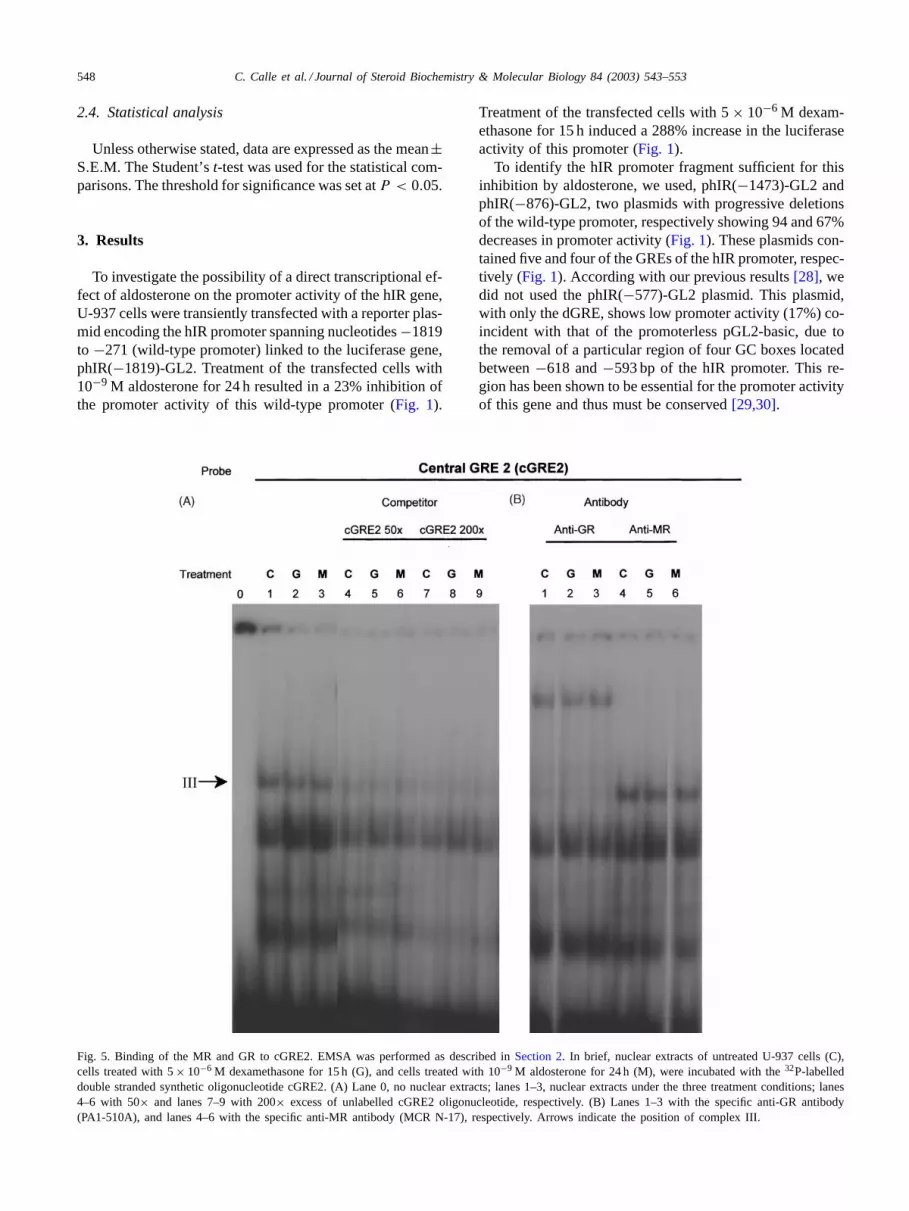
Fig. 5. Binding of the MR and GR to cGRE2. EMSA was performed as described inSection 2. In brief, nuclear extracts of untreated U-937 cells (C),cells treated with 5× 10⁻⁶ M dexamethasone for 15 h (G), and cells treated with 10−9 M aldosterone for 24 h (M), were incubated with the32P-labelleddouble stranded synthetic oligonucleotide cGRE2. (A) Lane 0, no nuclear extracts; lanes 1–3, nuclear extracts under the three treatment conditions; lanes4–6 with 50× and lanes 7–9 with 200× excess of unlabelled cGRE2 oligonucleotide, respectively. (B) Lanes 1–3 with the specific anti-GR antibody(PA1-510A), and lanes 4–6 with the specific anti-MR antibody (MCR N-17), respectively. Arrows indicate the position of complex III.
Treatment of the transfected cells with 5× 10⁻⁶ M dexam-ethasone for 15 h induced a 288% increase in the luciferaseactivity of this promoter (Fig. 1).
To identify the hIR promoter fragment sufficient for thisinhibition by aldosterone, we used, phIR(−1473)-GL2 andphIR(−876)-GL2, two plasmids with progressive deletionsof the wild-type promoter, respectively showing 94 and 67%decreases in promoter activity (Fig. 1). These plasmids con-tained five and four of the GREs of the hIR promoter, respec-tively (Fig. 1). According with our previous results[28], wedid not used the phIR(−577)-GL2 plasmid. This plasmid,with only the dGRE, shows low promoter activity (17%) co-incident with that of the promoterless pGL2-basic, due tothe removal of a particular region of four GC boxes locatedbetween−618 and−593 bp of the hIR promoter. This re-gion has been shown to be essential for the promoter activityof this gene and thus must be conserved[29,30].

C. Calle et al. / Journal of Steroid Biochemistry & Molecular Biology 84 (2003) 543–553 549
Once the cells were transfected with these plasmids andexposed to the treatments, luciferase determinations revealedthat the promoter activity of the fragments spanning upto −1473 and−876 bp, were inhibited 23, and 31% re-spectively, by aldosterone (Fig. 1). In addition, the activi-ties of the fragments spanning up to−1473 and−876 bp,were induced 260 and 174%, respectively, by dexametha-sone (Fig. 1). These data together with those obtained withthe wild-type promoter suggest that while dexamethasoneneeds at least the fragment spanning up−1473 bp for fulltranscriptional stimulation of the hIR gene, only the portionspanning up to−876 bp is needed for complete transcrip-tional inhibition by aldosterone.
We next explore the interaction of MR and GR with eachof the functional GREs of the hIR gene promoter[18,19]by EMSA. As a positive control of MR binding, first weused the Vit-GRE as oligoprobe on nuclear extracts of un-
Fig. 6. Binding of the MR and GR to cGRE3. EMSA was performed as described inSection 2. In brief, nuclear extracts of untreated U-937 cells (C),cells treated with 5× 10⁻⁶ M dexamethasone for 15 h (G), and cells treated with 10−9 M aldosterone for 24 h (M), were incubated with the32P-labelleddouble stranded synthetic oligonucleotide cGRE3. (A) Lane 0, no nuclear extracts; lanes 1–3, nuclear extracts under the three treatment conditions; lanes4–6 with 50× and lanes 7–9 with 200× excess of unlabelled cGRE3 oligonucleotide, respectively. (B) Lanes 1–3 with the specific anti-GR antibody(PA1-510A), and lanes 4–6 with the specific anti-MR antibody (MCR N-17), respectively. Arrows indicate the position of the complexes (I–III).
treated, aldosterone-treated or dexamethasone-treated cells.Vit-GRE has been previously demonstrated to specificallybind MR in this assay[31]. As shown inFig. 2A, two majorprotein–DNA complexes (I and III) were formed under thesethree experimental conditions (lanes 1–3). Complex I wasof less intensity than complex III, possibly reflecting differ-ences in the expression level of MR and GR in U-937 cells[16,17,22]. The fact that the intensity of both complexes wasapparently unmodified by the treatments may be explainedby the background of both receptors in these cells. The ad-dition of excess (50× and 200×) unlabelled oligonucleotideled to competition for both complexes (lanes 4–6, 7–9, re-spectively).Fig. 2B shows that the use of a specific anti-body against MR blocked the formation of the protein–DNAcomplex I, indicating the presence of MR in this complex(lanes 4–6), while the addition of a specific antibody againstGR caused supershifting of the protein–DNA complex III,

550 C. Calle et al. / Journal of Steroid Biochemistry & Molecular Biology 84 (2003) 543–553
indicating the presence of GR in this complex (lanes 1–3).Thus, Vit-GRE was able to bind two complexes with easilydistinguishable electrophoretic mobilities due to the differ-ent molecular mass of MR and GR[1]. These complexespresumably corresponded to MR–MR and GR–GR homod-imers [6–9]. This binding pattern was used as a control insubsequent experiments.
Using dGRE as the next oligoprobe, two main delayedcomplexes (I and III) were detected under the three ex-perimental conditions (Fig. 3A, lanes 1–3). The intensityof both complexes was unmodified by the treatments. Theaddition of 50× and 200× excess unlabelled oligonu-cleotide gave rise to partial competition for the formationof complex III but not complex I (lanes 4–6, 7–9, re-spectively). Further, using specific antibodies (Fig. 3B),complex III was partially supershifted by the anti-GR an-
Fig. 7. Binding of the MR and GR to pGRE. EMSA was performed as described inSection 2. In brief, nuclear extracts of untreated U-937 cells (C),cells treated with 5× 10⁻⁶ M dexamethasone for 15 h (G), and cells treated with 10−9 M aldosterone for 24 h (M), were incubated with the32P-labelleddouble stranded synthetic oligonucleotide pGRE. (A) Lane 0, no nuclear extracts; lanes 1–3, nuclear extracts under the three treatment conditions;lanes4–6 with 50× and lanes 7–9 with 200× excess of unlabelled pGRE oligonucleotide, respectively. (B) Lanes 1–3, with the specific anti-GR antibody(PA1-510A), and lanes 4–6 with the specific anti-MR antibody (MCR N-17), respectively. Arrows indicate the position of the complexes (II and III).
tibody (lanes 1–3), while complex I was unaltered by theanti-MR antibody (lanes 4–6). It therefore appears thatdGRE only partially binds GR, presumably as a GR–GRhomodimer.
We then went on to test cGRE1, cGRE2 and cGRE3 asoligoprobes. The oligonucleotide cGRE1 gave rise to threemain complexes (I–III) (Fig. 4A). The intensity of thesecomplexes was unmodified by the treatments. The additionof excess (50× and 200×) unlabelled nucleotide (lanes 4–6,7–9, respectively) resulted in competition for these com-plexes. Complex I appeared to be a MR–DNA complex(Fig. 4B), since it was blocked by the specific anti-MR anti-body (lanes 4–6). However, neither complex II nor III wereaffected by either antibody (Fig. 4B) (lanes 1–3, 4–6, re-spectively). Thus, cGRE1 was only able to bind MR, pre-sumably as a MR–MR homodimer.

C. Calle et al. / Journal of Steroid Biochemistry & Molecular Biology 84 (2003) 543–553 551
When the binding pattern of the cGRE2 was analysed,only one GR–DNA complex (III) was observed (Fig. 5A).This complex was efficiently competed with the additionof unlabelled oligonucleotide (lanes 4–6, 7–9, respectively)and supershifted by the specific anti GR-antibody (Fig. 5B)(lanes 1–3), while addition of the anti-MR antibody had noeffect (Fig. 5B) (lanes 4–6). Hence, cGRE2 was only ableto bind GR, presumably as a GR–GR homodimer.
The cGRE3 binding pattern indicated three major com-plexes (I–III)(Fig. 6A), which showed enhanced intensityafter treatment with either hormone. The formation of thesecomplexes was competed with the addition of 200×, but not50×, excess unlabelled oligonucleotide (lanes 4–6, 7–9, re-spectively). The specific anti-GR antibody caused a super-shift of the protein–DNA complex III (Fig. 6B) (lanes 1–3),indicating the presence of GR in this complex. The use of thespecific anti-MR antibody blocked the protein–DNA com-plex II but no complex I (Fig. 6B), possibly since cGRE3includes two overlapping GRE elements, as will be indi-cated later. Therefore, cGRE3 appears to bind MR and GR,presumably as MR–MR and GR–GR homodimers.
The pGRE binding pattern included two major complexes(II and III) that were competed with the addition of the un-labelled oligonucleotide (Fig. 7A) (lanes 4–6, 7–9, respec-tively). Both hormone treatments enhanced the intensity ofcomplex II. Addition of the anti-GR antibody supershiftedthe protein–DNA complexes II and III (Fig. 7B) (lanes 1–3),indicating the presence of GR in these two complexes. More-over, the use of the anti-MR antibody blocked the formationof protein–DNA complex II, also indicating the presence ofMR in this complex (Fig. 7B) (lanes 4–6). This complex IIof intermediate electrophoretic mobility containing both MRand GR, appears to be a heterodimer[8,9]. Thus, pGREwas able to bind a MR–GR heterodimer and a GR–GRhomodimer.
Table 1Comparison of the 3′ half-element and 5′ half-element of each of the five GREs of the human insulin receptor gene promoter (18, 19) with those of aconsensus GRE element (32, 33)
Base positions in the consensus GRE are indicated by numbers. Nucleotides identical to the consensus GRE are labelled by filled circles (�) or asterisks( ).
4. Discussion
In earlier studies[16,17], we observed that aldosteronereduced hIR gene expression by 30%, decreased the hIRnumber by 34% and was able to reduce several effects ofinsulin by 21–31% in U-937 cells. It would therefore appearthat this aldosterone-elicited decrease in hIR expression andinsulin action is the direct result of transcriptional inhibitionof the hIR gene. Herein, we addressed this question anddemonstrated that aldosterone inhibited the activity of thehIR wild-type promoter by 23%, and caused 23 and 31%reductions in the activity of progressive deletions of thispromoter, consisting of fragments spanning up to−1473 bpand up to−876 bp, respectively (Fig. 1). This indicates thatthe −876 to −271 bp region of the hIR promoter may besufficient for this transcriptional inhibition by aldosterone.
In this sense, we were able to observe that MR specif-ically recognized three of the four GREs of this region:cGRE1, cGRE3 and pGRE. In the case of cGRE1 andcGRE3, protein–DNA complexes I and II, respectively,probably representing MR–MR homodimers, were effi-ciently competed against by the addition of the correspond-ing unlabelled probe and blocked by the specific anti-MRantibody (Figs. 4 and 6). In the case of pGRE, we observed aprotein–DNA complex II, probably representing a MR–GRheterodimer that was efficiently competed against by theaddition of the unlabelled probe, blocked by the anti-MRantibody and supershifted by the anti-GR antibody. As isindicated inTable 1, cGRE1 has 5 conserved nucleotidesof the 12 nucleotides of a consensus GRE element[32,33],cGRE3 has 4 and 5 in each of the two overlapping ele-ments, while pGRE has 6. Thus, there has to be a minimalnumber of conserved nucleotides in the response elementto achieve MR recognition. These nucleotides appear to bethe two G, positions+2 and−5, and the C, position+5

552 C. Calle et al. / Journal of Steroid Biochemistry & Molecular Biology 84 (2003) 543–553
(Table 1) [33,34]. In addition, the relatively well conservedsequence of the pGRE, also includes the G, position−6,whose essential role in dimeric binding in GREs has beendemonstrated[35].
Conversely, MR was not recognized by dGRE (Fig. 3).This dGRE has a sequence with 7 conserved nucleotidesnot including the G, position−5, that we have postulatedas essential for MR recognition. Further, neither was MRrecognized by cGRE2 (Fig. 5), perhaps due to the presenceof three consecutive G, positions−3, −4, and−5, of the 5′half-site (Table 1). This is consistent with mutational datareported by other authors which indicate that substitution ofthe T, position−4, for G in this position leads to a 50%decrease in receptor recognition[33,35].
The inducibility of the hIR promoter by dexamethasonewas also tested. Dexamethasone induced a 288% increase inthe activity of the wild-type promoter (Fig. 1), which is inagreement with the level of induction reported by Lee andco-workers[18,19]in rat 208 F cells. Moreover, this increaseis in line with our previous observations of dexamethasonestimulation of hIR mRNA levels (230%), and high affinityIRs (250%) in U-937 cells[12,13]. In addition, the activitiesof the 5′deletions of this promoter (up to−1473 bp and upto −876 bp) were increased by 260 and 174%, respectively(Fig. 1). This indicates that the−1473 to−271 bp regionof the hIR promoter that contains the five GREs may besufficient for transcriptional induction by dexamethasone.
In this sense, we observed that GR specifically recognizeddGRE, cGRE2, cGRE3 and pGRE (Figs. 3 and 5–7). In eachcase, a protein–DNA complex (III), probably representing aGR–GR homodimer was efficiently competed against by theaddition of the corresponding unlabelled probe, and super-shifted by the specific anti-GR antibody (Figs. 3 and 5–7).Moreover, in the case of pGRE, GR also recognised this ele-ment as a protein–DNA complex (II), probably representinga MR–GR heterodimer. The fact that GR was not recognizedby cGRE1 may be attributable to a cell-specific differenceand/or to the presence of three consecutive G, positions+1,+2, and+3, of the 3′ half-site (Table 1). A comparison ofthe consensus GRE element inTable 1with the GREs ofthe hIR promoter, suggests that the minimal number of con-served nucleotides essential for GR recognition may be theG, position+2, and the C, position+5 [33–35].
Returning to the biological importance of the MR–GRheterodimer of the pGRE, in cells expressing both recep-tors, such as the cells used here, heterodimerization isfavoured [6,9,27,36]. Thus, this heterodimer would playthe most important role in the transcriptional modulationof the hIR gene by aldosterone and dexamethasone. In-deed, a MR–GR heterodimer mediates the transcriptionalregulation by mineralocorticoids and glucocorticoids of thehuman Na/K-ATPase�1 gene promoter[27,37], the humanNa/K-ATPase�1 gene promoter[38,39]and the rat 5-HT1Areceptor gene promoter[36]. Moreover, the possibility thatother factors could assist the formation of this heterodimercannot be ruled out. Thus, upstream from the pGRE region
(from −363 to −341 bp) there is a cluster of three GCboxes which coincide with two AP2-like sites (from−444to −433 bp) and (from−436 to−425 bp)[40]. Given theprevious description of cooperation of GR with AP-2 inthe activation of other promoters[41–43], it is possible thatMR and AP-2 also act together, though this remains to bedemonstrated.
In conclusion, to our knowledge ours is the first demon-stration of aldosterone causing the transcriptional inhibitionof the hIR gene. This inhibition is consistent with our ear-lier observations that aldosterone led to reduced hIR mRNAlevels, hIR number, and insulin-mediated effects in U-937cells. It was also shown that this transcriptional inhibitionof the hIR gene by aldosterone occurs by the direct inter-action of MR with some of the functional GREs elementslocated in the hIR promoter, specifically with the cGRE1and cGRE3 as MR–MR homodimers, and with pGRE as aMR–GR heterodimer. This heterodimer could be the mostrelevant and participate in the cross-talk between mineralo-corticoids, glucocorticoids and insulin signalling in U-937cells.
Acknowledgements
The authors are indebted to Drs. S.Y. Tsai and G. El-berg for their generous gift of the hIR promoter. J. Campiónand B.Maestro were holders of fellowships from Gobiernode Navarra and Complutense University of Madrid, respec-tively. This work was supported by research funds from theDGES grants (PB 96-0573; BMC2000/0765) and the CAM,grant (08.6/0010/98).
References
[1] J.W. Funder, Glucocorticoid and mineralocorticoid receptors: biologyand clinical relevance, Annu. Rev. Med. 48 (1997) 231–240.
[2] A. Aranda, A. Pascúal, Nuclear hormone receptors and geneexpression, Physiol. Rev. 81 (2001) 1269–1304.
[3] J.W. Funder, Aldosterone action: fact, failure and the future, Clin.Exp. Pharmacol. Physiol. 25 (1998) 547–550.
[4] R.E. Booth, J.P. Johnson, J.D. Stockand, Aldosterone, Adv. Physiol.Educ. 26 (2002) 8–20.
[5] F.M. Rogerson, P.J. Fuller, Mineralocorticoid action, Steroids 65(2000) 61–73.
[6] D. Pearce, A mechanistic basis for distinct mineralocorticoidand glucocorticoid receptor transcriptional specificities, Steroids 59(1994) 153–159.
[7] T. Trapp, R. Rupprecht, M. Castrén, J.M.H.M. Reul, F. Holsboer,Heterodimerization between mineralocorticoid and glucocorticoidreceptor: a new principle of glucocorticoid action in the CNS, Neuron13 (1994) 1457–1462.
[8] W. Liu, J. Wang, N.K. Sauter, D. Pearce, Steroid receptorheterodimerization demonstrated in vitro and in vivo, Proc. Natl.Acad. Sci. 92 (1995) 12480–12484.
[9] T. Trapp, F. Holsboer, Heterodimerization between mineralocorticoidand glucocorticoid receptors increases the functional diversity ofcorticosteroid action, Trends Pharmacol. Sci. 17 (1996) 145–149.

C. Calle et al. / Journal of Steroid Biochemistry & Molecular Biology 84 (2003) 543–553 553
[10] M.A. Leal, P. Aller, A. Torres, A. Picardo, N. Dávila, C. Calle, Insulinreceptor mRNA levels are modulated in a tissue-specific manner inCushing’s syndrome patients, Horm. Metab. Res. 26 (1994) 349–350.
[11] M.A. Leal, P. Aller, A. Más, M.C. Carranza, C. Calle,Tissue-specific changes in insulin receptor mRNA concentrations indexamethasone-treated and adrenalectomized rats, Endocrinol. J. 41(1994) 737–741.
[12] M.A. Leal, C. Cabañas, C. Rius, P. Aller, C. Calle, Modulation bydexamethasone of insulin binding and insulin receptor mRNA levelsin U-937 human promonocytic cells, Biochemie 74 (1992) 545–549.
[13] M.A. Leal, P. Aller, C. Calle, Effect of dexamethasone on insulinreceptor mRNA levels, RNA stability and isotype RNA pattern inU-937 human promonocytic cells, J. Endocrinol. Invest. 19 (1996)530–534.
[14] M.C. Carranza, A. Torres, C. Calle, Disminución en el numero yen la afinidad de los receptores de insulina en el tejido adipososubcutáneo de un paciente con hyperaldosteronismo primario, Rev.Clin. Esp. 188 (1991) 414–417.
[15] J. Campión, V. Lahera, V. Cachofeiro, B. Maestro, N. Dávila,M.C. Carranza, C. Calle, In vivo tissue-specific modulation ofrat insulin receptor gene expression in an experimental model ofmineralocorticoid excess, Mol. Cell Biochem. 185 (1998) 177–182.
[16] J. Campión, B. Maestro, F. Mata, N. Dávila, M.C. Carranza, C.Calle, Inhibition by aldosterone of insulin receptor mRNA levelsand insulin binding in U-937 human promonocytic cells, J. SteroidBiochem. Mol. Biol. 70 (1999) 211–218.
[17] J. Campión, B. Maestro, S. Molero, N. Dávila, M.C. Carranza, C.Calle, Aldosterone impairs insulin responsiveness in U-937 humanpromonocytic cells via the downregulation of its own receptor, CellBiochem. Funct. 20 (2002) 237–245.
[18] J.K. Lee, J.W.O. Tam, M.J. Tsai, S.Y. Tsai, Identification ofcis- andtrans-acting factors regulating the expression of the human insulinreceptor gene, J. Biol. Chem. 267 (1992) 4638–4645.
[19] J.K. Lee, S.Y. Tsai, Multiple hormone response elements can conferglucocorticoid regulation on the human insulin receptor gene, Mol.Endocrinol. 8 (1994) 625–634.
[20] C. Sundstrom, K. Nilsson, Establishment and characterization of ahuman histiocytic lymphoma cell line (U-937), Int. J. Cancer 17(1976) 565–570.
[21] A. Robert, G. Grunberger, J.L. Carpentier, J.M. Dayer, J.M. Orci,P. Gorden, The insulin receptor of a human monocyte-like cell line:characterization and function, Endocrinology 114 (1984) 247–250.
[22] D. Duval, P. Lynde, A. Hatzfeld, J. Hatzfeld, Dexamethasone-inducedstimulation of arachidonic acid release by U-937 cells grown indefined medium, Biochim. Biophys. Acta 887 (1986) 204–213.
[23] B. Maestro, S. Molero, S. Bajo, N. Dávila, C. Calle, Transcriptionalactivation of the human insulin receptor gene by 1,25-dihydro-xyvitamin D3, Cell Biochem. Funct. 20 (2002) 227–232.
[24] C. Bonnerot, D. Rocancourt, P. Briand, G. Grimber, J.F. Nicolas,A �-galactosidase hybrid protein targeted to nuclei as a marker fordevelopmental studies, Proc. Natl. Acad. Sci. 84 (1987) 6795–6799.
[25] V. Kumar, P.C. Chambon, The estrogen receptor binds thightly to itsresponsive element as a ligand-induced homodimer, Cell 55 (1988)145–156.
[26] E. Schreibert, P. Matthias, M.M. Múller, W. Schaffner, Identificationof a novel lymphoid specific octamer binding protein (OTF-2B)by proteolytic clipping badshift assay (PCBA), EMBO J. 7 (1988)4221–4229.
[27] A. Derfoul, N.M. Robertson, J.B. Lingrel, D.J. Hall, G. Litwack,Regulation of the human Na/K-ATPase�1 gene promoter bymineralocorticoid and glucocorticoid receptors, J. Biol. Chem. 273(1998) 20702–20711.
[28] B. Maestro, N. Dávila, M.C. Carranza, C. Calle, Identificationof a vitamin D response element in the human insulin receptorgene promoter, J. Steroid Biochem. Mol. Biol. 84 (2003) 223–230.
[29] E. Araki, T. Murakami, T. Shirotani, F. Kanai, Y. Shinohara, F.Shimada, M. Mori, M. Shichiri, Y. Ebina, A cluster of four Sp1binding sites required for efficient expression of the human insulinreceptor gene, J. Biol. Chem. 266 (1991) 3944–3948.
[30] K. Yoshizato, T. Shirotani, N. Furukawa, T. Taguchi, H. Motoshima,T. Toyonaga, Y. Hirashima, J. Kawashima, Y. Ebina, M. Shichiri, E.Araki, Identification of a cis-acting element and a novel transactingfactor of the human insulin receptor gene in HepG2 and rat livercells, Biochem. Bioph. Res. Co. 280 (2001) 428–434.
[31] M. Lombès, N. Binart, M.E. Oblin, V. Joulin, E.E. Baulieu,Characterization of the interaction of the human mineralocorticoidreceptor with hormone response elements, Biochem. J. 292 (1993)577–583.
[32] M. Beato, G. Chalepakis, M. Schauer, E.P. Slater, DNA regulatoryelements for steroid hormones, J. Steroid Biochem. 32 (1989) 737–747.
[33] C.C. Nelson, S.C. Hendy, R.J. Shukin, H. Cheng, N. Bruchousky,B.F. Koop, P.S. Rennie, Determinants of DNA sequence specificityof the androgen, progesterone and glucocorticoid receptors: evidencefor differential steroid receptor response elements, Mol. Endocrinol.13 (1999) 2090–2107.
[34] C. Scheidereit, H.M. Westphal, C. Carlson, H. Bosshard, M. Beato,Molecular model of the interaction between the glucocorticoidreceptor and the regulatory elements of inducible genes, DNA 5(1986) 383–391.
[35] S.K. Nordeen, B.J. Suh, B. Kuhnel, C.D. Hutchison, Structuraldeterminats of a glucocorticoid receptor recognition element, Mol.Endocrinol. 4 (1990) 1866–1873.
[36] X.M. Ou, J.M. Storring, N. Kushwaha, P.R. Alberti, Hetero-dimerization of mineralocorticoid and glucocorticoid receptors at anovel negative response element of the 5-HT1A receptor gene, J.Biol. Chem. 276 (2001) 14299–14307.
[37] A. Derfoul, N.M. Robertson, D.J. Hall, G. Litwack, TheN-terminal domain of the mineralocorticoid receptor modulates bothmineralocorticoid receptor- and glucocorticoid receptor-mediatedtransactivation from Na/K ATPase�1 target gene promoter,Endocrine 13 (3) (2000) 287–295.
[38] V. Kolla, N.M. Robertson, G. Litwack, Identification of a mineralo-corticoid/glucocorticoid response element in the human Na/K ATPase�1 gene promoter, Biochem. Bioph. Res. Co. 266 (1999) 5–14.
[39] V. Kolla, G. Litwack, Transcriptional regulation of the humanNa/K ATPase via the human mineralocorticoid receptor, Mol. CellBiochem. 204 (2000) 35–40.
[40] E. Wingender, X. Chen, E. Fricke, R. Geffers, R. Hehl, I. Liebich, M.Krull, V. Matys, H. Michael, R. Ohnhäuser, M. Prü�, F. Schacherer,S. Thiele, S. Urbach, The TRANSFAC system on gene expressionregulation, Nucleic Acids 29 (2001) 281–283.
[41] D.L. Wong, B.J. Siddall, S.N. Ebert, R.A. Bell, S. Her, Phenyl-ethanolamine N-methyltransferase gene expression: synergisticactivation by Egr-1, AP-2 and the glucocorticoid receptor, Brain Res.Mol. Brain Res. 61 (1–2) (1998) 154–161.
[42] P.A. Shaw, O. Chaparro, The 5′-flanking sequence and regulatoryelements of the cystatin S gene, Biochem. Bioph. Res. Co. 261(1999) 705–711.
[43] K. Hilger-Eversheim, M. Moser, H. Schorle, R. Buettner, Regulatoryroles of AP-2 transcription factors in vertebrate development,apoptosis and cell-cycle control, Gene 260 (2000) 1–12.