trace metals and micronutrients in bone tissues of the red fox vulpes vulpes (l., 1758)
TRANSCRIPT

ORIGINAL PAPER
Trace metals and micronutrients in bone tissues of the red foxVulpes vulpes (L., 1758)
Natalia Lanocha & Elzbieta Kalisinska &
Danuta I. Kosik-Bogacka & Halina Budis &
Kinga Noga-Deren
Received: 26 October 2011 /Accepted: 25 January 2012 /Published online: 10 February 2012# The Author(s) 2012. This article is published with open access at Springerlink.com
Abstract In this study we determined the levels of traceelements (zinc, copper, lead, cadmium and mercury) in threelayers of bones of the hip joint (cartilage, compact bone andspongy bone) of 30 red foxes (Vulpes vulpes) from north-western Poland. Concentrations of Cu, Zn, Pb and Cd weredetermined by atomic absorption spectrophotometry (ICP-AES) in inductively coupled argon plasma using a Perkin-Elmer Optima 2000 DV. Determination of Hg concentrationwas performed by atomic absorption spectroscopy. In carti-lage, compact bone and spongy bone samples from the redfox, median concentrations of the metals studied could bearranged in the following descending series: Zn > Cu > Pb >Cd > Hg, the values ranging from 142 to 0.002 mg/kg dw.There was a significant difference in Cu concentrations,among all the materials analyzed, with much more Cu foundin spongy bone than in compact bone. Significant differ-ences were also noted in the case of Hg concentrations incartilage with compact bone and the spongy bone, andbetween concentrations of this metal in compact bone andspongy bone. In males, the concentration of Hg in spongybone was greater than in females. Younger foxes had ahigher concentration of this metal in cartilage than adults.The strongest synergistic relationships were observed in
spongy bone between the Zn and Cu, Zn and Cd, as wellas between Cu and Cd. Statistically significant antagonisticrelationships were detected between zinc and lead in com-pact bone. In addition to monitoring studies conducted onthe abiotic environment, an urgent need exists for long-termmonitoring of concentrations of heavy metals with long-term effects on living organisms. An important addition isprovided by biomonitoring studies on domesticated andfree-living mammals, including Canidae.
Keywords Red fox . Bioindicator . Bone tissue . Traceelements
Introduction
Representatives of Canidae, namely the red fox Vulpes vulpes(Linnaeus, 1758), the domesticated dogCanis lupus familiaris(Linnaeus, 1758), the wild raccoon dog Nyctereutes procyo-noides (Gray 1834), and the wolf Canis lupus (Linnaeus,1758), are all common objects of ecotoxicological studies(Kalisinska et al. 2009; 2011; Millan et al. 2008; Shore et al.2001).
The fox belongs to a group of hunted mammals com-monly found in Eurasia and North America. It is a predatorpreferring food of animal origin including rodents, birds andsmall invertebrates (beetles, grubs, earthworms) (Kidawaand Kowalczyk 2011). The species occupies a high positionin the food pyramid and accumulates ingested substances. Italso exhibits a measurable response to environmental con-taminants, including heavy metals (Kalisinska et al. 2009;2011; Lopez-Alonso et al. 2007).
Many reports mention the neuro-, nephro- or hepatotoxiceffects of exposure to trace elements, but publications on theaccumulation of trace elements (including zinc, copper, lead,
Communicated by: Danuta I. Kosik-Bogacka
N. Lanocha : E. Kalisinska :D. I. Kosik-Bogacka (*) :H. BudisDepartment of Biology and Medical Parasitology,Pomeranian Medical University of Szczecin,Powstancow Wielkopolskich 72,70-111 Szczecin, Polande-mail: [email protected]
K. Noga-DerenDepartment of Preclinical Conservative Dentistry and PreclinicalEndodontics, Pomeranian Medical University of Szczecin,Powstancow Wielkopolskich 72,70-111 Szczecin, Poland
Acta Theriol (2012) 57:233–244DOI 10.1007/s13364-012-0073-1

cadmium and mercury) in cartilage and bone elements ofhuman and animal joints are much less numerous (Brodziak-Dopierala et al. 2007; Jankovska et al. 2010; Kalisinska et al.2007; Kwapulinski et al. 1995; Piskorova et al. 2003).
We examined bone tissues of the red fox in a risk assess-ment of exposure to trace elements, as they are subject to slowtransfer of some metals in the body. Due to its characteristicsand long renewal time, this tissue may reflect levels of chronicexposure and could be the basis of indirect environmentalassessment (Brodziak-Dopierala et al. 2007; 2009; Zaichickand Zaichick 2009; 2010; Zaichick et al. 2011).
Zinc (Zn) and copper (Cu)
Zinc and copper are involved in the formation and metabo-lism of bone tissue (Honda et al. 1997; Nielsen and Milne2004; Senczuk 2006; Yamaguchi 1998).
Zinc is essential for the correct ossification and mineral-ization of bones, especially in the basal part of the femur. Itis a cofactor for the enzyme affecting the synthesis ofvarious ossein components and plays a role in the regulationof bone resorption (Machalinski et al. 1996; NRC 1980;Puzanowska-Tarasiewicz et al. 2009; Smrcka 2005). Bothtoo high and too low concentrations of Zn contribute to thegradual reduction of bone mass and reduce the concentra-tion of calcium ions in bones and blood serum (Charles et al.2001).
Copper in mammals is involved in the process of thehardening of collagen, hair keratinization and also normalizesthe deposition of calcium and phosphorus in bones. Cooperinhibits bone resorption, which may occur due to a reductionin prostaglandin synthesis (Senczuk 2006). Copper deficiencyleads to reduced bone mass, resulting in a decrease in itsmechanical strength and subsequent fractures.
Studies on Cu and Zn supplementation in humans andanimals indicate that a deficiency of these micronutrientsleads to osteoporosis-like changes (Nielsen and Milne2004).
Lead (Pb), cadmium (Cd) and mercury (Hg)
The presence of Pb, Cd and Hg have been detected in alltissues of mammals, and even minimum concentrationshave been observed to cause metabolic disturbances, thusreducing physical efficiency, weakening immune and enzy-matic processes, and leading to many diseases and sometimesdeath.
Lead toxicity is manifested in anemia, impaired nervoussystem and kidney function and changes in bones such asdecreased bone mass (osteopenia) and delayed healing offractures (Gerhardsson et al. 2005; Kjellstrom 1992;Wiechulaet al. 2008). Accumulation of Pb in bone, in contrast to solidorgans, increases with age (Jurkiewicz et al. 2004).
Cadmium has mainly nephro- and hepatotoxic properties.The osteotoxic action of Cd was described in Japan as earlyas 1960 in 90% of older women (after menopause and manypregnancies) living in areas contaminated with Pb and Znores. The disease was called "Itai-Itai" (ouch-ouch disease).First, it was observed in the Japanese population of the JinzuRiver overflow area, consuming mainly rice grown in fieldsfertilized with silt derived from local plants (Starek 2007;Zhu et al. 2004). The concentration of Cd in that soil was8 mg/kg dw, and in rice up to 2.7 mg/kg. The patients hadosteomalacia resistant to vitamin D, accompanied by severepain around the sacrum, the lower limbs and ribs, sponta-neous fractures, as well as proteinuria, glycosuria and de-creased sodium reabsorption (Bernard 2008; Horiguchi et al.2010; Jarup and Akesson 2009; Umemura and Wako 2006).
Mercury is a potent neurotoxin, primarily disrupting thefunction of the central and peripheral nervous system(Scheuhammer et al. 2008). However, individual studieshave drawn attention to the deposition of this metal inskeletal elements (Yoo et al. 2002; Zaichick and Zaichick2010; Zaichick et al. 2011).
The determination of toxic element concentrations inliving organisms is one of the basic methods of indirectassessment of environmental pollution. Ecotoxicologicalstudies in Poland and the rest of the world are usuallycarried out on the liver and kidneys of warm-blooded verte-brates. However, there has been an increasing number ofbiomonitoring studies on bones. Apart from environmentalstudies, a large need exists for the monitoring of concen-trations of heavy metals in humans, which can be supple-mented with biomonitoring of both domesticated animals(such as the pig, sheep, cattle, horse and dog) and wildanimals (e.g., fox, boar, roe deer and deer) (Kalisinska etal. 2009; Lanocha et al. 2009; Lazarus et al. 2008; Liu2003).
The aim of this study was to determine the concentrationsof five elements: two micronutrients (Zn and Cu) and threetoxic trace metals (Pb, Cd and Hg) in three types of biolog-ical materials derived from the bones of the red fox V.vulpes, and to determine intraspecific differences betweenthe concentrations of trace elements in cartilage, spongybone and compact bone.
Materials and methods
Study area
The material was collected in north-western Poland, in theWest Pomeranian province including its capital, Szczecin.Most of the province's area is agricultural (38%) and forest-ed (35%), and several percent of the area is covered by water(numerous lakes, rivers, the Odra estuary with Dabie Lake,
234 Acta Theriol (2012) 57:233–244

and Szczecin Lagoon) (http://www.stat.gov.pl/cps/rde/xbcr/szczec/ASSETS_przegl_2.pdf).
Material
The material was collected in 2008–2009. Altogether itconsisted of 30 foxes from six districts of the West Pomer-anian province (municipalities of Szczecin, Choszczno,Stargard, Gryfice, Kamien Pomorski and Mysliborz —three, eight, seven, one, two, and two specimens, respec-tively, and seven remaining foxes from the Provincial Vet-erinary Inspectorate in Szczecin). In Poland, the fox isincluded in the list of animals for hunting (Journal of Law2005, no 45, pos. 433), and according to the Minister ofEnvironment, it may be hunted from 1st July to 31st March(Journal of Law 2005, no 48, pos. 459).
The acquisition of biological material from the foxes wasapproved by the Local Ethics Committee for Research onAnimals in Szczecin (Poland).
Fox age determination
Fox age categories were based on the examination of onesingle-root lower canine, with preserved anatomical crown,from 30 foxes. The teeth were placed immediately afterextraction in distilled water, and then dried. In order toobtain pantomographic images, all the teeth were glued oncardboard sheets. Radiographs were performed on a CranexCeph's Soredex digital pantomogram from a distance of120 cm (60 kV, 10 mA s). Measurements of linear param-eters were performed using digital radiography DIGORA2.1 software (Soredex-Orion, Helsinki, Finland).
They included the total width of the tooth (TW) andwidth of the pulp chamber (WC). According to the workof Knowlton and Whittemore (2001), the width of the ca-nine pulp chamber was measured at a standard distance of15 mm from the root apex. Canine width index (CWI) wascalculated as the ratio of the width of the pulp chamber tothe overall width of the lower canine, which allowed thedivision of subjects into two age categories (adultus [ad];immaturus [im]). It was assumed that immature foxes werein the range of CWI from 0.20 to 0.50, and adults from 0.05to 0.20 (Cavallini and Santini 1995). Among the specimenscollected for analysis, some teeth had a very large pulpchamber and others very narrow. Following CWI values itwas determined that the examined group included 18 im and12 ad foxes, respectively (Table 1).
Preparation of material for analysis of bone tissues
The head, neck and part of the femoral shaft were collectedfrom the foxes using a glass tool. Chemical analysis wasperformed on three materials: cartilage, compact bone and
spongy bone with directly adjacent compact bone. Bonetissue was dried to constant weight at 55°C and 105°C indrying oven with natural convection, ED 53 (Binder GmbH,Germany). This procedure was used to determine the watercontent (gravimetric method). Dried samples were ground inan agate mortar (Sigma-Aldrich, Poland).
Determination of zinc, copper, cadmium and lead
The samples were divided into doses, weighing from0.5 to 1.0 g. Bone tissue was mineralized by wetdigestion using a Velp Scientifica mineralizer (Italy)(Kalisinska et al. 2007).
Concentrations of Zn, Cu, Pb and Cd were determined byatomic absorption spectrophotometry (ICP-AES) in induc-tively coupled argon plasma, using a Perkin-Elmer Optima2000 DV. The device’s limits of detection for Zn, Cu, Pb, Cdwere 0.2, 0.4, 1 and 0.1 μg/L, respectively.
Determination of mercury
Total mercury (THg) concentrations were determined insamples dried at 55°C, using atomic absorption spectrosco-py. The assays were run in an AMA 254 mercury analyzer(Altach Ltd, Czech Republic). For the analysis, we collectedfrom 100 to 300 mg of the sample and then placed it in anickel nacelle in which it was automatically weighed anddried. The sample was thermally decomposed in a stream ofoxygen to obtain the gaseous form, and its degradationproducts were transferred to an amalgamator for the selec-tive off take of Hg. After determination of the parameters ofmeasurement, Hg vapor was released from the amalgamatorby brief heating. The amount of released Hg was measuredby atomic absorption (silicon UV diode detector in theAMA 254 analyzer) at a wavelength of 254 nm, in thearrangement of two measuring cells. The limit of detectionfor this method is 0.01 ng/100 mg of Hg in the sample. Foreach sample, two or three repetitions were performed, andthe statistical analysis used the average of the data,expressed in mg/kg dry mass (dw).
Validation of analytical proceedings
The reliability of the analytical procedure was controlled bythe determination of elements in two reference materialswith known concentrations: NIST SRM 1486 Bone Meal,and IAEA-407 Trace Elements and Methylmercury in Fish(National Institute of Standards and Technology [NIST] andthe International Atomic Energy Agency [IAEA]). Concen-trations of metals in the reference materials provided by themanufacturers and our own determinations are shown inTable 2.
Acta Theriol (2012) 57:233–244 235

Statistical analysis
The analysis used Statistica 9.0. StatSoft software. In orderto determine compliance with the expected normal distribu-tion of results, we used a Kolmogorov–Smirnov test withLillefors correction (p<0.05). In order to compare the im-pact of various environmental factors on the concentrationof metals in the bone material marrow test, we used aKruskall–Wallis test, and in the case of significant differences,a Mann–Whitney U-test (p<0.05).
In addition, we determined the Spearman rank correlationcoefficients between trace elements in different parts of thehip joint (cartilage, compact bone, spongy bone, cartilagewith compact bone).
Results
Basic data on the concentrations of metals in the fox bonematerial is presented in Table 3. Because two samples (com-pact bone and spongy bone) derived from two individualsexceeded Cu concentration found in the other samples manytimes, we also conducted statistical analysis of Cu withouttaking these samples into account. The concentration of Cuin these samples was 20.35 mg/kg dw in spongy bone and37.5 mg/kg dw in compact bone. In other samples, the
concentration of this metal did not exceed 2.5 mg/kg dw. Bothspecimens came from urban areas and had fed mostly fromtrash cans, which may be the reason for such high Cu levels.
The distribution of empirical data on the concentrations ofPb, Cd and Hg in the cartilage, compact bone and spongybone, and Zn in the fox compact bone, diverged from theexpected normal distribution, and was examined using a Kol-mogorov–Smirnov test (p>0.05) with Lillefors correction(p<0.05). For the concentration of Cu, the distribution of allresults was not consistent with the expected normal distribu-tion, but after removal of the two aforementioned exceptionalsamples, the distribution of the remaining results was consis-tent with the expected normal distribution (Table 3).
Among the micronutrients, Zn had the highest concentra-tion in samples obtained from the examined fox bones, withthe median values ranging from about 100 to 140 mg/kg dw,depending on the type of material. However, the differencesbetween Zn concentrations were not significant (Table 3).
The average concentration of Cu in different types ofsamples ranged from about 0.40 to 0.90 mg/kg dw, andthe Kolmogorov–Smirnov test revealed the existence of astatistically significant difference (p<0.05). The highestconcentration of Cu was observed in cartilage (0.88 mg/kg),and it was clearly higher compared to compact bone, cartilage,compact bone and spongy bone by 28%, 10% and 115%,respectively.
Table 1 Sizes of mandibularcanine teeth from immature (im)and adult (ad) foxes
WC width of the pulp chamber,TW total width, CWI caninewidth index, AM arithmeticmean, Med median, SD standarddeviation, CV coefficient of var-iation in %
Age category and number of specimens (n) Parameter WC TW CWI
im (n018) AM ± SD 1.49±0.58 4.95±0.91 0.29±0.10
Med 1.39 5.24 0.28
Range 0.72–3.0 2.5–5.7 0.19–0.60
CV 39.3 18.5 35.4
ad (n012) AM ± SD 0.65±0.33 5.64±0.70 0.11±0.05
Med 0.50 5.63 0.09
Range 51.1 12.4 47.9
CV 0.20–1.27 4.50–7.27 0.03–0.19
Total (n030) AM ± SD 1.15±0.65 5.23±0.89 0.22±0.13
Med 1.13 5.25 0.23
Range 0.2–3.0 2.50–7.27 0.03–0.60
CV 56.0 17.0 57.4
Table 2 Concentrations of se-lected elements in the certifiedreference materials in mg/kg dryweight
RV reference value, OD owndeterminationaEstimated value
Metal Bone Meal SRM NIST 1486 OD/RV (%) Fish Tissue IAEA-407 OD/RV (%)
RV OD (n07) RV OD (n08)
Zn 147.0±16.0 132.4±4.1 90.0 67.1 65.8±3.8 98.1
Cu 0.80a 0.74±0.01 92.5 3.28 3.12±0.28 95.1
Pb 1.335±0.014 1.190±0.306 89.1 0.12 0.11±0.03 91.7
Cd 0.003a 0.0020±0.0002 66.7 0.189 0.176±0.010 93.1
Hg – – – 0.222 0.237±0.002 106.8
236 Acta Theriol (2012) 57:233–244

In the group of highly toxic metals, Pb had the greatestlevels, with the median value ranging from about 0.45 mg/kg(compact bone) to about 0.80mg/kg dw (in cartilage), but withno statistically confirmed differences between the types ofsamples analyzed (Table 3). The maximum Pb concentrationin cartilage exceeded 10 mg/kg, and in half of the samples wasgreater than 1 mg/kg dw. In other types of samples maximumPb concentrations were not greater than 4.40 and6.15mg/kg in the compact bone and spongy bone, respectively(Table 3).
Mean Cd concentration in the analyzed materials rangedfrom 0.125 mg/kg (compact bone) to about 0.170 mg/kg(spongy bone), and the differences between them werestatistically significant. The highest Cd concentration wasfound in the spongy bone (0.169 mg/kg dw) and was highercompared to levels in cartilage, compact bone and cartilagewith adjacent compact bone by 4%, 35% and 13%,respectively.
The average Hg concentration in the various types ofsamples ranged from about 0.002 (in the spongy bone) tomore than 0.004 mg/kg dw (in cartilage) and the Mann–Whitney U-test revealed statistically significant differences(p<0.05). Significant differences were found between theconcentration of this metal in the cartilage with the adjacent
compact bone and in spongy bone. Such differences alsoexisted between the Hg concentration in the compact boneand spongy bone, with about 50% higher concentration ofthis metal in the compact bone underlying the cartilage(Tables 3 and 4). Maximum Hg concentration in the com-pact bone reached 0.0226 mg/kg, and in 17% of the samplesit was greater than 0.0100 mg/kg dw.
In fox cartilage, compact bone and spongy bone, concen-trations of the examined metals can be arranged in thefollowing order: Zn > Cu > Pb > Cd > Hg.
Comparative analysis, which included sex, showed thatbetween males and females, in principle, there were nostatistically proven differences in the concentrations of met-als determined in the corresponding bone material. Mercuryin the spongy bone is an exception, as its concentration infemales was 0.0017 mg/kg dw and was over 70% lowercompared to males, where it was on average 0.0029 mg/kgdw (U-test063.0, p<0.05).
In addition, a comparison of metal concentrations in thecorresponding bone material was carried out between foxesrepresenting two age categories — immature foxes (im) andthe adults (ad) (Table 5). Only in the case of Hg in cartilagewas there a confirmed statistical difference (U-test063.0,p<0.05); the average Hg concentration in young foxes was
Table 3 Concentrations of trace elements (in mg/kg dry weight) in four types of bone material coming from fox in the vicinity of Szczecin
Metal Parameter Cartilage Compact bone Cartilage with adjacentcompact bone
Spongy bone SignificanceK–W
Zn AM ± SD 134.3±55.6 125.1±56.3 129.7±55.0 111.04±48.74 NSMed 141.8 105.9 130.4 116.31
range 19.6–219.8 46.2–296.2 19.6–296.2 11.21–219.51
CV 41.4 45.0 42.9 43.9
Cua n030 n029 n030 n029 n030 n029 p<0.05AM ± SD 1.80±2.70 2.01±6.72 0.77±0.46 1.89±5.07 1.29±2.0 1.17±3.64 0.50±0.35
Med 0.88 0.72 0.69 0.82 0.80 0.43 0.41
range 0.10–9.66 0.13–37.5 0.13–2.18 0.10–34.49 0.10–9.66 0.07–20.36 0.07–1.18
CV 149.6 336.6 59.7 267.4 154.4 312.3 69.6
Pb AM ± SD 1.738±2.325 0.978±1.153 1.36±1.86 1.49±1.83 NSMed 0.788 0.447 0.469 0.610
range 0.165–11.017 0.150–4.354 0.150–11.017 0.069–6.147
CV 133.8 117.9 136.9 123.0
Cd AM ± SD 0.137±0.055 0.108±0.073 0.123±0.060 0.142±0.07 NSMed 0.163 0.125 0.150 0.169
range 0.028–0.198 0.002–0.226 0.002–0.226 0.034–0.260
CV 40.0 67.7 53.6 49.2
Hg AM ± SD 0.0060±0.0052 0.0054±0.0047 0.0057±0.0049 0.0029±0.0023 p<0.05Med 0.0044 0.0037 0.0038 0.0019
range 0.0016–0.0223 0.0012–0.0226 0.0011–0.0226 0.0013–0.0105
CV 87.2 87.2 86.7 77.2
AM arithmetic mean, SD standard deviation, Med median, CV coefficient of variation (in %); K–W Kruskall–Wallis test, p level of significance, NSdifference non-significanta Analysis for all specimens (n030), after removing samples with exceptionally high Cu concentration
Acta Theriol (2012) 57:233–244 237

about 100% higher than in adults (0.0054 and 0.0027 mg/kgdw, respectively). In addition, the cartilage Pb concentrationin the ad group was about 118% higher than in the im group(0.516 and 1.124 mg/kg dw), but this difference was notstatistically confirmed (p00.98).
Taking into account data from all subjects (n030), weexamined the relationship between concentrations of metalspresent in the same kind of bone material, and between thevarious groups. Table 6 presents the Spearman rank corre-lation coefficients (rs) and significance, and the relationshipsconcerning metals present in the same types of fox bone.The strongest synergistic relationships (rs>0.70) were ob-served in spongy bone between the Zn and Cu and Zn andCd, as well as between Cu and Cd. A similar although aslightly weaker relationship (rs in the range 0.50–0.70) wasfound in other types of bone between the Zn and Cd, and theweakest (rs<0.40) between concentrations of Cu and Hg incartilage and cartilage with adjacent compact bone. Antag-onistic statistically significant relationships were detectedbetween Zn and PB, while the absolute value of rs did notexceed 0.50 (Table 6).
The results of statistical analysis of the relationship be-tween the concentrations of micronutrients (Zn and Cu) andhighly toxic metals (Pb, Cd, Hg) occurring in various foxbone materials revealed a number of significant correlations(Tables 7 and 8).
In the case of micronutrients, these correlations werepurely synergistic, and strongest (rs>0.60) between Zn incartilage and in the compact bone and samples of cartilagewith adjacent compact bone, as well as between Zn incompact bone and cartilage with compact bone. Moreover,Zn concentration in the spongy bone was correlated with Znconcentration (rs>0.70). In the case of Cu, strong correla-tions (rs>0.60) were observed between its concentration incartilage and cartilage with adjacent compact bone, and Cuconcentration in compact bone and cartilage with adjacent
compact bone (Table 7). We found a few less expressedstatistically significant correlations (rs<0.50) between theconcentration of Cu in the cartilage and compact bone, andspongy bone and cartilage with adjacent compact bone.
In the group of toxic metals, we found both positive andnegative relationships between the metals present in varioustypes of bone samples (Table 8). Lead, with the greatestaffinity to bone among the studied metals, showed verystrong relationships between levels determined in all typesof samples. The rs values were very large (from about 0.87to 0.97) and highly statistically significant (p<0.0001). Re-gression equations were calculated for the strongestrelationships.
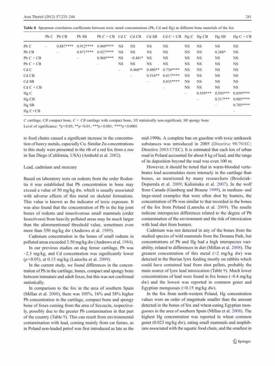
Also in the case of Cd, high values of the Spearmancorrelation coefficient were recorded, especially betweenthe concentration of this metal in the cartilage with adjacentcompact bone and its concentrations in cartilage, compactbone and spongy bone (rs showed a rising trend, from 0.73to 0.83). For the relationship between Cd concentration inthe cartilage with adjacent compact bone and concentrationof this metal in the spongy bone a regression equation wascalculated. Also, the concentration of Hg in cartilage withcompact bone strongly correlated with the cartilage, com-pact bone and spongy bone, albeit with a downward rs trendin this sequence (from about 0.86 to 0.70). Between differ-ent metals (Table 8) we reported only two statistically sig-nificant associations (p<0.05): antagonist between theconcentration of Pb in spongy bone and Cd in compact bone(rs0−0.401), and synergistic between Pb in compact boneand Hg in spongy bone (rs00.388).
Discussion
Studies on heavy metals in the environment include, amongother things, measuring concentrations in tissues and organsof animals living in land and water ecosystems. In mam-mals, including the red and arctic fox, toxic elements aredetermined mostly in the liver and kidneys which performdetoxification functions.
Warm blooded vertebrates are the subject of multifacetedstudies, mainly in Central Europe and especially in Poland,the Czech Republic and Slovakia (Jankovska et al. 2010;Kalisińska et al. 2011; Piskorova et al. 2003), and lessfrequently in the United States and Canada (Dehn et al.2006; Hoekstra et al. 2003), i.e., in areas or decades exposedto large amounts of anthropogenic toxic substances.
In Table 9, for comparison, we summarized data fromscientific literature concerning the concentrations of twomicronutrients (Zn and Cu) and three toxic metals (Pb, Cdand Hg) determined in different types of bones of long-livedmammals.
Table 4 Significance of differences between metal concentrations indifferent fox bone materials
Metal Parameter C vs. CB C vs. SB CB vs. SB C + CB vs. SB
Zn U NS NS NS NSn030 p
Cu U 297.0 217.0 293.0 510.0
n029 p 0.04 0.001 0.03 0.001
Pb U NS NS NS NSn030 p
Cd U NS NS 300.0 NSn030 p 0,03
Hg U NS 210.0 243.0 451.0
n030 p 0.001 0.002 0.002
C cartilage, CB compact bone, C + CB cartilage with compact bone,SB spongy bone, U Mann–Whitney U-test, p level of significance
238 Acta Theriol (2012) 57:233–244

In literature, there is little data on the concentrations reflect-ing chronic, sublethal and lethal intoxication with lead, cad-mium and mercury in mammal bones. The lowest leadconcentration in human bones is observed in newborns. Inchildren aged 11 months it is 1.5 mg/kg body weight, but thisconcentration usually increases even dozens of times over thelifespan, and the total concentration of this element in thebody of a 60- to 70-year-old male can exceed 200 mg/kg bodyweight (Ma 1996). The transition from a non-toxic to patho-logical state changes gradually, hence it is difficult to establisha clear line between non-toxic and toxic concentrations in
solid elements (Kabata-Pendias and Pendias 1999). Concen-trations of the three toxic elements in skeletal elements reflectchronic exposure, yet there is no proof whether the mobiliza-tion of elements accumulated in bones may occur so rapidlythat it may cause the symptoms of contamination.
Zinc and copper
In available publications, we found little data on concen-trations of Zn and Cu in the cartilage and bone of Canidae.In our earlier work (Lanocha et al. 2010) on dogs from
Table 5 Comparison of metal concentrations in analagous materials between immature and adult foxes
Immature, n018
Cartilage AM ± SD 136.6±52.8 1.90±1.34 1.876±2.755 0.147±0.129 0.0069±0.0053
Med 136.6 2.43 0.516 0.166 0.0054
range 48.3–219.8 0.16–3.82 0.178–11.01 0.028–0.198 0.0016–0.0223
CV 38.7 70.0 146.8 37.4 76.5
Compact bone n017
AM ± SD 108.8±71.02 0.85±0.51 1.016±1.273 0.118±0.077 0.0061±0.054
Med 67.6 0.75 0.415 0.149 0.0039
range 60.0–278.2 0.19–2.18 0.153–4.354 0.024–0.550 0.0016–0.0226
CV 65.3 60.6 125.3 65.0 88.7
Spongy bone n017
AM ± SD 118.4±47.4 0.99±1.04 1.39±1.853 0.149±0.071 0.0031±0.0022
Med 120.4 0.56 0.564 0.190 0.0019
range 11.7–219.5 0.01–2.75 0.096–5.993 0.035–0.226 0.0013–0.0091
CV 40.0 104.7 133.6 47.6 70.5
Adult, n012
Cartilage AM ± SD 130.8±61.70 0.123±3.439 1.530±1.564 0.123±0.054 0.0045±0.0049
Med 143.6 0.82 1.124 0.144 0.0027
range 19.6–205.4 0.104–9.45 0.165–4.827 0.031–0.184 0.0019–0.0186
CV 47.2 148.8 102.2 43.9 108.8
Compact bone n011
AM ± SD 108.4±48.8 0.67±0.37 0.922±0.372 0.093±0.068 0.0043±0.0033
Med 99.2 0.55 0.447 0.082 0.0030
range 50.04–202.1 0.13–1.28 0.150–2.826 0.022–0.191 0.0012–0.0104
CV 45.0 55.7 108.3 73.0 76.2
Spongy bone n011
AM ± SD 100.0±50.6 0.45±0.33 1.654±1.883 0.132±0.07 0.0028±0.0025
Med 105.0 0.35 0.927 0.131 0.0020
range 11.2–157.0 0.07–1.21 0.069–6.147 0.034–0.260 0.0013–0.0105
CV 50.7 74.1 113.8 53.2 91.1
Immature vs. adult
Cartilage U NS NS NS NS 63.0
p 0.98 0.05
Compact bone U NS NS NS NS NSp
Spongy bone U NS NS NS NS NSp
U Mann–Whitney U-test, NS statistically non-significant difference
Acta Theriol (2012) 57:233–244 239

north-western Poland, Zn concentrations in cartilage andspongy bone were similar and amounted to ~80 mg/kg,and the concentration of Cu was significantly higher incartilage than in spongy bone (2.3 and 1.8 mg/kg dw,respectively). Budis et al. (2009) examined the limb bonesof foxes from north-western Poland and observed that theconcentration of Cu in the compact bone was three timesgreater than in spongy bone (~2.8 compared to 0.9 mg/kg),but the difference between average values was not statisti-cally confirmed. They, however, did not take into accountthe age of the animals as we have done in this paper. In ourstudy, Cu concentrations in analogous materials were sig-nificantly lower, by approximately 300% and 120% in car-tilage and spongy bone, respectively. Comparison ofconcentrations of this metal in the compact and spongy bonebetween two age groups (immatures and adults), showedthat the Cu concentration was higher in immature foxes thanin adults by 36% and 60%, respectively, but the differencesbetween mean values were not statistically significant.
Millan et al. (2008) studied the concentrations of variousheavy metals, including Zn and Cu, in the bones of the fox,
the Iberian lynx (Lynx pardinus), common genet (Genettegenette), Egyptian mongooses (Herpestes ichneumon) andbadger (Meles meles) in Spain. These animals differed in diet,which could be one reason for the wide variation in concen-trations of trace elements in their bones (Millan et al. 2008).The largest concentrations of Zn (>170 mg/kg dw) weredetected in wheat-eating common genet feeding mostly oninsects, small vertebrates and fish. Smaller concentrations(~130 mg/kg dw) of this metal were found in fox and badger.
The concentration of Zn in the spongy bone of foxesfrom Western Pomerania was ~116 mg/kg and was about10% or so smaller than in foxes found in Spain. Copperconcentration in the bones of the fox from Spain (Millan etal. 2008) was almost 20 times greater than that observed inthe spongy bone from Western Pomeranian specimens. TheDonana Park in Spain, from which the animals originated inthe study by Millan et al. (2008), experienced an environ-mental disaster in 1998. Heavily polluted water from adamaged mine waste reservoir (located above the park)contaminated the park with heavy metals and penetratedits surface and ground waters. It is probable that the transfer
Table 6 Spearman rank coeffi-cient of variation of metal con-centrations in the examined bonematerials of the fox (n030)
NS statistically non-significant
Level of significance: *p<0.05;**p<0.01; ***p<0.001
Metal correlations Cartilage Compact bone Cartilage with compact bone Spongy bone
Zinc with:
Cu NS NS 0.275* 0.721***
Cd 0.536** 0.688*** 0.695 *** 0.828***
Pb –0.429* –0.451* –0.371** NS
Hg NS NS NS NS
Copper with:
Pb NS NS NS NS
Cd NS NS NS 0.713***
Hg NS 0.390* 0.275* NS
Lead with:
Cd NS NS NS NS
Hg NS NS NS NS
Cadmium with:
Hg NS NS NS NS
Table 7 Spearman rank corre-lation coefficient between twomicroelements — Zn and Cu —determined in various type ofbone material (n030) of the fox
C cartilage, CB compact bone, C+ CB cartilage with compactbone, NS values statisticallynon-significant, SB spongy bone
Levels of significance: *p<0.05;**p<0.01; ***p<0.001; ****p<0.0001
Zn C Zn CB Zn SB Zn C + CB Cu C Cu CB Cu SB Cu C + CB
Zn C – 0.633*** NS 0.895**** NS NS NS NS
Zn CB – 0.379* 0.882**** NS NS NS NS
Zn SB – 0.413* NS NS 0.721**** 0.483**
Zn C + CB – NS NS NS NS
Cu C – 0.367* NS 0.644***
Cu CB – NS 0.621****
Cu SB – 0.389*
Cu C + CB –
240 Acta Theriol (2012) 57:233–244
to food chains caused a significant increase in the concentra-tion of heavy metals, especially Cu. Similar Zn concentrationsto this study were presented in the rib of a red fox from a zooin San Diego (California, USA) (Arnhold et al. 2002).
Lead, cadmium and mercury
Based on laboratory tests on rodents from the order Roden-tia it was established that Pb concentration in bone mayexceed a value of 50 mg/kg dw, which is usually associatedwith adverse effects of this metal on skeletal formations.This value is known as the indicator of toxic exposure. Itwas also found that the concentration of Pb in the hip jointbones of rodents and insectivorous small mammals (orderInsectivora) from heavily polluted areas may be much largerthan the aforementioned threshold value, sometimes evenmore than 550 mg/kg dw (Andrews et al. 1989).
Cadmium concentration in the bones of small rodents inpolluted areas exceeded 3.50mg/kg dw (Andrews et al. 1984).
In our previous studies on dog femur cartilage, Pb was~2.3 mg/kg, and Cd concentration was significantly lower(p<0.05), at 0.13 mg/kg (Lanocha et al. 2009).
In the current study, we found differences in the concen-tration of Pb in the cartilage, bones, compact and spongy bonebetween immature and adult foxes, but this was not confirmedstatistically.
In comparison to the fox in the area of southern Spain(Millan et al. 2008), there was 105%, 16% and 58% higherPb concentration in the cartilage, compact bone and spongybone of foxes coming from the area of Szczecin, respective-ly, possibly due to the greater Pb contamination in that partof the country (Table 9). This can result from environmentalcontamination with lead, coming mainly from car fumes, asin Poland non-leaded petrol was first introduced as late as the
mid-1990s. A complete ban on gasoline with toxic antiknocksubstances was introduced in 2005 (Directive 98/70/EC;Directive 2003/17/EC). It is estimated that each km of urbanroad in Poland accounted for about 8 kg of lead, and the rangeof its deposition beyond the road was even 100 m.
However, it should be noted that in warm-blooded verte-brates lead accumulates more intensely in the cartilage thanbones, as mentioned by many researchers (Brodziak-Dopierala et al. 2009; Kalisinska et al. 2007). In the wolffrom Canada (Gamberg and Braune 1999), in medium- andlarge-sized examples that were often shot by hunters, theconcentration of Pb was similar to that recorded in the bonesof the fox from Poland (Lanocha et al. 2009). The resultsindicate interspecies differences related to the degree of Pbcontamination of the environment and the risk of intoxicationwith lead shot from hunters.
Cadmium was not detected in any of the bones from thestudied species of wild mammals from the Donana Park, butconcentrations of Pb and Hg had a high interspecies vari-ability, related to differences in diet (Millan et al. 2008). Thegreatest concentration of this metal (>2 mg/kg dw) wasdetected in the Iberian lynx feeding mostly on rabbits whichcould have contained lead from shot pellets, probably themain source of lynx lead intoxication (Table 9). Much lowerconcentrations of lead were found in fox bones (~0.4 mg/kgdw) and the lowest was reported in common genet andEgyptian mongooses (<0.15 mg/kg dw).
In the fox from north-western Poland, Hg concentrationvalues were an order of magnitude smaller than the amountdetected in the bones of fox and wheat-eating Egyptian mon-gooses in the area of southern Spain (Millan et al. 2008). Thehighest Hg concentration was reported in wheat commongenet (0.023 mg/kg dw), eating small mammals and amphib-ians associated with the aquatic food chain, and the smallest in
Table 8 Spearman correlation coefficients between toxic metal concentrations (Pb, Cd and Hg) in different bone materials of the fox
Pb C Pb CB Pb SB Pb C + CB Cd C Cd CB Cd SB Cd C + CB Hg C Hg CB Hg SB Hg C + CB
Pb C – 0.887**** 0.912**** 0.969**** NS NS NS NS NS NS NS NS
Pb CB – 0.871**** 0.927**** NS NS NS NS NS NS 0.388* NS
Pb C + CB – 0.960**** NS −0.401* NS NS NS NS NS NS
Pb C + CB – NS NS NS NS NS NS NS NS
Cd C – 0.466** 0.480** 0.730**** NS NS NS NS
Cd CB – 0.516** 0.817**** NS NS NS NS
Cd SB – 0.835**** NS NS NS NS
Cd C + CB – NS NS NS NS
Hg C – 0.559*** 0.595*** 0.859****
Hg CB – 0.517*** 0.805****
Hg SB – 0.705****
Hg C+CB –
C cartilage, CB compact bone, C + CB cartilage with compact bone, NS statistically non-significant, SB spongy bone
Level of significance: *p<0.05; **p<0.01; ***p<0.001; ****p<0.0001
Acta Theriol (2012) 57:233–244 241

the Egyptian mongoose (0.011 mg/kg dw), feeding on smallinvertebrates. The concentration of Hg in fox from Spain wasthree times higher compared to the compact bone of the foxpopulation originating from Western Pomerania.
Dey et al. (1999) determinedHg in the bones of two speciesof Felidae family from north-eastern India, Bengal cat (Felisbengalensis) and leopard (Panthera pardus), and in the Indiancivet (Viverra zebitha) from the civet family (Viverridae),which feeds on small mammals, snakes and fish. Their bonessometimes contained even over 30 mg/kg dw. In general, Hgconcentrations in the bones were very small and ranged from0.001 to 0.065 mg/kg dw (Table 9), but scientific papers onanimals occasionally report concentrations of this metal thatare 3–4 orders of magnitude larger (up to 30 mg/kg dw).
Although there are various studies on heavy metals inhumans and wild animals, knowledge of the concentrationsin bones and the effects on the osseous–articular system is stillinsufficient. There are many indications that the concentra-tions of elements in bones are strongly correlated with envi-ronmental conditions, diet and health status of variouspopulations of people and mammals, but more and moredetailed studies are required, also in Central Europe.
In addition to routine monitoring studies conducted onthe abiotic environment, an urgent need exists to monitorlong-term concentrations of heavy metals with long-termeffect on living organisms. An important addition is provid-ed by biomonitoring studies on domesticated and free-livingmammals, including Canidae.
Conclusion
In fox cartilage, compact bone and spongy bone, medianconcentrations of the metals studied could be arranged in thefollowing series: Zn > Cu > Pb > Cd > Hg, their valuesranging from 142 mg/kg to 0.002 mg/kg dw. The stron-gest synergistic relationships were observed in spongybone between the Zn and Cu and Zn and Cd, as well asbetween Cu and Cd. Statistically significant antagonisticrelationships were detected between zinc and lead incompact bone.
Bone tissue, due to its properties, may reflect levels ofchronic exposure and may be the basis of indirect assessmentof environmental exposure.
Table 9 Concentrations of trace elements (in mg/kg) in bone material of species from order Carnivova
Species Place Tissue Zn Cu Pb Cd Hg Age Sex n dw or ww Source
Family - Canidae
Red fox (Vulpes vulpes) Poland Compact bone – 2.8 – – – – F + M 24 dw Budis et al. 2009
West Pomeranian province Spongy bone 0.9 – – – –
Cartilage – – 1.96 0.16 – – F + M 24 dw Lanocha et al.
2009
Spain, Donana Park 142.1 4.2 0.385 ND 0.012 im F + M 17 dw Millan et al. 2008
ad
USA, San Diego, ZOO Cartilage 138.0 (121.4) – – – – im F + M dw (ww) Arnhold et al.
2002adSpongy bone
Domestic dog
(Canis lupus familiaris)
Poland Cartilage – – 2.28 0.13 – – F + M 15 dw Lanocha et al.
2009
West Pomeranian province Rib 81 2.3 – – – im F + M 15 dw Lanocha et al.
2010
Spongy bone 80 1.8 ad
Gray wolf (Canis lupus) Canada, Yukon Bone – – 2.12 – – im F + M 13 dw Gamberg and
Braune 1999ad
Family - Mustelidae
European badger (Meles meles) Spain, Donana Park Bone 128.3 1.02 0.73 ND – im M 1 dw Millan et al. 2008
Family - Viverridae
Common genet (Genetta genetta) Spain, Donana Park Bone 173.1 1.61 0.11 ND 0.023 ad F + M 4 dw Millan et al. 2008
Egyptian mongoose
(Herpestes ichneumon)
145.0 0.63 0.136 ND 0.011 11
Indian civet (Viverra zibetha) India Bone – – 3.9 – 25.0 – – 10 dw Dey et al. 1999
Family - Felidae
Iberian lynx (Lynx pardinus) Spain, Donana Park Bone 142.1 4.2 0.385 ND 0.012 im F + M 17 dw Millan et al. 2008
ad
Leopard cat (Felis bengalensis) India Bone – – 22.5 – 32.2 – – 10 dw Dey et al. 1999
Leopard (Panthera pardus) – – – – 18.3 – –
F female, M Male, DW dry weight, WW wet weight, ND-not detected
242 Acta Theriol (2012) 57:233–244

Acknowledgements The study was financed as research project no.NN 404 507738 by the Polish Ministry of Education from the resourcesfor the years 2010–2011.
Open Access This article is distributed under the terms of the CreativeCommons Attribution License which permits any use, distribution, andreproduction in any medium, provided the original author(s) and thesource are credited.
References
Andrews SM, Johnson MS, Cooke JA (1984) Cadmium in smallmammals from grassland established on metalliferous minewaste. Environ Pollut (Ser A) 33:153–162
Andrews SM, Johnson MS, Cooke JA (1989) Distribution of traceelements pollutants in a contaminated grassland ecosystem estab-lished on metalliferous fluorspar tailings: 1. Lead. Environ Pollut58:73–85. doi:10.1016/0269-7491(89)90229-7
Arnhold W, Anke M, Goebel S (2002) The copper, zinc and manga-nese status in opossum and gray fox. Z Jagdwiss 48:77–86.doi:10.1007/BF02192395
Bernard A (2008) Cadmium and its adverse effects on human health.Indian J Med Res 128:557–564
Brodziak-Dopierala B, Kwapulinski J, Rzepka J, Nogaj E, Bogunia M,Ahnert B (2007) Influence of smoking tobacco on the occurrencemetals in some parts and profiles of femur head. Przegl Lek64:720–722
Brodziak-Dopierala B, Kwapulinski J, Kusz D, Gajda Z, Sobczyk K(2009) Interactions between concentrations of chemical elementsIn human femoral heads. Arch Environ Contam Toxicol 57:203–210
Budis H, Kalisinska E, Lanocha N (2009) Manganese and copper inbone of red fox from Pomerania, Poland. Ecotoxicology in thereal world. Krakow, 16–19 September 2009, p 107
Cavallini P, Santini S (1995) Age determination in the red fox inMediterranean habitat. Z Saugetierkd 60:136–142
Charles CH, Cronin MJ, Conforti NJ, Dembling WZ, Petrone DM,McGuire JA (2001) Anticalculus efficacy of an antiseptic mouth-rinse containing zinc chloride. J Am Dent Assoc 132:94–98
Dehn LA, Follmann EH, Thomas DL, Sheffield GG, Rosa C, DuffyLK, O’Hara TM (2006) Trophic relationships in an Arctic foodweb and implications for trace metal transfer. Sci Total Environ362:103–123
Dey S, Stafford R, DebRoy MK, Bhattacharjee CR, Khathing DT,Bhattacharjee PC, Dkhar PS (1999) Metal toxicity and traceelement deficiency in some wild animal species from north-eastIndia, as revealed by cellular, bio-inorganic and behavioural studies.Curr Sci 77:267–280
Directive 2003/17/EC of the European Parliament and of the Councilof 3 March 2003 amending Directive 98/70/EC relating to thequality of petrol and diesel fuels
Directive 98/70/EC of the European Parliament and of the Council of13 March 1998 relating to the quality of petrol and diesel fuelsDirective 93/12/EC
Gamberg M, Braune BM (1999) Contaminant residue levels in arcticwolves (Canis lupus) from the Yukon Territory, Canada. Sci TotalEnviron 244:329–338
Gerhardsson L, Akantis A, Lundstrom NG, Nordberg GF, Schütz A,Skerfving S (2005) Lead concentrations in cortical and trabecularbones in deceased smelter workers. J Trace Elem Med Biol19:209–215. doi:10.1016/j.jtemb.2005.06.004
Hoekstra PF, Braune BM, Elkin B, Armstrong FAJ, Muir DCG (2003)Concentrations of selected essential and non-essential elements in
arctic fox (Alopex lagopus) and wolverines (Gulo gulo) from theCanadian Arctic. Sci Total Environ 309:81–92
Honda R, Tsuritani I, Ishizaki M, Yamada Y (1997) Zinc and copperlevels in ribs of cadmium-exposed persons with special reference toosteomalacia. Environ Res 75:41–48. doi:10.1016/enrs.1997.3747
Horiguchi H, Aoshima K, Oguma E, Sasaki S, Miyamoto K, Hosoi Y,Katoh T, Kayama F (2010) Latest status of cadmium accumula-tion and its effects on kidneys, bone, and erythropoiesis in inhab-itants of the formerly cadmium-polluted Jinzu River Basin inToyama, Japan, after restoration of rice paddies. Int Arch OccupEnviron Health 83:953–970. doi:10.1007/s00420-010-0510-x
Jankovska I, Miholova D, Bejcek V, Vadlejch J, Sulc M, Szakova J,Langrova I (2010) Influence of parasitism on trace element con-tents in tissues of red fox (Vulpes vulpes) and its parasites Meso-cestoides spp. (Cestoda) and Toxascaris leonina (Nematoda).Arch Environ Contam Toxicol 8:469–477. doi:10.1007/s00244-009-9355-2
Jarup L, Akesson A (2009) Current status of cadmium as an environ-mental health problem. Toxicol Appl Pharmacol 238:201–208.doi:10.1016/j.taap.2009.04.020
Jurkiewicz A, Wiechula D, Nowak R, Gazdzik T, Loska K (2004)Metal content in femoral head spongious bone of people living inregions of different degrees of environmental pollution in Southernand Middle Poland. Ecotoxicol Environ Saf 59:95–101.doi:10.1016/j.ecoenv.2004.01.002
Kabata-Pendias A, Pendias H (1999) Biogeochemistry of trace ele-ments. PWN, Warsaw
Kalisinska E, Salicki W, Kavetska KM, Ligocki M (2007) Trace metalconcentrations are higher in cartilage than in bones of scaup andpochard wintering in Poland. Sci Total Environ 388:90–103.doi:10.1016/j.scitotenv.2007.07.050
Kalisinska E, Lisowski P, Salicki W, Kavetska K, Kucharska T (2009)Mercury wild terrestrial carnivorous mammals from north-western Poland and unusual fish diet of red fox. Acta Theriol54:345–356. doi:10.4098/j.at.0001-7051.032.2008
Kalisińska E, Lisowski P, Kosik-Bogacka DI (2011) Red Fox Vulpesvulpes (L., 1758) as a bioindicator of mercury contamination interrestrial ecosystems of North-Western Poland. Biol Trace ElemRes. doi:10.1007/s12011-011-9181-z
Kidawa D, Kowalczyk R (2011) The effects of sex, age, season andhabitat on diet of the red fox Vulpes vulpes in northeastern Poland.Acta Theriol 56:209–218
Kjellstrom T (1992) Mechanism and epidemiology of bone effects ofcadmium. In: Nordberg G, Alessio L, Herber R (eds) Cadmium inthe human environment: toxicity and carcinogenicity. IARC SciPubl 118:301–310
Knowlton FF, Whittemore SL (2001) Pulp cavity-tooth width ratiosfrom known age and wild-caught coyotes determined by radiogra-phy. Wildl Soc Bull 29:239–244
Kwapulinski J, Miroslawski J, Wiechula D, Jurkiewicz A, TokarowskiA (1995) The femur capitulum as a biomarker of contaminationdue to indicating lead content in the air by participation of theother metals. Sci Total Environ 175:57–64. doi:10.1016/0048-9697(95)04844-8
Lanocha N, Kalisinska E, Budis H (2009) Comparison of copper andzinc concentrations in cartilage and cancellous bone of dogs fromSzczecin and surrounding areas. Analyst for the twenty-first cen-tury society. VIII Polish Conference on Analytical Chemistry,Cracow 4–9 July 2010, Warsaw, p. 347
Lanocha N, Kalisinska E, Budis H (2010) Cadmium and lead in femurcartilage of canids from north-western Poland. Ecotoxicology inthe real world. Krakow, 16–19 September 2009, p 111
Lazarus M, Orct T, Blanusa M, Vickovic I, Sostari CB (2008) Toxicand essential metal concentrations in four tissues of red deer(Cervus elaphus) from Baranja, Croatia. Food Addit Contam25:270–283. doi:10.1080/02652030701364923
Acta Theriol (2012) 57:233–244 243

Liu ZP (2003) Lead poisoning combined with cadmium in sheep andhorses in the vicinity of non-ferrous metal smelters. Sci TotalEnviron 309:117–126. doi:10.1016/S0048-9697(03)00011-1
Lopez-Alonso M, Miranda M, García-Partida P, Cantero F, HernandezJ, Benedito JL (2007) Use of dogs as indicators of metal exposurein rural and urban habitats in NW Spain. Sci Total Environ372:668–675. doi:10.1016/j.scitotenv.2006.10.003
Ma WC (1996) Lead in mammals. In: Beyer WN, Heinz Redmon-Norwood AW (eds) Environmental contaminants in wildlife: inter-preting tissue concentrations. CRC Press, Boca Raton, pp 281–296
Machalinski B, Machoy Z, Dabkowska E, Chlubek D, Ogonski T(1996) Zinc in developmental cycle of domestic hen exposed tohigh doses of sodium fluoride. Environ Sci 4:127–132
Millan J, Mateo R, Taggart MA, Lopez-Bao JV, Viota M, Monsalve L,Camarero PR, Blazquez E, Jimenez B (2008) Levels of heavymetals and metalloids critically endangered Iberian lynx and otherwild carnivores from Southern Spain. Sci Total Environ 399:193–201. doi:10.1016/j.scitotenv.2008.03.038
Nielsen FH, Milne DB (2004) A moderately high intake compared to alow intake of zinc depresses magnesium balance and alters indicesof bone turnover in postmenopausal women. Eur J Clin Nutr58:703–710. doi:10.1038/sj.ejcn.1601867
NRC (1980) Mineral tolerance of domestic animals. National Academyof Sciences, National Research Council, Washington, pp 93–276
Piskorova L, Vasilkova Z, Krupicer I (2003) Heavymetal residues in tissuesof wild boar (Sus scrofa) and red fox (Vulpes vulpes) in the CentralZemplin region of the SlovakRepublic. Czech JAnimSci 48:134–138
Puzanowska-Tarasiewicz H, Kuzmicka L, Tarasiewicz M (2009) Bio-logical functions of elements: III. Zinc-component and an activatorof enzymes. Pol Merk Lek 161:419–422
Scheuhammer AM, Basu N, Burgess NM, Elliott JE, Campbell GD,Wayland M, Champoux L, Rodrigue J (2008) Relationships amongmercury, selenium, and neurochemical parameters in common loons(Gavia immer) and bald eagles (Haliaeetus leucocephalus). Ecotox-icology 17:93–101. doi:10.1007/s10646-007-0170
Senczuk W (2006) Modern toxicology. Ed PZWL, Warsaw
Shore RF, Casulli A, Bologov V, Wienburg CL, Afsar A, Toyne P,Dell’Omo G (2001) Organochlorine pesticide, polychlorinatedbiphenyl and heavy metal concentrations in wolves (Canis lupusL. 1758) from north-west Russia. Sci Total Environ 280:45–54.doi:10.1016/S0048-9697(01)00802-6
Smrcka V (2005) Trace elements in bone tissue. Karolinum Press,Charles University in Prague, Prague
Starek A (2007) Organ toxicology. Wyd PZWL, WarsawUmemura T, Wako Y (2006) Pathogenesis of osteomalacia in Itai-itai
Disease. J Toxicol Pathol 19:69–74. doi:10.1293/tox.19.69Wiechula D, Jurkiewicz A, Loska K (2008) An assessment of
natural concentrations of selected metals in the bone tissues of thefemur head. Sci Total Environ 406:161–167. doi:10.1016/j.scitotenv.2008.07.068
Yamaguchi M (1998) Role of zinc in bone formation and bone resorp-tion. J Trace Elem Exp Med 11:119–135. doi:10.1002/(SICI)1520-670X(1998) 11:2/3<119::AID-JTRA5>3.0.CO;2-3
Yoo YCh, Lee SK, Yang JY, Kim KW, Lee SY, Oh SM, Chung KH(2002) Interrelationship between the concentration of toxic andessential elements in Korean tissues. J Health Sci 48:195–200
Zaichick V, Zaichick S (2009) Instrumental neutron activation analysisof trace element contents in the rib bone of healthy men. JRadioanal Nucl Chem 281:47–52. doi:10.1007/s10967-009-0084-9
Zaichick S, Zaichick V (2010) The effect of age and gender on 38chemical element contents in human iliac crest investigated byinstrumental neutron activation analysis. J Trace Elem Med Biol24:1–6. doi:10.1016/jtemb.2009.07.002
Zaichick S, Zaichick V, Karandashev VK, Moskvina IR (2011) Theeffect of age and gender on 59 trace-element contents in humanrib bone investigated by inductively coupled plasma mass spec-trometry. Biol Trace Elem Res 143:41–57. doi:10.1007/s12011-010-8837-4
Zhu G, Wang H, Shi Y, Weng S, Jin T, Kong Q, Nordberg GF (2004)Environmental cadmium exposure and forearm bone density. Bio-metals 17:499–503. doi:10.1023/B:BIOM.0000045728.80518.d9
244 Acta Theriol (2012) 57:233–244