toxicological impact of exposure to 2,3,7,8-
TRANSCRIPT
____________________________________________________________________________________________
*Corresponding author: Email: [email protected];
Annual Research & Review in Biology4(8): 1278-1289, 2014
SCIENCEDOMAIN internationalwww.sciencedomain.org
Toxicological Impact of Exposure to 2,3,7,8-tetrachlorodibenzo-p-dioxin (TCDD) on Some
Hormonal Profiles and HematologicalParameters in Goats
Azza M. Mohamed1*, A. O. Hegab2, Jehad M. Yousef1
and Manal E. A. El H alwagy1
1Biochemistry Department, Faculty of Science for Girls, King Abdulaziz University, Saudi Arabia.2Biology Department, Faculty of Science, Taif University, Saudi Arabia.
Authors’ contributions
This work was carried out in collaboration between all authors. Author AMM designed thestudy and wrote the first draft of the manuscript, author AOH wrote the protocol and
performed the statistical analysis, author JMY managed the biochemical analyses of thestudy and author MEAEH managed the literature searches. All authors read and approved
the final manuscript
Received 28th August 2013Accepted 23rd October 2013Published 2nd January 2014
ABSTRACT
Female Baladi goats were used to investigate the toxicological effects of 2,3,7,8-tetrachlorodibenzo-p-dioxin (TCDD) on some serum hormones and blood features.Animals were divided into two groups, group 1: served as control, group 2: animals wereorally administered with three repeated doses (0.23µg/Kg body weight) of TCDD with 2days interval between dosing.Results revealed that exposure to TCDD induced reduction in serum estradiol,progesterone and prolactin levels and elevation in glucocorticoid hormone cortisolthroughout the different studied periods (48h, 96h and 16 days commenced the lastintoxicated dose). The adverse impact of TCDD on goat reproductive hormones wasconfirmed by histopatholgical observations on their uteri and ovaries after 16 dayscommenced the last intoxicated dose. Intoxication of goats with TCDD showed alsomarked decreases in hemoglobin (Hb) concentration, red blood cell count (RBC), packedcell volume (PCV), mean corpuscular volume (MCV), mean corpuscular hemoglobin (MCH)
Original Research Article
Annual Research & Review in Biology, 4(8): 1278-1289, 2014
1279
and mean corpuscular hemoglobin concentrations (MCHC) compared with normal healthygroup. Pronounced decreases in total white blood cells (WBC) count as well as in itsdifferential percentages namely lymphocytes, monocytes and eosinophils were alsonoticed in intoxicated animals compared with normal ones, while the percentage ofneutrophils showed significant increase.In conclusion, the results of this study indicated that oral exposure of female goats torepeated doses of TCDD caused endocrine disruption which may lead to adverse impacton their reproductive performance. Moreover, toxicity of TCDD inducedimmunosuppressive effect and anemia as indicated by its deleterious action on differenthematological parameters.
Keywords: Female goats; tetrachlorodibenzo-p-dioxin; hormones; hematological parameters.
1. INTRODUCTION
Endocrine disrupters (EDs) are pollutants that can act as agonists or antagonists to naturalhormones. EDs have been intensively studied for their actions as estrogen mimetic whereinthey work by binding to the estrogen receptor, thus affecting estrogen-regulated cellular andreproductive processes [1].
Dioxins are a class of persistent polyhalogenated aromatic hydrocarbons (PHAHs) of whichpolychlorinated dibenzodioxins (PCDDs) and polychlorinated dibenzofurans (PCDFs), haveidentified as among the most globally distributed potent environmental pollutants [2]. Dioxinsare unwanted by products of many industrial processes and mainly come from industrial airemissions, waste incineration and combustion of fuels [3]. Moreover, they are slowlydegraded in environment and hence remain as persistent and toxic contaminants for longtime. Humans are exposed to dioxins through diet, particularly fish products, meat and fattymilk and [4]. TCDD has been reported to accumulate in body due to its lipophilic properties,slow metabolism and excretion [5].
Based on biochemical and toxic responses, 2, 3, 7, 8-tetrachlorodibenzo-p-dioxin (TCDD) isconsidered one of the most potent members of PCDDs group and a model compound forstudy of the mechanisms of PCDDs [6]. The compound is often referred simply as dioxin.
Exposure to acute and chronic toxic levels of TCDD in various animal species and man,causes a wide-variety of adverse effects in a tissue and species specific manner, includinghepato-toxicity, carcinogenicity, teratogenicity, interference with lipid metabolism, reductionof bone strength, neurobehavioral effects, endocrine disruption, wasting syndrome, thymicatrophy, developmental and reproductive toxicity and immunosuppression [7 -10]. Previousstudies on female reproductive system of nonhuman primates indicated that exposure toTCDD leads to long - term adverse effects on pregnancy and ovarian function [11-12] and ithas been implicated in development of endometriosis [13] and abortion [14].
Therefore, the aim of this study was to demonstrate the reproductive toxicity andhematotoxicity induced by TCDD on some serum hormones, blood features andhistomorphologic phenotypes of uteri and ovaries in female goats.
Annual Research & Review in Biology, 4(8): 1278-1289, 2014
1280
2. MATERIALS AND METHODS
2.1 Chemicals
All chemicals used in the current investigation were of high analytical grade and products ofthe Sigma and Merck companies (USA). TCDD (>99 % pure) was purchased from the UFAOil Institute (Ufa, Russia). It was mixed with corn oil. The mixture was stirred using amagnetic stirrer before dosing.
2.2 Experimental Animals
The study protocol was approved by the Animal Experiment local ethics Committee. Eightmature female baladi goats (2-2.5 years old and 20-25 kg) were obtained from NationalResearch Centre Experimental Farm (Abu Rawash, Giza, Egypt). The animals were fed oncommercial diet and water ad libitum.
2.3 Experimental Design
Animals were divided into two groups, each consisting of four animals, G1: Normalhealthy animals. G2: Animals administered three repeated doses of TCDD with two daysintervals. TCDD was given orally at a dose of 0.23µg/Kg body weight (equivalent to 1/3 ofLD50 of TCDD) [15]. Blood samples were collected from each animal of both control andTCDD intoxicated groups after 48 h, 96 h and 16 days commenced the last intoxicated doseinto sterilized tubes for serum separation and into tubes containing heparin for determinationof hematological parameters. Serum was separated by centrifugation at 3000× g for 10minutes and used for biochemical serum analysis. At the end of experimental period (16days) all animals were slaughtered, the uterus and ovary from each animal was collected forhistopathological examination.
2.4 Haematological Evaluation
Haematological parameters including, white blood cells (WBC) count and differentialpercentages, red blood cells (RBC) count, haemoglobin (Hb), packed cell volume (PCV),mean corpuscular haemoglobin (MCH), mean corpuscular haemoglobin concentration,(MCHC) and mean corpuscular volume (MCV) were determined using auto- analyzer(Abbott Diagnostics, USA).
2.5 Biochemical Serum Analysis
2.5.1 Hormonal assay
Serum estradiol, progesterone, prolactin and cortisol were assayed using availablecommercial Radioimmunoassay (RIA) kits according to the manufacturer’s instructions.
2.6 Histopathological Examination
A small specimens of ovaries and uterus were fixed by 4% formalin and then embeddedinto paraffin, sectioned for 5–6-μm thick, and mounted on the glass microscope slides using
Annual Research & Review in Biology, 4(8): 1278-1289, 2014
1281
standard histopathological techniques. The sections were stained with hematoxylin-eosinand examined by light microscopy [16].
2.7 Statistical Analysis
The obtained data were computed and statistically analyzed according to Snedecor andCochran [17]. Data were analyzed by comparing values obtained from animals after thestudied intoxicated periods (48h, 96h and 16 days) in an intoxicated group with the valuesfor individual controls. Results are expressed as mean ± S.D. The significant differencesamong values were analyzed using analysis of variance (one-way Anova) coupled with post-hoc (LSD). Results were considered significant at P< 0.05.
3. RESULTS
3.1 Clinical Signs
The Clinical symptoms of intoxicated goats with repeated oral dose of dioxin during dioxiningestion till the end of experimental period (16 days) were general weakness, loss ofappetites, weight loss, diarrhea. Respiratory manifestations were in the form of continuousnasal discharge and cough.
3.2 Hormonal Profiles and Histo-pathological Examination
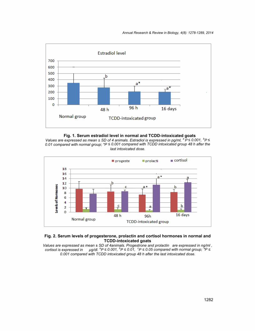
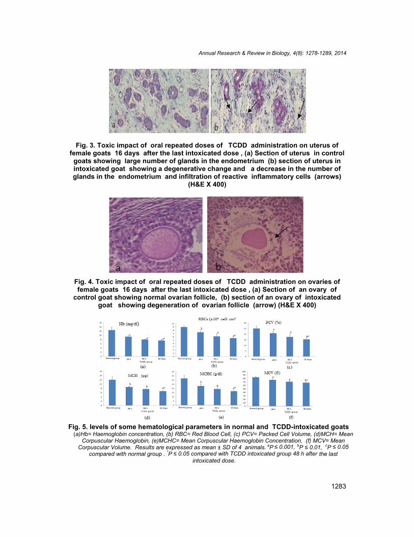
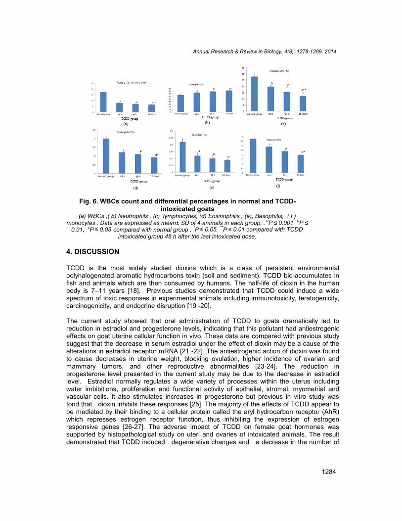
Figs. (1 and 2) summarize the adverse effect of TCDD on some hormones in serum offemale goats. The results revealed marked reduction in serum estradiol (Fig. 1) andprogesterone (Fig. 2) levels during the different exposure periods of goats to TCDD (48h, 96h and 16 days after the last intoxicated dose) compared with control healthy animals. Theresult also demonstrated that exposure to TCDD led to significant a reduction in the serumprolactin level, and an increase in the serum level of glucocorticoid steroid hormone,cortisol, during the same exposure periods of goats to this toxin compared with normal ones(Fig. 2). Histopathological observation showed that TCDD induced degenerative changesin uteri of intoxicated goats as observed by a decrease in the number of glands in theendometrium associated with infiltration of reactive inflammatory cells (Fig. 3). The ovariesof intoxicated goats also showed degenerative ovarian follicle (Fig. 4).
3.3 Haematological Parameters
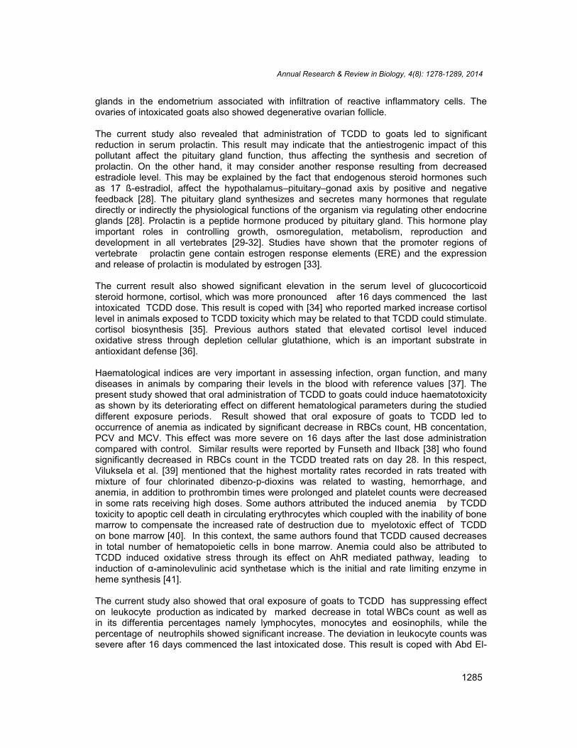
The effect of oral administration of TCDD on blood features of goats was illustrated in Fig. 5.Significant decreases in Hb concentration, RBCs count, PCV, MCV, MCH and MCHC wererecorded after the studied exposure periods compared with normal healthy group.Pronounced decreases in total WBCs count as well as in its differential percentages namelylymphocytes, monocytes and eosinophils were also noticed in intoxicated animals comparedwith normal ones, while the percentage of neutrophils showed significant increase (Fig. 6).
Annual Research & Review in Biology, 4(8): 1278-1289, 2014
1282
Fig. 1. Serum estradiol level in normal and TCDD-intoxicated goatsValues are expressed as mean ± SD of 4 animals. Estradiol is expressed in pg/ml, a P ≤ 0.001, bP ≤0.01 compared with normal group; *P ≤ 0.001 compared with TCDD intoxicated group 48 h after the
last intoxicated dose.
Fig. 2. Serum levels of progesterone, prolactin and cortisol hormones in normal andTCDD-intoxicated goats
Values are expressed as mean ± SD of 4animals. Progestrone and prolactin are expressed in ng/ml ,cortisol is expressed in µg/dl. aP ≤ 0.001, bP ≤ 0.01, cP ≤ 0.05 compared with normal group; *P ≤
0.001 compared with TCDD intoxicated group 48 h after the last intoxicated dose.
Annual Research & Review in Biology, 4(8): 1278-1289, 2014
1283
Fig. 3. Toxic impact of oral repeated doses of TCDD administration on uterus offemale goats 16 days after the last intoxicated dose , (a) Section of uterus in control
goats showing large number of glands in the endometrium (b) section of uterus inintoxicated goat showing a degenerative change and a decrease in the number ofglands in the endometrium and infiltration of reactive inflammatory cells (arrows)
(H&E X 400)
Fig. 4. Toxic impact of oral repeated doses of TCDD administration on ovaries offemale goats 16 days after the last intoxicated dose , (a) Section of an ovary of
control goat showing normal ovarian follicle, (b) section of an ovary of intoxicatedgoat showing degeneration of ovarian follicle (arrow) (H&E X 400)
Fig. 5. levels of some hematological parameters in normal and TCDD-intoxicated goats(a)Hb= Haemoglobin concentration, (b) RBC= Red Blood Cell, (c) PCV= Packed Cell Volume, (d)MCH= Mean
Corpuscular Haemoglobin, (e)MCHC= Mean Corpuscular Haemoglobin Concentration, (f) MCV= MeanCorpuscular Volume. Results are expressed as mean ± SD of 4 animals. aP ≤ 0.001, bP ≤ 0.01, cP ≤ 0.05
compared with normal group . *P ≤ 0.05 compared with TCDD intoxicated group 48 h after the lastintoxicated dose.
Annual Research & Review in Biology, 4(8): 1278-1289, 2014
1284
Fig. 6. WBCs count and differential percentages in normal and TCDD-intoxicated goats
(a) WBCs ,( b) Neutrophils , (c) lymphocytes, (d) Eosinophilis , (e), Basophilis, ( f )monocytes . Data are expressed as mean± SD of 4 animals in each group, . aP ≤ 0.001, bP ≤
0.01, cP ≤ 0.05 compared with normal group . *P ≤ 0.05, **P ≤ 0.01 compared with TCDDintoxicated group 48 h after the last intoxicated dose.
4. DISCUSSION
TCDD is the most widely studied dioxins which is a class of persistent environmentalpolyhalogenated aromatic hydrocarbons toxin (soil and sediment). TCDD bio-accumulates infish and animals which are then consumed by humans. The half-life of dioxin in the humanbody is 7–11 years [18]. Previous studies demonstrated that TCDD could induce a widespectrum of toxic responses in experimental animals including immunotoxicity, teratogenicity,carcinogenicity, and endocrine disruption [19 -20].
The current study showed that oral administration of TCDD to goats dramatically led toreduction in estradiol and progesterone levels, indicating that this pollutant had antiestrogeniceffects on goat uterine cellular function in vivo. These data are compared with previous studysuggest that the decrease in serum estradiol under the effect of dioxin may be a cause of thealterations in estradiol receptor mRNA [21 -22]. The antiestrogenic action of dioxin was foundto cause decreases in uterine weight, blocking ovulation, higher incidence of ovarian andmammary tumors, and other reproductive abnormalities [23-24]. The reduction inprogesterone level presented in the current study may be due to the decrease in estradiollevel. Estradiol normally regulates a wide variety of processes within the uterus includingwater imbibitions, proliferation and functional activity of epithelial, stromal, myometrial andvascular cells. It also stimulates increases in progesterone but previous in vitro study wasfond that dioxin inhibits these responses [25]. The majority of the effects of TCDD appear tobe mediated by their binding to a cellular protein called the aryl hydrocarbon receptor (AhR)which represses estrogen receptor function, thus inhibiting the expression of estrogenresponsive genes [26-27]. The adverse impact of TCDD on female goat hormones wassupported by histopathological study on uteri and ovaries of intoxicated animals. The resultdemonstrated that TCDD induced degenerative changes and a decrease in the number of
Annual Research & Review in Biology, 4(8): 1278-1289, 2014
1285
glands in the endometrium associated with infiltration of reactive inflammatory cells. Theovaries of intoxicated goats also showed degenerative ovarian follicle.
The current study also revealed that administration of TCDD to goats led to significantreduction in serum prolactin. This result may indicate that the antiestrogenic impact of thispollutant affect the pituitary gland function, thus affecting the synthesis and secretion ofprolactin. On the other hand, it may consider another response resulting from decreasedestradiole level. This may be explained by the fact that endogenous steroid hormones suchas 17 ß-estradiol, affect the hypothalamus–pituitary–gonad axis by positive and negativefeedback [28]. The pituitary gland synthesizes and secretes many hormones that regulatedirectly or indirectly the physiological functions of the organism via regulating other endocrineglands [28]. Prolactin is a peptide hormone produced by pituitary gland. This hormone playimportant roles in controlling growth, osmoregulation, metabolism, reproduction anddevelopment in all vertebrates [29-32]. Studies have shown that the promoter regions ofvertebrate prolactin gene contain estrogen response elements (ERE) and the expressionand release of prolactin is modulated by estrogen [33].
The current result also showed significant elevation in the serum level of glucocorticoidsteroid hormone, cortisol, which was more pronounced after 16 days commenced the lastintoxicated TCDD dose. This result is coped with [34] who reported marked increase cortisollevel in animals exposed to TCDD toxicity which may be related to that TCDD could stimulate.cortisol biosynthesis [35]. Previous authors stated that elevated cortisol level inducedoxidative stress through depletion cellular glutathione, which is an important substrate inantioxidant defense [36].
Haematological indices are very important in assessing infection, organ function, and manydiseases in animals by comparing their levels in the blood with reference values [37]. Thepresent study showed that oral administration of TCDD to goats could induce haematotoxicityas shown by its deteriorating effect on different hematological parameters during the studieddifferent exposure periods. Result showed that oral exposure of goats to TCDD led tooccurrence of anemia as indicated by significant decrease in RBCs count, HB concentation,PCV and MCV. This effect was more severe on 16 days after the last dose administrationcompared with control. Similar results were reported by Funseth and IIback [38] who foundsignificantly decreased in RBCs count in the TCDD treated rats on day 28. In this respect,Viluksela et al. [39] mentioned that the highest mortality rates recorded in rats treated withmixture of four chlorinated dibenzo-p-dioxins was related to wasting, hemorrhage, andanemia, in addition to prothrombin times were prolonged and platelet counts were decreasedin some rats receiving high doses. Some authors attributed the induced anemia by TCDDtoxicity to apoptic cell death in circulating erythrocytes which coupled with the inability of bonemarrow to compensate the increased rate of destruction due to myelotoxic effect of TCDDon bone marrow [40]. In this context, the same authors found that TCDD caused decreasesin total number of hematopoietic cells in bone marrow. Anemia could also be attributed toTCDD induced oxidative stress through its effect on AhR mediated pathway, leading toinduction of α-aminolevulinic acid synthetase which is the initial and rate limiting enzyme inheme synthesis [41].
The current study also showed that oral exposure of goats to TCDD has suppressing effecton leukocyte production as indicated by marked decrease in total WBCs count as well asin its differentia percentages namely lymphocytes, monocytes and eosinophils, while thepercentage of neutrophils showed significant increase. The deviation in leukocyte counts wassevere after 16 days commenced the last intoxicated dose. This result is coped with Abd El-
Annual Research & Review in Biology, 4(8): 1278-1289, 2014
1286
Nasser et al. [40] who demonstrated that oral exposure to TCDD resulted in severedecreases in leukocyte counts as well as in the percentages of lymphocytes, monocytes andeosinophils. The toxic effects of TCDD on total and differential leukocytes was previouslyexplained by some mechanisms. Murante and Gasiewicz [42] reported that TCDD affectsthe proliferation and/or differentiation processes of hemopoietic stem cells in bone marrow asit has a myelotoxic effects, thus it reduced the capacity of bone marrow to generate pro-lymphocytes. Also, it was found that marrow apoptosis and hypoplasia of bone marrow cellsare the major mechanisms of TCDD toxicity induced leucopenia [43]. In addition, Donald etal. [44] attributed this condition to immune suppressive effect of TCDD on bone marrow stemcells by mechanism mediated directly or indirectly through estrogenic action. Moreover, it wasfound that TCDD-treated hematopoietic stem cells almost lost long-term reconstitution activity[45]. Beside, some authors declared that TCDD has direct effect on the functional AhR thatis expressed by bone marrow stromal cells and plays an important role in the support anddirection of lymphopoiesis [43]. The authors added that TCDD treatment could alterlymphopoietic development, resulting in decrease of the total number of hematopoietic cellsand lymphocytes.
Neutrophilia recorded in this study during the term of TCDD exposure was not related todirect effect rather than as a compensatory response to the recoded lymphopenia. Byexamining the bone marrow in rats intoxicated with TCDD, Abd El-Nasser et al. [40]observed that significant decreases in lymphocytic, eosinophillic, and megakaryocyticseries, while neutrophillic series including myeloblast, metamyeloblast, promyloblast,segmented and band cell series showed significant increase.
5. CONCLUSION
The results of this study indicate that oral exposure of female goats to TCDD was found tohave antiestrogenic effect and induced adverse effects on progesterone and prolactin. Sucheffect may lead to endocrine disruption and subsequently affect the reproductiveperformance of animals. Moreover, this pollutant showed immunosupressive effect indicatedby its deleterious action on hematological parameters.
COMPETING INTERESTS
Authors have declared that no competing interests exist.
REFERENCES
1. Witorsch RJ. Endocrine disruptors: can biological eVects and environmental risks bepredicted? Regul. Toxicol. Pharmacol. 2002;36:118–130.
2. Scialli AR. Review, Tampons, dioxins and endometriosis. Reprod Toxicol2001;15:231-238.
3. Prange JA, Gaus C, Weber R, Papke O, Muller JF. Assessing forest fire as a potentialPCDD/F source in Queensland Australia. Environ. Sci. Technol. 2003;37:4325–4329.
4. Tuomisto J. Are dioxins a health problem in Finland? Duodecim. 2001;117:245–246.5. Enan E, El-Sabeawy F, Overstreet J, Matsumura F, Lasley B. Mechanisms of gender
specific TCDD induced toxicity in Guinea pig adipose tissue. Reprod Toxicol.1998;12:357–69.
Annual Research & Review in Biology, 4(8): 1278-1289, 2014
1287
6. El-Sabeawy F, Enanb E, Lasley B. Biochemical and toxic effects of 2,3,7,8-tetrachlorodibenzo-p-dioxin in immature male and female chickens. Comp BiochemPhysiol. 2001;129C:317-327
7. Birnbaum LS. The mechanism of dioxin toxicity: relationship to risk assessment.Environ. Health Perspect. 1994;102(Suppl 9):157–67.
8. Wormley DD, Ramesh A, Hood DB. Environmental contaminant-mixture effects onCNS development, plasticity, and behavior. Toxicol. Appl. Pharmacol. 2004;197:49–65.
9. Esser C, Steinwachs S, Herder C, Majora M, Lai ZW. Effects of a single dose of2,3,7,8-tetrachlorodibenzo-p-dioxin, given at post-puberty, in senescent mice.Toxicology Letters. 2005;157:89–98.
10. Finnila MAJ, Zioupos P, Herlin M, Miettinen HM, Simanainen U, Akansson HH,Tuukkanen J, Viluksela M, Jamsa J. Effects of 2,3,7,8-tetrachlorodibenzo-p-dioxinexposureonbone material properties. J Biomech. 2010;43:1097–1103
11. Li XL, Johnson DC, Rozman KK. Reproductive Effects of 2,3,7,8- Tetrachlorodibenzo-p-dioxin (TCDD) in Female Rats: Ovulation, Hormonal Regulation, and PossibleMechanism(s). Toxicol Appl Pharmacol. 1995;133:321-327.
12. Mora´n FMR, Tarara J, Chen S, Santos A, Cheney JW, Overstreet BL, Lasley C.Effect of dioxin on ovarian function in the cynomolgus macaque (M. fascicularis)Reprod Toxicol. 2001;15:377–383.
13. Rier S, Martin D, Bowman R, Dmowski W, Becker J. Endometriosis in rhesusmonkeys (Macaca mulatta) chronically exposed to 2, 3,7,8- tetrachlorodibenzo-p-dioxin. Fundam Appl. Toxicol. 1993;21:433– 41.
14. Li B, Liu HY, Dai LJ, Lu JC, Yang ZM, Huang L. The early embryo loss caused by2,3,7,8-Tetrachlorodibenzo-p-dioxin may be related to the accumulation of thiscompound in the uterus. Reprod Toxicol. 2006;21:301-306.
15. Kociba RI, Keyes DG, Beyer JE. Results of two years chronic toxicity and oncogenicitystudy of 2,3,7,8-Tetrachlorodibenzo-p-dioxin in rats. Toxicol Applied. 1978;46:279-303.
16. Bancroft JD, Stevens A, Turner DR. Theory and practice of histological techniques,4th Ed., Churchill Livingstone Co.; New York, London, San Francisco, Tokyo; 1996.
17. Snedecor, Cochran. Statistical Methods 7th Edn. Iowa State Univ; Press Ame IowaUSA; 1980.
18. Kreuzer PE, Csanady GA, Baur C, Kessler W, Pa¨ pke O, Greim H, Filser JG.2,3,7,8-tetrachlorodibenzo-p-dioxin (TCDD) and congeners in infants. A toxicokineticmodel of human lifetime body burden by TCDD with special emphasis on its uptake bynutrition. Arch Toxicol. 1997;71:383–400.
19. Poland A, Knutson J. 2,3,7,8-Tetrachlorodibenzo-p-dioxin, and related halogenatedaromatic hydrocarbons: examination of the mechanism of toxicity. Ann Rev PharmacolToxicol. 1982;22:517–54.
20. Pohjanvirta R, Tuomisto J. Short-term toxicity of 2, 3, 7, 8-tetrachlorodibenzo-p-dioxinin laboratory animals: effects, mechanisms, and animal models. Pharmacol Rev.1994;46:483–549.
21. Janz DM, Bellward GD. In Ovo 2,3,7,8-Tetrachlorodibenzo-p-dioxin Exposure in ThreeAvian Species: 2. Effects on Estrogen Receptor and Plasma Sex Steroid Hormonesduring the Perinatal Period. Toxicol Appl Pharmacol. 1996;139:292-300.
22. Elango A, Shepherd B, Chen TT. EVects of endocrine disrupters on the expression ofgrowth hormone and prolactin mRNA in the rainbow trout pituitary. Gen CompEndocrinol. 2006;145:116–127.
Annual Research & Review in Biology, 4(8): 1278-1289, 2014
1288
23. Gray Jr, LE, Kelce WR, Monosson E, Ostby JS, Birnbaum LS. Exposure to TCDDduring development permanently alters reproductive function in male Long Evans ratsand hamsters: reduced ejaculated and epididymal sperm numbers and sex accessorygland weights in offspring with normal androgenic status. Toxicol. Appl. Pharmacol.1995;131:108–118.
24. Ushinohama K, Son DS, Roby KF, Rozman KK, Paul F. Terranova PF. Impairedovulation by 2,3,7,8 tetrachlorodibenzo-p-dioxin (TCDD) in immature rats treated withequine chorionic gonadotropin. Reprod Toxicol. 2001;15:275–280.
25. Safe SH. Modulation of gene expression and endocrine response pathways by2,3,7,8-tetrachlorodibenzo-p-dioxin and related compounds. Pharmacol Ther.1995;67:247-81.
26. Laiosa MD, Lai ZW, Thurmond TS, Fiore NC, DeRossi C, Holdener BC, Gasiewicz TA,Silverstone AE. 2,3,7,8-tetrachlorodibenzo-p-dioxin causes alterations in lymphocytedevelopment and thymic atrophy in hemopoietic chimeras generated from micedeficient in ARNT2. Toxicol Sci. 2002;69(1):117-24.
27. Ohtake F, Takeyama K, Matsumoto T, Kitagawa H, Yamamoto Y, Nohara K,Tohyama C, Krust A, Mimura J, Chambon P, Yanagisawa J, Fuji-Kuriyama Y, KatoS. Modulation of oestrogen receptor signaling by association with the activated dioxinreceptor. Nature (London). 2003;423:545–550.
28. Arukwe A. Cellular and molecular responses to endocrine-modulators and the impacton Wsh reproduction. Mar. Poll. Bull. 2001;442:643– 655.
29. Sakamoto T, Shepherd BS, Madsen SS, Nishioka RS, Siharath K, Richman NHI,Grau EG, Bern HA. Osmoregulatory actions of growth hormone and prolactin in anadvanced teleost. Gen.Comp. Endocrinol. 1997;106:95–101.
30. Bole-Feysot C, GoYn V, Edery M, Binart N, Kelly PA. Prolactin (PRL) and its receptor:actions, signal transduction pathways and phenotypes observed in PRL receptorknockout mice. Endocr. Rev. 1998;19:225–268.
31. McCormick SD. Endocrine control of osmoregulation in teleost Wsh. Am. Zool.2001;41:781–794.
32. Manzon LA. The role of prolactin in Wsh osmoregulation: a review. Gen. Comp.Endocrinol. 2002;125:291–310.
33. Williams AJ, Wigham T. The regulation of prolactin cells in the rainbow trout(Oncorhynchus mykiss): 1. Possible roles for thyrotropin-releasing hormone (TRH)and oestradiol. Gen. Comp. Endocrinol. 1994;93:388–397.
34. Shridhar S, Farley A, Reid RL, Foster WG, Vugt DA V. The Effect of 2,3,7,8-Tetrachlorodibenzo-p-dioxin on Corticotrophin- Releasing Hormone, ArginineVasopressin, and Pro-opiomelanocortin mRNA Levels in the Hypothalamus of theCynomolgus Monkey. Toxicol Sci. 2001;63:181–188.
35. Li LA, Wang PW. PCB126 induces differential changes in androgen, cortisol, andaldosterone biosynthesis in human adrenocortical H295R cells. Toxicol Sci.2005;84:1–11.
36. McAnulty S, McAnulty L, Nieman D, Morrow J, Dumke C, Utter A. Carbohydrate effect:hormone and oxidative changes. Int J Sports Med. 2007;28(11):921- 927.
37. Scope A, Schwendenwein I, Gabler C. Short-Term Variations of BiochemicalParameters in Racing Pigeons (Columba livia). J Avian Med. Surg. 2002;16(1):10-15.
38. Funseth E, Ilback NG. Effects of 2,3,7,8-tetrachlorodibenzo-p-dioxin on blood andspleen natural killer (NK) cell activity in the mouse. Toxicol. Lett. 1992;60(3):247-56.
39. Viluksela M, Stahl BU, Birnbaum LS, Schramm KW, Kettrup A, Rozman KK.Subchronic/chronic toxicity of a mixture of four chlorinated dibenzo-p-dioxins in rats. I.Design, general observations, hematology,and liver concentrations Toxicol. Appl.Pharmacol. 1998;151(1):57-69.
Annual Research & Review in Biology, 4(8): 1278-1289, 2014
1289
40. Abd El-Nasser M, Salem DA, El-Sharkawy EE, Shehata A. Effect oftetrachlorodibenzo-p-dioxin (TCDD) on blood constituents after short and long termoral application in albino rats. Ass. Univ. Bull. Environ. Res. 2008;11:25-33.
41. Conolly RB, Anderson ME. Biologically based pharmacodynamic models; tools fortoxicological research and risk assessment. Annu. Rev. Pharmacol. Toxicol.1991;31:503-523.
42. Murante FG, Gasiewicz A. Hemopoietic progenitor cells are sensitive targets of2,3,7,8-Tetrachlorodibenzo-p-dioxin in C57BL/6J Mice. Toxicol Sci 2000;54:374-383.
43. Allavain, S W, Gosiewcz, TA. TCDD-dependent alterations in B cells lymphopoiesisare mediated at the lymphocytes level directly. SOT Annual Meeting, 1999; 1010-1031
44. Donald E, Frazier Jr, Allen E, Thomas A, Gasiewicz TA. 2,3,7,8-Tetrachlorodibenzo-p-dioxin -induced thymic atrophy and lymphocyte stem cell alterations by mechanismsindependent of the estrogen receptor. Biochemical Pharmacol. 1994;47:2039-2048.
45. Sakai R, Kajiume T, Inoue H, Kanno R, Miyazaki M, Ninomiya Y, Kanno M. TCDDtreatment eliminates the long term reconstitution activity of hematopoietic stem cells.Toxicol. Sci. 2003;72:84-91.
_________________________________________________________________________© 2014 Mohamed et al.; This is an Open Access article distributed under the terms of the Creative CommonsAttribution License (http://creativecommons.org/licenses/by/3.0), which permits unrestricted use, distribution, andreproduction in any medium, provided the original work is properly cited.
Peer-review history:The peer review history for this paper can be accessed here:
http://www.sciencedomain.org/review-history.php?iid=382&id=32&aid=2936