toxic effects of diazinon and its photodegradation products
TRANSCRIPT

This article appeared in a journal published by Elsevier. The attachedcopy is furnished to the author for internal non-commercial researchand education use, including for instruction at the authors institution
and sharing with colleagues.
Other uses, including reproduction and distribution, or selling orlicensing copies, or posting to personal, institutional or third party
websites are prohibited.
In most cases authors are permitted to post their version of thearticle (e.g. in Word or Tex form) to their personal website orinstitutional repository. Authors requiring further information
regarding Elsevier’s archiving and manuscript policies areencouraged to visit:
http://www.elsevier.com/copyright
Author's personal copy
Toxicology Letters 193 (2010) 9–18
Contents lists available at ScienceDirect
Toxicology Letters
journa l homepage: www.e lsev ier .com/ locate / tox le t
Toxic effects of diazinon and its photodegradation products
Mirjana Colovic a, Danijela Krstic b, Sandra Petrovic a, Andreja Leskovaca, Gordana Joksic a,Jasmina Savic a, Mladen Frankoc, Polonca Trebsec, Vesna Vasic a,∗
a Department of Physical Chemistry, Vinca Institute of Nuclear Sciences, P.O. Box 522, 11001 Belgrade, Serbiab University School of Medicine, Institute of Medicinal Chemistry, University of Belgrade, Belgrade, Serbiac Laboratory for Environmental Research, University of Nova Gorica, Nova Gorica, Slovenia
a r t i c l e i n f o
Article history:Received 6 July 2009Received in revised form13 November 2009Accepted 25 November 2009Available online 3 December 2009
Keywords:DiazinonPhotodegradationAcetylcholinesteraseNa+/K+-ATPaseLipid peroxidationCytogenetic damage
a b s t r a c t
The toxic effects of diazinon and its irradiated solutions were investigated using cultivated human bloodcells (lymphocytes and erythrocytes) and skin fibroblasts. Ultra Performance Liquid Chromatography(UPLC)–UV/VIS system was used to monitor the disappearance of starting diazinon during 115-minphotodegradation and formation of its by-products (diazoxon and 2-isopropyl-6-methyl-4-pyrimidinol(IMP)) as a function of time. Dose-dependent AChE and Na+/K+-ATPase inhibition by diazinon wasobtained for all investigated cells. Calculated IC50 (72 h) values, in M, were: 7.5 × 10⁻⁶/3.4 × 10−5,8.7 × 10−5/6.6 × 10−5, and 3.0 × 10⁻⁵/4.6 × 10−5 for fibroblast, erythrocyte and lymphocyte AChE/Na+/K+-ATPase, respectively. Results obtained for reference commercially purified target enzymes indicatesimilar sensitivity of AChE towards diazinon (IC50 (20 min)-7.8 × 10⁻⁵M), while diazinon concentra-tions below 10 mM did not noticeably affect Na+/K+-ATPase activity. Besides, diazinon and IMP inducedincreasing incidence of micronuclei (via clastogenic mode of action) in a dose-dependent manner up to2 × 10−6 M and significant inhibition of cell proliferation and increased level of malondialdehyde at allinvestigated concentrations. Although after 15-min diazinon irradiation formed products do not affectpurified commercial enzymes activities, inhibitory effect of irradiated solutions on cell enzymes increasedas a function of time exposure to UV light and resulted in significant reduction of AChE (up to 28–45%) andNa+/K+-ATPase (up to 35–40%) at the end of irradiation period. Moreover, photodegradation treatmentstrengthened prooxidative properties of diazinon as well as its potency to induce cytogenetic damage.
© 2009 Elsevier Ireland Ltd. All rights reserved.
1. Introduction
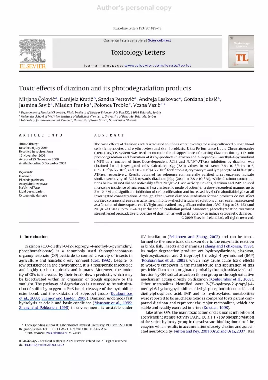
Diazinon (O,O-diethyl-O-(2-isopropyl-4-methyl-6-pyrimidinylphosphorothionate) is a commonly used thionophosphorousorganophosphate (OP) pesticide to control a variety of insects inagriculture and household environment (Cox, 1992). Despite itslow persistence in the environment, it is a nonspecific insecticideand highly toxic to animals and humans. Moreover, the toxic-ity of OPs is increased by their break-down products, which maybe bioactivated within an organism or trough exposure to thesunlight. The pathway of degradation is assumed to be substitu-tion of sulfur by oxygen in P S bond, cleavage of the pyrimidineester bond, and the oxidation of isopropyl group (Kouloumboset al., 2003; Shemer and Linden, 2006). Diazinon undergoes fasthydrolysis at acidic and basic conditions (Mansour et al., 1999;Zhang and Pehkonen, 1999) in environment, is unstable under
∗ Corresponding author at: Laboratory of Physical Chemistry, P.O. Box 522, 11001Belgrade, Serbia. Tel.: +381 11 2453 967; fax: +381 11 2447 207.
E-mail address: [email protected] (V. Vasic).
UV irradiation (Pehkonen and Zhang, 2002) and can be trans-formed to the more toxic diazoxon due to the enzymatic reactionin birds, fish, insects and mammals (Zhang and Pehkonen, 1999).Its major degradation products are hydroxydiazinon, diazoxon,hydroxydiazoxon and 2-isopropyl-6-methyl-4-pyrimidinol (IMP)(Kouloumbos et al., 2003), which may cause acute toxic effectsto workers employed in the manufacture and application of thispesticide. Diazoxon is originated probably through oxidative desul-furation by OH radical attack on thiono group or through oxidativemechanism acting directly on diazinon (Kouloumbos et al., 2003).Other metabolites identified were 2-(2′-hydroxy-2′-propyl)-4-methyl-6-hydroxypyrimidine, diethyl-phosphorothioic acid anddiethylphosphoric acid. IMP and its hydroxylated metabolitieswere reported to be much less toxic as compared to its parent com-pound diazinon and represent the major metabolites, which arestable and readily excreted in urine (Ku et al., 1998).
Like other OPs, the main toxic action of diazinon is inhibition ofacetylcholinesterase activity (AChE, EC 3.1.1.7) by phosphorylationof the serine hydroxyl group in the substrate-binding domain of theenzyme which results in accumulation of acetylcholine and associ-ated neurotoxicity (Fulton and Key, 2001; Oruc and Usta, 2007). It is
0378-4274/$ – see front matter © 2009 Elsevier Ireland Ltd. All rights reserved.doi:10.1016/j.toxlet.2009.11.022
Author's personal copy
10 M. Colovic et al. / Toxicology Letters 193 (2010) 9–18
Fig. 1. The chemical structures of diazinon and the products of its photochemically induced transformation.
established also that some OPs inhibit different ATPases, the groupof enzymes playing an important role in biochemical processes(Vasic et al., 2008) in various tissues (Rahman et al., 1997). Pes-ticides exert biological effect on the ATPase system by partitioningin the enzyme complex (Kinter et al., 1972) and it is also reportedthat an allosteric change results in a decrease of ATPase activity(Reddy et al., 1992). The proper functioning of these enzymes iscritical for cellular viability because they control many essentialcellular functions, and any inhibition of the activity may be con-sidered as a sensitive indicator of toxicity. Both Na+/K+-ATPase (EC3.6.1.37) and AChE are membrane bound enzymes and their activ-ities depend on the phospholipids environment of the membrane.Therefore, any change in the lipid component of the membrane willdirectly affect the activities of these enzymes (Sahoo et al., 1999).Besides, the pesticides may also induce oxidative stress and geneticmaterial damage leading to cell malformations, generation of freeradicals and cause lipid peroxidation (Banerjee et al., 1999; Goel etal., 2005; Kehrer, 1993).
Moreover, there is a concern that the risk of genotoxicitymight be appreciably greater than that predicted from standardtoxicity tests (Attia, 2007). Therefore, cytogenetic damage (chro-mosomal aberrations—CA, micronuclei—MN and sister-chromatidexchanges—SCE) in circulating lymphocytes has been widely usedas a biomarker of exposure and effects of pesticides. Measuringmicronuclei frequency is an approach used extensively in molecu-lar epidemiology and cytogenetics to evaluate the presence and theextent of the chromosomal damage in human populations exposedto genotoxic agents or bearing a susceptible genetic profile. DNAdamage induced by xenobiotics appears primarily in the form ofalterations to the phosphate backbone, sugar or base modificationssuch as alkylations, crosslinks, or formation of DNA adducts thatare substrates for DNA repair (Bajpayee et al., 2006) when missrepaired they are seen as increased incidence of CA, MN or SCE.Phosphorus moiety in the pesticides appears to be a good substratefor nucleophilic attack leading to phosphorylation of DNA which isan instance of DNA damage (Das et al., 2007). Micronuclei may be
induced by strand breaks in DNA due to oxidative stress (Fenech,1993).
Although diazinon is similar in basic chemical structure andmetabolite profile to other organophosphates, there is still signifi-cant gap both in data on diazinon and its metabolites toxicity andin genotoxicity studies on humans. In the present study we inves-tigated in vitro the toxicity potential and oxidative stress responsesof diazinon and its photo degradation products (diazoxon and IMP,formed in nature or during water purification processes using UVlight) by determining the activity of AChE and ATPases in har-vested human blood cells (erythrocytes, lymphocytes), human skinfibroblasts and purified enzymes, lipid peroxodation product mal-ondaldehyde (MDA), incidence of micronuclei and cell proliferationpotential (CBPI), comparatively.
2. Materials and methods
2.1. Chemicals
All chemicals were of analytical grade. Acethylcholinesterase(AChE, E.C. 3.1.1.7, specific activity 500 IU/mg protein) from humanerythrocytes, Na+/K+ ATPase from porcine cerebral cortex (specificactivity 25.8 �mol Pi/h/mg protein), acethylthiocholine iodide(AChI), 5,5′-dithio-bis-2-nitrobenzoic acid (DTNB), adenosinet-riphosphate (ATP), propidium iodide and Cytochalasin B werepurchased from Sigma Chemicals Co. (Germany). PB-max kary-otyping medium was purchased from Invitrogen-Gibco (UK).Lymphoprep (Lymphocyte separation medium) was from PAALaboratories (Austria). Proteinase K and RNAse were from Fermen-tas (Canada). FITC labeled �-satellite DNA probe was purchasedfrom ICN Biomedicals (USA). Other medium assay chemicals(magnesium chloride, Tris–HCl sodium chloride, potassium chlo-ride, stannous chloride, calcium chloride, potassiumhydrogenphosphate (K2HPO4 × 3H2O), sodium citrate, ammonium molyb-date and Giemsa were from Merck (Germany). Formamide was

Author's personal copy
M. Colovic et al. / Toxicology Letters 193 (2010) 9–18 11
purchased from AppliChem (Germany). Diazinon (O,O-diethyl-O-(2-isopropyl-6-methyl-pyrimidine-4-yl)phosphorothioate)(97.3%), O-analog diazinon (diazoxon) solution (100 ppm in ace-tonitrile) and 2-isopropyl-6-methyl-4-pyrimidinol (IMP) (93%)were from Pestanal (Germany). The chemical structures of studiedcompounds are collected in Fig. 1.
2.2. Photodegradation experiments
10 mL of absolute ethanol was added to 30.4 mg of diazinonand prepared 1 × 10⁻² M solution was then diluted 10 times bydeionised water. Photodegradation of 1 × 10⁻³ M (10% ethanol)diazinon solution (10 mL) was performed using a 125 W Cer-max Xenon parabolic lamp emitting light with wavelengths above200 nm, irradiating the quartz sample cell with the optical pathlength of 1.0 cm placed 65 cm from the light source during six dif-ferent periods of time: 0, 5, 15, 30, 60 and 115 min. These irradiatedsolutions were diluted 50 times in blood and fibroblast culturesas well as in incubation medium for determination of AChE andNa+/K+-ATPase activities.
2.3. UPLC analysis
Quantification of diazinon and its decomposition products (IMPand diazoxon) in aliquots of the irradiated solutions was performedon Waters ACQUITY Ultra Performance Liquid Chromatography(UPLC)–UV/VIS detection system, using an ACQUITY UPLCTM BEHC18, 1.7 �m, 50 mm × 2.1 mm column. The mobile phase was com-posed of 0.1% formic acid (phase A) and acetonitrile (phase B) inthe ratio 25:75 (for diazinon and diazoxon analysis), and 10:90 (forIMP analysis). Flow rate was 0.3 mL/min, injection volume 10 �Land the column temperature 25 ◦C. The separation of diazinon anddiazoxon was monitored in a single wavelength mode at 245 nm,while IMP determination was carried out at 230 nm. The reten-tion times for diazinon, diazoxon and IMP were 1.9 min, 1.2 minand 1.8 min, respectively. Limits of detection (LOD) for diazinonand diazoxon were 3 × 10⁻⁷ and 1 × 10⁻⁶ M for IMP. For quantifi-cation purposes calibration curves in the concentration range from1 × 10−6 to 1 × 10⁻³ M for diazinon and IMP and from 3 × 10⁻⁷ to1 × 10−4 M for diazoxon were prepared.
2.4. Blood cultures
Blood sample was obtained from healthy 30 years old male,non-smoking volunteer in the Medical Unit in accordance withthe current Health and Ethical regulations in Serbia (Law onHealth Care, 2005). Aliquots of heparinized whole blood (0.5 mL)were placed in cultures containing PB-max karyotyping medium.Two parallel sets of blood cultures were set up in the presenceof increasing doses of diazinon (2 × 10⁻⁸ to 2 × 10⁻⁴ M) and itsphotodegradation products yielded after irradiation of 2 × 10⁻⁵ Msolution. Micronuclei formation and cell proliferation potentialwere scored in the first sets of samples and the correspondingcontrols, whereas the second set was used for measurement ofAChE and Na+/K+-ATPase activities and MDA level. For that purpose,after 72 h of incubation harvested cells were separated on Lym-phoprep, lymphocytes were collected by centrifugation, washedin physiological saline, and frozen at −70 ◦C for later analysis.The erythrocytes were collected from the bottom and RBC mem-branes were prepared according to the method of (Post et al.,1960).
Medium of each culture in which cells were grown during incu-bation (incubation medium) was also kept for measurement ofMDA level.
2.5. Fibroblasts
Human skin fibroblasts were set up as parallel tissue. Whenbeing confluent, cells were expanded in 15× 2 flasks, and 1 h aftertripsinisation, cells were treated with diazinon and its by-productsas used for blood cells. The fibroblast cell density was 1 × 10⁵
cells in 75 cm2 flasks. Micronuclei formation and cell proliferationpotential were then scored in the first set of samples and the corre-sponding controls. In the second set of samples cells were collectedby centrifugation, washed in physiological saline, and frozen at−70 ◦C for later analysis of AChE and Na+/K+-ATPase activities aswell as MDA level.
2.6. AChE assay
The inhibition of AChE was measured in erythrocytes, lympho-cytes and fibroblasts treated with desired diazinon concentrationsand its irradiated solutions, while commercially available humanerythrocytes enzyme, in vitro exposed to the same solutions wasused as a reference. AChE activity was determined using Ellman’sprocedure (Ellman et al., 1961) and expressed as �A/(min × mgprotein). The in vitro experiments were performed by 20 min expo-sure of 0.02 IU commercial enzyme to investigated compounds infinal volume 0.650 mL. AChI was applied as the enzyme substratein combination with DTNB as a chromogenic reagent. The product5-thio-2-nitrobenzoate, formed in the reaction of thiocholineio-dide (product of enzymatic reaction) and DTNB was measuredspectrophotometrically (Perkin Elmer Lambda 35 UV-VIS spec-trophotometer) at 412 nm (in buffer solution). All experimentswere made in triplicates. Preliminary studies showed that diazi-non and its by-products did not interfere with quantization of theyellow product 5-thio-2-nitrobenzoate. Total protein was quanti-fied by the procedure of Lowry’s method (Lowry et al., 1951) usingFolin’s reagent, and bovine serum albumin as standard.
2.7. ATPase assay
The specific activity of Na+/K+-ATPase in erythrocytes, lympho-cytes and fibroblasts treated with desired diazinon concentrationsand its irradiated solutions was determined in a standard incu-bation medium (200 �L), containing 50 mM Tris–HCl (pH 7.4),100 mM NaCl, 20 mM KCl, 5 mM MgCl2, 2 mM ATP. The in vitroexperiments were performed by exposure of 0.0125 IU commer-cial porcine cerebral cortex Na+/K+ ATPase (reference model) toinvestigated compounds in the same medium assay. Incubationmixtures were preincubated at 37 ◦C and the reaction was startedby the addition of ATP, and allowed to proceed until stopped byice cold HClO4. The activity obtained in the presence of 2 mMouabain (without the NaCl and KCl) was attributed to Mg2+-ATPase(ecto-ATPase). Na+/K+-ATPase activity was calculated as a differ-ence between the total ATPase and Mg2+-ATPase activity. Theconcentration of adenosinediphosphate (ADP) liberated due to thehydrolysis of ATP was measured by UPLC method (Sudo et al.,2000). The mobile phase was composed of 4 mM tetrabutylammo-nium hydroxide (TBAH) in 4 mM phosphate buffer (phase A) andmethanol (phase B) in the ratio 75:25. Flow rate was 0.25 mL/min,injection volume 10 �L and the column temperature 40 ◦C. The sep-aration was monitored in a single wavelength mode at 254 nm. Inaddition, the concentration of liberated orthophosphate (Pi) wasmeasured using modified spectrophotometric procedure based onthe stannous chloride method (Jorgensen et al., 2003; Vasic et al.,1999), by reading the absorbance at 690 nm and was comparedto the concentration of ADP. Excellent agreement was obtained,confirming that only orthophosphates liberated from enzyme cat-alyzed ATP hydrolysis were detected.
Author's personal copy
12 M. Colovic et al. / Toxicology Letters 193 (2010) 9–18
2.8. Thiobarbituric acid (TBA) assay
Defrosted lymphocyte and fibroblast suspensions, their incuba-tion media and RBC membranes were used for measurement ofmalondyaldehyde, spectrophotometrically (Aruoma et al., 1989) atthe 532 nm wavelength. The values are expressed as nmol of thio-barbituric acid-reactive substance (MDA equivalent)/mg protein,using a standard curve of 1,1,3,3-tetramethoxypropane.
2.9. Micronuclei analysis
For micronuclei preparation, the cytokinesis block method ofFenech et al. was followed (Fenech, 1993) Cytochalasin B at a finalconcentration of 4 �g/mL was added to each sample for micronu-cleus assay. Cell suspension was prefixed in methanol/acetic acid3:1, washed 3 times with fixative and dropped onto clean slide.Slides were air dried and stained in alkaline Giemsa (2%). At least1000 binucleated (BN) cells per sample were scored. All slides wereanalyzed with an Axiophot-2 microscope using magnification 400×or 1000× when necessary. A minimum of 1000 BN cells were scoredto evaluate the percentage of cells with micronuclei.
2.10. In situ hybridization
Parallel unstained micronuclei slides were analyzed using Flu-orescent in situ hybridization with FITC labeled �-satellite DNAprobe (ICN Biomedicals, USA), hybridizing exclusively the cen-tromeres of all human chromosomes. Subsequently slides werepretreated with RNAse (100 �g/mL in 2xSSC (saline-sodium cit-rate buffer), for an hour, and proteinase K (0.5 �g/mL in 20 mMTRIS, 2 mM CaCl2. pH 7.5 for 15 min at 37 ◦C. The slides were dehy-drated in an ethanol series (70%, 90%, 100%), for 5 min and weredenatured in 70% formamide/2xSSC, pH 7.0 at 70 ◦C for 2 min. Afterdenaturation the slides were dehydrated in ethanol series. TargetDNA was denatured at 70 ◦C for 5 min, placed on ice for 10 minand than applied to slides. The slides were incubated for 16 h inmoist chamber at 37 ◦C. After posthybridization washes (5 min in50% formamide, 2xSSC) and two changes in 2xSSC for 2 min each,slides were dehydrated in cold ethanol series and were stainedwith propidium iodide (PI) antifade solution. Centromere micronu-clei were analyzed on Axiomager A1 fluorescent microscope usingFITC and PI filters and magnification 1450×. According to pres-ence of centromere’s signal in micronuclei, they were classifiedas centromere positive (MNC+) and centromere negative (MNC−)(Bolognesi, 2003).
2.11. Cell proliferation index
A cytokinesis-block proliferation index (CBPI) was calcu-lated according to method of Surrales et al. (1995) as follows:CBPI = MI + 2MII + 3(MIII + MIV)/N, where MI-MIV represent thenumber of cells with one to four nuclei, respectively, and N is thenumber of cells scored.
Table 1Concentration of products formed due to the exposure 1 × 10⁻³ M diazinon to UVirradiation as the function of irradiation time.
Irradiation time (min) Diazinon (M) Diazoxon (M) IMP (M)
0 1.0 × 10⁻³ – –5 9.0 × 10⁻⁴ 2.0 × 10⁻⁶ 3.2 × 10⁻⁵
15 7.2 × 10⁻⁴ 4.0 × 10⁻⁷ 6.4 × 10⁻⁵
30 4.1 × 10⁻⁴ – 1.1 × 10⁻⁴
60 2 × 10⁻⁴ – 2.7 × 10⁻⁴
115 – – 4.5 × 10⁻⁴
Note: The irradiated solutions were diluted 50 times in blood and fibroblast cul-tures as well as in incubation medium for determination of AChE and Na+/K+-ATPaseactivities.
2.12. Statistics
A statistical analysis of each of the parameters of interestwas carried out using statistical software package Statistics, ver-sion 6 for Microsoft Windows. Analysis considered the incidenceof micronuclei in binucleated cells, the level of produced mal-ondyaldehyde and cytokinesis-block proliferation index of treatedcells. Among parameters under consideration, Product-Momentand partial correlations and Student’s t-test were used. The Pvalue < 0.05 was considered significant.
3. Results
3.1. Photochemical degradation of diazinon
The absorption spectrum of diazinon, diazoxon and IMP dis-play strong absorption bands in the range from 200 to 280 nm. Thisfact implies that they have the potential to be photolyzed by anywavelengths below 280 nm. The concentrations of products (dia-zoxon and IMP) formed due to the exposure 1 × 10⁻³ M diazinonto UV irradiation as the function of irradiation time are presentedin Table 1. The results, obtained by UPLC analysis, indicate thecomplete degradation of the parent compound within 115 min ofirradiation. IMP was observed already after 5 min (3.2 × 10⁻⁵ M)and reached maximal concentration at the end of experiment when45% of irradiated diazinon was converted to IMP. Diazoxon wasonly detected at 5 min and 15 min, while after 15 min of irradiationits concentration was under LOD. Maximal diazoxon concentrationwas measured at 5 min of the experiment and corresponded to 0.2%of the initial parent compound concentration. It is obvious (Table 1)that the sum of concentrations of diazinon, diazoxon and IMP ineach moment of photodegradation is less than the initial concen-tration of diazinon suggesting the presence of other compounds(not detected) in the irradiated mixtures.
3.2. Effect of diazinon on AChE and Na+/K+-ATPase activity
The effect of diazinon on AChE and Na+/K+-ATPase was inves-tigated by determination of specific enzyme activities in humanlimfocytes, erythrocytes and fibroblasts which were cultivated dur-
Table 2Effect of diazinon on Na+/K+-ATPase and AChE activity in different human cells after 3 days exposure and in purified commercial preparations. All results present mean valueof at least two experiments done in triplicate.
Tissue Na+/K+-ATPase AChE
Specific activity (control), �mol Pi/h/mg protein IC50 (72 h), M Specific activity (control), �A412/min/mg protein IC50 (72 h), M
Fibroblasts 1.110 3.4 × 10⁻⁵ 0.109 7.5 × 10⁻⁶
Erythrocytes 0.189 6.6 × 10⁻⁵ 0.518 8.7 × 10⁻⁵
Lymphocytes 0.081 4.6 × 10⁻⁵ 0.929 3.0 × 10⁻⁵
Commercial purified enzyme* 25.8 >10−4** 2418 7.8 × 10⁻⁵**
* Na+/K+-ATPase from porcine cerebral cortex; AChE from human erythrocytes.** IC50 (20 min).

Author's personal copy
M. Colovic et al. / Toxicology Letters 193 (2010) 9–18 13
Fig. 2. (a) Inhibition of AChE activity in cells (solid symbols): fibroblasts (square),lymphocytes (asterisk) and erythrocytes (circle) induced by diazinon. Inset: Inhibi-tion of commercial human erythrocyte AChE (open symbols) induced by diazinon(square), diazoxon (triangle) and IMP (circle). (b) Inhibition of Na+/K+-ATPaseactivity in cells (solid symbols): fibroblasts (square), lymphocytes (triangle) anderythrocytes (circle) induced by diazinon. Inset: Inhibition of commercial porcinecerebral cortex Na+/K+-ATPase (open symbols) induced by diazinon (square), dia-zoxon (triangle) and IMP (circle).
ing 72 h in the absence (control) and presence of the inhibitor(concentration range from 1 × 10⁻⁹ to 2 × 10⁻⁴ M). Fig. 2(a and b)illustrates the dose-dependent diazinon induced enzyme activi-ties inhibition in all investigated cells. The dependence of AChEand Na+/K+-ATPase activity, expressed as a percentage of the con-trol value, on diazinon concentration fits a sigmoidal function inall cases. The concentrations of diazinon with capability to inhibit50% of the enzyme after given exposure time (IC50 values) weredetermined by sigmoidal fitting the experimental results and sum-marized in Table 2. It is obvious that fibroblasts AChE showsthe highest sensitivity (IC50 (72 h)—7.5 × 10⁻⁶ M), while the ery-throcytes AChE is one order of magnitude less sensitive towardthe investigated organophosphate (IC50 (72 h)—8.7 × 10⁻⁵ M), thatcould be attributed to lower capability of fibroblasts to detoxifydiazinon than blood. On the other hand, Na+/K+-ATPase shows simi-lar sensitivity in all examined cell types. Obtained IC50 (72 h) values,in M, are: 4.56 × 10⁻⁵, 6.60 × 10⁻⁵ and 3.41 × 10⁻⁵ for human lim-focytes, erythrocytes and fibroblasts, respectively.
The in vitro influence of diazinon on AChE and Na+/K+-ATPaseactivities was followed exposing the reference model systems(commercial purified: AChE from human erythrocytes and cere-bral cortex Na+/K+-ATPase) to the investigated compound in thesame concentration range. In addition, the inhibitory potencies ofdiazoxon and IMP (detected diazinon photoinduced by-products)were tested on commercial enzymes. The obtained concentration-dependent inhibition of AChE (Fig. 2a, inset), indicates the similarresponse in the presence of diazinon as in the examined celltypes. Moreover, similar IC50 values for commercial and humanerythrocytes AChE were obtained, while IC50 value for the targetenzyme from fibroblasts was ten times lower (Table 2). Obtainedresults show that diazoxon is about 1000 times more potentinhibitor than its parent compound (IC50 (20 min)—6.8 × 10⁻⁸ M),while the inhibitory effect of IMP was negligible at concentrationsbelow 2 mM (Fig. 2a, inset). Unlike AChE and Na+/K+-ATPase inthe investigated cells, activity of commercially purified referenceNa+/K+-ATPase was not affected by diazinon (as well as diazoxonand IMP) concentrations lower than 1 × 10⁻⁴ M (Fig. 2b, inset,Table 2).
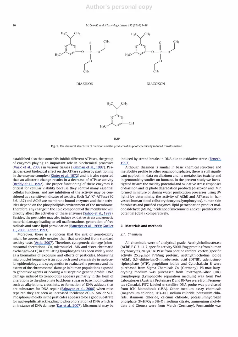
3.3. Influence of diazinon irradiated solutions on AChE andNa+/K+-ATPase activity
The influence of diazinon photochemical treatment on its AChEand Na+/K+-ATPase inhibitory potency was investigated by deter-mination of specific enzyme activities in the same model cells(erythrocytes, lymphocytes and fibroblasts). The cells were culti-vated during 72 h in the presence of previously photochemicallytreated (during several irradiation periods) diazinon solutionsunder the conditions described in Section 2. The initial irradiatedsolutions were diluted 50 times in cell incubation medium. In addi-tion, in vitro effects of these diluted irradiated solutions on thecommercial purified reference enzymes were followed. Fig. 3a illus-trates the effect of irradiation time of diazinon on AChE inhibition(expressed as a percentage of the control value) in all model sys-tems. As can be seen, inhibitory efficiency of diazinon irradiatedsolutions increased as a function of the exposure time to the UVlight in all investigated cells. Moreover, first 5 min of irradiationinduced a rapid decrease of enzyme activity (about 25%) com-pared to inhibition caused by non-irradiated 2 × 10⁻⁵ M diazinon,while a gradual decrease in AChE activity was obtained duringnext 110 min of photochemical treatment. Obviously, obtainedAChE fibroblasts inhibition in the presence of irradiated sampleswas higher than inhibition of blood cells AChE, as in the caseof non-irradiated diazinon. In addition, in the case of commer-cial purified enzyme the maximum inhibition was achieved after5 min irradiation. However, in samples with prolonged irradia-tion time (Fig. 3a, inset) the increasing commercial AChE activitywas noticed. Moreover, after 115 min the irradiated solution didnot noticeably affect enzymatic activity (96% of control activ-ity).
Fig. 3b illustrates that inhibitory efficiency of diazinon irradi-ated solutions increases as a function of photodegradation timein all investigated cells. Gradual decrease in Na+/K+-ATPase activ-ity was obtained during 115 min of photochemical treatment. Thepresence of 115-min irradiated solution resulted in diminishingNa+/K+-ATPase activity 35–40% in respect to the initial activity (inthe presence of unirradiated 2 × 10⁻⁵ M diazinon) (Fig. 3b). On theother hand, diazinon photochemical treatment did not remark-ably affect its influence on reference Na+/K+-ATPase (Fig. 3b, inset).Obtained Na+/K+-ATPase activity (% of control) ranged from 93 to105 in the presence of irradiated solutions containing diazinonand/or its by-products and corresponded to the Na+/K+-ATPaseresponse on unirradiated 2 × 10⁻⁵ M diazinon (Fig. 2b, inset).

Author's personal copy
14 M. Colovic et al. / Toxicology Letters 193 (2010) 9–18
Table 3Incidence of micronuclei (MN) and proliferation index in human lymphocytes and fibroblasts treated with diazinon and IMP.
Concentration (M) Control 2 × 10⁻⁸ 2 × 10⁻⁷ 2 × 10⁻⁶ 2 × 10⁻⁵ 2 × 10⁻⁴
DiazinonLymphocytesMN/1000 BN cells 4.61 10.18 14.07 15.04 12.9 –CBPI 1.53 1.42 1.35 1.27 1.26 –
FibroblastsMN/1000 cells 3.50 9.20 11.10 12.30 9.10 –CBPI 1.32 1.31 1.26 1.26 1.31 –
IMPLymphocytesMN/1000 cells 4.61 10.21 11.75 20.95 20.21 18.63CBPI 1.53 1.40 1.31 1.27 1.20 1.19
FibroblastsMN/1000 cells 3.50 8.30 9.20 16.10 15.30 15.10CBPI 1.32 1.29 1.28 1.18 1.17 1.12
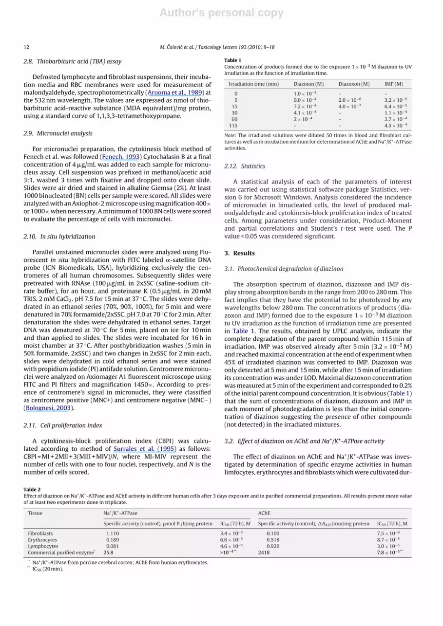
3.4. Effects of diazinon and its irradiated solutions on MDA level
The investigated diazinon concentrations up to 2 × 10⁻⁶ M didnot noticeably (p > 0.05) alter MDA level (compared to the control)in lymphocytes, as well as in incubation medium, while increasingMDA level was observed in the presence of diazinon concentrationsabove 2 × 10⁻⁶ M (Fig. 4a). The approximately 1.5- and 2.8-foldenhancement of MDA level (in respect to the control) was observedin lymphocytes and incubation medium, respectively, treated with2 × 10−4 M diazinon. In addition, the effect of IMP, the diazinonhydrolysis product formed during its photodegradation, on lipidperoxidation was tested in order to compare it to the parent com-pound (diazinon) effect. IMP also induced the increase in MDAlevel, both in lymphocytes and incubation medium, in a dose-dependent manner (Fig. 4a, inset). The MDA level in lymphocytesinduced by IMP at concentrations above 2 × 10⁻⁶ M was approxi-mately 50–80% higher (statistically significant, t = 11.32, p = 0.008)than that induced by the same concentrations of diazinon. Simi-lar results were obtained analyzing the diazinon and IMP effects inincubation medium.
Results obtained for lymphocytes and incubation mediumexposed to 2 × 10⁻⁵ M irradiated diazinon solutions are presentedin Fig. 4b. The statistically significant (t = −3.22, p = 0.018) gradualenhancement of MDA level was observed with the increasing irra-diation time and achieved the maximum value at the end of theirradiation process. The level of MDA at the end of photochemicaldegradation was approximately 2-fold higher in respect to the con-trol and level obtained in the presence of unirradiated 2 × 10⁻⁵ Mdiazinon solution.
Similar results were obtained for diazinon and its by-productseffects on lipid peroxidation in erythrocytes and fibroblasts (datanot shown).
3.5. Cytotoxic effects of diazinon and its irradiated solutions
The effects of diazinon and its by-product IMP on the incidenceof micronuclei and proliferation index in cell cultures (lympho-cytes and fibroblasts) are illustrated in Table 3. In lymphocytecultures, significant increase (p < 0.05) of the incidence of micronu-clei in a dose-dependent manner was observed in all samplestreated with diazinon and IMP at concentration range from 2 × 10⁻⁸
to 2 × 10⁻⁶ M, compared to the control. Even low concentrationlevels, below 2 × 10⁻⁶ M of both compounds (Table 3) displayclastogenic properties seen as induction of micronuclei and low-ering cell proliferation potential. IMP behaved as a more powerfulinducer of MN than diazinon. The incidence of micronuclei reached
the maximum at concentration 2 × 10⁻⁶ M, afterwards decreasedand further increase of diazinon as well as IMP concentrationconsequently inhibited cell proliferation potential, lowering all bio-chemical processes in cells. Concentration dependent inhibition ofCBPI in lymphocytes treated with diazinon and IMP was statisticallysignificant (t = 4.08, p = 0.009 and t = 4.61, p = 0.004, respectively).The incidence of micronuclei and CBPI in cultures treated withdiazinon as well as with IMP correlated inversely, statistically sig-nificant (r = −0.92, p = 0.027 and r = −0.93, p = 0.008, respectively).
The similar trend was observed in fibroblasts treated withincreasing concentrations of diazinon and IMP. The incidence ofmicronuclei significantly increased in a dose-dependent mannerin fibroblasts treated with both, diazinon and IMP, reaching themaximum at concentration of 2 × 10⁻⁶ M (t = −7.37, p = 0.02 andt = −3.12, p = 0.035, respectively).
The increasing concentration of IMP significantly lowered(t = 2.49, p = 0.047) proliferation potential of fibroblasts. On the con-trary, treatment with diazinon did not induce significant changesin CBPI at the investigated concentration range (p > 0.05). Prolif-eration potential of cells was slightly reduced at concentration2 × 10−6 M (compared to control) and remained almost unchangedwith increasing the dose.
In lymphocyte cultures treated with IMP positive correlationbetween micronuclei and the level of MDA was observed (r = 0.91,p = 0.03), whereas the level of MDA and CBPI correlated inversely(r = −0.93, p = 0.02).
The data of lymphocyte and fibroblast micronuclei monitoringas well as CBPI in samples treated with irradiated diazinon (initialconcentration 2 × 10⁻⁵ M), are presented in Table 4. Micronucleiincidence significantly increased with irradiation time in fibrob-lasts (t = −2.49, p = 0.047) and at the border of significance inlymphocytes. In both tissues, maximal incidences of micronu-
Table 4Incidence of micronuclei (MN) and proliferation index in human lymphocytes andfibroblasts treated with irradiated diazinon (initial concentration 2 × 10⁻⁵ M).
Irradiation time (min) 0 5 15 30 60 115
LymphocytesMN/1000 cells 12.90 15.20 21.44 24.22 43.74 49.45CBPI 1.26 1.20 1.17 1.15 1.11 1.10MNC+ 7 6 5 6 5 5MNC− 6 8 17 18 39 45FibroblastsMN/1000 cells 9.10 13.00 16.00 17.00 29.00 31.00CBPI 1.31 1.24 1.22 1.28 1.24 1.20MNC+ 7 6 5 6 5 5MNC− 2 7 11 11 24 26
Author's personal copy
M. Colovic et al. / Toxicology Letters 193 (2010) 9–18 15
Fig. 3. (a) Inhibition of AChE activity in cells (solid symbols): fibroblasts (square),lymphocytes (triangle) and erythrocytes (circle) induced by 2 × 10⁻⁵ M diazinonirradiated solutions. Inset: Inhibition of commercial human erythrocyte AChEinduced by 2 × 10⁻⁵ M diazinon irradiated solutions. Note: Irradiated solutions of1 × 10−3 M diazinon were diluted 50 times in blood and fibroblast cultures aswell as in incubation medium for determination of AChE activity. (b) Inhibitionof Na+/K+-ATPase activity in cells (solid symbols): fibroblasts (square), lympho-cytes (triangle) and erythrocytes (circle) induced by 2 × 10⁻⁵ M diazinon irradiatedsolutions. Inset: Inhibition of commercial porcine cerebral cortex Na+/K+-ATPaseinduced by 2 × 10⁻⁵ M diazinon irradiated solutions. Note: Irradiated solutions of1 × 10−3 M diazinon were diluted 50 times in blood and fibroblast cultures as wellas in incubation medium for determination of Na+/K+-ATPase activity.
clei were observed at 115 min of irradiation. The proliferationpotential of cells significantly decreased with prolonged irradia-tion time in both tissues (t = 4.35, p = 0.005 and t = 3.78, p = 0.009,for lymphocytes and fibroblasts, respectively). In lymphocyte cul-tures, statistically significant inverse correlation between incidenceof micronuclei and proliferation index was observed (r = −0.91,p = 0.012). The level of MDA and incidence of micronuclei correlatedpositively (r = 0.94, p = 0.005).
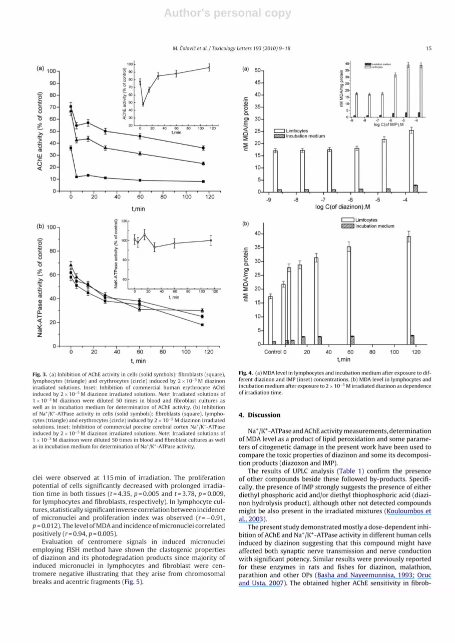
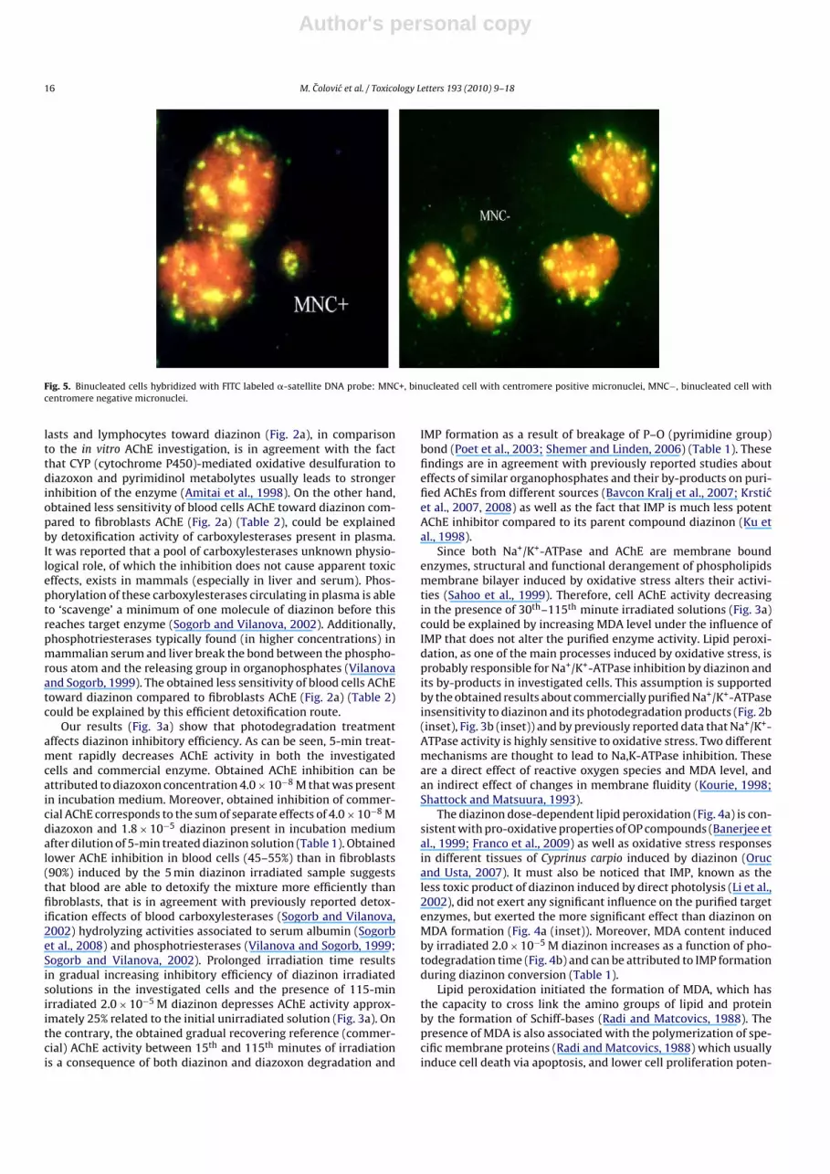
Evaluation of centromere signals in induced micronucleiemploying FISH method have shown the clastogenic propertiesof diazinon and its photodegradation products since majority ofinduced micronuclei in lymphocytes and fibroblast were cen-tromere negative illustrating that they arise from chromosomalbreaks and acentric fragments (Fig. 5).
Fig. 4. (a) MDA level in lymphocytes and incubation medium after exposure to dif-ferent diazinon and IMP (inset) concentrations. (b) MDA level in lymphocytes andincubation medium after exposure to 2 × 10⁻⁵ M irradiated diazinon as dependenceof irradiation time.
4. Discussion
Na+/K+-ATPase and AChE activity measurements, determinationof MDA level as a product of lipid peroxidation and some parame-ters of citogenetic damage in the present work have been used tocompare the toxic properties of diazinon and some its decomposi-tion products (diazoxon and IMP).
The results of UPLC analysis (Table 1) confirm the presenceof other compounds beside these followed by-products. Specifi-cally, the presence of IMP strongly suggests the presence of eitherdiethyl phosphoric acid and/or diethyl thiophosphoric acid (diazi-non hydrolysis product), although other not detected compoundsmight be also present in the irradiated mixtures (Kouloumbos etal., 2003).
The present study demonstrated mostly a dose-dependent inhi-bition of AChE and Na+/K+-ATPase activity in different human cellsinduced by diazinon suggesting that this compound might haveaffected both synaptic nerve transmission and nerve conductionwith significant potency. Similar results were previously reportedfor these enzymes in rats and fishes for diazinon, malathion,parathion and other OPs (Basha and Nayeemunnisa, 1993; Orucand Usta, 2007). The obtained higher AChE sensitivity in fibrob-
Author's personal copy
16 M. Colovic et al. / Toxicology Letters 193 (2010) 9–18
Fig. 5. Binucleated cells hybridized with FITC labeled �-satellite DNA probe: MNC+, binucleated cell with centromere positive micronuclei, MNC−, binucleated cell withcentromere negative micronuclei.
lasts and lymphocytes toward diazinon (Fig. 2a), in comparisonto the in vitro AChE investigation, is in agreement with the factthat CYP (cytochrome P450)-mediated oxidative desulfuration todiazoxon and pyrimidinol metabolytes usually leads to strongerinhibition of the enzyme (Amitai et al., 1998). On the other hand,obtained less sensitivity of blood cells AChE toward diazinon com-pared to fibroblasts AChE (Fig. 2a) (Table 2), could be explainedby detoxification activity of carboxylesterases present in plasma.It was reported that a pool of carboxylesterases unknown physio-logical role, of which the inhibition does not cause apparent toxiceffects, exists in mammals (especially in liver and serum). Phos-phorylation of these carboxylesterases circulating in plasma is ableto ‘scavenge’ a minimum of one molecule of diazinon before thisreaches target enzyme (Sogorb and Vilanova, 2002). Additionally,phosphotriesterases typically found (in higher concentrations) inmammalian serum and liver break the bond between the phospho-rous atom and the releasing group in organophosphates (Vilanovaand Sogorb, 1999). The obtained less sensitivity of blood cells AChEtoward diazinon compared to fibroblasts AChE (Fig. 2a) (Table 2)could be explained by this efficient detoxification route.
Our results (Fig. 3a) show that photodegradation treatmentaffects diazinon inhibitory efficiency. As can be seen, 5-min treat-ment rapidly decreases AChE activity in both the investigatedcells and commercial enzyme. Obtained AChE inhibition can beattributed to diazoxon concentration 4.0 × 10⁻⁸ M that was presentin incubation medium. Moreover, obtained inhibition of commer-cial AChE corresponds to the sum of separate effects of 4.0 × 10⁻⁸ Mdiazoxon and 1.8 × 10⁻⁵ diazinon present in incubation mediumafter dilution of 5-min treated diazinon solution (Table 1). Obtainedlower AChE inhibition in blood cells (45–55%) than in fibroblasts(90%) induced by the 5 min diazinon irradiated sample suggeststhat blood are able to detoxify the mixture more efficiently thanfibroblasts, that is in agreement with previously reported detox-ification effects of blood carboxylesterases (Sogorb and Vilanova,2002) hydrolyzing activities associated to serum albumin (Sogorbet al., 2008) and phosphotriesterases (Vilanova and Sogorb, 1999;Sogorb and Vilanova, 2002). Prolonged irradiation time resultsin gradual increasing inhibitory efficiency of diazinon irradiatedsolutions in the investigated cells and the presence of 115-minirradiated 2.0 × 10⁻⁵ M diazinon depresses AChE activity approx-imately 25% related to the initial unirradiated solution (Fig. 3a). Onthe contrary, the obtained gradual recovering reference (commer-cial) AChE activity between 15th and 115th minutes of irradiationis a consequence of both diazinon and diazoxon degradation and
IMP formation as a result of breakage of P–O (pyrimidine group)bond (Poet et al., 2003; Shemer and Linden, 2006) (Table 1). Thesefindings are in agreement with previously reported studies abouteffects of similar organophosphates and their by-products on puri-fied AChEs from different sources (Bavcon Kralj et al., 2007; Krsticet al., 2007, 2008) as well as the fact that IMP is much less potentAChE inhibitor compared to its parent compound diazinon (Ku etal., 1998).
Since both Na+/K+-ATPase and AChE are membrane boundenzymes, structural and functional derangement of phospholipidsmembrane bilayer induced by oxidative stress alters their activi-ties (Sahoo et al., 1999). Therefore, cell AChE activity decreasingin the presence of 30th–115th minute irradiated solutions (Fig. 3a)could be explained by increasing MDA level under the influence ofIMP that does not alter the purified enzyme activity. Lipid peroxi-dation, as one of the main processes induced by oxidative stress, isprobably responsible for Na+/K+-ATPase inhibition by diazinon andits by-products in investigated cells. This assumption is supportedby the obtained results about commercially purified Na+/K+-ATPaseinsensitivity to diazinon and its photodegradation products (Fig. 2b(inset), Fig. 3b (inset)) and by previously reported data that Na+/K+-ATPase activity is highly sensitive to oxidative stress. Two differentmechanisms are thought to lead to Na,K-ATPase inhibition. Theseare a direct effect of reactive oxygen species and MDA level, andan indirect effect of changes in membrane fluidity (Kourie, 1998;Shattock and Matsuura, 1993).
The diazinon dose-dependent lipid peroxidation (Fig. 4a) is con-sistent with pro-oxidative properties of OP compounds (Banerjee etal., 1999; Franco et al., 2009) as well as oxidative stress responsesin different tissues of Cyprinus carpio induced by diazinon (Orucand Usta, 2007). It must also be noticed that IMP, known as theless toxic product of diazinon induced by direct photolysis (Li et al.,2002), did not exert any significant influence on the purified targetenzymes, but exerted the more significant effect than diazinon onMDA formation (Fig. 4a (inset)). Moreover, MDA content inducedby irradiated 2.0 × 10⁻⁵ M diazinon increases as a function of pho-todegradation time (Fig. 4b) and can be attributed to IMP formationduring diazinon conversion (Table 1).
Lipid peroxidation initiated the formation of MDA, which hasthe capacity to cross link the amino groups of lipid and proteinby the formation of Schiff-bases (Radi and Matcovics, 1988). Thepresence of MDA is also associated with the polymerization of spe-cific membrane proteins (Radi and Matcovics, 1988) which usuallyinduce cell death via apoptosis, and lower cell proliferation poten-
Author's personal copy
M. Colovic et al. / Toxicology Letters 193 (2010) 9–18 17
tial. The obtained cell Na+/K+-ATPase inhibition, induced probablyvia oxidative membrane damage by diazinon and its decomposi-tion products, may be responsible for the observed dose-dependentdecrease of CBPI. This assumption is in agreement with previ-ously published findings that some modulators of Na+/K+-ATPaseaffect cell proliferation (Gentile et al., 1997; Vasic et al., 2008). Freeradicals influence gene expression, regulate cellular responses tocytokines, as well as proliferative capability of a cell. It is possiblethat enhanced levels of MDA can lead to cell deregulation and resultin apoptosis (Jia and Misra, 2007).
It should be pointed out that dizinon and IMP induce micronu-clei in a dose-dependent manner via clastogenic mode of action,inducing single and double strand breaks on DNA molecule. Theclastogenic mechanism of action of organophosphate pesticideswas also observed in in vivo studies in mice (Cicchetti et al., 1999).These findings are consistent with a number of reports in whichpesticide exposure has been associated with increases in micronu-clei incidence in cultured lymphocytes isolated from peripheralblood taken from exposed individuals (Bull et al., 2006; Bolognesi,2003). The maximum of toxic activity was observed at concentra-tion of 2 × 10⁻⁶ M (Table 3), afterwards the incidence of micronucleidecreases because a very few cells survive further overtoxic con-centration. The decrease of micronuclei incidence at concentrationabove 2 × 10⁻⁶ M is accompanied with significant inhibition of cellproliferation and increased level of MDA.
Photodegradation product of diazinon, IMP, possesses strongergenotoxic potential than diazinon alone. In all samples treated withirradiated solutions of diazinon significant increase of the levelof MDA and micronuclei incidence was observed, particularly forproduct yielded at 115 min of irradiation, where the level of MDAis 80% higher than that observed at the beginning of the irradiation(0 min). The same product induces almost 4-fold enhancement ofmicronuclei incidence. Incidence of micronuclei and level of MDAcorrelated positively, statistically significant (p = 0.005).
In conclusion, despite no observed effect of diazinon (diazoxon)photodegradation products on the activity of the purified targetenzymes, the experiments performed with human cell culturesindicate their significant toxicity which is particularly seen in sig-nificant enhancement of the MDA level, cytogenetic damage andreduction of AChE and Na+/K+-ATPase activity. The human lympho-cyte micronucleus test is considered a well-established system todetect genotoxic activity of different agents, especially after devel-opment of FISH techniques to unequivocally distinguish the originof the micronuclei. Therefore, determination of these parameters inexposed cell cultures could be recommended for toxicity evaluationof pesticides and their by-products.
Conflict of interest statement
The authors declare that there is no conflict of interest.
Acknowledgments
Authors would like to thank to the Ministry of Science and Tech-nological Development of the Republic of Serbia for their financialsupport (Project No. 142051). The work was also supported bythe Ministry of Higher Education, Science and Technology of theRepublic of Slovenia
References
Amitai, G., Moorad, D., Adani, R., Doctor, B.P., 1998. Inhibition of acetylcholinesteraseand butyrylcholinesterase by chlorpyrifos-oxon. Biochemical Pharmacology 56,293–299.
Aruoma, O.I., Halliwell, B., Laughton, M.J., Quinlan, G.J., Gutteridge, J.M.C., 1989. Themechanism of initiation of lipid peroxidation. Evidence against a requirementfor an iron (II)-iron (III) complex. Biochemical Journal 258, 617–620.
Attia, S., 2007. Chromosomal composition of micronuclei in mouse bone marrowtreated with rifampicin and nicotine, analyzed by multicolor fluorescence insitu hybridization with pancentromeric DNA probe. Toxicology 235, 112–118.
Bajpayee, M., Pandey, A.K., Zaidi, S., Musarrat, J., Parmar, D., Mathur, N., Seth, P.K.,Dhawan, A., 2006. DNA damage and mutagenicity induced by endosulfan andits metabolites. Environmental and Molecular Mutagenesis 47, 682–692.
Banerjee, B.D., Seth, V., Bhattacharya, A., Pasha, S.T., Chakraborty, A.K., 1999. Bio-chemical effects of some pesticides on lipid peroxidation and free-radicalscavengers. Toxicology Letters 107, 33–47.
Basha, P.M., Nayeemunnisa, 1993. Effect of methyl parathion on Na+-K+ and Mg2+
adenosine triphosphatase activity in developing central nervous system in rats.Indian Journal of Experimental Biology 31, 785–787.
Bavcon Kralj, M., Franko, M., Trebse, P., 2007. Photodegradation of organophos-phorus insecticides—investigations of products and their toxicity using gaschromatography–mass spectrometry and AChE-thermal lens spectrometricbioassay. Chemosphere 67, 99–107.
Bolognesi, C., 2003. Genotoxicity of pesticides: a review of human biomonitoringstudies. Mutation Research 543, 251–272.
Bull, S., Fletcher, K., Boobis, A.R., Battershill, J.M., 2006. Evidence for genotoxicity ofpesticides in pesticide applicators: a review. Mutagenesis 21, 93–103.
Cicchetti, R., Bari, M., Argentin, G., 1999. Induction of micronuclei in bone marrowby two pesticides and their differentiation with CREST staining: an in vivo studyin mice. Mutation Research 439, 239–248.
Cox, C., 1992. Diazinon. Journal of Pesticide Reform 12 (3), 30–35.Das, P.P., Shaik, A.P., Jamil, K., 2007. Genotoxicity induced by pesticide mixtures: in-
vitro studies on human peripheral blood lymphocytes. Toxicology and IndustrialHealth 7, 449–458.
Ellman, G.L., Courtney, K.D., Andreas, V., Featherstone, R.M., 1961. A new and rapidcolorimetric determination of acetylcholinesterase activity. Biochemical Phar-macology 7, 88–90.
Fenech, M., 1993. The cytokinesis blocks micronucleus technique: a detailed descrip-tion on the method and its application to genotoxicity studies in humanpopulation. Mutation Research 285, 35–44.
Franco, L.J., Posser, T., Mattos, J.J., Trevisan, R., Brocardo, S.P., Rodrigues, S.A.L., Leal,B.R., Farina, M., Marques, R.F.M., Bainy, C.D.A., Dafre, L.A., 2009. Zinc reversesmalathion-induced impairment in antioxidant defenses. Toxicology Letters 187,137–143.
Fulton, M.H., Key, P.B., 2001. Acetylcholinesterase inhibition in estuarine fish andinvertebrates as an indicator of organophosphorus insecticide exposure andeffects. Environmental Toxicology and Chemistry 20, 37–45.
Gentile, A.D., Henry, J., Katz, J.A., Skoner, P.D., 1997. Inhibition of peripheral bloodmononuclear cell proliferation by cardiac glycosides. Annals of Allergy, Asthma& Immunology 78, 466–472.
Goel, A., Dani, V., Dhawan, D.K., 2005. Protective effects of zinc on lipid peroxida-tion, antioxidant enzymes and hepatic histoarchitecture in chlorpyrifos-inducedtoxicity. Chemico-biological Interactions 156, 131–140.
Jia, Z., Misra, P.H., 2007. Reactive oxygen species in in vitro pesticide induced neu-ronal cell (SH-SY5Y) cytotoxicity: Role of NF�B and caspase-3. Free RadicalBiology and Medicine 42 (2), 288–298.
Jorgensen, P.L., Hakansson, K.O., Karlish, S.J.D., 2003. Structure and mechanism of Na,K-ATPase: functional sites and their interactions. Annual Review of Physiology65, 817–849.
Kehrer, J.P., 1993. Free radicals as mediators of tissue injury and disease. CriticalReviews in Toxicology 23, 21–48.
Kinter, W.B., Merkens, L.S., Janicki, R.H., Guarino, A.M., 1972. Studies on the mech-anism of toxicity of DDT and polychlorinated biphenyls (PCB’s): disruption ofosmoregulation in marine fish. Environmental Health Perspectives 1, 169–173.
Kouloumbos, V.N., Tsipi, D.F., Hiskia, A.E., Nikolic, D., van Breemen, R.B., 2003. Iden-tification of photocatalytic degradation products of diazinon in TiO2 aqueoussuspensions using GC/MS/MS and LC/MS with quadrupole time-of-flight massspectrometry. Journal of the American Society for Mass Spectrometry 14 (8),803–817.
Kourie, J.I., 1998. Interaction of reactive oxygen species with ion transport mecha-nisms. American Journal of Physiology 275, C1–C24.
Krstic, Z.D., Colovic, M., Krinulovic, S.K., Djuric, M.D., Vasic, M.V., 2007. Inhibitionof AChE by single and simultaneous exposure to malathion and its degradationproducts. General Physiology and Biophysics 26, 247–253.
Krstic, Z.D., Colovic, M., Bavcon Kralj, M., Franko, M., Krinulovic, S.K., Trebse, P.,Vasic, M.V., 2008. Inhibition of AChE by malathion and some structurally similarcompounds. Journal of Enzyme Inhibition and Medicinal Chemistry 23, 562–573.
Ku, Y., Chang, J.L., Cheng, S.C., 1998. Effect of solution pH on the hydrolysis andphotolysis of diazinon in aqueous solution. Water Air and Soil Pollution 108,445–456.
Law on Health Care, 2005. Official Gazette of the Republic of Serbia, Parliament ofthe Republic of Serbia. 107, pp. 112–161.
Li, P.C.H., Swanson, E.J., Gobas, F.A.P.C., 2002. Diazinon and its degradation productsin agricultural water courses in British Columbia, Canada. Bulletin of Environ-mental Contamination and Toxicology 69, 59–65.
Lowry, O.H., Rosebrough, N.J., Farr, A.L., Randall, R.J., 1951. Protein measurementwith the Folin-Phenol reagents. Journal of Biological Chemistry 193, 265–275.
Mansour, M., Feicht, E.A., Behechti, A., Schramm, K.W., Kettrup, A., 1999. Determi-nation photostability of selected agrochemicals in water and soil. Chemosphere39, 575–585.
Oruc, E.O., Usta, D., 2007. Evaluation of oxidative stress responses and neurotoxic-ity potential of diazinon in different tissues of Cyprinus carpio. EnvironmentalToxicology and Pharmacology 23, 48–55.
Author's personal copy
18 M. Colovic et al. / Toxicology Letters 193 (2010) 9–18
Pehkonen, S.O., Zhang, Q., 2002. The degradation of organophosphorus pesticides innatural waters: A critical review. Critical Reviews in Environmental Science andTechnology 32, 17–72.
Poet, T.S., Wu, H., Kousba, A.A., Timchalk, C., 2003. In vitro rat hepatic and intesti-nal metabolism of the organophosphate pesticides chlorpyrifos and diazinon.Toxicological Sciences 72, 193–200.
Post, R.I., Merit, C.R., Kosolving, C.R., Albbright, C.D., 1960. Membrane adenosinetriphosphatase as a participant in the active transport of sodium and potassiumin the human erythrocyte. Journal of Biological Chemistry 235, 1796–1802.
Radi, A.A., Matcovics, B., 1988. Effects of metal ions on the antioxidant enzymeactivities, protein contents and lipid peroxidation of carp tissues. ComparativeBiochemistry and Physiology 90, 69–72.
Rahman, M.F., Siddiqui, M.K.J., Mustafa, M., 1997. Effects of a new phosphorothionate(RPR-V) on ATPases and acetylcholinesterase in rat brain by subchronic dosing.Journal of Applied Toxicology 17 (5), 273–278.
Reddy, A.N., Venugopal, N.B.R.K., Reddy, S.L.N., 1992. Effect of endosulfan 35 EC onATPases in the tissues of a freshwater field crab Barytelphusa guerini. Bulletin ofEnvironmental Contamination and Toxicology 48 (2), 216–222.
Sahoo, A., Samanta, L., Das, A., Patra, K.S., Chainy, B.N.G., 1999.Hexachlorocyclohexane-induced behavioural and neurochemical changesin rat. Journal of Applied Toxicology 19, 13–18.
Shattock, M.J., Matsuura, H., 1993. Measurement of Na+, K+ pump current inisolated rabbit ventricular myocytes using the whole cellvoltage-clamp tech-nique. Inhibition of the pump by oxidant stress. Circulation Research 72, 91–101.
Shemer, H., Linden, K.G., 2006. Degradation and by-product formation of diazinonin water during UV and UV/H2O2 treatment. Journal of Hazardous Materials B136, 553–559.
Sogorb, M.A., Garcia-Arguelles, S., Carrera, V., Vilanova, E., 2008. Serum albuminis as efficient as paraxonase in the detoxication of paraoxon at toxicologicallyrelevant concentrations. Chemical Research in Toxicology 21, 1524–1529.
Sogorb, M.A., Vilanova, E., 2002. Enzymes involved in the detoxification oforganophosphorus, carbamate and pyrethroid insecticides through hydrolysis.Toxicology Letters 128, 215–228.
Sudo, J., Terui, J., Iwase, H., Kakuno, K., 2000. Assay of ATPase and Na,K-ATPaseactivity using high-performance liquid chromatographic determination of ADPderived from ATP. Journal of Chromatography B 744, 19–23.
Surrales, J., Xamena, N., Creus, A., Marco, R., 1995. The suitability of the micronucleusassay in human lymphocytes as a new biomarker of excision repair. MutationResearch 342, 43–59.
Vasic, V., Jovanovic, D., Krstic, D., Nikezic, G., Horvat, A., Vujisic, L.J., Nedeljkovic, N.,1999. Prevention and recovery of CuSO4 induced inhibition of Na,K-ATPase andMg-ATPase in rat brain synaptosomes by EDTA. Toxicology Letters 110, 95–104.
Vasic, M.V., Momic, G.T., Petkovic, M., Krstic, Z.D., 2008. Na+,K+-ATPase as the targetenzyme for organic and inorganic compounds. Sensors 8, 8321–8360.
Vilanova, E., Sogorb, M.A., 1999. The role of phosphotriesterases in the detoxicationof organophosphorus compounds. Critical Reviews in Toxicology 29, 21–57.
Zhang, Q., Pehkonen, S.O., 1999. Oxidation of diazinon by aqueous chlorine: kinetics,mechanisms, and product studies. Journal of Agricultural and Food Chemistry47, 1760–1766.