detoxification of toxic phorbol esters from malaysian
TRANSCRIPT
Int. J. Mol. Sci. 2014, 15, 2274-2288; doi:10.3390/ijms15022274
International Journal of
Molecular Sciences ISSN 1422-0067
www.mdpi.com/journal/ijms
Article
Detoxification of Toxic Phorbol Esters from Malaysian Jatropha curcas Linn. Kernel by Trichoderma spp. and Endophytic Fungi
Azhar Najjar 1, Norhani Abdullah 2,3,*, Wan Zuhainis Saad 1, Syahida Ahmad 2,
Ehsan Oskoueian 1, Faridah Abas 4 and Youssuf Gherbawy 5,6
1 Department of Microbiology, Faculty of Biotechnology and Biomolecular Sciences, Universiti
Putra Malaysia, Serdang 43400, Selangor, Malaysia; E-Mails: [email protected] (A.N.);
[email protected] (W.Z.S.); [email protected] (E.O.) 2 Department of Biochemistry, Faculty of Biotechnology and Biomolecular Sciences, Universiti
Putra Malaysia, Serdang 43400, Selangor, Malaysia; E-Mail: [email protected] 3 Institute of Tropical Agriculture, Universiti Putra Malaysia, Serdang 43400, Selangor, Malaysia 4 Department of Food Science, Faculty of Food Science and Technology, Universiti Putra Malaysia,
Serdang 43400, Selangor, Malaysia; E-Mail: [email protected] 5 Department of Biology, Faculty of Science, Taif University, P.O. Box: 888-Taif, Taif 21974,
Saudi Arabia 6 Botany Department, Faculty of Science, South Valley University, Qena 83523, Egypt;
E-Mail: [email protected]
* Author to whom correspondence should be addressed; E-Mail: [email protected];
Tel.: +603-8947-1147; Fax: +603-8938-1612.
Received: 18 November 2013; in revised form: 6 December 2013 / Accepted: 3 January 2014 /
Published: 5 February 2014
Abstract: The presence of phorbol esters (PEs) with toxic properties limits the use of
Jatropha curcas kernel in the animal feed industry. Therefore, suitable methods to detoxify
PEs have to be developed to render the material safe as a feed ingredient. In the present
study, the biological treatment of the extracted PEs-rich fraction with non-pathogenic fungi
(Trichoderma harzianum JQ350879.1, T. harzianum JQ517493.1, Paecilomyces sinensis
JQ350881.1, Cladosporium cladosporioides JQ517491.1, Fusarium chlamydosporum
JQ350882.1, F. chlamydosporum JQ517492.1 and F. chlamydosporum JQ350880.1) was
conducted by fermentation in broth cultures. The PEs were detected by
liquid chromatography-diode array detector-electrospray ionization mass spectrometry
(LC-DAD-ESIMS) and quantitatively monitored by HPLC using phorbol-12-myristate
OPEN ACCESS
Int. J. Mol. Sci. 2014, 15 2275
13-acetate as the standard. At day 30 of incubation, two T. harzianum spp., P. sinensis and
C. cladosporioides significantly (p < 0.05) removed PEs with percentage losses of
96.9%–99.7%, while F. chlamydosporum strains showed percentage losses of
88.9%–92.2%. All fungal strains could utilize the PEs-rich fraction for growth. In the
cytotoxicity assay, cell viabilities of Chang liver and NIH 3T3 fibroblast cell lines were
less than 1% with the untreated PEs-rich fraction, but 84.3%–96.5% with the fungal treated
PEs-rich fraction. There was no inhibition on cell viability for normal fungal growth
supernatants. To conclude, Trichoderma spp., Paecilomyces sp. and Cladosporium sp. are
potential microbes for the detoxification of PEs.
Keywords: phorbol esters detoxification; phorbol esters-rich fraction utilization; fungal
treatment; mycelial dry weight; cytotoxicity; cell lines
1. Introduction
Jatropha curcas Linn. belongs to the family Euphorbiaceae, which was originally native to South
America but is now found in abundance in South and Central America, Africa and Asia. Different
parts of the plant have been shown to possess biological activities that could be associated with the
presence of phenolics, terpenoids and flavonoids [1]. The main interest in this plant is as a source of
kernel oil. Ripe kernel contains about 60% oil, which can be converted to biodiesel. However,
previous studies have shown that J. curcas kernel contained considerable levels of phorbol esters
(PEs). These esters were found to have carcinogenic and mutagenic properties [2]. The amount of PEs
in the kernel oil varies. The Malaysian J. curcas seed oil had a lower content (0.23%) than that of seed
oil from India and Indonesia which had 0.58% and 1.58%, respectively [3].
Several approaches to eliminate PEs from both Jatropha kernel and oil have been reported.
Physiochemical treatments of Jatropha meal could only reduce the level of PEs to 76% [4]. Organic
solvents however have been used to detoxify PEs in defatted Jatropha kernel meal up to an
undetectable level [5]. Unfortunately these procedures are not only costly, but also have some negative
impacts on the environment. The physiochemical procedure is a non-specific process which is based
on combination of heat and chemical applications to eliminate PEs from Jatropha kernel [6].
Removal of PEs by microbial treatment is largely dependent on the types of microbes, incubation
periods and substrates used [7,8]. The complete removal of PEs from J. curcas seed cake was observed
after nine days incubation with Pseudomonas aeruginosa [1]. Also, a decrease of 20% of the
concentration of PEs in seed cake of J. curcas was observed in the treatment with
Ganoderma resinaceum, but up to 91% and 97% in the treatments with Bjerkandera adusta and
Phlebia rufa, respectively, after 30 days [6]. Although, these studies showed potential removal of PEs
following microbial treatments, the pathogenicity of some microorganisms limit their applications in
the detoxification of Jatropha kernel. Thus, safer microorganisms have to be considered in treating
Jatropha kernel in the production of an edible feed source. In recent years, there has been an
increasing interest in some non-pathogenic and non-toxic Ascomycota fungi, particularly Trichoderma
genera and endophytic fungi, such as Cladosporium, Fusarium and Paecilomyces for the degradation
Int. J. Mol. Sci. 2014, 15 2276
of complex organic compounds in soil. For instance, T. harzianum has been used to degrade the
cellulose and lignin from olive pomace within 50 days [9]. Cladosporium cladosporioides has been
shown to degrade and transform aquatic humic substances by producing laccase enzyme [10].
Many species of Trichoderma and endophytes are widely used for numerous agricultural, medical,
and pharmaceutical applications [11,12]. However, these fungi have not been evaluated for detoxifying
the PEs present in Jatropha kernel. Hence, the present study was focused on T. harzianum, P. sinensis,
C. cladosporioides and F. chlamydosporum strains in the degradation of PEs from J. curcas kernel.
2. Results and Discussion
2.1. Detection of PEs-Rich Fraction
The PEs-rich fraction obtained from Jatropha kernel was analyzed by liquid chromatography-diode
array detector-electrospray ionization mass spectrometry (LC-DAD-ESIMS) to detect the presence of
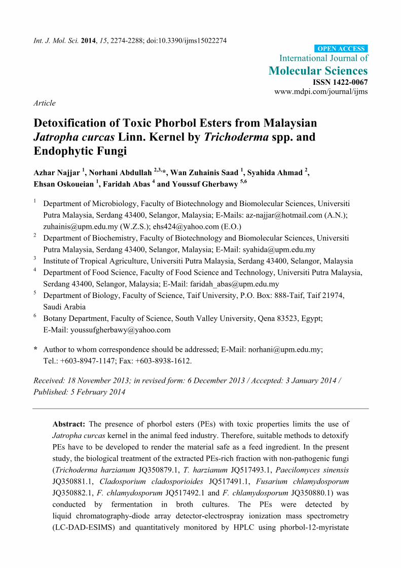
PEs in the fraction. As shown in Figure 1, the total ion chromatogram (TIC) profile showed four peaks
at retention times between 33.32 and 37.30 min, corresponding to four derivatives of PE [1]. The
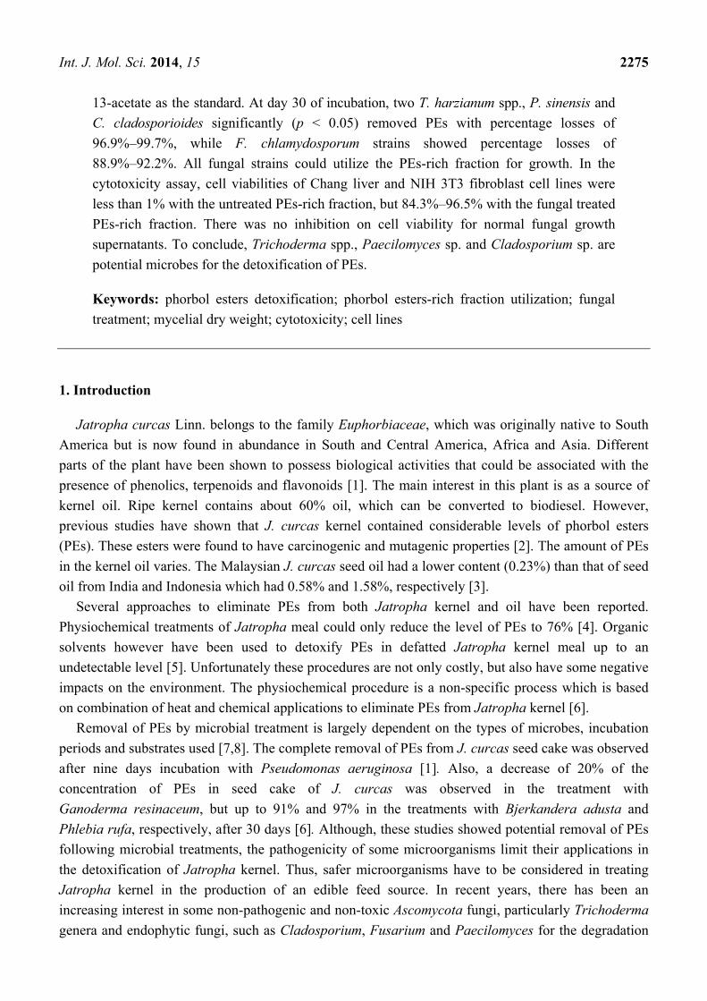
deprotonated molecular ion [M − H]− spectrum and UV chromatogram (UV λmax at 280 nm) of Peak 1
is shown in Figure 2. The mass value of Peak 1 was m/z 709 [M − H]−, which corresponds to
12-deoxy-16-hydroxyphorbol, that was reported to be m/z 711.5 [M + H]+ by positive ion mode [1]. Peaks
2–4 are PE compounds with the same diterpene moiety, namely, 12-deoxy-16-hydroxyphorbol [13]. In
the present study, only four PE derivatives were detected, whereas in previous studies, six derivatives
of PEs in J. curcus kernel were reported [13,14]. Similarly, two and five PE derivatives have been
observed from different varieties of J. curcas [1,3]. It has been suggested that the number of PE
derivatives present in J. curcas depends on analytical techniques, variety, soil and climatic conditions.
Figure 1. Total ion chromatogram (TIC) by negative ion mode electrospray ionization
mass spectrometry (ESI/MS) of the phorbol esters (PEs)-rich fraction. Peaks 1–4 indicate
the four derivatives of PE at retention times between 33.32 and 37.30 min.
Int. J. Mol. Sci. 2014, 15 2277
Figure 2. The deprotonated molecular ion [M − H]− spectrum and UV chromatogram
(UV λmax at 280 nm) of Peak 1.
2.2. Phorbol Esters Degradation by Fungal Strains
The amount of PEs in the rich fraction obtained from the seed kernel was 66.08 mg PMA
equivalent/g dry matter of the PEs-rich fraction. This value is equivalent to 2.78 mg of PEs/g dry
matter of kernel. The levels of PEs may vary according to the samples analyzed. A value of 2.79 mg/g
kernel has been reported [15], but earlier reports indicated the levels of PEs in different provenances of
J. curcas to be in the range of 0.8–3.3 mg/g kernel [16].
The levels of PEs in the extract of the control flasks (samples without fungal strains) were similar at
all incubation times (7, 14, 21 and 30 days). The original levels of PEs were maintained, while extracts
from fungal treated flasks showed significant (p < 0.05) reduction of PEs. Figure 3a shows a
representative of an HPLC elution profile of PEs from J. curcas treated with T. harzianum
JQ350879.1. The four peaks eluted at 21.8, 22.4, 23.4 and 24.3 min under the specified running
conditions corresponded to the four different PE derivatives. The retention times of PEs are different
according to the techniques used. Analysis by HPLC gave shorter retention times compared to that of
LC-DAD-ESIMS analysis. However, the two analytical techniques revealed similar findings, in that
only four PE derivatives were detected.
Int. J. Mol. Sci. 2014, 15 2278
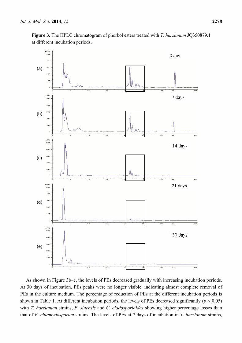
Figure 3. The HPLC chromatogram of phorbol esters treated with T. harzianum JQ350879.1
at different incubation periods.
As shown in Figure 3b–e, the levels of PEs decreased gradually with increasing incubation periods.
At 30 days of incubation, PEs peaks were no longer visible, indicating almost complete removal of
PEs in the culture medium. The percentage of reduction of PEs at the different incubation periods is
shown in Table 1. At different incubation periods, the levels of PEs decreased significantly (p < 0.05)
with T. harzianum strains, P. sinensis and C. cladosporioides showing higher percentage losses than
that of F. chlamydosporum strains. The levels of PEs at 7 days of incubation in T. harzianum strains,
(a)
(b)
(d)
(c)
(e)
Int. J. Mol. Sci. 2014, 15 2279
P. sinensis and C. cladosporioides cultures were reduced by 28.4%, 23.5%, 25.0% and 16.5%,
respectively, and by 13.7%, 11.5% and 13.2% in the treatments with F. chlamydosporum strains,
respectively. The same trend in percentage losses was observed at 14 and 21 days, but with higher
values. At 30 days of incubation, PE losses were 99.7%, 99.5%, 99.0% and 96.4% for treatment
with T. harzianum strains, P. sinensis and C. cladosporioides, respectively. The results clearly
demonstrated that T. harzianum strains and P. sinensis could degrade PEs almost completely. Previous
studies have reported the ability of these fungal strains to produce enzymes that could degrade tigliane
diterpene esters and other closely related esters [1]. Phorbol esters are compounds with fatty acid
moieties esterified to the hydroxyl groups of the tigliane diterpenes. Enzymes such as lipases and
esterases are involved in hydrolyzing these ester bonds [6]. Fungal strains like those used in the
present study have been reported to produce lipases [9,17,18] and esterases [9,19,20] that in high
probability hydrolyze the PEs to fatty acids and the phorbol moiety.
Table 1. Percentage degradation of phorbol esters (PEs) by fungal strains in potato
dextrose broth (PDB) medium at different incubation times.
Fungal strains Percentage loss of PEs
7 days 14 days 21 days 30 days
T. harzianum JQ350879.1 28.4 a,v ± 1.44 51.0 b,v ± 0.80 90.0 c,v ± 0.27 99.7 d,v ± 0.01 T. harzianum JQ517493.1 23.4 a,v ± 0.25 47.6 b,w ± 0.57 88.8 c,v ± 0.34 99.4 d,v ± 0.05
P. sinensis JQ350881.1 24.9 a,v ± 1.96 45.3 b,x ± 0.33 86.2 c,w ± 0.38 98.9 d,v ± 0.02 C. cladosporioides JQ517491.1 16.4 a,w ± 0.88 42.9 b,y ± 0.20 78.0 c,x ± 0.15 96.9 d,w ± 0.03 F. chlamydosporum JQ350882.1 13.7 a,w ± 1.97 33.6 b,z ± 0.27 70.1 c,y ± 0.50 92.2 d,x ± 0.40 F. chlamydosporum JQ517492.1 11.5 a,w ± 1.97 34.8 b,z ± 1.23 69.8 c,y ± 0.24 89.1 d,y ± 0.29 F. chlamydosporum JQ350880.1 13.1 a,w ± 1.97 33.6 b,z ± 0.27 66.9 c,z ± 0.21 88.9 d,y ± 0.60
Mean values of three replicates ± standard error. a,b,c,d, letters show significant difference (p < 0.05) within
row. v,w,x,y,z show significant difference (p < 0.05) within column.
It is essential for industrial and agricultural applications to remove the PEs which are the main toxic
compounds present in Jatropha kernel for safe and maximum utilization. It has been reported that the
PE level in the Jatropha kernel of between 0.02 and 0.11 mg/g is considered safe for human and
animal consumption [6]. In the present study, the level of PEs has been reduced by 99%, equivalent to
a level of 0.02 mg/g of PEs remaining in the kernel. Accordingly, this level of PEs is considered safe
as a feed resource.
A number of studies have been conducted to evaluate the effects of treated Jatropha cake (defatted
kernel) in feeding trial experiments in goats, fish and poultry. Treated Jatropha cake with 77% of
PE reduction by Aspergillus niger and Penicillium sp. could replace 50% of soybean meal in the goat
diet [21]. It was also observed that fish growth performance increased five times in the group fed with
detoxified PEs in Jatropha kernel meal by physiochemical treatment [5]. Recently, it has been
suggested that Jatropha seed cake can be an alternative feed for broiler after PE detoxification of
79.7% through biological treatment with A. niger and Neurospora sitophila [22]. These reports
nevertheless, indicate the possibility of using treated Jatropha cake as a feed ingredient. Although,
Jatropha seed also contained other anti-nutritional compounds like trypsin inhibitor, lectins, phytate
and saponins, they could be easily destroyed by heat [4].
Int. J. Mol. Sci. 2014, 15 2280
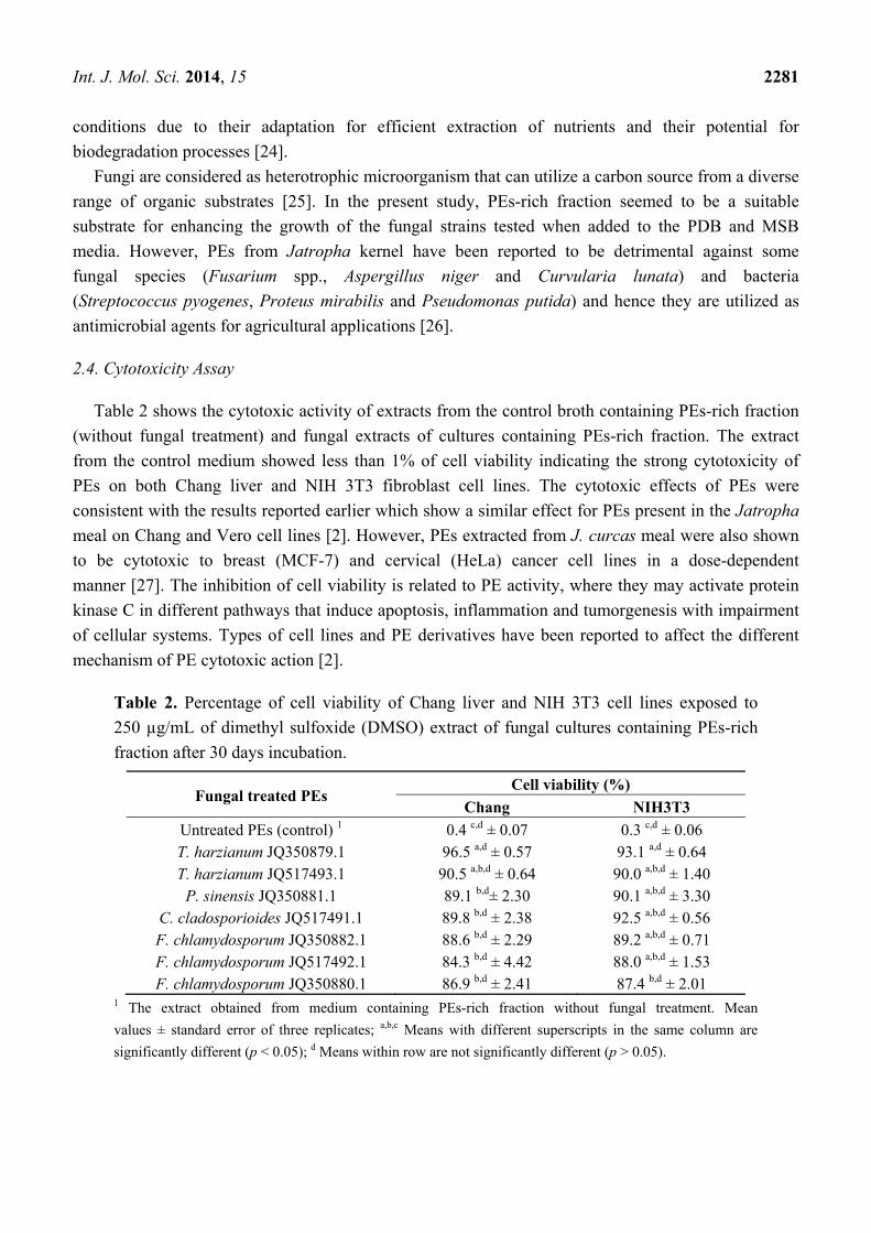
2.3. Utilization of PEs-Rich Fraction as Carbon Source for Fungal Growth
The dry weight (DW) of all the fungal strains grown for 30 days in potato dextrose broth (PDB) and
mineral salts broth (MSB) media are shown in Figure 4a,b, respectively. Interestingly, the presence of
PEs-rich fraction in both media (treated sample) significantly (p < 0.05) enhanced the fungal growth to
average values of 2.19 and 2.14 g, respectively, when compared to the fungal growth without PEs-rich
fraction (control sample) with values of 0.24 and 0.13 g, respectively. Both T. harzianum and
C. cladosporioides showed higher fungal growth than the other fungal strains. The presence of PEs in
the media did not cause any inhibition on the fungal growth, indicating the non-toxicity of PEs on
these microbes. As observed, the ability of fungal strains to utilize the compounds present in the
PEs-rich fraction was clearly demonstrated in PDB medium when there was significant (p < 0.05)
enhancement in growth, although this medium is an enrichment culture medium of carbon source [23].
On the other hand, growth was lower in MSB due to the absence of other organic carbon sources.
Figure 4. Dry weight (DW) of fungal strains grown in (a1) PDB; (a2) PDB + phorbol
esters-rich fraction (PERF); (b1) Mineral salts broth (MSB); (b2) MSB + PERF and (c)
PERF only after 30 days incubation. Treated media contained 2 g of PERF. The standard
error bars were calculated from three replicates of each sample.* indicates significant
difference between treated and control (p < 0.05).
0
1
2
3
4
T. harzianumJQ 350879.1
T. harzianum JQ 517493.1
P. sinensisJQ 350881.1
C. cladosporioidesJQ517491.1
F. chlamydosporum JQ 350882.1
F. chlamydosporumJQ 517492.1
F. chlamydosporumJQ 350880.1
Fungus (control) Fungus (control)Fungus+ PERF (treated) Fungus+ PERF (treated) Fungus (control) Fungus+ PERF
(c)(a1) (a2)* (b1) (b2)*
Fun
gal
D.W
.(g× 1
03)
The ability to grow in the media containing PEs augurs well with the ability to degrade the PEs.
However, the enhancement of fungal growth could be the additive effect of both PEs and other
nutrients present in the rich fraction. Lipase and esterase activity could be involved in both PE
degradation and the oil components present in the fraction. The utilization of the PEs-rich fraction by
some fungi has been shown to be due to their ability to produce degradative enzymes [9,19,22]. In
addition, some fungal strains have been reported to grow and survive on different compounds and
Int. J. Mol. Sci. 2014, 15 2281
conditions due to their adaptation for efficient extraction of nutrients and their potential for
biodegradation processes [24].
Fungi are considered as heterotrophic microorganism that can utilize a carbon source from a diverse
range of organic substrates [25]. In the present study, PEs-rich fraction seemed to be a suitable
substrate for enhancing the growth of the fungal strains tested when added to the PDB and MSB
media. However, PEs from Jatropha kernel have been reported to be detrimental against some
fungal species (Fusarium spp., Aspergillus niger and Curvularia lunata) and bacteria
(Streptococcus pyogenes, Proteus mirabilis and Pseudomonas putida) and hence they are utilized as
antimicrobial agents for agricultural applications [26].
2.4. Cytotoxicity Assay
Table 2 shows the cytotoxic activity of extracts from the control broth containing PEs-rich fraction
(without fungal treatment) and fungal extracts of cultures containing PEs-rich fraction. The extract
from the control medium showed less than 1% of cell viability indicating the strong cytotoxicity of
PEs on both Chang liver and NIH 3T3 fibroblast cell lines. The cytotoxic effects of PEs were
consistent with the results reported earlier which show a similar effect for PEs present in the Jatropha
meal on Chang and Vero cell lines [2]. However, PEs extracted from J. curcas meal were also shown
to be cytotoxic to breast (MCF-7) and cervical (HeLa) cancer cell lines in a dose-dependent
manner [27]. The inhibition of cell viability is related to PE activity, where they may activate protein
kinase C in different pathways that induce apoptosis, inflammation and tumorgenesis with impairment
of cellular systems. Types of cell lines and PE derivatives have been reported to affect the different
mechanism of PE cytotoxic action [2].
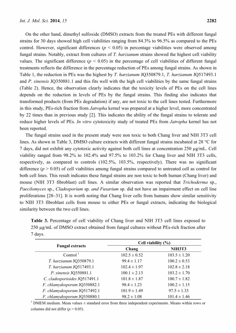
Table 2. Percentage of cell viability of Chang liver and NIH 3T3 cell lines exposed to
250 µg/mL of dimethyl sulfoxide (DMSO) extract of fungal cultures containing PEs-rich
fraction after 30 days incubation.
Fungal treated PEs Cell viability (%)
Chang NIH3T3
Untreated PEs (control) 1 0.4 c,d ± 0.07 0.3 c,d ± 0.06 T. harzianum JQ350879.1 96.5 a,d ± 0.57 93.1 a,d ± 0.64 T. harzianum JQ517493.1 90.5 a,b,d ± 0.64 90.0 a,b,d ± 1.40
P. sinensis JQ350881.1 89.1 b,d± 2.30 90.1 a,b,d ± 3.30 C. cladosporioides JQ517491.1 89.8 b,d ± 2.38 92.5 a,b,d ± 0.56 F. chlamydosporum JQ350882.1 88.6 b,d ± 2.29 89.2 a,b,d ± 0.71 F. chlamydosporum JQ517492.1 84.3 b,d ± 4.42 88.0 a,b,d ± 1.53 F. chlamydosporum JQ350880.1 86.9 b,d ± 2.41 87.4 b,d ± 2.01
1 The extract obtained from medium containing PEs-rich fraction without fungal treatment. Mean
values ± standard error of three replicates; a,b,c Means with different superscripts in the same column are
significantly different (p < 0.05); d Means within row are not significantly different (p > 0.05).
Int. J. Mol. Sci. 2014, 15 2282
On the other hand, dimethyl sulfoxide (DMSO) extracts from the treated PEs with different fungal
strains for 30 days showed high cell viabilities ranging from 84.3% to 96.5% as compared to the PEs
control. However, significant differences (p < 0.05) in percentage viabilities were observed among
fungal strains. Notably, extract from cultures of T. harzianum strains showed the highest cell viability
values. The significant difference (p < 0.05) in the percentage of cell viabilities of different fungal
treatments reflects the difference in the percentage reduction of PEs among fungal strains. As shown in
Table 1, the reduction in PEs was the highest by T. harzianum JQ350879.1, T. harzianum JQ517493.1
and P. sinensis JQ350881.1 and this fits well with the high cell viabilities by the same fungal strains
(Table 2). Hence, the observation clearly indicates that the toxicity levels of PEs on the cell lines
depends on the reduction in levels of PEs by the fungal strains. This finding also indicates that
transformed products (from PEs degradation) if any, are not toxic to the cell lines tested. Furthermore
in this study, PEs-rich fraction from Jatropha kernel was prepared at a higher level, more concentrated
by 22 times than in previous study [2]. This indicates the ability of the fungal strains to tolerate and
reduce higher levels of PEs. In vitro cytotoxicity study of treated PEs from Jatropha kernel has not
been reported.
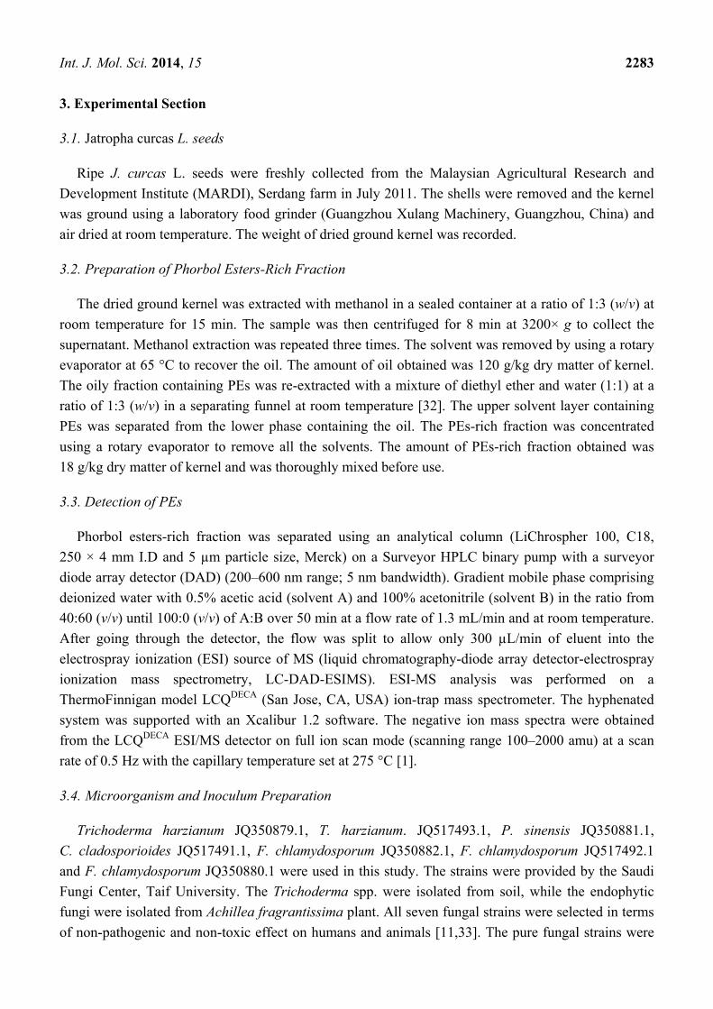
The fungal strains used in the present study were non toxic to both Chang liver and NIH 3T3 cell
lines. As shown in Table 3, DMSO culture extracts with different fungal strains incubated at 28 °C for
7 days, did not exhibit any cytotoxic activity against both cell lines at concentration 250 µg/mL. Cell
viability ranged from 98.2% to 102.4% and 97.5% to 103.2% for Chang liver and NIH 3T3 cells,
respectively, as compared to controls (102.5%, 103.5%, respectively). There was no significant
difference (p > 0.05) of cell viabilities among fungal strains compared to untreated cell as control for
both cell lines. This result indicates these fungal strains are non toxic to both human (Chang liver) and
mouse (NIH 3T3 fibroblast) cell lines. A similar observation was reported that Trichoderma sp.,
Paecilomyces sp., Cladosporium sp. and Fusarium sp. did not have an impairment effect on cell line
proliferations [28–31]. It is worth noting that Chang liver cells from humans show similar sensitivity
to NIH 3T3 fibroblast cells from mouse to either PEs or fungal extracts, indicating the biological
similarity between the two cell lines.
Table 3. Percentage of cell viability of Chang liver and NIH 3T3 cell lines exposed to
250 µg/mL of DMSO extract obtained from fungal cultures without PEs-rich fraction after
7 days.
Fungal extracts Cell viability (%)
Chang NIH3T3
Control 1 102.5 ± 0.52 103.5 ± 1.20 T. harzianum JQ350879.1 99.4 ± 1.17 100.2 ± 0.53 T. harzianum JQ517493.1 102.4 ± 1.97 102.8 ± 2.18
P. sinensis JQ350881.1 100.1 ± 2.13 103.2 ± 1.70 C. cladosporioides JQ517491.1 101.8 ± 1.87 100.7 ± 1.82 F. chlamydosporum JQ350882.1 98.4 ± 1.23 100.2 ± 1.15 F. chlamydosporum JQ517492.1 101.9 ± 1.49 97.5 ± 1.33 F. chlamydosporum JQ350880.1 98.2 ± 1.08 101.4 ± 1.46
1 DMEM medium. Mean values ± standard error from three independent experiments. Means within rows or
columns did not differ (p > 0.05).
Int. J. Mol. Sci. 2014, 15 2283
3. Experimental Section
3.1. Jatropha curcas L. seeds
Ripe J. curcas L. seeds were freshly collected from the Malaysian Agricultural Research and
Development Institute (MARDI), Serdang farm in July 2011. The shells were removed and the kernel
was ground using a laboratory food grinder (Guangzhou Xulang Machinery, Guangzhou, China) and
air dried at room temperature. The weight of dried ground kernel was recorded.
3.2. Preparation of Phorbol Esters-Rich Fraction
The dried ground kernel was extracted with methanol in a sealed container at a ratio of 1:3 (w/v) at
room temperature for 15 min. The sample was then centrifuged for 8 min at 3200× g to collect the
supernatant. Methanol extraction was repeated three times. The solvent was removed by using a rotary
evaporator at 65 °C to recover the oil. The amount of oil obtained was 120 g/kg dry matter of kernel.
The oily fraction containing PEs was re-extracted with a mixture of diethyl ether and water (1:1) at a
ratio of 1:3 (w/v) in a separating funnel at room temperature [32]. The upper solvent layer containing
PEs was separated from the lower phase containing the oil. The PEs-rich fraction was concentrated
using a rotary evaporator to remove all the solvents. The amount of PEs-rich fraction obtained was
18 g/kg dry matter of kernel and was thoroughly mixed before use.
3.3. Detection of PEs
Phorbol esters-rich fraction was separated using an analytical column (LiChrospher 100, C18,
250 × 4 mm I.D and 5 µm particle size, Merck) on a Surveyor HPLC binary pump with a surveyor
diode array detector (DAD) (200–600 nm range; 5 nm bandwidth). Gradient mobile phase comprising
deionized water with 0.5% acetic acid (solvent A) and 100% acetonitrile (solvent B) in the ratio from
40:60 (v/v) until 100:0 (v/v) of A:B over 50 min at a flow rate of 1.3 mL/min and at room temperature.
After going through the detector, the flow was split to allow only 300 µL/min of eluent into the
electrospray ionization (ESI) source of MS (liquid chromatography-diode array detector-electrospray
ionization mass spectrometry, LC-DAD-ESIMS). ESI-MS analysis was performed on a
ThermoFinnigan model LCQDECA (San Jose, CA, USA) ion-trap mass spectrometer. The hyphenated
system was supported with an Xcalibur 1.2 software. The negative ion mass spectra were obtained
from the LCQDECA ESI/MS detector on full ion scan mode (scanning range 100–2000 amu) at a scan
rate of 0.5 Hz with the capillary temperature set at 275 °C [1].
3.4. Microorganism and Inoculum Preparation
Trichoderma harzianum JQ350879.1, T. harzianum. JQ517493.1, P. sinensis JQ350881.1,
C. cladosporioides JQ517491.1, F. chlamydosporum JQ350882.1, F. chlamydosporum JQ517492.1
and F. chlamydosporum JQ350880.1 were used in this study. The strains were provided by the Saudi
Fungi Center, Taif University. The Trichoderma spp. were isolated from soil, while the endophytic
fungi were isolated from Achillea fragrantissima plant. All seven fungal strains were selected in terms
of non-pathogenic and non-toxic effect on humans and animals [11,33]. The pure fungal strains were
Int. J. Mol. Sci. 2014, 15 2284
preserved in potato dextrose agar (PDA) (Oxoid, Basingstoke, UK) slant and stored at −20 °C [34].
Glycerol stock was prepared for the maint of the fungi on PDA and stored at 4 °C. Fungal strains were
transferred on PDA plates, incubated at 28 °C for 7 days before used. One 5-mm agar plug from each
strain in the PDA plates was estimated to contain around 1–5 × 10⁵ spores/mL by using the serial
dilution counting chamber method.
3.5. Degradation of PEs
One liter of potato dextrose broth (PDB) containing 4 g potato extract and 20 g dextrose was
prepared according to the instruction manual (Oxoid, Basingstoke, UK). The pH of medium was
adjusted to pH 5.5 with 12 M hydrochloric acid or 10 M sodium hydroxide. The PEs-rich fraction was
mixed with 30 mL PDB in a ratio 1:15 (w/v) to obtain a concentration of 101.9 mg PEs in 250 mL
glass bottles (Schott, Elmsford, NY, USA) and autoclaved. Two plugs (5-mm) of each fungus grown
on PDA plates at 28 °C for 7 days were inoculated into each bottle (treatment). Untreated PEs control
(without fungus) and PEs treated (with fungus) were incubated for 7, 14, 21 and 30 days at
28 °C. The samples were freeze dried at −45 °C under vapor pressure of 0.129 mBar (Thermo Electron
Corporation Modulyod, Newington, NH, USA). The PEs residues were extracted with 5 mL methanol
and filtered using a syringe filter (0.22 µm) for quantitative analyses.
The concentration of PEs in the sample was determined using phorbol-12-myristate 13-acetate
(PMA) as the standard by HPLC (Agilent Technologies-1200, Waldbronn, Germany) [15] analysis. A
standard PMA calibration curve was prepared in a concentration range of 20–100 µg/mL with five
different levels. The HPLC analytical conditions (flow rate and column) were the same as described
above. The sample injection volume was 20 µL. The gradient was a combination of deionized water
(solvent A) and absolute acetonitrile (solvent B) and increased from 40:60 (v/v) to 100:0 (v/v). The
peak detection was at the UV wave length of 280 nm and the elution time was approximately 25 min.
3.6. Utilization of PEs-Rich Fraction as Carbon Source for Fungal Growth
In a separate experiment, fungal growth in two different media was determined to evaluate the
ability of fungal strains to utilize PEs-rich fraction as a carbon source. Potato dextrose broth (PDB)
was used as the conventional carbon source and mineral salts broth (MSB) was formulated by
replacing the carbon source with minerals. The MSB medium contained 0.005 g of NaCl, 1.0 g of
NH4NO3, 0.002 g of FeSO4 7H2O, 0.002 g of ZnSO4 7 H2O, 0.7 g of K2HPO4, 0.7 g of KH2PO4, 0.7 g
of MgSO4 7 H2O and 0.001 g of MnSO4 (Difco Laboratories, Detroit, MI, USA) in one liter of
deionized water [35]. Control media were without the PEs-rich fraction, while treated media contained
the PEs-rich fraction in a ratio of 1:15 (w/v). All media were inoculated with two plugs (5-mm) of the
seven fungal strains separately and incubated for 30 days at room temperature under shaking
(150 rpm) conditions [36]. The fungal dry weight (DW) produced in PDB and MSB media by each
fungal strain was aseptically filtered through a glass microfiber filter (Schleider and Schuell GF50,
Dassel, Germany) and washed twice with 10 mL of hot distilled water. The biomass was determined
gravimetrically after drying the filters in an oven at 80 °C to a constant weight.
Int. J. Mol. Sci. 2014, 15 2285
3.7. Cytotoxicity Assay
A cytotoxicity assay was carried out using Chang liver (human hepatocytes, CCL-13) and NIH 3T3
(Swiss mouse fibroblasts, CRL-1658) cell lines obtained from the American Type Culture Collection
(ATCC). After 30 days incubation, the cytotoxic activity of PEs-rich fraction with fungal strains
(treated), as well as untreated PEs-rich fraction (control), was evaluated for each fungal strain. A
30 mL PDB sample in a 250 mL Schott bottle was inoculated with two plugs (5-mm) of each fungal
strain and incubated at 28 °C for 7 days to evaluate fungal cytotoxicity. All samples of untreated or
treated PEs-rich fraction and fungal cultures were freeze dried under antiseptic condition (−40 °C and
vapor pressure of 0.129 mBar). Dimethyl sulfoxide (DMSO) in the ratio 1:100 (v/w) was added to the
dried residues to prepare DMSO extract (stock solution). A syringe filter was used to filter the extract.
Each DMSO extract at 250 µg/mL was used for the assay. Cells were seeded into 96-well
micro-culture plates (5 × 10³/100 µL) in Dulbecco’s Modified Eagle Media (DMEM) after treatment
with 0.25% trypsin. The cells were incubated at 37 °C in a humidified atmosphere containing 5% CO2
for 24 h. Thiazolyl blue tetrazolium bromide dye (MTT) was used to estimate the cell viability [2].
3.8. Statistical Analysis
Analysis of variance (ANOVA) was used to analyze the data and mean values were tested by
Dunnett’s Multiple Range test at p < 0.05. All statistical analyses were done by Graph Pad Prism 5
software (GraphPad Software Inc., San Diego, CA, USA).
4. Conclusions
In conclusion, the present study demonstrated that all the seven fungal strains were able to remove
the four phorbol ester derivatives extracted from local J. curcas Linn. kernel in the range of
88.9%–99.7% after 30 days of incubation. The increase in fungal growth in PDB and MSB media
containing the PEs-rich fraction clearly showed the ability of the fungi to utilize the PEs as well as
other nutrients present in the fraction. The cytotoxic activity of the fungal extracts grown in medium
containing the PEs-rich fraction was markedly decreased in accordance with the percentage removal
of PEs. The strains, notably T. harzianum JQ350879.1, T. harzianum JQ517493.1 and P. sinensis
JQ350881.1 could be promising biological agents to remove the toxic PEs present in the
J. curcas kernel.
Acknowledgments
The authors would like to thank the Ministry of Science, Technology and Innovation of Malaysia
for the grant under the Science Fund Project No. 02-01-04-SF1132 and Universiti Putra Malaysia for
the facilities provided.
Conflicts of Interest
The authors declare no conflict of interest.
Int. J. Mol. Sci. 2014, 15 2286
References
1. Joshi, C.; Mathur, P.; Khare, S. Degradation of phorbol esters by Pseudomonas aeruginosa PseA
during solid-state fermentation of deoiled Jatropha curcas seed cake. Bioresour. Technol. 2011,
102, 4815–4819.
2. Oskoueian, E.; Abdullah, N.; Ahmad, S. Phorbol esters isolated from Jatropha meal induced
apoptosis-mediated inhibition in proliferation of Chang and Vero cell lines. Int. J. Mol. Sci. 2012,
13, 13816–13829.
3. Ahmed, W.A.; Salimon, J. Phorbol ester as toxic constituents of tropical Jatropha curcas seed oil.
Eur. J. Sci. Res. 2009, 31, 429–436.
4. Oskoueian, E.; Abdullah, N.; Saad, W.Z.; Omar, A.R.; Ahmad, S.; Kuan, W.B.; Zolkifli, N.A.;
Hendra, R.; Ho, Y.W. Antioxidant, anti-inflammatory and anticancer activities of methanolic
extracts from Jatropha curcas Linn. J. Med. Plants Res. 2011, 5, 49–57.
5. Kumar, V.; Makkar, H.P.; Amselgruber, W.; Becker, K. Physiological, haematological and
histopathological responses in common carp (Cyprinus carpio L.) fingerlings fed with differently
detoxified Jatropha curcas kernel meal. Food Chem. Toxicol. 2010, 48, 2063–2072.
6. De Barros, C.R.M.; Ferreira, L.M.M.; Nunes, F.M.; Bezerra, R.M.F.; Dias, A.A.; Guedes, C.V.;
Cone, J.W.; Marques, G.S.M.; Rodrigues, M.A.M. The potential of white-rot fungi to degrade
phorbol esters of Jatropha curcas L. seed cake. Eng. Life Sci. 2011, 11, 107–110.
7. Abdel-Hafez, A.A.M.; Nakamura, N.; Hattori, M. Biotransformation of phorbol by human
intestinal bacteria. Chem. Pharm. Bull. 2002, 50, 160–164.
8. Jain, A.; Morlok, C.K.; Henson, J.M. Comparison of solid-state and submerged-state fermentation
for the bioprocessing of switchgrass to ethanol and acetate by Clostridium phytofermentans. Appl.
Microbiol. Biotechnol. 2013, 97, 905–917.
9. Haddadin, M.S.Y.; Haddadin, J.; Arabiyat, O.I.; Hattar, B. Biological conversion of olive pomace
into compost by using Trichoderma harzianum and Phanerochaete chrysosporium.
Bioresour. Technol. 2009, 100, 4773–4782.
10. Claus, H. Laccases: structure, reactions, distribution. Micron 2004, 35, 93–96.
11. Harman, G.E.; Howell, C.R.; Viterbo, A.; Chet, I.; Lorito, M. Trichoderma species—Opportunistic,
avirulent plant symbionts. Nat. Rev. Microb. 2004, 2, 43–56.
12. Yan, X.; Sikora, R.A.; Zheng, J. Potential use of cucumber (Cucumis sativus L.) endophytic fungi
as seed treatment agents against root-knot nematode Meloidogyne incognita. J. Zhejiang Uni. Sci.
2011, 12, 219–225
13. Haas, W.; Sterk, H.; Mittelbach, M. Novel 12-Deoxy-16-Hydroxyphorbol diesters isolated from
the seed oil of Jatropha curcas. J. Nat. Prod. 2002, 65, 1434–1440.
14. Hirota, M.; Suttajit, M.; Suguri, H.; Endo, Y.; Shudo, K.; Wongchai, V.; Hecker, E.; Fujiki, H. A
new tumor promoter from the seed oil of Jatropha curcas L., an intramolecular diester of
12-Deoxy-16-Hydroxyphorbol. Cancer Res. 1988, 48, 5800–5804.
15. Makkar, H.P.; Kumar, V.; Oyeleye, O.O.; Akinleye, A.O.; Angulo-Escalante, M.A.; Becker, K.
Jatropha platyphylla, a new non-toxic Jatropha species: Physical properties and chemical
constituents including toxic and antinutritional factors of seeds. Food Chem. 2011, 125, 63–71.
Int. J. Mol. Sci. 2014, 15 2287
16. Makkar, H.P.S.; Becker, K.; Sporer, F.; Wink, M. Studies on nutritive potential and toxic
constituents of different provenances of Jatropha curcas. J. Agric. Food Chem. 1997, 45,
3152–3157.
17. Gopinath, S.C.B.; Anbu, P.; Hilda, A. Extracellular enzymatic activity profiles in fungi isolated
from oil-rich environments. Mycoscience 2005, 46, 119–126.
18. Ward, O.P. Production of recombinant proteins by filamentous fungi. Biotechnol. Adv. 2012, 30,
1119–1139.
19. De Lima Damásio, A.R.; da Silva, T.M.; Maller, A.; Jorge, J.A.; Terenzi, H.F.; Polizeli, M.L.
Purification and partial characterization of an exo-polygalacturonase from Paecilomyces variotii
liquid cultures. Appl. Biochem. Biotechnol. 2010, 160, 1496–1507.
20. Stamatis, H.; Christakopoulos, P.; Kekos, D.; Macris, B.; Kolisis, F. Studies on the synthesis of
short-chain geranyl esters catalysed by Fusarium oxysporum esterase in organic solvents.
J. Molec. Catal. B 1998, 4, 229–236.
21. Belewu, M.; Akande, B. Biological upgrading of the nutritional quality of Jatropha curcas kernel
cake: Effect on performance characteristics of goat. Int. Res. J. Biotech. 2010, 1, 19–22.
22. Kurniati, T. Detoxification through fermentation by consortium of Aspergillus niger and
Neurospora sitophila towards the degree of forbol esther and nutrition value of Jatropha curcas L.
for broilers feed. J. Asian Sci. Res. 1998, 2, 317–324.
23. Gabriel-Ajobiewe, R.A.; Akinyele, B.; Mirrila, E. Basal media formulation using
Canavalia ensiformis as carbon and nitrogen source for the growth of some fungi species
J. Microbiol. Biotech. Food Sci. 2012, 1, 1136–1151.
24. Arfi, Y.; Chevret, D.; Henrissat, B.; Berrin, J.; Levasseur, A.; Record, E. Characterization of
salt-adapted secreted lignocellulolytic enzymes from the mangrove fungus Pestalotiopsis sp. Nat.
Commun. 2013, 4, 1810–1838.
25. Preiss, K.; Adam, I.K.; Gebauer, G. Irradiance governs exploitation of fungi: fine-tuning of
carbon gain by two partially myco-heterotrophic Orchids. Proc. R. Soc. B 2010, 277, 1333–1336.
26. Devappa, R.K.; Angulo-Escalante, M.A.; Makkar, H.P.S.; Becker, K. Potential of using phorbol
esters as an insecticide against Spodoptera frugiperda. Ind. Crops Prod. 2012, 38, 50–53.
27. Oskoueian, E.; Abdullah, N.; Ahmad, S. Phorbol esters from Jatropha meal triggered apoptosis,
activated PKC-δ, caspase-3 proteins and down-regulated the proto-oncogenes in Mcf-7 and Hela
cancer cell lines. Molecules 2012, 17, 10816–10830.
28. Calvert, T.; Aidoo, K.; Candlish, A.G.G.; Fuat, A.R.M. Comparison of in vitro cytotoxicity of
Fusarium mycotoxins, deoxynivalenol, T-2 toxin and zearalenone on selected human epithelial
cell lines. Mycopathologia 2005, 159, 413–419.
29. Flemming, J.; Hudson, B.; Rand, T. Comparison of inflammatory and cytotoxic lung responses in
mice after intratracheal exposure to spores of two different Stachybotrys chartarum strains.
Toxicol. Sci. 2004, 78, 267–275.
30. Shi, M.; Wang, H.N.; Xie, S.T.; Luo, Y.; Sun, C.Y.; Chen, X.L.; Zhang, Y.Z. Antimicrobial
peptaibols, novel suppressors of tumor cells, targeted calcium-mediated apoptosis and autophagy
in human hepatocellular carcinoma cells. Mol. Cancer 2010, 9, 26–40.
Int. J. Mol. Sci. 2014, 15 2288
31. Toledo Marante, F.J.; Mioso, R.; Bermejo Barrera, J.; González González, J.E.;
Santana Rodríguez, J.J.; Bravo de Laguna, I.H. Structural characterization and metabolite
profiling of the facultative marine fungus Paecilomyces variotii. Ann. Microbiol. 2012, 62,
1601–1607.
32. Makkar, H.P.S.; Becker, K. Are Jatropha curcas phorbol esters degraded by rumen microbes?
J. Sci. Food Agric. 2010, 90, 1562–1565.
33. Moore, J.E.; Xu, J.; Millar, B.; Elshibly, S. Edible dates (Phoenix dactylifera), a potential source
of Cladosporium cladosporioides and Sporobolomyces roseus: Implications for public health.
Mycopathologia 2002, 154, 25–28.
34. Najjar, A.A. Determination Mycobiota and Mycotoxins of Air-borne Dust in Some Animal
Houses and Computer Laboratories in Jeddah Province, Master’s Thesis. King Abdul-Aziz
University, 4 March 2007.
35. Roberts, W.; Davidson, P. Growth characteristics of selected fungi on polyvinyl chloride film.
Appl. Environ. Microbiol. 1986, 51, 673–676.
36. Hammer, E.C.; Nasr, H.; Wallander, H. Effects of different organic materials and mineral
nutrients on arbuscular mycorrhizal fungal growth in a mediterranean saline dryland. Soil Biol.
Biochem. 2011, 7, 76–81.
© 2014 by the authors; licensee MDPI, Basel, Switzerland. This article is an open access article
distributed under the terms and conditions of the Creative Commons Attribution license
(http://creativecommons.org/licenses/by/3.0/).