tick-borne diseases in north carolina: is “rickettsia amblyommii” a possible cause of...
TRANSCRIPT
VECTOR-BORNE AND ZOONOTIC DISEASESVolume 8, Number �, 2008© Mary Ann Liebert, Inc.DOI: 10.1089/vbz.2007.0271
Tick-Borne Diseases in North Carolina: Is “Rickettsia amblyommii” a Possible Cause of
Rickettsiosis Reported as Rocky Mountain Spotted Fever?
CHARLES S. APPERSON,1 BARRY ENGBER,2 WILLIAM L. NICHOLSON,3DANIEL G. MEAD,4 JEFFREY ENGEL,5 MICHAEL J. YABSLEY,4 KATHY DAIL,5
JOEY JOHNSON,5 and D. WESLEY WATSON1
ABSTRACT
Cases of Rocky Mountain spotted fever (RMSF) in North Carolina have escalated markedly since 2000. In 2005,we identified a county in the Piedmont region with high case numbers of RMSF. We collected ticks and exam-ined them for bacterial pathogens using molecular methods to determine if a novel tick vector or spotted fevergroup rickettsiae (SFGR) might be emerging. Amblyomma americanum, the lone star tick, comprised 99.6% of 6,502specimens collected in suburban landscapes. In contrast, Dermacentor variabilis, the American dog tick, a princi-pal vector of Rickettsia rickettsii, comprised � 1% of the ticks collected. Eleven of 25 lone star tick pools testedwere infected with “Rickettsia amblyommii,” an informally named SFGR. Sera from patients from the same countywho were presumptively diagnosed by local physicians with a tick-borne illness were tested by an indirect im-munofluorescence antibody (IFA) assay to confirm clinical diagnoses. Three of six patients classified as probableRMSF cases demonstrated a fourfold or greater rise in IgG class antibody titers between paired acute and conva-lescent sera to “R. amblyommii” antigens, but not to R. rickettsii antigens. White-tailed deer, Odocoileus virgini-anus, are preferred hosts of lone star ticks. Blood samples collected from hunter-killed deer from the same countywere tested by IFA test for antibodies to Ehrlichia chaffeensis and “R. amblyommii.” Twenty-eight (87%) of 32 deerwere positive for antibodies to E. chaffeensis, but only 1 (3%) of the deer exhibited antibodies to “R. amblyommii,”suggesting that deer are not the source of “R. amblyommii” infection for lone star ticks. We propose that somecases of rickettsiosis reported as RMSF may have been caused by “R. amblyommii” transmitted through the biteof A. americanum. Key Words: Rocky Mountain spotted fever—Amblyomma americanum—lone star tick—spottedfever group rickettsiae—“Rickettsia amblyommii”—Rickettsia rickettsii — Ehrlichia chaffeensis
1
INTRODUCTION
SPOTTED FEVER GROUP RICKETTSIAE are obligateintracellular bacteria transmitted by ticks.
Tick-borne rickettsioses are an important causeof human morbidity worldwide (Parola et al.2005). Rocky Mountain spotted fever (RMSF) isa rickettsiosis of significant public health im-
portance, because of the severity of its pathol-ogy. It is a zoonotic disease that is maintainedin nature in a cycle involving a tick vector andreservoir, with various wildlife species con-tributing to new lines of ticks infected with thepathogen (Azad and Beard 1998). Humans areincidentally infected. The disease is mostprevalent in the eastern United States. In 2006,
1Department of Entomology, North Carolina State University, Raleigh, North Carolina.2Public Health Pest Management Section, Department of Environment and Natural Resources, Raleigh, North Car-
olina.3Rickettsial Zoonoses Branch, Centers for Disease Control and Prevention, Atlanta, Georgia.4Southeastern Cooperative Wildlife Disease Study, University of Georgia, Athens, Georgia.5Division of Public Health, North Carolina State Department of Health and Human Services, Raleigh, North Car-
olina.
approximately 42% of RMSF cases reported tothe Centers for Disease Control and Prevention(CDC) were from North Carolina (NC). Thehigh proportion of cases reported from NC rep-resents an escalating trend, reflecting an ap-proximate 10-fold rise, from 78 cases in 2000 to 842 cases in 2006 (www.epi.state.nc.us/epi/gcdc.html).
Significantly, most reported cases of RMSFin NC have not been confirmed by laboratorytests of paired acute and convalescent sera,but are classified as probable cases, based onexposure to ticks, occurrence of appropriateclinical symptoms and, a single acute phaseserum sample producing a titer of � 1/64 forpolyvalent antibodies (all immunoglobulinclasses) by indirect immunofluorescence an-tibody assay or comparable optical densitymeasured by enzyme-linked immunosorbentassay (ELISA). Test results can vary from lab-oratory to laboratory and standardization ofmethods is not well established. Conse-quently, the true burden of disease fromRMSF in NC is unknown.
There are several plausible explanations forthe observed increases. Tick vectors may be in-creasing in abundance in residential landscapes(Shriefer and Azad 1994; Childs and Paddock2002); a novel tick vector could be emerging orincreasing in prevalence (Demma et al. 2005);and an unrecognized rickettsial disease couldbe emerging in NC (Carpenter et al. 1999; Pad-dock et al. 2004; Whitman et al. 2007). Further-more, it is conceivable that some people expe-riencing a summer flu-like illness or reportingexposure to ticks may perhaps exhibit falsepositive laboratory tests for RMSF because ofpre-existing antibody titers resulting from in-fection from other SFG rickettisae (Paddock2005).
Accordingly in 2005, we initiated a tick-borne disease surveillance and tick ecologyproject that involved physician-based sur-veillance for tick-borne illness, coupled withlaboratory confirmation, collection of ticksfrom suburban woodlands, and analysis ofsera from hunter-killed white-tailed deer in asingle county in the Piedmont region of NC.Our objectives were to determine the com-parative abundance of tick species on resi-dential properties, to estimate the prevalence
of infection for selected bacterial pathogens ina sample of the ticks, to determine exposurerate of white-tailed deer (Oedocoileus virgini-anus) to selected pathogens, and to confirmthrough laboratory testing the occurrence ofRMSF among patients diagnosed with a tick-borne illness.
MATERIALS AND METHODS
Study area
Chatham County, NC, was chosen as the lo-cation for our investigation. The county isrepresentative of other counties in the Pied-mont region that have experienced rapid sub-urbanization (http://quickfacts.census.gov/qfd/ states/37/37037.html). In 2000, the pop-ulation density averaged 27.9 persons persquare kilometer in Chatham County while thestatewide average was 63.8 persons per squarekilometer. However, from 2000 to 2004, the res-ident population increased by approximately15.6% compared to an increase of 6.1% for theentire state. The total number of cases of RMSFreported during this period was 33 cases inChatham County, while the total number ofcases averaged 14.2 cases per county for all 100 counties in NC (http://www.epi.state.nc.us/epi/gcdc/pdf/CDbyDiseasebyYear2000-2005.pdf).
Collection and processing of ticks
Ticks were collected from April 9 to July 24,2005 in vegetation around residences (n � 32)of people expressing concern about heavy in-festations on their property. In general, thelandscape consisted of oak-pine woodlandswith an understory of leaf litter and low-grow-ing shrubs. A flag constructed of corduroycloth was pulled over and through vegetation,and attached ticks were removed and immedi-ately preserved in 95% ethanol. Quantitativecollections were not made, but attempts weremade to collect at least 100 ticks at each resi-dence. Subsequently, ticks from each residencewere sorted by species and life stage into poolsfor molecular analyses for pathogens as de-scribed below.
APPERSON ET AL.2
Collection and storage of deer blood samples
Exposure of white-tailed deer to tick-bornepathogens was assessed from blood collectedfrom hunter-killed deer. Blood from each deerwas adsorbed onto Nobuto blood filter strips(Toyo Roshi Kaisha, Ltd., Tokyo, Japan) within1 hour of the time the deer was killed. Stripswere air dried and then placed in individual la-beled plastic envelopes. Within 2 days of col-lection, strips were stored at –18oC until theywere processed as described below.
Molecular analyses of ticks
Dermacentor variabilis and Ixodes scapulariswere processed individually, except for one D.variabilis pool that contained three ticks. At siteswhere these tick species were collected, a smallnumber of A. americanum pools were also ana-lyzed. Because of the large overall total, a sam-ple of A. americanum pools at other sites was ran-domly chosen and tested. Ticks were maceratedindividually or in pools of up to 20 ticks/poolin microcentrifuge tubes containing two coppermetal pellets and 0.5 mL sterile PBS in a MixerMill 300 (Qiagen Inc, Valencia, CA). Total nu-cleic acids (NA) were extracted from individualsamples using a Qiagen Viral RNA Kit (QiagenInc, Valencia, CA) according to the manufac-turer’s instructions. Quality control measuresincluded negative controls (water) that were ex-tracted and amplified in parallel with all speci-mens. Positive control DNA for each agent wasincluded in each assay run. To minimize the po-tential for NA contamination, three separate,designated areas were used for NA extractionand preparation of primary and secondaryPCRs. Additionally, two thermal cyclers (PTC-100TM, MJ Research, Inc.), designated for eitherprimary or secondary amplification, were used.
The 16S rRNA gene was targeted for the de-tection of Ehrlichia chaffeensis and Anaplasmaphagocytophilum using 2.5 �L of DNA templatein a primary polymerase chain reaction (PCR)with external primers ECC and ECB (Andersonet al. 1991), which amplify all Ehrlichia andAnaplasma spp. expected to be found in the ge-ographic area of our investigation. For sec-ondary PCR amplification, 1 �L of primaryproduct was used in reactions with species-spe-cific primers HE1and HE3 to detect E. chaffeen-
sis (Dawson et al. 1994), producing a 389-bpamplicon, and in reactions with primersGA1UR and GE9F to detect A. phagocytophilum(Chen et al. 1994; Little et al. 1998), producinga 400-bp amplicon. External primers FLALLand FLARL and internal primers FLALS andFLARS, which amplify a 330-bp region of theflaB gene of all species in the genus Borrelia,were used in a nested PCR assay as describedelsewhere (Barbour et al. 1994; Moore et al.2003). Primer pairs 17 kDa1/17 kDa2 (external)and 17 kDa3/17 kDa4 (internal) were used toamplify a 434-bp portion of the 17kDa genefrom Rickettsia spp. (Skeyova et al. 2001).
Products amplified with internal primer setsHE1/HE3, GA1UR/GE9F, FLALS/FLARS,and 17kDa3/17kDa4 were purified with aQiagen Gel Purification Kit (Qiagen Inc., Va-lencia, CA) and sequenced in both the 3’ and5’ directions with a PerkinElmer ABI Prism3700 automated DNA sequencer at the Molec-ular Genetics Instrumentation Facility at theUniversity of Georgia. The sequences were as-sembled and edited with the Sequencher soft-ware package, version 4.0.5 (Gene Codes Corp.,Ann Arbor, MI), and a nucleotide–nucleotideBLAST (BLASTN) search was performed to de-termine the most similar GenBank sequences.
Tick-borne disease surveillance and laboratoryconfirmation of clinical diagnoses
Five physician practices were recruited intoour prospective investigation for a 3-month pe-riod from June 1 to August 31, 2005. Participat-ing physicians agreed to report all suspectedcases of tick-borne illness to the ChathamCounty Health Department. To be considereda suspect case of tick-borne illness, the affectedpatient had to have had a reasonable opportu-nity for tick exposure (bite was not necessary),a fever or rash, and one other compatible symp-tom not explained by another illness. Self-re-ported symptoms were considered by physi-cians in making a diagnosis. Physicians alsoagreed to provide a clinical assessment for pa-tients, and to collect acute and convalescentserum samples for testing in the North CarolinaState Laboratory of Public Health. Sera weretested by an indirect immunofluorescence an-tibody (IFA) assay against a panel of bacterial
IS R. AMBLYOMMII A CAUSE OF RICKETTSIOSIS REPORTED AS RMSF? 3
antigens, including R. rickettsii, R. typhi, andEhrlichia chaffeensis (CDC Biologics Branch, At-lanta, GA). Polyvalent conjugate to both heavyand light chains of IgG was used; therefore, theantibody titers reflect the summary reaction ofall immunoglobulin classes. A case was classi-fied as probable when the patient exhibitedsymptoms of a tick-borne illness (CDC 2006)and the IFA test produced a serologic titer of�1/64 for an acute phase serum sample (CDC2006). A confirmed case of tick-borne illness re-quired a � 4-fold increase in antibody titer be-tween paired sera (CDC 2006). Serum samplesfrom all patients that were tested for antibod-ies to rickettsial and ehrlichial pathogens werealso sent to the CDC (Fort Collins, CO) for test-ing by ELISA and Western blot against Borre-lia burgdorferi antigens. Subsequently, pairedsera were sent to CDC (Atlanta) for additionalrickettsial testing as described in the Resultssection.
Testing of deer serum samples
Each Nobuto strip was eluted separately inenough diluent to produce an estimated 1/20dilution. Eluates were briefly refrigerated untilassayed for antibodies to various agents. Forscreening, the elutions were diluted threefoldjust prior to testing to make a 1/60 dilution. Sub-sequent twofold serial dilutions were made intoa PBS (pH 7.38) diluent containing 1% rabbitserum and 1% bovine serum albumin. Indirectimmunofluorescence antibody tests were carriedout as described previously for A. phagocyto-philum (Petrovec et al. 2002), but with E. chaf-feensis or rickettsial antigens. Titers are expressedas the reciprocal of the dilution of the final wellshowing distinct immunofluorescence.
RESULTS
Comparative abundance of tick species
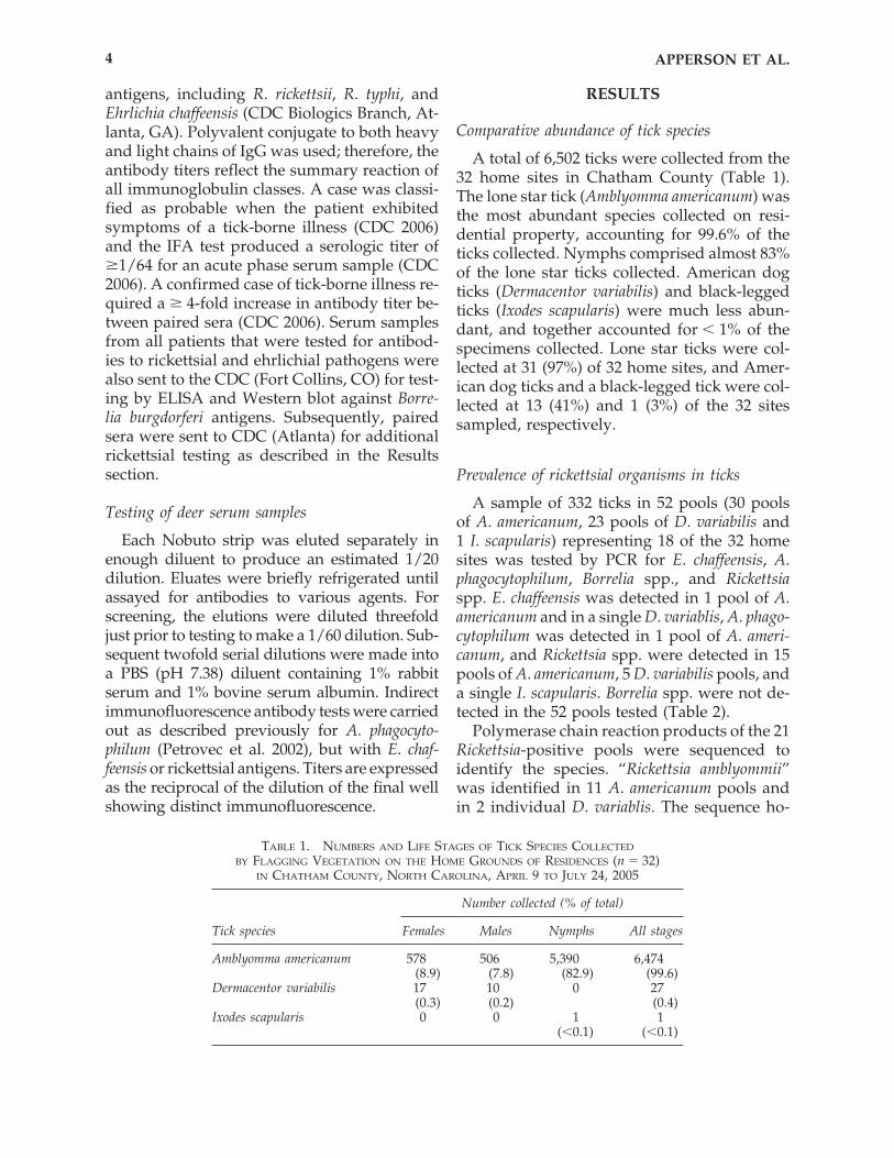
A total of 6,502 ticks were collected from the32 home sites in Chatham County (Table 1).The lone star tick (Amblyomma americanum) wasthe most abundant species collected on resi-dential property, accounting for 99.6% of theticks collected. Nymphs comprised almost 83%of the lone star ticks collected. American dogticks (Dermacentor variabilis) and black-leggedticks (Ixodes scapularis) were much less abun-dant, and together accounted for � 1% of thespecimens collected. Lone star ticks were col-lected at 31 (97%) of 32 home sites, and Amer-ican dog ticks and a black-legged tick were col-lected at 13 (41%) and 1 (3%) of the 32 sitessampled, respectively.
Prevalence of rickettsial organisms in ticks
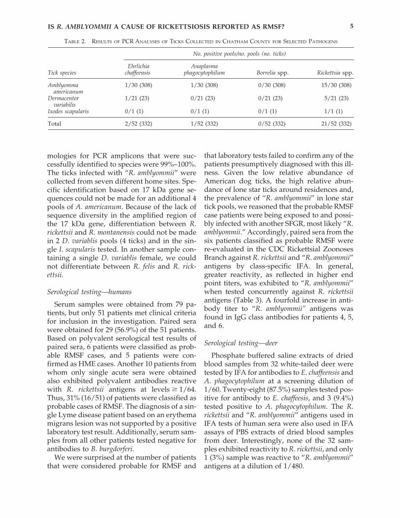
A sample of 332 ticks in 52 pools (30 poolsof A. americanum, 23 pools of D. variabilis and1 I. scapularis) representing 18 of the 32 homesites was tested by PCR for E. chaffeensis, A.phagocytophilum, Borrelia spp., and Rickettsiaspp. E. chaffeensis was detected in 1 pool of A.americanum and in a single D. variablis, A. phago-cytophilum was detected in 1 pool of A. ameri-canum, and Rickettsia spp. were detected in 15pools of A. americanum, 5 D. variabilis pools, anda single I. scapularis. Borrelia spp. were not de-tected in the 52 pools tested (Table 2).
Polymerase chain reaction products of the 21Rickettsia-positive pools were sequenced toidentify the species. “Rickettsia amblyommii”was identified in 11 A. americanum pools andin 2 individual D. variablis. The sequence ho-
APPERSON ET AL.4
TABLE 1. NUMBERS AND LIFE STAGES OF TICK SPECIES COLLECTED
BY FLAGGING VEGETATION ON THE HOME GROUNDS OF RESIDENCES (n � 32) IN CHATHAM COUNTY, NORTH CAROLINA, APRIL 9 TO JULY 24, 2005
Number collected (% of total)
Tick species Females Males Nymphs All stages
Amblyomma americanum 578 506 5,390 6,474(8.9) (7.8) (82.9) (99.6)
Dermacentor variabilis 17 10 0 27(0.3) (0.2) (0.4)
Ixodes scapularis 0 0 1 1(�0.1) (�0.1)
mologies for PCR amplicons that were suc-cessfully identified to species were 99%–100%.The ticks infected with “R. amblyommii” werecollected from seven different home sites. Spe-cific identification based on 17 kDa gene se-quences could not be made for an additional 4pools of A. americanum. Because of the lack ofsequence diversity in the amplified region ofthe 17 kDa gene, differentiation between R.rickettsii and R. montanensis could not be madein 2 D. variablis pools (4 ticks) and in the sin-gle I. scapularis tested. In another sample con-taining a single D. variablis female, we couldnot differentiate between R. felis and R. rick-ettsii.
Serological testing—humans
Serum samples were obtained from 79 pa-tients, but only 51 patients met clinical criteriafor inclusion in the investigation. Paired serawere obtained for 29 (56.9%) of the 51 patients.Based on polyvalent serological test results ofpaired sera, 6 patients were classified as prob-able RMSF cases, and 5 patients were con-firmed as HME cases. Another 10 patients fromwhom only single acute sera were obtainedalso exhibited polyvalent antibodies reactivewith R. rickettsii antigens at levels � 1/64.Thus, 31% (16/51) of patients were classified asprobable cases of RMSF. The diagnosis of a sin-gle Lyme disease patient based on an erythemamigrans lesion was not supported by a positivelaboratory test result. Additionally, serum sam-ples from all other patients tested negative forantibodies to B. burgdorferi.
We were surprised at the number of patientsthat were considered probable for RMSF and
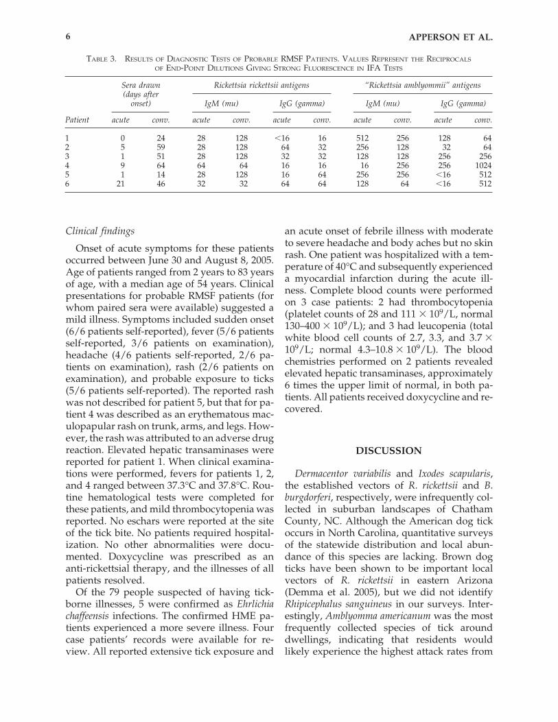
that laboratory tests failed to confirm any of thepatients presumptively diagnosed with this ill-ness. Given the low relative abundance ofAmerican dog ticks, the high relative abun-dance of lone star ticks around residences and,the prevalence of “R. amblyommii” in lone startick pools, we reasoned that the probable RMSFcase patients were being exposed to and possi-bly infected with another SFGR, most likely “R.amblyommii.” Accordingly, paired sera from thesix patients classified as probable RMSF werere-evaluated in the CDC Rickettsial ZoonosesBranch against R. rickettsii and “R. amblyommii”antigens by class-specific IFA. In general,greater reactivity, as reflected in higher endpoint titers, was exhibited to “R. amblyommii”when tested concurrently against R. rickettsiiantigens (Table 3). A fourfold increase in anti-body titer to “R. amblyommii” antigens wasfound in IgG class antibodies for patients 4, 5,and 6.
Serological testing—deer
Phosphate buffered saline extracts of driedblood samples from 32 white-tailed deer weretested by IFA for antibodies to E. chaffeensis andA. phagocytophilum at a screening dilution of1/60. Twenty-eight (87.5%) samples tested pos-itive for antibody to E. chaffeesis, and 3 (9.4%)tested positive to A. phagocytophilum. The R.rickettsii and “R. amblyommii” antigens used inIFA tests of human sera were also used in IFAassays of PBS extracts of dried blood samplesfrom deer. Interestingly, none of the 32 sam-ples exhibited reactivity to R. rickettsii, and only1 (3%) sample was reactive to “R. amblyommii”antigens at a dilution of 1/480.
IS R. AMBLYOMMII A CAUSE OF RICKETTSIOSIS REPORTED AS RMSF? 5
TABLE 2. RESULTS OF PCR ANALYSES OF TICKS COLLECTED IN CHATHAM COUNTY FOR SELECTED PATHOGENS
No. positive pools/no. pools (no. ticks)
Ehrlichia AnaplasmaTick species chaffeensis phagocytophilum Borrelia spp. Rickettsia spp.
Amblyomma 1/30 (308) 1/30 (308) 0/30 (308) 15/30 (308)americanum
Dermacentor 1/21 (23) 0/21 (23) 0/21 (23) 5/21 (23)variabilis
Ixodes scapularis 0/1 (1) 0/1 (1) 0/1 (1) 1/1 (1)
Total 2/52 (332) 1/52 (332) 0/52 (332) 21/52 (332)
Clinical findings
Onset of acute symptoms for these patientsoccurred between June 30 and August 8, 2005.Age of patients ranged from 2 years to 83 yearsof age, with a median age of 54 years. Clinicalpresentations for probable RMSF patients (forwhom paired sera were available) suggested amild illness. Symptoms included sudden onset(6/6 patients self-reported), fever (5/6 patientsself-reported, 3/6 patients on examination),headache (4/6 patients self-reported, 2/6 pa-tients on examination), rash (2/6 patients onexamination), and probable exposure to ticks(5/6 patients self-reported). The reported rashwas not described for patient 5, but that for pa-tient 4 was described as an erythematous mac-ulopapular rash on trunk, arms, and legs. How-ever, the rash was attributed to an adverse drugreaction. Elevated hepatic transaminases werereported for patient 1. When clinical examina-tions were performed, fevers for patients 1, 2,and 4 ranged between 37.3°C and 37.8°C. Rou-tine hematological tests were completed forthese patients, and mild thrombocytopenia wasreported. No eschars were reported at the siteof the tick bite. No patients required hospital-ization. No other abnormalities were docu-mented. Doxycycline was prescribed as an anti-rickettsial therapy, and the illnesses of allpatients resolved.
Of the 79 people suspected of having tick-borne illnesses, 5 were confirmed as Ehrlichiachaffeensis infections. The confirmed HME pa-tients experienced a more severe illness. Fourcase patients’ records were available for re-view. All reported extensive tick exposure and
an acute onset of febrile illness with moderateto severe headache and body aches but no skinrash. One patient was hospitalized with a tem-perature of 40°C and subsequently experienceda myocardial infarction during the acute ill-ness. Complete blood counts were performedon 3 case patients: 2 had thrombocytopenia(platelet counts of 28 and 111 � 109/L, normal130–400 � 109/L); and 3 had leucopenia (totalwhite blood cell counts of 2.7, 3.3, and 3.7 �109/L; normal 4.3–10.8 � 109/L). The bloodchemistries performed on 2 patients revealedelevated hepatic transaminases, approximately6 times the upper limit of normal, in both pa-tients. All patients received doxycycline and re-covered.
DISCUSSION
Dermacentor variabilis and Ixodes scapularis,the established vectors of R. rickettsii and B.burgdorferi, respectively, were infrequently col-lected in suburban landscapes of ChathamCounty, NC. Although the American dog tickoccurs in North Carolina, quantitative surveysof the statewide distribution and local abun-dance of this species are lacking. Brown dogticks have been shown to be important localvectors of R. rickettsii in eastern Arizona(Demma et al. 2005), but we did not identifyRhipicephalus sanguineus in our surveys. Inter-estingly, Amblyomma americanum was the mostfrequently collected species of tick arounddwellings, indicating that residents wouldlikely experience the highest attack rates from
APPERSON ET AL.6
TABLE 3. RESULTS OF DIAGNOSTIC TESTS OF PROBABLE RMSF PATIENTS. VALUES REPRESENT THE RECIPROCALS
OF END-POINT DILUTIONS GIVING STRONG FLUORESCENCE IN IFA TESTS
Sera drawn(days after
onset) IgM (mu) IgG (gamma) IgM (mu) IgG (gamma)
Patient acute conv. acute conv. acute conv. acute conv. acute conv.
1 0 24 28 128 �16 16 512 256 128 642 5 59 28 128 64 32 256 128 32 643 1 51 28 128 32 32 128 128 256 2564 9 64 64 64 16 16 16 256 256 10245 1 14 28 128 16 64 256 256 �16 5126 21 46 32 32 64 64 128 64 �16 512
“Rickettsia amblyommii” antigensRickettsia rickettsii antigens
this species. The probable risk of pathogen ac-quisition from lone star and other tick speciesis likely correlated with their distribution andlocal abundance. Amblyomma americanum is thevector of E. chaffeensis (Childs and Paddock2003), and laboratory confirmation of HME pa-tients is congruent with the high frequency ofoccurrence of lone star ticks around the resi-dences included in our tick survey. Indeed,some patients thought to be RMSF cases mayhave actually been infected with Ehrlichia chaf-feensis. Ehrlichial and rickettsial infection canhave similar acute clinical appearances and canbe confused if laboratory testing is not com-pleted (Carpenter et al 1999). Rickettsia parkerihas been recently identified as another SFGRthat may be misclassified as RMSF, particularlysince cross-reactive antibodies may be seen(Paddock et al 2004; Paddock 2005; Whitman etal. 2007).
In view of the large numbers of RMSF casesfrom North Carolina reported to CDC, we weresurprised that none of the patients presump-tively diagnosed with RMSF were confirmedthrough initial testing completed in the NCState Laboratory of Public Health. Clinical eval-uations of patients presumptively diagnosed asRMSF indicated that they experienced a mildfebrile illness without a maculopapular or pe-techial rash and did not require hospitalization.All patients reported sudden onset of illness,and 5 of 6 patients reported a tick bite. Addi-tionally, serological testing confirmed thatthese patients had been infected with a SFGR.
All six patients who reported a tick biteshowed higher endpoint titers to “R. ambly-ommii” over those to R. rickettsii in assays runat the same time, using the same reagents, andread by the same investigator. Patients 4, 5, and6 demonstrated clear IgG seroconversions to“R. amblyommii.” Class-specific antibody test-ing clarified the results of the initial laboratorytesting (via polyvalent conjugate); notably, sig-nificant seroconversions against R. rickettsiiantigens were still lacking. These results pro-vide support for infection with “R. amblyommii”or a closely related SFGR as the cause of theseillnesses, while lessening evidence for R. rick-ettsii infection. It also should be noted that pa-tient 5 exhibited a fourfold change in titer toIgG class antibodies to R. rickettsii antigen. This
patient would have been classified as a con-firmed RMSF patient if additional testing with“R. amblyommii” antigen had not been carriedout. Notably, Burgdorfer et al. (1981) reportedthat “R. amblyommii”–infected voles and guineapigs did not develop antibodies that cross-re-acted with R. rickettsii antigen. “Rickettsia am-blyommii”–infected mice produced antibodiesthat cross-reacted only with R. rickettsii antigenand at lower titer (homologous titer: 1/4,096versus heterologous titer: 1/16). Antisera to R.rickettsii and R. parkeri cross-reacted with “R.amblyommii” antigen only at low titers (1/16and 1/8, respectively).
Patients 1, 2, and 3 showed high titers of IgMand IgG class antibodies in acute specimens,but they did not exhibit significant rises in titersin convalescent sera. One or more of the fol-lowing explanations might account for this ob-servation. First, the patients could have beeninfected during the preceding year but sawtheir physicians for an unrelated illness. Sec-ond, patients might have been infected with aSFGR other than “R. amblyommii” or R. rickettsii.Third, the patients could have developed animmune response during an asymptomatic pe-riod prior to their perceived date of “onset,”but acute sera was still drawn and antibiotictreatment given before a full immune responseoccurred. Generally, it is suggested that anti-body levels in RMSF sera will not reach diag-nostic levels before one week of onset and earlyantibiotic therapy can interfere with antibodyproduction.
Spotted fever group rickettsiae (Goddardand Norment 1986; Goddard et al. 2003; Kar-datzke et al. 1992; Solberg et al. 1996), includ-ing “R. amblyommii” (Burgdorfer et al. 1981;Stromdahl et al. 2008; Labruna et al. 2004, 2007;Kelly et al. 2005; Mixson et al. 2006), have beendetected previously in Amblyomma spp. ticks.Amblyomma americanum is apparently com-monly infected with “R. amblyommii”. Whenthis SFGR was first identified, prevalences ashigh as 42% were noted (Burgdorfer et al. 1981).In a recent survey encompassing 9 U.S. states,prevalence estimates of “R. amblyommii” in lonestar ticks ranged from 0% to 84%, and averaged41.2% (Mixson et al. 2006). “Rickettsia ambly-ommii” was detected in lone star ticks collectedfrom 7 (38.8%) of the 18 residences for which
IS R. AMBLYOMMII A CAUSE OF RICKETTSIOSIS REPORTED AS RMSF? 7
ticks were tested in our survey, indicating thatthe organism is prevalent in A. americanumpopulations in central NC.
When the SFGR known now as “R. ambly-ommii” was originally isolated from A. ameri-canum ticks and designated WB-8-2, the inves-tigators (Burgdorfer et al. 1981) discussed thepathogenic potential of this agent. In laboratorystudies, it was nonpathogenic to laboratory an-imals. Because of this finding and the frequentexposures of humans to commonly infectedticks with no apparent subsequent illness, theysuggested that the organism was nonpatho-genic to humans (Burgdorfer et al. 1981). How-ever, nonpathogenic strains may be importantin the evolution of pathogenic SFG rickettsiaethrough changes in genes encoding antigenicsurface proteins (Weller et al. 1998). Stromdahland coworkers (2008) speculate that bites from“R. amblyommii”–infected lone star ticks mightaccount for the seroprevalence of antibodies toSFGR, which often exceeds reported cases ofRMSF. Patients experiencing mild febrile ill-nesses following tick bites and developing an-tibodies reactive with SFGR have been reportedpreviously in NC (Carpenter et al. 1999; Sextonet al. 1998) and other eastern states (Sanchez1992; Dasch et al. 1993; Yevich et al. 1995; Mc-Call et al. 2001). Anecdotally, “R. amblyommii”infections have been suggested as the cause ofunexplained fever following tick bite, based onserologic reactivity of patient sera with rick-ettsial antigens in Western blot analyses (Daschet al. 1993). Recently, “R. amblyommii” DNAwas detected by PCR in an engorged tick of thespecies A. americanum that was removed froma woman who subsequently developed a mac-ular rash at the site of the tick bite (Billeter etal. 2008).
The high prevalence of antibodies to E. chaf-feensis in white-tailed deer sampled during ourinvestigation is concordant with its status as awildlife reservoir and its close association withlone star ticks, which are vectors of thepathogen (Paddock and Childs 2003). White-tailed deer seropositve for antibodies to E. chaf-feensis have been reported previously from NC(Yabsley et al. 2005). For the deer that we tested,a smaller percentage (9.4%) were positive forantibodies to A. phagocytophilum. A previousserosurvey reported finding 19% of white-
tailed deer samples from NC positive for anti-bodies to A. phagocytophilum (Dugan et al.2006). In contrast to the high prevalence of an-tibodies to E. chaffeensis in white-tailed deer, wefound only 1 (3%) of 32 deer serum samples tobe positive for antibodies reactive with “R. am-blyommii” and none to be positive for R. rick-ettsii. The lack of antibodies to R. rickettsii wasexpected, as deer are not commonly hosts of D.variabilis. However, the low prevalence of anti-bodies to “R. amblyommii” is surprising becauseA. americanum feeds heavily on deer (Childsand Paddock 2003). The lack of antibodies to“R. amblyommii” was likely not due to failureof ticks to transmit the organism, becauseBurgdorfer et al. (1981) reported finding dis-seminated infections of “R. amblyommii” in lonestar ticks. It is more likely that “R. amblyommii”is not infectious to deer, that deer exhibit im-mune tolerance, or that the production of anti-bodies is downregulated by heavy exposure tothe agent (Semu et al. 2001). White-tailed deerare established reservoirs for E. chaffeensis (Pad-dock and Yabsley 2007), but the reservoir sta-tus of deer for “R. amblyommii” is unknown.Spotted fever group rickettsiae are maintainedvertically by transovarial transmission (Azadand Beard 1998). It may be that there is no vertebrate reservoir for “R. amblyommii,” asBurgdorfer et al. (1981) reported that 100% oflone star tick progeny from infected females arealso infected.
CONCLUSIONS
Our findings suggest that an emerging SFGRmay be the cause of some cases of rickettsiosisreported as RMSF in NC. Amblyomma ameri-canum, an aggressive tick that readily attackshumans, is the most abundant tick species inrural and suburban landscapes. High preva-lence of infection of lone star ticks with “R. am-blyommii” suggests that this SFG rickettsialspecies might be responsible for some of the ill-ness reported as RMSF, and it could accountfor the recent escalation in numbers of cases re-ported as RMSF. Our findings support previ-ous suggestions that the pathogenicity of “R.amblyommii” to humans warrants further in-vestigation (Mixson et al. 2006; Labruna et al.
APPERSON ET AL.8
2007). These studies would include cell cultureand molecular evaluation of human specimensfrom clinically ill patients to provide specificidentity of the etiologic agent.
ACKNOWLEDGMENTS
The assistance of the Chatham County(North Carolina) Health Department was in-valuable. We thank Al Cooke of the ChathamCounty Cooperative Extension Center for as-sistance in locating tick sampling sites. HollyColeman of the Chatham County Health De-partment helped us obtain some of the white-tailed deer blood samples. Assistance providedby personnel of the NC State Laboratory ofPublic Health is gratefully acknowledged. Wethank Amanda Loftis for the “R. amblyommii”antigen used in our investigation.
REFERENCES
Anderson, BE, Dawson, JE, Jones, DC, Wilson, KH.Ehrlichia chaffeensis, a new species associated with hu-man ehrlichiosis. J Clin Microbiol 1991; 29:2838–2842.
Azad, AF, Beard, CB. Rickettsial pathogens and theirarthropod vectors. Emerg Infec Dis 1998; 4:179–186.
Barbour, AG, Maupin, GO, Teltow, GJ, Carter, CJ. Iden-tification of an uncultivable Borrelia species in the hardtick Amblyomma americanum: possible agent of a Lymedisease-like illness. J Infect Dis 1996; 173:403–409.
Billeter, SA, Blanton, HL, Little, SE, Levy, MG, et al. De-tection of “Rickettsia amblyommii” in association with atick bite rash. Vector-Borne Zoonotic Dis 2007;7:607–610.
Burgdorfer, W, Hayes, SF, Thomas, LA, Lancaster, JL, Jr.A new spotted fever group rickettsia from the lone startick, Amblyomma americanum. In: Burgdorfer, W,Anacker, RL, eds. Rickettsiae and rickettsial diseases. NewYork, Academic Press, 1981:595–602.
Carpenter, CF, Gandhi, TK, Kong, LK, Corey, GR, et al.The incidence of ehrlichial and rickettsial infection inpatients with unexplained fever and recent history oftick bite in central North Carolina. J Infec Dis 1999;180:900–903.
Centers for Disease Control and Prevention. Diagnosisand management of tickborne rickettsial diseases:Rocky Mountain spotted fever, ehrlichiosis, andanaplasmosis–United States: a practical guide forphysicians and other health-care and public health pro-fessionals. MMWR 2006; 55 (No. RR-4):1–29.
Chen, SM, Dumler, JS, Bakken, JS, Walker, DH. Identifi-cation of a granulocytotropic Ehrlichia species as the eti-
ologic agent of human disease. J Clin Microbiol 1994;32:589–595.
Childs, JE, Paddock, CD. Passive surveillance as an in-strument to identify risk factors for fatal Rocky Moun-tain spotted fever: Is there more to learn? Am J TropMed Hyg 2002; 66:450–457.
Childs, JE, Paddock, CD. The ascendancy of Amblyommaamericanum as a vector of pathogens affecting humansin the United States. Ann Rev Entomol 2003;48:307–338.
Dasch, GA, Kelly, DJ, Richards, AL, Sanchez, JL, et al.Western blotting analysis of sera from military person-nel exhibiting serological reactivity to spotted fevergroup rickettsiae [abstract 242]. In Program and ab-stracts of the Joint Annual Meeting of the American So-ciety of Tropical Medicine and Hygiene and the Amer-ican Society of Parasitologists (Atlanta). Am J Trop MedHyg. 1993; 49(Suppl):220.
Dawson, JE, Childs, JE, Biggie, KL, Moore, C, et al. White-tailed deer as a potential reservoir of Ehrlichia spp. JWildl Dis 1994; 30:162–168.
Demma, LJ, Traeger, MS, Nicholson, WL, Paddock, CD,et al. Rocky Mountain spotted fever from an unex-pected tick vector in Arizona. N Engl J Med 2005;353:587–594.
Dugan, VG, Yabsley, MJ, Tate, CM, Mead, DG, et al. Eval-uation of white-tailed deer (Odocoileus virginianus) asnatural sentinels for Anaplasma phagocytophilum. VectorBorne Zoonotic Dis 2006; 6:192–207.
Goddard, J, Norment, BR. Spotted fever group rickettsiaein the lone star tick, Amblyomma americanum (Acari: Ixo-didae). J Med Entomol 1986; 23:465–473.
Goddard, J, Sumner, JW, Nicholson, WL, Paddock, CD,et al. Survey of ticks collected in Mississippi for Rick-ettsia, Ehrlichia, and Borrelia species. J Vector Ecol 2003;28:184–189.
Kardatzke, JT, Neidhardt, K, Dzuban, DP, Sanchez, JL, etal. Cluster of tick-borne infections at Fort Chaffee,Arkansas: Rickettsiae and Borrelia burgdorferi in ixodidticks. J. Med Entomol 1992; 29:669–672.
Kelly, DJ, Carmichael, JR, Booton, GC, Poetter, KF, et al.Novel spotted fever group rickettsiae (SFGR) infectingAmblyomma americanum in Ohio, USA. Ann NY AcadSci 2005; 1063:352–355.
Labruna, MB, Pacheco, RB, Nava, S, Brandão, PE, et al.Infection by Rickettsia bellii and Candidatus “Rickettsiaamblyommii” in Amblyomma neumanni ticks from Ar-gentina. Micro Ecol 2007; 54:126–133.
Labruna, MB, Whitworth, T, Bouyer, DH, McBride, J, etal. Rickettsia belli and Rickettsia amblyommii in Ambly-omma ticks from the State of Rondônia, Western Ama-zon, Brazil. J Med Entomol 2004; 41:1073–1081.
Little, SE, Stallknecht, DE, Lockhart, JM, Dawson, JE. Nat-ural coinfection of a white tailed deer (Odocoileus vir-ginianus) population with three Ehrlichia spp. J Parasitol1998; 84:897–901.
McCall, CL, Curns, AT, Rotz, LD, Singelton, JA, et al. FortChaffee revisited: The epidemiology of tick-borne rick-ettsial and ehrlichial diseases in a natural focus. VectorBorne Zoonotic Dis 2001; 1:119–127.
IS R. AMBLYOMMII A CAUSE OF RICKETTSIOSIS REPORTED AS RMSF? 9
Mixson, TR, Campbell, SR, Gill, JS, Ginsberg, HS, et al.Prevalence of Ehrlichia, Borrelia, and rickettsial agentsin Amblyomma americanum (Acari: Ixodidae) collectedfrom nine states. J Med Entomol 2006; 43:1261–1268.
Moore, VA, Varela, AS, Yabsley, MJ, Davidson, WR. De-tection of Borrelia lonestari, putative agent of southerntick-associated rash illness, in white-tailed deer(Odocoileus virginianus) from the Southeastern UnitedStates. J Clin Microbiol 2003; 41:424–427.
Paddock, CD. Rickettsia parkeri as a paradigm for multi-ple causes of tick-borne spotted fever in the westernhemisphere. Ann NY Acad Sci 2005; 1061:315–326.
Paddock, CD, Childs, JE. Ehrlichia chaffeensis: A prototyp-ical emerging pathogen. Clin Microb Rev 2003;16:37–64.
Paddock, CD, Sumner, JW, Comer, JA, Zaki, SR, et al. Rick-ettsia parkeri: A newly recognized cause of spotted feverrickettsiosis in the United States. Clin Infec Dis 2004;38:805–811.
Paddock, CD, Yabsley, MJ. Ecological havoc, the rise ofwhite-tailed deer, and the emergence of Amblyommaamericanum-associated zoonoses in the United States.CTMI 2007; 315:289–324.
Parola, P, Paddock, CD, Raoult, D. Tick-borne rick-ettsioses around the world: Emerging diseases chal-lenging old concepts. Clin Microbiol Rev 2005;18:719–756.
Petrovec, M, Bidovec, A, Sumner, JW, Nicholson, WL, etal. Infection with Anaplasma phagocytophila in cervidsfrom Slovenia: evidence for two genotypic lineages.Wien Klin Wochenschr 2002; 114:641–647.
Sanchez, JL, Candler, WH, Fishbein, DB, Greene, CR, etal. A cluster of tick-borne infections: association withmilitary training and asymptomatic infections due toRickettsia rickettsii. Trans Royal Soc trop Med Hyg 1992;86:321–325.
Schriefer, ME, Azad, AF. Changing ecology of RockyMountain spotted fever. In: Sonenshine, DE, Mather,TN, eds. Ecological Dynamics of Tick-Borne Zoonoses. Ox-ford, Oxford University Press, 1994:314–326.
Sekeyova, Z, Roux, V, Raoult, D. Phylogeny of Rickettsiaspp. inferred by comparing sequences of ‘gene D’,which encodes an intracytoplasmic protein. Int J SystEvol Microbiol 2001; 51:1353–1360.
Semu, SM, Peter, TF, Mukwedeya, D, Barbet, AF, et al.Antibody responses to MAP 1B and other Cowdria ru-minantium antigens are down regulated in cattle chal-lenged with tick-transmitted heartwater. Clin DiagnLab Immunol 2001; 8:388–396.
Sexton, DJ. Corey, GR, Carpenter, C, Kong, LQ, et al. Dualinfection with Ehrlichia chaffeensis and a spotted fevergroup rickettsia: A case report. Emerg Inf Dis 1998;4:311–316.
Solberg, VB, Olson, JG, Boobar, LR, Burge, JR, et al. Preva-lence of Ehrlichia chaffeensis, spotted fever group rick-ettsia, and Borrelia spp. infections in ticks and rodentsat Fort Bragg, North Carolina. J Vector Ecol 1996;21:81–84.
Stromdahl, EY, Vince, MA, Billingsley, PM, Dobbs, NA,et al. Rickettsia amblyommii infecting Amblyomma ameri-canum larvae. Vector Borne Zoonotic Dis 2008; 8:15–24.
Weller, SJ, Baldridge, GD, Munderloh, UG, Noda, H, etal. Phylogenetic placement of Rickettsiae from the ticksAmblyomma americanum and Ixodes scapularis. J Clin Mi-crobiol 1998; 36:1305–1317.
Whitman, TJ, Richards, AL, Paddock, CD, Tamminga, CL,et al. Rickettsia parkeri infection after tick bite, Virginia.Emerg Infect Dis 2007; 13:334–336.
Yabsley, MJ, Winberly, MC, Stallknecht, DE, Little, SE, etal. Spatial analysis of the distribution of Ehrlichia chaf-feensis, causative agent of human monocytotropic ehrli-chiosis, across a multi-state region. Am J Trop Med Hyg2005; 72:840–850.
Yevich, SJ, Sanchez, JL, DeFraites, RF, Rives, CC, et al.Seroepidemiology of infections due to spotted fevergroup rickettsiae and Ehrlichia species in military per-sonnel exposed in areas of the United States where suchinfections are endemic. J Infect Dis 1995; 171:1266–1273.
Address reprint requests to:Charles S. Apperson
Department of EntomologyBox 7647
North Carolina State UniversityRaleigh, NC 27695–7647
E-mail: [email protected]
APPERSON ET AL.10