tibial plateau fractures: compared outcomes between arif
TRANSCRIPT
ORIGINAL ARTICLE
Tibial plateau fractures: compared outcomes between ARIFand ORIF
C. Dall’Oca • T. Maluta • F. Lavini •
M. Bondi • G. M. Micheloni • P. Bartolozzi
Received: 23 July 2011 / Accepted: 8 October 2012 / Published online: 20 October 2012
� The Author(s) 2012. This article is published with open access at Springerlink.com
Abstract The purpose of this study is to compare
arthroscopic assisted reduction internal fixation (ARIF)
treatment with open reduction internal fixation (ORIF)
treatment in patients with tibial plateau fractures. We
studied 100 patients with tibial plateau fractures (54 men
and 46 women) examined by X-rays and CT scans, divided
into 2 groups. Group A with associated meniscus tear was
treated by ARIF technique, while in group B ORIF tech-
nique was used. The follow-up period ranged from 12 to
116 months. The patients were evaluated both clinically
and radiologically according to the Rasmussen and HSS
(The Hospital for Special Surgery knee-rating) scores. In
group A, the average Rasmussen clinical score is
27.62 ± 2.60 (range, 19–30), while in group B is
26.81 ± 2.65 (range, 21–30). HSS score in group A was
76.36 ± 14.19 (range, 38–91) as the average clinical result,
while in group B was 73.12 ± 14.55 (range, 45–91).
According to Rasmussen radiological results, the average
score for group A was 16.56 ± 2.66 (range, 8–18), while in
group B was 15.88 ± 2.71 (range, 10–18). Sixty-nine of
100 patients in our study had associated intra-articular
lesions. We had 5 early complications and 36 late com-
plications. The study suggests that there are no differences
between ARIF and ORIF treatment in Schatzker type I
fractures. ARIF technique may increase the clinical out-
come in Schatzker type II–III–IV fractures. In Schatzker
type V and VI fractures, ARIF and ORIF techniques have
both poor medium- and long-term results but ARIF
treatment, when indicated, is the best choice for the lower
rate of infections.
Keywords Tibial plateau fractures � ARIF �Arthroscopically assisted reduction � Post-traumatic
arthrosis � External fixation
Introduction
Tibial plateau fractures are complex injuries of the articular
and the metaphyseal segments. Surgery is challenging due
to the fracture patterns and the associated complications.
The displacement of the bony fragments and pattern of
involvement of subchondral bone and cartilage character-
ize the severity of the lesion and treatment strategy. The
associated soft-tissue damage, knee instability, meniscal
lesions and possibility of compartment syndrome also
influence treatment methods [1–11].
Open reduction and internal fixation (ORIF) with plates
and screws is an established method of treatment for
complex fractures (Schatzker types V–VI). ORIF strategy
has undergone refinement with the advent of external fix-
ators into the treatment plan and introduction of low profile
plates and anatomic periarticular implants [12–14]. Exter-
nal circular fixation is used for complex fractures with soft-
tissue damage because of the advantage of being minimally
invasive and a potential to reduce deep infection [8, 15–23].
External fixators are also used as a temporary stabilization
frames across the knee joint for pain relief, provisional
reduction and soft-tissue control; CT scans are obtained for
pre-operative planning [17, 24, 25]. Recently, we have
observed a progressive modification of the treatment from
ORIF to arthroscopic assisted reduction and internal fixa-
tion (ARIF) [14, 24, 26–28]. Some authors recommend
C. Dall’Oca (&) � T. Maluta � F. Lavini � M. Bondi �G. M. Micheloni � P. Bartolozzi
Department of Surgery, Orthopaedic and Traumatology Clinic,
G.B. Rossi Hospital, University of Verona,
Piazzale Scuro 10, 37134 Verona, Italy
e-mail: [email protected]
123
Strat Traum Limb Recon (2012) 7:163–175
DOI 10.1007/s11751-012-0148-1
ARIF for Schatzker type I, II, III, IV whilst a few have
done the same for type V and VI fractures [6, 9, 14, 23, 28–
31].
The aim of this retrospective study was to compare the
results obtained by ARIF versus ORIF treatment.
Materials and methods
The study relates to patients with tibial plateau fractures
treated between March 2000 and December 2009. There
were 100 patients, 54 men and 46 women with a mean age
of 51 years (range 13–77), who underwent operative sur-
gery. There were 14 cases of type I fracture, 12 type II, 44
type III, 8 type IV, 12 type V and 10 type VI, according to
the Schatzker classification [32]. In order to decide treat-
ment, all patients were assessed using X-rays and CT scans
[25, 33]. If an associated meniscal tear was present, the
patient was treated with the ARIF technique. Otherwise the
ORIF technique was used, avoiding arthrotomy where
possible. For Schatzker V and VI fractures, the ARIF
technique was used only in selected cases where a low
degree of comminution was present.
There were two groups: group A (ARIF; composed of
50 patients of whom 23 were males) and group B (ORIF;
composed of 50 patients of whom 31 were males). The
exclusion criteria were: open fractures; pathologic frac-
tures; significant pre-existing degenerative joint disease;
severe systemic illness (active cancer, chemotherapy, renal
failure or other comorbidities that contraindicate surgery)
or a neurological condition that would interfere with
rehabilitation. The follow-up period ranged from 12 to
116 months, with a mean of 73, 27 months. No patients
were lost to follow-up.
Sixty-four patients were injured in traffic accidents, 24
in sport injuries (ski, motorbike, bicycle and rugby) and 12
by a simple fall. Eighteen of them had associated fractures
(2 clavicle fractures, 12 distal radial fractures and 4 prox-
imal humerus fractures) all of which were treated with
conservative methods. The patients were evaluated as fol-
lows: soft-tissue condition using the Tscherne classifica-
tion, sensorimotor function of the limb by a clinical
neurological examination and vascular status by Doppler.
The mean time between day of admission and surgery
was 4 days (range 2–10 days) and the timing of surgery
was influenced by the patient’s general and the soft-tissue
envelope conditions, in particular significant oedema and
skin blisters [34].
Type I fractures were treated using cannulated screws,
type II by plates and screws, type III by cannulated screws
or plates and screws. Type IV, V and VI were treated by
plates and screws with cannulated screws added if needed;
Table 1 Patient’s data, treatment and associated lesions
Classification Schatzker I
(n.14)
Schatzker II
(n.12)
Schatzker III
(n.44)
Schatzker IV
(n.8)
Schatzker V
(n.12)
Schatzker VI
(n.10)
Treatment ARIF ORIF ARIF ORIF ARIF ORIF ARIF ORIF ARIF ORIF ARIF ORIF
Patient 4 10 7 5 26 18 5 3 4 8 4 6
Age 33.29 44.14 54.33 53.67 51.64 48.5 53.75 64.25 51.33 45 38.8 51.4
Gender
M 2 6 4 3 10 13 3 2 3 3 1 4
F 2 4 3 2 16 5 2 1 1 5 3 2
Side
R 1 7 2 1 14 14 3 1 6 4 3
L 3 3 5 4 12 4 2 2 4 2 3
Treatment
Cannulated screws 4 10 14 7 1 2 3 5 3 4
Plate ? screws 7 5 12 11 4 4 3 7 2 4
Circular external fixation 1 1 2 2
Transarticular external fixation 2 3 2 3
Associated lesions
None 10 2 11 8
Meniscus 4 5 2 13 10 3 2 4 7 4 3
ACL 2 2 2 1 1 2 2 2
PCL 1 1 1
MCL 1 2 2 2
LCL 1
164 Strat Traum Limb Recon (2012) 7:163–175
123
in three Schatzker V and three Schatzker VI fractures,
double plates were used through two incisions; for the
remainder of type V and VI fractures, we used a circular
external fixator as definitive treatment as the metaphysis
was highly comminuted or the soft-tissue conditions were
of Tscherne grade 3 [35]. With high energy fractures, it
was necessary to wait for better local conditions in order to
perform internal synthesis and for this reason temporary
bridging external fixators were used.
In this series, 6 cases (2 Schatzker V and 4 Schatzker VI
fracture) were treated with circular fixators and 10 (5
Schatzker V and 5 Schatzker VI fracture) with temporary
bridging external fixators to be followed by ORIF.
Joint distension during ARIF treatment was accom-
plished by intra-articular fluid infusion by gravity with a
third portal used for venting to prevent extravasation
increases in joint pressure.
Bone grafts taken from the iliac crest were used in 3
cases (3 %).
Knee motion was allowed 10 days after surgery in both
groups. Partial weight bearing was permitted at an average
of 6.3 weeks post-operatively and full weight bearing at
9.0 weeks in both groups (Table 1).
The patients were evaluated clinically and radiologically
using the Rasmussen and HSS (The Hospital for Special
Surgery knee-rating score) systems. This provided a record
Table 2 Rasmussen, HSS scores and complications
Classification Schatzker I
(n.14)
Schatzker II
(n.12)
Schatzker III
(n.44)
Schatzker IV
(n.8)
Schatzker V
(n.12)
Schatzker VI
(n.10)
Treatment ARIF ORIF ARIF ORIF ARIF ORIF ARIF ORIF ARIF ORIF ARIF ORIF
Patient 4 10 7 5 26 18 5 3 4 8 4 6
Rasmussen clinical assessment
Pain 5.75 5.7 5.42 5.2 5.69 5.44 5 4.33 4.75 4.5 4.5 4.67
Walking capacity 6 6 5.29 5.2 5.69 5.67 5.6 4.67 4.5 4.75 4.5 4.67
Extension 6 6 5.71 5.6 5.54 5.67 5.6 5.33 4.5 4.75 4.5 4.33
ROM 6 5.9 5.29 5.2 5.69 5.22 4.8 4.67 5 4.5 4.75 4.5
Stability 6 6 6 6 6 6 5.4 5.67 5.75 5.38 5.25 5.17
TOT 29.75 29.6 27.71 27.2 28.62 28 26.4 24.67 24.5 23.84 23.5 23.3
Rasmussen radiological assessment
Depression 6 6 6 5.2 5.77 5.67 5.2 4 4 4.75 4 4.33
Condylar widening 6 6 5.43 5.6 5.77 5.89 6 5.33 5 5 4 4
Angulation (varus/valgus) 6 6 5.71 5.6 5.85 5.78 5.2 5.33 5 4 3.5 2.67
TOT 18 18 17.14 16.4 17.38 17.33 16.4 14.67 14 13.75 11.5 11
HSS score
Pain 30 30 21.43 18 22.31 20.28 13 8.33 8.75 8.75 7.5 8.33
Function 12 11.8 11.14 11.6 11.54 11.67 11.2 10 10 10.5 9 8.67
ROM 17.5 17.4 16.57 16.8 17.23 16.44 15.2 15.33 15.25 13.75 14.5 14.5
Muscle strength 10 9.8 9.14 9.2 9.69 9.56 9.2 7.33 8.5 8.25 8 9
Flexion deformity 10 10 8.57 9.2 9.42 9.17 9 8.67 7.5 6.63 7.5 7.67
Instability 10 10 10 10 10 10 9 8.67 7.5 8.13 8.75 6.67
Subtraction 1 1 1 1 1.04 1 1 1 1.25 1.25 1.25 1
TOT 90.5 90 77.86 75.8 81.23 78.11 67.6 59.33 58.75 57.25 56.5 55.83
Complications
Early complications
SPE stupor 1
TVP 1 1
Superficial infection 2
Late complications
Deep infection 1 1
Algodystrophy 2 2
Intolerance fixation 2 3 4 6 2 2 1 5 2 3
Strat Traum Limb Recon (2012) 7:163–175 165
123
of functional and anatomic results after treatment [36, 37].
The follow-up protocol included analysis of subjective
complaints and objective clinical findings. Radiographic
evaluations were done pre-operatively, at 3, 6 months and
1 year post-operatively. Standing X-rays of the knee were
evaluated at each year interval from surgery in order to
detect joint depression, articular degeneration and axial
changes.
Results
In group A, the average Rasmussen clinical score is
27.62 ± 2.60 (range 19–30). Scores related to each
Schatzker type of fractures are reported in Table 2. The
following scores were obtained: 29.75, 27.71, 28.62, 26.4,
24.5 and 23.5, respectively, for Schatzker I, II, III, IV, V
and VI types of fracture. In group B, the average Ras-
mussen clinical score is 26.81 ± 2.65 (range 21–30).
Analysing the clinical scores for each type of fracture,
29.6, 27.2, 28, 24.67, 23.84 and 23.3 were obtained,
respectively, for Schatzker I, II, III, IV, V and VI types.
Using the HSS score, group A had 76.36 ± 14.19 (range
38–91) on average.
The HSS scores for each type of fracture were 90.5,
77.86, 81.23, 67.6, 58.75 and 56.5, respectively, for
Schatzker I, II, III, IV, V and VI types.
In group B, the average HSS score was 73.12 ± 14.55
(range 45–91).
The scores for each type of fracture were 90, 75.8,
78.11, 59.33, 57.25 and 55.83, respectively, for Schatzker
I, II, III, IV, V and VI types.
According to Rasmussen radiological results, the aver-
age score for group A is 16.56 ± 2.66 (range 8–18). The
scores for each type of fracture were 18, 17.14, 17.38, 16.4,
14 and 11.5, respectively, for Schatzker I, II, III, IV, V and
VI types. In group B, the average score was 15.88 ± 2.71
(range 10–18). The scores for each type of fracture were
18, 16.4, 17.33, 14.67, 13, 75 and 11, respectively, for
Schatzker I, II, III, IV, V and VI types.
Associated injuries and procedures
Sixty-nine of 100 patients in our study had associated intra-
articular lesions. Of the remaining 31 patients, without
associated lesions, 20 of them belonged to group B while
11 patients belonged to group A. A lesion of the meniscus
was found in 57 knees: a medial meniscus tear in 13 knees;
a lateral meniscus tear in 34 knees and bilateral meniscal
tears in 10 knees. Thirty-two menisci were sutured, 21
partially resected and 4 totally removed.
Ruptures or avulsions of ligaments were found in 25
knees, including 14 anterior cruciate ligament avulsions, 3
posterior cruciate ligament ruptures, 1 lateral collateral
ligament avulsion at the fibular insertion, 7 medial collat-
eral ligament partial ruptures and 3 combination of anterior
cruciate ligament and medial collateral ligament partial
ruptures. Eight anterior cruciate ligament lesions were
treated arthroscopically, 6 lesions were treated with a
Fig. 1 Schatzker type IV fracture, pre-op X-rays (AP)
Fig. 2 Schatzker type IV fracture, pre-op X-rays (LL)
166 Strat Traum Limb Recon (2012) 7:163–175
123
secondary reconstruction of the ligament. The lateral col-
lateral ligament avulsion was fixed.
Complications
There were no complications directly associated with
arthroscopic procedures in group A. There were two cases
of deep vein thrombosis: one in group A and one in group
B. One patient who underwent ORIF treatment developed a
common peroneal nerve neurapraxia which recovered fully
in 4 months. There are no post-operative incidences of
compartment syndrome in either group.
Two patients in group B had a superficial infection treated
with antibiotic therapy after sample culture and identification of
bacteria. Two deep infections occurred in ORIF group: one in a
type V and one in a type VI fracture. The first (type V) was
probably due to a proximal pin site infection from the tempo-
rary external fixator and was treated by removal of the device
and substitution with an antibiotic-embedded cement spacer.
The spacer was maintained for 8 months and, when there was
no evidence of infection relapse through labelled-leucocyte
scintigraphy and serological markers, a knee prosthesis was
inserted. The second (Schatzker VI) healed in a cast with
ongoing chronic infection despite fixation implant removal.
Completion of treatment was not feasible in this case due to
mental health issues and lack of compliance with the patient.
There were 10 cases of intolerance to the medial plates and
20 to the lateral plates: six patients in group B and four in
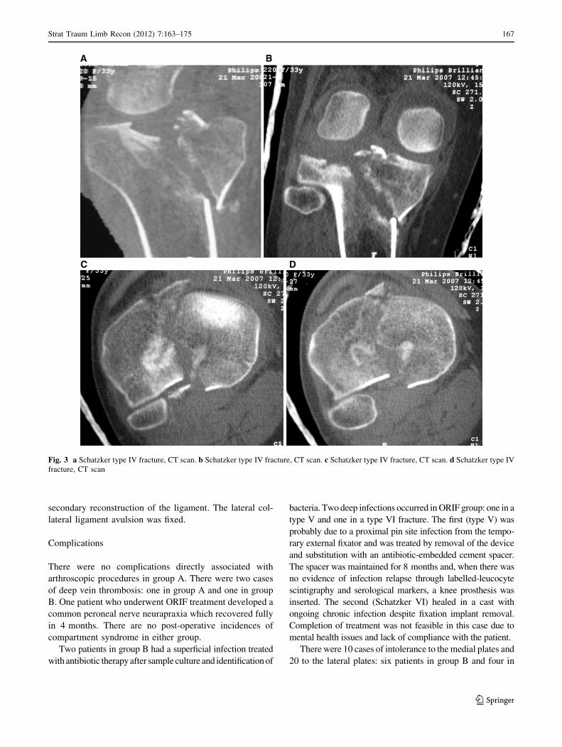
Fig. 3 a Schatzker type IV fracture, CT scan. b Schatzker type IV fracture, CT scan. c Schatzker type IV fracture, CT scan. d Schatzker type IV
fracture, CT scan
Strat Traum Limb Recon (2012) 7:163–175 167
123
group A needed the medial plates removed and 13 patients in
group B and seven in group A needed the same for the lateral
plates. No mechanical failures were observed. Four patients in
group B developed algodystrophy which was treated with
hyperbaric oxygen therapy and anti-osteoporotic drugs.
One case in group A (41 years old) had residual valgus
angulation and arthritis after 1 year which was treated with
uni-compartmental knee prosthesis. Two cases in group B
(67 and 69 years old) developed degenerative arthritis with
a significant post-traumatic valgus alignment and were
treated with a total knee prosthesis (Table 2).
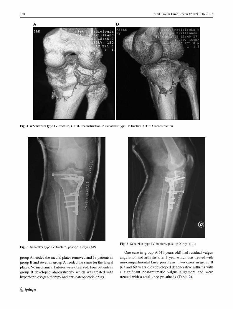
Fig. 4 a Schatzker type IV fracture, CT 3D reconstruction. b Schatzker type IV fracture, CT 3D reconstruction
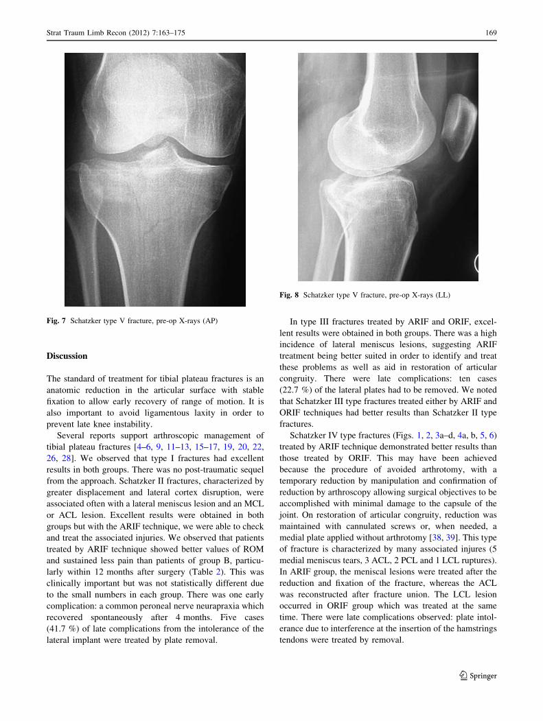
Fig. 5 Schatzker type IV fracture, post-op X-rays (AP)Fig. 6 Schatzker type IV fracture, post-op X-rays (LL)
168 Strat Traum Limb Recon (2012) 7:163–175
123
Discussion
The standard of treatment for tibial plateau fractures is an
anatomic reduction in the articular surface with stable
fixation to allow early recovery of range of motion. It is
also important to avoid ligamentous laxity in order to
prevent late knee instability.
Several reports support arthroscopic management of
tibial plateau fractures [4–6, 9, 11–13, 15–17, 19, 20, 22,
26, 28]. We observed that type I fractures had excellent
results in both groups. There was no post-traumatic sequel
from the approach. Schatzker II fractures, characterized by
greater displacement and lateral cortex disruption, were
associated often with a lateral meniscus lesion and an MCL
or ACL lesion. Excellent results were obtained in both
groups but with the ARIF technique, we were able to check
and treat the associated injuries. We observed that patients
treated by ARIF technique showed better values of ROM
and sustained less pain than patients of group B, particu-
larly within 12 months after surgery (Table 2). This was
clinically important but was not statistically different due
to the small numbers in each group. There was one early
complication: a common peroneal nerve neurapraxia which
recovered spontaneously after 4 months. Five cases
(41.7 %) of late complications from the intolerance of the
lateral implant were treated by plate removal.
In type III fractures treated by ARIF and ORIF, excel-
lent results were obtained in both groups. There was a high
incidence of lateral meniscus lesions, suggesting ARIF
treatment being better suited in order to identify and treat
these problems as well as aid in restoration of articular
congruity. There were late complications: ten cases
(22.7 %) of the lateral plates had to be removed. We noted
that Schatzker III type fractures treated either by ARIF and
ORIF techniques had better results than Schatzker II type
fractures.
Schatzker IV type fractures (Figs. 1, 2, 3a–d, 4a, b, 5, 6)
treated by ARIF technique demonstrated better results than
those treated by ORIF. This may have been achieved
because the procedure of avoided arthrotomy, with a
temporary reduction by manipulation and confirmation of
reduction by arthroscopy allowing surgical objectives to be
accomplished with minimal damage to the capsule of the
joint. On restoration of articular congruity, reduction was
maintained with cannulated screws or, when needed, a
medial plate applied without arthrotomy [38, 39]. This type
of fracture is characterized by many associated injures (5
medial meniscus tears, 3 ACL, 2 PCL and 1 LCL ruptures).
In ARIF group, the meniscal lesions were treated after the
reduction and fixation of the fracture, whereas the ACL
was reconstructed after fracture union. The LCL lesion
occurred in ORIF group which was treated at the same
time. There were late complications observed: plate intol-
erance due to interference at the insertion of the hamstrings
tendons were treated by removal.
Fig. 7 Schatzker type V fracture, pre-op X-rays (AP)
Fig. 8 Schatzker type V fracture, pre-op X-rays (LL)
Strat Traum Limb Recon (2012) 7:163–175 169
123
Cassard et al. described 26 patients with Schatzker types
I–IV fractures treated arthroscopically and concluded that
the results were as good as or better than from ORIF [40].
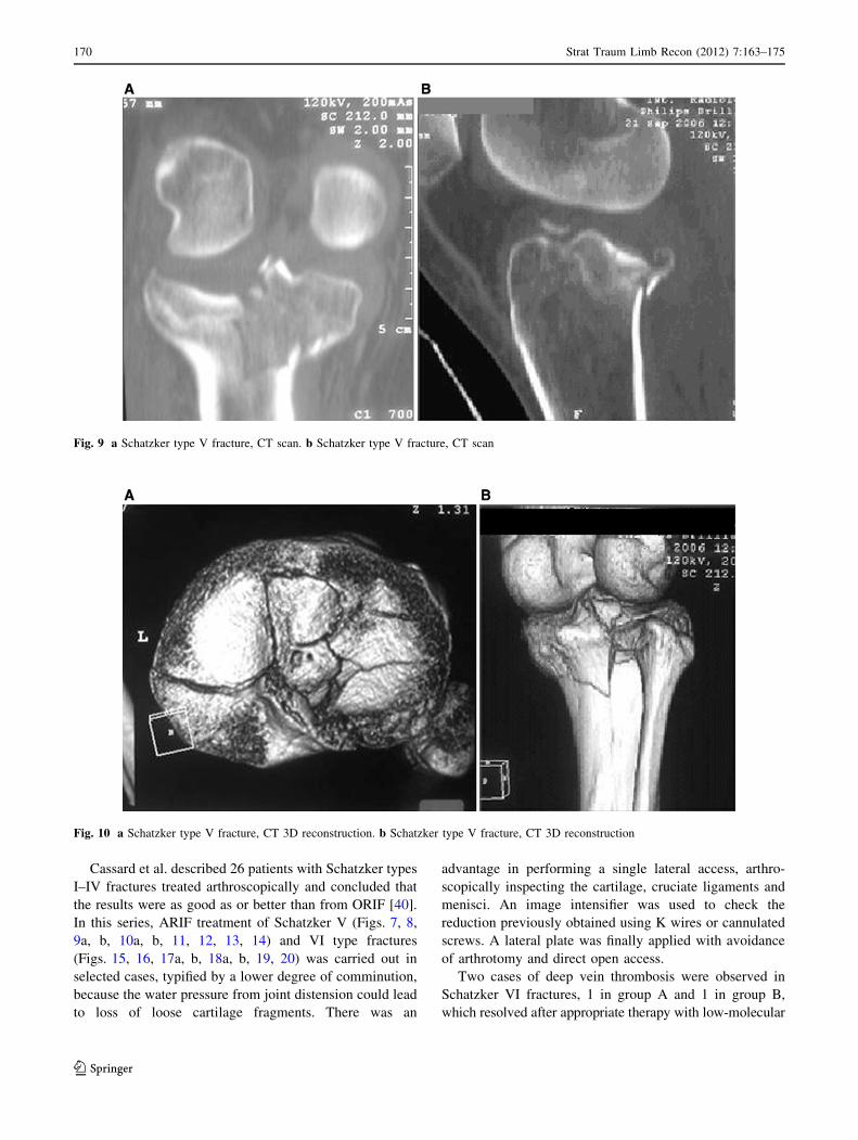
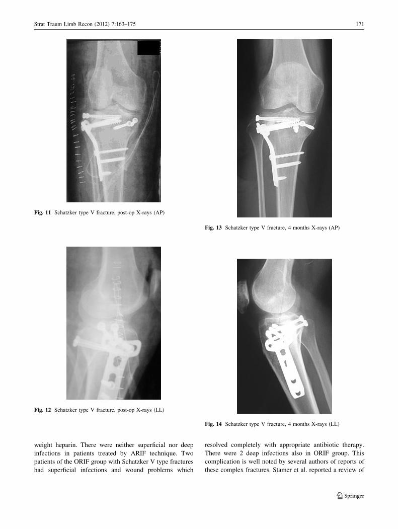
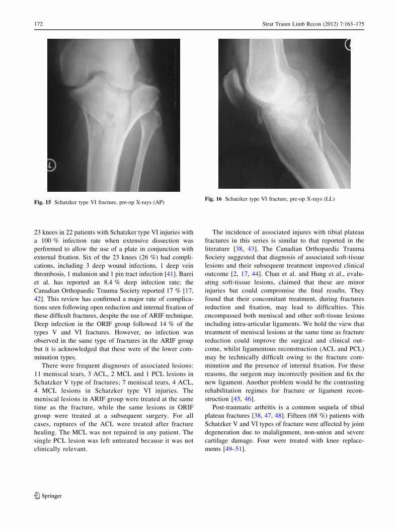
In this series, ARIF treatment of Schatzker V (Figs. 7, 8,
9a, b, 10a, b, 11, 12, 13, 14) and VI type fractures
(Figs. 15, 16, 17a, b, 18a, b, 19, 20) was carried out in
selected cases, typified by a lower degree of comminution,
because the water pressure from joint distension could lead
to loss of loose cartilage fragments. There was an
advantage in performing a single lateral access, arthro-
scopically inspecting the cartilage, cruciate ligaments and
menisci. An image intensifier was used to check the
reduction previously obtained using K wires or cannulated
screws. A lateral plate was finally applied with avoidance
of arthrotomy and direct open access.
Two cases of deep vein thrombosis were observed in
Schatzker VI fractures, 1 in group A and 1 in group B,
which resolved after appropriate therapy with low-molecular
Fig. 9 a Schatzker type V fracture, CT scan. b Schatzker type V fracture, CT scan
Fig. 10 a Schatzker type V fracture, CT 3D reconstruction. b Schatzker type V fracture, CT 3D reconstruction
170 Strat Traum Limb Recon (2012) 7:163–175
123
weight heparin. There were neither superficial nor deep
infections in patients treated by ARIF technique. Two
patients of the ORIF group with Schatzker V type fractures
had superficial infections and wound problems which
resolved completely with appropriate antibiotic therapy.
There were 2 deep infections also in ORIF group. This
complication is well noted by several authors of reports of
these complex fractures. Stamer et al. reported a review of
Fig. 11 Schatzker type V fracture, post-op X-rays (AP)
Fig. 12 Schatzker type V fracture, post-op X-rays (LL)
Fig. 13 Schatzker type V fracture, 4 months X-rays (AP)
Fig. 14 Schatzker type V fracture, 4 months X-rays (LL)
Strat Traum Limb Recon (2012) 7:163–175 171
123
23 knees in 22 patients with Schatzker type VI injuries with
a 100 % infection rate when extensive dissection was
performed to allow the use of a plate in conjunction with
external fixation. Six of the 23 knees (26 %) had compli-
cations, including 3 deep wound infections, 1 deep vein
thrombosis, 1 malunion and 1 pin tract infection [41]. Barei
et al. has reported an 8.4 % deep infection rate; the
Canadian Orthopaedic Trauma Society reported 17 % [17,
42]. This review has confirmed a major rate of complica-
tions seen following open reduction and internal fixation of
these difficult fractures, despite the use of ARIF technique.
Deep infection in the ORIF group followed 14 % of the
types V and VI fractures. However, no infection was
observed in the same type of fractures in the ARIF group
but it is acknowledged that these were of the lower com-
minution types.
There were frequent diagnoses of associated lesions:
11 meniscal tears, 3 ACL, 2 MCL and 1 PCL lesions in
Schatzker V type of fractures; 7 meniscal tears, 4 ACL,
4 MCL lesions in Schatzker type VI injuries. The
meniscal lesions in ARIF group were treated at the same
time as the fracture, while the same lesions in ORIF
group were treated at a subsequent surgery. For all
cases, ruptures of the ACL were treated after fracture
healing. The MCL was not repaired in any patient. The
single PCL lesion was left untreated because it was not
clinically relevant.
The incidence of associated injures with tibial plateau
fractures in this series is similar to that reported in the
literature [38, 43]. The Canadian Orthopaedic Trauma
Society suggested that diagnosis of associated soft-tissue
lesions and their subsequent treatment improved clinical
outcome [2, 17, 44]. Chan et al. and Hung et al., evalu-
ating soft-tissue lesions, claimed that these are minor
injuries but could compromise the final results. They
found that their concomitant treatment, during fractures
reduction and fixation, may lead to difficulties. This
encompassed both meniscal and other soft-tissue lesions
including intra-articular ligaments. We hold the view that
treatment of meniscal lesions at the same time as fracture
reduction could improve the surgical and clinical out-
come, whilst ligamentous reconstruction (ACL and PCL)
may be technically difficult owing to the fracture com-
minution and the presence of internal fixation. For these
reasons, the surgeon may incorrectly position and fix the
new ligament. Another problem would be the contrasting
rehabilitation regimes for fracture or ligament recon-
struction [45, 46].
Post-traumatic arthritis is a common sequela of tibial
plateau fractures [38, 47, 48]. Fifteen (68 %) patients with
Schatzker V and VI types of fracture were affected by joint
degeneration due to malalignment, non-union and severe
cartilage damage. Four were treated with knee replace-
ments [49–51].
Fig. 15 Schatzker type VI fracture, pre-op X-rays (AP)Fig. 16 Schatzker type VI fracture, pre-op X-rays (LL)
172 Strat Traum Limb Recon (2012) 7:163–175
123
Conclusions
There were no differences between ARIF and ORIF
treatment for type I tibial plateau fractures. We found
ARIF treatment preferable when meniscal tears were
present as it gave opportunity for simultaneous treatment.
In cases of Schatzker II, III and IV fractures, there was a
small difference in clinical outcomes in favour of ARIF but
not statistically significant. In Schatzker V or VI fractures,
ARIF treatment was limited to less comminuted fractures
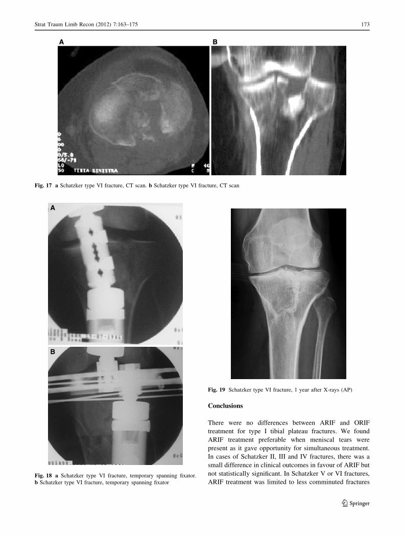
Fig. 17 a Schatzker type VI fracture, CT scan. b Schatzker type VI fracture, CT scan
Fig. 18 a Schatzker type VI fracture, temporary spanning fixator.
b Schatzker type VI fracture, temporary spanning fixator
Fig. 19 Schatzker type VI fracture, 1 year after X-rays (AP)
Strat Traum Limb Recon (2012) 7:163–175 173
123
and showed less incidence of infection. Mid-to-long-term
clinical results are influenced by the development of post-
traumatic arthritis and this itself was related to the severity
of the initial cartilage damage, subsequent malalignment
and non-union.
Open Access This article is distributed under the terms of the
Creative Commons Attribution License which permits any use, dis-
tribution, and reproduction in any medium, provided the original
author(s) and the source are credited.
References
1. Belanger M, Fadale P (1997) Compartment syndrome of the leg
after arthroscopic examination of a tibial plateau fracture. Case
report and review of the literature. Arthroscopy 13:646–651
2. Bennett WF, Browner B (1994) Tibial plateau fractures: a study
of associated soft tissue injuries. J Orthop Trauma 8:183–188
3. Crist BD, Della Rocca GJ, Stannard JP (2010) Compartment
syndrome surgical management techniques associated with tibial
plateau fractures. J Knee Surg 23:3–7
4. Dirschl DR, Dawson PA (2004) Injury severity assessment in
tibial plateau fractures. Clin Orthop Relat Res 423:85–92
5. Dirschl DR, Del Gaizo D (2007) Staged management of tibial
plateau fractures. Am J Orthop (Belle Mead NJ) 36:12–17
6. Duan XJ, Yang L, Guo L, Chan GX, Dai G (2008) Arthroscop-
ically assisted treatment for Schatzker type I-V tibial plateau
fractures. Chin J Traumatol 11(5):288–292
7. Lachiewicz PF, Funcik T (1990) Factors influencing the results of
open reduction and internal fixation of tibial plateau fractures.
Clin Orthop Relat Res 259:210–215
8. Rommens PM, Coosemans W, Broos PL (1989) The difficult
healing of segmental fractures of the tibial shaft. Arch Orthop
Trauma Surg 108:238–242
9. Scheerlinck T, Ng CS, Handelberg F, Casteleyn PP (1998)
Medium term results of percutaneous, arthroscopically-assisted
osteosynthesis of fractures of the tibial plateau. J Bone Joint Surg
Br 80:959–964
10. Weinlein J, Schmidt A (2010) Acute compartment syndrome in
tibial plateau fracture—beware! J Knee Surg 23:9–16
11. Zhou Z (2009) Arthroscopic percutaneous osteosynthesis of low
energy tibial plateau fractures. Zhongguo Xiu Fu Chong Jian Wai
Ke Za Zhi 23(11):1316–1318
12. Biggi F, Di Fabio S, D’Antimo C, Trevisani S (2010) Tibial
plateau fractures internal fixation with locking plates and the
MIPO technique. Injury 41(11):1178–1182
13. Farouk O, Krettek C, Miclau T et al (1997) Minimally invasive
plate osteosynthesis and vascularity: preliminary results of a
cadaver injection study. Injury 28(1):A7–A12
14. Musahl V, Tarkin I, Kobbe P et al (2009) New trends and tech-
niques in open reduction and internal fixation of fractures of the
tibial plateau. J Bone Joint Surg Br 91:426–433
15. Ali AM, Saleh M, Bolongaro S, Yang L (2003) The strength of
different fixation techniques for bicondylar tibial plateau frac-
tures—a biomechanical study. Clin Biomech 18:864–870
16. Ali AM, Saleh M, Eastell R, Wigderowitz CA, Rigby AS, Yang L
(2006) Influence of bone quality on the strength of internal and
external fixation of tibial plateau fractures. J Orthop Res
24:2080–2086
17. Canadian Orthopaedic Trauma Society (2006) Open reduction
and internal fixation compared with circular fixator application
for bicondylar tibial plateau fractures. Results of a multicenter,
prospective, randomized clinical trial. J Bone Joint Surg Am
88:2613–2623
18. Davies R, Holt N, Nayagam S (2005) The care of pin sites with
external fixation. J Bone Joint Surg [Br] 87-B:716–719
19. Giotakis N, Panchani SK, Narayan B, Larkin JJ, Al Maskari S,
Nayagam S (2010) Segmental fractures of the tibia treated by
circular external fixation. J Bone Joint Surg 92-B:687–692
20. Mallik AR, Covall DJ, Whitelaw GP (1992) Internal versus
external fixation of bicondylar tibial plateau fractures. Orthop
Rev 21:1433–1436
21. Marsh JL, Smith ST, Do TT (1995) External fixation and limited
internal fixation for complex fractures of the tibial plateau.
J Bone Joint Surg Am 77:661–673
22. Rademakers MV, Kerkhoffs GM, Sierevelt IN et al (2007)
Operative treatment of 109 tibial plateau fractures: five- to
27-year follow-up results. J Orthop Trauma 21:5–10
23. Young MJ, Barrack RL (1994) Complications of internal fixation
of tibial plateau fractures. Orthop Rev 23:149–154
24. Mui LW, Engelsohn E, Umans H (2007) Comparison of CT and MRI
in patients with tibial plateau fracture: can CT findings predict lig-
ament tear or meniscal injury. Skeletal Radiol 36:145–151
25. Te Stroet MA, Holla M, Biert J, Van Kampen A (2011) The value
of a CT scan compared to plain radiographs for the classification
and treatment plan in tibial plateau fractures. Emerg Radiol
18(4):279–283
26. Buchko GM, Johnson DH (1996) Arthroscopically assisted
operative management of tibial plateau fractures. Clin Orthop Rel
Res 332:29–36
27. Gill TJ, Moezzi DM, Oates KM, Sterett WI (2001) Arthroscopic
reduction and internal fixation of tibial plateau fractures in skiing.
Clin Orthop Rel Res 383:243–249
28. Siegler J, Galissier B, Marcheix PS, Charissoux JL, Mabit C,
Arnaud JP (2011) Percutaneous fixation of tibial plateau fractures
under arthoscopy: a medium term perspective. Orthop Traumatol
Surg Res 97:44–50
29. Hsu CJ, Chang WN, Wong CY (2001) Surgical treatment of tibial
plateau fracture in elderly patients. Arch Orthop Trauma Surg
121:67–70
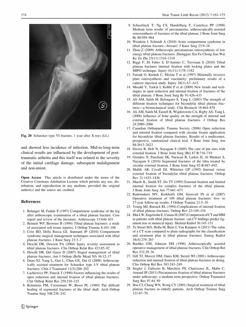
Fig. 20 Schatzker type VI fracture, 1 year after X-rays (LL)
174 Strat Traum Limb Recon (2012) 7:163–175
123
30. Lubokitz JH, Elson WS, Guttmann D (2004) Part I: arthroscopic
management of tibial plateau fractures. Arthroscopy 20(12):1063–1070
31. O’Dwyer KJ, Bobic VR (1992) Arthroscopic management of
tibial plateau fractures. Injury 23:261–264
32. Schatzker J, McBroom R, Bruce D (1979) The tibial plateau
fracture. The Toronto experience 1968–1975. Clin Orthop Relat
Res 138:94–104
33. Oestern HJ, Tscherne H (1984) Pathophysiology and classifica-
tion of soft tissue injuries associated with fractures. In: Tscherne
H, Gotzen L (eds) Fractures with soft tissue injuries. Springer,
Berlin
34. Egol KA, Tejwani NC, Capla EL, Wolinsky PL, Koval KJ (2005)
Staged management of high-energy proximal tibial fractures
(OTA types 41): the results of a prospective, standardized pro-
tocol. J Orthop Trauma 19:448–455
35. Tscherne H, Lobenhoffer P (1993) Tibial plateau fractures:
management and expected results. Clin Orthop Relat Res
292:87–100
36. Insall JN, Ranawat CS, Aglietti P, Shine J (1976) A comparison
of four models of total knee replacement prostheses. J Bone Joint
Surg [Am] 58-A:754–765
37. Rasmussen PS (1973) Tibial condylar fractures. Impairment of
knee joint function as an indication for surgical treatment. J Bone
Joint Surg Am 55:1331–1350
38. Chan Y-S, Chiu C-H, Lo Y-P, Chen AC-Y, Hsu K-Y, Wang C-J,
Chen W-J (2008) Arthroscopy-assisted surgery for tibial plateau
fractures: 2- to 10-year follow-up results. Arthroscopy 24:760–768
39. Chan Y-S, Yuan L-J, Hung S-S, Wang C-J, Shang-Won Y, Chen
C-Y, Chao E-K, Lee M (2003) Arthroscopic-assisted reduction
with bilateral buttress plate fixation of complex tibial plateau
fractures. Arthroscopy 19:974–984
40. Cassard X, Beaufils P, Blin JL, Hardy P (1999) Osteosynthesis
under arthroscopic control of separated tibial plateau fractures. 26
case reports. Rev Chir Orthop 85:257–266
41. Stamer DT, Schenk R, Staggers B, Aurori K, Aurori B, Behrens
FF (1994) Bicondylar tibial plateau fractures treated with a
hybrid ring external fixator: a preliminary study. J Orthop Trauma
8:455–461
42. Barei DP, Nork SE, Mills WJ, Henley MB, Benirschke SK (2004)
Complications associated with internal fixation of high-energy
bicondylar tibial plateau fractures utilizing a two-incision tech-
nique. J Orthop Trauma 18:649–657
43. Mohamed Zaki A-H, Chung-Hsun C, Yi-Sheng C, Yang-Pin L,
Jau-Wen H, Kuo-Yao H, Ching-Jen W (2006) Arthroscopic
evaluation of soft tissue injuries in tibial plateau fractures: ret-
rospective analysis of 98 cases. Arthroscopy 22:669–675
44. Holt MD, Williams LA, Dent CM (1995) MRI in the manage-
ment of tibial plateau fractures. Injury 26:595–599
45. Chan YS, Yuan LJ, Hung SS et al (2003) Arthroscopically-
assisted reduction with bilateral buttress plate fixation of complex
tibial plateau fractures. Arthroscopy 19:974–984
46. Hung SS, Chao EK, Chan YS et al (2003) Arthroscopically-
assisted osteosynthesis for tibial plateau fractures. J Trauma
54:356–363
47. Giannoudis PV, Tzioupis C, Papathanassopoulos A, Obakponovwe
O, Roberts C (2010) Articular step-off and risk of post-traumatic
osteoarthritis. Evidence today. Injury Int J Care Injured 41:986–995
48. Marsh JL, Buckwalter J, Gelberman R et al (2002) Articular
fractures: does an anatomic reduction really change the result?
J Bone Joint Surg Am 84-A:1259–1271
49. Honkonen SE (1995) Degenerative arthritis after tibial plateau
fractures. J Orthop Trauma 9:273–277
50. Moore TM, Patzakis MJ, Harvey JP (1987) Tibial plateau
fractures: definition, demographics, treatment rationale, and long-
term results of closed traction management or operative reduc-
tion. J Orthop Trauma 1:97–119
51. Woll TS, Duwelius PJ (1992) The segmental tibial fracture. Clin
Orthop 281:204–207
Strat Traum Limb Recon (2012) 7:163–175 175
123