the use of high-sensitivity assays for c-reactive protein in clinical practice
TRANSCRIPT

THE USE OF HIGH SENSITIVITY C-REACTIVE PROTEIN INCLINICAL PRACTICE
Kiran Musunuru*, Brian G Kral, Roger S Blumenthal, Valentin Fuster, Catherine YCampbell, Ty J Gluckman, Richard A Lange, Eric J Topol, James T Willerson, Milind YDesai, Michael H Davidson, and Samia Mora
SUMMARYMeasurement of the inflammatory biomarker high sensitivity C-reaction protein (hsCRP) has beenproposed for assessment of risk for cardiovascular disease (CVD). It remains unclear which patientpopulations would benefit from and should be targeted for hsCRP testing. Current data indicate thathsCRP levels are independently associated with risk of CVD, including both coronary events andstroke, in various asymptomatic populations; add predictive power to current coronary risk scoresfor some intermediate risk individuals; and are associated with clinical outcomes in high riskindividuals treated with statin therapy. HsCRP levels are also associated with incident diabetes andCVD outcomes in patients with the metabolic syndrome. There is a growing body of evidence tosupport recommendations for measurement of hsCRP in selected asymptomatic individuals deemedto be at intermediate risk of CVD according to traditional risk factor assessment and who do notalready warrant treatment with chronic aspirin and statin therapy, and selected secondary CVDprevention patients for further risk stratification in combination with LDL cholesterol.
Keywordscoronary disease; diabetes mellitus; prevention; risk factors; stroke
* Correspondence Johns Hopkins Ciccarone Preventive Cardiology Center, 600 North Wolfe Street/Blalock 524C, Baltimore, MD21287, USA [email protected] INTERESTS K Musunuru has declared an association with Alnylam Pharmaceuticals. TJ Gluckman has declaredassociations with the following companies: Sanofi-Aventis, Pfizer and Merck. MH Davidson has declared associations with diaDexus.See the article online for full details of the relationships. The other authors declared no competing interests.[Aus: to appear online: Dr. Musunuru has served as a consultant for Alnylam Pharmaceuticals within the last year. Dr. Gluckman hasreceived honoraria from Sanofi-Aventis and Pfizer and has served as a consultant for Merck within the last year. Dr. Davidson hasreceived honoraria from and served as a consultant for diaDexus within the last year. We declare no conflicts of interest pertaining tothis topic.]REVIEW CRITERIA We performed a comprehensive review of peer-reviewed publications that were identified through searches ofMEDLINE and the Cochrane Database from January 1990 through December 2007 using the search term “C-reactive protein”, incombination with one of the following: “heart disease”, “stroke”, “hypertension”, “metabolic syndrome” and “stroke”. Bibliographiesfrom these references were also reviewed, and additional studies were identified by experts. Initially identified papers were Englishlanguage, with the subject of the paper being the clinical risk prediction of cardiovascular disease or diabetes mellitus. All studies wereconsidered in our analysis. In analyzing the association of C-reactive protein (whether high-sensitivity or not) with cardiovascular disease(coronary heart disease or stroke) or diabetes in asymptomatic populations, we selected studies that used multivariate adjustment for atleast four traditional cardiovascular disease or diabetes risk factors. We excluded studies in which the populations had significantprevalence of comorbidities (>10% with, e.g., systolic heart failure, diabetes). In studies that included both men and women, whenseparate data were available for each sex we considered them separately rather than as a single population. When multiple publicationsreporting data from the same cohort were available, we chose the most recent publication.
NIH Public AccessAuthor ManuscriptNat Clin Pract Cardiovasc Med. Author manuscript; available in PMC 2009 April 1.
Published in final edited form as:Nat Clin Pract Cardiovasc Med. 2008 October ; 5(10): 621–635. doi:10.1038/ncpcardio1322.
NIH
-PA Author Manuscript
NIH
-PA Author Manuscript
NIH
-PA Author Manuscript

INTRODUCTIONMore than 800,000 individuals suffer a myocardial infarction annually in the US, and another700,000 experience a stroke.1 Of these events, nearly half occur in patients with no overtevidence of hyperlipidemia and 15% to 20% occur in patients with none of the major traditionalrisk factors.2,3 At the opposite end of the spectrum, a disproportionate number of events occurin individuals with a history of myocardial infarction, indicating the high level of risk forrecurrent events in these patients.
Although half of women and two-thirds of men in the US are affected by cardiovascular disease(CVD) after the age of 40,1, 4 only a small proportion of asymptomatic adults (<1% of womenand approximately 5% of men) are classified as at ̀ high risk' for CVD using contemporary riskscores. This discrepancy has been coined the ̀ detection gap'.5 In the US, 10% of asymptomaticwomen (~7 million) and 40% of asymptomatic men (~26 million) are considered to be atintermediate risk.5, 6 As the level of risk determines the intensity of preventive interventions,there is a clear need for better risk assessment in asymptomatic individuals, particularly thoseat intermediate risk.
The National Cholesterol Education Program Adult Treatment Panel (NCEP ATP) IIIguidelines7 provide a global risk score for `hard' coronary heart disease (CHD) events(myocardial infarction and death caused by coronary heart disease), and the 1998 Framinghamrisk score equation estimates total CHD events (myocardial infarction, cardiac death, coronaryinsufficiency);8, 9 a 10-year absolute risk of a hard coronary event less than 10% is consideredto be low risk, 10-20% is intermediate risk, and greater than 20% is high risk.7 It has beenproposed that the intermediate risk category be extended to include individuals who have a 10-year absolute risk between 5% and 20%. Proponents of this change argue that this lower cut-off identifies a group of individuals, especially women, who could gain more benefit fromaspirin and more aggressive lipid-lowering therapy and lifestyle modification than those with10-year absolute risk of less than 5%.5, 10 Increasingly, the intermediate risk category is beingfurther divided into `low' and `high' subgroups (i.e. 5-<10% and ≥10-<20%).
Numerous biomarkers have been proposed for improving CVD risk prediction. A biomarkeris felt to be useful if the following criteria are met: it adds to clinical knowledge; it providesrisk information that is independent of established predictors; it is easy to measure and interpretin a primary care setting; it is accurate, reproducible and internationally standardized; and ithas a favorable cost-benefit ratio.11 Screening biomarkers should also improve patientmanagement, particularly through more accurate risk classification and guidance in choice oftherapy.11 C-reactive protein (CRP) is an easily measured and widely investigated biomarkerof inflammation. The link between inflammation and atherosclerosis is well established;inflammation is a key element of the atherosclerotic process, contributing to all of its stages(initiation, growth, and plaque rupture).12-14 Thus, it would not be surprising if serum levelsof inflammatory markers such as CRP improve prediction of CHD and stroke risk in at leastsome patient populations.
Although a number of Reviews related to the high-sensitivity CRP test (hsCRP) have beenpublished in recent years, no review has comprehensively addressed the relevance of hsCRPin a variety of scenarios encountered in clinical practice—primary prevention of CVD, strokeand diabetes mellitus, and secondary prevention of CVD. We summarize the available dataand assess whether they support proposed guidelines for clinical hsCRP measurement. We willconsider CRP as a means to improve risk stratification and enable a better match betweentherapy and level of risk, not as a therapeutic target in its own right. Data are lacking as towhether CRP reduction per se reduces cardiovascular risk independent of other modifiable riskfactors.
Musunuru et al. Page 2
Nat Clin Pract Cardiovasc Med. Author manuscript; available in PMC 2009 April 1.
NIH
-PA Author Manuscript
NIH
-PA Author Manuscript
NIH
-PA Author Manuscript

PUBLISHED DATAPrimary prevention
In 2003, the Centers for Disease Control and Prevention (CDC) and the AHA issuedrecommendations regarding the use of inflammatory biomarkers for CVD detection,prevention and treatment.15 At the time, the body of evidence regarding the clinical use ofhsCRP measurements was modest, and most recommendations were given an ACC/AHA classII level of support, indicating that the weight of evidence was favorable but that more data wasneeded before general consensus could be reached.
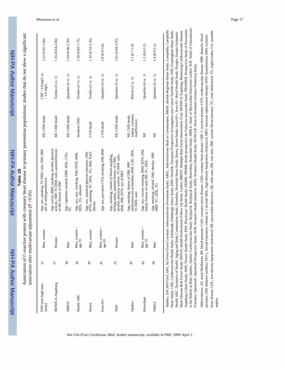
Since 2003, considerable data has been published regarding the use of hsCRP in improvingthe assessment of cardiovascular risk in primary prevention patients. We found at least 20prospective studies of distinct cohorts demonstrating that elevated hsCRP levels are associatedwith elevated risk of future coronary events after adjustment for at least four traditional riskfactors, including Framingham risk factors and/or diabetes and obesity (Table 1A and 1B).16-36 This association applied both to men and women across a wide age range (e.g. frommiddle-aged to elderly). Some studies stratified patients by hsCRP level—less than 1 mg/l, 1-3mg/l, and greater than 3 mg/l—and showed that these cutoffs correspond with lower, moderateand higher risk groups, respectively, although the risk was fairly linear across a wide range ofCRP levels. A small number of studies reported a positive association between hsCRP andcoronary event rate but none reached statistical significance after adjustment for at least fourother risk factors (Table 1A and 1B).25, 27, 37-44 Initial analysis of data from the FraminghamStudy found that CRP levels did not provide clear incremental value over the Framingham riskscore;37 however, the assay used to measure CRP was not high sensitivity. When the analysiswas repeated using a high-sensitivity assay, there was a positive correlation between CRP andCVD; after multivariate adjustment hsCRP levels greater than 3 mg/l were significantlyassociated with increased incident CVD (Table 1A and 1B).23
Although informative, individual studies are subject to variation and interpreting risk data canbe difficult. To date there has been one formal meta-analysis, but there is a clear need for furtherpooled investigations such as that from the Emerging Risk Factors Collaboration. Meta-analysis of 22 prospective studies found that after adjusting for traditional risk factorsindividuals in the top tertile of hsCRP levels (>3 mg/l) have a odds ratio of 1.45 for majorcardiac events (95% CI 1.25-1.68) compared with those in the lowest tertile (<1 mg/l).31 Thismeta-analysis incorporated studies that individually demonstrate a statistically significantassociation between hsCRP levels and cardiac events (including many of the studies listed inTable 1A) as well as a number of studies that did not show a statistically significant association(among those listed in Table 1B), suggesting that the overall conclusion of the study was notconfounded by publication bias.
CRP versus traditional risk factors: does CRP add incremental value?—Interestingly, in studies in which traditional risk factors underwent rigorous multivariateanalysis to assess the strength of association with CVD risk, the magnitude of the associationbetween incident CVD and hsCRP was comparable with that between CVD and LDL-cholesterol level, systolic blood pressure, or smoking behavior (Table 2). However, even if theassociation between elevated hsCRP levels and increased CVD risk is similar to that ofindividual traditional risk factors, the burden is on proponents of hsCRP measurement todemonstrate that the addition of hsCRP measurement to CVD risk prediction strategies has aclinical impact, even if only in limited patient populations. Recent data from the Women'sHealth Study suggest that adding CRP level to the NCEP ATP III global risk score improvesthe accuracy of CVD risk assessment in some asymptomatic individuals. In this largeprospective cohort study of asymptomatic middle-aged women, the addition of hsCRP to theATP III global risk score reclassified many intermediate risk individuals as higher or lower
Musunuru et al. Page 3
Nat Clin Pract Cardiovasc Med. Author manuscript; available in PMC 2009 April 1.
NIH
-PA Author Manuscript
NIH
-PA Author Manuscript
NIH
-PA Author Manuscript

risk; following hsCRP assessment 32% of women with a 5-<10% 10-year risk of `hard'coronary events and 42% of women with a 10-<20% 10-year absolute risk were reclassifiedinto a lower or higher-risk group.45
Using the same cohort of women, a more recent study that analyzed 35 cardiovascular riskfactors found that hsCRP provided the best prediction model for incident CVD events(myocardial infarction, stroke, revascularization, and CVD-related death) when used alongwith traditional risk factors (age, cholesterol, blood pressure, smoking and diabetes) andparental history of myocardial infarction before age 60 years.46 When this expanded riskalgorithm (the Reynolds Risk Score) was validated in a separate group of 8,158 womenfollowed up for 10 years, it provided more accurate risk assessment than did the smaller set oftraditional risk factors; 44% of women in intermediate risk categories (5-<10% and 10-<20%10-year risk) were reclassified as higher risk (27%) or lower risk (18%).46 It remains to beseen whether the Reynolds Risk Score will yield similar results in more diverse populations.In a cohort of middle-aged asymptomatic men, Koenig et al. showed that hsCRP providedincremental information regarding risk beyond that obtained using the Framingham risk score,particularly in those at intermediate risk.47 Additional studies show that hsCRP levels providerisk information incremental to the Framingham risk score in elderly men at intermediate riskand elderly women at high risk.20, 23
The c-statistic—The reclassification of intermediate-risk individuals to a different riskcategory could have important implications for preventive pharmacotherapy in these patients.It remains to be seen, however, whether such reclassification improves patient outcomes. Inthe absence of long-term, prospective studies, statistical criteria are being used to evaluate theincremental utility of hsCRP measurement. In a 2006 publication from the FraminghamOffspring Study, elevated baseline levels of hsCRP were associated with higher overallmortality during 7-year follow-up.48 Despite the higher mortality, the c-statistic (derived fromthe receiver-operator curve [ROC] whereby a value of 0.5 signifies a test of no utility and avalue of 1.0 signifies a test with perfect discrimination) of the risk prediction model did notchange with the addition of hsCRP. Indeed, most studies have not found the inclusion of hsCRPin models to increase the c-statistic significantly.
Whether the c-statistic is more suited to retrospective case-control studies than for prospectiverisk prediction models and whether criteria other than the c-statistic could be more appropriatefor assessing risk models is under debate.49-51 Although improvement of the c-statistic is onecriterion by which a biomarker can be judged to be ̀ ideal', relying solely on the c-statistic couldbe misleading and force the exclusion of clearly useful risk factors—the addition or subtractionof blood pressure and lipid profile individually from a model based on Framingham risk factorsdoes not significantly change the c-statistic.51 As more risk factors are incorporated into amodel it becomes increasingly difficult for a risk factor to increase the c-statistic, even if thatrisk factor carries as strong an association with the disease in question as the other risk factors.
The optimal set of parameters by which to judge the additive value of a biomarker to riskprediction algorithms is a subject of active investigation. A summary quantitative measure ofmodel fit that compares the proportion of individuals moving up or down in risk categorieswith the use of a biomarker (net reclassification index [NRI])52 has been used to assess whetherhsCRP adds information to traditional risk factors. In the Women's Health Study, the NRI usinghsCRP was 6%,113 whereas in the Framingham study, the NRI using hsCRP was 9%.23 Evenif one accepts that the c-statistic is the gold standard by which to assess the utility of hsCRP,most analyses of hsCRP have considered the change in the c-statistic when the test is appliedto a population as a whole, rather than just to intermediate-risk patients. In a cohort of middle-aged asymptomatic men, addition of hsCRP to the Framingham risk model improved the c-statistic from 0.735 to 0.750 when calculated for the whole study population—a modest change
Musunuru et al. Page 4
Nat Clin Pract Cardiovasc Med. Author manuscript; available in PMC 2009 April 1.
NIH
-PA Author Manuscript
NIH
-PA Author Manuscript
NIH
-PA Author Manuscript

that could be interpreted as being of little clinical importance. By contrast, when calculated forjust the intermediate-risk individuals with a 10-year CVD risk of 11-14% the c-statisticincreased from 0.725 to 0.776, and increased from 0.695 to 0.751 for patients with a 10-yearrisk of 15-19%—considerable improvements that support hsCRP use in these subgroups.47More analyses of this kind in different cohorts could help determine the appropriateness of thec-statistic in establishing the value of a biomarker.
In summary, hsCRP assessment in asymptomatic individuals seems most useful for those thatare at intermediate risk based on traditional risk factors (e.g. ATP III global risk score of 5-20%)and who do not already warrant chronic treatment with aspirin and statin therapy. In individualsat very low risk, even a doubling or tripling of risk (e.g. from 1% to 3%) would not changetheir risk classification and should not greatly change physician or patient behavior as theabsolute CVD risk remains low. Conversely, high-risk individuals are candidates for chronicaspirin and lipid-lowering therapy regardless of their hsCRP level. Among individuals atintermediate risk, however, reclassification to a higher or lower CVD risk category on the basisof hsCRP levels could influence decisions on whether to use more-aggressive or less-aggressive preventive strategies.
There are substantially more data now supporting the measurement of hsCRP in selectasymptomatic patients than there were in 2003, when the CDC and AHA guidelines werepublished. Nevertheless, more data are needed to establish the utility of hsCRP in creatingimproved risk prediction strategies—such as the Reynolds Risk Score—and validation of thosestrategies in intermediate-risk individuals in numerous cohorts.
Stroke and hypertensionIn numerous prospective studies, elevated hsCRP levels have correlated with an increased riskof stroke, even after adjusting for multiple traditional risk factors (Table 3).21, 24, 30, 34,53-57 Although as with coronary events there are some studies that fail to demonstrate astatistically significant association.39, 55, 57-60 Considering these studies together, therelative risk associated with elevated hsCRP levels is comparable to the relative risk of otherestablished risk factors for stroke, with as much as a three-fold increase in risk in high-CRPstrata compared to low-CRP strata. As a result of this strong association, consideration can begiven towards the measurement of hsCRP for the primary prevention of stroke in individualswith other risk factors for stroke who would not otherwise receive preventive therapy. Dataare, however, lacking on what proportions of individuals would be appropriately reclassifiedas being of higher or lower risk for stroke following incorporation of hsCRP.
Among individuals with blood pressure above desired goals as specified by the Joint NationalCommittee on Prevention, Detection, Evaluation, and Treatment of High Blood Pressure (JNC7) guidelines, the concomitant finding of elevated hsCRP should encourage the use ofantihypertensive therapy and more-aggressive lifestyle modification. However, it is reasonablethat all patients with hypertension and other risk factors for stroke should be counseled toundertake lifestyle modification. In conclusion, there are not yet specific data to support hsCRPmeasurement in addition to traditional strategies for stroke risk prediction.
The metabolic syndrome and diabetes mellitusIn individuals with the metabolic syndrome, elevated hsCRP levels correlated with both anincreased risk of developing non-insulin-dependent diabetes and the development of bothdiabetes and CVD.61-77 Multiple prospective cohort studies have confirmed hsCRP to beassociated with incident non-insulin-dependent diabetes independent of other risk factors suchas obesity, particularly in women (Table 4).61-73 In addition, CRP levels are associated with
Musunuru et al. Page 5
Nat Clin Pract Cardiovasc Med. Author manuscript; available in PMC 2009 April 1.
NIH
-PA Author Manuscript
NIH
-PA Author Manuscript
NIH
-PA Author Manuscript

CVD risk among those with the metabolic syndrome and appear to differentiate individuals athigh risk for both incident diabetes mellitus and CVD events from those at low risk.
Among individuals already diagnosed with diabetes, hsCRP levels can further stratifycardiovascular risk,72, 74, 75 underscoring the pathophysiologic link between insulinresistance, inflammation and CVD.66, 76, 77 As noted for stroke risk factors, it is importantto recommend more aggressive lifestyle modification to all individuals meeting criteria for themetabolic syndrome, regardless of whether they have elevated CRP levels.
Lifestyle interventionsWeight reduction, smoking cessation and exercise are recommended for patients at risk forCVD or diabetes, and these interventions have been shown to lower hsCRP levels.78-82,111, 112 It should be noted, however, that data are lacking as to whether hsCRP reduction perse reduces cardiovascular risk independent of other modifiable risk factors. Lifestyleinterventions should emphasize both increasing physical activity and dietary interventions thathelp the patient obtain and maintain an ideal body weight. In a large study of asymptomaticadult women and adjusted for cardiovascular risk factors, the relative risks for having hsCRPlevel greater than 3 mg/l were 1.3 for inactive, normal weight women, 2.7 for active, overweightwomen, 3.1 for inactive, overweight women, 8.3 for active, obese women, 9.9 for those whowere inactive and obese.81 In a comprehensive review of 40 observational studies and 12randomized clinical trials, most of which were in healthy individuals, both lower fitness andhigher fatness contributed to raised inflammation and hsCRP levels. Baseline levels of hsCRPmay be important in the overall changes observed with intervention studies, with the greatestchanges seen in those individuals with high baseline levels and, in some studies, no changesseen in individuals with low baseline levels.83
Statin therapyMany studies have shown that statin therapy lowers hsCRP levels, with relatively littlecorrelation between the degree of LDL-cholesterol reduction and hsCRP reduction inindividual patients.84-87 These data are consistent with laboratory studies demonstrating thatstatins have anti-inflammatory as well as lipid-lowering effects.88-90 As statins seem to besomewhat unique in this regard compared with other classes of lipid-lowering agents, theyshould be used preferentially over other lipid-lowering agents in the hypercholesterolemicpatient with elevated hsCRP. A greater degree of CVD event reduction with statin therapyoccurs in hypercholesterolemic patients with an elevated level of hsCRP than those with similarcholesterol levels and low levels of hsCRP;16, 86, 87, 91 however, whether CRP is raised ornot, all hypercholesterolemic patients should receive lipid-lowering therapy. Whether statinsprevent CVD events in individuals who have elevated hsCRP levels without hyperlipidemiawas the subject of a large-scale clinical trial (Justification for the Use of Statins in PrimaryPrevention: an Intervention Trial Evaluating Rosuvastatin [JUPITER]) that was recentlystopped early due to overwhelming benefit of rosuvastatin therapy in reducing adverse clinicaloutcomes.92 Until the results of JUPITER are published, statin therapy cannot be routinelyrecommended to patients with low levels of LDL-cholesterol and high levels of hsCRP;however, it is nonetheless reasonable to encourage substantial lifestyle changes (i.e. exercise,weight loss and complete smoking cessation) if not already undertaken. Of note, other agents,including metformin, thiazolidinediones, insulin, angiotensin-receptor blockers, andcombinations of agents, such as ezetimibe-statin combination therapy, are known to lowerhsCRP levels, but their optimum roles in primary prevention remain to be determined.
Secondary preventionA number of studies have demonstrated the prognostic utility of hsCRP in patients with acutecoronary syndromes,93-98 even when troponin is undetectable.97 When such high-risk
Musunuru et al. Page 6
Nat Clin Pract Cardiovasc Med. Author manuscript; available in PMC 2009 April 1.
NIH
-PA Author Manuscript
NIH
-PA Author Manuscript
NIH
-PA Author Manuscript

patients receive statin therapy, the best long-term clinical outcomes occur among those thatachieve very low levels of LDL cholesterol (<1.8 mmol/l [70 mg/dl]) and hsCRP (<2 mg/l).In the Pravastatin or Atorvastatin Evaluation and Infection Therapy - Thrombolysis inMyocardial Infarction 22 (PROVE IT - TIMI 22) and Aggrastat to Zocor (A to Z) trials, interms of reduction of coronary events and improvement in survival, achievement of low CRPlevels was as significant as reaching low LDL-cholesterol levels.87, 96 Furthermore, betteroutcomes were seen in individuals with both low LDL-cholesterol and low CRP levels than inthose with low LDL-C and high hsCRP levels.
Similar results have also been found in individuals with stable coronary artery disease, andthose who achieve low hsCRP levels on statin therapy have reduced risk of stroke99 andregression of atherosclerosis on intravascular ultrasonography.86 These data indicate thatachieving low levels of hsCRP after initiation of statin therapy could be an importanttherapeutic goal along with very low levels of LDL-cholesterol. It seems reasonable to considerhsCRP measurement in patients with a history of CVD who have achieved LDL-cholesterolgoals on low or moderate statin therapy—the finding of a high hsCRP level could help guidedecisions to further intensify statin therapy, although this strategy remains to be formallyvalidated in a prospective trial.
PRACTICAL CONSIDERATIONSTesting
CRP cutoffs of less than 1 mg/l, 1-3 mg/l, and greater than 3 mg/l are commonly used forcardiovascular risk discrimination and correspond to approximate tertile risk values inCaucasian populations.84 These same levels also discriminate risk of incident diabetes andvascular events among those with the metabolic syndrome.
Limited information is available regarding the utility of these hsCRP tertile levels in minoritypopulations,84 although evidence indicates that hsCRP levels are often higher in AfricanAmericans than in Caucasian and Asian Americans.100, 101 Of note, rates of CVD are alsoraised in African Americans compared with Caucasians.1 The effect of different treatmentstrategies across different ethnic groups based on hsCRP risk stratification is currentlyunknown.
The relationship between hsCRP and CVD risk is linear across the full range of CRP levels.An alternative system that divides hsCRP levels into five categories (<0.5 mg/l, 0.5-1.0 mg/l,1.0-3.0 mg/l, 3.0-5.0 mg/l, and >5.0 mg/l) could provide further discrimination,100 much inthe same manner that five categories are currently recommended for stratification of bloodpressure and lipids.102 The use of tertiles of hsCRP for risk stratification is consistent withrisk discrimination in major population studies but could lead to confusion and inconvenienceif applied in clinical practice. For primary prevention, therefore, the more conservativerecommendation is that high hsCRP levels be defined as 3 mg/l or greater, which readilyidentifies a group that has substantially increased risk compared with those individuals whohave hsCRP below 1 mg/l. For secondary prevention, levels of 2 mg/l or greater are consistentwith higher risk in patients with established coronary disease and ongoing treatment withstatins.103
Individuals with CRP levels that are consistently greater than 10 mg/l are at particularly highrisk for developing CVD.100, 102 CRP levels greater than 10 mg/l should not be viewed,therefore, as uninformative; patients with an hsCRP level in this range should undergo repeatassessment at a later date to see if the level remains elevated, which would suggest increasedlong-term vascular risk and perhaps warrant treatment.100, 102-104
Musunuru et al. Page 7
Nat Clin Pract Cardiovasc Med. Author manuscript; available in PMC 2009 April 1.
NIH
-PA Author Manuscript
NIH
-PA Author Manuscript
NIH
-PA Author Manuscript

Whereas most epidemiologic studies have relied on single hsCRP measurements per patient,in practice the clinical value of hsCRP could be improved if individuals with an initially highvalue undergo repeat assessment at least a month later. As hsCRP levels are not affected byintraindividual circadian variation or recent food ingestion, a blood sample for determinationof hsCRP level can be obtained at any time of the day, and a fasting sample is not required.74 Variation in hsCRP levels is comparable to that seen in cholesterol measurements.74 Itremains unclear whether there is significant seasonal variation in hsCRP levels and how thismight affect vascular risk.105, 106 As patients in the midst of an acute phase response canhave transiently elevated hsCRP levels, repeat testing is recommended for all values in excessof 5 mg/l. If the second blood sample yields a reduced hsCRP level, then the second valueshould be used in the assessment of CVD risk. When persistently high hsCRP values areobtained, vascular risk seems to be high regardless of the cause of the underlying inflammation.100, 102-104
Although older assays are capable of detecting high levels of CRP during the acute phaseresponse, these assays are not sensitive enough to detect the low levels of inflammation neededfor vascular risk prediction. As many hospital-based and outpatient laboratories offer CRPtesting to assess the presence systemic inflammatory states (i.e. collagen vascular disease,rheumatologic conditions, endocarditis) and hsCRP testing for cardiovascular evaluation,physicians need to specify an `hsCRP' test when they seek information concerning vascularrisk. Multiple commercial assays for hsCRP are available and have been standardized toprovide consistent clinical information in inpatient and outpatient settings.74 A comprehensiveprogram for standardization of commercial hsCRP assays was completed in 2003, so that allhsCRP results are now reported in mg/l.
Specificity for CVDIt is important to note that CRP is a marker of general inflammation and therefore couldhighlight the presence of chronic inflammatory conditions other than atherosclerosis. Case-control and retrospective studies have found associations between cancer and elevated hsCRPlevels, however, prospective studies have not confirmed this association.107 Elevated hsCRPlevels in cancer patients most likely reflect prevalent disease rather than being a marker offuture risk.107 Of note, hsCRP was shown to predict all-cause mortality in two recent studies.108, 109 In both studies CVD was the most common cause of death underlying all-causemortality, as it is in the general population, accounting for >60% of adult deaths. Even in lightof the possibility that CRP is not specific to vascular mortality but may also predict nonvascularmortality, in intermediate-risk patients with multiple CVD risk factors elevated CRP shouldbe regarded as a clear signal of CVD risk and can guide therapy specificially intended to reducevascular mortality, e.g., statin therapy.
Cost-effectivenessCost-effectiveness is an important consideration when assessing new biomarkers as screeningall patients has severe cost implications. A 2003 cost-effectiveness analysis examined theincremental cost-effectiveness of hsCRP screening followed by targeted statin therapy forindividuals with elevated levels, compared with dietary counseling alone, for the primaryprevention of cardiovascular events among patients with low or normal LDL cholesterol levels.110 The investigators found that using hsCRP screening to target statin therapy for the primaryprevention of CVD among individuals without overt hyperlipidemia was a cost-effective option—US$48,100 per quality-adjusted life-year (QALY) for 58-year-old men and $94,400 perQALY for 58-year-old women. In some scenarios, hsCRP was even cost-saving. Their resultsvaried by level of baseline cardiovascular risk and the cost and efficacy of statin therapy inpatients with high hsCRP levels. In light of the early termination of the JUPITER trial, theefficacy of statins might be much higher in asymptomatic individuals than originally expected.
Musunuru et al. Page 8
Nat Clin Pract Cardiovasc Med. Author manuscript; available in PMC 2009 April 1.
NIH
-PA Author Manuscript
NIH
-PA Author Manuscript
NIH
-PA Author Manuscript

In the primary prevention setting, a screening strategy that always requires lipids to bemeasured before hsCRP evaluation would probably not be cost-effective. In a patient knownto have a Framingham risk estimate of 5-20% on the basis of prior evaluations (i.e. intermediaterisk) and who is not receiving aspirin and/or statin therapy, concomitant hsCRP measurementat the time of lipid evaluation may be appropriate. As the cost of hsCRP is low, this approachmay be more efficient and more cost-effective than using a second physician visit and additionalphlebotomy after lipid results have been obtained.
CONCLUSIONSCRP levels when assessed by the high-sensitivity assay are associated with CVD in multiplepatient groups and add predictive power to traditional risk scores for some intermediate-riskindividuals. CRP data could also assist with targeting of lifestyle modification andpharmacologic preventive therapies. The available data support selective use of hsCRPmeasurement to improve risk prediction in the primary prevention setting in individuals atintermediate CVD risk according to traditional risk scores and who do not already warrantchronic aspirin and statin therapy. Data supporting the selective use of hsCRP levels to guidetreatment in secondary prevention patients not already on maximal statin therapy is currentlylimited. In both contexts, further validation studies will be needed before these strategies areuniversally endorsed.
BiographyK Musunuru is Clinical Fellow, BG Kral is Clinical Fellow, RS Blumenthal is Professor ofMedicine, CY Campbell is Clinical Fellow, TJ Gluckman is Clinical Fellow, and RA Langeis E. Cowles Andrus Professor of Cardiology at the Johns Hopkins Ciccarone PreventiveCardiology Center, Johns Hopkins University School of Medicine, Baltimore, MD, USA. VFuster is Director of the Zena and Michael A Wiener Cardiovascular Institute and the Marie-José and Henry R Kravis Center for Cardiovascular Health, Mount Sinai School of Medicine,New York, NY. E Topol is Director of Scripps Genomic Medicine at Scripps TranslationalScience Institute, La Jolla, CA. JT Willerson is President-Elect and Medical Director at St.Luke's Episcopal Hospital/Texas Heart Institute, Houston, TX, USA. MY Desai is AssistantProfessor of Medicine at the Department of Cardiovascular Medicine, Cleveland ClinicFoundation, and Lerner College of Medicine, Case Western Reserve University, Cleveland,OH. MH Davidson is Director of Preventive Cardiology and Atherosclerosis Research andClinical Professor of Medicine at University of Chicago School of Medicine, Chicago, IL. SMora is Associate Physician and Assistant Professor of Medicine at the Divisions of Preventiveand Cardiovascular Medicine, Brigham and Women's Hospital, Harvard Medical School,Boston, MA.
REFERENCES1. Thom T, et al. Heart disease and stroke statistics—2006 update: a report from the American Heart
Association Statistics Committee and Stroke Statistics Subcommittee. Circulation 2006;113:e85–e151. [PubMed: 16407573]
2. Greenland P, et al. Major risk factors as antecedents of fatal and nonfatal coronary heart disease events.JAMA 2003;290:891–897. [PubMed: 12928465]
3. Khot UN, et al. Prevalence of conventional risk factors in patients with coronary heart disease. JAMA2003;290:898–904. [PubMed: 12928466]
4. Lloyd-Jones DM, et al. Lifetime risk of developing coronary heart disease. Lancet 1999;353:89–92.[PubMed: 10023892]
5. Pasternak RC, et al. 34th Bethesda conference: task force #1—identification of coronary heart diseaserisk: is there a detection gap? J Am Coll Cardiol 2003;41:1863–1874. [PubMed: 12798553]
Musunuru et al. Page 9
Nat Clin Pract Cardiovasc Med. Author manuscript; available in PMC 2009 April 1.
NIH
-PA Author Manuscript
NIH
-PA Author Manuscript
NIH
-PA Author Manuscript

6. Ford ES, et al. The distribution of 10-year risk for coronary heart disease among US adults: findingsfrom the National Health and Nutrition Examination Survey III. J Am Coll Cardiol 2004;43:1791–1796. [PubMed: 15145101]
7. National Cholesterol Education Program (NCEP) Expert Panel on Detection, Evaluation, andTreatment of High Blood Cholesterol in Adults (Adult Treatment Panel III). Third report of theNational Cholesterol Education Program (NCEP) Expert Panel on Detection, Evaluation, andTreatment of High Blood Cholesterol in Adults (Adult Treatment Panel III) final report. Circulation2002;106:3143–3421. [PubMed: 12485966]
8. Wilson PW, et al. Prediction of coronary heart disease using risk factor categories. Circulation1998;97:1837–1847. [PubMed: 9603539]
9. D'Agostino RB Sr, et al. Validation of the Framingham coronary heart disease prediction scores: resultsof a multiple ethnic groups investigation. JAMA 2001;286:180–187. [PubMed: 11448281]
10. Berman DS, Wong ND. Implications of estimating coronary heart disease risk in the US population.J Am Coll Cardiol 2004;43:1797–1798. [PubMed: 15145102]
11. Vasan RS. Biomarkers of cardiovascular disease: molecular basis and practical considerations.Circulation 2006;113:2335–2362. [PubMed: 16702488]
12. Hansson GK. Inflammation, atherosclerosis, and coronary artery disease. N Engl J Med2005;352:1685–1695. [PubMed: 15843671]
13. Ross R. Atherosclerosis—an inflammatory disease. N Engl J Med 1999;340:115–126. [PubMed:9887164]
14. Libby P, Ridker PM. Inflammation and atherothrombosis from population biology and bench researchto clinical practice. J Am Coll Cardiol 2006;48(9 Suppl):A33–A46.
15. Pearson TA, et al. Markers of inflammation and cardiovascular disease: application to clinical andpublic health practice: a statement for healthcare professionals from the Centers for Disease Controland Prevention and the American Heart Association. Circulation 2003;107:499–511. [PubMed:12551878]
16. Ridker PM, et al. Measurement of C-reactive protein for the targeting of statin therapy in the primaryprevention of acute coronary events. N Engl J Med 2001;344:1959–1965. [PubMed: 11430324]
17. Ballantyne CM, et al. Lipoprotein-associated phospholipase A2, high-sensitivity C-reactive protein,and risk for incident coronary heart disease in middle-aged men and women in the AherosclerosisRisk in Communities (ARIC) study. Circulation 2004;109:837–842. [PubMed: 14757686]
18. Danesh J, et al. Low grade inflammation and coronary heart disease: prospective study and updatedmeta-analyses. BMJ 2000;321:199–204. [PubMed: 10903648]
19. Lowe GD, et al. C-reactive protein, fibrin D-dimer, and risk of ischemic heart disease: the Caerphillyand Speedwell studies. Arterioscler Thromb Vasc Biol 2004;24:1957–1962. [PubMed: 15308549]
20. Cushman M, et al. C-reactive protein and the 10-year incidence of coronary heart disease in oldermen and women: the cardiovascular health study. Circulation 2005;112:25–31. [PubMed: 15983251]
21. Tzoulaki I, et al. Relative value of inflammatory, hemostatic, and rheological factors for incidentmyocardial infarction and stroke: the Edinburgh Artery Study. Circulation 2007;115:2119–2127.[PubMed: 17404162]
22. Boekholdt SM, et al. C-reactive protein levels and coronary artery disease incidence and mortality inapparently healthy men and women: the EPIC-Norfolk prospective population study 1993-2003.Atherosclerosis 2006;187:415–422. [PubMed: 16257408]
23. Wilson P, et al. Increased CRP and long term risk for cardiovascular events in middle age men andwomen. Circulation 2006;II:28a.
24. Sakkinen P, et al. C-reactive protein and myocardial infarction. J Clin Epidemiol 2002;55:445–451.[PubMed: 12007546]
25. Pai JK, et al. Inflammatory markers and the risk of coronary heart disease in men and women. N EnglJ Med 2004;351:2599–2610. [PubMed: 15602020]
26. Laaksonen DE, et al. C-reactive protein in the prediction of cardiovascular and overall mortality inmiddle-aged men: a population-based cohort study. Eur Heart J 2005;26:1783–1789. [PubMed:15821003]
27. Koenig W, et al. Increased concentrations of C-reactive protein and IL-6 but not IL-18 areindependently associated with incident coronary events in middle-aged men and women: results from
Musunuru et al. Page 10
Nat Clin Pract Cardiovasc Med. Author manuscript; available in PMC 2009 April 1.
NIH
-PA Author Manuscript
NIH
-PA Author Manuscript
NIH
-PA Author Manuscript

the MONICA/KORA Augsburg case-cohort study, 1984-2002. Arterioscler Thromb Vasc Biol2006;26:2745–2751. [PubMed: 17008587]
28. Ridker PM, et al. Inflammation, aspirin, and the risk of cardiovascular disease in apparently healthymen. N Engl J Med 1997;336:973–979. [PubMed: 9077376]
29. Luc G, et al. C-reactive protein, interleukin-6, and fibrinogen as predictors of coronary heart disease:the PRIME study. Arterioscler Thromb Vasc Biol 2003;23:1255–1261. [PubMed: 12775578]
30. Sattar N, et al. C-reactive protein and prediction of coronary heart disease and global vascular eventsin the Prospective Study of Pravastatin in the Elderly at Risk (PROSPER). Circulation 2007;115:981–989. [PubMed: 17283264]
31. Danesh J, et al. C-reactive protein and other circulating markers of inflammation in the prediction ofcoronary heart disease. N Engl J Med 2004;350:1387–1397. [PubMed: 15070788]
32. Tice JA, et al. The relation of C-reactive protein levels to total and cardiovascular mortality in olderU.S. women. Am J Med 2003;114:199–205. [PubMed: 12637134]
33. Pradhan AD, et al. Inflammatory biomarkers, hormone replacement therapy, and incident coronaryheart disease: prospective analysis from the Women's Health Initiative observational study. JAMA2002;288:980–987. [PubMed: 12190368]
34. Ridker PM, et al. Comparison of C-reactive protein and low-density lipoprotein cholesterol levels inthe prediction of first cardiovascular events. N Engl J Med 2002;347:1557–1565. [PubMed:12432042]
35. Lowe GD, et al. Interleukin-6, fibrin D-dimer, and coagulation factors VII and XIIa in prediction ofcoronary heart disease. Arterioscler Thromb Vasc Biol 2004;24:1529–1534. [PubMed: 15205218]
36. Ridker PM, et al. Non-HDL cholesterol, apolipoproteins A-I and B100, standard lipid measures, lipidratios, and CRP as risk factors for cardiovascular disease in women. JAMA 2005;294:326–333.[PubMed: 16030277]
37. Wilson PW, et al. C-reactive protein and risk of cardiovascular disease in men and women from theFramingham Heart Study. Arch Intern Med 2005;165:2473–2478. [PubMed: 16314543]
38. Kuller LH, et al. Relation of C-reactive protein and coronary heart disease in the MRFIT nested case-control study. Multiple risk factor intervention trial. Am J Epidemiol 1996;144:537–547. [PubMed:8797513]
39. Cesari M, et al. Inflammatory markers and onset of cardiovascular events: results from the HealthABC study. Circulation 2003;108:2317–2322. [PubMed: 14568895]
40. Jager A, et al. Von Willebrand factor, C-reactive protein, and 5-year mortality in diabetic andnondiabetic subjects: the Hoorn Study. Arterioscler Thromb Vasc Biol 1999;19:3071–3078.[PubMed: 10591689]
41. Harris TB, et al. Associations of elevated interleukin-6 and C-reactive protein levels with mortalityin the elderly. Am J Med 1999;106:506–512. [PubMed: 10335721]
42. Pirro M, et al. Age and duration of follow-up as modulators of the risk for ischemic heart diseaseassociated with high plasma C-reactive protein levels in men. Arch Intern Med 2001;161:2474–2480.[PubMed: 11700160]
43. van der Meer IM, et al. The value of C-reactive protein in cardiovascular risk prediction: the RotterdamStudy. Arch Intern Med 2003;163:1323–1328. [PubMed: 12796068]
44. Doggen CJ, et al. C-reactive protein, cardiovascular risk factors and the association with myocardialinfarction in men. J Intern Med 2000;248:406–414. [PubMed: 11123505]
45. Cook NR, et al. The effect of including C-reactive protein in cardiovascular risk prediction modelsfor women. Ann Intern Med 2006;145:21–29. [PubMed: 16818925]
46. Ridker PM, et al. Development and validation of improved algorithms for the assessment of globalcardiovascular risk in women: the Reynolds Risk Score. JAMA 2007;297:611–619. [PubMed:17299196]
47. Koenig W, et al. C-reactive protein modulates risk prediction based on the Framingham Score:implications for future risk assessment: results from a large cohort study in southern Germany.Circulation 2004;109:1349–1353. [PubMed: 15023871]
48. Wang TJ, et al. Multiple biomarkers for the prediction of first major cardiovascular events and death.N Engl J Med 2006;355:2631–2639. [PubMed: 17182988]
Musunuru et al. Page 11
Nat Clin Pract Cardiovasc Med. Author manuscript; available in PMC 2009 April 1.
NIH
-PA Author Manuscript
NIH
-PA Author Manuscript
NIH
-PA Author Manuscript

49. Gail MH, Pfeiffer RM. On criteria for evaluating models of absolute risk. Biostatistics 2005;6:227–239. [PubMed: 15772102]
50. Harrell, FE. Regression Modeling Strategies: With Applications to Linear Models, LogisticRegression, and Survival Analysis. Springer; New York: 2001.
51. Cook NR. Use and misuse of the receiver operating characteristic curve in risk prediction. Circulation2007;115:928–935. [PubMed: 17309939]
52. Pencina MJ, et al. Evaluating the added predictive ability of a new marker: from area under the ROCcurve to reclassification and beyond. Stat Med 2008;27:157–172. [PubMed: 17569110]
53. Ballantyne CM, et al. Lipoprotein-associated phospholipase A2, high-sensitivity C-reactive protein,and risk for incident ischemic stroke in middle-aged men and women in the atherosclerosis risk incommunities (ARIC) study. Arch Intern Med 2005;165:2479–2484. [PubMed: 16314544]
54. Cao JJ, et al. C-reactive protein, carotid intima-media thickness, and incidence of ischemic stroke inthe elderly: the Cardiovascular Health Study. Circulation 2003;108:166–170. [PubMed: 12821545]
55. Rost NS, et al. Plasma concentration of C-reactive protein and risk of ischemic stroke and transientischemic attack: the Framingham study. Stroke 2001;32:2575–2579. [PubMed: 11692019]
56. Wakugawa Y, et al. C-reactive protein and risk of first-ever ischemic and hemorrhagic stroke in ageneral Japanese population: the Hisayama Study. Stroke 2006;37:27–32. [PubMed: 16306468]
57. Ford ES, Giles WH. Serum C-reactive protein and self-reported stroke: findings from the ThirdNational Health and Nutrition Examination Survey. Arterioscler Thromb Vasc Biol 2000;20:1052–1056. [PubMed: 10764672]
58. Kistorp C, et al. N-terminal pro-brain natriuretic peptide, C-reactive protein, and urinary albuminlevels as predictors of mortality and cardiovascular events in older adults. JAMA 2005;293:1609–1616. [PubMed: 15811980]
59. Gussekloo J, et al. C-reactive protein is a strong but nonspecific risk factor of fatal stroke in elderlypersons. Arterioscler Thromb Vasc Biol 2000;20:1047–1051. [PubMed: 10764671]
60. Bos MJ, et al. High serum C-reactive protein level is not an independent predictor for stroke: theRotterdam Study. Circulation 2006;114:1591–1598. [PubMed: 17015791]
61. Duncan BB, et al. Low-grade systemic inflammation and the development of type 2 diabetes: theatherosclerosis risk in communities study. Diabetes 2003;52:1799–1805. [PubMed: 12829649]
62. Wang Z, Hoy WE. C-reactive protein and the risk of developing type 2 diabetes in AboriginalAustralians. Diabetes Res Clin Pract 2007;76:37–43. [PubMed: 16952410]
63. Barzilay JI, et al. The relation of markers of inflammation to the development of glucose disordersin the elderly: the Cardiovascular Health Study. Diabetes 2001;50:2384–2389. [PubMed: 11574423]
64. Spranger J, et al. Inflammatory cytokines and the risk to develop type 2 diabetes: results of theprospective population-based European Prospective Investigation into Cancer and Nutrition (EPIC)-Potsdam Study. Diabetes 2003;52:812–817. [PubMed: 12606524]
65. Doi Y, et al. Elevated C-reactive protein is a predictor of the development of diabetes in a generalJapanese population: the Hisayama Study. Diabetes Care 2005;28:2497–2500. [PubMed: 16186286]
66. Festa A, et al. Elevated levels of acute-phase proteins and plasminogen activator inhibitor-1 predictthe development of type 2 diabetes: the insulin resistance atherosclerosis study. Diabetes2002;51:1131–1137. [PubMed: 11916936]
67. Nakanishi S, et al. Elevated C-reactive protein is a risk factor for the development of type 2 diabetesin Japanese Americans. Diabetes Care 2003;26:2754–2757. [PubMed: 14514575]
68. Laaksonen DE, et al. C-reactive protein and the development of the metabolic syndrome and diabetesin middle-aged men. Diabetologia 2004;47:1403–1410. [PubMed: 15309290]
69. Han TS, et al. Prospective study of C-reactive protein in relation to the development of diabetes andmetabolic syndrome in the Mexico City Diabetes Study. Diabetes Care 2002;25:2016–2021.[PubMed: 12401749]
70. Thorand B, et al. Sex differences in the prediction of type 2 diabetes by inflammatory markers: resultsfrom the MONICA/KORA Augsburg case-cohort study, 1984-2002. Diabetes Care 2007;30:854–860. [PubMed: 17392546]
71. Hu FB, et al. Inflammatory markers and risk of developing type 2 diabetes in women. Diabetes2004;53:693–700. [PubMed: 14988254]
Musunuru et al. Page 12
Nat Clin Pract Cardiovasc Med. Author manuscript; available in PMC 2009 April 1.
NIH
-PA Author Manuscript
NIH
-PA Author Manuscript
NIH
-PA Author Manuscript

72. Pradhan AD, et al. C-reactive protein, interleukin 6, and risk of developing type 2 diabetes mellitus.JAMA 2001;286:327–334. [PubMed: 11466099]
73. Freeman DJ, et al. C-reactive protein is an independent predictor of risk for the development ofdiabetes in the West of Scotland Coronary Prevention Study. Diabetes 2002;51:1596–1600.[PubMed: 11978661]
74. Ridker PM, et al. Should C-reactive protein be added to metabolic syndrome and to assessment ofglobal cardiovascular risk? Circulation 2004;109:2818–2825. [PubMed: 15197153]
75. Malik S, et al. Cardiovascular disease in U.S. patients with metabolic syndrome, diabetes, and elevatedC-reactive protein. Diabetes Care 2005;28:690–693. [PubMed: 15735209]
76. Wannamethee SG, et al. The metabolic syndrome and insulin resistance: relationship to haemostaticand inflammatory markers in older non-diabetic men. Atherosclerosis 2005;181:101–108. [PubMed:15939060]
77. Yudkin JS, et al. C-reactive protein in healthy subjects: associations with obesity, insulin resistance,and endothelial dysfunction: a potential role for cytokines originating from adipose tissue?Arterioscler Thromb Vasc Biol 1999;19:972–978. [PubMed: 10195925]
78. Bazzano LA, et al. Relationship between cigarette smoking and novel risk factors for cardiovasculardisease in the United States. Ann Intern Med 2003;138:891–897. [PubMed: 12779299]
79. Bermudez EA, et al. Relation between markers of systemic vascular inflammation and smoking inwomen. Am J Cardiol 2002;89:1117–1119. [PubMed: 11988205]
80. Esposito K, et al. Effect of weight loss and lifestyle changes on vascular inflammatory markers inobese women: a randomized trial. JAMA 2003;289:1799–1804. [PubMed: 12684358]
81. Mora S, et al. Association of physical activity and body mass index with novel and traditionalcardiovascular biomarkers in women. JAMA 2006;295:1412–1419. [PubMed: 16551713]
82. Tchernof A, et al. Weight loss reduces C-reactive protein levels in obese postmenopausal women.Circulation 2002;105:564–569. [PubMed: 11827920]
83. Hamer M. The relative influences of fitness and fatness on inflammatory factors. Prev Med 2007;44:3–11. [PubMed: 17064760]
84. Ridker PM. Clinical application of C-reactive protein for cardiovascular disease detection andprevention. Circulation 2003;107:363–369. [PubMed: 12551853]
85. Tsimikas S, et al. C-reactive protein and other emerging blood biomarkers to optimize riskstratification of vulnerable patients. J Am Coll Cardiol 2006;47:C19–C31. [PubMed: 16631506]
86. Nissen SE, et al. Statin therapy, LDL cholesterol, C-reactive protein, and coronary artery disease. NEngl J Med 2005;352:29–38. [PubMed: 15635110]
87. Ridker PM, et al. C-reactive protein levels and outcomes after statin therapy. N Engl J Med2005;352:20–28. [PubMed: 15635109]
88. Arnaud C, et al. Statins reduce interleukin-6-induced C-reactive protein in human hepatocytes: newevidence for direct antiinflammatory effects of statins. Arterioscler Thromb Vasc Biol 2005;25:1231–1236. [PubMed: 15790934]
89. Jain MK, Ridker PM. Anti-inflammatory effects of statins: clinical evidence and basic mechanisms.Nat Rev Drug Discov 2005;4:977–987. [PubMed: 16341063]
90. Schonbeck U, Libby P. Inflammation, immunity, and HMG-CoA reductase inhibitors: statins asantiinflammatory agents? Circulation 2004;109:II18–II26. [PubMed: 15173059]
91. Ridker PM, et al. Inflammation, pravastatin, and the risk of coronary events after myocardial infarctionin patients with average cholesterol levels. Cholesterol and Recurrent Events (CARE) investigators.Circulation 1998;98:839–844. [PubMed: 9738637]
92. Ridker PM. Rosuvastatin in the primary prevention of cardiovascular disease among patients withlow levels of low-density lipoprotein cholesterol and elevated high-sensitivity C-reactive protein:rationale and design of the JUPITER trial. Circulation 2003;108:2292–2297. [PubMed: 14609996]
93. Haverkate F, et al. Production of C-reactive protein and risk of coronary events in stable and unstableangina. European Concerted Action on Thrombosis and Disabilities Angina Pectoris Study Group.Lancet 1997;349:462–466. [PubMed: 9040576]
Musunuru et al. Page 13
Nat Clin Pract Cardiovasc Med. Author manuscript; available in PMC 2009 April 1.
NIH
-PA Author Manuscript
NIH
-PA Author Manuscript
NIH
-PA Author Manuscript

94. Lindahl B, et al. Markers of myocardial damage and inflammation in relation to long-term mortalityin unstable coronary artery disease. FRISC Study Group. Fragmin during Instability in CoronaryArtery Disease. N Engl J Med 2000;343:1139–1147. [PubMed: 11036119]
95. Liuzzo G, et al. The prognostic value of C-reactive protein and serum amyloid a protein in severeunstable angina. N Engl J Med 1994;331:417–424. [PubMed: 7880233]
96. Morrow DA, et al. Clinical relevance of C-reactive protein during follow-up of patients with acutecoronary syndromes in the Aggrastat-to-Zocor Trial. Circulation 2006;114:281–288. [PubMed:16847150]
97. Morrow DA, et al. C-reactive protein is a potent predictor of mortality independently of and incombination with troponin T in acute coronary syndromes: a TIMI 11A substudy. Thrombolysis inMyocardial Infarction. J Am Coll Cardiol 1998;31:1460–1465. [PubMed: 9626820]
98. Sabatine MS, et al. Multimarker approach to risk stratification in non-ST elevation acute coronarysyndromes: simultaneous assessment of troponin I, C-reactive protein, and B-type natriuretic peptide.Circulation 2002;105:1760–1763. [PubMed: 11956114]
99. Mega JL, et al. Cholesterol, C-reactive protein, and cerebrovascular events following intensive andmoderate statin therapy. J Thromb Thrombolysis 2006;22:71–76. [PubMed: 16786236]
100. Albert MA, et al. C-reactive protein levels among women of various ethnic groups living in theUnited States (from the Women's Health Study). Am J Cardiol 2004;93:1238–1242. [PubMed:15135696]
101. Albert MA, Ridker PM. C-reactive protein as a risk predictor: do race/ethnicity and gender make adifference? Circulation 2006;114:e67–e74. [PubMed: 16880331]
102. Ridker PM, Cook N. Clinical usefulness of very high and very low levels of C-reactive protein acrossthe full range of Framingham Risk Scores. Circulation 2004;109:1955–1959. [PubMed: 15051634]
103. Sabatine MS, et al. Prognostic significance of the Centers for Disease Control/American HeartAssociation high-sensitivity C-reactive protein cut points for cardiovascular and other outcomes inpatients with stable coronary artery disease. Circulation 2007;115:1528–1536. [PubMed:17372173]
104. Zieske AW, et al. Elevated serum C-reactive protein levels and advanced atherosclerosis in youth.Arterioscler Thromb Vasc Biol 2005;25:1237–1243. [PubMed: 15802624]
105. Fröhlich M, et al. Lack of seasonal variation in C-reactive protein. Clin Chem 2002;48:575–577.[PubMed: 11861455]
106. Sung KC. Seasonal variation of C-reactive protein in apparently healthy Koreans. Int J Cardiol2006;107:338–342. [PubMed: 16503255]
107. Heikkilä K, et al. A systematic review of the association between circulating concentrations of Creactive protein and cancer. J Epidemiol Community Health 2007;61:824–833. [PubMed:17699539]
108. Koenig W, et al. Prospective study of high-sensitivity C-reactive protein as a determinant ofmortality: results from the MONICA/KORA Augsburg Cohort Study, 1984-1998. Clin Chem2008;54:335–342. [PubMed: 18156284]
109. Marsik C, et al. C-reactive protein and all-cause mortality in a large hospital-based cohort. ClinChem 2008;54:343–349. [PubMed: 18156283]
110. Blake GJ, et al. Potential cost-effectiveness of C-reactive protein screening followed by targetedstatin therapy for the primary prevention of cardiovascular disease among patients without overthyperlipidemia. Am J Med 2003;114:485–494. [PubMed: 12727581]
111. Wannamethee SG, et al. Physical activity and hemostatic and inflammatory variables in elderly men.Circulation 2002;105:1785–1790. [PubMed: 11956120]
112. Wannamethee SG, et al. Associations between cigarette smoking, pipe/cigar smoking, and smokingcessation, and haemostatic and inflammatory markers for cardiovascular disease. Eur Heart J2005;26:1765–1773. [PubMed: 15817606]
113. Cook NR. Comments on `Evaluating the added predictive ability of a new marker: from area underthe ROC curve to reclassification and beyond' by M. J. Pencina et al., Statistics in Medicine. StatMed 2008;27:191–195. [PubMed: 17671959]
Musunuru et al. Page 14
Nat Clin Pract Cardiovasc Med. Author manuscript; available in PMC 2009 April 1.
NIH
-PA Author Manuscript
NIH
-PA Author Manuscript
NIH
-PA Author Manuscript

NIH
-PA Author Manuscript
NIH
-PA Author Manuscript
NIH
-PA Author Manuscript
Musunuru et al. Page 15Ta
ble
1AA
ssoc
iatio
n of C
-rea
ctiv
e pro
tein
with
coro
nary
hear
t dis
ease
in pr
imar
y pre
vent
ion p
opul
atio
ns; s
tudi
es th
at sh
ow a
sign
ifica
nt as
soci
atio
naf
ter m
ultiv
aria
te a
djus
tmen
t (P
<0.0
5).
Stud
yR
efPo
pula
tion
Adj
uste
d fo
rE
ndpo
int
Com
pari
son
RR
or
OR
(95%
conf
iden
cein
terv
al)
AFC
APS
/Tex
CA
PS16
Men
, wom
enA
ge, s
ex, s
mok
ing,
HTN
, par
enta
lhi
stor
y of
CA
D, l
ipid
leve
lsM
I, C
HD
dea
th, U
AQ
uarti
les (
per o
ne-
quar
tile
incr
ease
)1.
17 (1
.03-
1.33
)
AR
IC17
Men
, wom
enA
ge, s
ex, r
ace,
smok
ing,
HTN
, DM
,LD
L, H
DL
Inci
dent
CH
DTe
rtile
s (3
vs. 1
)1.
72 (1
.24-
2.39
)
BR
HS
18M
en
Age
, tow
n, sm
okin
g, B
P, T
C, H
DL,
TG, B
MI,
occu
patio
n, h
ousi
ngte
nure
, mar
ital s
tatu
s, ca
row
ners
hip,
chi
ldho
odso
cioe
cono
mic
fact
ors
MI,
CH
D d
eath
Terti
les (
3 vs
. 1)
2.13
(1.3
8-3.
28)
Cae
rphi
lly +
Spe
edw
ell
19M
enA
ge, a
rea,
smok
ing,
BM
I, D
BP,
TC, e
vide
nce
of is
chem
ia a
tba
selin
eM
I, C
HD
dea
thQ
uint
iles (
5 vs
. 1)
1.72
(1.1
4-2.
58)
CH
S20
Men
, wom
en >
age
65
Age
, sex
, rac
e, fi
eld
cent
er, H
TN,
DM
, sm
okin
g, B
MI,
wai
stci
rcum
fere
nce,
TC
, HD
L, a
spiri
nus
eM
I, C
HD
dea
thC
RP
> 3.
0 m
g/L
vs.
< 1.
0 m
g/L
1.45
(1.1
4-1.
86)
Edin
burg
h21
Men
, wom
enA
ge, s
ex, s
ubcl
inic
al d
isea
se (A
BI)
,pa
ck-y
ears
smok
ing,
DM
, BM
I,TC
/HD
L ra
tio, p
hysi
cal a
ctiv
ityM
I, st
roke
, rev
ascu
lariz
atio
nTe
rtile
s (3
vs. 1
)1.
62 (1
.11-
2.38
)
EPIC
-Nor
folk
22M
en, w
omen
Age
, sex
, sm
okin
g, D
M, B
MI,
SBP,
LDL,
HD
LC
AD
Qua
rtile
s (4
vs. 1
)1.
66 (1
.31-
2.12
)
FHS
(hig
h-se
nsiti
vity
CR
P as
say)
23M
en, w
omen
Age
, sex
, sm
okin
g, T
C/H
DL
ratio
,D
M, S
BP,
use
of a
ntih
yper
tens
ives
CV
DC
RP
> 3.
0 m
g/L
vs.
< 1.
0 m
g/L
1.74
(1.1
5-2.
63)
Hon
olul
u24
Men
Age
, sm
okin
g, a
lcoh
ol, T
C, H
TN,
DM
, BM
I, ph
ysic
al a
ctiv
ity in
dex
MI
Qua
rtile
s (4
vs. 1
)1.
6 (1
.1-2
.2)
HPF
US
25M
enA
ge, s
mok
ing,
mon
th o
f blo
odsa
mpl
ing,
par
enta
l his
tory
of C
HD
,al
coho
l, ph
ysic
al a
ctiv
ity, T
C/H
DL
ratio
, BM
I, D
M, H
TNM
I, C
HD
dea
thQ
uint
iles (
5 vs
. 1)
2.55
(1.4
0-4.
65)
Kuo
pio
26M
en
Age
, yea
r of e
xam
, sm
okin
g, L
DL,
HD
L, S
BP,
use
of
antih
yper
tens
ives
, die
t, fa
stin
gin
sulin
, fas
ting
gluc
ose,
wai
st g
irth,
exer
cise
, alc
ohol
, soc
ioec
onom
icst
atus
CV
D d
eath
Terti
les (
3 vs
. 1)
1.71
(1.1
6-2.
54)
MO
NIC
A-A
ugsb
urg
27M
enA
ge, s
urve
y, B
MI,
smok
ing,
alco
hol,
phys
ical
activ
ity, S
BP,
TC
/H
DL
ratio
, par
enta
l his
tory
of M
I,hi
stor
y of
DM
MI,
CH
D d
eath
Terti
les (
3 vs
. 1)
1.89
(1.2
8-2.
77)
Nat Clin Pract Cardiovasc Med. Author manuscript; available in PMC 2009 April 1.

NIH
-PA Author Manuscript
NIH
-PA Author Manuscript
NIH
-PA Author Manuscript
Musunuru et al. Page 16
Stud
yR
efPo
pula
tion
Adj
uste
d fo
rE
ndpo
int
Com
pari
son
RR
or
OR
(95%
conf
iden
cein
terv
al)
PHS
28M
enA
ge, B
MI,
DM
, his
tory
of H
TN,
fam
ily h
isto
ry o
f CA
DM
IQ
uarti
les (
4 vs
. 1)
2.6
(1.6
-4.4
)
PRIM
E29
Men
Age
, sm
okin
g, D
M, H
TN, L
DL,
HD
L, T
GM
I, C
HD
dea
thTe
rtile
s (3
vs. 1
)2.
16 (1
.26-
3.72
)
PRO
SPER
30M
en, w
omen
>ag
e 70
Age
, sex
, ran
dom
ized
trea
tmen
t,co
untry
, cur
rent
and
pas
t sm
okin
g,SB
P, D
BP,
use
of
antih
yper
tens
ives
, LD
L, H
DL,
TG
,D
M, B
MI
MI,
CH
D d
eath
, stro
keTe
rtile
s (3
vs. 1
)1.
51 (1
.17-
1.95
)
Rey
kjav
ik31
Men
, wom
enA
ge, s
ex, y
ear o
f enr
ollm
ent,
smok
ing,
SB
P, T
C, T
G, B
MI,
FEV
1, D
M, s
ocio
econ
omic
stat
usM
I, C
HD
dea
thTe
rtile
s (3
vs. 1
)1.
45 (1
.25-
1.68
)
SOF
32W
omen
> a
ge65
Age
, HTN
, LD
L, H
DL,
DM
,sm
okin
g, B
MI,
estro
gen
use,
educ
atio
n le
vel,
clin
ical
site
CV
D d
eath
Qua
rtile
s (4
vs. 1
)8.
0 (2
.2-2
9)
WH
I33
Wom
en
Age
, eth
nici
ty, s
mok
ing,
leng
th o
ffo
llow
-up,
TC
/HD
L ra
tio, B
MI,
hist
ory
of H
TN, f
amily
his
tory
of
prem
atur
e C
AD
, DM
, exe
rcis
efr
eque
ncy,
alc
ohol
, use
of H
RT
MI,
CH
D d
eath
Qua
rtile
s (4
vs. 1
)2.
1 (1
.1-4
.1)
WH
S34
Wom
enA
ge, s
mok
ing,
DM
, BP,
use
of H
RT
MI,
isch
emic
stro
ke, c
oron
ary
reva
scul
ariz
atio
n, C
VD
dea
thQ
uint
iles (
5 vs
. 1)
2.3
(1.6
-3.4
)
WO
SCO
P35
Men
Age
, sta
tin tr
eatm
ent,
DM
, HTN
,an
gina
, BM
I, SB
P, T
C, L
DL,
HD
L,TG
MI,
CH
D d
eath
,re
vasc
ular
izat
ion
Qui
ntile
s (5
vs. 1
)1.
49 (1
.00-
2.22
)
Nat Clin Pract Cardiovasc Med. Author manuscript; available in PMC 2009 April 1.
NIH
-PA Author Manuscript
NIH
-PA Author Manuscript
NIH
-PA Author Manuscript
Musunuru et al. Page 17Ta
ble
1BA
ssoc
iatio
n of
C-r
eact
ive
prot
ein
with
cor
onar
y he
art d
isea
se in
prim
ary
prev
entio
n po
pula
tions
; stu
dies
that
do
not s
how
a si
gnifi
cant
asso
ciat
ion
afte
r mul
tivar
iate
adj
ustm
ent (
P >0
.05)
FHS
(non
-hig
h se
ns.
assa
y)37
Men
, wom
enA
ge, s
ex, s
mok
ing,
TC
/HD
L ra
tio, D
M, S
BP,
use
of a
ntih
yper
tens
ives
MI,
CH
D d
eath
CR
P >
3.0
mg/
L vs
.<
3.0
mg/
L1.
22 (0
.81-
1.84
)
MO
NIC
A-A
ugsb
urg
27W
omen
Age
, sur
vey,
BM
I, sm
okin
g, al
coho
l, ph
ysic
alac
tivity
, SB
P, T
C/H
DL
ratio
, par
enta
l his
tory
of M
I, hi
stor
y of
DM
MI,
CH
D d
eath
Terti
les (
3 vs
. 1)
1.35
(0.6
4-2.
84)
MR
FIT
38M
enA
ge, c
igar
ette
s sm
oked
, DB
P, H
DL,
LD
L,TG
MI,
CH
D d
eath
Qua
rtile
s (4
vs. 1
)1.
54 (0
.96-
2.50
)
Hea
lth A
BC
39M
en, w
omen
>ag
e 70
Age
, sex
, rac
e, sm
okin
g, D
M, H
TN, B
MI,
HD
L, T
G, a
lbum
inIn
cide
nt C
HD
Terti
les (
3 vs
. 1)
1.20
(0.8
3-1.
75)
Hoo
rn40
Men
, wom
enA
ge, s
ex, i
mpa
ired
gluc
ose
tole
ranc
e, D
M,
HTN
, sm
okin
g, T
C, H
DL,
TG
, IH
D, P
AD
,ob
esity
CV
D d
eath
Terti
les (
3 vs
. 1)
1.32
(0.5
2-3.
35)
Iow
a 65
+41
Men
, wom
en >
age
65A
ge, s
ex, p
reva
lent
CV
D, s
mok
ing,
DM
, BM
IC
VD
dea
thQ
uarti
les (
4 vs
. 1)
1.8
(0.9
-3.6
)
NH
S25
Wom
enA
ge, s
mok
ing,
mon
th o
f blo
od sa
mpl
ing,
fast
ing
stat
us, p
aren
tal h
isto
ry o
f CH
D,
alco
hol,
phys
ical
act
ivity
, TC
/HD
L ra
tio,
BM
I, D
M, H
TN, u
se o
f HR
TM
I, C
HD
dea
thQ
uint
iles (
5 vs
. 1)
1.61
(0.8
4-3.
07)
Que
bec
42M
enA
ge, s
mok
ing,
his
tory
of D
M, S
BP,
med
icat
ion
use
at b
asel
ine,
BM
I, LD
L, H
DL,
TC/H
DL
ratio
MI,
CH
D d
eath
,an
gina
, cor
onar
yin
suff
icie
ncy
Hal
ves (
2 vs
. 1)
1.1
(0.7
-1.6
)
Rot
terd
am43
Men
, wom
en >
age
55A
ge, s
ex, c
urre
nt sm
okin
g, B
MI,
HTN
, DM
,fa
mily
his
tory
of e
arly
MI,
TC, H
DL
MI
Qua
rtile
s (4
vs. 1
)1.
2 (0
.6-2
.2)
SMIL
E44
Men
Age
, sm
okin
g, a
lcoh
ol, D
M, o
besi
ty, S
BP,
DB
P, T
C, H
DL,
TG
MI
Qui
ntile
s (5
vs. 1
)1.
4 (0
.9-2
.1)
Stud
ies:
AFC
APS
/Tex
CA
PS, A
ir Fo
rce/
Texa
s Cor
onar
y A
ther
oscl
eros
is P
reve
ntio
n St
udy;
AR
IC, A
ther
oscl
eros
is R
isk
in C
omm
uniti
es; B
RH
S, B
ritis
h R
egio
nal H
eart
Stud
y; C
aerp
hilly
, Cae
rphi
llyH
eart
Stud
y; C
HS,
Car
diov
ascu
lar H
ealth
Stu
dy; E
dinb
urgh
, Edi
nbur
gh A
rtery
Stu
dy; E
PIC
-Nor
folk
, Eur
opea
n Pr
ospe
ctiv
e In
vest
igat
ion
into
Can
cer N
orfo
lk S
tudy
; FH
S, F
ram
ingh
am H
eart
Stud
y;H
ealth
AB
C, D
ynam
ics o
f Hea
lth, A
ging
and
Bod
y C
ompo
sitio
n St
udy;
Hon
olul
u, H
onol
ulu
Hea
rt St
udy;
Hoo
rn, H
oorn
Stu
dy; I
owa
65+,
Iow
a 65
+ R
ural
Hea
lth S
tudy
; Kuo
pio,
Kuo
pio
Isch
aem
icH
eart
Dis
ease
Ris
k Fa
ctor
Stu
dy; H
PFU
S, H
ealth
Pro
fess
iona
ls F
ollo
w U
p St
udy;
Lei
den,
MR
FIT,
Mul
tiple
Ris
k Fa
ctor
Inte
rven
tion
Tria
l; M
ON
ICA
-Aug
sbur
g, M
onito
ring
Car
diov
ascu
lar D
isea
seA
ugsb
urg
Coh
ort S
tudy
; NH
S, N
urse
s' H
ealth
Stu
dy; P
HS,
Phy
sici
ans'
Hea
lth S
tudy
; PR
IME,
PR
IME
(étu
de p
rosp
ectiv
e du
l'in
farc
tus m
yoca
rde)
Stu
dy; P
RO
SPER
, Pro
spec
tive
Stud
y of
Pra
vast
atin
in th
e El
derly
at R
isk;
Que
bec,
Que
bec
Car
diov
ascu
lar S
tudy
; Rey
kjav
ik, R
eykj
avik
Stu
dy; R
otte
rdam
, Rot
terd
am S
tudy
; SM
ILE,
Stu
dy o
f Myo
card
ial I
nfar
ctio
ns L
eide
n; S
OF,
Stu
dy o
f Ost
eopo
rotic
Frac
ture
s; S
peed
wel
l, Sp
eedw
ell P
rosp
ectiv
e St
udy;
WH
I, W
omen
's H
ealth
Initi
ativ
e; W
HS,
Wom
en's
Hea
lth S
tudy
; WO
SCO
P, W
est o
f Sco
tland
Cor
onar
y Pr
otec
tion
Stud
y.
Abb
revi
atio
ns: A
F, a
trial
fibr
illat
ion;
BP,
blo
od p
ress
ure;
CA
D, c
oron
ary
arte
ry d
isea
se; C
HD
, cor
onar
y he
art d
isea
se; C
RP,
C-r
eact
ive
prot
ein;
CV
D, c
ardi
ovas
cula
r dis
ease
; DB
P, d
iast
olic
blo
odpr
essu
re; D
M, d
iabe
tes m
ellit
us; F
EV1,
forc
ed e
xpira
tory
vol
ume
in 1
seco
nd; H
DL,
hig
h-de
nsity
-lipo
prot
ein
chol
este
rol;
HR
T, h
orm
one
repl
acem
ent t
hera
py; H
TN, h
yper
tens
ion;
IHD
, isc
hem
iche
art d
isea
se; L
DL,
low
-den
sity
-lipo
prot
ein
chol
este
rol;
MI,
myo
card
ial i
nfar
ctio
n; O
R, o
dds r
atio
; RR
, ris
k ra
tio; S
BP,
syst
olic
blo
od p
ress
ure;
TC
, tot
al c
hole
ster
ol; T
G, t
rigly
cerid
es; U
A, u
nsta
ble
angi
na.
Nat Clin Pract Cardiovasc Med. Author manuscript; available in PMC 2009 April 1.
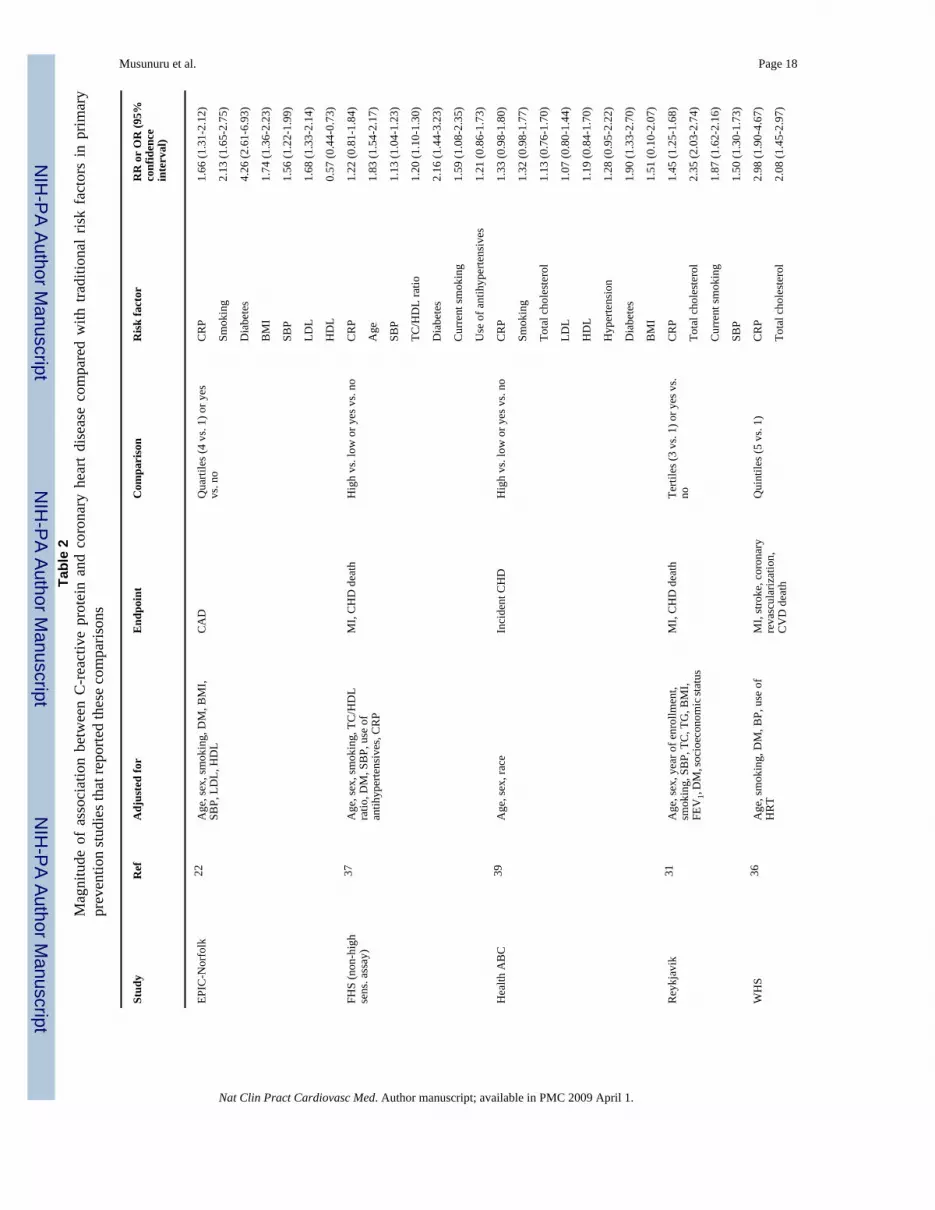
NIH
-PA Author Manuscript
NIH
-PA Author Manuscript
NIH
-PA Author Manuscript
Musunuru et al. Page 18Ta
ble
2M
agni
tude
of
asso
ciat
ion
betw
een
C-r
eact
ive
prot
ein
and
coro
nary
hea
rt di
seas
e co
mpa
red
with
tra
ditio
nal
risk
fact
ors
in p
rimar
ypr
even
tion
stud
ies t
hat r
epor
ted
thes
e co
mpa
rison
s
Stud
yR
efA
djus
ted
for
End
poin
tC
ompa
riso
nR
isk
fact
orR
R o
r O
R (9
5%co
nfid
ence
inte
rval
)
EPIC
-Nor
folk
22A
ge, s
ex, s
mok
ing,
DM
, BM
I,SB
P, L
DL,
HD
LC
AD
Qua
rtile
s (4
vs. 1
) or y
esvs
. no
CR
P1.
66 (1
.31-
2.12
)
Smok
ing
2.13
(1.6
5-2.
75)
Dia
bete
s4.
26 (2
.61-
6.93
)
BM
I1.
74 (1
.36-
2.23
)
SBP
1.56
(1.2
2-1.
99)
LDL
1.68
(1.3
3-2.
14)
HD
L0.
57 (0
.44-
0.73
)
FHS
(non
-hig
hse
ns. a
ssay
)37
Age
, sex
, sm
okin
g, T
C/H
DL
ratio
, DM
, SB
P, u
se o
fan
tihyp
erte
nsiv
es, C
RP
MI,
CH
D d
eath
Hig
h vs
. low
or y
es v
s. no
CR
P1.
22 (0
.81-
1.84
)
Age
1.83
(1.5
4-2.
17)
SBP
1.13
(1.0
4-1.
23)
TC/H
DL
ratio
1.20
(1.1
0-1.
30)
Dia
bete
s2.
16 (1
.44-
3.23
)
Cur
rent
smok
ing
1.59
(1.0
8-2.
35)
Use
of a
ntih
yper
tens
ives
1.21
(0.8
6-1.
73)
Hea
lth A
BC
39A
ge, s
ex, r
ace
Inci
dent
CH
DH
igh
vs. l
ow o
r yes
vs.
noC
RP
1.33
(0.9
8-1.
80)
Smok
ing
1.32
(0.9
8-1.
77)
Tota
l cho
lest
erol
1.13
(0.7
6-1.
70)
LDL
1.07
(0.8
0-1.
44)
HD
L1.
19 (0
.84-
1.70
)
Hyp
erte
nsio
n1.
28 (0
.95-
2.22
)
Dia
bete
s1.
90 (1
.33-
2.70
)
BM
I1.
51 (0
.10-
2.07
)
Rey
kjav
ik31
Age
, sex
, yea
r of e
nrol
lmen
t,sm
okin
g, S
BP,
TC
, TG
, BM
I,FE
V1,
DM
, soc
ioec
onom
ic st
atus
MI,
CH
D d
eath
Terti
les (
3 vs
. 1) o
r yes
vs.
noC
RP
1.45
(1.2
5-1.
68)
Tota
l cho
lest
erol
2.35
(2.0
3-2.
74)
Cur
rent
smok
ing
1.87
(1.6
2-2.
16)
SBP
1.50
(1.3
0-1.
73)
WH
S36
Age
, sm
okin
g, D
M, B
P, u
se o
fH
RT
MI,
stro
ke, c
oron
ary
reva
scul
ariz
atio
n,C
VD
dea
th
Qui
ntile
s (5
vs. 1
)C
RP
2.98
(1.9
0-4.
67)
Tota
l cho
lest
erol
2.08
(1.4
5-2.
97)
Nat Clin Pract Cardiovasc Med. Author manuscript; available in PMC 2009 April 1.

NIH
-PA Author Manuscript
NIH
-PA Author Manuscript
NIH
-PA Author Manuscript
Musunuru et al. Page 19
Stud
yR
efA
djus
ted
for
End
poin
tC
ompa
riso
nR
isk
fact
orR
R o
r O
R (9
5%co
nfid
ence
inte
rval
)
LDL
1.62
(1.1
7-2.
25)
Non
-HD
L2.
51 (1
.69-
3.72
)
HD
L0.
43 (0
.30-
0.61
)
Stud
yR
isk
fact
orR
R o
r O
R (9
5% c
onfid
ence
inte
rval
)
EPIC
-Nor
folk
22,a
CR
P1.
66 (1
.31-
2.12
)
Smok
ing
2.13
(1.6
5-2.
75)
Dia
bete
s4.
26 (2
.61-
6.93
)
BM
I1.
74 (1
.36-
2.23
)
SBP
1.56
(1.2
2-1.
99)
LDL
1.68
(1.3
3-2.
14)
HD
L0.
57 (0
.44-
0.73
)
FHS
(non
-hig
h se
ns. a
ssay
)37,b
CR
P1.
22 (0
.81-
1.84
)
Age
1.83
(1.5
4-2.
17)
SBP
1.13
(1.0
4-1.
23)
TC/H
DL
ratio
1.20
(1.1
0-1.
30)
Dia
bete
s2.
16 (1
.44-
3.23
)
Cur
rent
smok
ing
1.59
(1.0
8-2.
35)
Use
of a
ntih
yper
tens
ives
1.21
(0.8
6-1.
73)
Hea
lth A
BC
39,c
CR
P1.
33 (0
.98-
1.80
)
Smok
ing
1.32
(0.9
8-1.
77)
Tota
l cho
lest
erol
1.13
(0.7
6-1.
70)
LDL
1.07
(0.8
0-1.
44)
HD
L1.
19 (0
.84-
1.70
)
Hyp
erte
nsio
n1.
28 (0
.95-
2.22
)
Dia
bete
s1.
90 (1
.33-
2.70
)
BM
I1.
51 (0
.10-
2.07
)
Rey
kjav
ik31
,dC
RP
1.45
(1.2
5-1.
68)
Tota
l cho
lest
erol
2.35
(2.0
3-2.
74)
Nat Clin Pract Cardiovasc Med. Author manuscript; available in PMC 2009 April 1.

NIH
-PA Author Manuscript
NIH
-PA Author Manuscript
NIH
-PA Author Manuscript
Musunuru et al. Page 20
Stud
yR
isk
fact
orR
R o
r O
R (9
5% c
onfid
ence
inte
rval
)
Cur
rent
smok
ing
1.87
(1.6
2-2.
16)
SBP
1.50
(1.3
0-1.
73)
WH
S36,e
CR
P2.
98 (1
.90-
4.67
)
Tota
l cho
lest
erol
2.08
(1.4
5-2.
97)
LDL
1.62
(1.1
7-2.
25)
Non
-HD
L2.
51 (1
.69-
3.72
)
HD
L0.
43 (0
.30-
0.61
)
Stud
ies:
EPI
C-N
orfo
lk, E
urop
ean
Pros
pect
ive
Inve
stig
atio
n in
to C
ance
r Nor
folk
Stu
dy; F
HS,
Fra
min
gham
Hea
rt St
udy;
Hea
lth A
BC
, Dyn
amic
s of H
ealth
, Agi
ng a
nd B
ody
Com
posi
tion
Stud
y;R
eykj
avik
, Rey
kjav
ik S
tudy
; WH
S, W
omen
's H
ealth
Stu
dy.
Abb
revi
atio
ns: B
MI,
body
mas
s ind
ex; B
P, b
lood
pre
ssur
e; C
AD
, cor
onar
y ar
tery
dis
ease
; CH
D, c
oron
ary
hear
t dis
ease
; CR
P, C
-rea
ctiv
e pr
otei
n; C
VD
, car
diov
ascu
lar d
isea
se; D
M, d
iabe
tes m
ellit
us;
FEV
1, fo
rced
exp
irato
ry v
olum
e in
1 se
cond
; HD
L, h
igh-
dens
ity-li
popr
otei
n ch
oles
tero
l; H
RT,
hor
mon
e re
plac
emen
t the
rapy
; LD
L, lo
w-d
ensi
ty-li
popr
otei
n ch
oles
tero
l; M
I, m
yoca
rdia
l inf
arct
ion;
OR
, odd
s rat
io; R
R, r
isk
ratio
; SB
P, sy
stol
ic b
lood
pre
ssur
e; T
C, t
otal
cho
lest
erol
; TG
, trig
lyce
rides
.
Abb
revi
atio
ns: B
MI,
body
mas
s ind
ex; B
P, b
lood
pre
ssur
e; C
AD
, cor
onar
y ar
tery
dis
ease
; CH
D, c
oron
ary
hear
t dis
ease
; CR
P, C
-rea
ctiv
e pr
otei
n; C
VD
, car
diov
ascu
lar d
isea
se; D
M, d
iabe
tes m
ellit
us;
EPIC
-Nor
folk
, Eur
opea
n Pr
ospe
ctiv
e In
vest
igat
ion
into
Can
cer N
orfo
lk S
tudy
; FEV
1, fo
rced
exp
irato
ry v
olum
e in
1 se
cond
; FH
S, F
ram
ingh
am H
eart
Stud
y; H
ealth
AB
C, D
ynam
ics o
f Hea
lth, A
ging
and
Bod
y C
ompo
sitio
n St
udy;
HD
L, h
igh-
dens
ity-li
popr
otei
n ch
oles
tero
l; H
RT,
hor
mon
e re
plac
emen
t the
rapy
; LD
L, lo
w-d
ensi
ty-li
popr
otei
n ch
oles
tero
l; M
I, m
yoca
rdia
l inf
arct
ion;
OR
, odd
s rat
io;
RR
, ris
k ra
tio; S
BP,
syst
olic
blo
od p
ress
ure;
TC
, tot
al c
hole
ster
ol; T
G, t
rigly
cerid
es; W
HS,
Wom
en's
Hea
lth S
tudy
.
a Adj
uste
d fo
r age
, sex
, sm
okin
g, D
M, B
MI,
SBP,
LD
L, H
DL.
End
poin
t was
CA
D. C
ompa
red
quar
tiles
(4 v
s. 1)
or y
es v
s. no
.
b Age
, sex
, sm
okin
g, T
C/H
DL
ratio
, DM
, SB
P, u
se o
f ant
ihyp
erte
nsiv
es, C
RP.
End
poin
ts w
ere
MI a
nd C
HD
-rel
ated
dea
th. C
ompa
red
high
vs.
low
or y
es v
s. no
c Age
, sex
, rac
e. E
ndpo
int w
as in
cide
nt C
HD
. Com
pare
d hi
gh v
s. lo
w o
r yes
vs.
no
d Age
, sex
, yea
r of e
nrol
lmen
t, sm
okin
g, S
BP,
TC
, TG
, BM
I, FE
V1,
DM
, soc
ioec
onom
ic st
atus
. End
poin
ts w
ere
MI a
nd C
HD
dea
th. C
ompa
red
terti
les (
3 vs
. 1) o
r yes
vs.
no
e Age
, sm
okin
g, D
M, B
P, u
se o
f HR
T. E
ndpo
ints
wer
e M
I, st
roke
, cor
onar
y re
vasc
ular
izat
ion
and
CV
D d
eath
. Com
pare
d qu
intil
es (5
vs.
1).
Nat Clin Pract Cardiovasc Med. Author manuscript; available in PMC 2009 April 1.

NIH
-PA Author Manuscript
NIH
-PA Author Manuscript
NIH
-PA Author Manuscript
Musunuru et al. Page 21Ta
ble
3A
ssoc
iatio
n of
C-r
eact
ive
prot
ein
with
stro
ke in
prim
ary
prev
entio
n po
pula
tions
Stud
yR
efPo
pula
tion
Adj
uste
d fo
rE
ndpo
int
Com
pari
son
RR
or
OR
(95%
conf
iden
cein
terv
al)
Stud
ies t
hat s
how
a si
gnifi
cant
ass
ocia
tion
afte
r mul
tivar
iate
adj
ustm
ent (
p <
0.05
)
AR
IC53
Men
, wom
enA
ge, s
ex, r
ace,
smok
ing,
LD
L,H
DL,
DM
, BM
I, SB
P, u
se o
fan
tihyp
erte
nsiv
esIs
chem
ic st
roke
CR
P >
3.0
mg/
L vs
. < 1
.0m
g/L
1.97
(1.1
4-3.
39)
CH
S54
Men
, wom
en >
age
65A
ge, s
ex, r
ace,
smok
ing,
DM
,H
TN, S
BP,
TC
, int
erna
l + co
mm
onca
rotid
IMT
Isch
emic
stro
keC
RP
> 3.
0 m
g/L
vs. <
1.0
mg/
L1.
45 (1
.14-
1.86
)
Edin
burg
h21
Men
, wom
en
Age
, sex
, sub
clin
ical
dis
ease
(AB
I), p
ack-
year
s sm
okin
g, D
M,
BM
I, TC
/HD
L ra
tio, p
hysi
cal
activ
ity, M
I, st
roke
, ang
ina,
inte
rmitt
ent c
laud
icat
ion
Stro
keTe
rtile
s (3
vs. 1
)2.
18 (1
.30-
3.66
)
FHS
55W
omen
Age
, sm
okin
g, T
C, H
DL,
DM
, SB
PIs
chem
ic st
roke
or T
IAQ
uarti
les (
4 vs
. 1)
2.1
(1.1
9-3.
83)
His
ayam
a56
Men
, wom
enA
ge, s
ex, s
mok
ing,
alc
ohol
, SB
P,EC
G ab
norm
aliti
es, D
M, B
MI,
TC,
HD
L, p
hysi
cal a
ctiv
ityIs
chem
ic st
roke
Qui
ntile
s (5
vs. 1
)3.
11 (1
.04-
9.32
)
Hon
olul
u24
Men
Age
, sm
okin
g, a
lcoh
ol, T
C, H
TN,
DM
, BM
I, ph
ysic
al a
ctiv
ity in
dex
Thro
mbo
embo
lic st
roke
Qua
rtile
s (4
vs. 1
)1.
6 (1
.1-2
.4)
NH
AN
ES57
Wom
enA
ge, r
ace
or e
thni
city
, sm
okin
g,ed
ucat
ion,
SB
P, T
C, H
DL,
DM
,B
MI,
phys
ical
act
ivity
Stro
keTe
rtile
3 v
s. un
dete
ctab
leC
RP
1.82
(1.1
0-2.
99)
PHS
28M
enA
ge, H
TN, D
M, B
MI,
fam
ilyhi
stor
y of
CA
DSt
roke
Qua
rtile
(4 v
s.1)
1.6
(1.0
-3.1
)
PRO
SPER
30M
en, w
omen
>ag
e 70
Age
, sex
, ran
dom
ized
trea
tmen
t,co
untry
, cur
rent
and
pas
t sm
okin
g,SB
P, D
BP,
use
of
antih
yper
tens
ives
, LD
L, H
DL,
TG
,D
M, B
MI,
CH
D, P
AD
, stro
ke, T
IA
Stro
keTe
rtile
s (3
vs. 1
)1.
37 (1
.00-
1.86
)
WH
S34
Wom
enA
ge, s
mok
ing,
DM
, BP,
use
of
HR
TIs
chem
ic st
roke
Terti
les (
3 vs
. 1)
2.0
(1.3
-3.1
)
Stud
ies t
hat d
o no
t sho
w a
sign
ifica
nt a
ssoc
iatio
n af
ter m
ultiv
aria
te a
djus
tmen
t (p
> 0.
05)
FHS
55M
enA
ge, s
mok
ing,
TC
, HD
L, D
M, S
BP
Isch
emic
stro
ke o
r TIA
Qua
rtile
s (4
vs. 1
)1.
6 (0
.87-
3.13
)
Hea
lth A
BC
39M
en, w
omen
>ag
e 70
Age
, sex
, rac
e, sm
okin
g, D
M,
HTN
, BM
I, H
DL,
TG
, alb
umin
Stro
keTe
rtile
s (3
vs. 1
)1.
41 (0
.73-
2.71
)
Leid
en 8
5+59
Men
, wom
enA
ge, s
ex, c
urre
nt sm
okin
g, u
se o
fN
SAID
s, TC
, HTN
, DM
, pre
viou
sca
rdio
vasc
ular
eve
nts
Fata
l stro
keC
RP
5-10
mg/
L vs
. < 5
mg/
L2.
1 (0
.9-5
.4)
Nat Clin Pract Cardiovasc Med. Author manuscript; available in PMC 2009 April 1.

NIH
-PA Author Manuscript
NIH
-PA Author Manuscript
NIH
-PA Author Manuscript
Musunuru et al. Page 22
Stud
yR
efPo
pula
tion
Adj
uste
d fo
rE
ndpo
int
Com
pari
son
RR
or
OR
(95%
conf
iden
cein
terv
al)
NH
AN
ES57
Men
Age
, rac
e or
eth
nici
ty, s
mok
ing,
educ
atio
n, S
BP,
TC
, HD
L, D
M,
BM
I, ph
ysic
al a
ctiv
itySt
roke
Terti
le 3
vs.
unde
tect
able
CR
P1.
62 (0
.79-
3.32
)
Rot
terd
am60
Men
, wom
en >
age
55
Age
, sex
, SB
P, u
se o
fan
tihyp
erte
nsiv
es, D
M, s
mok
ing,
CH
D, A
F, L
VH
, int
ima-
med
iath
ickn
ess
Stro
keQ
uarti
les (
4 vs
. 1)
1.09
(0.8
4-1.
41)
Stud
ies:
AR
IC, A
ther
oscl
eros
is R
isk
in C
omm
uniti
es; C
HS,
Car
diov
ascu
lar H
ealth
Stu
dy; D
enm
ark,
Fre
derik
sber
g U
nive
rsity
Hos
pita
l, C
open
hage
n, D
enm
ark
coho
rt; E
dinb
urgh
, Edi
nbur
gh A
rtery
Stud
y; F
HS,
Fra
min
gham
Hea
rt St
udy;
Hea
lth A
BC
, Dyn
amic
s of H
ealth
, Agi
ng a
nd B
ody
Com
posi
tion
Stud
y; H
isay
ama,
His
ayam
a St
udy;
Hon
olul
u, H
onol
ulu
Hea
rt St
udy;
Lei
den
85-P
lus S
tudy
;N
HA
NES
, Thi
rd N
atio
nal H
ealth
and
Nut
ritio
n Ex
amin
atio
n Su
rvey
; PH
S, P
hysi
cian
s' H
ealth
Stu
dy; P
RO
SPER
, Pro
spec
tive
Stud
y of
Pra
vast
atin
in th
e El
derly
at R
isk;
Rot
terd
am, R
otte
rdam
Stu
dy;
WH
S, W
omen
's H
ealth
Stu
dy.
Abb
revi
atio
ns: A
BI,
ankl
e-br
achi
al in
dex;
AF,
atri
al fi
brill
atio
n; B
MI,
body
mas
s ind
ex; B
P, b
lood
pre
ssur
e; C
HD
, cor
onar
y he
art d
isea
se; C
RP,
C-r
eact
ive
prot
ein;
DB
P, d
iast
olic
blo
od p
ress
ure;
DM
, dia
bete
s mel
litus
; EC
G, e
lect
roca
rdio
gram
; FEV
1, fo
rced
exp
irato
ry v
olum
e in
1 se
cond
; HD
L, h
igh-
dens
ity-li
popr
otei
n ch
oles
tero
l; H
RT,
hor
mon
e re
plac
emen
t the
rapy
; HTN
, hyp
erte
nsio
n;IM
T, in
tima-
med
ia th
ickn
ess;
LD
L, lo
w-d
ensi
ty-li
popr
otei
n ch
oles
tero
l; LV
H, l
eft v
entri
cula
r hyp
ertro
phy;
MI,
myo
card
ial i
nfar
ctio
n; N
SAID
, non
ster
oida
l ant
i-inf
lam
mat
ory
drug
; OR
, odd
s rat
io;
PAD
, per
iphe
ral a
rtery
dis
ease
; RR
, ris
k ra
tio; S
BP,
syst
olic
blo
od p
ress
ure;
TC
, tot
al c
hole
ster
ol; T
G, t
rigly
cerid
es; T
IA, t
rans
ient
isch
emic
atta
ck.
Nat Clin Pract Cardiovasc Med. Author manuscript; available in PMC 2009 April 1.

NIH
-PA Author Manuscript
NIH
-PA Author Manuscript
NIH
-PA Author Manuscript
Musunuru et al. Page 23Ta
ble
4A
ssoc
iatio
n of
C-r
eact
ive
prot
ein
with
dia
bete
s in
prim
ary
prev
entio
n po
pula
tions
Stud
yR
efPo
pula
tion
Adj
uste
d fo
rE
ndpo
int
Com
pari
son
RR
or
OR
(95%
conf
iden
cein
terv
al)
AR
IC61
Men
, wom
enA
ge, s
ex, c
ente
r, et
hnic
ity, B
MI,
fast
ing
gluc
ose,
fast
ing
insu
lin, w
eigh
t/hip
ratio
,H
TN, p
aren
tal h
isto
ry o
f DM
Inci
dent
DM
Qua
rtile
s (4
vs. 1
)1.
23 (0
.74-
2.03
)
Aus
tralia
n62
Men
, wom
enA
ge, s
ex, b
asel
ine
gluc
ose,
BM
I, sm
okin
g,al
coho
l, ur
ine
albu
min
/cre
atin
ine
ratio
, TC
,SB
PIn
cide
nt D
MTe
rtile
s (3
vs. 1
+2)
1.75
(1.1
9-2.
56)
CH
S63
Men
, wom
en >
age
65A
ge, s
ex, f
astin
g gl
ucos
e, fa
stin
g in
sulin
,B
MI,
subc
linic
al C
VD
, use
of t
hiaz
ide
diur
etic
sIn
cide
nt D
MQ
uarti
les (
4 vs
. 1)
1.83
(1.2
4-2.
86)
EPIC
-Pot
sdam
64M
en, w
omen
Age
, sex
, BM
I, w
aist
/hip
ratio
, sm
okin
g,al
coho
l, sp
ortin
g ac
tiviti
es, e
duca
tiona
lat
tain
men
t, H
gbA
1cIn
cide
nt D
MH
alve
s (2
vs. 1
)1.
9 (1
.2-3
.2)
His
ayam
a65
Wom
enA
ge, f
amily
his
tory
of d
iabe
tes,
fast
ing
insu
lin, B
MI,
smok
ing,
alc
ohol
, phy
sica
lac
tivity
, TC
, HD
L, T
G, S
BP
Inci
dent
DM
Terti
les (
3 vs
. 1)
2.25
(1.0
1-5.
01)
His
ayam
a65
Men
Age
, fam
ily h
isto
ry o
f dia
bete
s, fa
stin
gin
sulin
, BM
I, sm
okin
g, a
lcoh
ol, p
hysi
cal
activ
ity, T
C, H
DL,
TG
, SB
PIn
cide
nt D
MTe
rtile
s (3
vs. 1
)2.
63 (1
.23-
5.65
)
IRA
S66
Men
, wom
enA
ge, s
ex, c
linic
site
, fas
ting
insu
lin, s
mok
ing
Inci
dent
DM
Per 1
SD
incr
ease
1.34
(1.1
1-1.
61)
Japa
nese
-Am
eric
an67
Wom
enA
ge, B
MI,
smok
ing,
FH
, BM
I, in
sulin
resi
stan
ce, g
luco
se to
lera
nce
test
ing,
HR
TIn
cide
nt D
MQ
uarti
les (
4 vs
. 1)
3.11
(1.2
5-7.
75)
Japa
nese
-Am
eric
an67
Men
Age
, BM
I, sm
okin
g, F
H, B
MI,
insu
linre
sist
ance
, glu
cose
tole
ranc
e te
stin
gIn
cide
nt D
MQ
uarti
les (
4 vs
. 1)
2.84
(1.0
9-7.
39)
Kuo
pio
68M
en
Age
, fas
ting
insu
lin, f
astin
g gl
ucos
e, T
G,
wai
st/h
ip ra
tio, s
mok
ing,
alc
ohol
, phy
sica
lac
tivity
, pre
senc
e of
CV
D, s
ocio
econ
omic
stat
us, f
amily
his
tory
of D
M, S
BP,
use
of
antih
yper
tens
ives
Inci
dent
DM
CR
P >
3.0
mg/
L vs
. <1.
0 m
g/L
2.30
(1.0
4-5.
07)
Mex
ico
69W
omen
Age
, sm
okin
g, a
lcoh
ol, p
hysi
cal a
ctiv
ityIn
cide
nt D
MTe
rtile
s (3
vs. 1
)4.
1 (2
.1-8
.0)
Mex
ico
69M
enA
ge, s
mok
ing,
alc
ohol
, phy
sica
l act
ivity
Inci
dent
DM
Terti
les (
3 vs
. 1)
0.8
(0.4
-2.0
)
MO
NIC
A-A
ugsb
urg
70W
omen
Age
, sur
vey,
BM
I, sm
okin
g, al
coho
l, ph
ysic
alac
tivity
, TC
/HD
L ra
tio, p
aren
tal h
isto
ry o
fD
MIn
cide
nt D
MTe
rtile
s (3
vs. 1
)2.
74 (1
.47-
5.11
)
MO
NIC
A-A
ugsb
urg
70M
enA
ge, s
urve
y, B
MI,
smok
ing,
alco
hol,
phys
ical
activ
ity, T
C/H
DL
ratio
, par
enta
l his
tory
of
DM
Inci
dent
DM
Terti
les (
3 vs
. 1)
1.09
(0.7
1-1.
66)
NH
S71
Wom
enA
ge, r
ace,
fast
ing
stat
us, t
ime
bloo
d dr
awn,
smok
ing,
alco
hol,
BM
I, ph
ysic
al ac
tivity
, die
t,In
cide
nt D
MQ
uint
iles (
5 vs
. 1)
4.36
(2.8
0-6.
80)
Nat Clin Pract Cardiovasc Med. Author manuscript; available in PMC 2009 April 1.

NIH
-PA Author Manuscript
NIH
-PA Author Manuscript
NIH
-PA Author Manuscript
Musunuru et al. Page 24
Stud
yR
efPo
pula
tion
Adj
uste
d fo
rE
ndpo
int
Com
pari
son
RR
or
OR
(95%
conf
iden
cein
terv
al)
fam
ily h
isto
ry o
f DM
, men
opau
sal s
tatu
s,H
RT
WH
S72
Wom
enA
ge, f
astin
g st
atus
, fas
ting
insu
lin, B
MI,
smok
ing,
alc
ohol
, phy
sica
l act
ivity
, fam
ilyhi
stor
y of
DM
, HR
TIn
cide
nt D
MQ
uarti
les (
4 vs
. 1)
4.3
(1.1
-17.
1)
WO
SCO
P73
Men
Age
, fas
ting
gluc
ose,
BM
I, sm
okin
g, a
lcoh
ol,
SBP,
HD
L, T
C, T
G, s
tatin
ther
apy,
WC
CIn
cide
nt D
MQ
uint
iles (
5 vs
. 1)
2.46
(1.2
0-5.
04)
Stud
ies:
AR
IC, A
ther
oscl
eros
is R
isk
in C
omm
uniti
es; A
ustra
ilian
, Aus
tralia
n A
borig
ine
coho
rt; C
HS,
Car
diov
ascu
lar H
ealth
Stu
dy; E
PIC
-Nor
folk
, Eur
opea
n Pr
ospe
ctiv
e In
vest
igat
ion
into
Can
cer
Pots
dam
Stu
dy; H
isay
ama,
His
ayam
a St
udy;
IRA
S, In
sulin
Res
ista
nce
and
Ath
eros
cler
osis
Stu
dy; J
apan
ese-
Am
eric
an, J
apan
ese-
Am
eric
an c
ohor
t; K
uopi
o, K
uopi
o Is
chae
mic
Hea
rt D
isea
se R
isk
Fact
or S
tudy
; Mex
ico,
Mex
ico
City
Dia
bete
s Stu
dy; M
ON
ICA
-Aug
sbur
g, M
onito
ring
Car
diov
ascu
lar D
isea
se A
ugsb
urg
Coh
ort S
tudy
; NH
S, N
urse
s' H
ealth
Stu
dy; W
HS,
Wom
en's
Hea
lth S
tudy
;W
OSC
OP,
Wes
t of S
cotla
nd C
oron
ary
Prot
ectio
n St
udy.
Abb
revi
atio
ns: B
MI,
body
mas
s ind
ex; C
RP,
C-r
eact
ive
prot
ein;
CV
D, c
ardi
ovas
cula
r dis
ease
; DM
, dia
bete
s mel
litus
; HD
L, h
igh-
dens
ity-li
popr
otei
n ch
oles
tero
l; H
gbA
1c, h
emog
lobi
n A
1c; H
RT,
horm
one
repl
acem
ent t
hera
py; H
TN, h
yper
tens
ion;
HR
T, h
orm
one
repl
acem
ent t
hera
py; O
R, o
dds r
atio
; RR
, ris
k ra
tio; S
BP,
syst
olic
blo
od p
ress
ure;
SD
, sta
ndar
d de
viat
ion;
TC
, tot
al c
hole
ster
ol;
TG, t
rigly
cerid
es; W
CC
, whi
te c
ell c
ount
.
Nat Clin Pract Cardiovasc Med. Author manuscript; available in PMC 2009 April 1.