the t4 phage sf1b helicase dda is structurally optimized to perform dna strand separation
TRANSCRIPT
Structure
Article
The T4 Phage SF1B Helicase Dda Is StructurallyOptimized to Perform DNA Strand SeparationXiaoping He,1,5 Alicia K. Byrd,2,5 Mi-Kyung Yun,1 Charles W. Pemble IV1,6 David Harrison,2 Laxmi Yeruva,2,7
Christopher Dahl,2 Kenneth N. Kreuzer,3 Kevin D. Raney,2 and Stephen W. White1,4,*1Department of Structural Biology, St. Jude Children’s Research Hospital, 262 Danny Thomas Place, Memphis, TN 38105, USA2Department of Biochemistry and Molecular Biology, University of Arkansas for Medical Sciences, Little Rock, AR 72205, USA3Department of Biochemistry, Duke University Medical Center, Nanaline Duke, Durham, NC 27710, USA4Department of Microbiology, Immunology and Biochemistry, University of Tennessee Health Science Center, Memphis, TN 38163, USA5These authors contributed equally to this work6Present address: Duke Human Vaccine Institute, Duke University, Durham, NC 27710, USA7Present address: Department of Pediatrics, University of Arkansas for Medical Sciences, Little Rock, AR 72205, USA
*Correspondence: [email protected]
DOI 10.1016/j.str.2012.04.013
SUMMARY
Helicases move on DNA via an ATP binding andhydrolysis mechanism coordinated by well-charac-terized helicase motifs. However, the translocationalong single-stranded DNA (ssDNA) and the strandseparation of double-stranded (dsDNA) may beloosely or tightly coupled. Dda is a phage T4 SF1Bhelicase with sequence homology to the Pif1 familyof helicases that tightly couples translocation tostrand separation. The crystal structure of the Dda-ssDNA binary complex reveals a domain referred toas the ‘‘pin’’ that was previously thought to remainstatic during strand separation. The pin containsa conserved phenylalanine that mediates a transientbase-stacking interaction that is absolutely requiredfor separation of dsDNA. The pin is secured at its tipby protein-protein interactions through an extendedSH3 domain thereby creating a rigid strut. Theconserved interface between the pin and the SH3domain provides the mechanism for tight couplingof translocation to strand separation.
INTRODUCTION
Helicases are ATP-dependent motor proteins that play key roles
in DNA and RNA metabolism (Delagoutte and von Hippel, 2003;
Lohman et al., 2008; Patel and Donmez, 2006; Pyle, 2008). Clas-
sically, helicases unwind double-stranded substrates, but they
can also modulate nucleic acid architecture in processes such
as recombination, chromatin remodeling, and RNA transport.
The fact that �2% of the yeast genome encodes helicase-like
proteins reflects their importance. Many diseases, typically
associated with cancer and/or premature aging, are linked to
mutations in helicase genes and defective DNA repair (Bohr,
2008; Brosh et al., 2000; Ellis, 1997; Stevnsner et al., 2008). Hel-
icases have also emerged as important therapeutic targets for
the treatment of cancer (Aggarwal and Brosh, 2009) and viral
infections (Frick, 2007; Lescar et al., 2008).
Structure 20, 1
Helicases are classified into four superfamilies based on the
presence of helicase motifs (Gorbalenya and Koonin, 1993;
Singleton et al., 2007). The monomeric SF1 and SF2 helicases
share an architecture based on paired RecA-like domains, while
the SF3 and SF4 helicases form hexameric rings. SF1 is the
largest class and is subdivided into the SF1A and SF1B enzymes
that translocate 30-to-50 and 50-to-30, respectively, on single-
stranded DNA (ssDNA) (Singleton et al., 2007). This report
focuses on the SF1B subfamily that includes members that are
conserved from bacteria to humans. Examples include Dda
from bacteriophage T4, RecD2 from bacteria (Saikrishnan
et al., 2009), Pif1 from eukaryotes (Bochman et al., 2010), and
human DNA helicase B (Gu et al., 2004). Although key informa-
tion has recently become available (Saikrishnan et al., 2008,
2009), SF1B helicases are poorly understood compared to their
SF1A and SF2 counterparts despite their emerging importance
in key biological functions. Notably, human DNA helicase B is
involved in DNA repair and replication (Gu et al., 2004; Taneja
et al., 2002), and the Pif1 helicase has roles in telomere regula-
tion (Boule et al., 2005; Boule and Zakian, 2010) and mitochon-
drial DNA repair (Cheng et al., 2007).
The Dda helicase from phage T4 is an ideal model system for
understanding the basic mechanism of the SF1B class, although
its biological role is still somewhat obscure. Dda knockouts are
fully viable, and the helicase is not needed for phage replication,
recombination, or repair (Behme and Ebisuzaki, 1975). Nonethe-
less, Dda-deficient phages show a delay in origin usage and
overall DNA replication, and replication is compromised during
infections with mutants deficient in both Dda and gp59, which
loads the replicative helicase gp41 (Gauss et al., 1994). These
results suggest that Dda has an overlapping function with gp59
in loading gp41 at T4 replication origins (Gauss et al., 1994).
In vitro experiments have suggested additional roles for Dda
that have yet to be verified in vivo. First, Dda can promote
DNA polymerase strand switching that could play a key role in
survival after DNA damage (Kadyrov and Drake, 2004). Second,
Dda can displace proteins from DNA suggesting that it helps
clear the DNA template for replication (Bedinger et al., 1983;
Byrd and Raney, 2006). Third, Dda has been shown to both
inhibit and stimulate recombination strand invasion reactions
(Kodadek, 1991; Kodadek and Alberts, 1987) suggesting a role
in homologous recombination. Consistent with this, Dda binds
189–1200, July 3, 2012 ª2012 Elsevier Ltd All rights reserved 1189
Table 1. Data Collection and Refinement Statistics
Dda/K38A(SeMet)
SAD (peak) Higher Resolution
Data Collectiona
Space group C2 C2
Cell dimensions
a, b, c (A) 223.9, 106.7, 85.3 225.5, 107.3, 85.5
a, b, g (0) 90.0, 94.3, 90.0 90.0, 94.1, 90.0
Wavelength (A) 0.9793 1.0
Resolution (A) 50.0–3.80
(3.87–3.80)
40.0–3.30
(3.42–3.30)
No. of reflections
Measured 120,589 208,930
Unique 19,895 29,389
Completeness (%) 99.5 (98.4) 95.8 (71.4)
Redundancy 6.1 (4.9) 7.1 (4.5)
I/s(I) 16.6 (2.6) 19.82 (2.05)
Rsym (%) 11.7 (48.2) 9.2 (47.3)
Phasing
Resolution (A) 3.8
FOM 0.29
FOM after density
modification
0.63
Refinement
Resolution (A) 32.3–3.3
No. reflections 27,990
Completeness (%) 91.1
Rwork (%) 20.4
Rfree (%)b 25.8
Rmsds
Bonds lengths(A) 0.004
Bonds angles(o) 0.916
No. of protein residues 1317
No. of dT nucleotides 24
No. of water molecules 10
Average B-factor (A2) 110
Ramachandran plot (%)
Most favored regions 89.3
Additional allowed regions 10.0
Generously allowed regions 0.7
Disallowed regions 0.0aHighest resolution shell is shown in parenthesis.bRfree is the R value obtained for a test set of reflections consisting of
randomly selected 5% subset of the data set excluded from refinement.
Structure
Structural Analysis of T4 Helicase Dda
both UvsX (the T4 recombinase) and gp32 (the T4 ssDNA-
binding protein) (Formosa and Alberts, 1984; Hacker and Al-
berts, 1992). Dda single mutants show no obvious recombina-
tion phenotype (Behme and Ebisuzaki, 1975) but phenotypes
do appear in double knockout phages. Most notably, a Dda/
gp61 (the T4 primase) double knockout phage was found to be
lethal due to poor DNA packaging (Belanger, 1997). Primase-
deficient infections are known to generate excessive ssDNA
that, in turn, promotes recombination and the overproduction
of DNA branches that inhibit packaging. In this scenario, it has
been suggested that Dda acts as an antirecombinase that facil-
itates packaging by reducing the levels of DNA branches.
In contrast, the enzymology of the Dda helicase has been well
characterized. Dda unwinds DNA as a monomer (Nanduri et al.,
2002) in a 50-to-30 direction at a rate of �250 bp/s (Eoff and Ra-
ney, 2006) and can efficiently displace proteins in its path (Morris
and Raney, 1999). In the absence of impediments, Dda mole-
cules function independently (Eoff and Raney, 2010), but when
impediments are present, displacement efficiency is augmented
by multiple copies of Dda working in tandem via protein-protein
interactions (Byrd and Raney, 2004). This is true for the displace-
ment of streptavidin (Byrd and Raney, 2004), the removal of
proteins (Byrd and Raney, 2006), and movement past chemical
perturbations in the DNA (Eoff et al., 2005). Helicases are
described as being active or passive depending on whether
the enzyme actively separates the base pairs or simply seques-
ters ssDNA that forms due to thermal fraying (Manosas et al.,
2010). The ratio of the rate of unwinding double-stranded
(dsDNA) to the rate of translocation on ssDNA distinguishes
between the two; a helicase is considered highly active when
the ratio is close to 1 and passive with ratios less than 0.25 (Man-
osas et al., 2010). Recently, we determined that Dda translo-
cates on ssDNA at nearly the same rate as it unwinds dsDNA,
making it one of the most ‘‘active’’ helicases yet studied (Byrd
et al., 2012).
Although Dda shares the same helicase motifs as other super-
family 1 helicases, the highly active nature of Dda suggests that
alternative or perhaps additional mechanisms are present to
explain the extraordinary efficiency for dsDNA unwinding.
Here, we present the crystal structure of the Dda-ssDNA binary
complex. The structure, together with biochemical and muta-
genesis data, supports the proposed 50-to-30 translocation
mechanism of the SF1B helicases (Saikrishnan et al., 2009)
and reveals a new mechanism by which translocation is directly
coupled to strand separation of duplex DNA. The results provide
the structural basis for distinguishing a highly active helicase
from a passive helicase.
RESULTS
Structure DeterminationDda is difficult to express in a soluble form, but the active site
point mutant K38A could be readily expressed and purified.
The mutation does not affect the DNA-binding properties of
Dda, and a purified complex with a dT8 oligonucleotide
produced crystals that diffract to 3.5 A. The structure was deter-
mined using SAD phasing with selenomethionine-substituted
protein. There are three molecules in the C2 asymmetric unit
(ASU). We subsequently grew crystals that diffract to 3.3 A,
1190 Structure 20, 1189–1200, July 3, 2012 ª2012 Elsevier Ltd All rig
and refined the structure to Rwork and Rfree values of 0.204 and
0.258, respectively. Data collection and refinement statistics
are shown in Table 1.
Description of the StructureThe Dda structure is shown in Figure 1A and it is most similar to
the SF1B helicase RecD2 (Saikrishnan et al., 2009). At the heart
of the molecule are the two signature RecA-like domains 1A
hts reserved
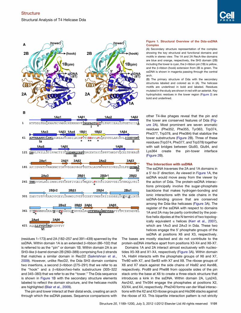
Figure 1. Structural Overview of the Dda-ssDNA
Complex
(A) Secondary structure representation of the complex
showing the key structural and functional domains and
motifs in stereo view. The 1A and 2A RecA-like domains
are blue and orange, respectively, the SH3 domain (2B)
including the tower is cyan, the b-ribbon pin (1B) is yellow,
and the b-ribbon (hook) extension from 2B is green. The
ssDNA is shown in magenta passing through the central
arch.
(B) The primary structure of Dda with the secondary
structures labeled and colored as in (A). The helicase
motifs are underlined in bold and labeled. Residues
mutated in the study are shown in red with an asterisk. Key
hydrophobic residues in the tower region (Figure 2) are
bold and underlined.
Structure
Structural Analysis of T4 Helicase Dda
(residues 1–173) and 2A (182–257 and 391–439) spanned by the
ssDNA. Within domain 1A is an extended b-ribbon (86–102) that
is referred to as the ‘‘pin’’ or domain 1B. Within domain 2A is an
SH3-like b-barrel domain 2B (260–389) comprising five b strands
that matches a similar domain in RecD2 (Saikrishnan et al.,
2009). However, unlike RecD2, the Dda SH3 domain contains
two insertions, a second b-ribbon (275–291) that we refer to as
the ‘‘hook’’ and a b-ribbon/two-helix substructure (305–322
and 345–383) that we refer to as the ‘‘tower.’’ The Dda sequence
is shown in Figure 1B with the secondary structure elements
labeled to reflect the domain structure, and the helicase motifs
are highlighted (Blair et al., 2009).
The pin and tower interact at their distal ends, creating an arch
through which the ssDNA passes. Sequence comparisons with
Structure 20, 1189–1200, July 3,
other T4-like phages reveal that the pin and
the tower are conserved features of Dda (Fig-
ure 2A). Most prominent are seven aromatic
residues (Phe352, Phe355, Tyr363, Trp374,
Phe377, Trp378, and Phe384) that stabilize the
tower substructure (Figure 2B). Three of these
residues (Trp374, Phe377, and Trp378) together
with salt bridges between Glu93, Glu94, and
Lys364 create the pin-tower interface
(Figure 2B).
The Interaction with ssDNAThe ssDNA traverses the 2A and 1A domains in
a 50-to-30 direction. As viewed in Figure 1A, the
ssDNA would move away from the viewer by
the action of Dda. The protein-ssDNA interac-
tions principally involve the sugar-phosphate
backbone that makes hydrogen-bonding and
ionic interactions with the side chains in the
ssDNA-binding groove that are conserved
among the Dda-like helicases (Figure 3A). The
register of the ssDNA with respect to domains
1A and 2A may be partly controlled by the posi-
tive helix dipoles at the N termini of two topolog-
ically equivalent a helices (Kerr et al., 2007),
which are 1Aa3 and 2Aa3 in Dda. These two
helices engage the 50 phosphate groups of the
ssDNA at positions X6 and X3, respectively.
The bases are mostly stacked and do not contribute to the
protein-ssDNA interface apart from positions X3-X4 and X6-X7.
Domains 1A and 2A interact almost exclusively with nucleo-
tides X5-X8 and X1-X4, respectively (Figure 3A). Within domain
1A, His64 interacts with the phosphate groups of X6 and X7,
Thr80 with X7, and Ser83 with X7 and X8. The ribose groups of
X6 and X7 stack against the side chains of His82 and Asn88,
respectively. Pro89 and Phe98 from opposite sides of the pin
stack onto the base at X6 to create a three-stack structure that
introduces a kink in the ssDNA. Within domain 2A, Lys243,
Asn242, and Thr394 engage the phosphates at positions X2,
X3/X4, and X4, respectively. Phe240 forms van der Waal interac-
tionswith the X2 and X3 ribose groups andHis396 stacks against
the ribose of X3. This bipartite interaction pattern is not strictly
2012 ª2012 Elsevier Ltd All rights reserved 1191
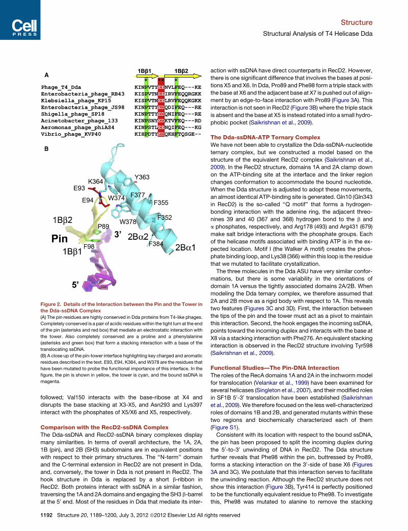
Figure 2. Details of the Interaction between the Pin and the Tower in
the Dda-ssDNA Complex
(A) The pin residues are highly conserved in Dda proteins from T4-like phages.
Completely conserved is a pair of acidic residueswithin the tight turn at the end
of the pin (asterisks and red box) that mediate an electrostatic interaction with
the tower. Also completely conserved are a proline and a phenylalanine
(asterisks and green box) that form a stacking interaction with a base of the
translocating ssDNA.
(B) A close up of the pin-tower interface highlighting key charged and aromatic
residues described in the text. E93, E94, K364, andW378 are the residues that
have been mutated to probe the functional importance of this interface. In the
figure, the pin is shown in yellow, the tower is cyan, and the bound ssDNA is
magenta.
Structure
Structural Analysis of T4 Helicase Dda
followed; Val150 interacts with the base-ribose at X4 and
disrupts the base stacking at X3-X5, and Asn293 and Lys397
interact with the phosphates of X5/X6 and X5, respectively.
Comparison with the RecD2-ssDNA ComplexThe Dda-ssDNA and RecD2-ssDNA binary complexes display
many similarities. In terms of overall architecture, the 1A, 2A,
1B (pin), and 2B (SH3) subdomains are in equivalent positions
with respect to their primary structures. The ‘‘N-term’’ domain
and the C-terminal extension in RecD2 are not present in Dda,
and, conversely, the tower in Dda is not present in RecD2. The
hook structure in Dda is replaced by a short b-ribbon in
RecD2. Both proteins interact with ssDNA in a similar fashion,
traversing the 1A and 2A domains and engaging the SH3 b-barrel
at the 50 end. Most of the residues in Dda that mediate its inter-
1192 Structure 20, 1189–1200, July 3, 2012 ª2012 Elsevier Ltd All rig
action with ssDNA have direct counterparts in RecD2. However,
there is one significant difference that involves the bases at posi-
tions X5 and X6. In Dda, Pro89 and Phe98 form a triple stack with
the base at X6 and the adjacent base at X7 is pushed out of align-
ment by an edge-to-face interaction with Pro89 (Figure 3A). This
interaction is not seen in RecD2 (Figure 3B) where the triple stack
is absent and the base at X5 is instead rotated into a small hydro-
phobic pocket (Saikrishnan et al., 2009).
The Dda-ssDNA-ATP Ternary ComplexWe have not been able to crystallize the Dda-ssDNA-nucleotide
ternary complex, but we constructed a model based on the
structure of the equivalent RecD2 complex (Saikrishnan et al.,
2009). In the RecD2 structure, domains 1A and 2A clamp down
on the ATP-binding site at the interface and the linker region
changes conformation to accommodate the bound nucleotide.
When the Dda structure is adjusted to adopt these movements,
an almost identical ATP-binding site is generated. Gln10 (Gln343
in RecD2) is the so-called ‘‘Q motif’’ that forms a hydrogen-
bonding interaction with the adenine ring, the adjacent threo-
nines 39 and 40 (367 and 368) hydrogen bond to the b and
a phosphates, respectively, and Arg178 (493) and Arg431 (679)
make salt bridge interactions with the phosphate groups. Each
of the helicase motifs associated with binding ATP is in the ex-
pected location. Motif I (the Walker A motif) creates the phos-
phate binding loop, and Lys38 (366) within this loop is the residue
that we mutated to facilitate crystallization.
The three molecules in the Dda ASU have very similar confor-
mations, but there is some variability in the orientations of
domain 1A versus the tightly associated domains 2A/2B. When
modeling the Dda ternary complex, we therefore assumed that
2A and 2B move as a rigid body with respect to 1A. This reveals
two features (Figures 3C and 3D). First, the interaction between
the tips of the pin and the tower must act as a pivot to maintain
this interaction. Second, the hook engages the incoming ssDNA,
points toward the incoming duplex and interacts with the base at
X8 via a stacking interaction with Phe276. An equivalent stacking
interaction is observed in the RecD2 structure involving Tyr598
(Saikrishnan et al., 2009).
Functional Studies—The Pin-DNA InteractionThe roles of the RecA domains 1A and 2A in the inchwormmodel
for translocation (Velankar et al., 1999) have been examined for
several helicases (Singleton et al., 2007), and their modified roles
in SF1B 50-30 translocation have been established (Saikrishnan
et al., 2009). We therefore focused on the less well-characterized
roles of domains 1B and 2B, and generatedmutants within these
two regions and biochemically characterized each of them
(Figure S1).
Consistent with its location with respect to the bound ssDNA,
the pin has been proposed to split the incoming duplex during
the 50-to-30 unwinding of DNA in RecD2. The Dda structure
further reveals that Phe98 within the pin, buttressed by Pro89,
forms a stacking interaction on the 30-side of base X6 (Figures
3A and 3C). We postulate that this interaction serves to facilitate
the unwinding reaction. Although the RecD2 structure does not
show this interaction (Figure 3B), Tyr414 is perfectly positioned
to be the functionally equivalent residue to Phe98. To investigate
this, Phe98 was mutated to alanine to remove the stacking
hts reserved

Figure 3. The ssDNA-Binding Interactions in Dda
(A) The ssDNA traverses the RecA-like domains 1A and 2A, and P89 and F98 form a triple stack interaction with the DNA base at position X6. Note that the
N termini of helices 1Aa3 (domain 1A) and 2Aa3 (domain 2A) point directly at the DNA 50 phosphates at positions X6 and X3, respectively. The coloring scheme
matches that shown in Figure 1.
(B) The open conformation of the RecD2-ssDNA complex (Saikrishnan et al., 2009) centered on position X6. To facilitate the comparison with (A), the two
structures are aligned and equivalently colored and labeled. Note that the bases X5, X6, and X7 are in different orientations in the two structures with X5 stacked
onto X6 in Dda but rotated into a pocket in RecD2.
(C) The observed open conformation structure of the Dda-ssDNA complex highlighting the locations of the pin (yellow), the hook (green), and helices 1Aa3 (blue
cylinder), and 2Aa3 (orange cylinder) relative to ssDNA. The arrow indicates the direction in which a helix 2Aa3 moves relative to ssDNA during domain closure.
(D) A model of the closed Dda-ssDNA complex based on the structure of the RecD2 ternary complex (Saikrishnan et al., 2009). Due to domain closure and
rotation, Phe98 no longer stacks onto the base at X6, and the hook becomes aligned along the axis of the ssDNA toward the incoming duplexwith Phe276 forming
a new stacking interaction with the base at position X8. Note that helix 2Aa3 has moved closer to helix 1Aa3 by one phosphate group compared to (C).
Structure
Structural Analysis of T4 Helicase Dda
interaction. F98A bound to a ss/dsDNA partial duplex with lower
affinity than the wild-type (wt) enzyme with an �6-fold higher
equilibrium dissociation constant, KD, and the ssDNA-stimulated
ATPase activity was reduced by less than 2-fold (Figure 4A,
Table 2). Hence, replacement of Phe98 with alanine reduces
the affinity of Dda for ssDNA, but does not greatly reduce the
inherent ability of the enzyme to hydrolyze ATP in a ssDNA-stim-
ulated manner. The stacking interaction between F98 and Pro89
was investigated by creating P98A. The resulting DNA binding
and ATPase activities were reduced, but only by 2- to 3-fold (Fig-
ure 4, Table 2).
DNA unwinding was measured under multiple-cycle condi-
tions to determine whether any unwinding was observed for
the Dda variants (Figure 5A). A substrate containing a short
duplex (12 bp) was chosen to detect even low levels of strand
separation. F98A was devoid of unwinding activity but P89A
was able to unwind the short substrate. To determine the
rates for unwinding by the variant forms of Dda, single-cycle
Structure 20, 1
DNA unwinding experiments were conducted using a well-
characterized partial duplex substrate containing 7 nt of
ssDNA and 16 bp (Figures 5B and 5C). Wild-type Dda
unwound the substrate at a rate of 258 ± 17 bp/s. No product
was observed with F98A Dda, as expected, but P89A Dda
unwound the substrate in a single kinetic step at a rate of
96 ± 13 bp/s. Hence, whereas loss of stacking interaction
between P89 and F98 reduces efficiency of DNA unwinding,
removal of the stacking interactions between F98 and the nu-
cleobases completely disrupts DNA unwinding. This has not
been accounted for in any helicase mechanism thus far
proposed.
Functional Studies—The Pin-Tower InteractionThree variant proteins were produced to test the importance of
the pin-tower interface (Figure S1), and these were examined
for DNA binding, ssDNA-stimulated ATPase activity, and DNA
unwinding activity. These data reveal that the pin-tower
189–1200, July 3, 2012 ª2012 Elsevier Ltd All rights reserved 1193
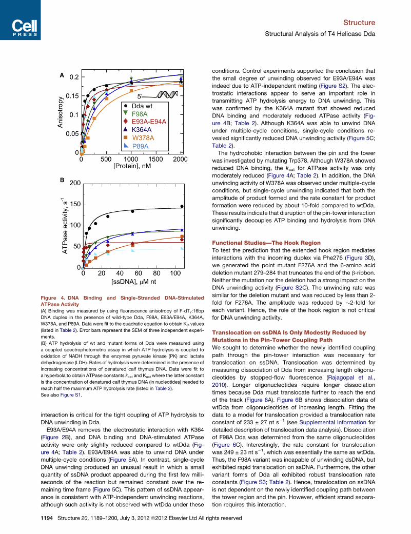
Figure 4. DNA Binding and Single-Stranded DNA-Stimulated
ATPase Activity
(A) Binding was measured by using fluorescence anisotropy of F-dT7:16bp
DNA duplex in the presence of wild-type Dda, F98A, E93A/E94A, K364A,
W378A, and P89A. Data were fit to the quadratic equation to obtain KD values
(listed in Table 2). Error bars represent the SEM of three independent experi-
ments.
(B) ATP hydrolysis of wt and mutant forms of Dda were measured using
a coupled spectrophotometric assay in which ATP hydrolysis is coupled to
oxidation of NADH through the enzymes pyruvate kinase (PK) and lactate
dehydrogenase (LDH). Rates of hydrolysis were determined in the presence of
increasing concentrations of denatured calf thymus DNA. Data were fit to
a hyperbola to obtain ATPase constants kcat and Kact where the latter constant
is the concentration of denatured calf thymus DNA (in nucleotides) needed to
reach half the maximum ATP hydrolysis rate (listed in Table 2).
See also Figure S1.
Structure
Structural Analysis of T4 Helicase Dda
interaction is critical for the tight coupling of ATP hydrolysis to
DNA unwinding in Dda.
E93A/E94A removes the electrostatic interaction with K364
(Figure 2B), and DNA binding and DNA-stimulated ATPase
activity were only slightly reduced compared to wtDda (Fig-
ure 4A; Table 2). E93A/E94A was able to unwind DNA under
multiple-cycle conditions (Figure 5A). In contrast, single-cycle
DNA unwinding produced an unusual result in which a small
quantity of ssDNA product appeared during the first few milli-
seconds of the reaction but remained constant over the re-
maining time frame (Figure 5C). This pattern of ssDNA appear-
ance is consistent with ATP-independent unwinding reactions,
although such activity is not observed with wtDda under these
1194 Structure 20, 1189–1200, July 3, 2012 ª2012 Elsevier Ltd All rig
conditions. Control experiments supported the conclusion that
the small degree of unwinding observed for E93A/E94A was
indeed due to ATP-independent melting (Figure S2). The elec-
trostatic interactions appear to serve an important role in
transmitting ATP hydrolysis energy to DNA unwinding. This
was confirmed by the K364A mutant that showed reduced
DNA binding and moderately reduced ATPase activity (Fig-
ure 4B; Table 2). Although K364A was able to unwind DNA
under multiple-cycle conditions, single-cycle conditions re-
vealed significantly reduced DNA unwinding activity (Figure 5C;
Table 2).
The hydrophobic interaction between the pin and the tower
was investigated by mutating Trp378. Although W378A showed
reduced DNA binding, the kcat for ATPase activity was only
moderately reduced (Figure 4A; Table 2). In addition, the DNA
unwinding activity of W378A was observed under multiple-cycle
conditions, but single-cycle unwinding indicated that both the
amplitude of product formed and the rate constant for product
formation were reduced by about 10-fold compared to wtDda.
These results indicate that disruption of the pin-tower interaction
significantly decouples ATP binding and hydrolysis from DNA
unwinding.
Functional Studies—The Hook RegionTo test the prediction that the extended hook region mediates
interactions with the incoming duplex via Phe276 (Figure 3D),
we generated the point mutant F276A and the 6-amino acid
deletion mutant 279–284 that truncates the end of the b-ribbon.
Neither the mutation nor the deletion had a strong impact on the
DNA unwinding activity (Figure S2C). The unwinding rate was
similar for the deletion mutant and was reduced by less than 2-
fold for F276A. The amplitude was reduced by �2-fold for
each variant. Hence, the role of the hook region is not critical
for DNA unwinding activity.
Translocation on ssDNA Is Only Modestly Reduced byMutations in the Pin-Tower Coupling PathWe sought to determine whether the newly identified coupling
path through the pin-tower interaction was necessary for
translocation on ssDNA. Translocation was determined by
measuring dissociation of Dda from increasing length oligonu-
cleotides by stopped-flow fluorescence (Rajagopal et al.,
2010). Longer oligonucleotides require longer dissociation
times because Dda must translocate further to reach the end
of the track (Figure 6A). Figure 6B shows dissociation data of
wtDda from oligonucleotides of increasing length. Fitting the
data to a model for translocation provided a translocation rate
constant of 233 ± 27 nt s�1 (see Supplemental Information for
detailed description of translocation data analysis). Dissociation
of F98A Dda was determined from the same oligonucleotides
(Figure 6C). Interestingly, the rate constant for translocation
was 249 ± 23 nt s�1, which was essentially the same as wtDda.
Thus, the F98A variant was incapable of unwinding dsDNA, but
exhibited rapid translocation on ssDNA. Furthermore, the other
variant forms of Dda all exhibited robust translocation rate
constants (Figure S3; Table 2). Hence, translocation on ssDNA
is not dependent on the newly identified coupling path between
the tower region and the pin. However, efficient strand separa-
tion requires this interaction.
hts reserved
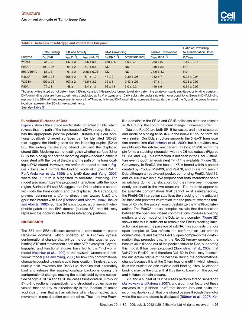
Table 2. Activities of Wild-Type and Variant Dda Enzymes
Enzyme
DNA Binding ATPase Activity DNA Unwinding ssDNA Translocase
Ratio of Unwinding
to Translocation Rates
KD (nM) kcat (s�1) Kact (mM, nt) ku (bp s�1) Amplitude (nM) ktrans (nt s
�1) ku/ktrans
wtDda 34 ± 3 147 ± 3 3.5 ± 0.5 258 ± 17 5.6 ± 0.1 233 ± 27 1.10 ± 0.15
F98A 192 ± 26 94 ± 2 6.7 ± 0.6 ND ND 249 ± 23 ND
E93A/E94A 55 ± 5 61 ± 2 0.26 ± 0.05 ND ND 77.0 ± 4.9 ND
K364A 298 ± 38 106 ± 2 15.1 ± 1.0 47 ± 9 0.39 ± .05 212 ± 7 0.22 ± 0.04
W378A 409 ± 72 107 ± 2 46.5 ± 3.9 36 ± 8 0.43 ± .05 157 ± 11 0.23 ± 0.05
P89A 72 ± 8 48 ± 1 5.4 ± 1.1 96 ± 13 3.0 ± 0.2 149 ± 6 0.64 ± 0.09
Those activities listed as not determined (ND) indicate too little product formed to reliably determine a rate constant, amplitude, or binding constant.
DNA unwinding data are from experiments conducted at 1 mM enzyme and 10 nM substrate under single-turnover conditions. Errors in DNA binding
represent the SEM of three experiments, errors in ATPase activity and DNA unwinding represent the standard error of the fit, and the errors in trans-
location represent the SD of three experiments.
See also Table S1.
Structure
Structural Analysis of T4 Helicase Dda
Functional Surfaces of DdaFigure 7 shows the surface electrostatic potential of Dda, which
reveals that the path of the translocated ssDNA through the arch
has the appropriate positive potential (surface S1). Four addi-
tional positively charged surfaces can be identified (S2–S5)
that suggest the binding sites for the incoming duplex (S2 or
S3), the exiting translocating strand (S4) and the displaced
strand (S5). Modeling cannot distinguish whether surface S2 or
S3 is the binding site for the incoming duplex because either is
consistent with the role of the pin and the path of the translocat-
ing ssDNA strand. However, we prefer the model shown in Fig-
ure 7 because it mirrors the binding mode of duplex DNA in
PcrA (Velankar et al., 1999) and UvrD (Lee and Yang, 2006)
where the 90� turn is suggested to facilitate unwinding. The
model also maximizes the proposed interactions with the hook
region. Surfaces S4 and S5 suggest that Dda maintains contact
with both the translocating and the displaced DNA strands, to
prevent reannealing and/or to direct them toward UvsX and
gp32 that interact with Dda (Formosa and Alberts, 1984; Hacker
and Alberts, 1992). Surface S4 leads toward a conserved hydro-
phobic patch on the SH3 domain (surface S6), and this may
represent the docking site for these interacting partners.
DISCUSSION
The SF1 and SF2 helicases comprise a core motor of paired
RecA-like domains, which undergo an ATP-driven cyclical
conformational change that brings the domains together upon
binding ATP andmoves them apart after ATP hydrolysis. Crystal-
lographic and functional studies have led to the ‘‘inchworm’’
model (Velankar et al., 1999) or the revised ‘‘wrench and inch-
worm’’ model (Lee and Yang, 2006) for how this conformational
change is coupled to nucleic acid translocation. Single-stranded
nucleic acid traverses the RecA-like domains that alternately
bind and release the sugar-phosphate backbone during the
conformational change, moving the nucleic acid by one nucleo-
tide per cycle. SF1A andSF1B helicases translocate in 30-to-50 or50-to-30 directions, respectively, and structural studies have re-
vealed that the key to directionality is the location of amino
acid side chains that act as ratchets which favor nucleic acid
movement in one direction over the other. Thus, the two RecA-
Structure 20, 1
like domains in the SF1A and SF1B helicases bind and release
ssDNA during the conformational change in reversed order.
Dda and RecD2 are both SF1B helicases, and their structures
and mode of binding to ssDNA in the non-ATP bound form are
very similar. Our Dda structure supports the 50-to-30 transloca-tion mechanism (Saikrishnan et al., 2009) but it provides new
insights into the ratchet mechanism. In Dda, Phe98 within the
pin forms a stacking interaction with the X6 nucleobase (Figures
2B, 3A, and 3C). This interaction is not seen in the RecD2 struc-
ture even though an equivalent Tyr414 is available (Figure 3B).
Conversely, in RecD2, the base at X5 is bound within a pocket
created by Pro389, Met439, and Val470, and this is not seen in
Dda although an equivalent pocket comprising Pro62, Met119,
and Val150 is available. We propose that both interactions serve
as ratchets during translocation, but they have been indepen-
dently observed in the two structures. The ratchets appear to
be alternate conformations that cannot exist simultaneously;
the Phe98-X6 interaction stabilizes the stacking of the adjacent
X5 base and prevents its rotation into the pocket, whereas rota-
tion of X5 into the pocket would destabilize the Phe98-X6 inter-
action. The RecD2 ternary complex reveals that the transition
between the open and closed conformations involves a twisting
motion, and our model of the Dda ternary complex (Figure 3D)
shows that this is sufficient to remove the Phe98 stacking inter-
action and permit the passage of ssDNA. This suggests that our
open complex of Dda reflects the conformation just prior to
domain closure and that the RecD2 open complex is the confor-
mation that precedes this. In the RecD2 ternary complex, the
base at X5 is flipped out of the pocket similar to Dda, supporting
this model. It has been proposed (Saikrishnan et al., 2009) that
Val470 in RecD2, and therefore Val150 in Dda, may ‘‘sense’’
the nucleotide status of the helicase during the conformational
change because it is at the C terminus of motif III which directly
links the nucleotide and nucleic acid binding sites. Nucleotide
binding may be the trigger that flips the X5 base from the pocket
and initiates domain closure.
SF1 and a subset of SF2 helicases perform strand separation
(Jankowsky and Fairman, 2007), and a common feature of these
enzymes is a b-ribbon ‘‘pin’’ that inserts into and splits the
incoming duplex such that one strand passes through the motor
while the second strand is displaced (Buttner et al., 2007; Kim
189–1200, July 3, 2012 ª2012 Elsevier Ltd All rights reserved 1195

Figure 5. DNA Unwinding Activity
(A) DNA Unwinding under multiple-cycle conditions. DNA, 10 nM dT8:12bp substrate was incubated with helicase (100 nM) and the reaction was initiated by
mixing with ATP, Mg2+, phosphoenol pyruvate, pyruvate kinase/lactate dehydrogenase, and DNA trap. ssDNA was separated from dsDNA on a 20% native
polyacrylamide gel and visualized by phosphorimager analysis.
(B) A 16 bp DNA substrate is unwound by Dda in a three-step unwinding scheme which was used to fit data using KinTek Explorer (Johnson et al., 2009). Starting
with enzyme (E) and substrate (S) prebound, the enzyme can convert the substrate to product in a series of three identical steps, defined by rate constant (ku). At
each step, the enzyme can also dissociate from the substrate with rate constant kd and bind to the DNA trap (T).
(C) DNA unwinding under single-cycle conditions. Unwinding of 10 nM dT7:16bp substrate by 1 mM Dda. Enzyme and substrate were preincubated, and the
reaction was initiated by mixing with ATP, Mg2+, DNA trap, and protein trap. Data were fit to the three-step sequential mechanism shown in B for wt, W378A and
K364A forms of Dda. A single exponential was used to fit unwinding data for the P89A variant. The rate constants and amplitudes of unwinding are listed in
Table 2.
See also Figure S2.
Structure
Structural Analysis of T4 Helicase Dda
et al., 1998; Korolev et al., 1997; Lee and Yang, 2006; Pike et al.,
2009; Theis et al., 1999; Velankar et al., 1999). In the 30-to-50 hel-icases thus far studied, the pin is associated with the 2A RecA
domain to facilitate interaction with the incoming duplex. In
contrast, the pin in Dda andRecD2 is part of the 1A RecA domain
to accommodate the entry of the duplex from the opposite direc-
tion. In the RecD2 ternary complex (Saikrishnan et al., 2009),
Tyr598 within a short loop in the SH3 domain acts as an
additional ratchet to prevent ‘‘backsliding’’ of the ssDNA as
the 1A domain repositions to the ‘‘open’’ conformation, and
Phe276 is equivalently placed to perform this same role in the
extended hook region. However, mutations of Phe276 and the
hook region reveal that these are not essential for unwinding,
consistent with its lack of conservation among T4-like Dda
sequences.
The biological roles of Dda during T4 infection have yet to be
firmly established, but a number of in vitro studies suggest that
1196 Structure 20, 1189–1200, July 3, 2012 ª2012 Elsevier Ltd All rig
its function is related to the ability to efficiently unwind dsDNA
roadblocks (Byrd and Raney, 2004, 2006; Eoff et al., 2005).
This is consistent with our kinetic data which show that Dda is
essentially an optimally active helicase, meaning that it sepa-
rates dsDNA at the same rate as it translocates on ssDNA
(ku/ktrans z1, Table 2) (Manosas et al., 2010). Our structure of
the Dda-ssDNA binary complex suggests that the elongated
pin and its tight interaction with the tower region at its tip and
the ssDNA at its base are the key features that contribute to
this efficiency. The tower region within domain 2B is rigidly con-
nected to domain 2A and appears to be specifically designed for
the task of supporting the extended pin. We suggest that 2A and
2B move as one unit during the ATP-driven translocation of
ssDNA while maintaining contact with the pin. In this scenario,
the pin-tower interaction can be considered as an additional
‘‘transmission’’ site that serves to more efficiently couple the
energy from ATP binding and hydrolysis to the unwinding of
hts reserved

Figure 6. ssDNA Translocase Activity
(A) A translocation scheme was used to fit data from DNA translocation
experiments using KinTek Explorer (Johnson et al., 2009). The enzyme was
equally distributed among all DNA binding sites (ED1 to EDL-7) where L is the
length of ssDNA to allow for random binding to the substrate and account for
the binding site size of 8 nucleotides based on the structure. The enzyme can
translocate on the substrate in a series of identical steps, defined by rate
constant (ktrans). At each step, the enzyme can also dissociate from the
substrate with rate constant kd and bind to the dextran sulfate trap.
(B) Change in fluorescence of Dda upon dissociation of wild-type Dda from
T30, T40, T55, T70, T85, and T100 oligonucleotides. Enzyme and DNA were
preincubated, and the reaction was initiated by mixing with ATP and dextran
sulfate. Data were fit to the mechanism shown in (A). The rate constants and
standard deviations for translocation and dissociation are 233 ± 27 nt/s and
5.75 ± 1.60 s�1, respectively.
(C) Change in fluorescence upon dissociation of F98A Dda from T30, T40, T55,
T70, T85, and T100 oligonucleotides. Enzyme and DNA were preincubated,
and the reaction was initiated by mixing with ATP and dextran sulfate. Data
were fit to the mechanism shown in (A). The rate constants for translocation
and dissociation are 249 ± 23 nt/s and 20.4 ± 2.3 s�1, respectively.
See also Figure S3.
Structure
Structural Analysis of T4 Helicase Dda
dsDNA. Movie S1 shows a model of the proposed Dda translo-
cation/unwinding mechanism. Point mutations designed to
break this energy transmission pathway support this model.
Structure 20, 1
Disruption of the pin-tower interface in K364A and W378A
reduces the processivity for DNA unwinding by more than 10-
fold and partially uncouples the translocase and helicase activi-
ties. However, Phe98 plays a more important role in unwinding.
Phe98 is required for DNA unwinding presumably due to its
ability to engage the duplex through stacking interactions with
an incoming base. The role of the pin-tower interface appears
to allow the pin to form a more rigid body through interaction
with two domains of the enzyme. The pin is thereby made
responsive to ATP driven conformational changes that modulate
the ability of Phe98 to actively engage the duplex. Disruption of
the pin-tower interaction reduces this activity whereas mutation
of Phe98 to Ala eliminates this activity. Translocation of F98A
remains intact because splitting the DNA is not necessary for
translocation of ssDNA. The fact that E93A/E94A has a greater
effect on ssDNA translocation than F98A may be surprising.
However, translocation measurements may be affected by the
enhanced, ATP-independent sliding activity of E93A/E94A,
which is not exhibited by F98A. Architecturally, Dda resembles
the HCV NS3 RNA helicase, which also contains an extended
pin attached at its distal end to a helical domain (Kim et al.,
1998), and mutations at the interaction surface produced similar
results to ours (Lam et al., 2003).
Two positively charged surfaces are appropriately located to
act as transient binding sites for the two emerging DNA strands
to prevent reannealing and, potentially, to contribute to
unwinding efficiency. Dda, like RecD2, contains an SH3 domain
that typically mediates protein-protein interactions, and the SH3
surface may be the docking site for the T4 recombinase UvsX
and/or the T4 ssDNA-binding protein gp32 (Formosa and Al-
berts, 1984; Hacker and Alberts, 1992). A hydrophobic patch
on the SH3 domain could mediate these interactions, and one
of the positively charged surfaces is appropriately positioned
to channel the translocated strand of DNA toward this patch (Fig-
ure 7). Our model of the Dda closed ternary complex (Figure 3D)
also suggested that the hook region and Phe276 at the base of
the hook might contribute to unwinding efficiency, but mutagen-
esis studies did not support this. However, the hook is well posi-
tioned to facilitate the displacement of bound proteins from the
incoming dsDNA, which is a well-characterized property of
Dda (Bedinger et al., 1983; Byrd and Raney, 2006; Morris and
Raney, 1999). These features are also shown in Movie S1. We
are in an excellent position to characterize these potential inter-
actions now that the structure of Dda is available.
EXPERIMENTAL PROCEDURES
Dda(K38A) Expression and Purification
The ATPase deficient Dda mutant, K38A was cloned as an N-terminally 6xHis-
tagged protein in pET28b. The construct was transformed into BL21 (DE3)
cells (Novagen, Madison, WI), and cells were grown in LB medium containing
20 mg/ml kanamycin at 37�C until the OD600 reached 0.4–0.6. The culture was
grown at 16�C for 1 hr before the cells were induced for 16 hr by adding iso-
propyl-b-D-thiogalactoside (0.5 mM). Cells were harvested by centrifugation
at 2,000 3 g for 20 min. The pellet was resuspended in lysis buffer (20 mM
Tris-HCl [pH 8.0], 100 mM NaCl, 5% glycerol, 1 mM imidazole, 5 mM b-mer-
captoethanol, 0.4 mg/ml 4-[2-aminoethyl] benzenesulfonyl fluoride supple-
mented with EDTA-free proteinase inhibitor cocktail [Roche]) and lysed using
a microfluidizer. NaCl was added to a final concentration of 400 mM, and the
suspension was centrifuged at 154,0003 g for 1 hr before application to a Ni2+
chelation column (GEHealthcare) in buffer A (20mMTris-HCl [pH 8.0], 500mM
189–1200, July 3, 2012 ª2012 Elsevier Ltd All rights reserved 1197
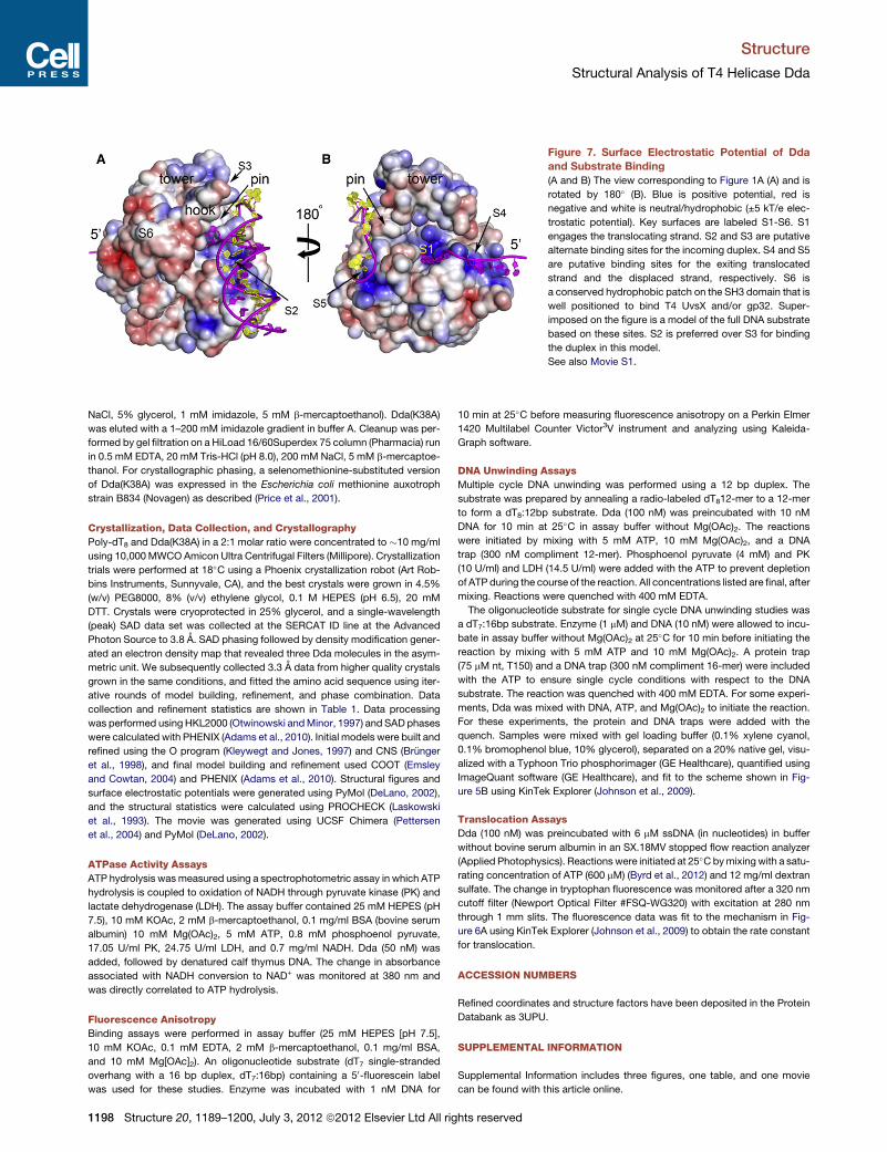
Figure 7. Surface Electrostatic Potential of Dda
and Substrate Binding
(A and B) The view corresponding to Figure 1A (A) and is
rotated by 180� (B). Blue is positive potential, red is
negative and white is neutral/hydrophobic (±5 kT/e elec-
trostatic potential). Key surfaces are labeled S1-S6. S1
engages the translocating strand. S2 and S3 are putative
alternate binding sites for the incoming duplex. S4 and S5
are putative binding sites for the exiting translocated
strand and the displaced strand, respectively. S6 is
a conserved hydrophobic patch on the SH3 domain that is
well positioned to bind T4 UvsX and/or gp32. Super-
imposed on the figure is a model of the full DNA substrate
based on these sites. S2 is preferred over S3 for binding
the duplex in this model.
See also Movie S1.
Structure
Structural Analysis of T4 Helicase Dda
NaCl, 5% glycerol, 1 mM imidazole, 5 mM b-mercaptoethanol). Dda(K38A)
was eluted with a 1–200 mM imidazole gradient in buffer A. Cleanup was per-
formed by gel filtration on a HiLoad 16/60Superdex 75 column (Pharmacia) run
in 0.5 mM EDTA, 20 mM Tris-HCl (pH 8.0), 200 mM NaCl, 5 mM b-mercaptoe-
thanol. For crystallographic phasing, a selenomethionine-substituted version
of Dda(K38A) was expressed in the Escherichia coli methionine auxotroph
strain B834 (Novagen) as described (Price et al., 2001).
Crystallization, Data Collection, and Crystallography
Poly-dT8 and Dda(K38A) in a 2:1 molar ratio were concentrated to �10 mg/ml
using 10,000MWCOAmicon Ultra Centrifugal Filters (Millipore). Crystallization
trials were performed at 18�C using a Phoenix crystallization robot (Art Rob-
bins Instruments, Sunnyvale, CA), and the best crystals were grown in 4.5%
(w/v) PEG8000, 8% (v/v) ethylene glycol, 0.1 M HEPES (pH 6.5), 20 mM
DTT. Crystals were cryoprotected in 25% glycerol, and a single-wavelength
(peak) SAD data set was collected at the SERCAT ID line at the Advanced
Photon Source to 3.8 A. SAD phasing followed by density modification gener-
ated an electron density map that revealed three Dda molecules in the asym-
metric unit. We subsequently collected 3.3 A data from higher quality crystals
grown in the same conditions, and fitted the amino acid sequence using iter-
ative rounds of model building, refinement, and phase combination. Data
collection and refinement statistics are shown in Table 1. Data processing
was performed using HKL2000 (Otwinowski andMinor, 1997) and SADphases
were calculated with PHENIX (Adams et al., 2010). Initial models were built and
refined using the O program (Kleywegt and Jones, 1997) and CNS (Brunger
et al., 1998), and final model building and refinement used COOT (Emsley
and Cowtan, 2004) and PHENIX (Adams et al., 2010). Structural figures and
surface electrostatic potentials were generated using PyMol (DeLano, 2002),
and the structural statistics were calculated using PROCHECK (Laskowski
et al., 1993). The movie was generated using UCSF Chimera (Pettersen
et al., 2004) and PyMol (DeLano, 2002).
ATPase Activity Assays
ATP hydrolysis wasmeasured using a spectrophotometric assay in which ATP
hydrolysis is coupled to oxidation of NADH through pyruvate kinase (PK) and
lactate dehydrogenase (LDH). The assay buffer contained 25 mM HEPES (pH
7.5), 10 mM KOAc, 2 mM b-mercaptoethanol, 0.1 mg/ml BSA (bovine serum
albumin) 10 mM Mg(OAc)2, 5 mM ATP, 0.8 mM phosphoenol pyruvate,
17.05 U/ml PK, 24.75 U/ml LDH, and 0.7 mg/ml NADH. Dda (50 nM) was
added, followed by denatured calf thymus DNA. The change in absorbance
associated with NADH conversion to NAD+ was monitored at 380 nm and
was directly correlated to ATP hydrolysis.
Fluorescence Anisotropy
Binding assays were performed in assay buffer (25 mM HEPES [pH 7.5],
10 mM KOAc, 0.1 mM EDTA, 2 mM b-mercaptoethanol, 0.1 mg/ml BSA,
and 10 mM Mg[OAc]2). An oligonucleotide substrate (dT7 single-stranded
overhang with a 16 bp duplex, dT7:16bp) containing a 50-fluorescein label
was used for these studies. Enzyme was incubated with 1 nM DNA for
1198 Structure 20, 1189–1200, July 3, 2012 ª2012 Elsevier Ltd All rig
10 min at 25�C before measuring fluorescence anisotropy on a Perkin Elmer
1420 Multilabel Counter Victor3V instrument and analyzing using Kaleida-
Graph software.
DNA Unwinding Assays
Multiple cycle DNA unwinding was performed using a 12 bp duplex. The
substrate was prepared by annealing a radio-labeled dT812-mer to a 12-mer
to form a dT8:12bp substrate. Dda (100 nM) was preincubated with 10 nM
DNA for 10 min at 25�C in assay buffer without Mg(OAc)2. The reactions
were initiated by mixing with 5 mM ATP, 10 mM Mg(OAc)2, and a DNA
trap (300 nM compliment 12-mer). Phosphoenol pyruvate (4 mM) and PK
(10 U/ml) and LDH (14.5 U/ml) were added with the ATP to prevent depletion
of ATP during the course of the reaction. All concentrations listed are final, after
mixing. Reactions were quenched with 400 mM EDTA.
The oligonucleotide substrate for single cycle DNA unwinding studies was
a dT7:16bp substrate. Enzyme (1 mM) and DNA (10 nM) were allowed to incu-
bate in assay buffer without Mg(OAc)2 at 25�C for 10 min before initiating the
reaction by mixing with 5 mM ATP and 10 mM Mg(OAc)2. A protein trap
(75 mM nt, T150) and a DNA trap (300 nM compliment 16-mer) were included
with the ATP to ensure single cycle conditions with respect to the DNA
substrate. The reaction was quenched with 400 mM EDTA. For some experi-
ments, Dda was mixed with DNA, ATP, and Mg(OAc)2 to initiate the reaction.
For these experiments, the protein and DNA traps were added with the
quench. Samples were mixed with gel loading buffer (0.1% xylene cyanol,
0.1% bromophenol blue, 10% glycerol), separated on a 20% native gel, visu-
alized with a Typhoon Trio phosphorimager (GE Healthcare), quantified using
ImageQuant software (GE Healthcare), and fit to the scheme shown in Fig-
ure 5B using KinTek Explorer (Johnson et al., 2009).
Translocation Assays
Dda (100 nM) was preincubated with 6 mM ssDNA (in nucleotides) in buffer
without bovine serum albumin in an SX.18MV stopped flow reaction analyzer
(Applied Photophysics). Reactions were initiated at 25�Cbymixingwith a satu-
rating concentration of ATP (600 mM) (Byrd et al., 2012) and 12 mg/ml dextran
sulfate. The change in tryptophan fluorescence was monitored after a 320 nm
cutoff filter (Newport Optical Filter #FSQ-WG320) with excitation at 280 nm
through 1 mm slits. The fluorescence data was fit to the mechanism in Fig-
ure 6A using KinTek Explorer (Johnson et al., 2009) to obtain the rate constant
for translocation.
ACCESSION NUMBERS
Refined coordinates and structure factors have been deposited in the Protein
Databank as 3UPU.
SUPPLEMENTAL INFORMATION
Supplemental Information includes three figures, one table, and one movie
can be found with this article online.
hts reserved
Structure
Structural Analysis of T4 Helicase Dda
ACKNOWLEDGMENTS
This work was supported by NIH grants GM066934 (to S.W.W. and K.N.K.) and
GM098922 (to K.D.R.), UL1RR029884 from the NCRR, Cancer Center core
grant CA21765 and the American Lebanese Syrian Associated Charities
(ALSAC). The content is solely the responsibility of the authors and does not
necessarily represent the official views of the NIH. Data were collected at
the Southeast Regional Collaborative Access Team (SER-CAT) 22-ID beam-
line at the Advanced Photon Source, Argonne National Laboratory. Supporting
institutions may be found at http://www.ser-cat.org/members.html. Use of the
Advanced Photon Source was supported by the U. S. Department of Energy,
Office of Science, Office of Basic Energy Sciences, under contract No. W-31-
109-Eng-38. We thank Rebecca DuBois and Darcie Miller for technical
assistance.
Received: March 9, 2012
Revised: April 19, 2012
Accepted: April 21, 2012
Published online: May 31, 2012
REFERENCES
Adams, P.D., Afonine, P.V., Bunkoczi, G., Chen, V.B., Davis, I.W., Echols, N.,
Headd, J.J., Hung, L.W., Kapral, G.J., Grosse-Kunstleve, R.W., et al. (2010).
PHENIX: a comprehensive Python-based system for macromolecular struc-
ture solution. Acta Crystallogr. D Biol. Crystallogr. 66, 213–221.
Aggarwal, M., and Brosh, R.M., Jr. (2009). Hitting the bull’s eye: novel directed
cancer therapy through helicase-targeted synthetic lethality. J. Cell. Biochem.
106, 758–763.
Bedinger, P., Hochstrasser, M., Jongeneel, C.V., and Alberts, B.M. (1983).
Properties of the T4 bacteriophage DNA replication apparatus: the T4 dda
DNA helicase is required to pass a bound RNA polymerase molecule. Cell
34, 115–123.
Behme, M.T., and Ebisuzaki, K. (1975). Characterization of a bacteriophage T4
mutant lacking DNA-dependent ATPase. J. Virol. 15, 50–54.
Belanger, K.G. (1997). Origin-dependent DNA replication in bacteriophage T4:
a mechanism for initiation. PhD thesis, Duke University Medical Center,
Durham, NC.
Blair, L.P., Tackett, A.J., and Raney, K.D. (2009). Development and evaluation
of a structural model for SF1B helicase Dda. Biochemistry 48, 2321–2329.
Bochman, M.L., Sabouri, N., and Zakian, V.A. (2010). Unwinding the functions
of the Pif1 family helicases. DNA Repair (Amst.) 9, 237–249.
Bohr, V.A. (2008). Rising from the RecQ-age: the role of humanRecQ helicases
in genome maintenance. Trends Biochem. Sci. 33, 609–620.
Boule, J.B., and Zakian, V.A. (2010). Characterization of the helicase activity
and anti-telomerase properties of yeast Pif1p in vitro. Methods Mol. Biol.
587, 359–376.
Boule, J.B., Vega, L.R., and Zakian, V.A. (2005). The yeast Pif1p helicase re-
moves telomerase from telomeric DNA. Nature 438, 57–61.
Brosh, R.M., Jr., Li, J.L., Kenny, M.K., Karow, J.K., Cooper, M.P., Kureekattil,
R.P., Hickson, I.D., and Bohr, V.A. (2000). Replication protein A physically
interacts with the Bloom’s syndrome protein and stimulates its helicase
activity. J. Biol. Chem. 275, 23500–23508.
Brunger, A.T., Adams, P.D., Clore, G.M., DeLano, W.L., Gros, P., Grosse-
Kunstleve, R.W., Jiang, J.S., Kuszewski, J., Nilges, M., Pannu, N.S., et al.
(1998). Crystallography & NMR system: A new software suite for macromolec-
ular structure determination. Acta Crystallogr. D Biol. Crystallogr. 54, 905–921.
Buttner, K., Nehring, S., and Hopfner, K.P. (2007). Structural basis for DNA
duplex separation by a superfamily-2 helicase. Nat. Struct. Mol. Biol. 14,
647–652.
Byrd, A.K., and Raney, K.D. (2004). Protein displacement by an assembly of
helicase molecules aligned along single-stranded DNA. Nat. Struct. Mol.
Biol. 11, 531–538.
Byrd, A.K., and Raney, K.D. (2006). Displacement of a DNA binding protein by
Dda helicase. Nucleic Acids Res. 34, 3020–3029.
Structure 20, 1
Byrd, A.K., Matlock, D.L., Bagchi, D., Aarattuthodiyil, S., Harrison, D.,
Croquette, V., and Raney, K.D. (2012). Dda helicase tightly couples transloca-
tion on single-stranded DNA to unwinding of duplex DNA: Dda is an optimally
active helicase. J. Mol. Biol. Published online Apr 11, 2012. 10.1016/j.jmb.
2012.04.007.
Cheng, X., Dunaway, S., and Ivessa, A.S. (2007). The role of Pif1p, a DNA heli-
case in Saccharomyces cerevisiae, in maintaining mitochondrial DNA.
Mitochondrion 7, 211–222.
Delagoutte, E., and von Hippel, P.H. (2003). Helicase mechanisms and the
coupling of helicases within macromolecular machines. Part II: Integration of
helicases into cellular processes. Q. Rev. Biophys. 36, 1–69.
DeLano,W.L. (2002). The PyMOLMolecular Graphics System (SanCarlos, CA,
USA: DeLano Scientific).
Ellis, N.A. (1997). DNA helicases in inherited human disorders. Curr. Opin.
Genet. Dev. 7, 354–363.
Emsley, P., and Cowtan, K. (2004). Coot: model-building tools for molecular
graphics. Acta Crystallogr. D Biol. Crystallogr. 60, 2126–2132.
Eoff, R.L., and Raney, K.D. (2006). Intermediates revealed in the kinetic mech-
anism for DNA unwinding by a monomeric helicase. Nat. Struct. Mol. Biol. 13,
242–249.
Eoff, R.L., and Raney, K.D. (2010). Kinetic mechanism for DNA unwinding by
multiple molecules of Dda helicase aligned on DNA. Biochemistry 49, 4543–
4553.
Eoff, R.L., Spurling, T.L., and Raney, K.D. (2005). Chemically modified DNA
substrates implicate the importance of electrostatic interactions for DNA
unwinding by Dda helicase. Biochemistry 44, 666–674.
Formosa, T., and Alberts, B.M. (1984). The use of affinity chromatography to
study proteins involved in bacteriophage T4 genetic recombination. Cold
Spring Harb. Symp. Quant. Biol. 49, 363–370.
Frick, D.N. (2007). The hepatitis C virus NS3 protein: amodel RNA helicase and
potential drug target. Curr. Issues Mol. Biol. 9, 1–20.
Gauss, P., Park, K., Spencer, T.E., and Hacker, K.J. (1994). DNA helicase
requirements for DNA replication during bacteriophage T4 infection.
J. Bacteriol. 176, 1667–1672.
Gorbalenya, A.E., and Koonin, E.V. (1993). Helicases - Amino-Acid-Sequence
Comparisons and Structure-Function-Relationships. Curr. Opin. Struct. Biol.
3, 419–429.
Gu, J., Xia, X., Yan, P., Liu, H., Podust, V.N., Reynolds, A.B., and Fanning, E.
(2004). Cell cycle-dependent regulation of a humanDNAhelicase that localizes
in DNA damage foci. Mol. Biol. Cell 15, 3320–3332.
Hacker, K.J., and Alberts, B.M. (1992). Overexpression, purification, sequence
analysis, and characterization of the T4 bacteriophage dda DNA helicase.
J. Biol. Chem. 267, 20674–20681.
Jankowsky, E., and Fairman, M.E. (2007). RNA helicases—one fold for many
functions. Curr. Opin. Struct. Biol. 17, 316–324.
Johnson, K.A., Simpson, Z.B., and Blom, T. (2009). Global kinetic explorer:
a new computer program for dynamic simulation and fitting of kinetic data.
Anal. Biochem. 387, 20–29.
Kadyrov, F.A., and Drake, J.W. (2004). UvsX recombinase and Dda helicase
rescue stalled bacteriophage T4 DNA replication forks in vitro. J. Biol.
Chem. 279, 35735–35740.
Kerr, I.D., Sivakolundu, S., Li, Z., Buchsbaum, J.C., Knox, L.A., Kriwacki, R.,
and White, S.W. (2007). Crystallographic and NMR analyses of UvsW and
UvsW.1 from bacteriophage T4. J. Biol. Chem. 282, 34392–34400.
Kim, J.L., Morgenstern, K.A., Griffith, J.P., Dwyer, M.D., Thomson, J.A.,
Murcko, M.A., Lin, C., and Caron, P.R. (1998). Hepatitis C virus NS3 RNA heli-
case domain with a bound oligonucleotide: the crystal structure provides
insights into the mode of unwinding. Structure 6, 89–100.
Kleywegt, G.J., and Jones, T.A. (1997). Model building and refinement prac-
tice. Methods Enzymol. 277, 208–230.
Kodadek, T. (1991). Inhibition of protein-mediated homologous pairing by
a DNA helicase. J. Biol. Chem. 266, 9712–9718.
189–1200, July 3, 2012 ª2012 Elsevier Ltd All rights reserved 1199
Structure
Structural Analysis of T4 Helicase Dda
Kodadek, T., and Alberts, B.M. (1987). Stimulation of protein-directed strand
exchange by a DNA helicase. Nature 326, 312–314.
Korolev, S., Hsieh, J., Gauss, G.H., Lohman, T.M., and Waksman, G. (1997).
Major domain swiveling revealed by the crystal structures of complexes of
E. coli Rep helicase bound to single-stranded DNA and ADP. Cell 90, 635–647.
Lam, A.M., Keeney, D., and Frick, D.N. (2003). Two novel conserved motifs in
the hepatitis C virus NS3 protein critical for helicase action. J. Biol. Chem. 278,
44514–44524.
Laskowski, R.A., MacArthur, M.W., Moss, D.S., and Thornton, J.M. (1993).
PROCHECK: a program to check the stereochemical quality of protein struc-
tures. J. Appl. Crystallogr. 26, 283–291.
Lee, J.Y., and Yang, W. (2006). UvrD helicase unwinds DNA one base pair at
a time by a two-part power stroke. Cell 127, 1349–1360.
Lescar, J., Luo, D., Xu, T., Sampath, A., Lim, S.P., Canard, B., and Vasudevan,
S.G. (2008). Towards the design of antiviral inhibitors against flaviviruses: the
case for themultifunctional NS3 protein fromDengue virus as a target. Antiviral
Res. 80, 94–101.
Lohman, T.M., Tomko, E.J., and Wu, C.G. (2008). Non-hexameric DNA heli-
cases and translocases: mechanisms and regulation. Nat. Rev. Mol. Cell
Biol. 9, 391–401.
Manosas, M., Xi, X.G., Bensimon, D., and Croquette, V. (2010). Active and
passive mechanisms of helicases. Nucleic Acids Res. 38, 5518–5526.
Morris, P.D., and Raney, K.D. (1999). DNA helicases displace streptavidin from
biotin-labeled oligonucleotides. Biochemistry 38, 5164–5171.
Nanduri, B., Byrd, A.K., Eoff, R.L., Tackett, A.J., and Raney, K.D. (2002). Pre-
steady-state DNA unwinding by bacteriophage T4 Dda helicase reveals
a monomeric molecular motor. Proc. Natl. Acad. Sci. USA 99, 14722–14727.
Otwinowski, Z., and Minor, W. (1997). Processing of X-ray diffraction data
collected in oscillation mode. Methods Enzymol. 276, 307–326.
Patel, S.S., and Donmez, I. (2006). Mechanisms of helicases. J. Biol. Chem.
281, 18265–18268.
Pettersen, E.F., Goddard, T.D., Huang, C.C., Couch, G.S., Greenblatt, D.M.,
Meng, E.C., and Ferrin, T.E. (2004). UCSF Chimera—a visualization system
for exploratory research and analysis. J. Comput. Chem. 25, 1605–1612.
1200 Structure 20, 1189–1200, July 3, 2012 ª2012 Elsevier Ltd All rig
Pike, A.C., Shrestha, B., Popuri, V., Burgess-Brown, N., Muzzolini, L.,
Costantini, S., Vindigni, A., and Gileadi, O. (2009). Structure of the human
RECQ1 helicase reveals a putative strand-separation pin. Proc. Natl. Acad.
Sci. USA 106, 1039–1044.
Price, A.C., Zhang, Y.M., Rock, C.O., and White, S.W. (2001). Structure of
beta-ketoacyl-[acyl carrier protein] reductase from Escherichia coli: negative
cooperativity and its structural basis. Biochemistry 40, 12772–12781.
Pyle, A.M. (2008). Translocation and unwinding mechanisms of RNA and DNA
helicases. Annu Rev Biophys 37, 317–336.
Rajagopal, V., Gurjar, M., Levin, M.K., and Patel, S.S. (2010). The protease
domain increases the translocation stepping efficiency of the hepatitis C virus
NS3-4A helicase. J. Biol. Chem. 285, 17821–17832.
Saikrishnan, K., Griffiths, S.P., Cook, N., Court, R., and Wigley, D.B. (2008).
DNA binding to RecD: role of the 1B domain in SF1B helicase activity.
EMBO J. 27, 2222–2229.
Saikrishnan, K., Powell, B., Cook, N.J., Webb, M.R., and Wigley, D.B. (2009).
Mechanistic basis of 50-30 translocation in SF1B helicases. Cell 137, 849–859.
Singleton, M.R., Dillingham, M.S., and Wigley, D.B. (2007). Structure and
mechanism of helicases and nucleic acid translocases. Annu. Rev. Biochem.
76, 23–50.
Stevnsner, T., Muftuoglu, M., Aamann, M.D., and Bohr, V.A. (2008). The role of
Cockayne Syndrome group B (CSB) protein in base excision repair and aging.
Mech. Ageing Dev. 129, 441–448.
Taneja, P., Gu, J., Peng, R., Carrick, R., Uchiumi, F., Ott, R.D., Gustafson, E.,
Podust, V.N., and Fanning, E. (2002). A dominant-negative mutant of human
DNA helicase B blocks the onset of chromosomal DNA replication. J. Biol.
Chem. 277, 40853–40861.
Theis, K., Chen, P.J., Skorvaga, M., Van Houten, B., and Kisker, C. (1999).
Crystal structure of UvrB, a DNA helicase adapted for nucleotide excision
repair. EMBO J. 18, 6899–6907.
Velankar, S.S., Soultanas, P., Dillingham,M.S., Subramanya, H.S., andWigley,
D.B. (1999). Crystal structures of complexes of PcrA DNA helicase with a DNA
substrate indicate an inchworm mechanism. Cell 97, 75–84.
hts reserved