analysis of the bacteriophage t4 dna replication complex
TRANSCRIPT

ANALYSIS OF THE BACTERIOPHAGE T4 DNA REPLICATION COMPLEX
by
MAUREEN M. MUNN
DISSERTATION
Submitted in partial satisfaction of the requirements for the degree of
DOCTOR OF PHILOSOPHY
in
BIOCHEMISTRY AND BIOPHYSICS
in the
GRADUATE DIVISION
of the
UNIVERSITY OF CALIFORNIA
San Francisco
Approved:
Committee in Charge
Deposited in the Library, University of California, San Francisco
- - -Dat. . . . . . . . . . . . . . . . University Lib rarian
-

TO MICHAEL
AND
MY PARENTS
FOR ALL THEIR LOVE AND SUPPORT

iV
ACKNOWLEDGEMENTS
I would like to thank Bruce Alberts for the wonderful guidance
he has given me throughout my graduate career. Bruce has provided
an endless source of inspiration through his extensive knowledge and
insightful understanding of science, his dedication to his work, and
his general good humour and kindness.
I am grateful to the members of the Alberts lab for providing
intellectual stimulation, friendship and good fun. I would especially
like to thank Jack Barry for sharing his extensive knowledge of pro
tein purification with me.
I am indebted to Kathy Raneses and Leslie Spector for their
expert typing abilities and to Carol Morita and Gert Weil for making
such beautiful figures.
I would like to extend a hearty thanks to my family and friends
for all their encouragement and support.

ANALYSIS OF THE BACTERIOPHAGE T4 DNA REPLICATION COMPLEX
BY MAUREEN M. MUNN
ABSTRACT
The bacteriophage T4 DNA replication system consists of a highly
organized multi-enzyme complex of at least seven T4-encoded proteins.
This thesis uses two different approaches to analyze the functional
interactions of these proteins within this complex.
Chapter 2 describes the analysis of replication fork pausing on a
duplex fo DNA template by the core replication complex (polymerase,
its accessory proteins, and the helix destabilizing protein) in the
absence or presence of either the dola helicase or the gene 41 helicase.
The core system pauses transiently in the course of DNA replication at
unique positions along the fo template. Fine mapping of two repli
cation pause-sites indicates that in each case the replication complex
pauses at a cluster of discrete sites; the precise locations of several of
these pause-sites correlate with four base palindromic sequences. We
propose that the replication fork pauses at these sites because the
nascent 3' end becomes transiently base-paired to the analogous
sequence on the displaced DNA strand. The 41 helicase causes the
elimination of replication pausing, while the dola helicase does not.
The interactions of polymerase, its accessory proteins, and the
helix destabilizing protein on a primer-template junction are probed in
Chapters 3 and 4, using DNA footprinting techniques. The DNA sub
strates used for these experiments are two perfect hairpin helices with
long single-stranded 5' extensions. These two DNA molecules consti
tute primer-template junctions, with the region from the 3' terminus to

Vi
the hairpin loop acting as the "primer strand" and the region from the
loop to the 5' terminus forming the "template strand". In the presence
of the ATP analogue, ATP*S, the polymerase accessory proteins bind
each of these DNA molecules in the region of the 3' terminus. This
binding is facilitated 100-fold by the presence of the helix destabilizing
protein on the single-stranded region of the template strand, indi
cating the importance of protein-protein interactions in the assembly of
this complex. DNA polymerase also binds in the region of the 3'
terminus, protecting 15 bases on the primer strand and 14 bases on
template strand from DNasel cleavage. The helix destabilzing protein
increases the affinity of the polymerase for its binding site at the
primer-template junction by a factor of about thirty. In the presence
of ATP*S, the DNA polymerase and the accessory proteins compete for
binding at the 3' terminus, and which protein is bound depends on
their relative concentrations.
Similar experiments demonstrate the formation of a complex
between the DNA polymerase and the gene 45 protein (one of the
accessory proteins), with the 45 protein binding behind the polymerase
on the duplex portion of the DNA molecule. No effect of the other
accessory protein, the 44/62 complex, is observed in this system,
either in the presence of ATP or in the absence of ribonucleotide.
The expected 44/62 footprint can be estimated by comparing the 45
protein footprint seen in the presence of polymerase with the 44/62
and 45 protein footprint that requires ATP&S. In this way, a model
can be derived for a functioning replication complex containing poly
merase and all three of its accessory proteins. Our data reveal that
this complex has a much lower affinity for DNA when the DNA poly
Vii
merase is stalled than when the polymerase is moving. This finding is
reasonable, once one considers the biological role of the accessory
proteins, and it suggests a specific model in which both ATP hydro
lysis and energy derived from each polymerase cycle are required to
keep the accessory protein clamp on the DNA.

Viii
TABLE OF CONTENTS
CHAPTER ONE: INTRODUCTION 1
The Bacteriophage T4 DNA Replication System 2The T4 Replication ProteinsMechanism of Initiation of DNA Synthesis In Vivo 10The E. coli DNA Replication System 14Eucaryotic DNA Replication Systems 23DNA Replication by Multienzyme Complexes 31The Use of DNA Protection Experiments to Study 36
a Multienzyme Complex
CHAPTER TWO : SPECIFIC ONA SEOUENCES ACT AS PAUSE SITES 41DURING THE IN VITRO REPLICATION OF ADOUBLE-STRANDED DNA TEMPLATE
Abstract 42Introduction 43Materials and MethodsResults 45Discussion 56Tables and Figures 61
CHAPTER THREE: THE T4 DNA POLYMERASE FORM A SPECIFIC 73COMPLEX WITH THE HELIX DESTAB | LIZINGPROTEIN ON A PRIMER-TEMPLATE JUNCT |ON
Abstract 74Introduction 75Materials and Methods 78Results 82Discussion 95Tables and Figures 99
CHAPTER FOUR INTERACTIONS OF THE T4 DNA POLYMERASE 111WITH ITS ACCESSORY PROTEINS AND THEHELIX DESTABILIZING PROTEIN ON APRIMER-TEMPLATE JUNCTION
Abstract 112Introduction 114Materials and Methods 117Results 120Discussion 134Tables and Figures 143
REFERENCES 157

CHAPTER ONE
|NTRODUCTION

THE BACTERIOPHAGE T4 DNA REPLICATION SYSTEM
Bacteriophage T4 provides an invaluable system for the study and
understanding of the DNA replication process. Thorough genetic
analysis has identified more than twenty T4 genes that are required
for DNA replication (Wood and Revel, 1976) and has indicated that this
bacteriophage encodes most, if not all of the proteins required for its
own DNA synthesis. In the course of a T4 infection there can be as
many as two hundred replication forks per T4-infected cell (McCarthy
et al., 1976), and correspondingly high concentrations of the
necessary replication proteins are produced. The complexity of thissystem, as defined by the genetic work, and the comparative ease of
purification of relatively large amounts of the relevant proteins make
T4 a good model system for understanding the mechanisms of this
process. The similarity of the problems which must be dealt with in
any replication system dictates that the approaches used by this
procaryotic bacteriophage will be similar in design, if not in detail, to
the approaches used by the more sophisticated eucaryotic systems.
Thirteen of the T4 replication proteins have been purified in the
laboratory of Bruce Alberts, where the research for this thesis was
carried out. Where possible, the initial purification procedures utilized
the assay of a specific activity or physical property for the selection
of a particular species. However, for several of the genes identified
genetically to be required for DNA replication a specific activity was
not known. These proteins were purified on the basis of their ability
to complement DNA synthesis carried out by a crude cell lysate made
from E. coli cells infected with a T4 mutant in that gene. In the
presence of exogenously added labelled deoxynucleotides and the

missing factors, these crude cell lysates can incorporate label into
endogenous DNA (Barry and Alberts, 1972; Barry et al., 1973). More
recently, the application of cloning techniques has allowed the cloning
and overexpression of many of the T4 replication proteins, providing
these proteins in sufficient quantities to make various physical studies
of this system much more feasible.
The T4 Replication Proteins
The central enzyme to this process is the DNA polymerase, the
product of gene 43 (de Waard et al., 1965; Warner and Barnes, 1966).
This 110,000 dalton protein can catalyze both the polymerization of
deoxynucleoside triphosphates onto a primed single-stranded DNA
template and the excision of mononucleotides from the 3' terminus of a
single- or double-stranded DNA template (Englund, 1971; Huang and
Lehman, 1972; Goulian et al., 1968; Hershfield and Nossal, 1972).
This 3' to 5’ exonuclease is much faster on a single-stranded than a
duplex template. This observed preference for a non-base paired 3'
terminus led to the hypothesis that this 3' to 5’ exonuclease activity
provides a mechanism of proofreading the most recently incorporated
nucleotide (Brutlag and Kornberg, 1972; Muzyczka et al., 1972). This
hypothesis has been verified by the characterization of polymerase
mutants with an altered exonuclease activity; mutants with reduced
exonuclease activity are characteristically mutator in phenotype, while
those with increased exonuclease activity show a reduction in mutation
frequency (Bessman et al., 1974; Muzyczka et al., 1972).
Although the T4 polymerase can elongate a primed single-stranded
DNA template in vitro, it does so at a rate that is slower and less

processive than the rate of replication fork movement measured in vivo
(Das and Fujimura, 1979; Mace and Alberts, 1984b; Newport et al.,
1980). Furthermore, the T4 polymerase cannot utilize a double
stranded DNA template, which is the physiological substrate of this
replication system. Finally, the T4 polymerase is unable to initiate
unprimed DNA synthesis and so requires primer formation on the
lagging strand of the replication fork. Thus, the activities of several
other proteins are required for efficient DNA synthesis in vivo.
These include a single-stranded DNA binding protein, proteins which
modify the rate and processivity of the polymerase, proteins which
contribute to the unwinding of the double helix, and the RNA primase,
which is required for the priming of discontinuous synthesis on the
lagging strand.
The most abundant T4 replication protein is the helix destabilizing
protein, the product of gene 32. This 34,000 dalton protein binds
cooperatively to the single-stranded regions of the DNA template,
leaving the bases accessible to the polymerase and eliminating short
regions of secondary structure (Alberts et al., 1969; Alberts and
Frey, 1970). This protein stimulates DNA synthesis by the polymerase
on a primed single-stranded template, both through its effect on the
structure of the DNA template and through a direct interaction with
the T4 polymerase (Huang et al., 1981; Huberman et al., 1971). Its
activity is required for DNA synthesis on a double-stranded template;
in this reaction the 32 protein binds and stabilizes single-stranded
DNA as the double helix "breathes" ahead of the growing replication
fork (Alberts et al., 1980). Genetic analysis has shown that gene
32 protein is involved in recombination as well as replication (Epstein

et al., 1963; Tomigawa et al., 1966). The use of affinity chromato
9raphy using an agarose column matrix to which the 32 protein has
been covalently attached has identified interactions between 32 protein
and at least nine different T4 proteins involved in DNA replication,
recombination and repair (Alberts et al., 1983; Formosa et al., 1983).
Thus, the role of 32 protein in each of these processes is not a
passive one, but rather involves direct interactions with many of the
protein components involved.
The polymerase accessory proteins were shown genetically to be
required for DNA synthesis (Epstein et al., 1963) and were originally
purified on the basis of the complementation assay described earlier
(Barry and Alberts, 1972; Morris et al., 1979; Nossal. 1979). The
accessory proteins consist of a 163,000 dalton complex of the products
of genes 44 and 62 (probably four 44 subunits and one 62 subunit;
Splicer et al., 1984) and the 49,000 dalton dimer of the product of
gene 45. The 44/62 protein has DNA-dependent ATPase activity that
resides in the 44 subunits. This activity is further stimulated by an
interaction with the 45 protein, which may be mediated via the 62
protein (Alberts et al., 1975; Lin et al., 1985; Mace and Alberts,
1984a; Piperno et al., 1978).
On a primed single-stranded DNA template, these proteins inter
act with the DNA polymerase in a reaction that requires ATP hydroly
sis by the 44/62 complex and that results in an increase in both the
rate and processivity of DNA synthesis by the polymerase molecule
(Alberts et al., 1975; Mace and Alberts, 1984c; Piperno et al., 1978).
The accessory proteins are absolutely required for efficient DNA
synthesis by the T4 polymerase on a nicked double-stranded template,

in addition to gene 32 protein (Liu et al., 1979; Nossal and Peterlin,
1979). The accessory proteins or low concentrations of 32 protein also
stimulate the rate and processivity of the 3' to 5’ exonuclease on a
double-stranded template; when these proteins are added in conjunc
tion, stimulation appears synergistic (Bedinger and Alberts, 1983;
Venkatesan and Nossal, 1982). The activity of the accessory proteins
is most consistent with the formation of a complex with the polymerase,
with the accessory proteins acting as a "sliding clamp" that holds the
polymerase on the 3' end of the nascent DNA strand. ATP hydrolysis
is required for the formation of this complex (Alberts et al., 1980;
Newport et al., 1980), although reports differ as to how frequently
ATP must be hydrolyzed to maintain it. One report states that this
interaction is stable for up to 10 min on a primed single-stranded
template (Huang et al., 1981), while other studies show that ATP
hydrolysis is required at least every 30 s on a primed fo template or
10 s on a nicked double-stranded template (Alberts et al., 1980;
Nossal and Alberts, 1983). Although less than one ATP molecule is
hydrolyzed for every ten nucleotides incorporated on a single-stranded
or duplex template (Liu et al., 1979; Piperno and Alberts, 1978), the
complex remains associated for the incorporation of tens of thousands
of nucleotides (Alberts et al., 1980). To explain this phenomenon, it
has been proposed that this complex functions as two "half clamps"
either of which can hold the polymerase on the DNA substrate, but
which require ATP hydrolysis, to reconstitute the functional "whole
clamp" (Alberts et al., 1980).
At physiological concentrations of 32 protein, yet another protein
is required for maximum rates of synthesis on a double-stranded

template. This protein, the product of gene 41, is a GTP- or ATP
dependent DNA helicase (Liu and Alberts, 1981a; Morris et al., 1979;
Nossal, 1979; Venkatesan et al., 1982). The gene 41 helicase func
tions as a dimer of 58,000 dalton subunits. It can catalyze the release
of 50 to 400 nucleotide long fragments which are hybridized to a
homologous strand. This activity initiates from the 3' terminus of the
released fragments, indicating that the 41 protein propagates along the
complementary sequence in a 5' to 3’ direction. This helicase activity
is stimulated by the polymerase accessory proteins and by another
replication protein, the product of gene 61. When all of these proteins
are added in concert the effect is greater that additive, indicating
synergism in their activities (Venkatesan et al., 1982). The gene 41
helicase stimulates the rate of DNA synthesis on a double-stranded
template by utilizing the energy of GTP or ATP hydrolysis to unwind
the DNA helix ahead of the advancing replication complex (Alberts,
1980; Liu et al., 1979; Venkatesan et al., 1982). The directionality of
this helicase is consistent with its association with the lagging strand.
In addition to its helicase activity, 41 protein is a component of
the RNA priming complex, along with the product of gene 61.
Together these proteins synthesize the RNA pentamers which are
essential for the priming of discontinuous DNA synthesis on the
lagging strand (Liu and Alberts, 1980; Liu and Alberts, 1981b;
Nossal, 1980; Silver and Nossal, 1979). The actual synthesis of
primers is catalyzed by the 61 protein, although this activity is
stimulated and modified in its template specificity by the 41 protein
(Cha and Alberts, J. Biol. Chem. , in press (1986)).
Another DNA helicase which interacts with the T4 replication
machinery is the T4-encoded dola helicase. This 48,000 dalton protein
is also a 5' to 3' helicase, and like the gene 41 helicase it utilizes the
energy of ATP hydrolysis to speed up the rate of fork movement on a
double-stranded DNA template (Krell et al., 1979; Jongeneel, Bedinger
and Alberts, 1984). However, these two proteins differ in this func
tion in two fundamental ways. The unwinding and release of a DNA
fragment by the dola helicase requires stoichiometric amounts of this
protein in a ratio of about one dola molecule per three base pairs
unwound, while the 41 helicase is required in catalytic amounts
(Jongeneel, Formosa and Alberts, 1984). In the presence of a repli
cation complex on a double-stranded template, the 41 helicase acts in a
highly processive manner, apparently as an integral component of the
replication complex, while the dola helicase operates in a highly dis
tributive manner (Jongeneel, Bedinger and Alberts, 1984). Experi
ments in which DNA replication and transcription were performed on
the same template have shed light on the probable physiological role of
the dola protein. In the absence of the dola protein, a growing repli
cation fork becomes stalled behind an RNA polymerase molecule which
is bound at a promoter site on the same DNA template. This block is
released in the presence of the dola protein, but not in the presence
of the gene 41 helicase. Thus, the physiological role of dola protein
may be to remove DNA-bound protein molecules ahead of the growing
replication fork (Alberts et al., 1983; Bedinger et al., 1983).
Using all of the proteins just described it is possible to synthe
size DNA in vitro at rates similar to those observed in vivo, to cata
lyze discontinuous synthesis on the lagging strand, and to remove

protein obstacles ahead of the replication fork. Thus, this in vitro
system probably contains all of the components required for DNA
synthesis on leading and lagging strands. Although the mechanistic
details of this system are not yet deciphered, the assembly of the
necessary components has made this task a resolvable one.

10
Mechanisms Of Initiation Of DNA Synthesis In Vivo
One aspect of the replication process in T4 which has not yet
been well characterized is its initiation. At least three different
mechanisms have been identified for the initiation of replication
in vivo. Early in infection, replication initiates at one or a few
specific origins on the T4 genome, starting from replication bubbles
that require the host RNA polymerase for their formation (Luder and
Mosig, 1982; Snyder and Montgomery, 1974). This primary mode of
initiation is used only in the first few minutes of infection and then is
replaced by secondary or recombinogenic initiation. This process is
independent of the host RNA polymerase and requires the T4 recom
bination pathway. It has been proposed that the intermediates of
recombination act as primers for the synthesis of DNA (Luder and
Mosig, 1982). The most recently discovered tertiary initiation is a
site-specific process which is independent of both the host RNA poly
merase and the recombination proteins, the products of genes 46 and
47, which are required for the secondary mode (Kreuzer and Alberts,
1985). Each of these processes is currently under investigation using
several novel techniques which will be discussed below.
Efforts to reconstruct primary synthesis in vitro have been
unsuccessful, possibly due to missing protein factors. The missing
components may function in a manner analogous to the n' or dria.A
proteins of E. coli or the O and P proteins of lambda; that is, these
factors may be required to position the T4 priming proteins at the
replication origin. Genes that encode proteins involved in related
functions tend to be clustered within the T4 genome; thus a 3 kb
region of T4 containing both the 41 and 61 genes was sequenced

11
(M. Nakanishi and B.M. Alberts, unpublished data). The sequence
reveals several unidentified reading frames (URFs) that could encode
small molecular weight proteins. In order to determine whether these
URFs encode proteins that are essential for replication initiation, a
technique has been devised to introduce site-specific mutations into
this region. This technique uses a sophisticated shuttle vector which
allows the introduction of a mutant T4 gene into a T4-infected cell and
provides a means of selecting for the recombinant integration of that
gene. This system should allow the identification of genes that are
essential but have not yet been identified by genetic analysis (Selick
and Alberts, 1985; H. Selick, personal communication).
The positions of primary origin sequences have been determined
by hybridizing DNA fragments synthesized early in infection to known
T4 sequences (Kozinski, 1983; Mosig, 1983). A 3 kb region adjacent
to the 41-61 region just described, which contains the origin sequence
denoted oria, has been sequenced (Macdonald and Mosig, 1984).
Analysis of this sequence has revealed an unknown gene, denoted
gene 69, which could encode a 46 kb basic protein. A second in
phase promoter within this gene may be the start site for an abbre
viated form of the 69 protein, which has been denoted 69*. A muta
tion which maps to gene 69 has a strong DNA delay phenotype which,
together with the coincidence of this gene with an origin sequence,
implicates its product in the initiation of replication (Macdonald and
Mosig, 1984). The prediction of a hydrophobic domain has led to the
postulate that gene 69 protein may be required to attach a T4 initiation
complex to the cell membrane (Mosig and Macdonald, 1985).
12
Secondary initiation was originally identified as a recombinogenic
process by genetic techniques (Luder and Mosig, 1982). The strong
binding of the two T4 recombination proteins, uvs)K and uvs.Y., to a
32 protein affinity column provided an initial method of purifying these
proteins (Formosa and Alberts, 1984; Formosa, 1985). The uvsX
protein is a DNA-dependent ATPase that binds to both single- and
double-stranded DNA. This enzyme can catalyze homologous pairing
between a linear double-stranded fragment and a homologous single
stranded DNA circle in a reaction that requires ATP hydrolysis. This
activity can be coupled to DNA replication in an in vitro assay that
mimics the model for recombinogenic initiation proposed by Luder and
Mosig (1982). This assay contains closed supercoiled M13 DNA and a
linear single-stranded M13 fragment, deoxy NTPs, the "core" repli
cation proteins (polymerase, its accessory proteins, and 32 protein),
the dola helicase, and uvs)K. The uvs)K-mediated homologous pairing
between the 3' region of the single-stranded fragment and the comple
mentary region on the duplex circle provides a primer for DNA syn
thesis; this base paired 3' terminus is then elongated by the repli
cation system in a reaction that is stimulated by the dola helicase
(Formosa, 1985). Surprisingly, the rate of DNA synthesis is not
stimulated by the T4 topoisomerase, indicating that topological con
straints do not inhibit this process. Examination of the products of
DNA synthesis by E.M. has revealed double-stranded circles with
single-stranded tails that increase in length with incubation. These
results suggest that the newly synthesized DNA strand is displaced
from the template strand, probably in a uvs)K-dependent reaction.
This implies that DNA replication in T4 is, at least at times, a con

13
servative process (Formosa, 1985).
The existence of a third mechanism of initiation was first demon
strated by selecting for regions of the T4 genome which, when inser
ted in a plasmid, could replicate autonomously (Kreuzer and Alberts,
1985). Two such regions were identified. The characterization of the
replication of these plasmids indicated that this process is independent
of both the host RNA polymerase and the T4 recombination proteins,
gene products 46 and 47, and thus is unique from both primary and
secondary initiation. The two original isolates map at about 151 and
114 kb on the T4 genome. Deletion analysis has defined each of these
origins to fragments which are 248 and 120 base pairs long, and which
are located in the tail fiber gene 34 and the promoter region of the
uvs)K gene, respectively. A comparison of the sequence of these two
regions has revealed a 9 base pair homology which occurs once in each
region and an 8 base pair homology which occurs twice in ori (uvs.Y.)
and once in ori(34) (Menkens and Kreuzer, 1985). Based on these
results, it has been proposed that the uvs.Y protein is required for
this initiation process and binds specifically to the 8 or 9 base pair
sequences. The occurrence of these putative regulatory sequences in
the promoter of the uvs.Y gene may reflect a self regulatory mechanism
(Menkens and Kreuzer, 1985).

14
THE E. COLI DNA REPLICATION SYSTEM
The central enzyme in the E. coli DNA replication system is the
polymerase l l l holoenzyme (reviewed by McHenry, 1985). This enzyme
is purified as a multicomponent complex that contains at least seven
subunits. The contributions of each of these subunits have been
defined through the purification and characterization of subsets of
holoenzyme which are missing one or more subunits. The catalytic
core of this enzyme contains three subunits, a, e and 6 (McHenry and
Crow, 1979). The 140,000 dalton a subunit contains the 5' to 3'
polymerase activity (Spanos et al., 1981), and the 25,000 dalton E
subunit contains the 3' to 5’ exonuclease activity that is required for
proofreading (Livingston and Richardson, 1975). These roles have
recently been verified by cloning and expressing the individual sub
units (Shepard et al., 1984; Scheuerman and Echols, 1984). No
activity has as yet been ascribed to the 10,000 dalton 6 subunit.
The poll l l core can catalyze limited DNA synthesis on a primed
single-stranded DNA substrate. Its activity is enhanced and modified
by the presence of at least four other subunits, denoted 3, 3, 5 and
t. Each of these subunits enhances the processivity of the poll || core
and makes its activity more holoenzyme-like in nature, based on
measurements made using holoenzyme sub-species which are missing
one or more of these subunits (Fay et al., 1981; 1982).
DNA polymerase III', which consists of a complex of the core
enzyme and the t subunit, is active in the presence of spermidine,
similar to the holoenzyme and unlike the core enzyme (McHenry, 1982).
Sedimentation analysis indicates that the poll I l' complex consists of a
dimer of the core polymerase and of the t subunit. Thus, the t

15
subunit appears to mediate the dimerization of the core polymerase
(McHenry, 1982). DNA polymerase | | Iº, which lacks only the B sub
unit (Wickner et al., 1973), is the most processive of the holoenzyme
subcomplexes. Like holoenzyme, it is stimulated by SSB protein and
spermidine (Fay et al., 1982).
Addition of the 3 subunit to poll II* is sufficient to reconstitute
holoenzyme (Wickner et al., 1973). Holoenzyme activity requires the
hydrolysis of ATP or dATP; in the absence of this, holoezyme displays
the processivity and salt sensitivity of the poll I lº (Wickner and
Kornberg, 1982a; 1982b). ATP hydrolysis is required for the forma
tion of an initiation complex between holoenzyme and the DNA template;
subsequent DNA synthesis does not require further ATP hydrolysis
(Burgers and Kornberg, 1982a). However, the recycling of holo
enzyme to a new DNA template does require further ATP hydrolysis
(Burgers et al., 1982b). The ATP binding site appears to be on the
poll II* species rather than the B subunit, although ATP binding is
greatly stimulated by the presence of the B subunit (Burgers and
Kornberg, 1982a). In the absence of ATP hydrolysis, excess B sub
unit can support maximum holoenzyme processivity. However, this
reaction differs from the ATP-stimulated reaction in terms of the lower
stability of the complex formed and its increased sensitivity to salt
(Crute et al., 1983). Thus, the authors suggest that ATP binding
stabilizes the formation of the complex between the 3 subunit and
poll II* and that excess B drives the formation of this complex by mass
action (Crute et al., 1983). The ATP analogue, ATP*S, can substi
tute for ATP in the stimulation of holoenzyme activity and is also
hydrolyzed in this reaction. However, net DNA synthesis in the

16
presence of ATPºS is half that achieved in the presence of ATP
(Johanson and McHenry, 1984). The authors propose the existence of
two holoenzyme species in their preparation, which reflects the occur
rence of an asymmetric dimeric polymerase that catalyses coupled
replication on leading and lagging strands of the replication fork
(Johanson and McHenry, 1984).
Like the T4 polymerase, the poll l l holoenzyme requires the acti
vity of several additional proteins for in vivo DNA replication. These
include the single-stranded DNA binding protein (SSB), which is the
E. coli equivalent of gene 32 protein, ATP-dependent DNA helicases,
and several proteins which constitute or are involved in the assembly
of the primosome. These will be discussed in some detail below.
The E. coli single-stranded DNA binding protein was first puri
fied on the basis of its tight binding to single-stranded DNA-cellulose
(Sigal et al., 1972). This 18,500 dalton protein functions as a
tetramer (Weiner et al., 1975). It binds cooperatively to single
stranded DNA, lowers the melting temperature of duplex DNA, and can
melt A-T rich regions of double-stranded DNA (Sigel et al., 1972). It
stimulates DNA synthesis on a primed single-stranded template by the
E. coli poll I, the poll || holoenzyme, and poll II*, while inhibiting the
activity of the poll ll core, and the T4 DNA polymerase (Faye et al.,
1982; Sigel et al., 1972; Weiner et al., 1975). As well as increasing
the processivity of poll II* and the poll I I holoenzyme, SSB removes
regions of secondary structure which otherwise result in the pausing
of the polymerase (LaDuca et al., 1983).
Four different ATP-dependent DNA helicases, designated heli
cases I, II and lll, and the rep helicase, have been purified from

17
E. coli (Abdel-Monem and Hoffmann-Berling, 1976; Abdel-Monem,
Chanal and Hoffmann-Berling, 1977; Scott and Kornberg, 1979;
Yarranton et al., 1979a). Helicases I through l l l invade the double
helix by binding to the single-stranded DNA adjacent to the duplex
region and propagating in the 5' to 3’ direction (Abdel-Monem,
Durwald and Hoffmann-Berling, 1977; Abdel-Monem et al., 1977;
Yarranton et al., 1979b), while the rep helicase unwinds in a 3’ to 5'
direction (Yarranton and Gefter, 1979). Helicase I is encoded by a
portion of the F factor transfer region, and thus its activity is limited
to F factor metabolism (Abdel-Monem, 1983). The role of helicase ||
has been partially defined by its identification as the product of the
uvrD gene (Hickson et al., 1983; Oeda et al., 1982; Taucher-Scholz
and Hoffmann-Berling, 1983). The activities of helicase II and the
E. coli polymerase I are required in vitro, in conjunction with the
uvr/ABC proteins, for the excision of UV-damaged fragments of the
DNA substrate (Caron et al., 1985; Husain et al., 1985). Whether
this enzyme plays an addition role in DNA replication has yet to be
determined. The function of helicase l l l is not known. This study
has been hampered by the absence of known mutants in this gene.
However, the recent indentification of the gene encoding this helicase
on a plasmid containing a fragment of the E. coli chromosome will make
genetic studies possible (Matson and Wood, 1985). Although the rep
helicase is involved in the replication of the E. coli chromosome, as
well as several bacteriophage, its requirement in the former is not
absolute (Denhardt et al., 1967; Lane and Denhardt, 1975). Thus,
another helicase, possibly helicase l l or I I I, is implicated in the
replication of the E. coli chromosome.

18
The replication of several E. coli-specific bacteriophage, including
4X174, requires the activity of the rep helicase (Denhardt, 1975).
The activity of this helicase has been characterized in vitro during the
conversion of RFI 4X174 to single-stranded (*) circles. In addition to
the rep helicase, this reaction requires the phage-encoded gene A
protein, the poll l l holoenzyme, and the single-stranded DNA binding
protein (Scott et al., 1977). This reaction is initiated by the site
specific cleavage of the DNA template by gene A protein, which results
in the covalent linkage of gene A protein to the 5' phosphoryl end
(Eisenberg and Kornberg, 1979). Rep protein binds the gene A
protein-RFI 1 complex in an interaction that may be required for rep
protein helicase activity (Kornberg et al., 1978). The addition of SSB
and ATP to this system results in the unwinding of the DNA helix,
which is stabilized by the coating of the single-stranded DNA by SSB
(Arai and Kornberg, 1981). In the complete reaction, containing RFI
DNA, gene A protein, rep protein, holoenzyme, SSB, ATP and dYTPs,
a looped rolling circle DNA intermediate is formed (Arai and Kornberg,
1981). After the onset of DNA replication, a DNA-bound protein
complex can be isolated which contains all of the added proteins and
which can sustain DNA synthesis (Arai and Kornberg, 1981). Thus,
rep protein appears to function as a processive component of this
replication complex.
The initiation and priming of the E. coli replication system has
been defined, in part, by in vitro studies of the replication of several
simple bacteriophage, G4, 4X174, and M13. Many of the components
required for the site-specific priming of these phage, which are
largely dependent on host functions for their replication, were later

19
found to be required for the site-specific initiation of the E. coli
chromosome.
The simplest system to reconstruct in vitro is the conversion of
single-stranded G4 circles to double-stranded circles. This reaction
required SSB, poll l l holoenzyme, and primase, the product of DnaG
(Rowen and Kornberg, 1978). In the presence of rNTPs and SSB
coated single-stranded G4 DNA, the primase synthesizes a unique 29
base RNA primer (Bouche et al., 1978). This primer is encoded by
the region of G4 known to be required for origin function, and it can
be elongated by the poll || holoenzyme to form duplex circles. The
sequence of this primer indicates that the DNA strand encoding it
contains a hairpin region, which is resistant to coating by SSB.
Thus, this region may be important for the site-specific recognition of
the G4 origin by the primase (Bouche et al., 1978).
The assembly of the primosome which is required for the conver
sion of single-stranded 4X circles to RF circles is a complex process
that requires the ordered assembly of seven subunits. The initial step
involves the formation of a complex between the pre-priming proteins
n, n' and n'' and the SSB-coated $X DNA. This probably occurs at
the origin of complementary strand synthesis, which is recognized by
the n' protein. The subunits dnaC, i, and dma B are then added to
this complex (Arai et al., 1981). The interaction of dnaB with this
system is mediated through either dna C or i protein. The dma B
protein, which functions as a hexamer, interacts with the single
stranded DNA in a reaction that requires ATP binding and which
probably involves the wrapping of the DNA around the dmaB hexamer
(Arai and Kornberg, 1981). The resulting DNA structure is recog

20
nized by the primase, and primer synthesis is initiated.
Replication of the E. coli chromosome is initiated at a unique site
and proceeds bidirectionally (Masters and Broda, 1971). The region
shown biologically to be required for initiation of DNA synthesis has
been cloned and sequenced (Sugimoto et al., 1979; Meijer et al.,
1979). Origin function is specified by a 245 base pair region which
has been denoted oric (Oka et al., 1980).
Factors required for oriC-specific initiation have been identified
by genetic techniques and by the reconstruction of an in vitro system
that catalyses oriC dependent initiation of replication. Genetic analysis
has implicated both the Dna A gene product and RNA polymerase in this
process (Bagdasarian et al., 1977; Hansen and Rasmussen, 1977).
The inhibition by rifampicin of a new cycle of chromosomal replication
is further evidence for the participation of RNA polymerase in this
process (Lark, 1972). Each of these proteins is an important com
ponent of the in vitro oriC-dependent replication system developed in
the Kornberg laboratory. This system originally consisted of an
ammonium sulfate cut of a crude cell lysate which catalyzed specific
initiation at oriC (Fuller et al., 1981). The components necessary for
this process have been defined, and activity can be reconstituted
using highly purified proteins, as described below.
RNA primer synthesis in the reconstituted system can be cata
lyzed by primase alone, RNA polymerase, or primase plus RNA poly
merase (Ogawa et al., 1985). The efficiency of each of these reactions
depends on the relative concentrations of the other components.
Besides these primer-synthesizing molecules, the necessary components
are the poll l l holoenzyme, DnaA, DnaB and Dna C proteins, SSB,
21
RNaseH, topoisomerase I, and the histone-like Hu protein. The
primase-dependent reaction is efficient in the absence or at low con
centrations of Hu protein, topol, and RNaseH. Only low levels of
initiation occurs in the presence of RNA polymerase as the sole priming
protein, while the reaction containing both primase and RNA poly
merase depends on high concentrations of RNaseH, Hu protein, and
topol. Although the combined reaction is more efficient than that with
primase alone in the presence of these auxiliary proteins, it is not as
efficient as primase alone in their absence. Thus, primase may be the
sole necessary priming protein for this reaction. The role of RNaseH
and topol appears to be to limit oric-independent initiation, which is
only a problem in the presence of RNA polymerase (Ogawa et al.,
1985).
The primase-dependent reaction can be divided into four stages:
the formation of a pre-priming complex; the synthesis of a primed
template by primase; DNA synthesis to form unit-length molecules; and
the amplification of DNA synthesis to form large undefined structures
(vander Ende et al., 1985). The pre-priming complex contains oriC,
dnaA, dnaB, and dmaC proteins, gyrase, SSB, and ATP. The site
specific binding of oriC by dmaA protein is probably an important step
in the assembly of this complex (Fuller et al., 1984). It has been
proposed that the role of gyrase is to release superhelical tension
resulting from the formation of a multi-enzyme complex (van der Ende
et al., 1985). This pre-priming complex appears analogous to the one
necessary for the in vitro priming of 4X174 single-stranded DNA,
described earlier. However, the proteins n, n', n'', and i, which are
required for that system, do not affect the oriC-dependent system

23
EUCARYOTIC DNA REPLICATION SYSTEMS
Three types of DNA polymerases, denoted a, 3 and Y, have been
identified in various eucaryotic tissues (reviewed by Weissbach, 1975).
Several lines of evidence indicate that the a polymerase is the species
involved in the replication of the eucaryotic genome, and this
discussion will focus on that enzyme. DNA polymerase a, constituting
up to 80-90% of the total cell polymerase, is located primarily in the
cytoplasm (=75%) (Weissbach, 1975). The variations in its concentra
tion correlate with the replication of the chromosome, increasing during
late G1 phase into S phase, and then declining in late S phase (Chiu
and Baril, 1975). It is inhibited by the drug aphidicolin, which
specifically blocks mitotic division (Ikegami et al., 1978).
The purification of the a polymerases has been characterized by a
high degree of heterogeneity due to proteolysis (Brakel and
Blumenthal, 1977; Grosse and Krauss, 1980). The use of multiple
protease inhibitors and rapid processing of crude extracts has, in
many systems, alleviated this problem. The a polymerase purified from
Drosophila embryos consists of four subunits of molecular weights
182,000 (a subunit), 73,000, 60,000 (3 subunit), and 50,000 ($ sub
unit) (Kaguni et al., 1983). Earlier purification procedures had
yielded an a subunit with a MW of 148,000 (Banks et al., 1979). To
ensure that the larger subunit described above represented the intact
a subunit, antibodies directed against this protein were blotted on a
polyacrylamide gel on which a crude extract of embryos had been
resolved. The results confirmed that this was the correct size for this
subunit (Kaguni et al., 1983). The lack of common fragments after
limited digestion by S. cerevisiae protease among the various poly

24
merase subunits indicates that these represent distinct protein species,
rather than proteolytic products of one or more species (Villani et al.,
1980).
The molecular species observed for the Drosophila polymerase are
fairly representative of most of the eucaryotic a polymerases studied,
which characteristically have a large subunit of approximately
150,000 daltons and several small subunits in the size range of 40
60,000 daltons (Mechali et al., 1980; Grosse and Krauss, 1981;
Yamaguchi et al., 1982; Masaki et al., 1982). In several cases these
subunits have been separated and assayed for polymerase activity
(Kaguni et al., 1983; Mechali et al., 1980; Yamaguchi et al., 1982).
Polymerase activity is located in the largest subunit of these com
plexes. The extent of activity of this purified subunit was generally
lower than in the presence of the auxiliary proteins, indicating that
these components contribute to the activity of the polymerase. The
presence of a 3’ to 5’ exonuclease activity is generally not observed in
association with the eucaryotic a polymerases. Several other activities
have been isolated, either as components of the polymerase complex or
as separate species, which stimulate or otherwise interact with this
enzyme. These proteins are described below.
Single-stranded DNA binding proteins have been purified from a
number of eucaryotic sources. These generally fall into two size
ranges, around 25,000 daltons and 30-35,000 daltons and are charac
terized by their relatively high concentrations in the cell (0.1 to 1x10°copies/cell), their non-cooperative binding of single-stranded DNA,
and their stimulatory effect on DNA polymerases (Duguet et al., 1977;
Herrick and Alberts, 1976a; 1976b; Tsai and Green, 1973). Recently,
25
two single-stranded DNA binding proteins of 61,000 and 48,000 daltons
have been purified from calf thymus. These proteins were purified by
a rapid procedure that employed strong protease inhibitors, which
probably attributes for the larger size, compared to previously puri
fied single-stranded DNA binding proteins. Nevertheless, limited
proteolysis has probably occurred, since antibodies directed against
the 48,000 dalton protein also bind the 61,000 dalton protein, as well
as a 25,000 dalton protein that partially copurifies with the others.
These two proteins bind single-stranded DNA in a non-cooperative
fashion. The 61,000 dalton protein can stimulate DNA synthesis by
the calf thymus a polymerase on an activated DNA template. At
optimal concentrations, the SSB results in an increase in the number
of DNA primers being extended, without affecting the length of
extension. The activity of this protein is most consistent with its
binding to the single-stranded region of the DNA template and ster
ically preventing unproductive binding of the a polymerase to these
regions. At higher concentrations of SSB, polymerase activity is
inhibited, apparently due to the steric hindrance of polymerase binding
in regions adjacent primers. There does not appear to be any
physical interaction between the polymerase and this SSB.
In the course of purification, the activity of the o-polymerase
from HeLa cells and the African Green monkey cell line CV-1 changes.
In early purification steps the a-polymerase is able to utilize both a
denatured DNA template and an activated double-stranded template
(double-stranded DNA incubated with DNasel to produce short gaps).
As purification proceeds, the ability to utilize the denatured DNA
template is lost. Two protein factors, Cl and C2, have been purified
26
from each of these sources based on their ability to restore polymerase
activity on the denatured template (Novak et al., 1978; Lamothe et al.,
1981; Pritchard and DePamphilis, 1983). Under certain conditions the
C1C2 proteins can be purified as a complex with the a polymerases;
when the purified C1C2 proteins are added back to the a polymerase
from the same species, a complex is formed which has the same activity
as the original complex. The C1G2 proteins of one species do not
stimulate the polymerase of the other species, indicating that a specific
interaction is required. The c. polymerase-cic, complex utilizes a
variety of DNA substrates which have a low primer to template ratio
with a lower KM and a higher YMAx than the purified polymerase.
Furthermore, the C1C2 proteins allow the polymerase to utilize a
shorter RNA or DNA primer. Finally, these proteins increase the rate
of incorporation of the first deoxynucleotide by the a-polymerase. The
role of these proteins has been further defined by the activities which
they lack. They do not increase the processivity of the a polymerase
or the rate of DNA synthesis through regions of secondary structure.
Thus, they do not appear to be analogous to the T4 polymerase
accessory proteins. The KM of the polymerase for decky ribonucleo
tides is not altered in the presence of these proteins; nor is the
sensitivity of the polymerase to the reagents aphidicolin and N-ethyl
maleimide (Pitchard et al., 1983).
The activity of these proteins indicates that they help the poly
merase to find a primer end. The a polymerase has a high affinity for
single-stranded DNA which results in frequent non-productive inter
actions with a DNA template such as denatured DNA, which contains a
large amount of single-stranded DNA. The activity of the C1C2 pro
27
teins suggests that they may decrease the dissociation of the poly
merase from the DNA template after it has bound to a single-stranded
region and thus increase the probability that the polymerase will "find"
a base paired 3' terminus. This model is supported by the ability of
these proteins to bind single-stranded, but not double-stranded DNA
(Pitchard et al., 1983).
An RNA primase activity copurifies with the a polymerases from
Drosophila, mouse, HeLa cell, and calf thymus (Gronostajski et al.,
1984; Grosse and Krauss, 1985; Kaguni et al., 1983; Tseng and
Ahlem, 1983). This activity appears to reside in a component which is
separate from the polymerase subunit, based either on its ability to be
separated from the polymerase subunit (Kaguni et al., 1983; Tseng
and Ahlem, 1983) or on the differential sensitivities of these two
activities to various denaturing treatments (Gronostajski et al., 1984;
Grosse and Krauss, 1985). The primers synthesized by these enzymes
generally initiate with a purine residue, are between 7 and 15 nucleo
tides in length, and can prime the synthesis of DNA fragments which
are several hundred nucleotides long by the corresponding polymerase
(Gronostajski et al., 1984; Grosse and Krauss, 1985; Yamaguchi et al.,
1985). These RNA-primed DNA fragments are similar to the Okazaki
fragments synthesized in vivo in terms of primer and DNA length and
in the 5' terminal primer nucleotide, and thus these primer-polymerase
complexes may be the species required for Okazaki fragment synthesis
in vivo.
A protein factor has been purified from Ehrlich mouse ascite
tumor cells which stimulates de novo DNA synthesis by the mouse
replicase (Yegura et al., 1983). This protein stimulates the frequency
28
of primer synthesis by the mouse replicase, which contains an RNA
priming activity as well as DNA polymerase activity. This stimulating
factor does not effect the length of the RNA primer or the products of
DNA synthesis. It is not species specific, but rather is able to
stimulate primer synthesis by other eucaryotic replicases to a similar
extent as the mouse replicase (Nishizawa et al., 1983). This charac
teristic, in combination with the fact that this factor has a variable
stimulatory effect on different DNA templates, has led the authors to
hypothesize that its activity is due to an interaction with the DNA
template rather than the polymerase molecule itself.
Early in its purification, DNA polymerase a from calf thymus can
be spearated into four peaks, designated A through D, after passage
over a DEAE-cellulose column (Ottiger and Hubscher, 1984). Each
protein peak contains a polymerase holoenzyme activity (the ability to
elongate a primed, long single-stranded DNA template), as well as
several other activities which are commonly associated with a replica
tion complex. These other activities are differentially distributed
among the four protein peaks, suggesting that these peaks of activity
represent different types of replisome assemblies. Peak A contains a
double-stranded DNA-dependent ATPase, a type l l DNA topoisomerase,
RNaseH activity as well as a low amount of 3' to 5’ exonuclease acti
vity. Peaks B, C and D all possess primase and type || topoisomerase
activities. Peak B also contains RNaseH activity, while peaks C and D
are enriched in 3' to 5’ exonuclease and DNA methyltransferase. The
purified holoenzyme contains only polymerase, primase, and 3' to 5'
exonuclease activities. Based on these associated activities, peak A
may represent a leading strand complex while peaks B, C, and D are

29
different forms of the lagging strand complex (Ottiger and Hubscher,
1984).
Considering the large amount of proteolytic degradation that
accompanies many of the preparations of eucaryotic replication pro
teins, it is important to be cautious in defining the physical and
biochemical characteristics of these proteins. The absence of cooper
ativity and of direct interactions with the polymerase which have been
observed for the calf thymus SSB may be due to the loss of a protein
domain through proteolysis. Since this protein was selected on the
basis of its DNA binding characteristics, the loss of such a domain
could escape detection. Limited proteolysis of the T4 helix destabil
izing protein results in the loss of two domains, one which is required
for cooperative interactions and the other which interacts with other
T4 replication proteins (Hosida and Moise, 1978; Williams and
Konigsberg, 1979). The approach described by Kaguni et al. (1983)
of blotting antibodies directed against a particular protein subunit to a
crude cell extract which has been resolved by polyacrylamide electro
phoresis provides a method of checking the molecular weight of the
protein before purification. This method should be systematically
applied to all eucaryotic replication proteins for which antibodies exist.
The a polymerase of several species are capable of synthesizing
RNA-primed DNA molecules in vitro in the size of the range appropri
ate for an Okazaki fragment (40-300 nucleotides of DNA primed by 8-12
nucleotides of RNA). However, synthesis on the leading strand is
catalyzed in a continuous manner that requires a much more processive
polymerase species. Furthermore, these in vitro experiments have
been performed on single-stranded DNA templates, which are not a
30
good approximation of the double-stranded templates utilized in vivo.
Clearly, a number of components remain to be identified in these
eucaryotic systems. Among these one would expect proteins which
increase the processivity of the a polymerase, DNA helicases, and
possibly the missing 3' to 5’ exonuclease. There are approximately
100 replication forks per chromosome in an S phase mammalian cell
(Huberman and Riggs, 1968) and an appropriately large concentration
of polymerase molecules. Thus, like the T4 system, the accessory
components of the polymerase molecule may associate with it by rela
tively weak interactions that are not stable to purification. This
potential feature should be considered in trying to isolate additional
factors.

31
DNA REPLICATION BY MULTIENZYME COMPLEXES
The utilization of multienzyme complexes for enzymatic pathwayshas been well documented. Such complexes have been described for
fatty acid synthesis, the mitochondrial F1 ATPase, pyruvate dehydro
genase of the glycolytic pathway, RNA synthesis, and protein synthe
sis. The major advantage to this approach is that the reactions are
not limited by the rate of diffusion of the substrate. In systems such
as fatty acid synthesis the enzymes that catalyze successive steps in
the synthetic pathway are juxtaposed so that the product of one
reaction can be readily passed onto the next enzyme. The auxiliary
components of RNA polymerase provide specificity and control to the
transcription process, demonstrating a second advantage to this
approach.
Various lines of evidence indicate that the many components of
the DNA replication process function as a highly ordered complex. In
some systems proteins bind tightly enough to copurify through a few
or many purification steps. This is true for the holoenzymes of
E. coli polymerase l l l and many eukaryotic a polymerases studied.
The T4 priming enzyme, 61 protein, binds tightly to 32 protein on a
single-stranded DNA cellulose column, indicating a tight association
between these proteins. Although most T4 proteins do not copurify,
many specific protein-protein interactions have been demonstrated by
affinity chromatography using an agarose matrix to which a T4 protein
had been covalently bound. Other interactions are implied by the
ability of some proteins to stimulate the enzymatic activities of others.
Two examples already cited are the stimulation of the 41 helicase by
both the accessory proteins and 61 protein and the effect of accessory

32
proteins and 32 protein on the activity of the polymerase.
The assembly of the components of replication into a multienzyme
complex has several advantages. It allows the coupling of DNA poly
merization to the opening of the double-helix, which prevents the
wasteful hydrolysis of nucleotides by helicases in regions that do not
contain a replication fork. As in the case of RNA polymerase, the uti
lization of a multi-component system provides a mechanism for the
control of this process, through the association of control factors at
specific stages, such as initiation. The utilization of the components
of replication in other cellular processes provides a method of coor
dinating replication with these processes. The best characterized
example of this in T4 is the requirement of gene 45 protein for late
transcription by the T4-modified host RNA polymerase.
It has been proposed that DNA synthesis on leading and lagging
strands is coupled. This mechanism would allow efficient replication of
the lagging strand by preventing the dissociation of the necessary
components after an Okazaki fragment has been completed. This
mechanism is supported by the demonstration that polymerase molecules
on the lagging strand are recycled during Okazaki fragment synthesis.
This was shown in the T4 system by the following experiment. DNA
synthesis was carried out on a nicked double-stranded DNA template in
the presence of ribo- and deoxyribonucleotides and a seven protein
system containing the T4 polymerase, accessory proteins, 32 protein
and the 41/61 priming proteins. Elongation of the 3' ends at nicks by
this system results in strand displacement. The displaced strand can
be replicated after priming by the 41/61 complex. To test whether
polymerase was recycled on this strand, this experiment was performed

33
at various concentrations of polymerase. If polymerase is recycled on
the lagging strand then the length of Okazaki fragments should remain
constant, even at very low concentrations, while if polymerase is not
recycled then the Okazaki fragments should become larger at dilute
polymerase concentrations because of a lower frequency of reinitiation.
The former is true, indicating that polymerase is recycled on the
lagging strand. T4 DNA polymerase molecules have been shown to self
aggregate by affinity chromatography, which suggests that there may
be a direct physical linkage between the polymerase molecules on the
two sides of the replication fork. The dimerization of both 45 protein
and the helicase may also be important for this process. The observed
dimerization of the E. coli poll || holoenzyme, which is mediated by the
tau subunit, indicates that this is a general phenomenon.
One of the apparent ironies of the T4 replication complex is the
weakness of the interactions that maintain it. In contrast to the
E. coli poll l l holoenzyme, the T4 holoenzyme, consisting of the poly
merase and its accessory proteins, is relatively unstable and only
seems to associate in the presence of DNA substrate. At least one
reason for this difference is the difference in concentration of the
holoenzyme components in these two systems. There are estimated to
be 10-20 copies of the poll || holoenzyme in an E. coli cell, and this
low concentration necessitates that its subunits associate with high
affinity. In contrast, a T4-infected cell contains high concentrations
of the T4 replication proteins, and this feature can be used to drive
the assembly of a complex that is maintained by relatively weak inter
actions.

34
The utilization of weak interactions in the assembly of complexes
has been observed in both procaryotic and eucaryotic replication
systems, and several examples have been cited in the previous sec
tions. This type of interaction provides flexibility to the system, which
is important for several reasons. The T4 replication process must
interface with at least three modes of initiation, each utilizing different
host and T4-encoded factors. Because the replication complex is not a
tightly-bound species, each of its components can potentially be
utilized in different types of complexes at these different initiation
sites. The assembly of the pre-priming complex during $X-174 repli
cation in E. coli relies on the successive addition of several com
ponents which are maintained in a weak complex at a specific site on
the chromosome. The requirement for several components and the site
specificity of this process both provide means of controlling the
eventual assembly of the primosome on the DNA strand. The coor
dination of replication and late transcription through gene 45 protein is
possible because this protein is not stably bound to a rigid replication
complex.
Two different approaches are described in this thesis to analyze
the interactions of the various components of the multienzyme T4 DNA
replication complex. The first approach, presented in Chapter 3, is to
analyze the sequence-specific pausing of DNA replication on a double
stranded template by various combinations of the replication proteins.
This study was performed in collaboration with Dr. Patricia Bedinger
when she was a graduate student in the Alberts lab. The second
approach utilizes DNA footprinting techniques to analyze the inter
actions of the replication proteins on a stalled replication fork. These

36
THE USE OF DNA PROTECTION EXPERIMENTS TO STUDY A MULTI
ENZYME COMPLEX
The use of DNA cleaving reagents to probe specific protein-DNA
interactions has been well characterized (Galas and Schmitz, 1979;
Johnson et al., 1979). The basis of the DNA "protection" or "foot
printing" experiment is the following. A DNA binding protein is incu
bated with an end-labelled DNA fragment which it binds at a specific
site. The region of the DNA molecule which is bound by protein is
then determined by a limited digestion with a DNA cleaving reagent
(less than one cleavage per DNA molecule), followed by resolution of
the cleavage products on a denaturing gel. In the absence of DNA
binding protein the resulting pattern is ideally a ladder of labelled
bands corresponding to each nucleotide position on the DNA fragment,
while in the presence of DNA binding protein, certain bands are
missing which correspond to positions on the DNA molecule to which
the DNA binding protein was bound.
In addition to providing information about where a protein binds a
DNA molecule, this technique can provide an estimate of the strength
of that interaction. At equilibrium the dissociation constant for a
protein binding a DNA molecule is given by:
KG = [p] [D] (l)[pD]
where [p] = concentration of free protein; [D] = concentration of free
DNA; [pD] = concentration of the protein-DNA complex.
When the concentration of DNA is much lower than that of the
protein then the free protein concentration is approximately equal to
the total protein concentration. Rearranging (I) gives:

37
Kc = D (2)# #
When the protein concentration equals Kd, then half of the DNA mole
cules are bound by protein. Thus the Kd can be determined by deter
mining the concentration of protein required to half saturate the DNA
molecule.
This technique has been useful for the study of multi-component
systems. By titrating lambda repressor on the FRM promoter of
lambda, Johnson et al. (1979) were able to establish relative dissoci
ation constants for the interaction of repressor with each of the three
operator regions OR |-3. Using promoter fragments which contained
mutations in each of the three operator regions, as well as an abbre
viated lambda repressor which was missing its C-terminal domain, they
were able to show that cooperative interactions between repressor
molecules are important for binding to the wild type promoter. This
technique has also been useful for studying the interactions between
dissimilar proteins, as in the case of lac repressor and RNA poly
merase on the lac promoter (Galas and Schmitz, 1979). This study
showed that RNA polymerase binds to a promoter to which the lac
repressor is bound, but in an altered fashion. The footprinting tech
nique has been widely applied as an assay for the purification of
eucaryotic transcription factors.
The application of this technique to the T4 replication system
poses several unique problems. The DNA specificity of the replication
proteins is determined by the structure, rather than the sequence of
the DNA substrate. Most of these interactions are relatively weak,
which is reflected in binding constants that are several orders of

38
magnitude lower than those observed for repressor molecules or RNA
polymerase for their specific recognition sites. Most critical of all, the
T4 DNA polymerase can modify a DNA molecule through either 5' to 3'
polymerase or 3' to 5’ exonuclease activity, so there is a potential
danger of changing the DNA substrate in the course of the experi
ment.
To meet the structural requirements of this replication system,
these experiments were performed using two DNA fragments which are
equivalent to primer-template junctions. These fragments each consist
of a long perfectly base-paired hairpin helix which has a single
stranded 5' extension. The region from the 5' terminus to the hairpin
loop constitutes the "template strand", while the region from the loop
to the 3' terminus is the fully base-paired "primer strand". The two
DNA fragments are almost equivalent, except that the longer of the
two has an additional 8 nucleotides on its primer strand (which are
base-paired to the template strand).
These DNA fragments are well suited to these experiments for
several reasons. As primer-template junctions, they constitute the
DNA substrate of a replication system. The double- and single
stranded regions of these molecules are large enough that they should
accommodate the T4 replication complex. Most important, DNA polymer
ase can be stalled at a unique position on these DNA fragments in the
presence of three deoxynucleoside triphosphates. Under these condi
tions, polymerase undergoes cycles of excision and incorporation
adjacent to the position where the missing triphosphate would be
incorporated. The fact that there is only one 3' terminus on each DNA
molecule minimizes the extent of modification through excision and/or

39
elongation and ensures that only one replication complex will assemble
per DNA molecule. Furthermore, the covalent linking of primer and
template strands allows the contact regions on both strands to be
analyzed in the same footprinting experiment.
As just described, it is possible to stall the DNA polymerase at a
unique position on the DNA substrate. However, in the course of an
incubation with DNasel, many more 3' hydroxyl termini are created
which are also potential substrates for the T4 polymerase. When the
DNA substrate is labelled at its 5' end, excision or elongation of the 3'
terminus produced by DNasel cleavage would obliterate the protection
pattern. Two different approaches are used to deal with this problem.
One is to use DNA cleaving reagents that produce modified 3' termini
which are resistant to excision and elongation by the T4 polymerase.
Two reagents, bleomycin (Blm) and neocarzinostatin (NCS), were
found to meet this requirement. These two molecules are naturally
occurring antibiotics that bind and cleave double-stranded DNA. The
bleomycins are a family of iron-binding glycopeptides of approximately
1000 daltons, which are purified from Streptomyces verticillus (Umezawa
et al., 1966). NCS is produced by Str. carzinostaticus and consists of
a functionally active chromophore which is stabilized by binding to a
10,700 dalton protein (Kappen et al., 1980). DNA cleavage by either of
these reagents results in the destruction of the nucleotide at the site
of cleavage and produces a 5' fragment which terminates in a 3' phos
phate for NCS and 3' phosphoglycolic acid for Blm (Giloni et al., 1981;
Kappen and Goldberg, 1978). When 5' labelled DNA which has been
cleaved with either of these reagents is incubated with T4 DNA poly
merase under the reaction conditions used for footprinting no excision

40
of the antibiotic-cleaved fragment is observed.
A second approach to this problem is to use a DNA fragment
which has been labelled near its 3' end, in conjunction with DNasel
cleavage. Because the DNA fragment is labelled on its 3' terminus, the
3' hydroxyl termini produced by DNasel cleavage are not on the
labelled fragments. (The labeled fragments terminate in 5' phosphates.)
Thus, excision or elongation at these 3' termini does not affect the
appearance of the footprinting pattern. Because the labeled nucleotide
is several positions removed from the 3' end, it is protected from
excision due to the presence of the nucleotides on its 3' side.
These protection studies are presented in Chapters 3 and 4. In
Chapter 3 the interactions of polymerase accessory proteins and 32
protein on both of these DNA substrates are presented. The cleaving
reagents DNasel, the single-strand specific endonuclease of Neurospora
crassa, NCS, and Blm were used in this analysis. Chapter 4 describes
the characterization of the T4 DNA polymerase interactions with 32
protein and the accessory proteins on the 177-nucleotide fragment
using NCS and DNasel as cleaving reagents.

CHAPTER TWO
SPECIFIC ONA SEQUENCES ACT AS PAUSE SITES DURING THE
|N VI TRO REPLICATION OF A DOUBLE-STRANDED DNA TEMPLATE
42
ABSTRACT
We have been able to detect site-specific pausing during strand
displacement DNA replication catalyzed by a set of five bacteriophage
T4 replication proteins on a double-stranded DNA template. The two
strongest pause sites are in regions where hairpin helices are pre
dicted to form when the DNA is single-stranded. However, other
pause sites, including two that become prominent at low 32 protein
concentrations, are in regions that are not obviously involved in
secondary structure. The location of one pause in each class was
determined to within t2 nucleotides. Comparison of the DNA sequences
at these two sites suggests that pausing can occur at palindromic
sequences that are too short to form an extensive secondary structure.
However, these sequences might base pair transiently to the opposite
strand, forming a temporary obstruction to fork elongation. The
addition of the T4 gene 41 protein (a DNA helicase) to the replication
system greatly increases the rate of fork movement and eliminates
detectable pausing. In contrast, the addition of the T4 dola protein, a
second DNA helicase that increases the rate of fork movement to a
similar extent, has no affect on replication fork pausing. This
difference is likely to be due to the fact that, while both the dola and
the 41 proteins bind to the displaced strand and propel themselves
along it, only the 41 protein moves in a highly processive manner.

43
|NTRODUCTION
While the initiation of DNA replication at specific origin sequences
has been the subject of intense research in recent years, the pheno
mena of DNA replication fork pausing and termination have been
largely neglected. Through the use of deletion mutants it has been
demonstrated that neither bacteriophage X nor the mammalian virus
SV40 require specific DNA sequences for replication termination, which
occurs where the two replication forks meet 180° half way around the
circular chromosome from the bidirectional origin of replication (Lai and
Nathans, 1975; Valenzuela, Freifelder and Inman, 1976). Recently,
however, specific replication termination sequences have been identified
in E. coli (Kuempel and Duerr, 1979), B. subtilis (Weiss and Wake,
1984), plasmid R6K (Kolter and Helinski, 1978), plasmid Col.B1
(Tomizawa, 1978), and mammalian mitochondrial DNA (Doda et al.,
1981). The mechanism of replication termination at these sites in vivo
is not currently understood. In order to begin an examination of why
replication forks stop, we have investigated this process in the well
characterized bacteriophage T4 in vitro DNA replication system, using
a double-stranded DNA template of known nucleotide sequence. The
complete T4 replication system, consisting of seven highly purified
proteins, closely mimics in vivo replication in terms of substrate uti
lization (Nossal and Peterlin, 1979; Sinha et al. 1980), fidelity (Hibner
and Alberts, 1980; Sinha and Haimes, 1981), RNA primer synthesis
(Liu and Alberts, 1980) and rate of fork movement (Alberts et al.
1980; Barry and Alberts, in preparation). We report here that the
five-protein core of this multienzyme complex pauses at specific DNA
sequences during replication fork movement.
44
MATERIALS AND METHODS
Enzymes - Bacteriophage fol gene 2 protein was either the
generous gift of T. Meyer and K. Geider, purified as described
(Meyer and Geider, 1979), or a preparation made in this laboratory
using a modification of their protocol (J. Barry, unpublished result).
The T4 DNA replication proteins corresponding to genes 32, 44/62, 45,
41 and 43 (T4 DNA polymerase) were purified using published proce
dures (Bittner et al. 1979; Morris et al. 1979a, Morris et al. 1979b).
The T4 dola protein (DNA-dependent ATPase) was purified in our
laboratory by C. Victor Jongeneel according to a new protocol
(Jongeneel et al., 1984a). All of these preparations were nearly
homogeneous and free of detectable nuclease contamination. Restriction
enzymes were purchased from New England Biolabs.
DNA - Double-stranded supercoiled replicative form (RF) fa DNA
was purified from foi-infected E. coli cells using the method of Clewell
(1972). To nick this DNA at one specific site, fc gene 2 protein (4
units) was incubated with 2 ug fo RF DNA in 20 mM Tris HCl pH 8.1,
50 mM KCI, 2 mM MgCl2, 2 mM 3-mercaptoethanol, 200 ug human serum
albumin, and 5% glycerol at 30°C for 30 min. The gene 2 protein was
then inactivated by heating the reaction mixture to 65°C for 10 min
and the DNA extracted with buffer-saturated phenol. Analysis of the
products of this reaction by agarose gel electrophoresis showed that
more than 80% of the fo DNA was nicked in the reaction. The replica
tive forms of the cloning vector M13mp9, with and without an insert of
the plasmid R6K replication terminator site (Bastia et al. 1981a), were
kindly supplied by Dr. D. Bastia. The replicative form of M13mp7 was

45
the gift of M. Betlach, and the replicative form of M13mp8 was pur
chased from New England Biolabs. All of these DNAs contain the
gene 2 nicking site.
Replication Reactions - Unless stated otherwise, in vitro DNA
replication was carried out in the presence of 33 mM Tris-acetate,
pH 7.8, 66 mM potassium acetate, 10 mM magnesium acetate, 2.5 ug/ml
DNA, 100 ug/ml human serum albumin, 0.5 mM dithioth reitol, 0.5 mM
rATP, 0.1 mM each dATP, dGTP, dCTP, and a [**P]-TTP. In "core
replication system" reactions, the following T4 DNA replication proteins
were present at the indicated concentrations: T4 DNA polymerase, 2.5
ug/ml; T4 gene 32 protein, 80 to 100 ug/ml, T4 gene 44/62 protein,
20 ug/ml; and T4 gene 45 protein, 18 ug/ml. The "core-41 replication
system" reactions contained in addition 6 ug/ml of the T4 gene 41 pro
tein. Where indicated, the T4 dola protein was added to reactions to a
final concentration of 3 ug/ml.
Replication reactions were started synchronously at a specific nick
in the fo DNA template, as outlined in Figure 1. Reaction mixtures
lacking only replication proteins and dcTP were prepared at 4°C. All
of the proteins were added and the mixture incubated at 37°C for
1 min; dCTP (0.1 mM) was then added to allow replication forks to
proceed. At various intervals, aliquots were removed from the reac
tion into tubes containing sodium dodecyl sulfate (SDS) to produce a
final concentration of 3% SDS. After 10 min at room temperature, each
aliquot was "spin dialyzed" to remove unincorporated dRWTPs through
Sepharose CL6B (Pharmacia), as described by Neal and Florini (1973).
In preparation for agarose gel electrophoresis, buffer was added to
each aliquot to produce a final concentration of 20 mM Na2EDTA, 10%

46
sucrose and 0.1% bromcresol green dye and the DNA analyzed by
electrophoresis through a 0.6% agarose gel, using 30 mM NaOH and
2 mM Na2EDTA as the running buffer. Gels measuring 0.4 x 14 x
22 cm were electrophoresed at 30 volts for 40 hr. The gels were then
dried onto Whatman 3 MM filter paper and autoradiographed at −70°C
using Kodak XAR-2 film with a Dupont "Lightning-Plus" intensifying
screen. The sizes of the radioactively-labeled, newly synthesized DNA
strands were determined by comparison with **P-labeled restriction
fragments of bacteriophage T4 DNA of known size (O'Farrell et al.
1980).
Precise Mapping of Pause Sites - In order to map the pause sites
more precisely, the products of DNA synthesis were digested with one
of several restriction enzymes that cleave at a single site on the fo
genome. The samples were then analyzed by electrophoresis on either
a 1% alkaline agarose gel or on a 6% polyacrylamide sequencing gel
(Maxam and Gilbert, 1980). In preparation for alkaline agarose gel
electrophoresis, replication reactions stopped after 2 minutes incubation
were digested with Acc 1 (at position 6092), while reactions stopped
after 10 minutes were digested with either Hpa I or Bal I (at positions
6408 and 5083, respectively). Subsequently, SDS and Na2EDTA Were
added to 0.2% and 20 mM, respectively, removed by spin dialysis, and
the samples were prepared and loaded on 1% alkaline agarose gels as
described for 0.6% gels. Electrophoresis was performed at 15 V for 20
hours and the gels dried and autoradiographed as described above.
The sizes of radioactively labelled DNA fragments were determined by
comparison to *P-labelled standards, and these lengths were added to

47
the position of the restriction site to determine the positions of the
replication pauses. ldentities were assigned to these pause site
positions by comparison to the more approximate positions determined
on the 0.6% gels.
The locations of two of the replication pauses were further re
fined by electrophoresing restriction cleaved DNA replication products
on a 6% polyacrylamide gel. Samples were then withdrawn from repli
cation reactions at 2, 6 and 10 min and cleaved with Accl or 15 min
and cleaved with Accl or Bal l. This was followed by ethanol preci
pitation and resuspension in 80% formamide, 100mM Tris-borate, pH
8.3, 2 mM Na2EDTA and 0.1% each bromphenol blue and xylene
cyanole. These samples were electrophoresed at 1500 V until the
xylene cyanole dye had migrated twice the length of the gel. The gels
were then rinsed for 30 minutes in 10% methanol - 10% acetic acid,
dried onto 3 MM paper, and autoradiographed as above. The sizes of
the fragments were determined by comparison to dideoxy sequencing
ladders prepared by DNA synthesis on Bam Hl cleaved pb R322 DNA
that was primed with a complementary oligonucleotide at the Hinf I site
(Sanger et al., 1977). It is possible by this method to size fragments
up to about 400 nucleotides in length to within one or two nucleotides.
Only two of the pause sites could be readily analyzed by this tech
nique because of the lack of suitably located restriction sites in the
other cases.

48
RESULTS
Replication forks pause at specific sites on a double-helical DNA
template - The simplest in vitro DNA replication reactions catalyzed by
the T4 replication system on a double helical template utilize a mixture
of the T4 gene 32, 45, and 44/62 proteins in addition to T4 DNA
polymerase (the gene 43 product). Each of these five proteins of the
"core replication system" is required to achieve efficient DNA synthesis
starting from a nick on a double-stranded DNA template (Liu et al.,
1979; Nossal and Peterlin, 1979; Sinha et al., 1980). The replication
process begins by the covalent addition of deoxyribonucleotides to the
3'OH end at the nick; the strand containing the 5' phosphate at the
nick is displaced ahead of the growing DNA chain during this reaction,
producing long single-stranded DNA "tails." On a circular DNA
template, the length of these tails can greatly exceed the length of the
parental circular DNA, since replication proceeds in a "rolling circle"
mode that allows many rounds of copying.
In the experiments to be described, we have used the double
stranded circular, replicative form (RF) of the bacteriophage fo
genome as the template for replication. We treated this DNA with
purified fol gene 2 protein in order to create a uniquely-nicked DNA
template (nick at nucleotide 5781; Meyer et al. 1979). Previous work
has shown that this DNA is an effective template for the T4 in vitro
DNA replication system, and that replication proceeds in a rolling
circle mode (Meyer et al. 1981). In an attempt to synchronize the
starts on this template, the procedure shown in Figure 1 was adopted.
At various intervals after beginning DNA synthesis, the lengths of the
growing DNA strands were measured by alkaline agarose gel electro

49
phoresis as shown in Figure 2. It can be seen that discrete DNA
product sizes are observed, representing unique sites where synthesis
must have stopped preferentially. The smaller molecules in this set
are "chased" into molecules of greater length at longer incubation
times, demonstrating that the discrete DNA bands observed represent
replication pause sites; i.e., sequences where the replication fork has
been only temporarily arrested. An analysis of the DNA sizes on such
gels reveals that the replication fork pauses at the same DNA
sequences in each successive round of rolling circle replication
(Table I, first column). For example, a strong pause site that is
located just "upstream" from the gene 2 cutting site first produces a
DNA band at about 12,600 nucleotides and then a second band at about
18,800 nucleotides (12,600 nucleotides plus the unit length of the fa
genome) when the replication fork encounters this sequence for the
second time.
Replication must be initiated at a specific site in order to detect
discrete pauses - If the replication fork described above is pausing at
a restricted set of DNA sequences, rather than pausing after fixed
time intervals or distances of synthesis, a specific initiation site should
be required to detect the pauses by our methods. As a test, replica
tion was initiated at the random nicks that are present at a level of
about 10% in our fol RF DNA preparation, rather than at the specific
gene 2 protein nicking site. Figure 3 displays densitometer tracings
of autoradiographs that compare the length distribution of reaction
Products of two replication reactions, one initiated at these random
zº if c ks and the other initiated from the specific fo gene 2 nick used

50
previously. It can be seen that the broad distribution of DNA sizes in
the randomly-initiated reaction becomes sharply punctuated with
defined size classes of DNA products when a specific start site is
used. Presumably the randomly-initiated replication forks still pause
at the same specific sites, but the size of the resulting DNA products
is now heterogeneous.
The mapping and characterization of the replication pause sites -
In order to more accurately map the positions of the replication pause
sites, the products of replication were digested with a restriction
enzyme that cleaves the fo DNA at one site and then analyzed by
electrophoresis through either a 1% agarose gel or a 6% polyacrylamide
gel under denaturing conditions. The autoradiographs of these gels
are shown in Figure 4.
From the autoradiograph of the agarose gel shown in Figure 4A it
is possible to estimate the sizes of DNA fragments that are between
approximately 300 and 4000 bases in length. The position of each
pause site was determined from the results of cleavage with at least
two different restriction nucleases, and these values were averaged to
give each "approximate map site" presented in Table 1.
The approximate locations of these replication pauses on a double
stranded fo DNA template are shown graphically in Figure 5A, where
the sites are labelled A through F proceeding downstream from the
gene 2 nick site (as in Table 1). The two strongest pauses (E and F
in Figure 5A) correlate with regions that have been reported to form
hairpin-like secondary structures in single-stranded fo DNA (Huang
a ºn c Hearst, 1980, 1981). These structures have previously been

51
shown to act as kinetic blocks to DNA synthesis by the T4 replication
system on a uniquely primed single-stranded fo DNA template (Huang
and Hearst, 1980; Huang et al., 1981). The core replication system
also pauses (albeit more weakly) at sites not known to be involved in
secondary structure (sites A through D in Figure 5A).
In an attempt to better understand this phenomenon, we have
determined the precise location of two of the pause-sites: site "A"
which does not correlate with a region of secondary structure, and
site "F", which does. Figure 4B shows the autoradiograph of a 6%
polyacrylamide gel used to determine the positions of these two pause
sites. At this resolution (t2 nucleotides) it is apparent that each of
these pause-sites consists of one or two major components as well as
several minor ones. With increasing reaction times most of the frag
ments corresponding to pause-site "A" become fainter, in agreement
with the expectation that these pauses are transient so that the repli
cation forks are "chased" into larger molecules with prolonged incu
bation. Note, however, that one of the fragments on this autoradio
graph (indicated by a solid arrowhead) is unusual in becoming more
intense at longer replication times. This fragment does not originate
from a pause site, as explained in the legend to Figure 4.
The DNA sequences in the region of these two pause-sites are
shown in Figure 5B. Bold arrows are used to indicate the strong
pauses, while the minor pauses are marked by thin arrows. The major
pause site in F is located six nucleotides before the start of an
imperfect hairpin helix of 27 Watson-Crick basepairs (see Figure 5A of
Huang and Hearst, 1981). However, there is no obvious secondary
structure in the region of pause-site "A". A striking similarity occurs

52
at the sequences corresponding to the major locations of chain termina
tion for both of these pause-sites, namely the occurence of four base
palindromes (underlined in Figure 5B). Although these palindromic
regions are too short to form hairpins, they could conceivably base
pair with the analogous sequence on the displaced DNA strand. Our
results suggest that DNA replication may be temporarily blocked by
such a structure until the 3' end of the nascent DNA strand resumes
base pairing with the original template strand.
The genomes of bacteriophage M13 and fo are 95% homologous.
To further test the effect of regions of secondary structure, we
performed a similar experiment using as DNA templates two genetically
engineered derivatives of M13 DNA, namely M13pm? and M13mp8.
These two plasmids are nearly homologous and consist of the original
M13 DNA with an 831 or 832 (for M13mp7 and M13mp8, respectively)
base pair insert at position 5868, including a polylinker region. In
M13mp7 this linker region consists of a 48 base pair palindrome, while
in M13mp8 it contains no obvious secondary structure. The double
stranded circular forms of these two DNAs were specifically nicked
with gene 2 protein and then tested in our standard replication assay.
The products of DNA synthesis on these two templates were
cleaved with Ava || at a unique site, 316 base pairs upstream from the
start of the palindrome on M13mp7, and then were analyzed in adjacent
lanes on a 6% polyacrylamide gel. There were no major pause sites in
the region of the polylinker of either template (data not shown). This
result indicates that a region expected to form a strong hairpin helix
on single-stranded DNA does not cause pausing of DNA replication on
double-stranded DNA.

53
Recently the specific replication termination site in the plasmid
R6K has been cloned and shown to function as a terminator site during
the intracellular replication of several other DNA molecules, with a
cloned sequence as short as 216 base pairs retaining functional termin
ator activity (Kolter and Helinski, 1978; Bastia et al. 1981a). We
tested whether this terminator sequence would exert an effect on the
above in vitro DNA replication system by replicating a specifically
nicked DNA template that contained an inserted R6K terminator
sequence. The result indicates that the R6K replication termination
sequence does not cause the T4 replication fork to pause (data not
shown).
The failure of the R6K terminator to function in this in vitro
system is not surprising, as it is currently thought that the R6K
replication terminator sequence functions in conjunction with uniden
tified regulatory proteins that specifically recognize and bind to the
terminator region (Germino and Bastia, 1981).
Effects of T4 replication proteins on replication fork pausing -
Two T4 proteins that function in DNA replication in vivo have been
shown to influence the rate of replication fork movement in in vitro
reactions; the T4 gene 41 protein and the T4 dola protein. Using the
same assay described in Figure 1, we have examined the effect of
these proteins on the replication fork pausing, as shown in Figures 6
and 7.
The T4 gene 41 protein is a DNA-dependent GTPase (and
ATPase) that seems to function as a dimer with a molecular weight of
116,000 (Liu and Alberts, 1981). The addition of this protein to the

54
T4 core replication system leads to a dramatic increase in the rate of
fork movement on a double-stranded DNA template (Alberts et al.
1980; Barry and Alberts, manuscript in preparation). Since contin
uous GTP hydrolysis is required for this effect, the stimulation of fork
rate is believed to reflect the action of the 41 protein as a DNA heli
case that uses the energy of GTP hydrolysis to move along the lagging
strand template and melt the duplex DNA ahead of the fork (Liu and
Alberts, 1981; Venkatesan et al., 1982).
As shown in the autoradiogram in Figure 6, an analysis of
nascent DNA in reactions including 41 protein reveals a uniform smear
of long DNA product lengths from those forks containing 41 protein,
very unlike the discrete products seen when the 41 protein is omitted.
This result indicates that the addition of the T4 gene 41 protein to our
reactions eliminates (or greatly reduces) the pausing of the replication
fork seen previously. This result is internally controlled by the fact
that reactions catalyzed by the core-41 system always contain a mix
ture of replication forks with and without 41 protein; because the 41
protein is highly processive in its action and remains with the same
DNA molecule once bound, the products from these two types of forks
can be readily distinguished by their very different lengths (Alberts
et al., 1980). Thus, the removal of kinetic barriers in front of the
replication fork by the T4 gene 41 protein can be most clearly seen by
comparing the size distributions of the long DNA products observed
after a brief incubation with 41 protein present with the products of
the same size produced at later times in the slower reaction catalyzed
by the core system without 41 protein (compare the DNA in the size
range of 6 to 20 kilobases in Figure 6).

55
The T4 dola (DNA-dependent ATPase) protein has been shown to
be a DNA helicase that uses ATP hydrolysis energy to drive a DNA
duplex melting reaction while moving in the 5' to 3’ direction along an
adjacent DNA single-strand (Krell et al., 1979; Kuhn et al., 1979;
Jongeneel et al., 1984a). Similar to the effect obtained by addition of
the gene 41 protein, the addition of 3 ug/ml of the purified T4 dola
protein to the core replication system results in a 4-fold increase in
the rate of fork movement. However, in contrast with the results of
41 protein addition, replication pause sites are detected in these faster
forks that seem to be identical in strength and position with those
found without dola protein (Figure 7). Therefore, we conclude that
the elimination of kinetic barriers to replication is a specific effect
caused by the gene 41 protein, rather than being either a general
property of DNA helicase action at the fork, or the result of a
faster-moving DNA polymerase molecule per se (see Discussion).
56
DISCUSSION
Nature of the Pause Sites - The experiments reported here have
shown that the T4 DNA core replication apparatus pauses at specific
sites during the in vitro replication of double-stranded DNA templates.
Previous in vitro experiments have demonstrated that purified DNA
polymerases pause at sites of secondary structure when replicating a
single-stranded DNA template (Sherman and Gefter, 1976; Challberg
and Englund, 1979; Huang and Hearst, 1980; Weaver and DePamphilis,
1982; Kaguni and Clayton, 1982). Recent sensitive assays for pausing
carried out with purified T4 DNA polymerase, E. coli DNA polymerase
| | | holoenzyme, and eucaryotic DNA polymerase a have revealed that
additional DNA sequences that are not involved in obvious secondary
structures can also arrest DNA synthesis (Fairfield et al., 1983;
Weaver and DePamphilis, 1982; Kaguni and Clayton, 1982).
Our results indicate that the potential of forming a hairpin helix
in a DNA single strand does not necessarily cause the replication fork
to pause on a double-stranded DNA template. Pause sites "E" and "F"
are near regions that can form strong hairpins in single strands
(Huang and Hearst, 1980, 1981), and the fine-mapping of pause site
"F" DNA indicates that it is located close to the base of the predicted
hairpin. However, when M13mp7 was used as a template, no pausing
was detected in the vicinity of the 22 base pair perfect hairpin helix
that should form in the polylinker region. Thus, there is not a clear
correlation between these regions of potential secondary structure and
the occurrence of replication fork pausing.
The fine mapping of pause-sites "A" and "F" have revealed two
notable similarities. First, in both cases chain termination occurs at a
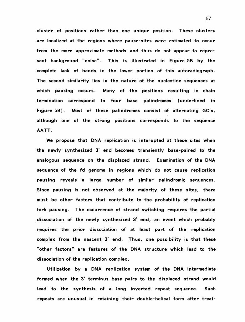
57
cluster of positions rather than one unique position. These clusters
are localized at the regions where pause-sites were estimated to occur
from the more approximate methods and thus do not appear to repre
sent background "noise". This is illustrated in Figure 5B by the
complete lack of bands in the lower portion of this autoradiograph.
The second similarity lies in the nature of the nucleotide sequences at
which pausing occurs. Many of the positions resulting in chain
termination correspond to four base palindromes (underlined in
Figure 5B). Most of these palindromes consist of alternating GC's,
although one of the strong positions corresponds to the sequence
AATT.
We propose that DNA replication is interupted at these sites when
the newly synthesized 3' end becomes transiently base-paired to the
analogous sequence on the displaced strand. Examination of the DNA
sequence of the fol genome in regions which do not cause replication
pausing reveals a large number of similar palindromic sequences.
Since pausing is not observed at the majority of these sites, there
must be other factors that contribute to the probability of replication
fork pausing. The occurrence of strand switching requires the partial
dissociation of the newly synthesized 3' end, an event which probably
requires the prior dissociation of at least part of the replication
complex from the nascent 3' end. Thus, one possibility is that these
"other factors" are features of the DNA structure which lead to the
dissociation of the replication complex.
Utilization by a DNA replication system of the DNA intermediate
formed when the 3' terminus base pairs to the displaced strand would
lead to the synthesis of a long inverted repeat sequence. Such
repeats are unusual in retaining their double-helical form after treat

58
ments that denature the DNA. This occurs because the complementary
strands are covalently joined at the point where the DNA polymerase
switched strands. Both the T7 DNA polymerase (form 1) and the
E. coli DNA polymerase I produce such "reversably denaturable" DNA
molecules during in vitro DNA synthesis (Lechner et al., 1983;
Lechner and Richardson, 1983). Results with the T4 in vitro repli
cation system vary, depending on which components are present
during DNA synthesis. Replication on a nicked duplex DNA template
by the T4 polymerase in the presence of 32 protein results in long
branched duplex products that have this characteristic renaturability
as a result of strand switching (Nossal, 1974). In contrast, DNA
synthesis catalyzed by the more complete 5-protein T4 "core" repli
cation complex or the "core * 41" complex on a duplex template results
in DNA products that are completely denaturable (Sinha et al., 1980).
Thus, the more complete replication complex that contains the poly
merase accessory proteins does not perform this abnormal type of DNA
synthesis. Our results indicate that the DNA intermediate that is
required for the synthesis of these "rapidly renaturable" DNA pro
ducts is formed in the presence of the 5-protein complex and results
in the temporary blocking of DNA replication. The fact that this
structure is not elongated in the presence of the polymerase accessory
proteins suggests that these proteins prevent DNA replication on a
template of improper structure.
The effects of T4 DNA helicases on replication fork pausing -
Both the dola protein and the gene 41 protein are DNA helicases that
utilize energy from nucleoside triphosphate hydrolysis to move in a 5'

59
to 3’ direction and melt DNA, thereby speeding the progress of the
replication fork (Jongeneel et al., 1984a; Alberts et al., 1980;
Venkatesan et al., 1982). While both of these proteins increase the
rate of replication fork movement, the effects of these two proteins on
replication fork pausing in our in vitro system are quite different.
The dola helicase does not affect the occurrence of pausing, whereas in
the presence of 41 protein, replication pausing is eliminated (Figures 6
and 7). This observation can be explained by the difference in the
manner that these two helicases interact with the T4 replication fork.
The 41 helicase acts in a highly processive manner and is probably a
tightly bound component of the T4 replication complex (Alberts et al.,
1980; Venkatesan et al., 1982; Barry, in preparation). Because the
"core * 41" protein replication complex is more processive than the
"core" complex, it is less likely to dissociate from the DNA and allow
the formation of a strand switch. In contrast, the dola protein oper
ates in a distributive fashion and thus interacts with the replication
complex in a transient manner. Although this protein results in an
increase in the rate of the replication fork, there is no indication that
it makes replication more processive. Thus, the replication complex
may dissociate from the DNA template with the same frequency regard
less of whether or not doia protein is present.
Possible importance of replication pausing - We have found that
in vitro T4 replication forks that lack gene 41 protein pause at specific
DNA sequences. Such "incomplete" replication forks could also exist
in vivo. The strong pauses seen during plasmid Col.F1 and mammalian
mitochondrial DNA replication could in fact be due to lack of particular

60
protein factors in the replication forks required to proceed past repli
cation barriers (Tomizawa, 1978; Doda et al., 1981).
In the case of DNA transcription, RNA polymerase pausing at
specific sites is thought to allow binding of ribosomes to "leader" RNA.
The subsequent synchrony of transcription and translation plays an
important role in the gene expression of trp operon in E. coli
(Yanofsky, 1981; Winkler and Yanofsky, 1982). While the extent of
specific replication fork pausing in vivo is far from clear, potential
functions for such events are conceivable. One process that could be
coordinated by pausing during T4 DNA replication would be the
assembly of the multienzyme T4 nucleotide-producing complex that
occurs at each newly-formed replication fork at a replication origin
(Reddy et al., 1977).

61
TABLE I: Locations of Pause Sites
Product Size Before
Letter Designation Restriction Cleavage Map Site
A 6900 + 100 24+2, 46:2%
B 8150 + 50 1055 + 15
C 8350 + 50 1360 + 20
D 9500 + 100 2255 + 65
E 10400 + 100 3330 + 55
F 12600 + 50 5494 + 2*
A 13250 + 50
B 14500 + 50
E 16600 + 100
F 18800 + 100
*These positions were determined after restriction digestion on a 6%
polyacrylamide gel, as shown in Figure 4B. The map sites of all the
other pause-sites were determined after restriction digestion on a 1%
denaturing agarose gel, as described in the legend to Figure 4A.

62
LEGENDS TO THE FIGURES
Figure 1. An assay that detects specific replication pause sites. The
conditions used are described in Materials and Methods. Supercoiled
fo RF was nicked at nucleotide 5781 by the fol gene 2 protein. Addi
tion of T4 replication proteins with dATP, dGTP and **P(TTP) allows
the insertion of 12 bases at the nick site as indicated. The delayed
addition of the missing deoxyribonucleotide, dCTP, allows further
synthesis to occur in a near synchronous manner, and replication
proceeds in a rolling circle mode. After unincorporated radioactivenucleotides are removed, the products of synthesis are analyzed by
denaturing gel electrophoresis, as described in Materials and Methods.
Figure 2. Rolling circle replication catalyzed by the T4 core replication
system pauses at specific DNA sites. Reactions were performed as
described in Figure 1 and Materials and Methods. Aliquots of 10 ul
were processed and electrophoresed as described. Shown here is an
autoradiograph of a gel analyzing DNA products of a standard reaction
after 5, 10 and 20 min.
Figure 3. Replication pause sites are detectable only when replication
is initiated at a specific site. This figure presents a microdensitometer
tracing of autoradiographs similar to those shown in Figure 2, and
compares products of reactions beginning at the random versus a
specific nick on the same fo DNA template.

63
Figure 4. Determination of locations of replication pause sites on
RF fo DNA.
A. Refinement of the locations of replication pause-sites. Prior to
electrophoresis on a 1% agarose gel, the DNA samples were digested
with a restriction enzyme that cleaves the fol genome at a single posi
tion. The first two lanes of the autoradiograph represent the DNA
products of a 2 min replication reaction, followed by no restriction
cleavage or cleavage with Acc I. The next three lanes represent
10 min replication products which were subsequently cleaved with no
enzyme, Hpal or Ball. The locations determined from this autoradio
graph are presented in column 4 of Table 1.
B. Precise determination of two replication pause sites. Samples were
prepared as described in Materials and Methods and then were resolved
on a 6% sequencing gel. The first four lanes on the autoradiograph
are heterologous markers consisting of dideoxy sequencing reactions of
pBR322 primed at the Hinfl site. To locate pause-site "A", replication
reactions stopped after 2, 6, 10 and 15 min incubation were cleaved
with Acc 1 at position 6092 (next four lanes). Pause-site "F" was
determed by cleaving the 15 min replication products with Ball at
postion 5083 (last lane). The positions determined from this experi
ment are presented in Figure 5B.
Note that each pause-site consists of a cluster of one or two maiorJ
positions and several minor ones. The time-course for pause-site "A"
reveals that most of these positions become fainter with increased incu

64
bation time, indicating that this effect is transient. The fragment
marked with a solid arrowhead, which becomes darker with increasing
incubation time, arises from the 3' end labeling of linear fo molecules
that are created at a low level at the gene 2 nicking site by double
strand scission. It became labelled by repeated cycles of exonuclease
excision and polymerization by the T4 polymerase (Englund, 1971). As
expected, this fragment is 313 bases long, which is the distance
between gene 2 nicking site and the Accl site.
Figure 5. Location of replication pause sites on RF fa DNA.
A. The approximate sites of pausing were located by measuring the
sizes of nascent DNA chains on 1% agarose gels after restriction cut
ting. The strongest pause sites, E and F, are in regions that are
thought to contain secondary structure in a single-stranded DNA
molecule, while the other pause sites (A-D) do not correlate with
regions of secondary structure. Sites A and B are particularly promi
nent in reactions with low 32 protein concentrations (see Figure 6A).
B. Precise locations of replication pause-sites "A" and "F". The DNA
sequence in the region of these two pause-sites is given, and the
positions that correspond to the pause-sites determined from the
experiment described in Figure 4B are indicated with arrows. Bold
arrows are used to denote major pauses and faint arrows the minor
ones. The numbers correspond to the position on the fo genome.

65
Figure 6. The effect of gene 41 protein on replication fork pausing.
The T4 gene 41 protein eliminates pausing of the replication fork.
Products of replication reactions catalyzed by the core replication
system without and with T4 gene 41 protein are compared. Gene 41
protein greatly increases the rate of fork movement, so in order to
compare DNA products of equivalent size, the core replication reaction
("5-protein reaction") included a high concentration (300 ug/ml) of
32 protein (the rate of 5-protein fork movement increases with increa
sing 32 protein concentration in this system). Additionally, aliquots
from the core replication reaction were taken at later times than those
from the core 41 replication reaction (5, 10, 15 min versus 1, 2,
5 min). The latter reactions ("6 protein reaction") include some
slower-moving DNA products, made on forks that lack 41 protein;
these display characteristic pausing, as indicated.
Figure 7. The effect of dola protein on replication fork pausing.
The T4 dola protein increases the rate of fork movement, but does not
eliminate replication fork pausing. Addition of purified T4 dola protein
to reactions catalyzed by the core replication system increases the rate
of fork movement about 4-fold under our conditions. Products of core
"5 protein" reactions after 8 min and reactions including dola after
2 min indicate similar, if not identical, pausing patterns.

66
Figure 1.
Incubate T4 replication 5'proteins (I min at 37°C) TTTwith all dnTP's *ATexcept dOTP *Gr
Hº- T °GA. . . .TTAATAGTGGAC .AAATTAT:A::: G . .fo DNA circlespecifically nickedof nucleotide 578] Limited DNA synthesis
Odd dcTP
Anolyze length of2. product DNA strond
3' 5' + single site by denoturingrestriction gel electrophoresisCleovodeSynchronous stort of Q
rolling circle replication
Figure 2.
— |9. I
– 16.6— 15. I
— |3. I
— ||.. 6
— |O.8
5 IO 2C)
Minutes of 37° C.

68
Figure 3.
gene II nick start \
rondom nick stort
\unit length fds,
| | | | | |6|OO 735O 795O 92OO ||OOO (22OO
DNA size (nucleotides)
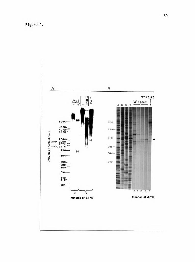
69
Figure 4.
5956–
43684O72–382O-T
264 O2464,225O—
237 22144, 21 6T
| 756–
"F"4-Bol I
"A"+AccI2–~
!-
oAcc I Clr—a T
+;
º--
1384–
996.<892–84OT
596–
440–4 12-T
284–
w 2 6 to 15 152 |O
Minutes at 37°C Minutes at 37°C
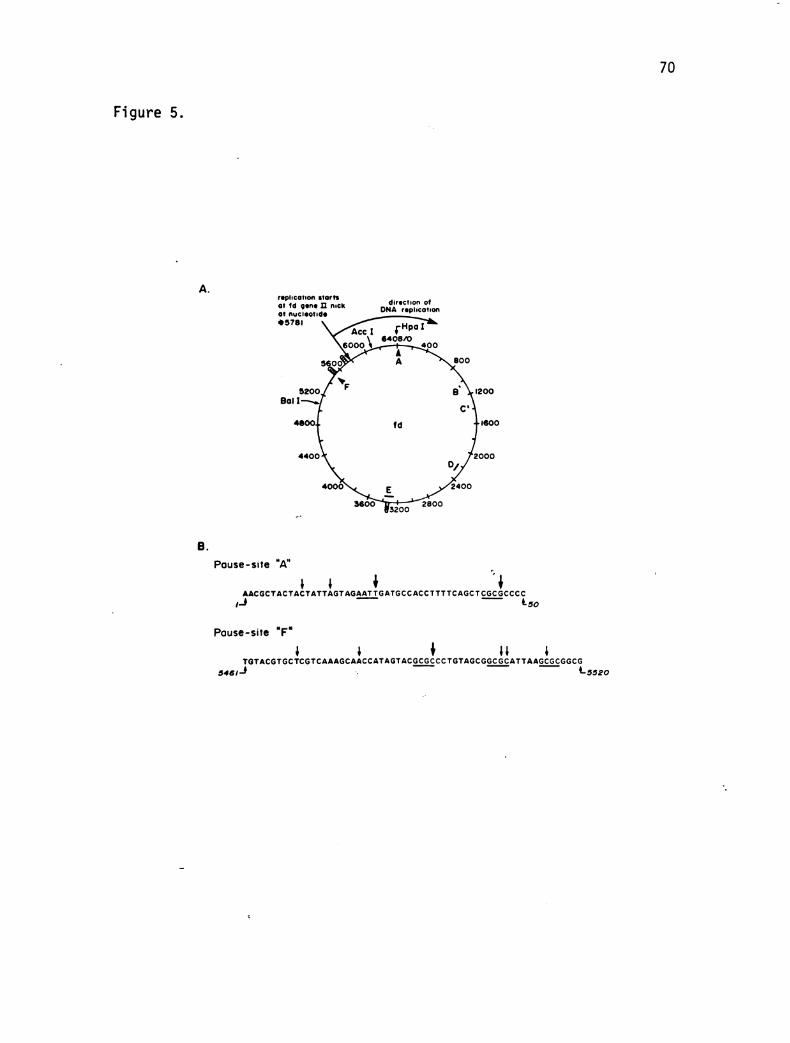
70
Figure 5.
replicotion storts direction ofof fo gene DI nuckof nucleotide DNA replication* 5781
44OO
Pouse-site "A"
AAcGct Act Act ATTAGT AGAATTGATGCCACCTTTTCAGCT cococcc.c1–3 *so
Pause-site "F."
to TAcGT GCTCGT CAAAGC AACCAT AGT ACGCGCCCTGTAGCGGCGC AT TAAGCGCGGCG54si-
-
*-5520

Figure 6.
Core plusCore 41 protein
|9. I —
| 6.6—|5. I —
i|3. I —
| | .6—
| O.8–
9.6—
8.9 —
i5.9—
5 |O |5
Minutes at 37°C.

Figure 7.
Core plusCore dod protein
-—17.4— |4.5— |3.O
-– | 2.4
— |O. 7
— 9.4
— 7.9
-
— 7.4
|-- – 6.2 8 2 8
Minutes of 37°C.

CHAPTER THREE
THE T4 DNA POLYMERASE ACCESSORY PROTEINS FORM A SPECIFIC
COMPLEX WITH THE HELIX DESTABILIZING PROTEIN
ON A PRIMER-TEMPLATE JUNCTION

74
ABSTRACT
Three proteins function as accessory proteins for the T4 DNA
polymerase: a complex of the products of genes 44 and 62, which is a
DNA-dependent ATPase, and the product of gene 45. All three
polypeptides are needed to form an ATP-dependent "sliding clamp" that
holds an active DNA polymerase molecule at the 3' terminus of a
growing DNA chain. In the absence of polymerase, the accessory
proteins can recognize and bind a primer-template junction. We have
used DNA footprinting techniques on special DNA template molecules to
characterize this interaction. We find that 19-20 nucleotides on the
primer strand just behind its 3' terminus and 27–28 nucleotides on the
opposite template strand are protected from DNasel cleavage by the
accessory proteins. A smaller region is protected from the antibiotic
cleaving reagents, bleomycin and neocarzinostatin, but this protected
region is also specifically located at the primer-template junction.
Protection of the DNA substrate requires both the 44/62 complex and 45
protein in stoichiometric amounts, as well as the non-hydrolysable ATP
analogue, ATP&S. Much lower concentrations of the accessory proteins
are required to form this complex when the T4 helix destabilizing
protein (gene 32 protein) is bound to the single-stranded portion of the
template strand. These results confirm earlier indirect evidence that
the accessory proteins recognize and bind specifically to a primer
template junction, and they indicate that the hydrolysis of ATP
promotes the release of this complex from the site on a DNA template
where the DNA polymerase is normally bound.

75
|NTRODUCTION
The bacteriophage T4 DNA replication machinery consists of a
multienzyme complex of at least seven T4-encoded proteins. These
include the DNA polymerase (product of gene 43), the helix destabiliz
ing protein (product of gene 32), the polymerase accessory proteins
(product of gene 45 and a complex of the products of genes 44 and
62), and the RNA priming proteins (products of genes 41 and 61).
Although the term "multienzyme complex" is used to describe this
system, a stable complex has never been isolated. Nevertheless there
is good biochemical and physical evidence for the existence of a specific
complex. The use of affinity columns consisting of a purified T4
protein covalently linked to an agarose matrix has been very useful for
identifying protein-protein interactions (Alberts et al., 1983; Formosa
et al., 1983), while other interactions are implied by the stimulatory
effect of certain proteins on the enzymatic activities of others. The
objective of this chapter is to analyze the interactions of a subset of
these proteins on a DNA molecule that is equivalent to a primer-template
junction (the DNA substrate of a replication complex), using DNA
footprinting techniques. The proteins to be tested here are the T4
helix destabilizing protein and the polymerase accessory proteins.
All of the polymerase accessory proteins have been cloned and
sequenced (Splicer et al., 1982; Splicer et al., 1984). The 44/62
complex is thought to consist of four 44 subunits (each 34,900 daltons)
and one 62 subunit (21,400 daltons) for a total molecular weight of
163,000. The 45 protein exists as a dimer of 49,000 daltons (Nossal
and Alberts, 1983).

76
The 44/62 complex has DNA-dependent ATPase activity that is
stimulated by 45 protein (Piperno et al., 1978; Mace and Alberts,
1984a). In a reaction that requires ATP (or dATP) hydrolysis, these
proteins make the DNA synthesis catalyzed by the T4 polymerase both
faster and more processive (Piperno and Alberts, 1978; Mace and
Alberts, 1984c). In addition, when dINTPs are omitted, the same three
proteins stimulate the rate of the 3' to 5’ exonuclease activity of the T4
DNA polymerase (Bedinger and Alberts, 1983; Venkatesan and Nossal,
1982). Both the stimulation of 5' to 3' polymerization and the
stimulation of 3' to 5’ excision are enhanced synergistically by the
presence of the 32 protein (Huang et al., 1981; Bedinger and Alberts,
1983).
The activity of the three accessory proteins suggests that they
form a complex with the T4 DNA polymerase, acting like a "sliding
clamp" that holds the polymerase on its DNA substrate. In order to
test whether these proteins can themselves recognize a primer-template
junction, the DNA cofactor requirement for their ATPase activity has
been studied. An investigation using synthetic DNA molecules as the
DNA cofactors has revealed that a polyda: oligodT hybrid stimulates the
ATPase activity much more effectively than either the oligonucleotide or
the polynucleotide alone. Furthermore, stimulation by the hybrid
molecule was much less sensitive to salt or to the presence of saturating
amounts of 32 protein (L. Paul, 1983). A study conducted in our
laboratory compared the stimulatory effect of DNA cleaved with restric
tion nucleases that produce various types of DNA ends. The greatest
stimulation of the accessory-protein ATPase activity is achieved in the
presence of DNA fragments terminating in 5' extensions, followed next
77
by fragments with blunt ends, and then those with 3' extensions. DNA
without ends has the least stimulatory effect. Thus, by this test the
double-stranded DNA end that most approximates a primer-template
junction appears to be the prefered cofactor for these proteins
(Jongeneel et al., 1984). In this chapter, we use more direct methods
to characterize their recognition of a primer-template junction.

78
MATERIALS AND METHODS
Enzymes - Polynucleotide kinase and the restriction enzymes Banl
and Hael II were purchased from New England Biolabs. Unless otherwise
noted, restriction digestions were performed as described by the manu
facturers. Pancreatic DNasel (DPFF) was purchased from Worthington
and the single-strand specific endonuclease from Neurospora crassa from
Boeringer-Mannheim. Neocarzinostatin was supplied by Kayaku
Antibiotics Research Co. and the commercial preparation of bleomycin
known as Blenoxane was provided by Bristol labs.
The T4 replication proteins, 32 protein, 44/62 protein, and DNA
polymerase were purified by published protocols (Bittner et al., 1979;
Morris et al., 1979). The gene 45 protein used in these experiments
was produced from cells containing an over-expression vector kindly
provided by Dr. W. Konigsberg; it was purified by a slight modification
of the standard protocol (Morris et al., 1979). In addition, a minor
exonuclease contaminant was removed from this protein by HPLC on an
anion exchange column (Pharmacia MonoC HR 5/5). The column was
loaded in 40 mM Tris-HCl, pH 7.4, 10% (vol/vol) glycerol, 10 mM BME
and 1 mM MgCl2, and eluted with a linear gradient from 0 to 0.4 M
NaCl. The 45 protein eluted from the column at 0.21 M NaCl.
Buffers - Footprinting and DNA replication reactions were
performed in our standard replication buffer, which contains 33 mM
Tris-acetate, pH 7.8, 66 mM potassium acetate, 10 mM magnesium
acetate, 0.5 mM DTT and 100 ug/ml human serum albumin.
Preparation of specific DNA templates - Single-stranded DNA from
the M13-derived cloning vector, M13mp7, was purified by the method of

79
Messing (1983). A 245 nucleotide Hael II restriction fragment containing
the palindromic polylinker region of M13mp7 was purified by the follow
ing method. M13mp7 DNA was cleaved to completion with Hael || using a
120-fold excess of enzyme relative to the amount recommended by the
manufacturer for double-stranded DNA (3U/ug DNA; 40 h incubation at
37°C). The products of digestion were resolved on a 2.2% agarose gel
and the fragment of interest was removed from the gel matrix by
electro-elution. Trace contaminants were removed from the purified
DNA fragment by extraction with phenol: chloroform (1:1) followed by
ethanol precipitation.
This 245 nucleotide fragment contains a 48 nucleotide palindrome
that can form a perfect hairpin; the additional nucleotides from a 192
nucleotide long 5' extension and a 5 nucleotide-long 3' extension from
this hairpin helix. Two steps were used to modify this fragment, as
illustrated in Figure 1A. In order to lengthen the duplex region, the
molecule was incubated with T4 DNA polymerase in the presence of 300
uM each dATP, dGTP, and dTTP and 0.04 um dCTP. (The reaction
mixture contained 2-3 ug/ml DNA, 2.5 ug/ml T4 DNA polymerase, our
standard replication buffer and the above nucleotides, and it was
incubated at 37°C for 1 min.) This results in the excision of the 3'
extension by the 3' to 5’ polymerase-exonuclease followed by a limited
polymerization: because of the strong nucleotide bias used in this
reaction, most of the DNA fragments are extended to a unique position.
The resulting DNA fragment is 262 nucleotides long and consists of a
perfect 44 base pair hairpin helix (plus terminal loop consisting of 4
nonbase-paired nucleotides) with a 5' single-stranded extension of 150
nucleotides. The 5' extension was "trimmed" by annealing this molecule

80
to a 5-fold excess of a 32 nucleotide synthetic oligomer complementary
to a region containing a Ban I site, followed by cleavage with Ban I.
The resulting fragment is 169 nucleotides long. This DNA molecule was
labelled on its 5' end using polynucleotide kinase, purified on a 5.5 %
polyacrylamide (1:30 bis: acrylamide) 7.5 M urea gel, and excised from
the gel by standard procedures (Maniatis et al., 1982). For some
experiments the duplex region was elongated by another 8 base pairs
by incubating the above molecule with 50 ug/ml T4 polymerase and 0.2
mM each d0TP, dGTP, and dTTP under the same conditions described
above, followed by gel purification (Figure 1B).
Footprinting reactions. DNA footprinting was performed in 20 ul
reactions containing replication buffer, 1 nM or less of the DNA frag
ment and, where indicated, 0.1-0.5 mM ATP or ATPMS. The polymerase
accessory proteins and/or 32 protein were added on ice and the reaction
mixture was incubated for 5 min at 37°C. DNasel was then added to a
final concentration of 0.1 ug/ml and incubation was continued for 2 min
at 37°C. The reactions were stopped with 20 ul of 0.5 mg/ml
Proteinase K, 0.2% SDS, 40 mM Na2EDTA, and 40 ug/ml sonicated calf3
thymus DNA; after incubation at 37°C for 10 min, they were extracted
with phenol: chloroform (1:1), followed by ethanol precipitation. The
dried pellets were resuspended in 1.5 u■ sample buffer containing 80%
formamide, 10 mM Tris-borate, pH 8.3, 0.2 mM Na2EDTA, and 0.1% each3
bromphenol blue and xylene cyanole. The samples were then electro
phoresed on 5.5 or 8% polyacrylamide (1:30 bis: acrylamide), 7.5 M urea
gels using as running buffer 0.1 M Tris-borate, pH 8.3, 2 mM
Na2EDTA.

81
For footprinting, gene 32 protein was used at a final concentration
of 10 ug/ml. In reactions containing accessory proteins alone, the
usual concentrations used were 400 ug/ml 44/62 protein and 60 ug/ml 45
protein. In the presence of 32 protein, these concentrations were
100 ug/ml 44/62 protein and 60 ug/ml 45 protein.
In certain experiments, bleomycin (Blm), neocarzinostatin (NCS),
or the single-strand-specific endonuclease from N. crassa was used as
the cleaving reagent in place of DNasel. The endonuclease, which is
provided as an ammonium sulfate suspension, was prepared for use by
centrifugation and resuspension of the pellet in a volume of replication
buffer equal to the volume of suspension used. It was used at a final
concentration of 1 ug/ml, and incubation was for 2 min. The reactions
were stopped and processed as for DNasel.
Just prior to use, Blm was mixed with an equimolar amount of
Fe(NH4)2(SO4)2, using a freshly made stock of the ferrous solution.
When Blm or NCS was used, the concentration of DTT was increased to
5 mM, using a freshly made stock of DTT. NCS was used to a final
concentration of 100 ug/ml and Blm: Fe(II) to concentrations of 0.5
uM:0.45 um; incubation times were 30 and 15 sec, resectively. The
stop mixture for reactions cleaved with Blm contained an additional
component: 17 um bathophenanthrolinedisulfonate. Otherwise reactions
with both of these reagents were processed as for the DNasel reactions.

82
RESULTS
DNA footprinting techniques require a specific interaction between
a DNA binding protein and a unique DNA molecule (Galas and Schmitz,
1979). Because the macromolecular substrate of a DNA replication
system is a primer-template junction, we have designed the DNA mole
cules shown in Figure 1 for our experiments. A perfect hairpin helix
with a long 5' extension contains both primer and template regions, with
the segment from the 3' hydroxyl end to the end of the hairpin loop
forming the "primer strand", and the segment from the loop to the 5'
terminus constituting the "template strand". Two different DNA
molecules of this type were used in these experiments: one is 169
nucleotides long and consists of a 44 base pair hairpin helix with a 77
nucleotide 5' extension, and the other is 177 nucleotides long and
consists of a 52 base pair hairpin helix with a 69 nucleotide 5' exten
sion.
The construction of each of these two DNA molecules is described
in detail in Materials and Methods. As illustrated in Figure 1A, the 245
base Hael II restriction fragment of single-stranded M13mp7 was gel
purified. Incubation of this molecule with T4 DNA polymerase in the
presence of a high concentration of dATP, dGTP, and dTTP and a low
concentration of dcTP results in the removal of a non-base paired 3'
extension by the 3' to 5’ exonuclease, followed by elongation to a
unique position on the DNA template. The low concentration of dCTP
used in this reaction is sufficient to allow incorporation of dOTP at two
positions containing isolated C residues; however on the majority of the
DNA templates the polymerase becomes stalled at a position where two
adjacent dOTP residues should be inserted. The resulting DNA

83
molecule is 262 nucleotides long and consists of a 44 base pair hairpin
helix with a 4 nucleotide loop at one end and a 170 nucleotide-long
5'extension at the other end. The long 5' extension was "trimmed" by
annealing a synthetic oligonucleotide to it that creates a Ban I site,
followed by cleavage with Ban I. The resulting DNA molecule is 169
nucleotides long. After purification on a denaturing polyacrylamide gel,
the 169mer was sometimes elongated another 8 nucleotides by incubating
with T4 DNA polymerase and 300 um each doTP, dGTP and dTTP. This
177mer is very similar in structure to the 169mer, bit it differs in its
nucleotide sequence at the 3' end (Figure 1B).
Cleavage agents used on the 169- and 177-nucleotide DNA frag
ments - Four different cleaving reagents were used for our footprinting
studies: pancreatic DNasel, the single-strand specific endonuclease of
Neurospora crassa, and two antibiotics, bleomycin (Blm) and
neocarzinostatin (NCS). DNasel and the single-strand endonuclease
preferentially cleave double- and single-stranded DNA, respectively,
producing a DNA fragment that terminates in a 3’ hydroxyl and a 5'
phosphate. NCS consists of a functionally active fluorescent chromo
phore which is stabilized by binding to a 10,700 dalton carrier protein
(Kappen et al., 1980), while the bleomycins are a family of iron binding
glycopeptides of approximately 1000 MW (Umezawa et al., 1966). For
these experiments we have used a commercial preparation called
Blenoxane, which consists mainly of bleomycins A2 and B2. Both Blm
and NCS are specific for double-stranded DNA. Although they react
by somewhat different mechanisms, they are similar in that cleavage
results in the destruction of the nucleotide at the site of cleavage, and
in the formation of a DNA fragment which terminates in a modified 3'

84
end (3' phosphate for NCS and 3' phosphoglycolic acid for Blm)
(Kappen and Goldberg, 1978; Giloni et al., 1981). Both molecules show
sequence specificity, with NCS cleaving at T, A and C, in order of
preference, and Blm predominantly at pyrimidines on the 3' side of G
(D'Andrea and Haseltine, 1978). Both Blm and the chromophore of NCS
have DNA intercalating properties, although in the case of Blm this
does not appear to be important for its DNA binding and cleaving
properties (Povirk et al., 1981a and b). These reagents were initially
used in our studies because it was found that only the modified 3' ends
produced by their cleavage are resistent to excision by the 3' to 5'
exonuclease activity of T4 DNA polymerase (M. Munn, unpublished
observation). However, even when the gene 43 protein is absent from
footprinting reactions, the results with these reagents have proven
interesting.
The format used for presenting data. The logic and procedures
that underlie DNA footprinting studies have been presented in detail by
Galas and Schmitz (1979). A uniquely-sized DNA fragment that is not
more than about 200 nucleotides long is radioactively labelled at one
end. This molecule is then cleaved by a reagent that can make many
cuts at random, and the pattern of cuts obtained is determined by
sizing the radioactive DNA fragment by high-resolution electrophoresis
through a polyacrylamide gel, followed by autoradiography. Care is
usually taken to make less than one cut on average per DNA molecule,
so that the position of each cut can be detected by the size of the
radioactive DNA fragment it creates. A comparison of the pattern of
cutting observed with and without a particular DNA-binding protein
present is used to reveal the positions on the DNA that are covered

85
when the protein binds. A reduced frequency of cutting is normally
observed at protein binding sites; however, local sites of enhanced
cleavage are occasionally caused by protein binding. Any effect of the
protein on the cleavage of the DNA is termed the "footprint" of that
protein.
The autoradiographs of the polyacrylamide gels used to analyze two
typical experiments are shown in Figures 2 and 3, in order to show the
quallity of the data obtained. The results obtained from many such
experiments are summarized in Figure 4 for the 169mer and in Figure 5
for the 177mer. In these summaries, the results for each of the four
cleaving reagents are presented just above the nucleotide sequence of
the template strand and just below the nucleotide sequence of the
primer strand. A plus symbol (*) is used to denote protection from
cleavage, while a minus symbol (-) denotes lack of protection. At the
positions where the cleaving reagent does not cut the DNA, even in the
absence of replication proteins, no symbol is used. Finally, at rare
positions where cleavage is enhanced in the presence of replication
proteins, a vertical arrow ( ) appears. For all cleaving agents, the
symbols refer to cleavage on the 5' side of the denoted nucleotide. The
positions of cleavage sites were determined by comparison to identical
samples cleaved by the sequence-specific chemical reactions of Maxam
and Gilbert (1980).
Protection by the helix destabilizing protein - A group of experi
ments that focus on probing protein binding at or near the single
stranded region adjacent the primer-template junction is presented in
Figure 2. Both the 169mer and the 177mer have been used in parallel
experiments here, so that any nucleotide sequence specific effects can

86
be unscrambled from the effect of the primer-template structure. The
results for 32 protein are summarized in Figures 4A and 5A. Not
surprisingly, this helix destabilizing protein binds and protects from
DNasel and NCS the entire single-stranded regions of both DNA
molecules. The protection from DNasel on the template strand extends
for 2 or 4 bases into the duplex region (for the 169mer and 177mer,
respectively), while none of the NCS sites on the duplex region of
either template is protected.
Although most of the single-stranded tail is protected from
cleavage by the single-strand specific endonuclease, the 32 protein
causes a strong enhancement of cleavage occurs at a few positions
adjacent to the primer-template junction. There is also a slight
enhancement of cleavage on the entire duplex region in the presence of
the 32 protein (not shown). This weak enhancement could reflect a
partial melting of the duplex region by the 32 protein, but it more
likely reflects a switch in endonuclease preference when the single
stranded regions are protected.
The cleavage patterns produced by all of these reagents in the ab
sence of replication proteins indicate that there may be some secondary
structure on the 20 nucleotides of single-stranded DNA adjacent to the
duplex region. Thus, there is intense cleavage in this region by both
DNasel and NCS, which in general prefer double-stranded over single
stranded DNA, while there is less cleavage in this region than in the
more distal single-stranded regions by the single-strand endonuclease
(Figure 2). This secondary structure may result from limited base
pairing within this region or, more interestingly, it may reflect an

87
extension of helical stacking in the single-stranded region of DNA that
is continuous with the double-helical region. *
Protection by the DNA polymerase accessory proteins-
In the
presence of the polymerase acessory proteins (both 44/62 and 45
proteins) and ATP*S, two regions of each DNA template are protected
from enzymatic or antibiotic cleavage (Figures 2 and 3). When the
protected sites are drawn on the sequences of the two hairpins, as
shown in Figures 4B and 5B, it becomes apparent that very similar
patterns of protection occur for the two DNA molecules. In both cases
the protected regions correspond to both strands of the duplex region
adjacent to the primer-template junction. A total of 24 nucleotides on
the template strand and 19-20 nucleotides of the primer strand in the
duplex region are protected from DNasel cleavage. Also, in both
cases, the 5' boundary of DNasel protection on the template strand
extends for 3-4 nucleotides into the single-stranded region. Similarly
up to 4 bases in the single-stranded region of the 177mer are protected
from the single-strand endonuclease (this endonuclease does not cleave
the 169mer in this region).
Residues which are protected from the antibiotic cleaving reagents,
Blm and NCS, are clustered about the primer-template junction, as
observed with the DNasel cleavage. However, a much smaller region is
protected from these reagents than from DNasel. On the template
strand, up to 3 nucleotides into the single-stranded region and 10 or 6
nucleotides into the duplex region (for 169mer and 177mer, respect
ively) are protected from NCS cleavage, while protection on the primer
strand extends for 9 and 6 bases, respectively. All sites which are
protected from Blm fall within these limits. The extent of protection is

88
not as neatly duplicated on the two DNA templates as for DNasel
protection; however, this is probably due to the more limited number of
cleavage sites for these reagents, which makes it difficult to ascertain
the exact boundaries of the protected region (see Figures 4B and 5B).
These results indicate that the polymerase accessory proteins
recognize and bind a specific DNA structure, independent of the DNA
sequence. The zone of protection is located at the primer-template
junction, extending predominantly into the duplex region and slightly
into the single-stranded region. Protection from DNasel by the acces
sory proteins is somewhat asymmetric, covering a slightly larger region
on the template strand than on the primer strand. This may reflect a
true asymmetry in the binding of these proteins to the DNA strands.
Alternately, this asymmetry may result from the DNasel cleavage reac
tion; that is, DNasel may be able to cleave closer to a DNA-bound
protein when it approches from the 5' direction than from the 3'
direction. The latter possibility was suggested by C. Joyce and
T. Steitz to explain an apparent disparity between the DNasel foot
printing results of Klenow fragment and the results of fitting the
structure of the DNA substrate into the known three-dimensional struc
ture of the Klenow fragment. Similar to our results, the DNasel foot
printing results for the Klenow fragment indicate that the 3' boundary
of protection on the template strand extends 3 bases beyond the corres
ponding 5' boundary on the primer strand. However, the model
building experiment does not confirm the asymmetry in the protein-DNA
contacts on the two strands (Joyce et al., 1985; C. Joyce and
T. Steitz, personal communication). A similar result is observed for
the DNasel protection by the A repressor or Cro protein of their 17

89
base pair recognition sequence on the right operator of A. The limits of
DNasel protection by either protein extends an additional 3 nucleotides
on the 5' side and 5 or 6 nucleotides on the 3' side of this 17 base pair
sequence (A. Johnson, personal communication; in preparation).
A much smaller zone of protection from NCS and Blm is observed,
but it is similarly located at the primer-template junction. It seems that
these smaller cleaving reagents can penetrate regions of the helix that
appear to be complexed with the accessory proteins based on the
DNasel results. The data suggests that there may be two zones within
the region of interaction between the accessory proteins and the DNA
template: one surrounding the base paired 3' end, which involves an
intimate contact between proteins and DNA, and one farther away in the
double helix, which involves a "looser" interaction that is detected by
DNasel but not chemical protections.
Protection in the presence of both the helix destabilizing protein
and the polymerase accessory proteins - When both 32 protein and the
accessory proteins are present in a single protection experiment, the
resulting "footprint" is the summation of the protection patterns
produced when these proteins are added separately. Thus, the entire
single-stranded region is protected, presumably due to the binding of
32 protein, and the duplex region is protected in the pattern
characteristic of the accessory proteins alone (Figures 4C abd 5C).
The fundamental difference between protection by accessory proteins in
the presence and absence of 32 protein is the concentration of the
accessory proteins required to achieve this effect. In the absence of
32 protein, half maximal protection from DNasel cleavage requires a
concentration of 400 ug/ml 44/62 protein when the concentration of 45

90
protein is 60 ug/ml. However, in the presence of 10 ug/ml 32 protein
and the same amount of 45 protein, the same degree of protection
occurs with 4 wg/m 44/62 protein (data not shown). In an experiment
using a different preparation of 44/62 protein a 3-fold lower concen
tration was required; however, the ratio of 44/62 protein required
without and with 32 was also 100. Thus, the presence of 32 protein on
the DNA template lowers the concentration of 44/62 protein required for
protection by a factor of 100 in the presence of a constant concentra
tion of 45 protein.
Requirements for protection of the DNA substrate by accessory
proteins with and without 32 protein - All of the experiments containing
accessory proteins described so far were performed in the presence of
both the 44/62 complex and the 45 protein, as well as the ATP
analogue, ATP&S. The requirements for protection by these proteins
have been determined by a number of experiments that are similar to
those already described. The requirements for 44/62 and 45 proteins
were examined by performing the standard footprinting reaction at
various concentrations of each of these proteins. Under the conditions
used, both 44/62 and 45 proteins are required for protection from
DNasel cleavage; neither 1 mg/ml 44/62 protein (5 uM complex) nor 0.12
mg/ml 45 protein (2.5 um dimer) provides any protection of the DNA
template. A similar result was obtained in the presence of 32 protein,
where maximum amounts of 0.1 mg/ml. 44/62 and 0.06 mg/ml 45 protein
were tested. Furthermore, at concentrations of accessory proteins that
are less than saturating, the intensity of the footprint varies as the
product of the concentrations of 44/62 and 45 proteins (with 32
protein), irrespective of their relative concentrations. Thus, for

91
example, the same protection was observed at 4 ug/ml 44/62 protein and
12 ug/ml 45 protein as at 20 ug/ml 44/62 protein and 2.4 ug/ml 45
protein. This data is presented in Table 1.
In order to determine whether the DNA binding reactions had
attained equilibrium prior to cleavage, the following experiment was
performed. Standard reactions containing non-saturating concentrations
of accessory proteins (with and without 32 protein) were mixed on ice
and divided into three aliquots. Each aliquot was then pre-incubated at
37°C for either 3, 5, or 7 min followed by the addition of DNasel to
0.8 ug/ml. Incubation was continued for 15 s and then the reactions
were stopped and processed as usual. Identical footprints were
obtained for each set of three samples, indicating that the 5 min pre
incubation used in our standard reaction is sufficient to allow
equilibrium to be reached. Thus, our data reveal that the DNA-bound
complex of accessory proteins forms according to the equation:
[Complex]=Q[45] [44/62], where Q is a constant, and thus that the
complex contains the 163,000 dalton 44/62 protein and the 45 protein
(49,000 dalton dimer) in equimolar amounts.
The nucleotide requirement for accessory protein binding was
investigated over a wide range of concentrations of these proteins, in
the presence and absence of 32 protein. Standard footprinting reac
tions were performed in the absence of nucleotide or the presence of
0.5 mM of either ATP or ATPMS. Absolutely no protection was
observed at any concentration of accessory proteins unless ATP&S was
present. It is possible that ADP produced by hydrolysis of ATP
inhibits the binding of accessory proteins to the DNA. To test this
possibility, 0.5 mM ADP was added to a standard footprinting reaction

92
containing 0.5 mM ATPMS. Identical footprints were obtained with and
without ADP present. Thus no inhibition of binding by ADP is
observed under the conditions used in this experiment. When both ATP
and ATP&S were present in the standard reaction mix containing
100 ug/ml. 44/62 protein, 60 ug/ml and 10 ug/ml 32 protein, only a slight
inhibition of binding was observed in the presence of a nine-fold excess
of ATP over ATPMS (0.45 mM ATP, 0.05 mM ATPMS); ATPºS is a
competitive inhibitor of the ATPase activity of the accessory proteins,
with a K. of about 40 um compared to the Km for ATP of 130 uM
(Bedinger and Alberts, 1983; Piperno et al., 1978).
Structure of the accessory protein complex on the primer-template
junction - A model depicting the interactions of the accessory proteins
and 32 protein on a primer-template junction is shown in Figure 6. Al
though this model is not likely to be correct in detail, it agrees in
concept with a variety of data. Gene 32 protein is shown binding to
the single-stranded tail in a cooperative fashion, with each molecule
protecting seven to ten nucleotides of the DNA strand (Alberts and
Frey, 1971; Kowalczykowski et al., 1981b; Bobst et al., 1982). The
accessory protein complex binds primarily on the adjacent duplex
region, in agreement with our footprinting data. Both the 44/62
complex and the 45 protein make contact with the DNA. The 44/62
complex is known to be a DNA binding protein, based on the DNA
dependence of its ATPase activity (Mace and Alberts, 1984; Piperno et
al., 1978). Although the gene 45 protein was previously believed not
to be a DNA binding protein, we have presented evidence in the next
chapter that indicates that it is. Recent laser cross-linking
experiments performed by Hockensmith et al. show that 45 protein binds

93
to single- and double-stranded DNA in the absence of other replication
proteins or the presence of the 44/62 complex (Hockensmith et al.,
1986; Hockensmith, personal communication). The 44/62 and 45 proteins
contact each other, in agreement with biochemical evidence that they
function as a complex (Mace and Alberts, 1984; Piperno et al., 1978).
In addition, the 44/62 protein contacts 32 protein, based on our results
and previous experiments which show that the 44/62 complex binds
preferentially to single-stranded DNA in the presence of 32 protein
(Mace and Alberts, 1984a). The position and extent of DNA coverage
by the 45 protein are based on the results presented in the next
chapter.
The details of the assembly of this complex are not understood.
The analysis of ATP hydrolysis by the 44/62 complex indicates that this
is an allosteric enzyme that is affected by the binding of both 45
protein and DNA (Piperno et al., 1978; Mace and Alberts, 1984a). In
order to form a stable complex on the DNA template in our experiments,
both the 44/62 and 45 proteins must be present. The order of inter
action of proteins and DNA substrate cannot be determined by these
results; however, the important feature is that the ternary complex,
containing DNA, 44/62, and 45 proteins, is much more stable than
either of the binary complexes, consisting of DNA complexed to either
44/62 protein or 45 protein.
The maintenance of this stable complex requires ATP*S, an ATP
analogue which is not hydrolysed by the accessory proteins (Piperno et
al-, 1978). No protection of the DNA substrate occurs in the presence
of ATP or in the absence of ribonucleotide. It is known that the
ATP-bound form of these proteins binds DNA, since ATP hydrolysis is

94
greatly stimulated by DNA. Assuming that the rate of formation of the
ternary complex is the same in the presence of ATP or ATP*S, then
these data suggest that ATP hydrolysis is coupled to the removal of
this complex from the primer-template junction.

95
DISCUSS|ON
The T4 DNA polymerase accessory proteins bind specifically to a
primer-template junction, protecting from DNasel cleavage a total of 28
nucleotides on the template strand and 20 nucleotides on the primer
strand. This interaction requires both the 44/62 and the 45 proteins in
stoichiometric amounts, as well as the nonhydrolysable ATP analogue,
ATP&S. When gene 32 protein is bound to the single-stranded portion
of the template strand, the concentration of accessory proteins required
to achieve this protection is reduced by a factor of 100.
The observation that the accessory proteins bind to a primer
template junction is not an unexpected result. However, the conditions
that are required to optomize this interaction reflect some interesting
characteristics of this system. Notably, the strong effect of 32 protein
on the binding of the accessory proteins to the DNA molecule indicates
the importance of protein-protein interactions for the stability of this
complex. A previous study using affinity chromatography had identi
fied an interaction between 32 protein and 45 protein; however, this
interaction appeared to be mediated by the DNA polymerase. No bind
ing of the 44/62 protein or of purified 45 protein to the 32 protein
column was observed (Formosa et al., 1983; Formosa, 1985). The major
effect of 32 protein on the accessory protein complex observed in these
protection experiments therefore would not have been predicted from
the affinity chromatography results. However, we have observed
previously that 44/62 protein will bind to DNA-bound 32 protein (Mace
and Alberts, 1984a). It is known that 32 protein undergoes conforma
tional changes on binding DNA (Kowalczykowski et al., 1981a); this
conformation may be required for a strong interaction. Alternately, the

96
32 protein may modify the structure of the DNA template on binding to
it and thereby increase its affinity for 44/62 protein. In either case,
we suspect that the stabilizing effect of 32 protein on the accessory
protein complex with a primer-template junction is mediated via 44/62
protein.
Another striking feature of these experiments is the requirement
for ATP&S (but not ATP) for DNA protection. Assuming that the
ATP&S-bound complex is a perfect mimic of the complex with ATP
bound, we can conclude that ATP hydrolysis causes a weakening of the
DNA-accessory protein complex. How this might be involved in the
function of the accessory proteins in DNA replication will discussed in
the following chapter.
In spite of biochemical evidence that a highly ordered complex is
the functional unit for the T4 replication complex, it has been very
difficult to isolate it intact. The results presented here , using a
limited subset of the replication proteins, demonstrate several princi
ples which might facilitate the site specific assembly of a replication
complex. A fundamental concept is that of gradual allosteric changes
that are stabilized as various components are added to the complex,
which facilitate the addition of other components. An example of this is
the preferential binding of a protein or protein complex to the DNA
bound form of another protein. Thus, two proteins that bind poorly in
solution can interact specifically on the DNA template. For example,
Lunder conditions in which 44/62 protein fails to bind either to 32
protein (Formosa et al., 1983) or single-stranded DNA alone, this
protein binds to a 32 protein-DNA complex (Mace and Alberts, 1984a).
This interaction of 44/62 and 32 proteins would seem to stabilize the

97
addition of the accessory proteins as a functional unit to the replication
complex. Coordinate assembly at a replication fork has also been
observed for the T4 DNA polymerase and the 41 protein DNA helicase
(J. Barry and B. Alberts, unpublished results).
Why should the replication complex be labile? The site-directed
assembly of a replication complex provides a means of utilizing the same
proteins in different types of complexes. In our in vitro replication
reactions, DNA synthesis is usually primed by either a DNA primer
strand that is base paired to the template DNA strand (equivalent to
normal chain elongation) or by RNA pentamers synthesized by the 41/61
priming complex in situ (equivalent to the priming of Okazaki fragments
on the lagging strand). In addition to these reactions, replication
forks can be initiated by various mechanisms within a T4-infected cell.
Thus, primary initiation is a site-specific process that requires the host
RNA polymerase in addition to T4-encoded proteins (Luder and Mosig,
1982, Kozinski, 1983); secondary initiation is a rifampicin-resistant
process that requires the T4-encoded recombination proteins, the
products of genes 46 and 47 (Luder and Mosig, 1982); and tertiary
initiation is a site-specific process which is independent of both host
RNA polymerase and the gene 46 and 47 proteins (Kreuzer and Alberts,
1985). The DNA replication system may need to interface with different
structures during each of these initiation processes. This would be
more readily accomplished if the replication apparatus were not locked
into a unique conformation, but rather was able to form various types
of complexes.
The existence of loosely associated complexes also provides a
mechanism for regulation and coordination with other processes. Gene
98
45 protein is known to interact with the T4-modified RNA polymerase to
stimulate the transcription of late mRNA and thus may provide a link
between replication and transcription (Ratner, 1974; Wu et al., 1975;
reviewed in Rabussay, 1983).

99
TABLE 1. Variation in Intensity of Protection with Concentrations of
Accessory Proteins in the Presence of 32 Protein
[44/62 Protein] [45 Protein] [44/62 Protein]*
Intensity With
(ug/ml) (ug/ml) x [45 Protein] Accessory ProteinsIntensity Without
Accessory Proteins
4 60 240 .19
20 12 240 . 12
100 2.4 240 .075
4 12 48 .39
20 2.4 48 .53
º:The intensities of DNA bands corresponding to nucleotide positions
that are protected by the accessory proteins were measured by densi
tometry. The values given represent the ratios of the average of
several relevant positions with and without accessory proteins.

100
LEGENDS TO THE FIGURES
Figure 1. (A) Construction of the DNA templates. Step 1: The
single-stranded viral DNA of the M13 derivative, M13mp7, was cleaved
to completion with Hael II, and the 245 nucleotide fragment containing a
48 nucleotide perfect palindrome was gel-purified. Step 2: The 245
nucleotide fragment forms a 22 base pair perfect hairpin helix with a 5
base 3' overhang and a 192 nucleotide-long 5' overhang. This fragment
was incubated with T4 DNA polymerase in the presence of 300 um each
dATP, dGTP and dTTP and 0.04 um dCTP. This results in the exci
sion of the 5 non-base paired nucleotides at the 3' end and subsequent
elongation to a unique position, resulting in a hairpin helix that is 44
nucleotides long. Step 3: The long single-stranded tail is "trimmed"
by annealing a 32 nucleotide synthetic oligomer to create a restriction
site and then cleaving with Ban I. The resulting DNA fragment is 169
nucleotides long. (B) After polyacrylamide gel purification, the DNA
fragment in (A) can be elongated another 8 nucleotides by incubating
with T4 DNA polymerase in the presence of dOTP, dGTP and dTTP.
The DNA sequence of both the 169 and 177 nucleotide-long DNA mole
cules is shown. Tthe 177 base fragment contains the additional 8
nucleotides enclosed by the box. Each molecule consists of a perfectly
base paired hairpin helix with a long single-stranded 5' extension. The
3' hydroxyl end is perfectly base paired.
Figure 2. Examination of the template strand near the primer-template
junction for binding by the polymerase accessory proteins or 32
protein. Shown here are the autoradiogaphs of two 8% polyacrylamide
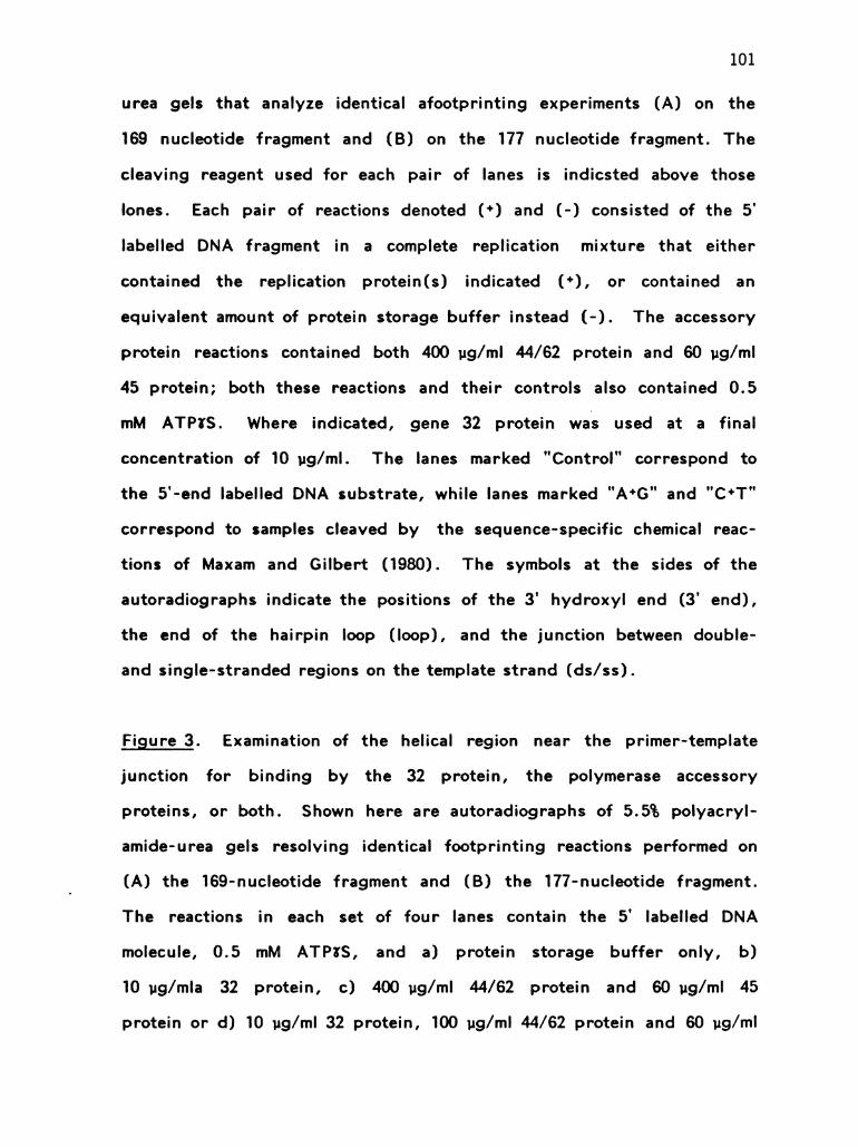
101
urea gels that analyze identical afootprinting experiments (A) on the
169 nucleotide fragment and (B) on the 177 nucleotide fragment. The
cleaving reagent used for each pair of lanes is indicsted above those
lones. Each pair of reactions denoted (*) and (-) consisted of the 5’
labelled DNA fragment in a complete replication mixture that either
contained the replication protein (s) indicated (*), or contained an
equivalent amount of protein storage buffer instead (-). The accessory
protein reactions contained both 400 ug/ml 44/62 protein and 60 ug/ml
45 protein; both these reactions and their controls also contained 0.5
mM ATP&S. Where indicated, gene 32 protein was used at a final
concentration of 10 ug/ml. The lanes marked "Control" correspond to
the 5'-end labelled DNA substrate, while lanes marked "A*G" and "C*T"
correspond to samples cleaved by the sequence-specific chemical reac
tions of Maxam and Gilbert (1980). The symbols at the sides of the
autoradiographs indicate the positions of the 3' hydroxyl end (3' end),
the end of the hairpin loop (loop), and the junction between double
and single-stranded regions on the template strand (ds/ss).
Figure 3. Examination of the helical region near the primer-template
junction for binding by the 32 protein, the polymerase accessory
proteins, or both. Shown here are autoradiographs of 5.5% polyacryl
amide-urea gels resolving identical footprinting reactions performed on
(A) the 169-nucleotide fragment and (B) the 177-nucleotide fragment.
The reactions in each set of four lanes contain the 5' labelled DNA
molecule, 0.5 mM ATP*S, and a) protein storage buffer only, b)
10 ug/mla 32 protein, c) 400 ug/ml 44/62 protein and 60 ug/ml 45
protein or d) 10 ug/ml 32 protein, 100 ug/ml 44/62 protein and 60 ug/ml

102
45 protein. Cleavage was with either NCS, DNasel or Blm-Fe(II), as
indicated above each set of four lanes. Reactions were stopped and
processed as described under Materials and Methods. The lanes marked
"Control" represent uncleaved samples of the two DNA fragments. The
positions of the 3' hydroxyl ends (3' end) and the loop of the hairpin
helix (loop) are indicated on the sides of the autoradiograph. The
cutting sites on the template strand near the double-strand single
strand junction have been run off the bottom of the gel in order to
increase the resolution of the larger DNA fragments (compare with
Figure 2).
Figure 4. A summary of protection observed on the 169 nucleotide DNA
molecule by (A) 32 protein alone, (B) polymerase accessory proteins in
the presence of ATP&S and (C) 32 protein plus the polymerase acces
sory proteins in the presence of ATPMS. The sequence of the DNA
fragment is represented as a hairpin, and the cleavage results for each
reagent at each nucleotide are presented above the template strand and
below the primer strand. The four cleaving reagents presented are
1) DNasel, 2) the single-stranded specific endonuclease, 3) NCS, and
4) Blm-Fe(II). Positions that are protected from cleavage are indicated
with a plus (*) symbol, while those positions which are not protected
are indicated with a minus (-) symbol. A vertical arrow ( ) is used
to denote any site at which cleavage is enhanced in the presence of
replication proteins (s). Nucleotides that lack any of these symbols
correspond to positions which are not cleaved by that reagent. For all
of the cleaving reagents, the symbols refer to cleavage on the 5' side
of the denoted nucleotide.

103
Although a general enhancement of cleavage throughout the
double-stranded region by the single-strand endonuclease occurs in the
presence of gene 32 protein, these enhanced cleavage sites have not
been indicated for the sake of simplicity, since they represent a non
specific effect of removing a competing good substrate. Cleavage by
the single-strand specific endonuclease in the presence of 32 protein
and the accessory proteins was not performed.
Figure 5. A summary of protection on the 177 nucleotide DNA molecule
by (A) 32 protein, (B) accessory proteins in the presence of ATP&S
and (C) 32 protein and accessory proteins in the presence of ATP&S.
The symbols used are identical to those used in Figure 4.
Positions 44 to 48 on the 169mer and positions 45 to 54 on the
177mer are partially protected from DNasel cleavage in the presence of
accessory proteins and ATP&S. These regions of protection are at
approximately the same nucleotide position on the two DNA substrates
but are in different positions relative to the primer-template junction.
Thus, the protection of these sites is most likely not caused by acces
sory protein binding at the primer-template junction. Instead, protec
tion in these regions probably reflects limited accessory protein binding
at these sequences due to the local secondary structure of the DNA.
The fact that the cleavage sites of NCS and the single-strand specific
endonuclease in this region are not protected supports that accessory
protein binding of these sequences is weak.
Figure 6. Model of the accessory protein complex on the primer
template junction. The single-stranded region of the template strand is

104
shown coated with 32 protein, while the accessory proteins interact with
the adjacent duplex region. Both the 163,000 dalton 44/62 complex and
the 49,000 dalton dimer of 45 protein make contact with the DNA, for
reasons discussed in the Results section. The 44/62 complex is shown
in contact with both the 32 protein and the 45 protein. The coverage
of the DNA by the accessory proteins is inside the boundary of DNasel
protection (shown as stippling on the DNA backbone), because some
steric hindrance to DNasel is assumed. No attempt has been made to
account for the limited protection from antibiotic cleavage in this data at
this time. However, as discussed earlier, the shapes of the accessory
proteins may be much more asymmetric than presented here.
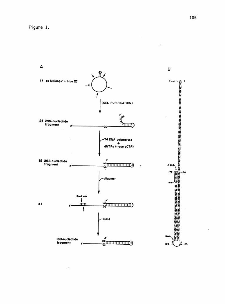
105
Figure 1.
A
1) ss M13mp7 + Hoe III
(GEL PURIFICATION)
2) 245-nucleotidefrogment 5' Go
T4 DNA polymerase+
dNTPs (trace dCTP)
3) 262-nucleotide s'HO
frogment 5’ ...[IIII DITILITIC3)
–s’|
4) s—IITT ..IITITITITITITIII.3)
|-169-nucleotide 3r
HO
frogment s—:TITITIIIHIIII.3
Bon I site
5' end-- 5; -I
3' end\
177– #
I69–
■
--
124 – rº shN->
- 7 o
23

106
IFigure 2.
Control |3' end
loop■
IIC
IOO
90
8O
ds/ss -
GO
I69-nucleotide fragment
Accessory proteins 32 proteinss ss
NCS DNose endo NCS DNase endo- + - +
--
--- -
--
---
+ - + - +-
i-H|+ A+G
Control
A+G C+T
|
º
H
|
I77-nucleotide fragment
Accessory proteins 32 protein
NCS DNose eño NCS DNose eño- + + - + + - + - +
-3' end
D loop
- O
-IOO
-90
-BO
-ds/ss
-6O.
-50
-40
-3C
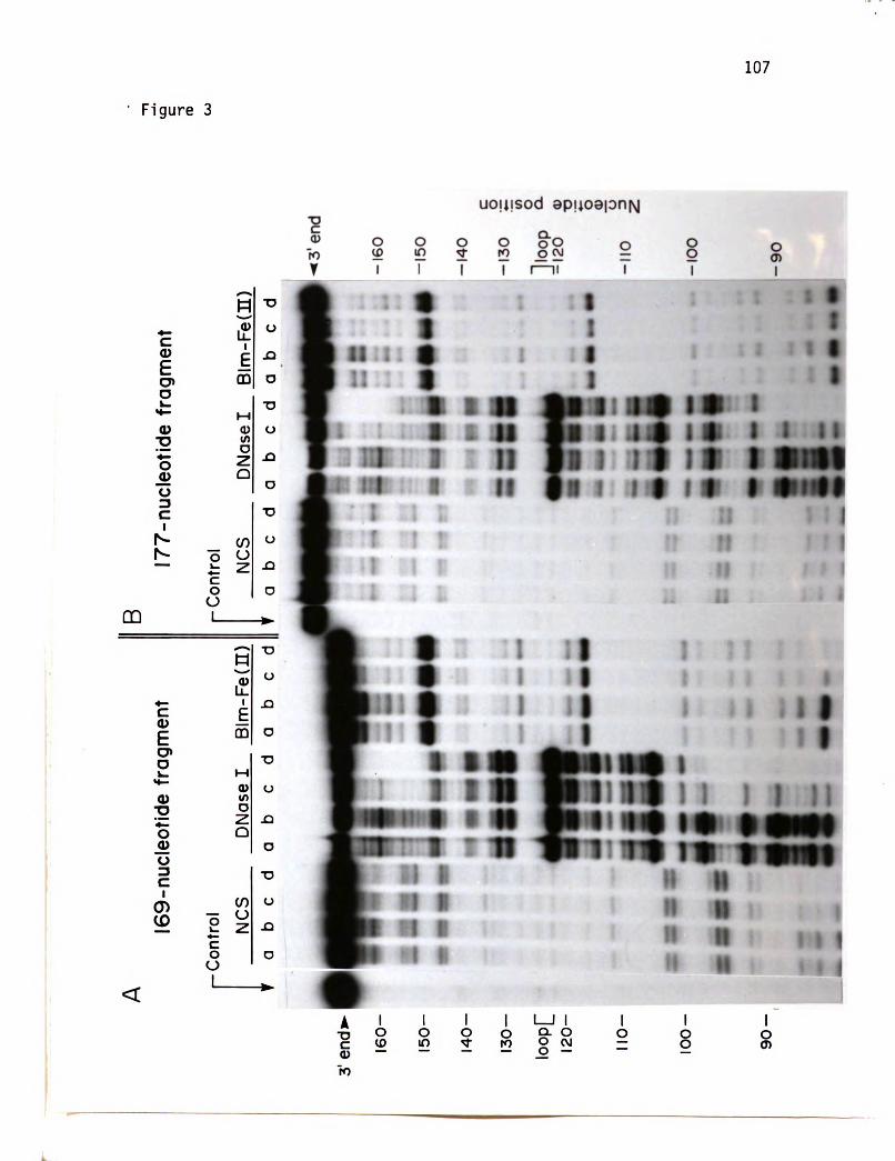
107
Figure 3
uou■ sod aploalon N
OO|-Oll
poqopoqopoqD
(II)-3-3-uulgI
3SDNQSON|Ou■ uOO
■ ueuuôou,ep■■ 03||Onu-/.../..|
|
(II)º-J-uuIGI
3SDNQSON|Ou■ uOO■ aeº■■+i■{
quæuuôou■ap■■ 03||Onu-69||-OOI-O1||-O2||IJdoo!-O2|–Ot>|–O9||--O9||~pu3
,2

108
Figure 4.
r-cº!NrcrI{hr—cºrn-r <rnfºocººr- r—cºrry<rtncQ-i
JV00105■ 595■ 0559591007901930IV990000990105■ 595■ 105595910079010001W99000JV00105■ 595■ 055959100790193)||Wºº)))
-__--___--*4*4+}Z---+++++++++
+I9ýWI■ 59■ všijyiºpijvi■ šp■ ŠILI
J11\/\/90■ /11W91W.00W91W109WOWWW99WOW0W0111WW3/WIW9909W91911WW99|19101101W19010990011091W
-+++++++++++++++++++
+++++++++-------
~--+++++++
++++++++++
--••++
SºdIV+SN13108dgh+Z9/hh+Z2
|-••••++
+--
---------++
w-cºurfºw-r
---++++++++++
9ýWI■ 59■ všijyiºpijvi■ šp■ ŠILI
J11
WW90W11W91W.00W91W109W0WWW99WOWOW011||WWOWWIW9909791911WW9919191.191W19010990011001\}
-++++++++++++++++•••
•.---+++++
+.--
----------+++++++
–-------------
--••++
SydIV+SN13108dgh+Z9/hh
h}
9yylijºiv■■ v■ 99■ v■■■ p■ 5111QIIVý9)||199■ VOJV01y10970'■ y■ º■ wy)\)||1■ VYOWIV900979491■ vyº!!!!!101101V10010000011001W-----------+++++++
+++++++++
{{{{{
{+++-+++++-+-+++++++++
----••••••••••••+
++++++++++
NI31088Z9
<rnfºocººr'9}'8<rrºncºr'W

109
Figure 5
<rNºncºr- *—cºrrº-r Sºrrºcºr- r—cººlnº)-rr<rNºncºr
----~------,*■Z––+++++++-+++■
3y99135595■ 55959ýWI■ 59■ v■■■■ 99■ jvi■ šp■ ŠIlijyiºi91991007901900|V90000}LIVÝ9)||199■ V0}\!\!\!0!■ 0WV90■ }{}V}11|y\}\\1\390979|01|W}}|}101191■ ||9013000011001W+++++++++++++++++++++++
++++++++++
-
---------
-++++++++++++++++++
<rrocºrSRdIV+SN13108dgh+Z9/hh+Z2'0
I--T--~----,*?•Z----+++++-++-++
+I
JV00105■ 595■ 1055950■ yl■ 59■ v■■■■■ 99■ jvišji■ Ilijyiºi9199100■ 001900|V9000001IVý90V11V9■ V0}\!\!\!}}VOVVV90■ )\}\\11■ WQwy■ yººººy!!!01!wy■ ||0101101V19010990011001W0\/99109W0999100ý901000V■ p■ 5959ýWI■ 59■ vIV9000001IVV90||
++++++++++++++------■
+++++++--
---
++++++-------------------------------
-------
-+++
+---------------
<!-NºncºrSºdIV+SN13108dgh+Z9/hh'8
VIŠ■■ 99■ v■ y■ n■ 511■ 5)■ 9■ 9191W91W00W91W109W0WWW99W070\10111WW3WWIW9909791911WW99|19101101W19010990011001\}-------
---+++++++++++++
++++++++++
t
II
l
º
!t
I
v-Hºnº-r"
||----++++++++++++++++++++++++++++++++
-++++++++++++++++
<rrºncºrNI31088Z9'W


CHAPTER FOUR
THE INTERACTIONS OF THE T4 DNA POLYMERASE WITH
|TS ACCESSORY PROTEINS AND THE HELIX DESTAB || LIZING
PROTEIN ON A PRIMER-TEMPLATE JUNCTION
112
ABSTRACT
DNA replication by bacteriophage T4 is catalyzed by a multi
enzyme complex that consists of at least seven T4-encoded proteins.
In this chapter we have used DNA footprinting techniques to analyze
the interactions of five of these proteins at a primer-template junction:
the T4 DNA polymerase, its three accessory proteins, and the T4 helix
destabilizing protein. Two different cleaving reagents, DNasel and the
antibiotic neocarzinostatin (NCS) have been used for the footprinting.
The DNA substrate used for these experiments is the special 177
nucleotide-long fragment described in the previous chapter. The T4
DNA polymerase binds this DNA molecule in the duplex region adjacent
to its 3' hydroxyl end, protecting 14-15 base pairs from DNasel clea
vage and partially protecting up to 6 base pairs from NCS cleavage.
Non-specific binding of the polymerase to the single-stranded region of
the template strand results in the complete protection of this region
from DNasel cleavage and partial protection from NCS cleavage. By
itself, the T4 helix destabilizing protein (product of gene 32) binds
and protects the entire single-stranded region of the template strand
from both DNasel and NCS cleavage. In the presence of both the T4
DNA polymerase and the 32 protein, the protection patterns are addi
tive, covering both the single-stranded region and the region of the
double helix that is covered by polymerase alone. The binding of 32
protein to the template strand facilitates the binding of the polymerase
to its site on the adjacent duplex region, reducing its Kd by a factor
of about thirty.
The DNasel protection experiments also demonstrate the formation
of a complex between the DNA polymerase and the gene 45 protein,

113
one of the three polymerase accessory proteins. This complex protects
a larger segment on the duplex region adjacent to the 3' terminus than
polymerase alone from DNasel cleavage: 32 nucleotides on the template
strand and 25 nucleotides on the primer strand (versus 14 and 15
nucleotides, respectively, for the polymerase alone). No effect of the
44/62 protein (another polymerase accessory protein) or ATP was
observed on the DNasel protection caused by polymerase and 45 pro
tein. The results of the DNasel protection experiments carried out in
the presence of ATP*S, accessory proteins and polymerase are most
consistent with a competition between two mutually-exclusive com
plexes, the DNA polymerase and the ATP&S-bound accessory protein
complex, for the same binding site. Based on this data, a model for
the assembly and maintenance of a functional complex between the DNA
polymerase and its accessory proteins is presented.

114
|NTRODUCTION
Although T4 DNA polymerase can by itself catalyze DNA synthesis
on a primed single-stranded DNA molecule, this activity is greatly
enhanced by several auxiliary proteins. Most of these auxiliary
components are absolutely required for extensive synthesis on a
double-stranded DNA template, which is the natural substrate for the
T4 DNA replication system. The helix destabilizing protein (product
of gene 32) is a single-stranded DNA binding protein that enhances
polymerase activity on a single-stranded DNA substrate by removing
secondary structure on the template strand ahead of the active
polymerase molecule (Huang et al., 1981; Huberman et al., 1971). On
a double-stranded substrate, the 32 protein is required to destabilize
the double helix ahead of the growing replication fork (Alberts et al.,
1980). The polymerase accessory proteins (the product of gene 45 and
a complex of the products of genes 44 and 62) interact with the DNA
polymerase in a reaction that requires ATP (or dATP) hydrolysis. This
interaction results in an increase in the rate and processivity of the
DNA synthesis catalyzed by the DNA polymerase (Alberts et al., 1975;
Piperno and Alberts, 1978; Mace and Alberts 1984c). In the absence
of deoxyribonucleoside triphosphates, the same combination of the
three accessory proteins and ATP increases the rate and the pro
cessivity of the 3' to 5' polymerase exonuclease (Venkatesan & Nossal,
1982; Bedinger & Alberts, 1982). In both cases, the activity of these
proteins is consistent with the formation of a complex with the poly
merase, such that the accessory proteins act like a "sliding clamp"
that increases the retention of the polymerase on its DNA substrate.

115
The functional complex between polymerase and its accessory proteins
constitute the "DNA polymerase holoenzyme".
Both the helix destabilizing protein and the accessory proteins
are required for efficient DNA synthesis by the polymerase on a
double-stranded template. The polymerase activity on a double
stranded template is further enhanced by the T4 DNA helicase (pro
duct of gene 41). This enzyme utilizes the energy of GTP or ATP
hydrolysis to run along the lagging strand and unwind the DNA helix
ahead of the growing replication fork (Alberts et al., 1980; Venkatesan
et al., 1982).
During replication, the components of the T4 replication system
interact in a highly ordered multi-enzyme complex. Although this
complex is not stable enough to be isolated intact by standard
methods, its existence is implied by the biochemical and physical
properties of the replication proteins. Based on these properties,
much can be inferred about the organization of this complex on its
DNA substrate, a primer-template junction. The DNA polymerase by
necessity must bind in the region of the 3' terminus, where it can
either incorporate the next nucleotide (polymerization) or remove the
previously incorporated one (exonuclease proofreading). The helix
destabilizing protein is thought to bind to the template strand just
ahead of the advancing polymerase molecule. A direct interaction
between the DNA polymerase and the 32 protein has been shown by
both sedimentation studies (Huberman et al., 1971) and protein affinity
chromatography (Alberts et al., 1983; Formosa et al., 1983). The
polymerase accessory proteins appear to interact directly with the
polymerase, based on their effect on its activity. Gene 45 protein

116
binds to a polymerase affinity column (Alberts et al., 1983), and thus
it may function to link polymerase to the 44/62 complex, to which 45
protein also binds. The 44/62 complex is known to bind DNA, as it
exhibits DNA-dependent ATPase activity (Piperno et al., 1978; Mace
and Alberts, 1984a). Therefore, this protein has been thought to be
the major DNA binding component of the accessory protein complex.
The other major protein complex that functions at the replication
fork centers on the gene 41 DNA helicase which catalyzes the
unwinding of the DNA helix (Liu & Alberts, 1981; Venkatesan et al.,
1982). This protein is also a central component of the T4
"primosome", together with the gene 61 RNA primase (Liu and Alberts,
1980; Silver and Nossal, 1979; Cha and Alberts, 1986).
In this chapter we have analyzed directly the interactions of the
DNA polymerase with its accessory proteins and the helix destabilizing
protein at a primer-template junction. As in the previous chapter,
DNA footprinting techniques have been used to probe these interac
tions. A major advantage to this technique is that the assembly of a
DNA-bound protein complex can be driven by using high concentra
tions of the protein species. It also provides a method for estimating
the Kd of a protein-DNA complex (Johnson et al., 1981). A similar
study on the Klenow fragment of the E. coli DNA polymerase I has
been performed (Joyce et al., 1985; Joyce and Grindley, in prepara
tion).

117
MATERIALS AND METHODS
Enzymes - Restriction enzymes, nucleases, alkaline phosphatase
and polynucleotide kinase were purchased from the same manufacturers
as described in the previous chapter. The T4 replication proteins,
DNA polymerase, 32 protein, 44/62 protein and 45 proteins were puri
fied by the methods described in Chapter 3.
Preparation of labelled DNA substrate - The construction of the
177-nucleotide long molecule has been described in detail in the pre
vious chapter. This 169-nucleotide fragment was 5' labelled with poly
nucleotide kinase (Boehringer-Mannheim) and then purified after
electrophoresis on a 5.5% polyacrylamide sequencing gel (Maniatis et
al., 1982). The duplex region of this molecule was then elongated
another 8 nucleotides by incubating for 1 min at 37°C with 50 ug/ml
T4 DNA polymerase in replication buffer containing 0.2 mM each d0 TP,
dGTP and dTTP. This 177-nucleotide fragment was relabelled on its 5'
end, as the efficiency of the previous labelling step was usually low,
and then it was repurified on a 5.5% polyacrylamide gel.
In order to label the 177 nucleotide DNA molecule at its 3' end,
the 169-nucleotide fragment was treated with calf alkaline phosphatase
to remove its 5' terminal labelled phosphate. This fragment was then
incubated for 1 min at 37°C with 50 ug/ml [a-**P] dcTP (Amersham,
greater than 3000 Ci/mMol), followed by gel purification as above.
NCS footprinting on the 5' end-labelled 177-nucleotide fragment-
Footprinting reactions were carried out in replication buffer containing
5 mM DTT (made with a freshly prepared DTT stock), less than 1 nM
5' labelled DNA, 0.3 mM each dCTP, dGTP and dTTP and, where
specified, 0.1 mM ATP or 0.5 mM ATP&S. Standard reaction volumes

118
were 20 ul. Gene 32, 44/62 and 45 proteins or equivalent amounts of
their respective storage buffers in their place were added on ice to
final concentrations of 10, 100 and 60 ug/ml, respectively, unless
otherwise noted. The reactions were incubated for 4.75 min at 37°C,
at which time DNA polymerase (or its storage buffer) was added.
After a delay of 15 sec, NCS was added to 0.1 mg/ml, and incubation
was continued for 30 sec at 37°C before the reaction was stopped with
an equal volume of stop mix (0.5 mg/ml Proteinase K, 0.2% SDS, 40 mM
Na2EDTA, and 40 ug/ml sonicated calf thymus DNA). After a further
10 min incubation at 37°C, the samples were extracted with phenol:
chloroform (1:1), rinsed with ether, and ethanol precipitated. The
dried precipitates were resuspended in 1.5 ul of sample buffer which
contained 80% formamide, 10 mM Tris-borate, pH 8.3, 0.2 mM
Na2EDTA, and 0.1% each bromphenol blue and xylene cyanole.
Samples were resolved by electrophoresis through 5.5% polacrylamide
(1:30 bis: acrylamide), 7.5 M urea gels using 0.1 M Tris-borate,
pH 8.3, 2 mM Na2EDTA as the running buffer. The A plus G and C
plus T sequencing reactions of Maxam and Gilbert (1980) were run
simultaneously as size markers. After drying onto Whatman 2 MM
paper, the gels were autoradiographed at -70°C using Kodak XAR-2
film with a Dupont "Lighning Plus" intensifying screen.
DNasel footprinting on the 3' end-labelled 177-nucleotide fragment
- The DNA template used for these experiments had been labelled near
its 3' end as described above. Reaction conditions were as described
above, except that DNasel was substituted for NCS. Dnasel was
added to 0.8 ug/ml and the incubation was continued for 15 sec. The
reactions were stopped and subsequently treated as described above,

119
and the DNA fragments then resolved by electrophoresis through 5.5%
or 10% polyacrylamide gels, as described above.

120
RESULTS
Biochemical evidence indicates that the T4 DNA polymerase forms
a specific complex with its accessory proteins and the helix destabi
lizing protein at a primer-template junction. For this reason we have
studied the binding of these proteins to a DNA molecule that is struc
turally equivalent to a primer-template junction. This 177-nucleotide
DNA fragment (Figure 1) consists of a perfect 52-base pair hairpin
helix (including a 4 base terminal loop) plus a 5' single-stranded
extension of 69 nucleotides. With respect to normal DNA synthesis, the
segment from the 5' end to the hairpin loop constitutes a "template
strand" while the region from the loop to the 3' end is a "primer
strand".
The construction of this molecule has been discussed in detail in
the previous chapter and is outlined under Materials and Methods. In
some of the experiments described here the molecule is labeled at its 5'
end using polynucleotide kinase, while in others it is labeled near the
3' end at the two boxed C residues in Figure 1, by incorporating
[a-**P] dOTP during its construction.
Footprinting in the presence of the T4 DNA polymerase is made
difficult by the potent 3' to 5’ exonuclease activity of this enzyme.
The 3' hydroxyl end of the 177-nucleotide fragment is a potential
substrate for this activity. However, in the presence of dOTP, dGTP,
and dTTP the polymerase stalls on this DNA substrate, remaining in
one place while it catalyzes successive rounds of excision and incor
poration of the terminal nucleotides. Since daTP is absent, elongation
does not continue beyond position 177. The subterminal positions for
the 3' label were chosen in order to prevent the loss of the label

121
during polymerase stalling.
A second difficulty arises because the 3' hydroxyl ends produced
by DNasel nicking are also susceptible to this 3' to 5’ exonuclease; as
a result, the fragments produced by DNasel cleavage during a foot
printing reaction are rapidly excised by the polymerase, resulting in
the destruction of the footprinting pattern. Two different tactics have
been used to circumvent this problem. The first is to use a cleaving
reagent that produces modified 3' ends that are resistant to the 3' to
5' exonuclease. The reagent chosen for this purpose was neocazino
station (NCS), an antibiotic that produces 3' phosphates as a result of
cleavage (Kappen and Goldberg, 1978). We find that the 5' end
labelled DNA fragments that terminate in 3' ends produced by NCS
cleavage are resistant to the exonuclease activity of the polymerase
(unpublished results). The second approach is to use a DNA fragment
labelled near its 3' end in conjunction with DNasel as the cleaving
reagent. Because the label is on the 3' end of the DNA molecule, the
DNA fragments terminating in 3’ hydroxyls that are produced by
DNasel cleavage are not labelled, and the degradation of these frag
ments leaves the original footprint unaltered. A potential problem with
this labelling strategy arose because the **P label was not precisely at
the 3' end in our experiments; thus, anomolous DNA bands are pro
duced when the fragment is cleaved between the 3' end and the
labelled nucleotides. However, as will be seen below, this does not
seriously affect the results.
NCS footprinting on the 5' end-labelled 177 nucleotide fragment-
Neocarzinostatin consists of a biologically active fluorescent chromo
phore that is stabilized by binding to a 10,700 dalton carrier protein.

122
It cleaves double-stranded DNA better than single-stranded DNA and
with the sequence specificity T greater than A greater than C
(Hatayama et al., 1978). Cleavage results in the destruction of the
nucleotide at the site of cleavage with the release of the free base; a
3' phosphate terminus is left on the DNA fragment on the 5' side of
the site of cleavage (Kappen and Goldberg, 1978).
A polyacrylamide gel analysis of the DNA fragments produced by
NCS cleavage of the 5' end-labelled DNA molecule in Figure I with and
without various T4 replication proteins present is shown in Figure 2B.
Each set of five lanes shows the result of adding increasing amounts of
T4 DNA polymerase in the absence or presence of other replication
proteins. Such titrations with the polymerase allow us to detect
changes in the affinity of the polymerase for the DNA substrate caused
by the presence of other proteins. The first four lanes in this auto
radiograph represent polymerase-containing controls in which water
was substituted for NCS. These lanes indicate that the polymerase is
not perfectly stalled in that minor amounts of specific DNA fragments
are created by the 3' to 5’ exonuclease. This background had to be
accounted for in analyzing the remaining experiments.
Evidence for polymerization dimerization - At low concentrations
of polymerase, an unusual phenomenon occurs which initially compli
cated the interpretation of the footprinting experiments. In Figure 2A
is shown a low exposure of the upper portion of the autoradiograph in
Figure 2B. The large uppermost band corresponds to the intact
177-nucleotide fragment. At low concentrations of polymerase, the
intensity of this band is greatly reduced and the bulk of the radio
activity is in a band at a slightly lower position on the gel. This

123
band corresponds to a DNA fragment which has had six nucleotides
removed from its 3' end. The appearance of this band is not depen
dent on NCS cleavage, since it is present in the polymerase-containing
control lanes which lack NCS (first four lanes of Figure 2). Thus we
conclude that it is due to a limited 3' to 5’ excision by the DNA poly
merase. The relative intensities of these two bands change as the
polymerase concentration increases, with less of the small band occur
ring the greater the polymerase concentration. This same phenomenon
occurs in the presence of the accessory proteins and ATP. However,
only the intact DNA band is seen at all concentrations of polymerase
when 32 protein or 32 protein plus accessory proteins are present.
The results in Figure 2A reveal that a change in the relative
polymerase and exonuclease activities occurs when the concentration of
the polymerase is varied. Because of its concentration-dependence, we
suggest the dimerization of the polymerase molecule at the 3' end is
involved. The limited excision seen in this reaction always stops at a
unique position seven bases from the 3' terminus. No such effect is
seen when a similar experiment is performed on the 169mer (data not
shown). Apparently, the string of alternating GTs at the 3' end is
somehow unusual, and the binding and activity of polymerase on this
sequence is different for the polymerase monomer and dimer. The
ability of 32 protein to completely obliterate this phenomenon may be
due either to its effect on the structure of the DNA or to its effect on
the conformation of the polymerase. In any case, whereas polymerase
footprint experiments are readily evaluated at any polymerase concen
tration in the presence of 32 protein, without this protein footprints
are best evaluated only at polymerase concentrations greater than

124
27 ug/ml, where little excision occurs.
Protection from NCS cleavage by the T4 DNA replication proteins
The results of the various footprinting experiments in Figure 2 are
summarized in Figure 4A. Protection results are shown above each
nucleotide for each combination of replication proteins studied, as
described in the legend. For the sake of comparison, the protection
caused by 32 protein plus accessory proteins in the presence of ATP&S
is also shown (data taken from the previous chapter).
The T4 polymerase alone partially protects most of the cleavage
sites on the single-stranded region of the DNA substrate, due to its
non-specific binding to DNA single strands. However, it also protects
two positions on the template strand on the adjacent duplex region (4
and 6 nucleotides away from the primer template junction). The 32
protein alone completely protects the single-stranded region of the
DNA molecule. Additional protection on the duplex region is provided
in the presence of 32 protein and DNA polymerase. The two positions
on the template strand correspond to the same positions protected by
polymerase alone and presumably are the result of polymerase binding
in this area. A third position protected is on the primer strand, six
nucleotides from the 3' terminus, which is also likely to correspond to
polymerase protection. (Protection of this position is presumably not
discernible with polymerase alone because a small amount of 171-nucleo
tide fragment is produced by limited 3' to 5’ excision that obscures
this area of the autoradiograph.) A much lower concentration of
polymerase is required in the presence of 32 protein to give a specific
amount of protection, as shown in Table I. For example, to achieve the
same amount of protection on the duplex region of the DNA template as

125
provided by 81 ug/ml of polymerase alone requires only 3 ug/ml poly
merase in the presence of 32 protein. Thus, 32 protein increases the
affinity of polymerase for this DNA site by a factor of about thirty.
A similar region of the DNA is protected from NCS cleavage in
the presence of 32 protein, the three polymerase accessory proteins,
and ATPºS (see Chapter 3 and Figure 3). Thus, the ATPºS-bound
accessory protein complex appears to bind to the same region of the
primer-template junction as the polymerase. However, there are too
few NCS cleavage sites in this region to determine whether these
proteins cover exactly the same nucleotides in these experiments.
Experiments containing accessory proteins and DNA polymerase
were first carried out in the presence of ATP, since ATP hydrolysis is
known to be required to observe an effect of the accessory proteins on
polymerase activities (Piperno and Alberts, 1978; Mace and Alberts,
1984c; Bedinger and Alberts, 1982). Although the results of foot
printing experiments are complicated by the occurrence of the limited
excision observed at low concentrations of polymerase, very little
difference in the polymerase footprint is seen with and without the
accessory proteins present.
Likewise, the polymerase titration performed in the presence of 32
protein plus accessory proteins and ATP gave essentially the same
footprinting results as the titration in the presence of 32 protein
alone. We conclude that, under our conditions, the polymerase acces
sory proteins neither effect the Kd of the polymerase for the DNA
template nor protect additional positions on the DNA that are not
covered by 32 protein and polymerase by themselves. Thus, there is
no indication from these NCS footprinting experiments that a complex

126
is formed on the DNA between polymerase and its accessory proteins in
the presence of ATP.
In summary, these experiments show that the DNA polymerase
binds to a primer-template junction in a way that protects the single
stranded tail and up to 6 base pairs of the adjacent duplex region
from NCS cleavage. As judged by its protection of the duplex region,
the polymerase binds at least ten times more tightly to the primer
template when 32 protein is bound to the single-stranded tail, which
presumably reflects the direct interaction between these two proteins
detected by sedimentation assays (Huberman et al., 1971) and by
affinity chromatography studies (Formosa et al., 1983). Moreover, our
results suggest that the DNA polymerase dimerizes at the junction over
the concentration range used in these experiments, consistent with
previous findings of a polymerase-polymerase affinity in solution
(Alberts et al., 1983). The surprising aspect of these experiments is
that no major effect of the polymerase accessory proteins is observed
on the polymerase-DNA complex.
DNasel footprinting on the 3' end-labelled 177-nucleotide fragment
- Our previous experience with the accessory proteins had shown that
protection from NCS cleavage could be much more limited that that
from DNasel cleavage. For this reason, we repeated the above type of
analysis on the 177-nucleotide fragment labelled near its 3' end, which
permitted the use of DNasel as the cleaving reagent. As just
described for the 5' labelled 177mer, little polymerase-induced excision
of the DNA template occurs in the presence of dOTP, dGTP and dTTP,
provided either that the concentration of DNA polymerase is kept high
to induce dimerization (greater than 27 mg/ml) or that 32 protein is

127
present. Therefore, the following experiments were performed either
at a high concentration of polymerase or in the presence of 32 protein.
We were initially concerned that the two labelled nucleotides near the
3' end would be removed as a result of limited excision and incorpora
tion; however, this did not prove to be a problem.
Protection from DNasel cleavage by the T4 DNA polymerase - The
autoradiograph of a polyacrylamide gel analyzing a typical experiment
is shown in Figure 4. Once again different polymerase concentrations
were tested for each set of conditions used. The positions of the
DNasel cleavages were determined by comparison to Maxam and Gilbert
sequencing lanes (1980). A summary of these results is presented in
Figure 5.
At 81 ug/ml, DNA polymerase alone protects all of the single
stranded tail and a segment of the adjacent duplex region which
extends for 14 bases on the template strand and 15 bases on the
primer strand. Although there appears to be enhanced cleavage of
several positions in the single-stranded region, comparison to the
polymerase control lacking DNasel (third from the last lane in
Figure 4) indicates that these fragments are caused by cleavage by an
unidentified minor nuclease contaminant in the polymerase stock.
Both the DNasel and the NCS results indicate that polymerase
interacts with the duplex region of the DNA fragment in a position
where it can act on the 3' terminus of the primer strand, as expected.
Both cleavage agents also detect a non-specific binding of the T4 DNA
polymerase to single-stranded DNA described previously (Newport,
1980). Because of this non-specific binding, an estimate for the
extent of protection of single-stranded DNA caused by a polymerase
128
bound to the 3' primer terminus can only be made by comparison to
data for DNasel protection caused by the Klenow fragment of E. coli
DNA polymerase I. When bound at a nick on a duplex template, the
Klenow fragment protects a region behind the 3' terminus that is
similar in size to that protected by the T4 polymerase on the 177mer.
The region of protection also extends ahead of the 3' terminus,
covering an additional 9 nucleotides on the nicked strand and 10
nucleotides on the template strand (Joyce et al., 1985). By analogy,
the protection by the T4 DNA polymerase probably extends a similar
distance into the single-stranded template strand ahead of the 3'
terminus.
Just as was seen for the ATPWS-bound accessory protein complex
(see Chapter 3), the DNA polymerase protects a much smaller segment
on the duplex region from NCS than from DNasel cleavage (6 base
pairs versus 14-15 base pairs). There are several factors that could
account for this difference. The protection from NCS cleavage may be
larger than that observed here, because of the lack of appropriate
cleavage sites to detect its limits. Also, because of its smaller size,
NCS can cleave closer to a DNA-bound protein molecule, and it may be
able to penetrate regions of the DNA helix that are partially covered
by a protein molecule.
DNasel protection by polymerase in conjunction with 32 protein
and the accessory proteins - Gene 32 protein protects the entire
single-stranded region of the 177mer. In the presence of polymerase
there is additional protection on the adjacent duplex region which
includes 14 bases on the template strand and 15 bases on the primer
strand, the same region protected by polymerase alone. Some other

129
changes in the DNA fragment pattern are observed, but these arise
from secondary causes, as explained in the legend to Figure 4.
The further addition of polymerase accessory proteins produced
interesting results. At the two lowest concentrations of polymerase,
the patterns of protection were very similar to those for the corres
ponding reactions lacking accessory proteins. However, as the
concentration of polymerase was increased from 9 to 81 ug/ml, two
additional regions of protection emerged. Each of these regions
corresponds to a strand of the duplex region adjacent to the region
protected by polymerase: an additional 18 nucleotides on the template
strand and 10 nucleotides on the primer strand. This additional pro
tection is incomplete.
Thus, in the presence of 32 protein, accessory proteins, ATP and
high concentrations of polymerase, a different kind of protein-DNA
complex is formed, which protects a total of 25 nucleotides on the
primer strand and 32 nucleotides on the template strand in the duplex
region. A series of experiments were performed to define the require
ment for this additional protection (data not shown). Surprisingly,
the only components required for the enlarged footprint are the DNA
polymerase and the gene 45 protein. No additional protection is
observed when 44/62 protein and/or ATP are included in a reaction
which contains polymerase and 45 protein, nor is the intensity of the
existing protection pattern altered by these additions. Increasing the
concentration of ATP to 0.3 mM and adding the accessory proteins at
the same time as the polymerase to prevent depletion of the ATP
concentration through extensive hydrolysis had no effect. Gene 32
protein is not required for the extended protection. In the absence of

130
polymerase, gene 45 protein does not protect any region of the 177
nucleotide fragment, either in the presence or absence of 32 protein.
Thus, this extended region of protection on the duplex region of the
177-mer represents the formation of a complex containing polymerase,
gene 45 protein and the DNA template.
A clear interaction between the T4 DNA polymerase and 45 protein
has previously been identified by affinity chromatography (Alberts et
al., 1983; Formosa et al., 1983; Formosa, 1985). At least two possible
mechanisms can be invoked to explain how this interaction results in
the extended DNasel protection described here. The formation of a
complex between polymerase and 45 protein may result in a dramatic
conformational change in the polymerase molecule such that it protects
a much larger region of the DNA substrate. More likely, the gene 45
component of a DNA-bound complex of polymerase and 45 protein
contacts the DNA substrate in this region, providing an additional 10
nucleotides of protection to DNasel cleavage on the primer strand and
an additional 18 nucleotides of protection on the template strand. The
formation of a complex between polymerase and 45 protein at the
primer-template junction may be sufficient to stabilize a relatively weak
interaction between 45 protein and the DNA template. Alternately, the
otherwise weak DNA binding protein may undergo allosteric changes on
binding the polymerase that increases its affinity for DNA. The
complex between polymerase and 45 protein is not affected by the
addition of 44/62 protein in the presence or absence of ATP, based on
the similarity of DNase protection with and without 44/62 protein.
This indicates that any interaction between 44/62 and 45 protein
and/or polymerase under our conditions is a weak one. The fact that

131
high concentrations of polymerase are required to see the protection
by 45 protein indicates that the complex between polymerase and 45
protein binds fairly weakly to DNA. Nevertheless, the complex
between 45 protein and polymerase is no doubt an important component
of the holoenzyme complex and is weakly bound only because the entire
complex is not assembled (see Discussion).
The DNA polymerase and the ATP&S-bound accessory protein
complex compete for the primer template junction - To test whether
ATP binding without ATP hydrolysis affects the formation of a DNA
bound complex containing polymerase, 44/62 protein and 45 protein,
the DNasel footprinting experiment was repeated in the presence of
ATP*S, an ATP analogue that binds but is not hydrolyzed by the
accessory proteins. The polymerase concentration was varied from 0
to 81 ug/ml in the presence of 32 protein, accessory proteins, and
ATP&S (lanes 24 to 29 in Figure 4). In the absence of polymerase,
the protection characteristic of 32 protein and the ATP&S-bound
accessory protein complex is obtained (all of the single-stranded region
and 24 and 20 bases, respectively, on the template and primer strands
in the duplex region). As the concentration of polymerase is increased
from 0 to 81 ug/ml, a progressive change occurs in the pattern of
protection on the duplex portion of the template strand, while the
protection on the primer strand remains constant. At 81 ug/ml poly
merase, the protection on the template strand resembles that achieved
by polymerase alone, extending a total of 14 nucleotides from the
junction between single- and double-stranded regions. At intermediate
concentrations of polymerase, the protection on the template strand is
a mixture of that given by accessory proteins in the presence of

132
ATP&S and that given by polymerase.
The most reasonable interpretation of these results is that the
protections observed are due to a competition between two non-inter
active assemblies, the ATP&S-bound accessory protein complex and the
DNA polymerase, for the same DNA binding site. For example, if,
instead, a holoenzyme were formed under these conditions, then the
presence of accessory proteins should decrease the Kd of polymerase
for this substrate. However, this is not observed. Moreover, the
extended region of protection that reflects the interaction between
polymerase and 45 protein found in the absence of ATP&S is never
generated with ATP&S present.
As an independent test for the competition just proposed, we
compared the ability of the T4 polymerase to use the l77-nucleotide
DNA fragment in the presence of ATP*S, with and without accessory
proteins present. The reactions contained 200 uM dCTP, dGTP and
dTTP, 10 uM dATP, 500 um ATP*S, and less than 1 nM DNA template
in the usual replication buffer. Under these conditions, 2.5 ug/ml T4
DNA polymerase alone causes all of the DNA molecules to be elongated
after a 15 sec incubation at 37°C. However, in the presence of 100
Mg/ml 44/62 protein and 60 ul/ml 45 protein, DNA synthesis is in hi
bited, and fewer than 10% of the DNA molecules are elongated after 15
sec. This inhibition is incomplete, and eventually all of the DNA
molecules are elongated to the same extent as when accessory proteins
are absent (data not shown). Thus, we conclude that the presence of
an ATP&S-bound accessory protein complex on the DNA template blocks
the function of the DNA polymerase.
133
These results presumably reflect the fact that ATP hydrolysis is
required for the formation of a functional holoenzyme complex between
the T4 DNA polymerase and its accessory proteins. In the presence of
an ATP analogue which cannot be hydrolyzed, the accessory proteins
appear to be locked in a conformation which binds the DNA substrate
at the primer template junction but which is unable to interact with the
polymerase. ATP hydrolysis is presumably coupled to a conformational
change in the 44/62 protein that positions this protein on the DNA
template in contact with the 45 protein- polymerase complex. Our
failure to detect the complete complex in the presence of ATP in our
experiments suggests that at least one other condition is necessary for
holoenzyme formation that is absent during our experiments. What is
believed to be absent is DNA polymerase movement, as will be discused
in the next section.

134
DISCUSSION
In this chapter we have demonstrated the formation of several
partial replication complexes on a primer-template junction, using DNA
footprinting techniques. As well as indicating the locations of some of
the T4 replication proteins on the replication fork, these experiments
have demonstrated the contribution of protein-protein interactions to
the formation of these complexes.
T4 DNA polymerase binds the DNA substrate adjacent to the 3'
terminus, protecting a segment of the duplex region from both NCS
and DNasel cleavage. The extent of protection on the single-stranded
template ahead of the 3' terminus due to the binding of polymerase on
the primer-template junction cannot be determined because of non
specific binding by other polymerase molecules along the entire length
of this single-stranded region. However, it is assumed that the DNasel
protection by a polymerase molecule bound at the primer-template
junction extends for several nucleotides into this region by comparison
to the protection produced by the Klenow fragment of the E. coli
polymerase I (Joyce et al., 1985).
A number of direct interactions between polymerase and other
replication proteins have been demonstrated by these experiments. The
presence of 32 protein on the single-stranded template strand lowers
the Kd of polymerase for its functional site on this DNA substrate by
a factor of about thirty. A direct interaction between these two
proteins has been demonstrated (Huberman et al., 1971; Formosa et al.,
1983), and this interaction is presumably important for aiding poly
merase binding.

135
These experiments have also demonstrated the formation of a
DNA-bound complex between the polymerase and the gene 45 protein, a
component of the polymerase accessory protein complex. As discussed
earlier, the extended DNasel protection in the presence of both poly
merase and 45 protein strongly suggests that 45 protein binds to the
DNA template just behind DNA-bound polymerase. This result is one of
the first indications that 45 protein binds DNA. Preliminary results
using a sensitive laser cross-linking technique have shown that 45
protein binds to single-stranded DNA even in the absence of other
replication proteins (Hockensmith et al., in press), although this
interaction is too weak to be detected by other methods (Mace and
Alberts, 1984a) The interaction detected between polymerase and 45
protein is likely to reflect the geometry of these proteins in the intact
holoenzyme complex.
Gene 45 protein is known to be required for the transcription of
late T4 genes and to bind to the T4-modified host RNA polymerase
(Ratner, 1974; Wu et al., 1975; Rabussay, 1983). It is presumably
advantageous for 45 protein not to bind DNA in a non-specific manner,
since this would effectively titrate it from solution. Thus, that 45
protein seems to be designed to bind DNA only when associated with a
replication or a transcription complex.
Our experiments also suggest that polymerase undergoes dimeriza
tion at the primer-template junction over the range of concentrations
used. The dimerization of polymerase agrees with the fact that it is
retained by a polymerase affinity column (Alberts et al., 1983). A
growing body of evidence indicates that the leading and lagging strand
replication forks of T4, E. coli and perhaps mammalian cells are phy

136
sically coupled during replication (Alberts et al., 1983; McHenry,
1985). The potential of molecules such as the T4 DNA polymerase, 45
protein, and the 4! helicase to dimerize provides potential mechanisms
for this coupling (Alberts et al., 1983).
Two different criteria have been used to search for the formation
of a holoenzyme complex in my experiments. The first is the occur
rence of a unique region of DNase I protection that absolutely requires
polymerase, 44/62 protein, 45 protein, and either ATP or ATP&S. The
second is a decrease in the Kq of polymerase for the DNA template in
the presence of all the accessory proteins and ATP or ATP&S. These
criteria are suggested by the biological activity of the accessory
proteins, namely the formation of a DNA-bound complex with the
polymerase that makes its DNA synthesis highly processive. By these
criteria, no holoenzyme forms under the reaction conditions used in the
footprinting experiments reported here.
A model for the holoenzyme complex - ln Figure 5A is shown a
representation of the polymerase-45 protein complex on the primer
template junction, based on our footprinting results. Polymerase binds
predominantly on the duplex region of the primer-template junction in
a position where it can interact with the 3' terminus. Gene 45 protein
binds distal to polymerase relative to the 3' end, in contact with both
polymerase and the DNA double helix. A hypothetical model for the
entire holoenzyme complex, based on the above model and that for the
accessory protein complex presented in the previous chapter, is shown
in Figure 5B. Polymerase is again shown in association with the 3'
terminus, while the accessory proteins are in a more distal position.
Gene 45 protein mediates the interaction between polymerase and the

137
44/62 complex, based on physical evidence that it binds to both
(Formosa et al., 1983; results of this and the previous chapter).
The instability of the stalled holoenzyme complex provides a
mechanism for the release of stalled replication complexes - A model for
the assembly and maintenance of the T4 DNA polymerase holoenzyme
complex is described below and is depicted in Figures 6 and 7. This
model tries to account for the footprinting data presented in this
chapter and the previous one, as well as a variety of published data
from this and other laboratories. Specifically, it accommodates the
observed requirement for ATP&S for the protection of the primer
template junction by accessory proteins, the apparent competition
between polymerase and the ATP&S-bound accessory protein complex
for the primer-template junction, and the absence of a stably-bound
holoenzyme complex containing polymerase, 44/62 protein and 45 pro
teins, in the presence of ATP in our experiments.
It is important to begin any analysis by recognizing that there is
a major difference in the stability of the holoenzyme complex on a
moving versus a stationary replication fork. Titrations of accessory
proteins during polymerization and excision have shown the accessory
proteins are very tightly bound. Thus, the 44/62 complex saturates
such reactions at a molar ratio of 1 or 2 to 1 with DNA primer ends,
with concentrations of 2 to 5x10° M 44/62 protein being used. Thus,
the accessory protein complex appears to be a stably-bound component
of a moving replication complex whether movement is forward or back
ward (Bedinger and Alberts, 1982; Newport, 1980). In contrast, a
stable complex is not observed on the stalled replication forks used in
these experiments at concentrations of accessory proteins that are

138
100,000 fold higher than the concentrations required to saturate a
moving replication complex. Moreover, different frequencies of ATP
hydrolysis are required during DNA replication on different DNA
substrates. The frequency of ATP hydrolysis has been estimated
during DNA synthesis on various DNA templates by measuring how
quickly accessory protein-stimulated synthesis is stopped by an excess
of ATP*S, which is a competitive inhibitor for this activity. On a
duplex DNA substrate, all synthesis stops within 15 sec of the addition
of ATPMS. However, on single-stranded DNA templates, the cessation
of activity can occur as quickly as 30 sec (after ATP&S addition or as
slowly as 10 min (Huang et al., 1981; Nossal and Alberts, 1983). The
model proposed to account for all of this data follows.
The assembly of the T4 polymerase holoenzyme complex requires
ATP hydrolysis by the 44/62 protein - A model for the assembly of
this complex is depicted in Figure 6. We propose that the initial step
of the assembly process is the recognition and binding of the primer
template junction by the ATP-bound accessory protein complex. This
is followed by the interaction of polymerase with this DNA bound
complex, resulting in the positioning of the polymerase at the primer
template junction and the simultaneous displacement of the accessory
proteins to the adjacent position on the duplex region of the primer
template hybrid. This reaction is coupled to hydrolysis of ATP by the
44/62 protein. The resulting accessory protein complex, which we
propose remains bound to ADP+ Pi, represents a high energy state and
constitutes the active polymerase clamp. In the absence of a moving
polymerase, the decay of this complex to release ADP and Pi leads to
its dissociation from the DNA molecule.

139
Maintenance of a stable holoenzyme complex requires transloca
tion by the T4 DNA polymerase - Although all of the obvious require
ments for the assembly of the holoenzyme complex, as represented in
the previous model, are present in these footprinting reactions, we do
not observe a stable complex on the DNA substrate by footprinting
techniques. For this reason we propose that the maintenance of this
complex requires a second component, namely the translocation of
polymerase in either the 5' to 3’ or 3' to 5' direction. Without poly
merase translocation, the accessory protein component of the holo
enzyme complex is unstable and quickly decays to a conformation that
does not bind the DNA substrate.
The details of the model for the maintenance of the holoenzyme
complex are presented in Figure 7A. The accessory protein complex is
represented as having three different energy states. The activated
state, denoted E**, is formed during the assembly of the holoenzyme
complex as a result of ATP hydrolysis. This form of the complex is
bound to ADP+ Pi, as well as the DNA-polymerase complex (denoted as
De P in Figure 7A). This high energy accessory protein complex
decays to a lower energy state, denoted E*, which remains bound to
ADP+ Pi and the DNA-polymerase complex. Further decay of the E*
complex results in the release of ADP+ Pi. In this low energy state the
accessory protein complex can either hydrolyze another molecule of
ATP, promoting it to the E* state, or it can dissociate from the DNA
substrate.
To account for the observed difference of stability of a paused
and a moving replication complex, it is necessary to provide an alter
nate mechanism besides ATP hydrolysis to maintain the E* state.

140
Therefore, we propose that the accessory protein complex can be
promoted from the E* state to the E* state in the course of poly
merase cycling in the 5' to 3' or 3' to 5' direction. Since both poly
merization and excision are exothermic processes, it is possible to
transfer energy to the accessory protein complex as a result of these
processes. Although the paused polymerase undergoes slow cycles of
polymerization and excision, we do not observe holoenzyme formation in
these experiments. Therefore we propose that accessory protein
activation is coupled to the translocation step that accompanies pro
cessive polymerization or excision. This aspect of the model is
presented in the cartoon in Figure 7B.
The decay from e” to E* introduces a lag-time before the
accessory proteins begin a second decay process to E that can lead to
their dissociation. This time-delay represents a form of clock
mechanism, which allows the accessory proteins to measure the time of
polymerase stalling in a manner that prevents them from dissociating
until a certain minimum stalling time is exceeded. Such clock
mechanisms require an input of energy as pointed out by Hopfield
(1974, 1980). Here that energy comes from the ATP hydrolysis that
initially establishes e” from E.
This model accounts for a variety of experimental results. The
requirement for ATP&S for protection of the DNA substrate by
accessory proteins reflects the coupling of accessory protein dissoci
ation to ATP hydrolysis in the absence of polymerase. Similarly, the
competition observed between polymerase and the ATP&S-bound
accessory protein complex at the primer-template junction is explained
by the requirement for ATP hydrolysis for the formation of a complex

141
between these proteins. The coupling of polymerase translocation to
the reactivation of a partially decayed accessory protein complex
provides an explanation for the enormous difference in the stability of
a stalled versus a moving holoenzyme complex. Furthermore, this
feature explains the observation that the frequency of ATP hydrolysis
by the accessory proteins during DNA synthesis varies, depending on
the DNA substrate. When replication is relatively slow, as on a
double-stranded DNA template, the accessory protein complex decays
to the E state more frequently and so more frequent ATP hydrolysis is
required to maintain the holoenzyme complex. On a single-stranded
DNA template, polymerase cycles frequently and so the accessory
protein complex can be maintained in the high energy state for a
longer period between cycles of ATP hydrolysis.
What is the logic behind such a mechanism and why would it be
advantageous to the cell? Note that, according to the mechanism pro
posed, the accessory proteins act to monitor polymerase activity,
bringing about the dissociation of the holoenzyme complex when it has
become stalled. This feature allows the removal of the replication
complex when it has replicated up to the next Okazaki fragment or
when it becomes stalled behind a region of DNA damage over which it
cannot replicate. The dissociation of the replication complex is
necessary in these cases to allow the repair of these regions of DNA
by other enzyme complexes.
Several predictions can be made as a consequence of this model.
The transfer of energy from polymerase to accessory proteins during
polymerization or excision should result in a decrease in the rates of
these activities. Presumably, this decrease in rates is normally more

142
than compensated for by the increased processivity mediated by the
accessory proteins. However, at infinitely high concentrations of
polymerase, where the polymerase binding site is virtually always
occupied in spite of frequent dissociation, the accessory proteins
should actually slow down the rates of excision and polymerization.
This model also predicts a difference in the rates of ATP hydrolysis
by the 44/62 complex during polymerization versus excision. The rate
of polymerization on a primed single-stranded DNA template is approxi
mately 100-fold faster than the rate of excision on a duplex DNA
template with a 5' extension in the presence of accessory proteins.
Thus, ATP hydrolysis by the 44/62 complex should be greater during
excision than during polymerization, with very little or no ATP hydro
lysis when the polymerase is moving rapidly over a single-stranded
template that is easy to replicate, such as a homopolymer with no
secondary structure.

143
TABLE 1. Intensity of NCS Protection at Positions 73 and 75 Caused
by Various Concentrations of Polymerase With and Without 32 Protein
º: º:[32 Protein] [Polymerase] Relative Intensity Relative Intensity
ug/ml ug/ml at Position 73 at Position 75
0 0 1 1
O 81 . 11 .22
10 O .82 .91
10 1 .44 .42
10 3 . 16 .30
10 9 .02 .08
10 27 ~0.003 0.01
“The intensity of NCS protection is presented relative to the intensity
in the absence of polymerase and 32 protein, which is given an arbi
trary value of 1. These values were normalized to account for the
slightly different amounts of DNA loaded in each lane, using the
intensity of the triplet band at positions 96 to 98.

144
LEGENDS TO FIGURES
FIGURE I. The sequence of the 177-nucleotide DNA fragment. The 3'
region of this molecule forms a 52-base pair hairpin helix with a 4
nucleotide terminal loop. The 5' extension of this hairpin is 69
nucleotides long and is single-stranded. For experiments using DNase I
as the cleaving reagent, this DNA fragment was labelled near its 3'
end at the two C residues which are denoted in the box.
FIGURE 2: (A). A reverse concentration effect for polymerase exonu
clease action on the 5' end-labelled 177-nucleotide fragment suggests
that the polymerase forms an active dimer. Shown here is a brief
exposure of the top-most portion of the autoradiograph presented in
Figure 2B. At low concentrations of polymerase, the DNA template is
hydrolyzed from the 3' terminus to a unique position which is seven
nucleotides away from the original 3' end (the NCS treatments are
irrelevant here). As the concentration of polymerase increases, the
proportion of DNA molecules that are shortened in this way decreases.
In the presence of 32 protein, this excision reaction is eliminated,
even at low polymerase concentrations. The presence of accessory
proteins plus ATP has no effect on this phenomenon. We suggest that
this hydrolysis is catalyzed by the polymerase monomer but not by the
polymerase dimer.
(B). Protection from NCS cleavage caused by protein binding to the
5' end-labelled 177-nucleotide fragment. Shown here is the autoradio
graph of a 5.5% polyacrylamide gel used to analyze the DNA cleavage
from a typical NCS footprinting experiment. In this experiment the

145
DNA polymerase concentration was increased from 0 to 27 or 81 ug/ml
in the presence of either a) no other replication proteins, b) 10 ug/ml
32 protein, c) 100 ug/ml 44/62 protein, 60 ug/ml 45 protein and 0.1 mM
ATP, or d) 10 ug/ml 32 protein, 100 ug/ml 44/62 protein, 60 ug/ml 45
protein and 0.1 mM ATP. The reactions were carried out as described
under Materials and Methods. Because the eighth lane of this gel was
under-loaded, the NCS cleavage pattern in the absence of polymerase
should be read from lane 18. The bands near the top of the the auto
radiograph correspond to fragments 167 to 176 nucleotides long, and
these are masked by the large band corresponding to uncleaved
177mer. In order to see these bands, the region of the gel containing
the uncleaved 177mer was first cut away, and then the gel was re
exposed to X-ray film (data not shown).
FIGURE 3. Protection from DNase I cleavage caused by protein
binding to the 3' end-labelled 177-nucleotide fragment. Presented here
is an autoradiograph of a 10% polyacrylamide gel used to analyze a
typical footprinting experiment. The DNA polymerase concentration was
increased from 0 to 81 ug/ml, as indicated in the presence of either a)
10 ug/ml 32 protein, b) 10 ug/ml 32 protein, 100 ug/ml 44/62 protein, 60
ug/ml 45 protein and 0. Imm ATP, or c) 10 ug/ml 32 protein, 100 ug/ml
44/62 protein, 60 ug/ml 45 protein and 0.5mm ATPºS. Control foot
printing reactions containing no replication proteins, 81 ug/ml poly
merase or 10 ug/ml 32 protein plus 81 ug/ml polymerase were also
carried out in the absence of nucleotide, in the presence of ATP, and
in the presence of ATP&S, in order to test for possible nucleotide

146
effects on the DNasel cleavage patterns. All reactions were carried out
as described under Materials and Methods.
There are several secondary changes in the fragment patterns
seen here. Thus, the positions in the single-stranded region that are
cleaved in the presence of polymerase alone, probably due to a con
taminating nuclease, are protected in the presence of 32 protein.
Several positions near the bottom of the autoradiograph in Figure 4
(indicated by a bracket) are darker in the present of 32 protein and a
low concentration of polymerase (1 and 3 ug/ml). These fragments
appear to have the following origin. At low concentrations of poly
merase, the strong DNasel cleavage sites at positions 168-170 are only
partially protected. The 8-10 nucleotide fragments that are produced
by these cleavages dissociate from the DNA helix and then are par
tially excised by the 3' to 5’ exonuclease, which results in the shift in
their migration. These bands are more intense at 1 ug/ml than at
3 ug/ml polymerase because the three DNasel cleavage sites are less
protected at the lower concentraion. In addition, a greater shift in
migration occurs at the higher polymerase concentration because more
excision occurs.
FIGURE 4. (A) A summary showing the NCS protection on the 177
nucleotide fragment by I) 10 ug/ml 32 protein, 2) 81 ug/ml polymerase,
3) 10 ug/ml 32 protein and 27 ug/ml polymerase, 4) 10 ug/ml 32 pro
tein, 27 ug/ml polymerase, 100 ug/ml 44/62 protein, 60 ug/ml 45 protein
and 0.1 mM ATP, 5) 27 ug/ml polymerase, 100 ug/ml 44/62 protein, 60
Mg/ml 45 protein and 0.1 mM ATP and 6) 10 ug/ml 32 protein, 100 ug/ml
44/62 protein, 60 ug/ml 45 protein, and 0.5 mM ATPºS. The protec

147
tion results are shown for each nucleotide and indicate the effect on
cleavage observed for the phosphodiester bond on the 5' side of that
nucleotide. A plus symbol (*) is used to denote protection from
cleavage, a minus symbol (-) to denote lack of protection, and a
vertical arrow to denote enhanced cleavage. The absence of a symbol
indicates that this is not an NCS cleavage site, so that no information
was obtained. A position is denoted as being protected if it is either
completely or partially protected from cleavage at the concentrations of
replication proteins indicated above. None of the sites of enhanced
cleavage are real; they correspond instead to discrete DNA fragments
created by the 3' to 5’ exonuclease activity of the DNA polymerase.
(B) A summary showing the DNasel protection on the l77-nucleotide
fragment by I) 10 ug/ml 32 protein, 2) 81 ug/ml polymerase, 3) 10
ug/ml 32 protein and 81 ug/ml polymerase, 4) 10 ug/ml 32 protein, 8!
ug/ml polymerase, 100 ug/ml 44/62 protein, 60 ug/ml 45 protein, and
0. Imm ATP, 5) 10 ug/ml 32 protein, 100 ug/ml 44/62 protein, 60 ug/ml
45 protein and 0.5mm ATP&S. The symbols used in this figure are the
same as those used in Figure 3. Once again a position is considered
protected if its 5' phosphodiester linkage is not cleaved. Most of the
sites of enhanced cleavage are not real, corresponding to fragments
created by the 3' to 5’ exonuclease (compare to the last three lanes of
Figure 4).
FIGURE 5. (A) Model of the polymerase-gene 45 complex on the
primer-template junction. The T4 DNA polymerase binds the primer
template junction, binding predominantly to the duplex region and
extending slightly into the single-stranded region. The position and

148
approximate shape of this molecule was determined by comparison to
the published three-dimensional structure of the E. coli polymerase I
Klenow fragment (Ollis et al., 1985), which protects a similar region on
the primer-template junction from DNase I cleavage (Joyce et al.,
1985). Gene 45 protein contacts the adjacent distal position on the
double-helix. The stippling on the DNA backbone represents the
additional cleavage over that by polymerase alone achieved in the
presence of polymerase and the 45 protein.
(B) Model of the polymerase holoenzyme complex on the primer-template
junction. Although this complex was not observed in our footprinting
results, a model is presented that combines the known interactions of
these five replication proteins. Polymerase contacts the primer-template
junction in the region of the 3' terminus and in contact with both 32
protein and 45 protein. Polymerase interaction with the 44/62 complex
is mediated through 45 protein. Although it is probable that some or
all of these proteins undergo conformational changes on forming this
complex, no attempt has been made to depict this. Thus, these mole
cules have the same shapes as in Figure 5 of the previous chapter and
Figure 5A of this chapter.
FIGURE 6. Assembly of the polymerase holoenzyme complex. The
initial step in the assembly of this complex is the recognition of the
primer-template junction by the ATP-bound accessory protein complex.
DNA polymerase associates with this complex and in a reaction that
requires ATP hydrolysis by the 44/62 complex, polymerase is posi
tioned on the primer-template junction at the 3' terminus and the
accessory proteins are displaced to the adjacent position. This ADP+

149
Pi-bound form of the accessory proteins constitutes the active "sliding
clamp".
FIGURE 7. (A). Maintenance of the polymerase holoenzyme. A model
depicting the coupling of polymerase translocation to the maintenance
of an activated holoenzyme complex is depicted in schematic form. The
accessory protein component of the holoenzyme complex is represented
in three energy states, denoted e”. E* and E, while the DNA-bound
polymerase component of this complex is denoted (D-P). Immediately
after holoenzyme formation, the accessory protein complex assumes the
highest energy state, denoted e”. In this state, the complex is bound
to ADP+ Pi and functions as an active "sliding clamp". This form of
the complex decays quickly to the E* form, which is also bound to
ADP+ Pi and is functionally active as a clamp. Further decay to the E
state results in release of the clamp. This form of the accessory
protein complex can either bind and hydrolyze another ATP molecule,
thereby returning to the e” state, or it can dissociate. An alternate
pathway for the activation from E* to e” is provided by the energetic
coupling of this reaction to polymerase translocation (either in the 5’
to 3’ direction of polymerization or the 3' to 5' direction of excision).
Thus, a moving holoenzyme complex is potentially more stable than a
stationary one.
(B) The coupling of accessory protein activation to polymerase trans
location. The decay of the accessory protein component of the holo
enzyme complex from the e” state to the E* state is shown in the
first step of this cartoon. The E* state is "spurred" into the e” state
by a translocating polymerase molecule, shown in the second step.

150
Figure 1.
loop 123t 7o |I | J-G
GCACCCCAGGCIIIACACIIIAIGCTICCGGCTCGIAIGIIGIGIGGAAITGIGAGCGGAIAACAAITICACACAGGAAACAGCIATGACCAIGATTACGAATICCCCGGAICCGICGACC )2-GIGIGICCITIGICGAIAEIGGIACIAAIGCIIAAGGGGCCIAGGCAGLIGE,5' end 3' end ºr A-C|
124

151
Figure 2.
No NCS 43 protein 43+32proteins 43+44/62, 43+32,controls Control 45 proteins 44/62,45proteins
C+T-
A+G[43] ºwn-3 s 27 el” O 3 9 27 8|| O || 3 9 27|O || 3 9 27| O I 3 9 27
--- -------------------- --- --
B |3' end
I6O- -- -
150- --- ---------------------- -----------------
4O- ----13O
loop C ------
|IO
- -----------------IOO
- - --------------------- - -
90
- --- - - -- - - - - - - - - - -
- - - -- - - - - - - - -8O H ---- - - - - -----
-- - - -
ds/ss- --
6O- =-EH- - -50- --
-
4O
w -- - -
-

152
Figure 3.
No ribonucleotide +O.1 mM ATP +0.5 mM ATP/S No DNose Icontrols
Control +32 protein +32 protein +32 proteinA+G
C+T | --~~~~+32 protein +32,44/62,45 proteins +32,44/62,45proteins **0 & 0 || 3 9 27 81 1 0 8, 81 0 1 3 9 27 81 1 0 8, 81 0 1 3 9 27 Bll 8, 8, 81–I43]ng/ml
5' end
5060
ds/sss80–
90
IOO
|IO ------
|20
loop■
130 - - - - - -- - - ------
---------------------------------------------
--------------------------
140 - - - - - - - - - - - - - - - - - -- - - - - - --- - - - - -
- - - - ----- - - - - - - - - - - - -
-
- - - - - - - - - -
- -------------------------- -
150--- --- --------------
- - - - - - - - - - - -
- - - ------
- -----
--------- -- - ---------- -
-
----------- ---
- ------ ----I60- --- - - -- -
-------- ---- --
-- -- - - --- -
- - - --
- - - - --

153
Figure 4.
r-cºrrºw-ri-nº-cUCLn-rrºrNCSJrr-CNNfn-ru■ hU.cUcLn-rrºrNcN
}
--------{------------+----|--------+----------+----++++++++++++--+-+--+-+
+ + + + +
+++++++++++++++
JV00105■ 095×1055555ïI■ 59■ v■■ v■ 99■ v■ y■ p■ 91■ 501919190100\})|0}}|vº}}}]]|1■ 907||vº|x0,y0!!0■■ 0■ !99!!0■■ 0!!1!■ !■ 1■ !39■ !0!!1!■ 0919101101v1}}10|-{----
}\--|||--
tI
CN- + + CN- +
JV00100×500■ 000009ww■ 001wij■ 991)VIÑ90■ 91■ 00■ 9191991007901900|V000000||VW90■ ||1991+ + + + +
r-Hoºn-rLnuov-icºrn-rint oyQJ79|■ 100■ 000\■ 01||wºwiyº■■ 919+++---+++++++++++++
}
-----}
-----}----+\-----
1 + + 1
--}}-+++--}}}+++++++++++++++++
-+++
++++
+ + + + +
+ + + + +
+ + + + +
+++++++++++ + + + +
+ + + + +
{{
{}}+++++++++
+ + + + +
N011031088I
BSVNICI11\}\/9910191.191W10010++++
++-}-}
++++
++++
+++}+
++++N011031088SON
UOLn-rrºncºrºUcun-rrºncºr'W


156
Figure 7.
Pol ymero;etroºm sloco+ion
(DPF,AT (D.P)-E---> (D-P) + E
ATP
157
REFERENCES
Abdul-Monem, M. and Hoffmann-Berling, H. (1976). Eur. J. Biochem.
65, 431-440.
Abdul-Monem, M., Chanal, M. -C. and Hoffmann-Berling, H. (1977).
Eur. J. Biochem. 79, 33-38.
Abdul-Monem, M., Durwald, H. and Hoffmann-Berling, H. (1979).
Eur. J. Biochem. 79, 39-45.
Abdul-Monem, M., Lauppe H. F., Kartenbeck, J., Durwald, H. and
Hoffmann-Berling, H. (1977). J. Mol. Biol. 110, 667-685.
Abdul-Monem, M., Taucher-Scholz, G. and Klinkert, M. -O. (1983).
Proc. Nat. Acad. Sci. USA 80, 4659-4663.
Alberts, B.M., Amodio, F.J., Jenkins, M., Guttman, E. D. and Ferris,
F. L. (1969). Cold Spring Harbor Symp. Quant. Biol. 33, 289-305.
Alberts, B.M. and Frey, L. (1970). Nature 227, 1313-1318.
Alberts, B., Morris, C. F., Mace, D., Sinha, N., Bittner, M. and
Moran, L. 1975). in "DNA Synthesis and its Regulation" (Goulian,
M., Hanawalt, P. and Fox, C. F., eds), Vol. 3, pp. 241-269,
W. H. Benjamin, Inc., Menlo Park, CA.
Alberts, B.M., Barry, J., Bedinger, P., Burke, R. L., Hibner, U.,
Liu, C.-C., and Sheridan, R. (1980) In Mechanistic Studies of
DNA Replication and Genetic Recombination, Vol. 19 ICN-UCLA
Symposia on Molecular and Cellular Biology (Alberts, B.M., ed.)
pp. 449-471. Academic Press, New York.
Arai, K. and Kornberg, A. (1981). J. Biol. Chem. 256, 5260-5266.
Arai, N. and Kornberg, A. (1981). J. Biol. Chem. 256, 5294-5298.

158
Arai, K., McMacken, R., Yasuda, S. and Kornberg, A. (1981). J.
Biol. Chem. 256, 528]-5286.
Bagdasarian, M.M., Isakowska, M. and Bagdasarian, M. (1977). J.
Bacteriol. 130, 577-582.
Banks, G. R., Boezi, J. A. and Lehman, I. R. (1979). J. Biol. Chem.
254, 9886-9892.
Barry, J., Hama-Inaka, H., Moran, L., Alberts, B. and Wiberg, J.
(1973). In "DNA Synthesis In Vitro" (R. D. Wells and R. B.
Inman, eds), University Park Press, Baltimore, MD., pp. 195–
214.
Barry, J. and Alberts, B. (1972). Proc. Nat. Acad. Sci. USA 69,
2717-2721.
Bastia, D., Germino, J., Crosa, J. H., and Ram, J. (1981) Proc. Natl.
Acad. Sci. USA 78, 2095-2099.
Bastia, D., Germino, J., Crosa, J. H. and Hale, P. (1981) Gene 14,
81-89.
Bedinger, P. and Alberts, B.M. (1983). J. Biol. Chem. 258,
96.49-9656.
Bedinger, P., Hochstrausser, M., Jongeneel, C.V. and Alberts, B.M.
(1983). Cell 34, 115-123.
Bessman, M.J., Muzycska, N., Goodman, M. F. and Schnaar, R. L.
(1974). J. Mol. Biol. 88, 409-421.
Bobst, A.M., Langemeier, P.W., Warwick-Koochaki, P. E., Bobst, E.V.
and Ireland, J.C. (1982). J. Biol. Chem. 257, 6184-6.193.
Bouche, J.-P., Rowen, L. and Kornberg, A. (1978). J. Biol. Chem.
253, 65-769.

159
Brakel, C. L. and Blumenthal, A. B. (1977). Biochemistry 16, 3137–
3143.
Brutlag, D. and Kornberg, A. (1972). J. Biol. Chem. 247, 241-248.
Burgers, P. and Kornberg, A. (1982). J. Biol. Chem. 257, 11468
11473.
Burgers, P. and Kornberg, A. (1982). J. Biol. Chem. 257, 11474
11478.
Caron, P. R., Kushner, S. R. and Grossman, L. (1985). Proc. Nat.
Acad. Sci. USA 82, 4925-4929
Cha, T. -A. and Alberts, B.M. (1986). J. Biol. Chem., in press.
Challberg, M.D., and Englund, P.T (1979) J. Biol. Chem. 254,7820–7826.
Chiu, R.W. and Baril, E. F. (1975). J. Biol. Chem. 250,
Clewell, D. B. (1972) J. Bacteriol. 110, 667-676.
Crute, J.J., LaDuca, R.J., Johanson, K.O., McHenry, C.S. and
Bambara, R.A. (1983). J. Biol. Chem. 258, 11344-11349.
D'Andrea, A. D. and Haseltine, W.A. (1978). Proc. Natl. Acad. Sci.
USA 75, 3608-3612.
Das, S.K. and Fujimura, R. K. (1979). J. Biol. Chem. 254, 1227-1232.
Denhardt, D.T., Dressler, D. H. and Hathaway, A. (1967). Proc. Nat.
Acad. Sci. USA 57, 813–820.
Denhardt, D.T. (1975). CRC Crit. Rev. Microbiol. 4, 161-223.
deWaard, A., Paul, A.V. and Lehman, I. R. (1965). Proc. Nat. Acad.
Sci. USA 54, 1241-1248.
Doda, J. N., Wright, C.T. and Clayton, D.A. (1981) Proc. Natl. Acad.
Sci. USA 78, 6116-6120.

160
Duguet, M., Soussi, T., Rossignol, J.M., Mechali, M. and de
Recondo, A.M. (1977). FEBS Letts. 79, 160-164.
Eisenberg, S. and Kornberg, A. (1979). J. Biol. Chem. 254, 5328
5332.
Englund, P.T. (1971). J. Biol. Chem. 246, 5684-5687.
Epstein, R. H., Bolle, A., Steinberg, C.M., Kellenberger, E., Boy de
la Tour, E., Chevalley, R., Edgar, R.S., Susman, M.,
Denhardt, G. H. and Lielausis, A. (1963). Cold Spring Harbor
Symp. Quant. Biol. 28, 375-394.
Fairfield. F. R., Newport, J.W., Dolejsi, M. K. and von Hippel, P. H.
(1983) J. Biomed. Struct. Dynamics 1, 715–727.
Fay, P.J., Johanson, K.O., McHenry, C.S. and Bambara, R. A.
(1981). J. Biol. Chem. 256, 976–983.
Fay, P.J., Johanson, K.O., McHenry, C. S. and Bambara, R. (1982).
J. Biol. Chem. 257, 5692-5699.
Formosa, T., Burke, R. L. and Alberts, B.M. (1983). Proc. Natl.
Acad. Sci. USA 80, 2442-2446.
Formosa, T. (1985). Ph.D. Thesis. University of California, San
Francisco.
Formosa, T. and Alberts, B.M. (1984). Cold Spring Harbor Symp.
Quant. Biol. 49, 363-370.
Fuller, R.S., Funnel, B. and Kornberg, A. (1984). Cell 38, 889-900.
Fuller, R.S., Kaguni, J.M. and Kornberg, A. (1981). Proc. Nat.
Acad. Sci. USA 78, 7370-7374.
Galas, D. and Schmitz, A. (1978). Nucl. Acids Res. 5, 3157-3170.
Germino, J. and Bastia, D. (1981) Cell 23, 681–687.

161
Gilbert, W. (1976) In RNA Polymerase (Losick, R. and Chamberlin,
M., eds) pp. 193–205. Cold Spring Harbor Laboratory, Cold
Spring Harbor, New York. pp. 196-205.
Giloni, L., Takeshita, M., Johnson, F., lclen, C. and Grollman, A. P.
(1981). J. Biol. Chem. 256, 8608-8615.
Goulian, M., Lucas, Z.J. and Kornberg, A. (1968). J. Biol. Chem.
243, 627-638.
Gronostaijski, R.M., Field, J. and Hurwitz, J. (1984). J. Biol. Chem.
259, 9479-9486.
Grosse, F. and Krauss, G. (1980). Nucl. Acids Res. 8, 5703-5714.
Grosse, F. and Krauss, G. (1981). Biochemistry 20, 5470-5475.
Grosse, F. and Krauss, G. (1985). J. Biol. Chem. 260, 1881-1888.
Hansen, F. G. and Rasmussen, K.V. (1977). Mol. Gen. Genet. 155,
219–225.
Herrick, G. and Alberts, B.M. (1976). J. Biol. Chem. 251, 2124-2132.
Herrick, G. and Alberts, B.M. (1976). J. Biol. Chem. 251, 2133-2I4].
Hershfield, M.S. and Nossal, N.G. (1972). J. Biol. Chem. 247,
3393-3404.
Hibner, U. and Alberts, B.M. (1980) Nature 285, 300-305.
Hickson, I. D., Arthur, H.M., Bramhill, D. and Emmerson, P. T.
(1983). Mol. Gen. Genet. 190, 265-270.
Hockensmith, J.W., Kubasek, W. L., Vorachek, W. R. and von Hippel,
P. H. (1986). J. Biol. Chem. , in press.
Hopfield, J.J. (1974). Proc. Nat. Acad. Sci. USA 71, 4135-4139.
Hosida, J. and Moise, H. (1978). J. Biol. Chem. 253, 7547-7555.
Huang, W.M. and Lehman, 1. R. (1972). J. Biol. Chem. 247, 3139-3146.

162
Huang, C.-C., and Hearst, J. E. (1980) Analytical Biochem. 103,
127-139.
Huang, C.-C. and Hearst, J.E. (1981) Nucl. Acids Res. 9, 5587-5599.
Huang, C.-C., Hearst, J. E., and Alberts, B.M. (1981) J. Biol.
Chem. 256, 4087-4094.
Huberman, J.A. and Riggs, A. D. (1968). J. Mol. Biol. 32, 327–341.
Husein, I., van Houten, B., Thomas, D.C., Abdel-Monem, M. and
Sancar, A. (1985). Proc. Nat. Acad. Sci. 82, 6774-6778.
Ikegami, S., Taguchi, T., Ohashi, M., Nagano, H. and Mano, Y.
(1978). Nature 275, 458–460.
Johanson, K.O. and McHenry, C.S. (1984). J. Biol. Chem. 259,
4589-4595.
Johnson, A. D., Meyer, B.J. and Ptashne, M. (1979). Proc. Nat.
Acad. Sci. USA 76, 5061-5065.
Jongeneel, C.V., Formosa, T. and Alberts, B.M. (1984) J. Biol.
Chem. 259, 12925-12932.
Jongeneel, C.V., Bedinger, P. and Alberts, B.M. (1984). J. Biol.
Chem. 259, 12933-12938.
Jongeneel, C.V., Formosa, T., Munn, M. and Alberts, B.M. (1984). in
"Proteins involved in DNA Replication" (Hubscher, U and
Spadari, S., eds). Plenum Publishing Corp., New York, pp.
|7-33.
Joyce, C.M., Ollis, D. L., Rush, J., Steitz, T.A., Konigsberg, W. H.
and Grindley, N. D. F. (1985). in "Protein Structure, Folding and
Design, UCLA Symp. Mol. Cell. Biol. , New Series, in press.
Kaguni, L.S. and Clayton, D.A. (1982) Proc. Natl. Acad. Sci. USA
79, 983-987.

163
Kaguni, L.S., Rossignol, J.-M., Conaway, R. C. and Lehman, I. R.
(1983). Proc. Nat. Acad. Sci. USA 80, 2221-2225.
Kappen, L.S. and Goldberg, I. H. (1978). Biochemistry IZ, 729-734.
Kappen, L.S., Napier, M.A. and Goldberg, I. H. (1980). Proc. Nat.
Acad. Sci. USA 77, 1970-1974.
Kolter, R., and Helinski, D. R. (1978) J. Mol. Biol. 124, 425-441.
Kornberg, A., Scott, J. F. and Bertsch, L. L. (1978). J. Biol. Chem.
253, 3298–3304.
Kowalczykowski, S.C., Bear, D.S. and von Hippel, P. H. (1981). in
"The Enzymes" (Boyer, P. D., ed). Academic Press, New York,
Vol. 14A, pp. 373–444.
Kozinski, A.W. (1983). In "Bacteriophage T4" (Mathews, C., Kutter,
W., Mosig, G. and Berget, P., eds), American Society for Micro
biology, Washington, D.C., pp. 111-119.
Krell, H., Durwald, H. and Hoffmann-Berling, H. (1979). Eur. J.
Biochem. 93, 387–395.
Kreuzer, K. N. and Alberts, B.M. (1985). Proc. Nat. Acad. Sci. USA
82, 3345-3349.
Kuempel, P. L., and Duerr, S.A. (1979) Cold Spring Harbor Symp.
Quant. Biol. 43, 563-567.
Kuhn, B., Abdel-Monem, M., Krell, H. and Hoffmann-Berling, H.
(1979) J. Biol. Chem. 254, 11323-11350.
LaDuca, R.J., Fay, P.J., Chuang, C., McHenry, C.S. and Bambara,
R.A. (1983). Biochemistry 22, 5177-5188.
Lai, C.-J., and Nathans, D. (1975) J. Mol. Biol. 97, 113-118.
Lamothe, P., Baril, B., Chi, A., Lee, L. and Boril, E. (1981). Proc.
Nat. Acad. Sci. USA 78, 4723-4727.
164
Lane, H. E. D. and Denhardt, D.T. (1975). J. Mol. Biol. 97, 99-112.
Lark, K.G. (1972). J. Mol. Biol. 64, 47-60.
Lechner, R. L., Engler, M.J. and Richardson, C.C. (1983) J. Biol.
Chem. 258, 11174-11184.
Lechner, R. L. and Richardson, C.C. (1983) J. Biol. Chem. 258,
1 1185-11196.
Lin, T. C., Rush, J., Mckim, I. and Konigsberg, W. (1985). Abstract
1985 Evergreen International T4 Meeting.
Liu, C.-C., Burke, R. L., Hibner, U., Barry, J. and Alberts, B.M.
(1979) Cold Spring Harbor Symp. Quant. Biol. 43, 465-487.
Liu, C.-C., and Alberts, B.M. (1980) Proc. Natl. Acad. Sci. USA 77,
5698-5702.
Liu, C.-C. and Alberts, B.M. (1981a). J. Biol. Chem. 256, 2813-2820.
Liu, C.-C. and Alberts, B.M. (1981b). J. Biol. Chem. 256, 2821-2829.
Livingston, D.M. and Richardson, C.C. (1975). J. Biol. Chem. 250,
470-478.
Luder, A. and Mosig, G. (1982). Proc. Nat. Acad. Sci. USA 79,
1101 - 1105.
MacDonald, P.M. and Mosig, G. (1984). EMBO J. 3, 2863–2871.
Mace, D.C. and Alberts, B.M. (1984a). J. Mol. Biol. 177, 279-293.
Mace, D.C. and Alberts, B.M. (1984b). J. Mol. Biol. 177, 294-312.
Mace, D.C. and Alberts, B.M. (1984c). J. Mol. Biol. 177, 313-327.
Maniatis, T., Fritsch, E. F. and Sambrook, J. (1982). in "Molecular
Cloning", Cold Spring Harbor Laboratory, Cold Spring Harbor,
New York, pp. 122-178.
Masaki, S., Koiwai, O. and Yoshida, S. (1982). J. Biol. Chem. 257,
7172-7177.

165
Masters, M. and Broda, P. (1971). Nature New Biol. 323, 137-140.
Matson, S.W. and Wood, E. R. (1985). J. Biol. Chem. 260, 11811
1 1816.
Maxam, A.M. and Gilbert, W. (1980). Meth. Enzymol. 65, 499-560.
McCarthy, D., Minner, C., Bernstein, H. and Bernstein, C. (1976).
J. Mol. Biol. 106, 963-981.
McHenry, C. and Crow, W. (1979). J. Biol. Chem. 254, 1748-1753.
McHenry, C.S. (1982). J. Biol. Chem. 257, 2657–2663.
McHenry, C.S. (1985). Mol. Cell. Biochem. (1985). 66, 71-85.
Mechali, M., Abadiedebat, J. and de Recondo, A.M. (1980). J. Biol.
Chem. 255, 2114-2122.-
Meijer, M., Beck, E., Hansen, F. G., Bergmans, H. E., Messer, W.,
von Menkens, A. E. and Kreuzer, K. N. (1985). Abstract, 1985
Evergreen International T4 Meeting.
Messing, J. (1983). Meth. Enzymol. IOI, 20-78.
Meyenberg, K. and Schaller, H. (1979). Proc. Nat. Acad. Sci. USA
76, 580-584.
Meyer, T. F., and Geider, K. (1979) J. Biol. Chem. 254, 12636-12641.
Meyer, T. F., Geider, K., Kurz, C., and Schaller, H. (1979) Nature
278, 365-367.
Meyer, T. F., Baumel, I., Geider, K., and Bedinger, P. (1981) J.
Biol. Chem., 256, 5810-5813.
Morris, C. F., Moran, L.A. and Alberts, B.M. (1979). J. Biol. Chem.
254, 6797-6802
Mosig, G. (1983). In "Bacteriophage T4" (Mathews, C., Kutter, E.,
Mosig, G. and Berget, P., eds), American Society for Micro
biology, Washington, D.C., pp. 120-130.

166
Mosig, G. and MacDonald, P. (1985). Abstract, 1985 Evergreen
International T4 Meeting.
Muzyczka, N., Poland, R. L. and Bessman, M.J. (1972). J. Biol.
Chem. 247, 7116-7122.
Newport, J.W. (1980). Ph. D. Thesis, Univ of Oregon, Eugene.
Newport, J.W., Kowalczykowski, S.C., Lonberg, N., Paul, L.S. and
von Hippel, P. H. (1980). In Mechanistic Studies of DNA Replica
tion and Genetic Recombination (Alberts, B.M. ed), Academic
Press, N.Y.
Nishizawa, M., Tanabe, K., Matsukage, A., Nakamura, H. and
Yagura, T. (1983). J. Biochem. (Tokyo) 93, 297-290.
Nossal, N.G. (1974) J. Biol. Chem. 249, 5668-5676.
Nossal, N.G. (1979). J. Biol. Chem. 254, 6026–6031.
Nossal, N.G. (1980). J. Biol. Chem. 255, 2176-2182.
Nossal, N.G. and Peterlin, B.M. (1979). J. Biol. Chem. 254, 6032
6037.
Nossal, N.G. and Alberts, B.M. (1983). In "Bacteriophage T4"
(Mathews, C., Kutter, E., Mosig, G. and Berget, P., eds),
American Society for Microbiology, Washington, D.C., pp. 71–81.
Novak, B. and Baril, E. F. (1978). Nucl. Acids Res. 5, 221-239.
O'Farrell, P. H., Kutter, E., and Nakanishi, M. (1980) Molec. gen.
Genet. 179, 421-435.
Oeda, K., Horiuchi, T. and Sekiguchi, M. (1982). Nature 298, 98-100.
Ogawa, T., Baker, T.A., van der Ende, A. and Kornberg, A.
(1985). Proc. Nat. Acad. Sci. USA 82, 3562-3566.
Oka, A., Sugimoto, K., Takanami, M. and Hirota, Y. (1980). Mol.
Gen. Genet. 178, 9-20.

167
Ollis, D. L., Brick, P., Hamlin, R., Xuong, N.G. and Steitz, T.A.
(1985). Nature 313, 762–766.
Ottiger, H.-P. and Hubscher, U. (1984). Proc. Nat. Acad. Sci. USA
81, 3993-3997.
Paul, L. (1983). Ph.D. Thesis, University of Oregon.
Piperno, J. R. and Alberts, B.M. (1978). J. Biol. Chem. 253,
5||74–5||79.
Piperno, J. R., Kallen, R. G. and Alberts, B.M. (1978). J. Biol. Chem.
253, 5180-5185.
Povirk, L. F., Hogan, M., Dattagupta, N. and Buechner, M. (1981).
Biochemistry 20, 665-671.
Povirk, L. F., Dattagupta, N., Warf, B.C. and Goldberg, I. H. (1981).
Biochemistry 20, 4007-4014.
Pritchard, C.G. and DePamphilis, M. L. (1983). J. Biol. Chem. 258,
9801-9809.
Pritchard, C. G., Weaver, D.T., Baril, E. F. and DePamphilis, M. L.
(1983). J. Biol. Chem. 258, 9810-9819.
Rabussay, D. (1983). in "Bacteriophage T4" (Matthews, C., Kutter,
E., Mosig, G. and Berget, P., eds), American Society for
Microbiology, Washington, D.C., pp. 167-173.
Ratner, D. (1974). J. Mol. Biol. 88, 373-383.
Reddy, G. P.V., Singh, A., Stafford, M. E., Mathews, C. K. (1977)
Proc. Natl. Acad. Sci. USA 74, 3152-3156.
Roth, A. C., Nossal, N. G., and Englund, P.T. (1982) J. Biol. Chem.
257, 1267-1273.
Rowen, L. and Kornberg, A. (1978). J. Biol. Chem. 253, 758-764.
Sanger, F., Nicklen, S. and Coulson, A. R. (1977) Proc. Natl. Acad.
168
Sci. USA 74, 5463-5467.
Scheuermann, R. H. and Echols, H. (1984). Proc. Nat. Acad. Sci. USA
81, 7747-775l.Scott, J. F., Eisenberg, S., Bertsch, L. L. and Kornberg, A. (1977).
Proc. Nat. Acad. Sci. USA 74, 193-197.
Scott, J. F. and Kornberg, A. (1978). J. Biol. Chem. 253, 3292-3297.
Selick, H. E. and Alberts, B.M. (1985). Abstract, 1985 Evergreen
International T4 Meeting.
Sherman, L.A., and Gefter, M. L. (1976) J. Mol. Biol. 103, 61-76.
Sinha, N. K., Morris, C. F., and Alberts, B.M. (1980) J. Biol. Chem.
255, 4290-4303.
Sigel, N., Delius, H., Kornberg, T., Gefter, M. L. and Alberts, B.
(1972). Proc. Nat. Acad. Sci. USA 69, 3537-3541.
Silver, L. L. and Nossal, N. G. (1979). Cold Spring Harbor Symp.
Quant. Biol. 43, 489-494.
Sinha, N. K., Morris, C. F. and Alberts, B.M. (1980). J. Biol. Chem.
255, 4290-4303.
Snyder, L. R. and Montgomery, D. L. (1974). Virology 62, 184-196.
Spanos, A., Sedgwick, S., Yarranton, G., Hubscher, U. and Banks,
G. (1981). Nucl. Acids Res. 9, 1825-1839.
Splicer, E. K., Noble, J. A., Nossal, N. G., Konigsberg, W. H. and
Williams, K. R. (1982). J. Biol. Chem. 259, 8972-8979.
Splicer, E. K., Nossal, N. G. and Williams, K. R. (1984). J. Biol. Chem.
259, 15425-15322.
Sugimoto, K., Oka, A., Sugisaka, H., Takanami, M., Nishimura, A.,
Yasuda, S. and Hirota, Y. (1979). Proc. Nat. Acad. Sci. USA
76, 575-579.

169
Taucher-Scholz, G. and Hoffmann-Berling, H. (1983). Eur. J.
Biochem. 137, 573-580.
Tomizawa, J. l. (1978) In DNA Synthesis: Present and Future
(Molineux, I. and Kohiyama, M., ed.) pp. 797-825, Plenum Press,
New York.
Tsai, R. L. and Green, H. (1973). J. Mol. Biol. 73, 307-316.
Tseng, B.Y. and Ahlem, C. N. (1983). J. Biol. Chem. 258, 9845–9849.
Tomizawa, J., Anraku, N. and Iwama, Y. (1966). J. Mol. Biol. 21,
247-253.
Tomizawa, J. I. (1978). in "DNA Synthesis: Present and Future
(Molineux, I. and Kohiyama, M. eds). Plenum Press, New York,
pp. 797-825.
Umezawa, H., Maeda, K., Takeuchi, T. and Okami, Y. (1966). J.
Antibiot. 19, 200-209.
Valenzuela, M.S., Freifelder, D., and Inman, R. B. (1976) J. Mol.
Biol. 102, 569-589.
van der Ende, A., Baker, T.A., Ogawa, T. and Kornberg, A.
(1985). Proc. Nat. Acad. Sci. USA 82, 3954–3958.
Venkatesan, M. and Nossal, N. G. (1982). J. Biol. Chem. 257,
12435–12443.
Venkatesan, M., Silver, L. L. and Nossal, N.G. (1982). J. Biol. Chem.
257, 12426-12434.
Villani, G., Sauer, B. and Lehman, I. R. (1980). J. Biol. Chem. 255,
94.79–9483.
Warner, H. R. and Barnes, J. E. (1966). J. Virol. 28, 100-107.
Weaver, D.T., and DePamphilis, M. L. (1982) J. Biol. Chem. 257,
2075-2086.

170
Weiner, J. H., Bertsch, L. L. and Kornberg, A. (1975). J. Biol. Chem.
250, 1972-1975.
Weiss, A.S. and Wake, R. G. (1984) Cell 39, 683-689.
Weissbach, A. (1975). Cell 5, 101-108.
Wickner, W. and Kornberg, A. (1973). Proc. Nat. Acad. Sci. USA 70,
3679-3683.
Wickner, W., Schekman, R., Geider, K. and Kornberg, A. (1973).
Proc. Nat. Acad. Sci. USA 70, 1764-1767.
Williams, K. R. and Konigsberg, W.H. (1978). J. Biol. Chem. 253,
2463-2470.
Winkler, M.E., and Yanofsky, C. (1981) Biochemistry 20, 3738-3744.
Wood, W. B. and Revel, H. R. (1976). Bacteriol. Rev. 40, 847-868.
Wu, R., Geiduschek, E. P. and Cascino, A. (1975). J. Mol. Biol. 96,
539-562.
Yagura, T., Kozu, T. and Seno, T. (1983). Nucl. Acids Res. 11,
6369–6380.
Yamaguchi, M., Hendrickson, E. A. and DePamphilis, M. L. (1985). J.
Biol. Chem. 260, 6254–6263.
Yamaguchi, M., Tanabe, K., Takahashi, T. and Matsukage, A.
(1982). J. Biol. Chem. 257, 4484-4489.
Yanofsky, C. (1981) Nature 289, 751–758.
Yarranton, G., Das, R. H. and Gefter, M. L. (1979a). J. Biol. Chem.
254, 11997-12001.
Yarranton, G., Das, R. H. and Gefter, M. L. (1979b). J. Biol. Chem.
254, 12002-12006.
Yarranton, G. T. and Gefter, M. L. (1979). Proc. Nat. Acad. Sci. USA
76, 1658-1662.

Rivº G in
*/ºO/][…] º
y
T- º, O–– v.
c)ºgy/ºncºcoLi BRARY sº . .”.º tºº.
*
*… Lºl &º sº C (/Cº
º,*
sº º y
*
S. %)
dºi/ */ºO) 2– sº *[…] º, LI B WA ■ º Y º- º[] º
º~/C
sº tººC •r [[] º■ ºvº an º ■
** 0.97.94/1-1/1/2º %.
sºN^2.sº
º- *** - cº
* *, .7//7.1/10 ■ .
O/le º
º **- O º N. * } --- rº.º [...] º, º /2 - sº […] °, L! B RA R_Y º -- r- ºQ- --J 2, -, * --
; : . ) "/2 -A- c)- -
º* - * * * * * **, * * **, *, *, * -y sº A- º º/ lº■ t ºt J º %4.
* x
J. : J-22, 2– ■ º IX-------, - .- - - 11
■ º °, L■ 0. RA R_Y º [] º, ..) ~ sº-* - º º9. _º* * , r- &
- x* [I] sº , , « L'il is [- -% -- sº ºvºi gin °, º ~ //? & -– º –,* > º *... sº vº **, *
-
º 00: º/”'º ºf Sºciº *.* tºJº <!º
º *z, - J/■ , ...” Cº. º --> %º 4. _* *g, *- º
* -
* sº C r ~ º* L_1 & ~ * \; º, & - - 27--- ºc ºvugin º- º
º, sº .y º, sº \\
-
º * * ~ * -- * , ºf -, *, *, * -- 7. º 7.21/1. º 2-- ---
s * ºncº . Nº <!.º * -S ºr c y, zºº. 7...", J
º 9. -S- 2. 4- sº º, º
•oC. sº -r* -- -
ºº'-' ºvugin º--- &L J's
cº
- * . *y) º * ,
r] º ..) A-2 S’ [...] º| |- ..)º --º, _* *- -
-:(C º [...] S ARVN 9 in *. L. *
L i B R ARY -* -
- -*.*
C - * *
-
º, cº, º/” º, sº o **, * * ~ *, sº º- ***** - J. J.J i■ ! - A 's º * -- - - - - - * > 0.2%º ** *2 SºØ■ ºme1.CO § 4.
- 4- sº º •), Zºº, ■ Wil■ º so, y? * * A - 1. 0. :~ 1- º *- -> º, . -> *;º, a. o º ** 1. sº * ,
BRARY sº [-º, O) le º T º, L. BRARY sº [-r] tº O/2 - Sº º, L* * -*- - -- -tº ■ º "º º ■ º I
- ~. ºf & -- º Lºl º º * * [. ~ * r.º'-' T.C. ººl ºvugin º-' s º■ C º' -- sºvºsº ºº, -º Sº f º * * * º,º, Nº. 12 ** º ** º S
2 º' - a º * {} * , ), ºr " 2 <> º - ... -- 12. -- - ** --- . . . .
sº cºnciº º 4. cºpiº º sº ºncº 1) º () Tº A PA,” º/ ! : º& 9. º ^*. º "... sº º, -
* cy & & o* * , º C º ~,[-º] …, L1BRARY is º, J/2 - sº […]” Li B RARY s º, * //~ &
°o LO & º, > --- º -r- sº 'o. ººf º ~y. * sº
--y L. * -
-| | -Rºgºi º – º!C º'-s' ºvugin’. º (/C * – 5 2.º, -º
º*2 º' º, S º ~~ --pº■ º, *2 Sº C■ .
a º º º, Sº C■ . *:. º ***.- - * - -* * * * - -- --- 4. ** * * * * -
- - - * - tºº sº * ºffin ■ º sº %. &º º, 77/7 º'clºp -S ºr0)- {}ºw,
*… º Lºr | | *o
º w º| BRARY Sº I -, *,
_\- | | º O/2- º tºº. Li B RARY sº | || "º.| | & •o —r- ~ º -* & -r º º | | ~" o r ~ º,2. – º ºvº gº º -- - C -- ºvugin º L– sº . I/Cº, S- ^ -
-º, & 7 º’ -42 Sº
-12, Nº 12 º'
-- *4 ** -y
-1. U}'■ 21/?? / / / 2- S._ z * ** r * , * *-a º º sº, C. ºwickºo sº, *º º º, c. º/Zºº
sº º, ...)/ º º &-
º, O) º º º, *~ *”, le Sº º, L. BRARY is º, &- sº º, Li B R ARY -■ º […] […] ºn ■ ºlº."* º º *º/C *-s ºvugin º' -/C *-s ºvºgº º Lºlº26 -> ºf s * C - º º42 sº * - sº * - º -
- --- ~~ º 0.7% ºf º/ºr 2.S.* ~ * jº * 27, , , º, º, sº *gº " º, º/wºo gº. "4 "Sº º A..)/le sº g *- ** s !1 BRARY Lº º* rºl Sº º
vo *
-f | | *A*ºvus 1-1 %, sºpº■ ºno ºsº-/ ºf . * - * * * --~~~~
ºSº º C■ .ºn, 1. CDJ/2 .
[T] s2 -- sº.*.
º
> Qe
º
J’ sº
!.sº * ,
sº º, Lcº º
[…] º,
cºpiniº/Quo
*sº
y
—r- * &~ * o ~
ºL J's -: /C º, I* C■ .
2sº º/rºncºcoº- y
■ º, Li B RARY— —
Sº
sº ºe
S- º
* (IC
sº
■ ºvºgri º
[] *.QC
[…]
[…]
%)
º
º, sº cºlº/”Sº&- 4. *
sº30
-r-
'4. * 2.2 < --~~~~~ *S
..Sº Sº i■ ■ ºlº sº ºsº º sº º*3.
--- º
■ ºvº gº º, L. s ~/Cº S*o º º &Sº, sº º * >
4. cºlºz º 2.9sº º .Nº ºº sºº cº,* Lºº.sº C.oºvugin º L– s C *%2. *-sº º,
- e
cº ºcºlº■ /no º*.
- **
y * º ~ * _*º * .
-*... * *
º, tºº sº, Ole s H -- * *
> º * - --
2- […]sº & ºNº. C ■ & -->cº *** * º, º
As
*Tº c *
0/2- ºr " BRARY º tºº. O/ 2–
* *** * -: * * * -->- º
1. C■ . g/wººd sº, 4., S- 9. * * .&º 0. º º *z...)/ 2– & […] º, L! B RA R_Y sº ■ ºr º º[…] sº ~- º, --- -
|-'º ºvºi giºi º Lºl s -* , , ºrº*:

~,> -->- - sº -- " - ºf S * * * !
- - º -%.S. 2 º' ". . . . . .
s"A c)4&// ºf Ciºl J 3. *: wº*/ *S cy.” ºc, ■ co º º, */ * \--- *- _* *2* .* *
y cº ** O º * , º ºp-º, tº L. Jº, /2 s Hº, tºº sº, Ole
-* - o - r- —s ©e .* - * º, - r- s ºr -- sºº º Lºl & c- --> C, | —- | sº
- -º L– lº º ■ T sº
ºu gº º sº t !C %, ºf Köv - 9 in º, cº & ■ /(' % -2- */ & **, º ~, !, s .* º º
* * !-- ~ * * , ºº * 1… ." -*… s * *
'- wº 2.S. c)-- º Cºl/11//ºC, os
-* * 0.2,
--> º º ■ º, ■ /.../?cºco & - -* - *Y.1//, /7 ºf Cººp & Cº.,
* sº º, *- º -S ºr ** sº º, *- *- º/7 ■ º *1.> * >
- > º *… O))- ~ es +, º y & º, * **,
'^2-5 sº […]”, LIBRARY sº [-r] tº *** A-2 lº Tºº, L15 RAR Y sº | *-*. _& " --- * * *
*
*-
4- - ". y
* >
T ** º, -r- sº *. […] &---
* , - *- _x- º,- - * º, ** * ** º-
* - *ºn tº ºc - sº tº sº-y */
-
> 12, sº º ** ~, º, - 42, N 4 º'
-4. N.º, º/wº ºf cºme 3 */º ºi & Zºº,
- aº *- sº *, C).7/7/777/7, A■ ºo sº º
- * sº tº C. º tº!.-
º º - *- t- 3.
__ * O/2- sº tº Le RARY is tºº. O/2 . sº ■ º, Li B R A R Y .- y x *
-> ^o _* º, --- º ºT S. º, | - re- | º- - * * - * º * 72 ~C - ■ lºganº -- -LC ºr 's ºvºgº --~~
º º ~ * ºº, sº º,- 7 º
- ** - - - º * wº
*- *z, * º/? º, sº º, sº * * * y ..), , , ;", º, sº --- - - - - - * ºl/?' I ■ ºlº (J º * * * *A. -: *- * > Cº.7.1/??-■ /-/.2% 2.s * * */ºi/10. To s º 0. º wº **, º dºctºo -S ºr -” f
º sº º cº*S * . z- _S- º -> º * n -º- * ,º, ‘.. * y º
- - * - ***
RARY sº [...] º, ..)/ le º Hº, LIBRARY sº ºr- º, ..)/ *~ sº ■ º º, Lº! ■ º […] ..." [...] ºn* - - - - *_is º■ C ºr ºvº an º-' s TAC º' -- ºvuginº
- - * 4. sº ~ '4. º * º, sº ***
> -º * - *S * ■sº 2 -º º - **** -- 4. Sº dº out!! ■ º, ■ ºsº cº 7-i/foºd sº º cº
* s º, ºftºn, ■ º sº %.-
■ º y* - f *- -
- - -. º 12 * *
-- ºº, L B ºº º [...] *. O)le sº ■ º-f °, | |º R_Y -* º [...] ºº, ºº
sº sº-- sº-
!, - * * *º -- * * U. ºcºnci■ co gº
-
*-- -
y
-º L. | * * º o” *} - r ** º,
ºg IT *, sº c ((C * [...] s Aºvº. 9 in *. L– sº ~/C ºº * *~ *- tº º º *
-sºº/º º, sº C■ º francisco *...* º ºSº tº s -
C. _* <,- A- > * >sº tº *~Vls sº tie Raº sº, ■ º J/lº sº, tºº sº, ■ º... [...] … ºº is […] º * […]” * L.
- - * 'o --- _s. C. º º -r- -> º,º sº Aºvº. 9 in °, L J's -/C º T º A 8vº. 9 in º L– sº // '
■ '
* -1. º º2 -º--! *
º sº C ºutº■ º º Sº C■ .a * tº jºijl■ º l/? ~,
-
S.-
y*z, º * * sº º,
jº, O/2– sº ■ º º, L. BRARY is L_ º, O) le s [...] tº Raº*
* S- 'o -r- & º- * **
º | | * * º & re sº º sºC - sº vºx; 9 in --- ~/C ■ ºlº AQ#vº G in * ---
12. Nº The n + , as ^{- sº 1. Nº-
tº º *
** > 0.0% put/? 7C - os S.
- * * * ** 21/11/ /ºr 2 º' ºfººd & 4.-
º * *z, º/? //ºncºco sº ºf 21/11//ºC Sºº, C \.*
º 42. 9/2 - s" º, *. s º,º º 4- º * ,RARY & L. º, t º […] °, Li BRARY sº [] º, t / le sº º, I-r .* º º, […] º ~~ o, - ºr- * sº t *.. T º *** * -
ºsº (/C *…*.*.* ºvº an º' L– sº -/C * - sº ºvºi gin º
~ º - t *. Sº-
y -ST -: *, * 12 |- - ** S’ * * º
sº
sº cºncº º, FoR REFERENCE F., ºvº/º- *"seº, Lie RARY sº. sº "… D/)
-
-- *, ºf º-
* Lºº, º is*T/*º- -
º L. |- &- r7 ºf 3 IT ºr
~r- ~, x
NOT TO BE TAKEN FROM THE ROOM | * º -
*> & sº & t /C *2. […] S. A.vººrto -
~, §§ cat". no. 23 at 2 wº. sº72 S.*/º 42.sº C■ …, zºº ºr ºf Zºº. 2 sºesº, Cº tºo sº ºv, º S. * * º fºrt■ , ■ o_º tº º sº º,
)/ le sº
”, Li BRARY sº tºº, ..) 2– sº Li B RARY sº---- º,
-- sº-- - -
º, ---- º º, [...] º - - - - *-* t -- s | || t -
*-'º ºvºgri * -- s' º■ C ºr 's ºugh * Lºs -C-y
> - ºº * º *y~~ */ º *as onº■ ºno ºs C. z º.º.
-
& Cº.*º
* •o
- -
2a nº ºf ■ º, º sº 7
