the stimulus-secretion coupling of glucose-induced insulin release. xxxix. long term effects of k+...
TRANSCRIPT
Biochem. J. (1980) 186, 183-190Printed in Great Britain
The Stimulus-Secretion Coupling of Glucose-Induced Insulin Release
METABOLISM OF GLUCOSE IN K+-DEPRIVED ISLETS
Abdullah SENER, Shoji KAWAZU and Willy J. MALAISSELaboratory ofExperimental Medicine, University ofBrussels, 1000 Brussels, Belgium
(Received 29 May 1979)
1. When pancreatic islets were exposed to a K+-free medium, the intracellularconcentration of K+ was decreased and that of Na+ increased. 2. In the K+-deprivedislets, the utilization of [5-3H]glucose, output of lactic acid and oxidation of [U-'4C]-glucose were decreased by about 30-40% below the control values found at normalextracellular K+ concentration (5.0mM). However, the oxidation of [U-14C]pyruvatewas unaffected. 3. The omission of extracellular K+ little affected the production of14CO2 from islets prelabelled with [U-14C]palmitate and incubated in the absence ofglucose, despite the fact that K+ deprivation significantly increased the ATP concentra-tion and ATP/ADP concentration ratio in the glucose-deprived islets. 4. At normal K+concentration, glucose increased the concentrations of phosphoenolpyruvate,NAD(P)H and ATP in the islets. In the glucose-stimulated islets, the concentration ofphosphoenolpyruvate, but not that of either NAD(P)H or ATP, was higher in theabsence than in the presence of extracellular K+. In islet homogenates, the activity ofpyruvate kinase (EC 2.7.1.40) was stimulated by K+ (optimal activity at 100-150mM-K+) and inhibited by Na+ (except at very low K+ concentrations). 5. K+ could be re-placed by NH4+, Rb+, Cs+ or Na+ to maintain, at least to some extent, pyruvate kinaseactivity in islet homogenates. Addition of Rb+ or Cs+, but not NH4+, to K+-deprivedmedia also increased [U-'4C]glucose oxidation by intact islets. 6. The omission of K+did not cause any obvious anomaly in the apparent dependency of 45Ca2+ net uptake onNAD(P)H concentration in the islets. 7. These data suggest that the coupling betweenmetabolic and ionic events in the islet cells involves feedback mechanisms through whichglucose oxidation may be modulated by cationic factors.
The hypothesis was recently advanced that the in-sulinotropic capacity of several nutrients depends ontheir ability to serve as a fuel in pancreatic islet cells(Malaisse et al., 1979b). This view raises the ques-tion as to the existence and nature of feedbackmechanisms through which the oxidation of nutri-ents would be regulated by the consumption ofenergy in islet cells. To evaluate the participation ofa K+-activated ATPase in such an energy expendi-ture, we have investigated to what extent the omis-sion of extracellular K+ may affect the metabolismof glucose in isolated pancreatic islets.
Materials and Methods
All experiments were performed with isolatedislets removed from fed female albino rats (Lacy &Kostianovsky, 1967) and incubated in a bicarbon-ate-buffered medium (Malaisse et al., 1970). The
Vol. 186
methods used for measuring the net uptake of 86Rb+(Malaisse et al., 1978a), 22Na+ (Kawazu et al.,1978) or 45Ca2+ (Malaisse-Lagae & Malaisse, 1971),the conversion of [5-3Hlglucose into 3H20 (Levy etal., 1976), the output of lactic acid (Sener &Malaisse, 1976), the oxidation of [U-14C]glucose(Malaisse et al., 1974) or [U-'4C]pyruvate (Sener etal., 1978), the production of '4CO2 from islets pre-labelled with [U-14C]palmitate (Sener et al., 1978)and the concentrations of ATP, ADP, AMP andNAD(P)H (Malaisse et al., 1978b) in the islets wereas previously described.The measurement of phosphoenolpyruvate con-
centration was performed by the method of Sugden& Ashcroft (1977), with some modifications.Groups of 15 islets each were incubated for 60minin 40,ul of incubation medium and then placed inliquid N2. After addition of 1044l of 0.1 M-HCl, theislets were homogenized by mechanical vibration
0306/3283/80/010183-08 $1.50!1
183
A. SENER, S. KAWAZU AND W. J. MALAISSE
(Malaisse et al., 1978b). The homogenate was mixedwith 40,l of a solution of 200 mM-triethanol-amine/HCl, pH 7.4, containing KCl (200 mM),MgSO4 (10mM), EDTA (1 mM), glucose (2.2mM)and ADP (0.01mM). After the mixture had beenheated for 5min at 900C, hexokinase [EC 2.7.1.1;2.4 munits (nmol of substrate transformed/min) in2,1 of the triethanolamine buffer] was added to thesolution, which was maintained for 45 min at roomtemperature (230C). The reaction was stopped byheating the solution for 5min at 900 C, and a firstsample (2Ou1) removed for measurement of residualATP. Pyruvate kinase [EC 2.7.1.40; 2.4 munits(nmol of substrate transformed/min) in 2,l of thetriethanolamine buffer] was then added to theremaining solution. After 20 min incubation at roomtemperature and 5min heating at 900C, the ATPconcentration was again measured in samples (20,ul)of the solution. The method used for the measure-ment of ATP by the luciferase technique is detailedelsewhere (Malaisse et al., 1978b). The concentra-tion of phosphoenolpyruvate was judged from thepaired difference in ATP concentration found beforeand after the final incubation with pyruvate kinase.Standard solutions of phosphoenolpyruvate con-taining 1-8pmol/40,u1 were prepared in the same
30
* 20
3.0
c)
.EE 101
0 1 2 4
Phosphoenolpyruvate (pmol)8
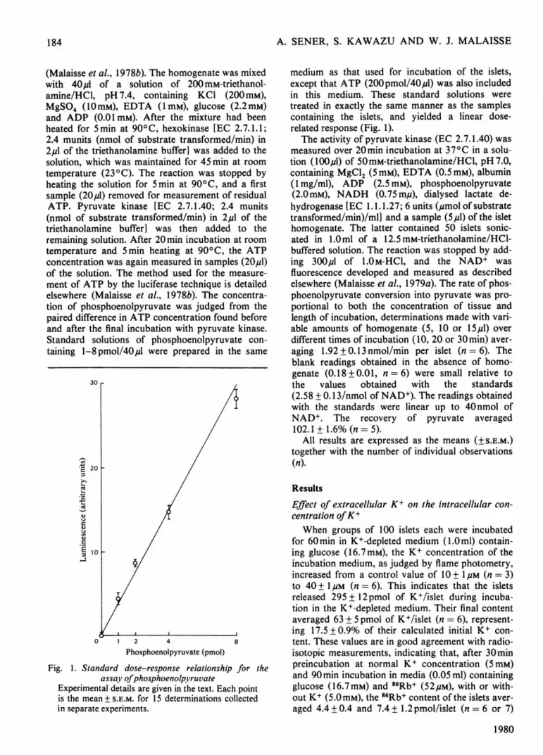
Fig. 1. Standard dose-response relationship for theassay ofphosphoenolpyruvate
Experimental details are given in the text. Each pointis the mean + S.E.M. for 15 determinations collectedin separate experiments.
medium as that used for incubation of the islets,except that ATP (200pmol/40,u1) was also includedin this medium. These standard solutions weretreated in exactly the same manner as the samplescontaining the islets, and yielded a linear dose-related response (Fig. 1).The activity of pyruvate kinase (EC 2.7.1.40) was
measured over 20min incubation at 370C in a solu-tion (lOO,ul) of 50mM-triethanolamine/HCl, pH 7.0,containing MgCl2 (5mM), EDTA (0.5mM), albumin(1 mg/ml), ADP (2.5 mM), phosphoenolpyruvate(2.0mM), NADH (0.75 m,u), dialysed lactate de-hydrogenase [EC 1.1.1.27; 6 units (pmol of substratetransformed/min)/mlI and a sample (5,u1) of the islethomogenate. The latter contained 50 islets sonic-ated in 1.0ml of a 12.5 mM-triethanolamine/HCI-buffered solution. The reaction was stopped by add-ing 300,u1 of 1.0 M-HCI, and the NAD+ wasfluorescence developed and measured as describedelsewhere (Malaisse et al., 1979a). The rate of phos-phoenolpyruvate conversion into pyruvate was pro-portional to both the concentration of tissue andlength of incubation, determinations made with vari-able amounts of homogenate (5, 10 or 15,u1) overdifferent times of incubation (10, 20 or 30min) aver-aging 1.92 + 0.13 nmol/min per islet (n = 6). Theblank readings obtained in the absence of homo-genate (0.18 + 0.01, n = 6) were small relative tothe values obtained with the standards(2.58 + 0.13/nmol of NAD+). The readings obtainedwith the standards were linear up to 40 nmol ofNAD+. The recovery of pyruvate averaged102.1+1.6%(n=5).
All results are expressed as the means (±S.E.M.)together with the number of individual observations(n).
Results
Effect of extracellular K+ on the intracellular con-centration ofK+When groups of 100 islets each were incubated
for 60min in K+-depleted medium (1.Oml) contain-ing glucose (16.7mM), the K+ concentration of theincubation medium, as judged by flame photometry,increased from a control value of 10 + IM (n = 3)to 40 + 1pM (n = 6). This indicates that the isletsreleased 295 + 12 pmol of K+/islet during incuba-tion in the K+-depleted medium. Their final contentaveraged 63 + 5 pmol of K+/islet (n = 6), represent-ing 17.5 + 0.9% of their calculated initial K+ con-tent. These values are in good agreement with radio-isotopic measurements, indicating that, after 30minpreincubation at normal K+ concentration (5 mM)and 90 min incubation in media (0.05 ml) containingglucose (16.7mM) and 86Rb+ (52,UM), with or with-out K+ (5.0mM), the 86Rb+ content of the islets aver-aged 4.4 + 0.4 and 7.4 + 1.2 pmol/islet (n = 6 or 7)
1980
184

STIMULATION-SECRETION COUPLING OF GLUCOSE-INDUCED INSULIN RELEASE
respectively. Taking into account the usual K+content of the islets (Malaisse et al., 1978a), and thepossible contamination by residual preincubationmedium (.3 p1), the final K+ content of the islets wasestimated to be 315 + 28 pmol/islet (n = 6) after in-cubation at normal K+ concentration and.41 + 7pmol/islet (n = 7) after exposure to theK+-depleted medium. The latter values were correc-ted for the ARb/AK ratio, which is close to 0.7 in theislets (Boschero & Malaisse, 1979) as well as inother tissues (Stieve & Hartung, 1977). They con-firm that the islets retain less than 20% of theirnormal K+ content when exposed to a K+-depletedincubation medium.
Over 90min incubation, the omission of extra-cellular K+ increased 22Na+ net uptake by the isletsfrom a control value (K+ 5.0mM) of 142+ 17 to332 ± 14 pmol/islet (n = 6) in the absence of glucose,and from 150 + 21 to 412 + 18pmol/islet (n = 10) inthe presence of glucose.
Effect ofK+ deprivation on glycolysisOver 120min incubation, the rate of [5-3H1-
glucose utilization, lactic acid production and [U-14C]glucose oxidation were significantly diminishedin K+-deprived islets (Table 1). Relative to the meancontrol value found at normal K+ concentration, therates of glucose utilization, lactate output andglucose oxidation in the K+-deprived islets averaged59.0+ 9.2, 67.0+4.1 and 57.8+3.9% respectively.None of these three values is significantly differentfrom any other, suggesting an unaltered depen-dency of glucose oxidation on the rate of glycolysis.This view is supported by the failure of K+ depriva-tion to affect significantly the oxidation of [U-'4C]-pyruvate.
Effect ofK+ deprivation on glucose oxidationAt normal K+ concentration, the oxidation of [U-
"4C]glucose increased as the glucose concentrationwas raised from 5.6 to 8.3 and 16.7mM (Table 2).
Table 1. Effects ofK+ deprivation on metabolic variablesAll data are expressed as pmol of substrate (glucose or pyruvate) metabolized/120 min per islet. Mean values + S.E.M.are shown together with the numbers of individual observations in parentheses and the significance of differencesdue to the change in K+ concentration (N.S., not significant).
SubstrateGlucose (16.7 mM)Glucose (16.7mM)Glucose (16.7mM)Pyruvate (20.0mM)
Metabolic parameter[5-3HIGlucose utilizationLactate output[U-_4C]Glucose oxidation[U-'4C]Pyruvate oxidation
Rate (pmol of substrate metabolized/120min per islet)
, A---- A
With 5.0mM-K+ With no K+210.4+ 17.7 (20) 124.1 + 19.3 (20)71.5 + 6.5 (22) 47.9 ± 2.9 (21)49.0 + 3.5 (23) 28.3 + 1.9 (23)40.8 + 4.1 (24) 36.6 + 5.7 (24)
p<0.005<0.001<0.001N.S.
Table 2. Glucose oxidation by isolated isletsThe rate of glucose oxidation was measured, with two different tracers, at three glucose concentrations andover two lengths of time of incubation in the presence or absence of K+, Rb+, Cs+ and NH4+. Control rates obtainedin the presence of K+ (5 mM) are expressed in absolute terms. Experimental findings are expressed as meanpercentages + S.E.M. of the mean control value found within the same experiment(s), and are shown together with thestatistical significance of differences between them and control values (N.S., not significant).
Concn.c
Tracer[U-'4C]Glucose (5.6mM)[U-14C]Glucose (5.6mM)[U-14C]Glucose (8.3 mM)[U-14C]Glucose (8.3 mM)[U-14C]Glucose (16.7mM)[U-14C]Glucose (16.7mM)[U-14C]Glucose (16.7mM)[U-14C]Glucose (16.7 mM)[U-'4CIGlucose (16.7 mM)[U-'4C]Glucose (16.7mM)[U-14C]Glucose (16.7 mM)[U-"4C]Glucose (16.7mM)[l-'4C]Glucose (16.7mM)[1_-4CIGlucose (16.7mM)
Vol. 186
Concn. Concn. Concn.)f K+ of Rb+ of Cs+ of NH4+ Time(mM) (mM) (mM) (mM) (min)5 120
1205 120
1205 30
305 120
1205 120
5 1205 120
50 1205 120
-_-- - 120
Glucose oxidation
(fmol/minper islet)
105+ 9 (11)(% of control) P
63.5 + 8.9 (12)208 + 12 (30)
71.2 + 4.8 (22)393 + 36 (24)
74.7+ 2.8 (7)408 + 32 (48)
61.3+3.6 (16)90.5 + 7.3 (16)90.8+ 5.9 (16)26.3 + 2.7 (8)25.0 + 2.0 (8)
353±31 (14)60.9 ±4.7 (15)
<0.010
<0.001
<0.005
<0.00 1N.S.N.S.
<0.001<0.001
<0.001
185

A. SENER, S. KAWAZU AND W. J. MALAISSE
Over 120min incubation, the oxidation of [U-"4Cl-glucose was diminished in K+-deprived media. Onpooling of the results obtained at three glucose con-centrations (5.6, 8.3 and 16.7mM), the degree ofinhibition averaged 33.8 + 3.1% (n = 50). An inhibi-tion of glucose oxidation was observed within thefirst 30min of exposure to the K+-depleted medium.At 16.7mM-glucose, the oxidations of [U-'4C1- and[1-14C1-glucose were affected to the same extent,suggesting that there was no major anomaly in therate of glucose metabolism through the pentosephosphate shunt relative to other oxidative path-ways.
Effect of K+ deprivation on the oxidation of endo-genous substratesWhen islets are prelabelled with [U-'4C]palmitate
and then incubated in the absence of glucose, therate of 14CO2 production is illustrative of theoxidation of endogenous substrates (Malaisse et al.,1979a; Sener et al., 1978). The omission of K+ hadlittle effect on the production of "4CO2 from the pre-labelled islets (Table 3), a significant fall in 14CO2output being observed in only one out of four separ-ate experiments.
Effect of K+ deprivation on the concentrations ofadenine and nicotinamide nucleotidesWhen islets are incubated in the absence of
glucose, the ATP concentration falls below its initialvalue (Ashcroft et al., 1973; Malaisse et al., 1976).The ATP concentration and ATP/ADP concentra-tion ratio were decreased to a smaller extent(P< 0.05 or less) when the glucose-deprived isletswere incubated in the absence of K+ (Table 4).When the incubation medium contained glucose, theconcentrations of adenine nucleotides were notsignificantly different in the presence and in theabsence of K+. Both in the presence and in theabsence of glucose the omission of K+ tended to de-crease the concentration of reduced nicotinamidenucleotides; however, this effect failed to achievestatistical significance in the absence of glucose. Theglucose-induced increment in NAD(P)H concentra-tion was evident both at normal K+ concentrationand in the absence of extracellular K+ (P < 0.02).
Effect of K+ deprivation on the concentration ofphosphoenolpyruvate
At normal K+ concentration an increase in glu-cose concentration from 1.7 to 16.7mm slightly but
Table 3. Output of "'CO2from islets prelabelled with I U-"4ClpalmitateThe outputs of "'CO2 were measured over a 120min incubation period in the absence of glucose, and are expressedas mean percentages+ S.E.M. of the final radioactivity content of the islets. The latter content averaged 4064 +206 c.p.m./islet (n = 77), corresponding to 2.31 + 0.12pmol of IU-"'Cpalmitate with the same specific radioactivityas that of the preincubation medium. The ratio 14CO2 output/14C content was also normalized relative to the meancontrol value (first row) found within the same experiment. Such a control value corresponded to a mean 14CO2output of 255.1 + 16.2 c.p.m./ 120 min per islet.
Concn. ofK+(mM)
S
Concn. of 14CO2 output/14C content of the isletsmycin A ,(mM) (%) (normalized) (n)
7.10+0.43 100.0+ 3.3 276.53 +0.51 94.3 + 5.1 31
0.01 2.11+0.16 31.0+ 1.8 19
antit
5
Table 4. Effects ofglucose and K+ on the concentrations ofadenine and reduced nicotinamide nucleotides in isolated isletsAll measurements were performed after 30 min incubation under the conditions indicated at the top of each column.Results are expressed as mean values + S.E.M. for the numbers of observations given in parentheses.
Concentration or concentration ratio
Glucose ... .
K+ . . . 5mMATP + ADP + AMP (pmol/islet) 10.8 + 0.5 (28)ATP (% ofATP + ADP + AMP) 50.4 + 2.3 (28)ADP (% ofATP + ADP + AMP) 26.0 + 1.3 (28)AMP (% of ATP + ADP + AMP) 23.6 + 1.9 (28)[ATPI/[ADPI ratio 2.15 +0.19 (26)[ATP + 0.5 ADPI/ 0.633 + 0.020 (28)[ATP+ADP+AMP] ratio
NADH + NADPH (fmol/islet) 284 + 37 (34)
9.9 + 0.8 (7)60.1 + 1.8 (7)18.0+ 1.4 (7)21.9+ 1.0 (7)3.46 + 0.34 (7)
0.700+0.017 (7)
16.7mM5mM
11.2+0.5 (32)68.0+ 1.3 (32)17.5 + 0.8 (32)14.5+ 1.1 (32)4.23 + 0.28 (32)
0.760 + 0.010 (32)
16.7mM
13.9+ 1.1 (10)67.3+2.4 (10)16.8+0.9 (10)15.9 + 2.5 (10)4.20+0.30 (10)
0.757 + 0.023 (10)
230 + 22 (18) 406 + 28 (32) 323 + 25 (30)
1980
186
STIMULATION-SECRETION COUPLING OF GLUCOSE-INDUCED INSULIN RELEASE
significantly (P < 0.05) augmented the concentra-tion of phosphoenolpyruvate in the islets from216 + 27 (n = 27) to 298 + 24 (n = 28)fmol/islet. Atthe high glucose concentration a further increase(P<0.01) in phosphoenolpyruvate concentration to399 ± 27 fmol/islet (n = 27) was observed when K+was omitted from the incubation medium.
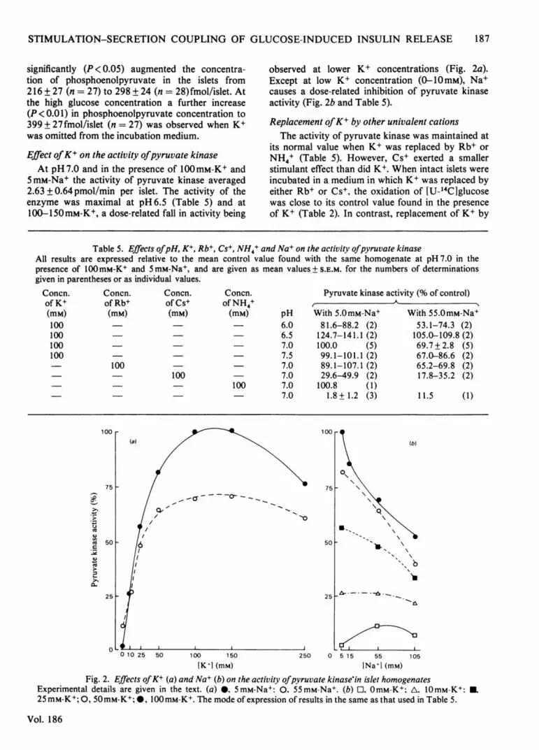
Effect ofK+ on the activity ofpyruvate kinaseAt pH 7.0 and in the presence of 100mM-K+ and
5 mM-Na+ the activity of pyruvate kinase averaged2.63 + 0.64 pmol/min per islet. The activity of theenzyme was maximal at pH 6.5 (Table 5) and at100-150mM-K+, a dose-related fall in activity being
observed at lower K+ concentrations (Fig. 2a).Except at low K+ concentration (0-10mM), Na+causes a dose-related inhibition of pyruvate kinaseactivity (Fig. 2b and Table 5).
Replacement ofK+ by other univalent cationsThe activity of pyruvate kinase was maintained at
its normal value when K+ was replaced by Rb+ orNH4+ (Table 5). However, Cs+ exerted a smallerstimulant effect than did K+. When intact islets wereincubated in a medium in which K+ was replaced byeither Rb+ or Cs+, the oxidation of [U-'4C]glucosewas close to its control value found in the presenceof K+ (Table 2). In contrast, replacement of K+ by
Table 5. Effects ofpH, K+, Rb+, Cs+, NH4+ and Na+ on the activity ofpyruvate kinaseAll results are expressed relative to the mean control value found with the same homogenate at pH 7.0 in thepresence of 100mM-K+ and 5 mM-Na+, and are given as mean values +S.E.M. for the numbers of determinationsgiven in parentheses or as individual values.
Pyruvate kinase activity (% of control)
(mM) pH With 5.0mM-Na+6.0 81.6-88.2 (2)6.5 124.7-141.1 (2)7.0 100.0 (5)7.5 99. 1-101.1 (2)7.0 89. 1-107.1 (2)7.0 29.6-49.9 (2)
100 7.0 100.8 (1)7.0 1.8 + 1.2 (3)
With 55.0mM-Na+53.1-74.3 (2)105.0-109.8 (2)69.7+ 2.8 (5)67.0-86.6 (2)65.2-69.8 (2)17.8-35.2 (2)
11.5 (1)
-a _---
I/
0 10 25 50 100 150
[K+] (mM)250
(b)
50
25 - - --.
o 15 55 105
[Na+l (mM)
Fig. 2. Effects ofK+ (a) and Na+ (b) on the activity ofpyruvate kinase in islet homogenatesExperimental details are given in the text. (a) 0, 5mM-Na+: 0. 55mM-Na+. (b) OmM-K+; A, 10mM-K+: U,
25 mM-K+; 0, 50mM-K+; 0, 100 mM-K+. The mode of expression of results in the same as that used in Table 5.
Vol. 186
Concn.of K+(mM)100100100100
Concn.of NH4+
Concn.of Rb+(mM)
100
Concn.of Cs+(mM)
100
100
75-
c 5Cl 50._
0~ctS
25
0
187
a,II
I ,
I&,"I \
b

A. SENER, S. KAWAZU AND W. J. MALAISSE
NH4+ provoked a dramatic inhibition of glucoseoxidation (Table 2).
Relationship between 4SCa2+ net uptake andmetabolic parameters
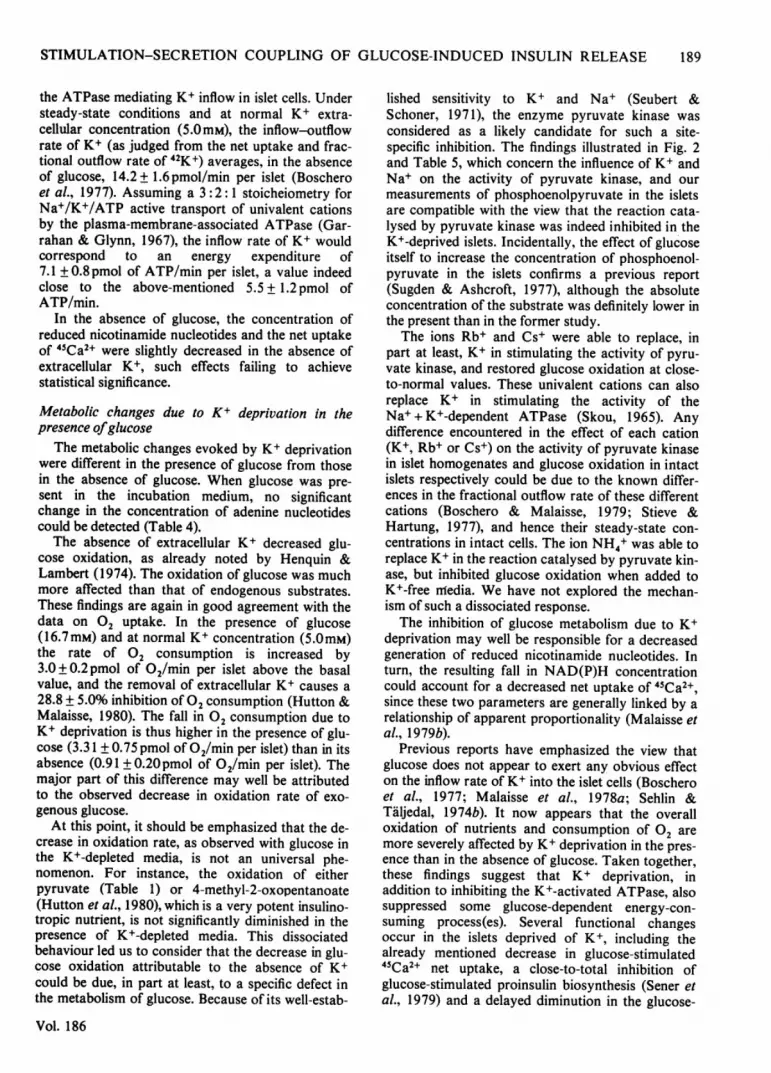
There was a significant correlation (P < 0.05) be-tween the net uptake of 45Ca2+ by the islets and theconcentration of NAD(P)H (Fig. 3). No correlationwas found between 4SCa2+ net uptake and the con-
centration of either ATP or phosphoenolpyruvate inthe islets.
Discussion
Cation concentrations in K+-deprived isletsIn the islets exposed to K+-depleted media, the
intracellular concentration of K+ was decreased toless than 20% of its normal value. Since the omis-sion of extracellular K+ is known to decrease thefractional outflow rate of K+ from the islets(Boschero & Malaisse, 1979), the decreased K+ con-
tent is obviously the consequence of a decrease inK+ influx.
4
1-u
E 3
1-
L
+ 2SCd
0 200 300 400
NAD(P)H (fmol/islet)500
Fig. 3. Correlation between the net uptake of 45Ca2+ andthe concentration of reduced nicotinamide nucleotides inislets incubated in the absence (O and 0) or presence (Aand A) of glucose (16.7mM) with (O and A) or without
( and A) K+ (5.0mM)Experimental details are given in the text. Meanvalues +±S.E.M. for 45Ca2+ net uptake refer to 12-55individual observations. The data for NAD(P)H are
taken from Table 4.
The absence of extracellular K+ is likely to causeinhibition of the Na+ + K+-activated ATPasethought to normally mediate, in part at least, the up-hill inflow of K+ and outflow of Na+ across theplasma membrane of the islet cells. In good agree-ment with the latter concept, the intracellular con-centration of Na+ was dramatically increased in theK+-deprived islets. In such islets the loss of intra-cellular K+ appeared to be matched by the increasein intracellular Na+. In islets exposed to glucose(16.7mM), the total concentration of these cationsaveraged 445 + 29 and 475 + l9pmol/islet in thepresence and absence of extracellular K+ respec-tively.The net uptake of 22Na+ is little affected (Sehlin &
Taljedal, 1974a) or slightly diminished (Kawazu etal., 1978) by glucose when the islets are incubatedat normal extracellular K+ concentration. WhenK+ was omitted from the incubation medium, thenet uptake of 22Na+ was higher (P < 0.01) in thepresence (412 + 18 pmol/islet) than absence(332 + 14pmol/islet) of glucose. A comparablesituation was previously found in islets incubated atnormal extracellular K+ concentration in the pres-ence of ouabain (Kawazu et al., 1978). The exportof Na+ being inhibited in either the absence of K+ orthe presence of ouabain, the glucose-induced incre-ment in 22Na+ net uptake may reflect a stimulantaction of the sugar on the inflow of Na+ into the isletcells (Kawazu et al., 1978).
Metabolic changes due to K+ deprivation in theabsence ofglucoseThe absence of extracellular K+ exerted several
effects on the metabolism of the islets. In the K+-deprived islets and in the absence of glucose, themetabolic situation was compatible with the exist-ence of a diminished energy expenditure, leading toless rapid or less severe falls in ATP concentrationand ATP/ADP concentration ratio than thoseusually seen when islets are incubated in the absenceof glucose at normal K+ concentration (Ashcroft etal., 1973; Malaisse et al., 1976).A minor decrease in the utilization of endogenous
nutrients might occur in the K+- and glucose-deprived islets (Table 3). This view is supported bythe fact that the removal of extracellular K+diminishes by 10.7 + 1.5% the rate of 02 consump-tion in islets not exposed to glucose (Hutton &Malaisse, 1980). The control rate of 02 consump-tion in the absence of glucose averaging8.5 + 1.5 pmol of 02/min per islet, the fall in °2 Up-take attributable to the absence of K+ amounts to0.91 + 0.20 pmol of 02/min per islet, correspondingto an estimated loss in ATP generation of5.5 + 1.2 pmol/min per islet. This last value is notvastly different (P> 0.3) from the energy thought tobe normally required for the appropriate function of
1980
188
STIMULATION-SECRETION COUPLING OF GLUCOSE-INDUCED INSULIN RELEASE
the ATPase mediating K+ inflow in islet cells. Understeady-state conditions and at normal K+ extra-cellular concentration (5.0mM), the inflow-outflowrate of K+ (as judged from the net uptake and frac-tional outflow rate of 42K+) averages, in the absenceof glucose, 14.2 + 1.6 pmol/min per islet (Boscheroet al., 1977). Assuming a 3 :2: 1 stoicheiometry forNa+/K+/ATP active transport of univalent cationsby the plasma-membrane-associated ATPase (Gar-rahan & Glynn, 1967), the inflow rate of K+ wouldcorrespond to an energy expenditure of7.1 + 0.8 pmol of ATP/min per islet, a value indeedclose to the above-mentioned 5.5 + 1.2 pmol ofATP/min.
In the absence of glucose, the concentration ofreduced nicotinamide nucleotides and the net uptakeof 45Ca2+ were slightly decreased in the absence ofextracellular K+, such effects failing to achievestatistical significance.
Metabolic changes due to K+ deprivation in thepresence ofglucose
The metabolic changes evoked by K+ deprivationwere different in the presence of glucose from thosein the absence of glucose. When glucose was pre-sent in the incubation medium, no significantchange in the concentration of adenine nucleotidescould be detected (Table 4).
The absence of extracellular K+ decreased glu-cose oxidation, as already noted by Henquin &Lambert (1974). The oxidation of glucose was muchmore affected than that of endogenous substrates.These findings are again in good agreement with thedata on 02 uptake. In the presence of glucose(16.7mM) and at normal K+ concentration (5.0mM)the rate of 02 consumption is increased by3.0+0.2pmol of 02/min per islet above the basalvalue, and the removal of extracellular K+ causes a28.8 + 5.0% inhibition of 02 consumption (Hutton &Malaisse, 1980). The fall in 02 consumption due toK+ deprivation is thus higher in the presence of glu-cose (3.31 + 0.75 pmol of 02/min per islet) than in itsabsence (0.91 + 0.20pmol of 02/min per islet). Themajor part of this difference may well be attributedto the observed decrease in oxidation rate of exo-genous glucose.At this point, it should be emphasized that the de-
crease in oxidation rate, as observed with glucose inthe K+-depleted media, is not an universal phe-nomenon. For instance, the oxidation of eitherpyruvate (Table 1) or 4-methyl-2-oxopentanoate(Hutton et al., 1980), which is a very potent insulino-tropic nutrient, is not significantly diminished in thepresence of K+-depleted media. This dissociatedbehaviour led us to consider that the decrease in glu-cose oxidation attributable to the absence of K+could be due, in part at least, to a specific defect inthe metabolism of glucose. Because of its well-estab-
Vol. 186
lished sensitivity to K+ and Na+ (Seubert &Schoner, 1971), the enzyme pyruvate kinase wasconsidered as a likely candidate for such a site-specific inhibition. The findings illustrated in Fig. 2and Table 5, which concern the influence of K+ andNa+ on the activity of pyruvate kinase, and ourmeasurements of phosphoenolpyruvate in the isletsare compatible with the view that the reaction cata-lysed by pyruvate kinase was indeed inhibited in theK+-deprived islets. Incidentally, the effect of glucoseitself to increase the concentration of phosphoenol-pyruvate in the islets confirms a previous report(Sugden & Ashcroft, 1977), although the absoluteconcentration of the substrate was definitely lower inthe present than in the former study.The ions Rb+ and Cs+ were able to replace, in
part at least, K+ in stimulating the activity of pyru-vate kinase, and restored glucose oxidation at close-to-normal values. These univalent cations can alsoreplace K+ in stimulating the activity of theNa+ + K+-dependent ATPase (Skou, 1965). Anydifference encountered in the effect of each cation(K+, Rb+ or Cs+) on the activity of pyruvate kinasein islet homogenates and glucose oxidation in intactislets respectively could be due to the known differ-ences in the fractional outflow rate of these differentcations (Boschero & Malaisse, 1979; Stieve &Hartung, 1977), and hence their steady-state con-centrations in intact cells. The ion NH4+ was able toreplace K+ in the reaction catalysed by pyruvate kin-ase, but inhibited glucose oxidation when added toK+-free nmedia. We have not explored the mechan-ism of such a dissociated response.The inhibition of glucose metabolism due to K+
deprivation may well be responsible for a decreasedgeneration of reduced nicotinamide nucleotides. Inturn, the resulting fall in NAD(P)H concentrationcould account for a decreased net uptake of 45Ca2+,since these two parameters are generally linked by arelationship of apparent proportionality (Malaisse etal., 1979b).
Previous reports have emphasized the view thatglucose does not appear to exert any obvious effecton the inflow rate of K+ into the islet cells (Boscheroet al., 1977; Malaisse et al., 1978a; Sehlin &Taljedal, 1974b). It now appears that the overalloxidation of nutrients and consumption of 02 aremore severely affected by K+ deprivation in the pres-ence than in the absence of glucose. Taken together,these findings suggest that K+ deprivation, inaddition to inhibiting the K+-activated ATPase, alsosuppressed some glucose-dependent energy-con-suming process(es). Several functional changesoccur in the islets deprived of K+, including thealready mentioned decrease in glucose-stimulated45Ca2+ net uptake, a close-to-total inhibition ofglucose-stimulated proinsulin biosynthesis (Sener etal., 1979) and a delayed diminution in the glucose-
189
190 A. SENER, S. KAWAZU AND W. J. MALAISSE
stimulated insulin release (Henquin & Lambert,1974). Some of these functional alterations may wellcontribute to the postulated decrease in ATP utiliza-tion evoked by K+ deprivation.
In conclusion, in the islet cells the coupling be-tween metabolic and cationic events, which, in ouropinion, represents an essential step in the glucose-stimulated secretory sequence, is susceptible to feed-back regulatory mechanisms through which a pri-mary change in cationic movements results in analteration of glucose metabolism. Other examples ofsuch a situation can be found in previous reports onthe influence of Na+ or Ca2+ deprivation on islet-cellmetabolism (Hellman et al., 1974; Malaisse et al.,1978c). In the K+-deprived islets, the inhibition ofglucose oxidation appears to be associated with aninhibition of pyruvate kinase and a decrease inenergy expenditure. Since glucose itself increases K+concentration (Malaisse et al., 1978a) and tends todecrease Na+ concentration (Kawazu et al., 1978) inthe islet cells, modulation of pyruvate kinase activityby univalent cations could play a role in thestimulus-secretion of glucose-induced insulin releaseat normal extracellular concentrations of Na+ andK+.
This work was supported in part by grants from theBelgian Foundation for Scientific Medical Research(Brussels, Belgium). We are grateful to Mrs. J. Schoon-heydt, Mr. A. Tinant and Mrs. M. Urbain for technicalassistance, and to Mrs. C. Demesmaeker for secretarialhelp. The present paper is the thirty-seventh in a series onthe stimulus-secretion coupling of glucose-induced insulinrelease.
References
Ashcroft, S. J. H., Weerasinghe, L. C. & Randle, P. J.(1973) Biochem.J. 132, 223-231
Boschero, A. C. & Malaisse, W. J. (1979) Am. J. Physiol.236, E139-E146
Boschero, A. C., Kawazu, S., Duncan, G. & Malaisse,W. J. (1977) FEBS Lett. 83, 151-154
Garrahan, P. J. & Glynn, I. M. (1967) J. Physiol.(London) 192, 217-235
Hellman, B., Idahl, L. A., Lernmark, A., Sehlin, J. &Taljedal, I.-B. (1974) Biochem. J. 138, 33-45
Henquin, J. C. & Lambert, A. E. (1974) Diabetologia 10,789-794
Hutton, J. C. & Malaisse, W. J. (1980) Diabetologia inthe press
Hutton, J. C., Sener, A., Herchuelz, A., Atwater, I.,Kawazu, S., Boschero, A. C., Somers, G., Devis, G. &Malaisse, W. J. (1980) Endocrinology in the press
Kawazu, S., Boschero, A. C., Delcroix, C. & Malaisse,W. J. ( 1978) Pfluigers A rch. 375, 197-206
Lacy, P. E. & Kostianovsky, M. (1967) Diabetes 16, 35-39
Levy, J., Herchuelz, A., Sener, A., Malaisse-Lagae, F. &Malaisse, W. J. (1976) Endocrinology 98, 429-437
Malaisse, W. J., Brisson, G. R. & Malaisse-Lagae, F.(1970) J. Lab. Clin. Med. 76, 895-902
Malaisse, W. J., Sener, A. & Mahy, M. (1974) Eur. J.Biochem. 47, 365-370
Malaisse, W. J., Sener, A., Levy, J. & Herchuelz, A.(1976) Acta Diabetol. Lat. 13, 202-215
Malaisse, W. J., Boschero, A. C., Kawazu, S. & Hutton,J. C. (1978a)Pflzigers Arch. 373, 237-242
Malaisse, W. J., Hutton, J. C., Kawazu, S. & Sener, A.(1978b) Eur. J. BiQchem. 87, 121-130
Malaisse, W. J., Hutton, J. C., Sener, A., Levy, J.,Herchuelz, A., Somers, G. & Devis, G. (1978c) J.Membr. Biol. 38, 193-208
Malaisse, W. J., Kawazu, S., Herchuelz, A., Hutton, J. C.,Somers, G., Devis, G. & Sener, A. (1979a) Arch. Bio-chem. Biophys. 194, 49-62
Malaisse, W. J., Sener, A., Herchuelz, A. & Hutton, J. C.(1979b) Metab. Clin. Exp. 28, 373-386
Malaisse-Lagae, F. & Malaisse, W. J. (1971) Endocrin-ology 88, 72-80
Sehlin, J. & Taljedal, I.-B. (1974a) FEBS Lett. 39, 209-213
Sehlin, J. & Taljedal, I.-B. (1974b) J. Physiol. (London)242, 505-5 15
Sener, A. & Malaisse, W. J. (1976) Biochem. Med. 15,34-41
Sener, A., Kawazu, S., Hutton, J. C., Boschero, A. C.,Devis, G., Somers, G., Herchuelz, A. & Malaisse, W. J.(1978) Biochem.J. 176, 217-232
Sener, A., Kawazu, S. & Malaisse, W. J. (1979) ExcerptaMed. Found. Int. Congr. Ser. 481, 210 (Abstract)
Seubert, W. & Schoner, W. (1971) Curr. Top. Cell.Regul. 3, 237-268
Skou, J. C. (1965) Physiol. Rev. 45, 596-617Stieve, H. & Hartung, K. (1977) Biochim. Biophys. Acta
465, 634-649Sugden, M. C. & Ashcroft, S. J. H. (1977) Diabetologia
13, 481-486
1980