the mechanism of inhibition of ran-dependent nuclear transport by cellular atp depletion
TRANSCRIPT

The Rockefeller University Press, 0021-9525/2002/06/963/12 $5.00The Journal of Cell Biology, Volume 157, Number 6, June 10, 2002 963–974http://www.jcb.org/cgi/doi/10.1083/jcb.200111077
JCB
Article
963
The mechanism of inhibition of Ran-dependent nuclear transport by cellular ATP depletion
Eric D. Schwoebel, Thai H. Ho, and Mary Shannon Moore
Department of Molecular and Cellular Biology, Baylor College of Medicine, Houston, TX 77030
an-dependent nuclear transport requires a nuclearpool of RanGTP both for the assembly of export com-plexes and the disassembly of import complexes.
Accordingly, in order for these processes to proceed, Ran-dependent nuclear import and export assays in vitro requirethe addition of GTP to produce RanGTP. Notably, no ATPrequirement can be detected for these transport processesin vitro. But in vivo, when cells are depleted of ATP by theaddition of sodium azide and 2-deoxyglucose to block ATPproduction by oxidative phosphorylation and glycolysis,
R
respectively, Ran-dependent nuclear import and export arerapidly inhibited. This raised the question of whether thereis an ATP requirement for these nuclear transport pathwaysin an intact cell that has remained undetected in vitro.Here we report that the free (but not total) GTP concentrationrapidly drops to an undetectable level upon ATP depletion asdoes the availability of RanGTP. Our conclusion is that theinhibition of Ran-dependent nuclear transport observed uponATP depletion in vivo results from a shortage of RanGTPrather than the inhibition of some ATP-dependent process.
Introduction
That carrier-dependent nuclear transport of cargo across thenuclear pore complex (NPC)* requires energy is well estab-lished, but how is this energy used? Of the
�
30 proteins thatcomprise the yeast NPC, none of them resemble an ATPase orGTPase, making it unlikely that the NPC itself has to hy-drolyze nucleoside triphosphates (NTPs) to power transport(Rout et al., 2000). Consistent with this, movement of a carrier(with or without bound cargo) from one side of the NPC tothe other does not require energy (Kose et al., 1997; Yokoyaet al., 1999; Ribbeck and Gorlich, 2001) and is believedto occur by repeated binding and release of the carrier tosequential NPC proteins in a process called “facilitateddiffusion” (for review see Talcott and Moore, 1999). Incontrast, however, the signal-mediated accumulation of importcargo in the nucleus or export cargo in the cytoplasm doesrequire energy, and we now know that in many cases, at least
part of the energy requirement can be linked to the smallGTPase Ran (Moore, 1998).
The majority of nuclear carriers belong to the karyopherin-
�
(Kap-
�
) superfamily, and the one diagnostic feature of carriersin this family is that they all bind Ran, but critically onlywhen Ran is in the GTP- rather than the GDP-bound form(Nakielny and Dreyfuss, 1999). Nuclear transport com-plexes containing a Kap-
�
carrier use the concentration ofRanGTP, which varies widely between the cytoplasm andnucleus, as a positional cue to regulate their assembly anddisassembly (Gorlich et al., 1996; Izaurralde et al., 1997).The concentration of RanGTP is kept low in the cytoplasmand high inside the nucleus by localizing the Ran GTPaseactivating protein (GAP) to the cytoplasm and the Ran guaninenucleotide exchange factor (GEF) (RCC1) to the nucleus.
Crucially, having bound RanGTP has opposite effects onimport versus export carriers on their ability to simulta-neously bind cargo (Gorlich et al., 1996). Kap-
�
carriersthat function in import will only bind their cargo in theabsence of RanGTP (i.e., in the cytoplasm) and the subse-quent binding of RanGTP inside the nucleus triggers therelease of cargo. Thus, import of a classical nuclear localizationsequence (cNLS)–containing cargo mediated by Kap-
�
1and its adaptor Kap-
�
requires both nuclear Ran and GTP,however, GTP hydrolysis by Ran is not required for entry ofthe cargo into the nuclear interior (Schwoebel et al., 1998;Englmeier et al., 1999). Instead, nuclear RanGTP is requiredfor the disassembly of an incoming transport complex andthe release of cargo triggered by the interaction of Kap-
�
1
Address correspondence to Mary Shannon Moore, Department of Mo-lecular and Cellular Biology, Baylor College of Medicine, One BaylorPlaza, Houston, TX 77030. Tel.: (713) 798-6656. Fax: (713) 798-7799.E-mail: [email protected]
E.D. Schwoebel’s current address is Massachusetts Institute of TechnologyLincoln Laboratories, Lexington, MA 02420.*Abbreviations used in this paper: CAS, cellular apoptosis susceptibility;GAP, GTPase activating protein; GEF, guanine nucleotide exchange factor;GST, glutathione-
S
-transferase; Kap-
�
, karyopherin-
�
; NEM,
N
-ethyl-maleimide; NES, nuclear export sequence; cNLS, classical nuclear lo-calization sequence; NPC, nuclear pore complex; NTP, nucleosidetriphosphate; RanBD, Ran binding domain; RT, room temperature.Key words: Ran; nuclear; transport; ATP; ribavirin
964 The Journal of Cell Biology
|
Volume 157, Number 6, 2002
with RanGTP (Kutay et al., 1997b). GTP hydrolysis by Ranin this import pathway occurs only when the RanGTP–Kap-
�
1 complex recycles back through the NPC andencounters the RanGAP in the cytoplasm. Thus, GTP hy-drolysis by Ran during Ran-dependent nuclear import is re-quired for recycling transport carriers after import, ratherthan for powering the movement of an import cargothrough the NPC into the nucleus. Export carriers exhibitthe reverse behavior, requiring bound RanGTP to simulta-neously bind their nuclear export sequence (NES)–contain-ing cargo with high affinity. Upon contact of the exportcomplex with the RanGAP in the cytoplasm, RanGTP hy-drolyzes its bound GTP to become RanGDP. As a result ofthis GTP hydrolysis, both RanGDP and cargo are releasedby the carrier, terminating export.
In vitro, neither Kap-
�
1–mediated import of cNLS-con-taining cargo nor Crm1-mediated export of leucine-richNES-containing cargo are inhibited by the addition of anonhydrolyzable ATP analogue (Schwoebel et al., 1998; En-glmeier et al., 1999
).
In vivo, however, ATP depletion ofcells by metabolic poisons that block oxidative phosphoryla-tion and/or glycolysis causes a rapid shutdown of cNLS-mediated import (Richardson et al., 1988; Shulga et al., 1996).We wanted to determine by what mechanism ATP deple-
tion inhibits Ran-dependent nuclear transport in vivo, par-ticularly to determine whether there is an ATP-dependentstep in these transport pathways in vivo that has remainedundetected in vitro. Here we report that the in vivo inhibi-tion of Ran-dependent nuclear transport observed uponATP depletion is likely caused by depletion of cellularRanGTP resulting from a lack of free GTP, rather than inhi-bition of an ATP-dependent step required for transport.
Results
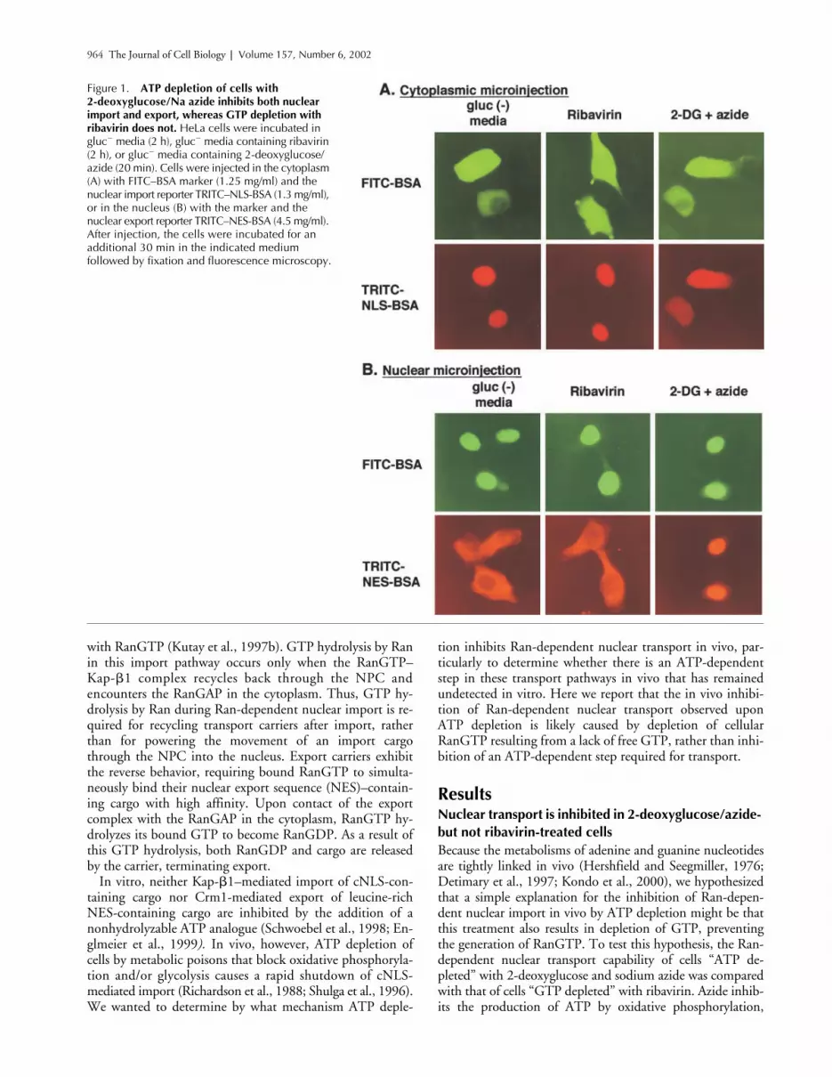
Nuclear transport is inhibited in 2-deoxyglucose/azide- but not ribavirin-treated cells
Because the metabolisms of adenine and guanine nucleotidesare tightly linked in vivo (Hershfield and Seegmiller, 1976;Detimary et al., 1997; Kondo et al., 2000), we hypothesizedthat a simple explanation for the inhibition of Ran-depen-dent nuclear import in vivo by ATP depletion might be thatthis treatment also results in depletion of GTP, preventingthe generation of RanGTP. To test this hypothesis, the Ran-dependent nuclear transport capability of cells “ATP de-pleted” with 2-deoxyglucose and sodium azide was comparedwith that of cells “GTP depleted” with ribavirin. Azide inhib-its the production of ATP by oxidative phosphorylation,
Figure 1. ATP depletion of cells with 2-deoxyglucose/Na azide inhibits both nuclear import and export, whereas GTP depletion with ribavirin does not. HeLa cells were incubated in gluc� media (2 h), gluc� media containing ribavirin (2 h), or gluc� media containing 2-deoxyglucose/azide (20 min). Cells were injected in the cytoplasm (A) with FITC–BSA marker (1.25 mg/ml) and the nuclear import reporter TRITC–NLS-BSA (1.3 mg/ml), or in the nucleus (B) with the marker and the nuclear export reporter TRITC–NES-BSA (4.5 mg/ml). After injection, the cells were incubated for an additional 30 min in the indicated medium followed by fixation and fluorescence microscopy.

ATP depletion and nuclear transport |
Schwoebel et al. 965
whereas 2-deoxyglucose inhibits ATP production by glycoly-sis. Ribavirin is an inhibitor of IMP dehydrogenase (the ratelimiting enzyme in de novo GTP biosynthesis) and ribavirintreatment is known to decrease intracellular GTP levels(Finch et al., 1993; Yalowitz and Jayaram, 2000).
After treatment with either ribavirin or 2-deoxyglucose/azide in gluc
�
medium, the labeled import substrateTRITC–NLS-BSA (containing the NLS of the SV40 T anti-gen) was injected into the cytoplasm of HeLa cells to monitorimport. In other cells, the labeled export substrate TRITC–NES-BSA (containing the Rev NES) was injected into thenucleus to monitor export. As a control, cells were injectedafter incubation in gluc
�
media. As expected from previousreports (Richardson et al., 1988; Shulga et al., 1996),TRITC–NLS-BSA was imported into the nuclei of controlcells, but not imported into the nuclei of cells treated with2-deoxyglucose/azide (Fig. 1 A). Likewise, nuclear injectedTRITC–NES-BSA was exported from the nuclei of controlcells, but not cells treated with 2-deoxyglucose/azide. Unex-pectedly, however, treatment with ribavirin to lower GTPlevels affected neither the nuclear import of TRITC–NLS-BSA nor the export of TRITC–NES-BSA (Fig. 1).
Total nucleotide levels in ATP- and GTP-depleted cells
To compare the nucleotide levels in cells after these treat-ments, nucleotides were isolated from control and treatedcells and quantitated by FPLC (Kremmer et al., 1989). Wefound that treatment with 2-deoxyglucose/azide in gluc
�
medium for 1 h decreased total cellular ATP to
�
40% ofthe level in cells incubated in just gluc
�
medium (8.22 vs.3.27 nmoles/10
6
cells; Table I). In trial experiments, whenFCS was omitted from the medium during treatment,2-deoxyglucose/azide treatment for 1 h resulted in muchlower ATP levels (
�
10% of normal; unpublished data). Wefound however that the cells were in such bad shape aftersuch severe ATP depletion that they were very difficult tomicroinject (unpublished data). We found that addition ofFCS to the medium during treatment made the cells moreamenable to microinjection and did not prevent the inhibi-tion of nuclear transport by 2-deoxyglucose/azide.
As hypothesized, ATP depletion with 2-deoxyglucose/azide also caused a decrease in the total GTP content from2.58 to 1.85 nmoles/10
6
cells (72% of control levels). GTPdepletion with ribavirin, however, decreased the GTP con-tent almost twice as much to 36% of control levels (0.87nmoles/10
6
cells) without significantly affecting the ATPconcentration (8.42 vs. 8.32 nmoles/10
6
cells). Yet, asshown in Fig. 2, these GTP-depleted cells are still transportcompetent unlike the ATP-depleted cells, indicating that in-hibition of nuclear transport in 2-deoxyglucose/azide-treated
cells is not the result of a decrease in the levels of total GTP.Note that although other cell types can contain higher ATP/GTP ratios than we found here (Franklin and Twose, 1977;Lee et al., 1985), our measured ATP/GTP ratios of 3.2–3.5in control cells are nearly identical to those previously deter-mined in HeLa cells (Finch et al., 1993).
Ran has a 10-fold higher affinity for GDP than GTP and al-though the RanGEF drastically increases the rate of nucleotideloading, it does not affect the relative affinity of Ran for GTPand GDP (Klebe et al., 1995). Other factors may modulate
Figure 2. The cellular localization of nuclear transport factors after 2-deoxyglucose/sodium azide or ribavirin treatment. Cells were treated with control gluc� media for 2 h, gluc� media containing ribavirin for 2 h, or gluc� media containing 2-deoxyglucose/sodium azide for 1 h. The cells were fixed and probed with antibodies against the indicated nuclear transport factor, followed by fluorescently labeled second antibody. Note that Kap-� accumulates in the nuclei of 2-deoxyglucose/azide-treated cells, whereas the distribution of the other transport factors remains essentially unchanged.
Table I.
Measurement of total nucleotide levels in HeLa cells after various treatments
ATP ADP GTP GDP ATP/ADP GTP/GDP
Glucose
�
media, 1 h 8.22
�
0.75 0.75
�
0.17 2.58
�
0.23 0.29
�
0.07 11.0 8.9Glucose
�
media, 2 h 8.32
�
0.59 0.69
�
0.05 2.4
�
0.2 0.21
�
0.03 12.1 11.4Ribavirin, 2 h 8.42
�
0.43 0.69
�
0.01 0.87
�
0.01 0.17
�
0.02 12.2 5.12-deoxyglucose
�
sodium azide, 1 h 3.27
�
0.60 0.53
�
0.13 1.85
�
0.29 0.27
�
0.07 6.2 6.9
Values are expressed as nmoles nucleotide/10
6
cells. Nucleotides were measured in total cellular extracts by FPLC as described in the Materials and methods.Each value represents the mean
�
SD of three independent experiments.

966 The Journal of Cell Biology
|
Volume 157, Number 6, 2002
this process in vivo but, based on our current knowledge, theratio of RanGTP versus RanGDP generated in a loading reac-tion is dependent on the ratio of free GTP to GDP in the im-mediate environment. Because of this, we then measured thetotal GDP (and ADP) levels to determine whether an alteredGTP/GDP ratio rather than the total concentration of GTPin the 2-deoxyglucose/azide-treated cells might be responsiblefor the observed transport inhibition. Treatment with 2-deoxy-glucose/azide did result in a decrease in the GTP/GDP ratiofrom 8.3 to 6.9. However, treatment with ribavirin affectedthe GTP/GDP ratio more severely, dropping it to 5.1. Thus,both the total GTP level and GTP/GDP ratio are lower afterribavirin treatment than 2-deoxyglucose/azide treatment (Ta-ble I), and yet ribavirin-treated cells remain transport compe-tent, whereas 2-deoxyglucose/azide treated cells do not. There-fore, it is not the decrease in total GTP or the GTP/GDP ratioafter 2-deoxyglucose/azide treatment that is responsible for theobserved nuclear transport inhibition.
The Mono-Q and Source 15Q FPLC columns used herefor the quantitation of nucleotides retain and fractionate allmono-, di-, and triphosphate nucleotides (Hartwick andBrown, 1975; Kremmer et al., 1989). After 2-deoxyglucose/azide treatment, we did not see a corresponding increase inany of the nucleotides (such as AMP) that might possibly rep-resent breakdown or interconversion products of ATP (un-published data). This is consistent with a previous report thata decrease in cellular ATP stimulated by anoxia correlates witha rise at time points similar to those observed here in the meta-bolic breakdown products of ATP, such as adenosine, that arenot retained on our FPLC columns (Grune and Siems, 1993).
2-deoxyglucose/azide treatment results in a buildup of Kap-
�
in the nucleus
To explore other possibilities for the 2-deoxyglucose/azideinhibition of nuclear transport, ribavirin- and 2-deoxyglu-cose/azide-treated cells were fixed and examined by indi-rect immunofluorescence microscopy to determine whetherthese treatments result in a change in the intracellular local-ization of proteins required for nuclear transport. We didnot observe any significant changes in the localization ofRan, the RanGAP, or the RanGEF (RCC1) after eithertreatment (Fig. 2). Note that cells treated with 2-deoxyglu-cose/azide tend to round up somewhat, resulting in a de-crease in the apparent cytoplasmic volume. This morpholog-ical change can result in a slightly altered appearance (see forexample the Ran localization) without actually changing thenucleocytoplasmic distribution of the transport factors (Fig.2). Kap-
�
1 in some experiments appeared to slightly con-centrate in the nucleus after 2-deoxyglucose/azide treatment,but this change was neither dramatic nor consistent. Kap-
�
,in contrast, was found to dramatically shift its localizationexclusively to the nuclei of 2-deoxyglucose/azide-treatedcells. Normally, Kap-
�
mainly appears either cytosolic orbound at the nuclear envelope as can be seen in the ribavi-rin- and gluc
�
-treated cells (Fig. 2).During cNLS-mediated import, Kap-
�
crosses the NPCinto the nucleus in a complex with cNLS-containing cargoand Kap-
�
1 (Gorlich et al., 1995). Upon contact withRanGTP in the nucleoplasm, this complex disassembles andKap-
�
is then recycled back to the cytoplasm by the CAS
(cellular apoptosis susceptibility) protein carrier (Kutay etal., 1997a). CAS requires bound RanGTP to bind Kap-
�
with high affinity, thus both the import of Kap-
�
com-plexed with cargo and Kap-
�
1 and the export of Kap-
�
me-diated by CAS require RanGTP (Melchior et al., 1993;Moore and Blobel, 1993; Kutay et al., 1997a). Accumula-tion of both the
Saccharomyces cerevisiae
homologue of Kap-
�
(Srp1p) and vertebrate Kap-
�
in the nucleus has been ob-served previously in cells predicted to have a low concentra-tion of nuclear RanGTP. Examples of this are cells expressinga mutant Ran that cannot be converted to the GTP-boundform, or when the Ran GEF is inactivated (Koepp et al., 1996;Tachibana et al., 2000). We wondered therefore whether thecells upon 2-deoxyglucose/azide treatment might be becom-ing deficient in RanGTP, thus explaining their transportdefect. Accordingly, we devised a method for directly mea-suring cellular RanGTP to see how this was affected by2-deoxyglucose/azide treatment.
Measurement of in vivo RanGTP levels
The percentage of cellular Ran in the GTP-bound form atsteady-state has never been measured, primarily for technical
Figure 3. NEM treatment of HeLa cellular lysates inhibits both RanGEF and RanGAP activity. Cellular lysates were prepared as described in the Materials and methods and treated with either (�) 10, (�) 20, (�) 40, or (�) 60 mM NEM for the indicated period of time, quenched with excess DTT, and measured as follows: (A) measurement of RanGEF activity in HeLa cell extracts after NEM treatment; (B) measurement of RanGAP activity in HeLa cell extracts after NEM treatment. Both RanGAP and GEF activity were measured as described in the Materials and methods.
ATP depletion and nuclear transport |
Schwoebel et al. 967
reasons. A major problem with measuring the intracellularconcentration of RanGTP is that upon cell lysis and prepara-tion of a cellular extract, Ran and its GAP and GEF becomemixed together instead of remaining spatially separate. In par-ticular, the mixing of RanGTP with the RanGAP results invery (artifactually) low levels of RanGTP that can be immu-noprecipitated from cellular extracts even when the extractsare kept cold (Lounsbury et al., 1996). Thus, we needed someway of inhibiting RanGAP and RanGEF activity in cellularlysates to accurately measure the amount of RanGTP that waspresent in the cells before lysis. In experiments not shown, wefound that the Ran GAP and GEF activities in the cellular ly-sates as measured with exogenously added Ran were notaltered after 2-deoxyglucose/azide treatment (unpublisheddata). This result indicated that 2-deoxyglucose/azide treat-ment does not affect RanGTP production by altering the ac-tivity of either of these enzymes by, for example, inhibiting anATP-dependent posttranslational modification.
We reported previously that the RanGEF activity ofRCC1 is inhibited by pretreatment of RCC1 with the alky-lating agent
N
-ethylmaleimide (NEM) (Schwoebel et al.,1998), and here we report that the RanGAP activity is alsoinhibited by treatment with NEM. We found that both
RanGAP and RanGEF activities in cellular lysates were in-hibited by NEM in a time- and dose-dependent fashion(Fig. 3, A and B). In every case, excess DTT was added afterNEM treatment to quench the activity of NEM before as-say. Note that despite the NEM sensitivity of the Ran GEFand GAP activities as shown here, Ran itself is known to beunaffected by NEM treatment (Moore and Blobel, 1992).
Having in hand a treatment to substantially reduce Ran-GAP and RanGEF activity in cellular lysates, we used thisNEM treatment in conjunction with a new assay that we de-vised to measure available RanGTP present in a cellular ly-sate. This assay utilizes the Ran binding domain (RanBD) ofRanBP1, which only binds RanGTP and not RanGDP(Beddow et al., 1995). After cell lysis and NEM treatment,the extract is passed over a desalting column to removeNEM and DTT. A glutathione-
S
-transferase (GST) pull-down is then performed with GST–RanBD, and bound Ranis determined by immunoblotting with an anti-Ran anti-body. Because only Ran in the GTP-bound form binds theRanBD, any Ran isolated in this pulldown must be in theGTP-bound form. Furthermore, only RanGTP not alreadycomplexed with RanBP1 or any other protein that binds tothis region of RanGTP will be isolated in this pulldown.
Figure 4. Isolation of RanGTP with a RanBD fused to GST. (A and B) NEM treatment of cellular lysate allows the recovery of RanGTP. Either cellular lysates or a solution of BSA were spiked with recombinant Ran loaded with either (A) [�-32P]GTP or (B)
[�-32P]GTP before pulldown of RanGTP with GST–RanBD. The radiolabeled Ran and NEM were added together with 2 mM GDP-�S upon cell lysis as described in the Materials and methods. Samples were either NEM treated to abolish RanGAP and GEF activity (100 mM NEM for 20 min) before quenching with 200 mM DTT, or mock treated in which the order of addition of NEM and DTT was reversed. After gel filtration to remove excess NEM and DTT, RanGTP was isolated in a pulldown with GST–RanBD and glutathione-agarose. In the indicated samples, GST was substituted for the GST–RanBD; these samples were mock treated. Shown are the CPM of radiolabeled Ran associated with the GST–RanBD glutathione-agarose beads after washing. (C) No free RanGTP can be detected in HeLa cells treated with 2-deoxyglucose/azide. HeLa cells were either untreated (lanes 1 and 6–8), incubated in gluc� media for either 1 (lane 2) or 2 h (lane 4), incubated with 2-deoxyglucose/azide in gluc� media for 1 h (lane 3), or incubated with ribavirin in gluc� media for 2 h (lane 5). After treatment, cells were scraped in buffer A containing 0.1% Triton X-100 and were either NEM treated as above to abolish RanGAP and GEF activity (lanes 1–7) or mock treated (lane 8). The unlabeled nucleotides added upon cellular lysis were 2 mM GDP-�S (lanes 1–5 and 8), GMP-PNP (lane 6), or GTP (lane 7). After NEM or mock treatment, RanGTP was isolated from the soluble cytosolic extract by incubation with GST–RanBD and glutathione-agarose. Bound protein was solubilized with SDS-PAGE sample buffer, separated on a 15% gel, transferred to nitrocellulose, and immunoblotted with an anti-Ran antibody. Note in particular the absence of detectable RanGTP in cells treated with 2-deoxyglucose/azide (lane 3). (D) Immunoblot of total cell extracts shows that the amount of total cellular Ran doesn’t change after these treatments.

968 The Journal of Cell Biology
|
Volume 157, Number 6, 2002
To confirm that a 20-min incubation with 100 mMNEM inhibits RanGAP and RanGEF activity sufficientlyin cellular extracts such that RanGTP can be quantitativelyrecovered in a pulldown with GST–RanBD, [
�
-
32
P]GTP-or [
�
-
32
P]GTP-loaded recombinant Ran was added (to-gether with NEM) to the lysate immediately upon celllysis. As a control for the efficiency of recovery of radio-labeled RanGTP in the absence of the RanGAP andRanGEF and endogenous unlabeled RanGTP, a BSA solu-tion was substituted for cellular extract in some samples. Asshown in Fig. 4, A and B, exogenously added [
32
P]GTP–Ran can be recovered by the GST–RanBD from cellularextracts to the same extent as from a BSA solution with,but not without, NEM treatment of the extract. A negligi-ble amount of counts was recovered when GST was substi-tuted for GST–RanBD. As the radiolabeled RanGTP andNEM were added together immediately upon cell lysis,these experiments confirmed that this NEM treatment wascapable of preserving RanGTP present in a cellular extractafter cell lysis.
Shown in Fig. 4 C are the amounts of free RanGTP thatcan be isolated from extracts of 2-deoxyglucose/azide- andribavirin-treated cells. In untreated cells (lane 1), or cellsincubated in gluc
�
media for 1 (lane 2) or 2 h (lane 4),substantial amounts of free RanGTP could be detected inthe GST–RanBD eluate. That NEM treatment is inhibit-ing the GAP and GEF activity normally present in cellularextracts can be seen by comparing the amount of Ran inlane 1 (NEM-inactivated extract from control cells) withthe amount in lane 8 (mock-inactivated extract from con-trol cells). The decreased RanGTP in lane 8 is presumablydue to GAP or GEF activity in the lysate. In all of thesesamples except those in lanes 6 and 7 (and in the experi-ment shown in Fig. 3), an excess of GDP
�
S (together withthe NEM) was added upon cell lysis. This was to ensurethat if any nucleotide exchange occurred on Ran before in-activation of the RanGEF, the Ran would be loaded intothe GDP- and not the GTP-bound form, thus avoiding anoverestimation of the cellular RanGTP. When GTP (lane7) or GMP-PNP (lane 6) were added in place of GDP
�
S, aslight increase in isolated RanGTP could be detected, indi-cating a slight amount of residual RanGEF activity thatwas not detected in the experiment shown in Fig. 3. Weconcluded that this NEM treatment was inhibiting theGAP and GEF activity in the lysate as much as was techni-cally possible, and that this treatment would allow us toobtain a good estimate of the relative levels of freeRanGTP present in the cell after these various treatments.The most significant result from this experiment was thatafter 1 h of treatment with 2-deoxyglucose/azide, noRanGTP could be detected in the lysate (compare lane 3with lanes 1 and 2). Immunoblotting with an anti-Ran an-tibody of the different extracts before incubation with theRanBD indicated that the total cellular Ran content wasunchanged after these different treatments (Fig. 4 D), indi-cating that only free RanGTP was lost, and not total Ran.Ribavirin treatment (2 h) resulted in a slight decrease inthe available RanGTP (Fig. 4 C, compare lanes 4 and 5),but the amount remaining was still significantly higherthan in the 2-deoxyglucose/azide-treated cells.
Buildup of Kap-
�
in the nucleus of2-deoxyglucose/azide-treated cells occurs concomitantly with a loss of RanGTP
Now that we had established that 2-deoxyglucose/azidetreatment for 1 h drops the available RanGTP to undetect-able levels, we wanted to see whether the disappearance ofRanGTP upon 2-deoxyglucose/azide treatment correlatedin time with the cessation of transport. A time course ex-amination of 2-deoxyglucose/azide treatment was per-formed, and at every time point, cells were either fixed andthe localization of Kap-
�
was determined by immunofluo-rescence microscopy, or lysed and measured for availableRanGTP as described above (Fig. 5). We found that theamount of available RanGTP in the lysate detectable inour assay dropped very quickly after the addition of2-deoxyglucose/azide, with a noticeable decrease in availableRanGTP observable just 30 s after addition. The amountof available RanGTP continued to decrease after 45 s and 2min of treatment, and after 5 min of treatment, availableRanGTP was no longer detectable. Available RanGTP re-mained undetectable by this assay from 5 min to 1 h oftreatment. Buildup of Kap-
�
in the nucleus was slightlyapparent after 2 min of treatment, more noticeable at 5min, and very striking after 10 min, with the cells at thistime point exhibiting some heterogeneity in their amountof Kap-
�
nuclear accumulation. Between 10 min and 1 hof treatment, there was a decrease in this heterogeneity,with every cell exhibiting very strong Kap-
�
nuclear accu-mulation after 1 h of treatment.
We also found that the reappearance of RanGTP duringrecovery from 2-deoxyglucose/azide treatment occurs con-comitantly with the movement of Kap-
�
out of the nucleus(Fig. 5 B). Deoxyglucose/azide-treated cells allowed to re-cover for only 1 min in gluc
�
media had a detectableamount of RanGTP and decreased Kap-
�
signal in the nu-cleus. After 10 min of recovery, the concentration of avail-able RanGTP was almost back to normal as was the cellulardistribution of Kap-
�
. Thus, the decrease in free RanGTPprecedes the nuclear accumulation of Kap-
�
upon treatmentwith 2-deoxyglucose/azide, and the reappearance of freeRanGTP during recovery from this treatment coincides withmovement of Kap-� out of the nucleus.
The effects on free (rather than total) GTP levels by 2-deoxyglucose/azide and ribavirin treatmentAs inactivation of the RanGEF resulting in a lack of RanGTPis known to result in a nuclear buildup of Kap-� (Tachibanaet al., 2000), we reasoned that the lack of RanGTP in 2-deoxy-glucose/azide-treated cells (Figs. 4 and 5) was likely to be trig-gering the same event. What was unclear from our data how-ever, is why free RanGTP was absent in the 2-deoxyglucose/azide-treated cells (Fig. 4), when these cells had significantlyhigher levels of GTP than ribavirin-treated cells (Table I). Wedecided to measure the free rather than the total GTP concen-tration, reasoning that it was the pool of free GTP rather thantotal GTP (free plus protein bound) that would be used toload Ran to form RanGTP.
To measure the concentration of free GTP, immediatelyafter lysis we briefly microfuged the sample, and then

ATP depletion and nuclear transport | Schwoebel et al. 969
passed the supernatant through a filter (10 kD mol wt cut-off) to remove free nucleotide from that which was boundto proteins over 10 kD. This filtered extract was then mea-sured for its GTP and ATP content. We found that theATP from control cells that passed through the filter (i.e.,free ATP) (4.48 nmoles/106 cells) was 55% of the cellulartotal (8.22 nmoles/106 cells) (Table II). Free GTP fromcontrol cells (0.93 nmoles/106 cells) represented 36% of thetotal (2.58 nmoles/106 cells). Notably, the amount of freeGTP present in control cells (0.93 nmoles/106 cells) de-creases after ribavirin treatment to 0.3 nmoles/106 cells, butdrops even further to an undetectable level after 2-deoxy-glucose/azide treatment (�0.1 nmoles/106 cells). Note thatthe latter value is actually an overestimation, as free GTPand ATP in extracts from 2-deoxyglucose/azide-treated cellswere undetectable by FPLC and the values in Table IItherefore represent the minimum we can detect by FPLC
rather than the actual nucleotide present in the samples.Even when twice the normal sample volume (containingfree nucleotide) was loaded from 2-deoxyglucose/azide-treated cells, there still was not a perceptible peak corre-sponding to GTP or ATP on the FPLC trace (unpublisheddata). Thus 2-deoxyglucose/azide treatment has a greater ef-fect on free GTP levels than GTP depletion with ribavirin.We believe that the absence of free GTP in the 2-deoxyglu-cose/azide-treated cells inhibits loading of Ran with GTP,and this lack of free GTP is likely to be the causative factorin the inhibition of Ran-dependent nuclear transport byATP depletion.
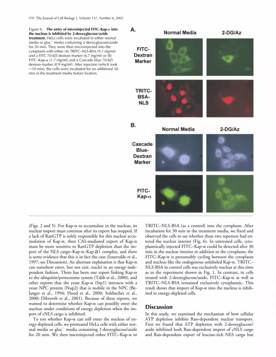
Entry of Kap-� into the nucleus is also inhibited by 2-deoxyglucose/azideWe had one final question concerning the nuclear accumu-lation of Kap-� seen upon 2-deoxyglucose/azide treatment
Figure 5. A time course of 2-deoxyglucose/azide treatment and recovery comparing the availability of RanGTP with the nuclear buildup of Kap-�. For each time point in A and B, the cellular localization of Kap-� as determined by indirect immunofluorescence microscopy (performed as in Fig. 2) was compared with the RanGTP from NEM-treated cellular extracts that bound to the RanBD (performed as in Fig. 4 C, lanes 2 and 3, arrow). (A) HeLa cells were incubated for the indicated time at 37C in gluc� media containing 2-deoxy-glucose/azide. (B) After treatment in 2-deoxyglucose/azide for 1 h, the cells were washed once in prewarmed gluc� media, and then transferred to prewarmed gluc� media and incubated at 37C for the indicated time. To emphasize the heterogeneity in Kap-� nuclear accumulation at longer time points, the same relatively short camera exposure time was used for all of these micrographs.
Table II. Measurement of free nucleotides in HeLa cells after various treatments
Free ATP Total ATP (from Table I) Free GTP Total GTP (from Table I)
Glucose� media, 1 h 4.48 � 0.27 8.22 � 0.75 0.93 � 0.22 2.58 � 0.232-deoxyglucose � sodium azide, 1 h �0.1a 3.27 � 0.60 �0.1a 1.85 � 0.29Ribavirin, 2 h 5.93 � 1.07 8.42 � 0.43 0.30 � 0.04 0.87 � 0.01
Values are expressed as nmoles nucleotide/106 cells. Free nucleotide was separated from the total cellular lysate by filtration as described in the Materialsand methods. Values for free ATP and GTP represent the mean � SD of three independent experiments; the values for total ATP and GTP were taken fromseparate experiments summarized in Table I.aBoth the free ATP and GTP from the cells treated with 2-deoxyglucose and sodium azide were essentially undetectable by FPLC, and thus below our low-est standards. The values given for these two measurements therefore indicate the amount of our lowest FPLC standards rather than the exact concentrationof nucleotide.
970 The Journal of Cell Biology | Volume 157, Number 6, 2002
(Figs. 2 and 5). For Kap-� to accumulate in the nucleus, itsnuclear import must continue after its export has stopped. Ifa lack of RanGTP is solely responsible for this nuclear accu-mulation of Kap-�, then CAS-mediated export of Kap-�must be more sensitive to RanGTP depletion than the im-port of the NLS cargo–Kap-�–Kap-�1 complex, and thereis some evidence that this is in fact the case (Izaurralde et al.,1997; see Discussion). An alternate explanation is that Kap-�can somehow enter, but not exit, nuclei in an energy-inde-pendent fashion. There has been one report linking Kap-�to the ubiquitin/proteosome system (Tabb et al., 2000), andother reports that the yeast Kap-� (Srp1) interacts with ayeast NPC protein (Nup2) that is mobile in the NPC (Be-langer et al., 1994; Hood et al., 2000; Solsbacher et al.,2000; Dilworth et al., 2001). Because of these reports, wewanted to determine whether Kap-� can possibly enter thenucleus under conditions of energy depletion when the im-port of cNLS cargo is inhibited.
To test whether Kap-� can still enter the nucleus of en-ergy-depleted cells, we pretreated HeLa cells with either nor-mal media or gluc� media containing 2-deoxyglucose/azidefor 20 min. We then microinjected either FITC–Kap-� or
TRITC–NLS-BSA (as a control) into the cytoplasm. Afterincubation for 30 min in the treatment media, we fixed andobserved the cells to see whether these two reporters had en-tered the nuclear interior (Fig. 6). In untreated cells, cyto-plasmically injected FITC–Kap-� could be detected after 30min in the nuclear interior in addition to the cytoplasm; theFITC–Kap-� is presumably cycling between the cytoplasmand nucleus like the endogenous unlabeled Kap-�. TRITC–NLS-BSA in control cells was exclusively nuclear at this timeas in the experiment shown in Fig. 1. In contrast, in cellstreated with 2-deoxyglucose/azide, FITC–Kap-� as well asTRITC–NLS-BSA remained exclusively cytoplasmic. Thisresult shows that import of Kap-� into the nucleus is inhib-ited in energy-depleted cells.
DiscussionIn this study, we examined the mechanism of how cellularATP depletion inhibits Ran-dependent nuclear transport.First we found that ATP depletion with 2-deoxyglucose/azide inhibited both Ran-dependent import of cNLS cargoand Ran-dependent export of leucine-rich NES cargo but
Figure 6. The entry of microinjected FITC–Kap-� into the nucleus is inhibited by 2-deoxyglucose/azide treatment. HeLa cells were incubated in either normal media or gluc� media containing 2-deoxyglucose/azide for 20 min. They were then microinjected into the cytoplasm with either (A) TRITC–NLS-BSA (9.1 mg/ml) and a FITC 70-kD dextran marker (6.7 mg/ml) or (B) FITC–Kap-� (1.7 mg/ml) and a Cascade blue 70-kD dextran marker (0.9 mg/ml). After injection (which took �10 min), the cells were incubated for an additional 30 min in the treatment media before fixation.

ATP depletion and nuclear transport | Schwoebel et al. 971
that GTP depletion with ribavirin inhibited neither (Fig.1). 2-deoxyglucose/azide (unlike ribavirin) treatment alsocaused an accumulation of Kap-� inside the nucleus (Fig.2). Measurement of the total nucleotide levels revealed thatafter 2-deoxyglucose/azide treatment, the total ATP contentdecreased to 40% of control, whereas the GTP content onlydecreased to 72% of control (Table I). In contrast, after ri-bavirin treatment, the total ATP concentration was un-changed (101% of control), but the total GTP content de-creased to 36% of control. Because ribavirin-treated cellsremain transport competent whereas 2-deoxyglucose/azide-treated cells do not, these nucleotide measurements did notsupport our original hypothesis that it is a drop in total GTPlevels upon ATP depletion that inhibits transport.
In further experiments, however, we found that 2-deoxy-glucose/azide treatment causes the amount of RanGTP avail-able for binding to a GST–RanBD construct to drop to un-detectable levels (Fig. 4). We further found that this decreasein RanGTP precedes the nuclear accumulation of Kap-�(Fig. 5). Finally, we found that 2-deoxyglucose/azide treat-ment resulted in the level of free (unlike total) GTP to dropto undetectable levels. Ribavirin treatment caused a decreasein free GTP to 32% of control (Table II), but this value issubstantially higher than the free GTP concentration in2-deoxyglucose/azide-treated cells. Treatment with ribavirinfor longer than 2 h was not tested, but because we did detecta decrease in free RanGTP after 2 h of ribavirin treatment(Fig. 4), it is possible that longer treatment times mighteventually cause the RanGTP concentration to drop to apoint where Ran-dependent nuclear transport is inhibited.
Because of these results, we believe it likely that the inhibi-tion of Ran-dependent nuclear transport seen with ATP de-pletion in fact results from a loss of free GTP needed to pro-duce RanGTP, rather than by an ATP-dependent step innuclear transport. Because we also found that free ATP alsodrops to undetectable levels upon 2-deoxyglucose/azidetreatment, it remains formally possible that free ATP (in ad-dition to free GTP) is also required for these transport path-ways in vivo. However, because all of the previously pub-lished in vitro data is completely consistent with an essentialrequirement for GTP to make RanGTP, and shows no de-tectable requirement for ATP or ADP, we favor the interpre-tation that it is the loss of free GTP rather than free ATPupon 2-deoxyglucose/azide treatment that is the criticalevent (Schwoebel et al., 1998; Englmeier et al., 1999).
In addition to the experiments shown, we did a num-ber of experiments to rule out other explanations of why2-deoxyglucose/azide inhibits Ran-dependent nuclear trans-port. First, we considered the possibility that sodium azidewas having a direct effect on transport rather than inhibit-ing it by lowering nucleotide levels. Azide inhibits oxidativephosphorylation by binding to cytochrome oxidase in themitochondria, however azide also binds and inhibits othermetal-containing proteins such as HRP (Holzwarth et al.,1988). We found, however, that the direct addition of so-dium azide at millimolar concentrations to digitonin-per-meabilized cells did not have the slightest effect on eitherthe in vitro import of TRITC–NLS-BSA or the in vitro ex-port of TRITC–NES-BSA (unpublished data). Further-more, we found that substitution of oligomycin, another
inhibitor of oxidative phosphorylation that works by a dif-ferent mechanism, gave identical results to sodium azide interms of Kap-� nuclear buildup (unpublished data). Wealso found that cytoplasmic microinjection of apyrase, anenzyme that cleaves any tri- or diphosphate nucleotide to itsmonophosphate derivative, resulted in identical nuclear ac-cumulation of Kap-� (unpublished data).
We considered the possibility that ATP depletion mightbe inhibiting some ATP-dependent posttranslational modi-fication of a protein involved in nuclear transport, such asan NPC protein or one of the transport factors. With re-gard to this, we found that neither the RanGEF or Ran-GAP activities in lysates from 2-deoxyglucose/azide-treatedcells were altered when compared with extracts from con-trol or ribavirin-treated cells (unpublished data). It is alsopossible that the export of large ribonucleoprotein com-plexes might require an additional energy source, unlikethe export of smaller substrates, to power an ATP-depen-dent helicase for example (Gatfield et al., 2001). We rea-soned that the absence of ATP might “jam” the NPC withpartially translocated RNPs, preventing the movement ofsmaller transport complexes. We found, however, that in-hibition of RNA synthesis with actinomycin D for severalhours before 2-deoxyglucose/azide treatment had no effecton the subsequent nuclear accumulation of Kap-� (unpub-lished data). We also found that with regard to potentialjamming of the NPC, 2-deoxyglucose/azide treatment didnot prevent the diffusion of a 25-kD protein through theNPC (unpublished data).
In summary, our results support the conclusion that inhi-bition of Ran-dependent transport by ATP depletion doesnot result from inhibition of an ATP-dependent step in thesetransport pathways. It should be noted that the results re-ported here do not rule out the possibility that other trans-port pathways, or the Kap-�1 and Crm1 pathways whentransporting much larger cargo than those tested here, mightrequire additional energy sources to facilitate movementacross the NPC. However, we believe the inhibition of thepathways studied here directly results from a lack of RanGTPand this lack is caused by a shortage of free GTP needed forthe loading of nuclear RanGDP. It is striking that the level offree GTP becomes undetectable after 2-deoxyglucose/azidetreatment when the pool of total GTP is still 72% of normal(Table II). One reason for this rapid loss of free GTP seenupon ATP depletion is probably the requirement for ATP inthe sequential phosphorylation of GMP to form GDP andGTP (Zalkin and Dixon, 1992). Thus, an absence of ATPshould completely inhibit the de novo synthesis of GTP. An-other key player likely to be involved in regulating relativeNTP levels is nucleoside diphosphokinase, which transfersthe terminal phosphate from any NTP to the terminus of anyNDP (Veron et al., 1994). This could have the effect of de-pleting GTP pools in parallel with ATP pools in cells under2-deoxyglucose/azide treatment. Contributing to the rapidloss of free GTP upon ATP depletion is probably the pres-ence of many cellular ATP-using proteins that can also useGTP, e.g., hexokinase (Ferguson et al., 1986). Under normalconditions, these proteins use the most available NTP, whichis ATP, but in the absence of free ATP, they can use GTP in-stead, which would rapidly deplete this pool as well.

972 The Journal of Cell Biology | Volume 157, Number 6, 2002
If the nuclear accumulation of Kap-� upon energy deple-tion is caused by a lack of RanGTP, this would have to meanthat its CAS-mediated export is more sensitive to RanGTPdepletion than its Kap-�1–mediated import. Evidence thatKap-� export is in fact more sensitive to RanGTP depletionthan cNLS import was shown by microinjecting radiolabeledproteins or RNAs into Xenopus oocytes followed by hand dis-section and observing their transport under conditions wherenuclear RanGTP is depleted (Izaurralde et al., 1997). Thisstudy found that the nuclear export of Kap-� is almost to-tally inhibited after nuclear injection of 10 M RanGAP(Rna1), but that the import of a cNLS-containing cargo(CBP80) is only slightly reduced by this GAP concentrationand requires the nuclear injection of 40 M RanGAP for to-tal inhibition. Thus, these findings support our conclusionthat it is the loss of RanGTP that inhibits Ran-dependentnuclear import and export upon ATP depletion rather thanthe inhibition of some ATP-dependent event.
Materials and methodsMaterialsRibavirin was from Sigma-Aldrich. The following antibodies were used:anti-Ran monoclonal antibody from Transduction Laboratories, anti-RanGAP monoclonal antibody 19C7 ascites fluid (Matunis et al., 1996) do-nated by M. Matunis (Johns Hopkins University, Baltimore, MD), anti–Kap-�(pendulin) antisera donated by M. Waterman (University of California,Irvine, CA), anti–Kap-�1 monoclonal antibody (Chi et al., 1995) donatedby S. Adams (Northwestern University Medical School, Chicago, IL), andanti-RCC1 rabbit antibody (Schwoebel et al., 1998). Rhodamine-conju-gated secondary antibodies against mouse and rabbit IgG were fromJackson ImmunoResearch Laboratories. TRITC–NLS-BSA was prepared aspreviously described (Moore and Blobel, 1992). TRITC–NES-BSA was pre-pared in the same manner, substituting peptides (CLPPLERLTL) containingthe Rev NES (in bold) (Fischer et al., 1995; Wen et al., 1995). Fluorescein-and Cascade blue–labeled 70-kD dextrans were from Molecular Probes.FITC–Kap-� was prepared by coupling His–Kap-�2 (Schwoebel et al.,1998) with a 1:2 molar ratio of fluorescein–5-isothiocyanate (MolecularProbes) according to the manufacturer’s instructions. After coupling, FITC–Kap-� was passed over a Nap-5 column equilibrated in 20 mM Hepes-KOH, pH 7.3, 100 mM potassium acetate, 2 mM DTT, snap frozen, andstored at �80C in single use aliquots. We found that labeling did not af-fect the ability of Kap-� to support cNLS-mediated import in permeabi-lized cells (unpublished data).
Cell culture and treatmentsFor energy depletion studies, HeLa cells were washed with PBS, and incu-bated in glucose-free DME (catalog no. 11966–025; GIBCO BRL) contain-ing penicillin (100 U/ml), streptomycin sulfate (100 g/ml), Hepes (10mM, pH 7.3), and 10% FCS (hereafter referred to as gluc� media). Whereindicated, cells were incubated in gluc� media containing either 10 mMsodium azide and 6 mM 2-deoxy-D-glucose, or 100 M ribavirin for thetimes indicated in the figure legends. For microinjection, coverslips wererinsed once in PBS, once in gluc� media, and incubated with gluc� mediacontaining 10 mM sodium azide and 6 mM 2-deoxyglucose for 20 min orgluc� media containing 100 m ribavirin for 90 min at 37C before micro-injection. Microinjection was for �10 min at room temperature (RT) intreatment media, and microinjected cells were then incubated for an addi-tional 30 min at 37C before fixation.
Immunofluorescence microscopyCells after treatment were placed on ice, washed with ice cold PBS, fixedfor 20 min on ice in 3% formaldehyde (diluted from a methanol-free stock)in PBS, rinsed twice with PBS, and permeabilized with 0.1% Triton in PBSfor 20 min on ice. Cells were blocked with 10% donkey serum in PBS for30 min at 37C, washed two times with PBS, and incubated with primaryantibody diluted in blocking serum for 1 h at RT. After washing, cells wereincubated with a labeled second antibody in blocking solution for 1 h atRT. After washing, coverslips were mounted and observed as previouslydescribed (Schwoebel et al., 1998).
Nucleotide isolation and quantitationTotal nucleotide. After treatment, cells were washed with PBS, trypsinized,and the cells were resuspended in ice cold PBS and centrifuged for 5 minat 200 g, 4C. Cells were resuspended in cold PBS, counted, centrifugedfor 5 min at 200 g, 4C, and resuspended in 0.7 M HClO4 (100 l/106
cells). Cells were vortexed for 30 s, centrifuged at 3,000 g for 20 min at4C, and the supernatants were neutralized with solid KHCO3 (10 mg/106
cells) (Kremmer et al., 1989). The precipitates were removed by microcen-trifugation twice for 10 min at 4C each time, and the supernatants werealiquoted and snap frozen in liquid N2. Aliquots of these supernatants(each representing 6.5 � 105 cells) were used for the measurement of nu-cleotides. Nucleotides were separated by FPLC and individual nucleotidelevels were quantitated by measurement of peak heights (Abs UV254) andcomparison to standard curves. GTP and ATP were separated on a Mono-Qcolumn with a gradient of 2–55% B (A � H2O; B � 1 M ammonium phos-phate, pH 7.0; Kremmer et al., 1989). With this protocol, ATP elutes at48.5% B, and GTP at 52% B. GDP and ADP were separated on a Source15Q column with a gradient of 0–60% B (A � 7 mM KH2PO4, pH 4.5; B �500 mM KH2PO4, 100 mM KCl, pH 5.0; Hartwick and Brown, 1975). Withthis protocol, GDP elutes at 23.5% B, and ADP elutes at 26% B.
Free nucleotide. After treatment, cells were washed with cold PBS andscraped in cold buffer A (20 mM Hepes, pH 7.3, 120 mM NaCl, 2 mMmagnesium acetate, 1 mM DTT). The scrapes from three 100-mm culturedishes were pooled and sonicated. An aliquot of the lysate was snap fro-zen in liquid nitrogen and saved for Bradford analysis. The cell numberwas calculated from the protein values compared with the previously de-termined protein content of a known number of HeLa cells. The remaininglysate was centrifuged in a Microcon centrifugal filter device with a 10,000mol wt cutoff (Millipore) at 12,000 g for 10 min at 4C. The resulting fil-trate was snap frozen in liquid nitrogen. The GTP and ATP content of thefiltrate was measured by FPLC as described above.
NEM treatment and measurement of free RanGTPPreparation of extracts and initial NEM experiments. All steps including in-cubation with NEM were performed on ice in a cold room using ice coldsolutions unless otherwise stated. HeLa cells were washed once with PBS,and then the cells were scraped using a rubber policeman into buffer Acontaining 0.1% Triton X-100. The lysate was vortexed for 15 s and NEM atdifferent concentrations was added to aliquots of the lysate. Aliquots wereremoved at different time points and a twofold molar excess of DTT overthe NEM in that particular sample was added to quench the NEM activity.
RanGAP and RanGEF assays. Before assay, each NEM-treated lysate(quenched with DTT) to be assayed for GAP or GEF activity was gel filteredon a Nap-5 column equilibrated in 20 mM Hepes, pH 7.3, 120 mM NaCl,5 mM magnesium acetate, 0.05% Tween, and 5 mM DTT. Human recom-binant untagged Ran (3 g) was loaded with radiolabeled nucleotide for2 h on ice in 20 mM Hepes, pH 7.3, 120 mM potassium acetate, 1 mMDTT, 10 mM EDTA, 1 mg/ml BSA, and either 5.5 pmol of 9,000 Ci/mmol[�-32P]GTP (NEN Life Science Products) or 3.3 pmol of [�-32P]GDP pre-pared as previously described (Schwoebel et al., 1998). The loading reac-tion was quenched by the addition of MgCl2 to 40 mM. The loaded Ranwas microfuged for 10 min at 4C and exchanged into buffer B (20 mMHepes, pH 7.3, 120 mM potassium acetate, 5 mm MgCl2, 1 mM DTT) bypassage over a NAP-5 desalting column. For the GAP assay, 40,000 CPMof GTP loaded Ran was added to each sample, vortexed, and placed at30C. The reaction was stopped after 15 min by the addition of 5 ml icecold buffer B. The sample was vacuum filtered onto 0.45-m nitrocellu-lose filters (Schleicher & Schuell), and the filters were washed with 12 mlcold buffer B. The filters were air dried, placed in scintillation fluid, andcounted. The GEF assay was performed in the same manner using 20,000CPM of [�-32P] GDP-loaded Ran per sample.
RanBD1 pulldowns of Ran[�-32P]GTP and Ran[�-32P]GTP. Human recom-binant untagged Ran (3 g) was loaded with radiolabeled nucleotide for2 h on ice in 20 mM Hepes, pH 7.3, 120 mM potassium acetate, 1 mMDTT, 10 mM EDTA, 1 mg/ml BSA, and either 34 pmol of 9,000 Ci/mmol[�-32P]GTP or 38.8 pmol of 3,000 Ci/mmol [�-32P]GTP (both from NENLife Science Products). The loading reaction was quenched by the additionof MgCl2 to 40 mM, and the loaded Ran was exchanged into buffer B by pas-sage over a NAP-5 column. HeLa cells grown to 70% confluence in 100-mM dishes were placed on ice in the cold room, washed in PBS, drained,and scraped in buffer B containing 0.1% Tween-20, 2.8 mM GDP-�S, and140 mM NEM (except mock-treated samples, which contained 280 mMDTT). Aliquots of the radiolabeled Ran were added immediately to thesecell lysates. In some samples, BSA (10 mg/ml in buffer B) was used in placeof cellular extract. After dilution, the final concentrations were 100 mMNEM and 2 mM nucleotide. The samples were incubated on ice for 20min, vortexed, and centrifuged at 13,000 g for 10 min at 4C. The superna-
ATP depletion and nuclear transport | Schwoebel et al. 973
tants were brought to 200 mM DTT (from a 2 M stock) and applied to aNAP-5 column equilibrated in buffer A. GST–RanBD or GST was added tothe NAP-5 eluates and the samples were rotated end over end for 30 minat 4C. Glutathione-agarose beads were added and the incubation contin-ued for an additional 30 min at 4C. Approximately 7 g of GST–RanBD orGST and 40 l of a 20% slurry of glutathione-agarose beads were addedper milligram of cellular extract. After incubation, the beads were sedi-mented by centrifugation at 1,000 g for 3 min at 4C. The supernatantswere discarded and the pellets washed four times in buffer A. Beads wereresuspended in SDS-PAGE sample buffer and aliquots of the resulting su-pernatant were placed in scintillation fluid and counted.
RanBD1 pulldowns of RanGTP from treated cells. After receiving the indi-cated treatment, HeLa cells grown to 70% confluence in 100-mM disheswere placed on ice in the cold room, washed in PBS, drained, and scrapedin buffer B containing 0.1% Tween-20, 2.8 mM nucleotide (GDP-�S ex-cept where indicated), and 140 mM NEM (except mock-treated samples,which contained 280 mM DTT). After dilution, the final concentrationswere 100 mM NEM and 2 mM nucleotide. After incubation for 20 min onice, the samples were quenched with DTT and processed for isolation offree RanGTP with the GST–RanBD and glutathione-agarose as describedabove. Beads were resuspended in SDS-PAGE sample buffer, and afterSDS-PAGE, the samples were transferred to nitrocellulose and immuno-blotted with an anti-Ran antibody.
We thank Marian Waterman, Mike Matunis, and Steve Adam for supplyingus with antibodies, Ian Macara (University of Virginia, Charlottesville, VA)for the GST–Ran BP plasmid, and Bob Schwartz (Baylor College of Medi-cine) for the use of his microinjector. We also thank Ian Cushman (BaylorCollege of Medicine) for the TRITC–NES-BSA conjugate and Joe Bryan(Baylor College of Medicine), Ted Wensel (Baylor College of Medicine),and Steen Pederson (Baylor College of Medicine) for helpful discussionsabout energy metabolism.
This work was supported by National Institutes of Health grant RO1GM53678 (M.S. Moore), a National Institute of Diabetes and Digestive andKidney Diseases Molecular Endocrinology training grant NIH DK07696(E.D. Schwoebel), and the Robert and Janice McNair Foundation (T.H. Ho).
Submitted: 21 November 2001Revised: 29 April 2002Accepted: 1 May 2002
ReferencesBeddow, A.L., S.A. Richards, N.R. Orem, and I.G. Macara. 1995. The Ran/TC4
GTPase-binding domain: identification by expression cloning and characteriza-tion of a conserved sequence motif. Proc. Natl. Acad. Sci. USA. 92:3328–3332.
Belanger, K.D., M.A. Kenna, S. Wei, and L.I. Davis. 1994. Genetic and physicalinteractions between Srp1p and nuclear pore complex proteins Nup1p andNup2p. J. Cell Biol. 126:619–630.
Chi, N.C., E.J. Adam, and S.A. Adam. 1995. Sequence and characterization of cy-toplasmic nuclear protein import factor p97. J. Cell Biol. 130:265–274.
Detimary, P., C. Xiao, and J.C. Henquin. 1997. Tight links between adenine andguanine nucleotide pools in mouse pancreatic islets: a study with mycophe-nolic acid. Biochem. J. 324:467–471.
Dilworth, D.J., A. Suprapto, J.C. Padovan, B.T. Chait, R.W. Wozniak, M.P.Rout, and J.D. Aitchison. 2001. Nup2p dynamically associates with the dis-tal regions of the yeast nuclear pore complex. J. Cell Biol. 153:1465–1478.
Englmeier, L., J.C. Olivo, and I.W. Mattaj. 1999. Receptor-mediated substratetranslocation through the nuclear pore complex without nucleotide triphos-phate hydrolysis. Curr. Biol. 9:30–41.
Ferguson, K.M., T. Higashijima, M.D. Smigel, and A.G. Gilman. 1986. The in-fluence of bound GDP on the kinetics of guanine nucleotide binding to Gproteins. J. Biol. Chem. 261:7393–7399.
Finch, R.A., G.R. Revankar, and P.K. Chan. 1993. Nucleolar localization of nu-cleophosmin/B23 requires GTP. J. Biol. Chem. 268:5823–5827.
Fischer, U., J. Huber, W.C. Boelens, I.W. Mattaj, and R. Luhrmann. 1995. TheHIV-1 Rev activation domain is a nuclear export signal that accesses an ex-port pathway used by specific cellular RNAs. Cell. 82:475–483.
Franklin, T.J., and P.A. Twose. 1977. Reduction in beta-adrenergic response ofcultured glioma cells following depletion of intracellular GTP. Eur. J. Bio-chem. 77:113–117.
Gatfield, D., H. Le Hir, C. Schmitt, I.C. Braun, T. Kocher, M. Wilm, and E.Izaurralde. 2001. The DExH/D box protein HEL/UAP56 is essential formRNA nuclear export in Drosophila. Curr. Biol. 11:1716–1721.
Gorlich, D., F. Vogel, A.D. Mills, E. Hartmann, and R.A. Laskey. 1995. Distinctfunctions for the two importin subunits in nuclear protein import. Nature.377:246–248.
Gorlich, D., N. Pante, U. Kutay, U. Aebi, and F.R. Bischoff. 1996. Identificationof different roles for RanGDP and RanGTP in nuclear protein import.EMBO J. 15:5584–5594.
Grune, T., and W.G. Siems. 1993. Reversed-phase high-performance liquid chro-matography of purine compounds for investigation of biomedical problems:application to different tissues and body fluids. J. Chromatogr. 618:15–40.
Hartwick, R.A., and P.R. Brown. 1975. The performance of microparticle chemi-cally-bonded anion-exchange resins in the analysis of nucleotides. J. Chro-matogr. 112:650–662.
Hershfield, M.S., and J.E. Seegmiller. 1976. Regulation of de novo purine biosynthe-sis in human lymphoblasts. Coordinate control of proximal (rate-determining)steps and the inosinic acid branch point. J. Biol. Chem. 251:7348–7354.
Holzwarth, J.F., F. Meyer, M. Pickard, and H.B. Dunford. 1988. Mechanism ofazide binding to chloroperoxidase and horseradish peroxidase: use of an io-dine laser temperature-jump apparatus. Biochemistry. 27:6628–6633.
Hood, J.K., J.M. Casolari, and P.A. Silver. 2000. Nup2p is located on the nuclearside of the nuclear pore complex and coordinates Srp1p/importin-� export.J. Cell Sci. 113:1471–1480.
Izaurralde, E., U. Kutay, C. von Kobbe, I.W. Mattaj, and D. Gorlich. 1997. Theasymmetric distribution of the constituents of the Ran system is essential fortransport into and out of the nucleus. EMBO J. 16:6535–6547.
Klebe, C., H. Prinz, A. Wittinghofer, and R.S. Goody. 1995. The kinetic mecha-nism of Ran—nucleotide exchange catalyzed by RCC1. Biochemistry. 34:12543–12552.
Koepp, D.M., D.H. Wong, A.H. Corbett, and P.A. Silver. 1996. Dynamic local-ization of the nuclear import receptor and its interactions with transport fac-tors. J. Cell Biol. 133:1163–1176.
Kondo, M., T. Yamaoka, S. Honda, Y. Miwa, R. Katashima, M. Moritani, K.Yoshimoto, Y. Hayashi, and M. Itakura. 2000. The rate of cell growth is reg-ulated by purine biosynthesis via ATP production and G(1) to S phase tran-sition. J. Biochem. (Tokyo). 128:57–64.
Kose, S., N. Imamoto, T. Tachibana, T. Shimamoto, and Y. Yoneda. 1997. Ran-unassisted nuclear migration of a 97-kD component of nuclear pore–target-ing complex. J. Cell Biol. 139:841–849.
Kremmer, T., E. Paulik, M. Boldizsar, and I. Holenyi. 1989. Application of thefast protein liquid chromatographic system and MonoQ HR 5/5 anion ex-changer to the separation of nucleotides. J. Chromatogr. 493:45–52.
Kutay, U., F.R. Bischoff, S. Kostka, R. Kraft, and D. Gorlich. 1997a. Export ofimportin � from the nucleus is mediated by a specific nuclear transport fac-tor. Cell. 90:1061–1071.
Kutay, U., E. Izaurralde, F.R. Bischoff, I.W. Mattaj, and D. Gorlich. 1997b. Domi-nant-negative mutants of importin-� block multiple pathways of import andexport through the nuclear pore complex. EMBO J. 16:1153–1163.
Lee, H.J., K. Pawlak, B.T. Nguyen, R.K. Robins, and W. Sadee. 1985. Biochemi-cal differences among four inosinate dehydrogenase inhibitors, mycophe-nolic acid, ribavirin, tiazofurin, and selenazofurin, studied in mouse lym-phoma cell culture. Cancer Res. 45:5512–5520.
Lounsbury, K.M., S.A. Richards, K.L. Carey, and I.G. Macara. 1996. Mutationswithin the Ran/TC4 GTPase. Effects on regulatory factor interactions andsubcellular localization. J. Biol. Chem. 271:32834–32841.
Matunis, M.J., E. Coutavas, and G. Blobel. 1996. A novel ubiquitin-like modifica-tion modulates the partitioning of the Ran-GTPase–activating proteinRanGAP1 between the cytosol and the nuclear pore complex. J. Cell Biol.135:1457–1470.
Melchior, F., B. Paschal, J. Evans, and L. Gerace. 1993. Inhibition of nuclear pro-tein import by nonhydrolyzable analogues of GTP and identification of thesmall GTPase Ran/TC4 as an essential transport factor. J. Cell Biol. 123:1649–1659.
Moore, M.S. 1998. Ran and nuclear transport. J. Biol. Chem. 273:22857–22860.Moore, M.S., and G. Blobel. 1992. The two steps of nuclear import, targeting to
the nuclear envelope and translocation through the nuclear pore, require dif-ferent cytosolic factors. Cell. 69:939–950.
Moore, M.S., and G. Blobel. 1993. The GTP-binding protein Ran/TC4 is re-quired for protein import into the nucleus. Nature. 365:661–663.
Nakielny, S., and G. Dreyfuss. 1999. Transport of proteins and RNAs in and outof the nucleus. Cell. 99:677–690.
Ribbeck, K., and D. Gorlich. 2001. Kinetic analysis of translocation through nu-clear pore complexes. EMBO J. 20:1320–1330.
Richardson, W.D., A.D. Mills, S.M. Dilworth, R.A. Laskey, and C. Dingwall.1988. Nuclear protein migration involves two steps: rapid binding at the nu-
974 The Journal of Cell Biology | Volume 157, Number 6, 2002
clear envelope followed by slower translocation through nuclear pores. Cell.52:655–664.
Rout, M.P., J.D. Aitchison, A. Suprapto, K. Hjertaas, Y. Zhao, and B.T. Chait.2000. The yeast nuclear pore complex: composition, architecture, and trans-port mechanism. J. Cell Biol. 148:635–651.
Schwoebel, E.D., B. Talcott, I. Cushman, and M.S. Moore. 1998. Ran-dependentsignal-mediated nuclear import does not require GTP hydrolysis by Ran. J.Biol. Chem. 273:35170–35175.
Shulga, N., P. Roberts, Z. Gu, L. Spitz, M.M. Tabb, M. Nomura, and D.S. Gold-farb. 1996. In vivo nuclear transport kinetics in Saccharomyces cerevisiae: arole for heat shock protein 70 during targeting and translocation. J. Cell Biol.135:329–339.
Solsbacher, J., P. Maurer, F. Vogel, and G. Schlenstedt. 2000. Nup2p, a yeast nu-cleoporin, functions in bidirectional transport of importin alpha. Mol. Cell.Biol. 20:8468–8479.
Tabb, M.M., P. Tongaonkar, L. Vu, and M. Nomura. 2000. Evidence for separablefunctions of Srp1p, the yeast homolog of importin � (Karyopherin �): role forSrp1p and Sts1p in protein degradation. Mol. Cell. Biol. 20:6062–6073.
Tachibana, T., M. Hieda, Y. Miyamoto, S. Kose, N. Imamoto, and Y. Yoneda.2000. Recycling of importin � from the nucleus is suppressed by loss ofRCC1 function in living mammalian cells. Cell Struct. Funct. 25:115–123.
Talcott, B., and M.S. Moore. 1999. Getting across the nuclear pore complex.Trends Cell Biol. 9:312–318.
Veron, M., A. Tepper, M. Hildebrandt, I. Lascu, M.L. Lacombe, J. Janin, S. Morera,J. Cherfils, C. Dumas, and M. Chiadmi. 1994. Nucleoside diphosphate ki-nase: an old enzyme with new functions? Adv. Exp. Med. Biol. 370:607–611.
Wen, W., J.L. Meinkoth, R.Y. Tsien, and S.S. Taylor. 1995. Identification of a sig-nal for rapid export of proteins from the nucleus. Cell. 82:463–473.
Yalowitz, J.A., and H.N. Jayaram. 2000. Molecular targets of guanine nucleotides indifferentiation, proliferation and apoptosis. Anticancer Res. 20:2329–2338.
Yokoya, F., N. Imamoto, T. Tachibana, and Y. Yoneda. 1999. �-Catenin can betransported into the nucleus in a Ran-unassisted manner. Mol. Biol. Cell. 10:1119–1131.
Zalkin, H., and J.E. Dixon. 1992. De novo purine nucleotide biosynthesis. Prog.Nucleic Acid Res. Mol. Biol. 42:259–287.