the intronic gabrg2 mutation, ivs6+2t->g, associated with childhood absence epilepsy altered...
TRANSCRIPT
Neurobiology of Disease
The Intronic GABRG2 Mutation, IVS6�2T3G, Associatedwith Childhood Absence Epilepsy Altered Subunit mRNAIntron Splicing, Activated Nonsense-Mediated Decay, andProduced a Stable Truncated �2 Subunit
Mengnan Tian ( )1,2 and Robert L. Macdonald1,2,3
Departments of 1Neurology, 2Pharmacology, and 3Molecular Physiology and Biophysics, Vanderbilt University Medical Center, Nashville, Tennessee 37212
The intronic GABRG2 mutation, IVS6�2T3G, was identified in an Australian family with childhood absence epilepsy and febrileseizures (Kananura et al., 2002). The GABRG2 intron 6 splice donor site was found to be mutated from GT to GG. We generated wild-typeand mutant �2 subunit bacterial artificial chromosomes (BACs) driven by a CMV promoter and expressed them in HEK293T cells andexpressed wild-type and mutant �2 subunit BACs containing the endogenous hGABRG2 promoter in transgenic mice. Wild-type andmutant GABRG2 mRNA splicing patterns were determined in both BAC-transfected HEK293T cells and transgenic mouse brain, and inboth, the mutation abolished intron 6 splicing at the donor site, activated a cryptic splice site, generated partial intron 6 retention, andproduced a frameshift in exon 7 that created a premature translation termination codon (PTC). The resultant mutant mRNA was eitherdegraded partially by nonsense-mediated mRNA decay or translated to a stable, truncated subunit (the �2-PTC subunit) containing thefirst six GABRG2 exons and a novel frameshifted 29 aa C-terminal tail. The �2-PTC subunit was homologous to the mollusk AChBP(acetylcholine binding protein) but was not secreted from cells. It was retained in the ER and not expressed on the surface membrane, butit did oligomerize with �1 and �2 subunits. These results suggested that the GABRG2 mutation, IVS6�2T3G, reduced surface ���2receptor levels, thus reducing GABAergic inhibition, by reducing GABRG2 transcript level and producing a stable, nonfunctional trun-cated subunit that had a dominant-negative effect on ���2 receptor assembly.
IntroductionEpilepsy is one of the most common neurological disorders, af-fecting up to 3% of the general population. One-third to one-halfof all epilepsy syndromes have a genetic basis (Berkovic et al.,2006), and patients with genetic epilepsy syndromes have ab-sence, myoclonic, and/or generalized tonic-clonic seizures(Beghi et al., 2006). Most mutations associated with geneticepilepsies have been identified in genes encoding voltage- orligand-gated ion channels (Noebels, 2003).
GABAA receptors mediate the majority of inhibitory neu-rotransmission in the CNS. Epilepsy mutations have been iden-
tified in GABAA receptor subunit genes (GABRs) GABRA1,GABRB3, and GABRG2 (Kang and Macdonald, 2009), but mostof the mutations are in GABRG2. The �1, �2, and �2 subunitsform the most abundant GABAA receptor subtype in the CNS(Sieghart and Sperk, 2002; Whiting, 2003; Farrant and Nusser,2005), and the �2 subunit plays a critical role in brain func-tion. In mouse brain, �75– 80% of GABAA receptors containthe �2 subunit (Olsen and Sieghart, 2008). Mice lacking �2 sub-units (�2�/� mice) died shortly after birth (Gunther et al., 1995).These �2�/� mice lost 94% of their benzodiazepine binding sites,but GABA binding sites were only decreased slightly. The �2subunit is required for maintaining postsynaptic GABAA recep-tor clustering (Essrich et al., 1998). Heterozygous �2�/� micehad significantly decreased benzodiazepine binding sites and in-creased extrasynaptic GABAA receptor radioligand binding sitesin the CNS, but unchanged muscimol binding sites (Sinkkonen etal., 2004), and these animals had decreased GABAA receptor clus-tering in hippocampus and cerebral cortex (Crestani et al., 1999).The �2�/� mice had increased anxiety (Crestani et al., 1999), abehavior recapitulated in �2 subunit knockdown mice (Chandraet al., 2005). Epilepsy, however, has not been reported in �2�/�
or �2�/� mice.GABR mutations have been associated with seizures ranging
from relatively benign absence and/or febrile seizures to severemyoclonic seizures (Macdonald et al., 2006). The most well char-acterized �2 subunit missense mutation is GABRG2(R82Q) asso-
Received Oct. 20, 2011; revised Feb. 24, 2012; accepted March 13, 2012.Author contributions: M.T. and R.L.M. designed research; M.T. performed research; M.T. contributed unpublished
reagents/analytic tools; M.T. analyzed data; M.T. and R.L.M. wrote the paper.This work was supported by NIH Grant R01 NS051590 (R.L.M.) and an Epilepsy Foundation predoctoral research training
fellowship(M.T.).ConfocalexperimentswereperformedinpartusingtheVanderbiltUniversityMedicalCenterCell ImagingShared Resource (supported by NIH Grants CA68485, DK20593, DK58404, HD15052, DK59637, and EY08126). Flow Cytom-etry experiments were performed in the Vanderbilt Medical Center Flow Cytometry Shared Resource, which is supported bythe Vanderbilt Ingram Cancer Center (NIH Grant P30 CA68485) and the Vanderbilt Digestive Disease Research Center (NIHGrant DK058404). We thank Xuan Huang, Wangzhen Shen, Ningning Hu, and Kelienne Verdier for technical assistance, Drs.Feng Huajun and Andre Lagrange for instructions in electrophysiology, Dr. Douglas Mortlock for sharing BAC recombineer-ing protocols, Cara Sutcliffe and Ping Mayo for instructions in TaqMan quantitative PCR, Sean Schaffer for help with confocalmicroscopy experiments, and Dr. Ron Emeson for helpful discussions and suggestions.
Correspondence should be addressed to Dr. Robert L. Macdonald, Vanderbilt University Medical Center, 6140 MedicalResearch Building III, 465 21st Avenue South, Nashville, TN 37232-8552. E-mail: [email protected].
DOI:10.1523/JNEUROSCI.5332-11.2012Copyright © 2012 the authors 0270-6474/12/325937-16$15.00/0
The Journal of Neuroscience, April 25, 2012 • 32(17):5937–5952 • 5937
ciated with childhood absence epilepsy and febrile seizures(Wallace et al., 2001). This mutation impaired �1�2�2 receptorassembly, retained mutant �2 subunits in the endoplasmic retic-ulum, and reduced receptor surface trafficking (Bianchi et al.,2002; Hales et al., 2005; Eugene et al., 2007). Knock-in mice har-boring the GABRG2(R82Q) mutation had reduced cell surface �2subunit expression and reduced cortical inhibition, even inheterozygous animals (Tan et al., 2007). Mice heterozygous forthe mutation also had absence seizures.
GABRG2(IVS6�2T3G) is a mutation of the intron 6 splice do-nor site from GT to GG identified in an Australian family with child-hood absence epilepsy and febrile seizures (Kananura et al., 2002).The basis for the epilepsy in this family results from the specificalteration in splicing of GABRG2(IVS6�2T3G) mRNA and onsubsequent translation of protein. To determine this splicing pat-tern, we generated wild-type and mutant GABRG2(IVS6�2T3G)bacterial artificial chromosomes (BACs) and determined how theIVS6�2T3G mutation altered intron 6 splicing and �2 subunitexpression in HEK293T cells and transgenic mouse brain. We thencharacterized the biogenesis and function of the translated mutant�2 subunit.
Materials and MethodsExpression vectors with GABAA receptor subunits. The coding sequences ofhuman �1, �2, �2S, and �2L GABAA receptor subunits from the trans-lation initiation codon ATG to the stop codon were cloned intopcDNA3.1 expression vectors (Invitrogen) or pLVX-IRES-ZsGreen1vectors (Clontech) as previously described (Gallagher et al., 2005). ThecDNA encoding the HA peptide, YPYDVPDYA, was introduced betweenthe fourth and fifth amino acids of mature �2S and �2L subunits, whichhas been reported to be a functionally silent position (Connolly et al.,1996). In recent studies, the position of the mutant and variant aminoacids in �1, �3, and � subunits have been specified in the immaturepeptide that includes the signal peptide, but mutations in �2 subunithave been reported in the mature peptide, excluding the signal peptide.For consistency, in this study, the positions of �2 subunit mutations weredesignated also in the immature peptide.
The BAC clone number RP11-1035I20 (BACPAC Resources; http://bacpac.chori.org) contains a human chromosome 5 fragment that in-cluded the wild-type human GABRG2 gene genomic sequence (and thusa complete intron 6) and 20 kb upstream and 40 kb downstream humanchromosome 5 sequences (see Fig. 1 A). The BAC sequence was con-firmed by restriction enzyme digestion and direct DNA sequencing. TheBAC clone was recombined with the pEHHG vector (Wade-Martins etal., 2001), which contained the eGFP reporter gene driven by the HSVearly gene promoter. In target cells expressing these BAC vectors, eGFPfluorescence was detected. In this BAC clone, hGABRG2 was predicted tobe driven by the promoter sequence in the 20 kb upstream human chro-mosome sequence, while the eGFP was driven by a separate HSV pro-moter, and thus, expression of eGFP was independent of expression ofhGABRG2. To introduce the point mutation in hGABRG2 at the IVS6�2position, we used galK-facilitated recombineering (Warming et al., 2005;Chandler et al., 2007). The galK gene encodes galactose kinase and pro-vides both positive and negative selection factors in this technique. UsinggalK-facilitated BAC recombineering, the human chromosome sequenceupstream of the GABRG2 translation initiation sequence ATG was re-placed with a CMV promoter (see Fig. 1 A). Unless otherwise specified,wt and mutant hGABRG2 BACs were driven by the CMV promoter andcontained the eGFP reporter gene. The oligo sequences for BAC recom-bineering are available upon request.
Cell culture, transfection, and RNAi. Human embryonic kidney cells(HEK293T) (ATCC; CRL-11268) and HeLa cells (ATCC; CCL-2) wereincubated at 37°C in humidified 5% CO2, 95% air and grown in DMEM(Invitrogen) supplemented with 10% fetal bovine serum, 100 IU/ml pen-icillin, and 100 �g/ml streptomycin (Invitrogen). Cells were transfectedwith cDNAs using the FuGENE 6 transfection reagent (Roche AppliedScience) or Lipofectamine 2000 (Invitrogen) at a DNA/transfection re-
agent ratio of 1:3 according to the manufacturer’s instructions. Thetransfected cells were harvested after 36 h in culture for the followingexperimental protocols.
Sprague Dawley rat cortex was dissected from E18 embryos and disso-ciated using 0.25% trypsin and mild trituration (Banker and Goslin,1998). Neurons were plated on poly-L-ornithine-coated coverslips inDMEM (Invitrogen) supplemented with 10% horse serum, 2 mM glu-tamine, and 1 mM Na-pyruvate. After 4 h, medium was replaced by 1 mlof serum-free culture medium containing Neurobasal with B27 supple-ment, glutamine (2 mM), and penicillin/streptomycin. Cultures weremaintained at 36°C in a humidified CO2 incubator for up to 4 weeks andfed once a week. Cultured neurons were transfected with Lipofectamine2000 (Invitrogen) at 7 DIV according to the manufacturer’s instructions.A mixture of 1 �g of DNA and 3 �l of Lipofectamine in 60 �l of Opti-MEM (Invitrogen) was added to the well. One hour after incubation, theculture medium containing the Lipofectamine/DNA complex was com-pletely replaced with fresh serum-free Neurobasal/B27 culture medium.Neurons were immunostained 7 d after transfection.
Nonsense-mediated mRNA decay (NMD) efficiency was decreased byknocking down the essential factor UPF1. Silencer select predesigned andvalidated siRNA (Ambion; siRNA ID s11926) was transfected to cellsusing Lipofectamine RNAiMax (Invitrogen) according to the manufac-turer’s manual. Twenty-four hours later, the same cells were transfectedagain with the wild-type or mutant BAC constructs and harvested 2 dlater for RT-PCR. The efficiency of UPF1 knockdown was confirmed byWestern blot.
RNA extraction, RT-PCR, and TaqMan real-time qPCR. Total RNAswere extracted from transfected HEK293T cells by using the PerfectPureRNA Cultured Cell kit (5 PRIME) following the manufacturer’s proto-col, total RNA in mouse brain tissue was expressed by TRIzol reagent(Invitrogen) and PureLink RNA mini kit (Invitrogen) according to man-ufacturer’s manual, and human total brain RNA was obtained from Am-bion. Two hundred nanograms of total RNA of each sample was reversetranscribed to cDNA in a 10 �l volume using the TaqMan MicroRNAReverse Transcription Kit (Applied Biosystems). The transcribed cDNAwas used as a template to perform regular PCR using Expand High Fi-delity PCR Kit (Roche Applied Science) following the manufacturer’smanual. One microliter of the 50� diluted transcribed cDNA was mixedwith TaqMan Universal PCR Master Mix (Applied Biosystems) and Taq-Man probes in a total volume of 5 �l for the TaqMan qPCR experiments.TaqMan probe detecting human GABRG2 gene mRNA, human GAPDHgene, 18S rRNA, or eGFP (part number 4331348; Custom TaqMan GeneExpression Assay Service) were used. The catalog number of each probeis available upon request. Each sample was run in triplicate, and theaverage threshold cycle (Ct) value of each sample was calculated by theSequence Detection System, version 2.3, Standard Edition (Applied Bio-systems). The average Ct values of GABRG2 gene mRNA were normal-ized to the endogenous human GAPDH, 18S rDNA, or eGFP amount,and the normalized Ct values of samples were compared to get the rela-tive RNA abundance.
Generation and maintenance of hGABRG2 BAC transgenic mice. Thecesium chloride density centrifugation purified BAC DNAs were micro-injected into the male pronucleus of C57BL/6J F1 fertilized mouse em-bryos and implanted into pseudopregnant ICR surrogate mice by theVanderbilt Transgenic/ES Cell Shared Resource Facility. Founder micewere bred to C57BL/6J mice to establish transgenic lines. All animals usedin these studies were handled in strict compliance with the guidelines ofthe American Association for Laboratory Animal Science and the Van-derbilt University Institutional Animal Care and Use Committee Protec-tion of Research Subjects.
Transgenic mouse genotyping PCR. Mouse tail samples collected at P14–P21 were extracted using red Extract-N-AMP tissue PCR kit (Sigma-Aldrich) according to the manufacturer’s manual. Forward primers bindingto either HA tag (primer sequence: TACCCCTACGACGTGCCCGAC-TACGCC) or intron 1/exon 2 border (GTAATCTATGTGTTTTTTGAC-CAATATGTTTTTTCTTAGCTTCACTAGCCAGAAATCTG) and reverseprimer binding to intron 2 (CACCTCTCCCACTCATAGGCCTGAATG)were used for genotyping. PCR cycling conditions were as follows: 95°C, 5
5938 • J. Neurosci., April 25, 2012 • 32(17):5937–5952 Tian and Macdonald • An Intronic Mutation Impaired �2 Subunit Splicing
min initial denature step; 95°C, 1 min/68°C, 1 min/72°C, 1 min (30 cycles);72°C, 5 min final step.
Immunohistochemistry. Brains were removed from CO2-killed mice,fresh frozen in powdered dry ice, and stored at �80°C until sectioned.Parasagittal sections, 20 �m thick, were prepared with a cryostat(CM1950; Leica Microsystems) and stored at �80°C until immunostain-ing (Schneider Gasser et al., 2006). Brain slices were fixed and permeabil-ized with 2% paraformaldehyde (Sigma-Aldrich) in PBS for 2 min, andwashed with PBS. Slices were incubated overnight in rabbit monoclonalanti-HA epitope-tagged antibodies (1:500; clone C29F4; Cell Signaling)in PBS with 0.2% Triton X (Sigma-Aldrich) to detect HA epitope-tagged�2 subunits, followed by 2 h incubation in IRDye800-conjugated donkeyanti-rabbit IgG secondary antibodies at 1:1000 dilution in PBS with 0.2%Triton X. Immunolabeled slices were scanned with Odyssey imagingsystem (LI-COR) after air dry. Scan parameters were as follows: resolu-tion, 21 �m; quality, highest; focus offset, 0.0 mm; intensity, 5.0 in both700 and 800 channels. The 700 channel fluorescence signal was scannedto show autofluorescence of the brain sections. Scanned images wereanalyzed with Odyssey, version 3.0 (LI-COR).
Immunocytochemistry and confocal microscopy. HEK293T cells wereplated on poly-L-ornithine-coated, glass-bottom imaging dishes at a den-sity of 3 � 10 5 cells/dish and cotransfected with 0.5 �g each of humansubunit plasmid. Cells were fixed with 1% paraformaldehyde for 15 minto stain surface proteins, or permeabilized with CytoPerm (BD Biosci-ences) for 15 min to stain total proteins. The fixed/permeabilized cellswere stained with rabbit polyclonal BIP antibodies (Abcam) for an hour,then a mixture of Alexa 568-conjugated donkey anti-rabbit secondaryantibodies, Alexa 488-conjugated mouse monoclonal HA antibodies,and Alexa 647-conjugated mouse monoclonal �1 subunit antibodies(Millipore) for an hour. BIP protein (GRP78) is an ER specific marker.BIP antibodies visualized ER in total staining and showed membraneintegrity in surface staining.
Neurons were fixed with 4% paraformaldehyde/4% glucose in PBS for15 min to stain surface proteins, or permeabilized with CytoPerm (BDBiosciences) for 15 min to stain total proteins. Coverslips were thenblocked for 1 h with 10% BSA in PBS, and incubated in mouse mono-clonal antibody against the HA epitope tag (Covance) and rabbit poly-clonal antibodies against ER marker BIP (Abcam) for 2 h, followed byAlexa 568-conjugated donkey anti-mouse IgG antibodies (Invitrogen)and Alexa 647-conjugated donkey anti-rabbit IgG antibodies (Invitro-gen) for 1 h. Antibodies were diluted in 4% BSA in PBS for surfacestaining, or in 4% BSA in PBS containing 0.2% Triton X-100 for totalstaining. Coverslips were mounted with 5% n-propyl gallate (Sigma-Aldrich) in PBS/glycerol. The ZsGreen translated from the pLVX-IRES-ZsGreen1 vector (Clontech) was a marker for transfected neurons.
Confocal experiments were performed in part using the VanderbiltUniversity Medical Center Cell Imaging Shared Resource. Images wereobtained using a Zeiss LSM 510 META inverted confocal microscope.Stained HEK293T cells or cultured neurons were excited with the 488 nmlaser for the Alexa 488 fluorophore or ZsGreen signal, 543 nm laser forthe Alexa 568 fluorophore signal, and 633 nm laser for the Alexa 647fluorophore signal. We adjusted the pinhole of all channels to obtain 1�m sections from HEK293T cells, or 2 �m sections from cultured neu-rons. In each experiment, we adjusted the laser intensity and detectorsensitivity to use the full linear range of detection. Images were obtainedwith 8 bit, 1024 � 1024 pixel resolution, and an average of four scans wastaken to decrease the background noise.
Flow cytometry. To collect cells for flow cytometry analysis, monolayercultures of HEK293T cells were dissociated by 37°C trypsin (Invitrogen) for2 min, and then isolated to single cell in 4°C PBS containing 2% fetal bovineserum and 0.05% sodium azide (FACS buffer) by pipette up and down 10times. Surface levels of each subunit were also quantified in 2 mM EDTAdissociated cells compared with trypsinized cells. The relative surface levelswere not affected by trypsinization (data not shown). To evaluate total sub-unit levels, cells were permeabilized with CytoPerm (BD Biosciences) for 15min, and washed with CytoWash (BD Biosciences).
Following washes with FACS buffer for surface staining or CytoWashfor total staining, cells were incubated with anti-HA epitope-tagged an-tibodies (clone 16B12; Covance) conjugated to the Alexa 647 fluoro-
phore (Invitrogen) for 1 h. Cells were then washed three times and fixedwith 2% paraformaldehyde. Flow cytometry experiments were per-formed in the Vanderbilt Medical Center Flow Cytometry Shared Re-source, which is supported by the Vanderbilt Ingram Cancer Center andthe Vanderbilt Digestive Disease Research Center. Data were acquiredusing FACSDiva 6.0 (BD Biosciences) and analyzed off-line using FlowJo7.5 (Tree Star). The mean fluorescence intensity of each sample wasevaluated and normalized to the 100% control (�1�2�2L HA or�1�2�2S HA as noted in each figure legend). The normalized mean fluo-rescence intensity was represented as a percentage of the 100% control.
Immunoblotting. Transgenic mouse brain tissue samples or culturedHEK293T cells were sonicated in radioimmunoprecipitation assay(RIPA) buffers (Pierce) and a protease inhibitor mixture (Sigma-Aldrich). Total tissue or cell lysates were cleaned by centrifugation at20,000 � g for 30 min in 4°C. The supernatants were mixed with NupageLDS sample buffer (Invitrogen), and then subjected to SDS-PAGE. Pro-teins in gels were transferred to Millipore Immobilon FL PVDF mem-brane (Millipore). Nonspecific binding on the membrane was blockedwith the Odyssey blocking buffer (LI-COR). Rabbit polyclonal anti-GABAA receptor �2 subunit antibodies (final concentration, 2 �g/ml;Alomone) and monoclonal anti-HA epitope-tagged antibodies (0.2 �g/ml; clone 16B12; Covance) were used to detect endogenous mouse �2subunits and HA epitope-tagged �2 subunits, respectively. Monoclonalanti-GABAA receptor �1 subunit antibodies (final concentration, 5 �g/ml; clone BD24; Millipore Bioscience Research Reagents) and monoclo-nal anti-GABAA receptor �2/3 antibodies (4 �g/ml; clone 62-3G1;Millipore) were used to detect wild-type human �1 and �2 subunits,respectively. The polyclonal anti-human Upf-1 (hUpf-1) antibodies(Abgent; AP1905c) were used at a final concentration of 125 ng/ml.Anti-Na �/K �-ATPase antibodies (0.2 �g/ml; clone ab7671; Abcam)were used to check loading variability. Following incubation with pri-mary antibodies, IRDye secondary antibodies were used at a 1:10,000�dilution (LI-COR) for visualization of specific bands with the Odysseyimaging system (LI-COR). The band intensities of scanned images werequantified with the Odyssey analysis software (LI-COR).
Glycosidase digestion. Whole-cell lysates obtained from 10 mM TrisRIPA buffer (10 mM Tris-HCl, 150 mM NaCl, 1.0 mM EDTA, 1% NonidetP-40, and 0.25% sodium deoxycholate) extraction were subjected toEndo H and peptide N-glycosidase-F digestion (New England Biolabs)following the manufacturer’s recommended protocol. The digestion re-actions were performed at 37°C for 3 h and terminated by addition ofsample buffer.
Immunoprecipitation. Protein complexes containing HA-tagged GABAA
receptor subunits were immunoprecipitated using EZview Red anti-HA M2beads (Sigma-Aldrich) for 30 min at room temperature following the man-ufacturer’s manual. After three washes with extracting RIPA buffer, proteincomplexes were eluted with 100 �g/ml HA peptide (Sigma-Aldrich).
Electrophysiology. Lifted whole-cell recordings were obtained fromtransfected HEK293T cells as previously described (Bianchi et al., 2002).Briefly, cells were bathed in an external solution consisting of the follow-ing (in mM): 142 NaCl, 8 KCl, 6 MgCl2, 1 CaCl2, 10 HEPES, 10 glucose,pH 7.4, 325 mOsm. Electrodes were fire-polished to resistances of 0.8 –1.5 M� and filled with an internal solution consisting of the following (inmM): 153 KCl, 1 MgCl2, 2 MgATP, 10 HEPES, 5 EGTA, pH 7.3, 300mOsm. The combination of internal and external solutions produced achloride equilibrium potential of �0 mV. For all recordings, cells werevoltage clamped at �20 mV. GABA (1 mM) was applied to cells for 4 s,and cells were then washed with external solution for 40 s. Zn 2� (10 �M)was then preapplied for 10 s followed by coapplication of GABA (1 mM)and Zn 2� (10 �M) for 4 s. Finally, cells were washed with external solu-tion for 10 s followed by application of GABA (1 mM) for 4 s. Whole-cellcurrents were low-pass filtered at 2–5 kHz and digitized at 10 kHz, andpeak current amplitudes were quantified using the pClamp9 softwaresuite (Molecular Devices).
Statistical analysis. Data are presented as means � SEM. We usedStudent’s t test for two group comparisons, and one-way or two-wayANOVA with Bonferroni’s multiple-comparison test for multiple com-parisons. Data were plotted and analyzed with GraphPad Prism 5(GraphPad Software).
Tian and Macdonald • An Intronic Mutation Impaired �2 Subunit Splicing J. Neurosci., April 25, 2012 • 32(17):5937–5952 • 5939
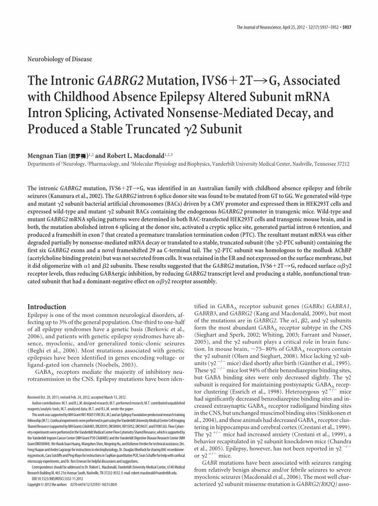
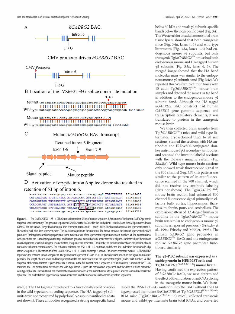
ResultsThe GABRG2(IVS6�2T3G) mutation generated a mutanthGABRG2(IVS6�2T3G) BAC transcript that retained a 53bp intron 6 fragmentThe GABRG2(IVS6�2T3G) mutation altered the GABRG2intron 6 splice donor site sequence. As a result, it was proposedthat intron 6 is spliced out either with the donor site fromanother intron, resulting in exon skipping, or with an alterna-tive donor site downstream of the wild-type site, resulting incryptic splice donor site activation and partial intron 6 reten-tion in the mutant mature mRNA (Kananura et al., 2002).However, the actual splice pattern of the mutant mRNA isunknown, and patient tissues or RNA samples are not avail-able. Our approach to determine the splicing pattern of themutant gene was to study splicing in vitro and in vivo of a BACconstruct that contained hGABRG2 genomic sequence, andthus a full-length intron 6 (Fig. 1 A) (for construct details, seeMaterials and Methods). Intron splicing is cell type specific, andthe optimal approach to study splicing of GABRG2 is to do so incells with endogenous GABRG2 expression. We used Lipo-fectamine 2000 to transfect either wild-type or mutant hGABRG2BACs containing their native promoter and the eGFP reporter geneinto PC12 cells, which have been reported to have endogenousGABRG2 expression (Tyndale et al., 1994). Although GFP expres-sion was observed in BAC-transfected PC12 cells, using RT-PCR wewere unable to demonstrate h�2 subunit mRNA. As an alternativestrategy, we replaced the hGABRG2 promoter with a CMV promoterand expressed the CMV promoter-driven hGABRG2 BAC inHEK293T cells (Fig. 1A) (for construct details, see Materials andMethods), and using RT-PCR, we were able to demonstrate h�2subunit mRNA expression. Thus, unless otherwise specified, allhGABRG2 BAC constructs in the remainder of the in vitro studiesused the CMV promoter.
To determine wild-type and mutant hGABRG2 splicing pat-terns, we expressed both wild-type hGABRG2 BAC and control�2S cDNA constructs in HEK293T cells and collected total RNA.DNA sequencing of hGABRG2 BAC RT-PCR products usingprimers binding to exons 5 and 9 of the GABRG2 coding se-quence showed that the intervening introns 6, 7, and 8 were com-pletely spliced out, and only �2S subunit mRNA was transcribedfrom the hGABRG2 BAC (data not shown). This was consistentwith the finding that the �2S subunit splice variant is the defaultsplicing product and that generation of the �2L subunit splicevariant requires positive regulation such as the function ofneuron-specific RNA binding protein Nova-1 (Zhang et al., 1996,1999; Dredge and Darnell, 2003).
The mutant hGABRG2(IVS6�2T3G) BAC transcript wasexpressed in HEK293T cells and cloned and sequenced (Fig.1 B). The mutant hGABRG2(IVS6�2T3G) BAC intron 6used a cryptic splice donor site 53 bp downstream of the wild-type splice donor site, and thus the mutant transcript retaineda 53 bp intron 6 fragment (Fig. 1C,D). None of the intronsplice donor site prediction models that we used detected thissite, suggesting that its sequence did not comply with generalsplice donor site rules. The mutant splice donor site was pre-dicted to be much weaker than the wild-type site (Carmel etal., 2004), having less hydrogen bonding with the splice ma-chinery, and hence forming a less stable mRNA–protein com-plex. These are all common properties of mutant splice donorsites (Buratti et al., 2007).
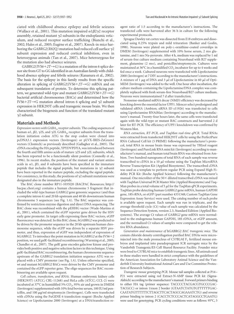
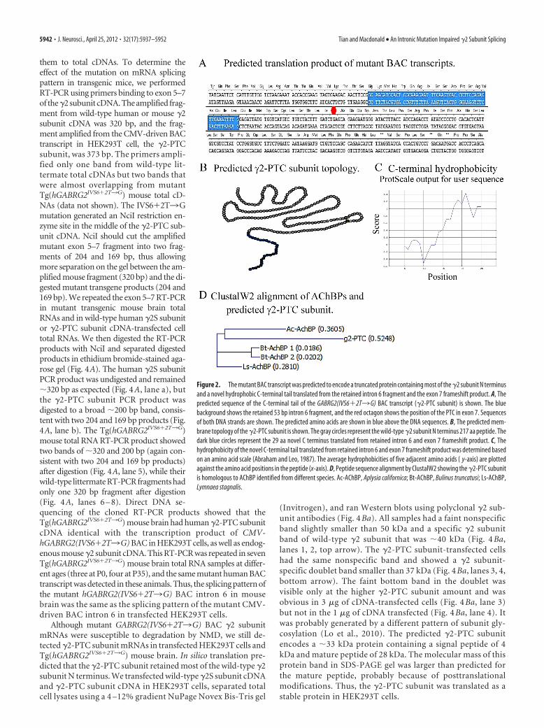
The mutant GABRG2(IVS6�2T3G) mRNA should betranslated to a truncated subunit containing the signalpeptide and N-terminal 217 aa of the wild-type �2 subunitIn silico translation, using Vector NTi (Invitrogen), showed thatthe mutant transcript should be translated to a polypeptide con-taining the signal peptide and N-terminal 217 aa of the wild-type�2 subunit. The retained 53 bp intron 6 fragment caused a frame-shift in exon 7, which generated a stop codon 33 bp from the endof the fragment. The retained intron 6 fragment and the exon 7frameshift sequence are predicted to be translated to a novel 29 aapeptide tail at the C terminus of the mutant protein (Fig. 2A), sothe mutant protein contained the N terminus of the wild-typesubunit and the novel C-terminal tail [�2-premature translationtermination codon (PTC) subunit] (Fig. 2B). The hydrophobic-ity of the 29 aa tail was evaluated by ProtScale at Expasy.org(Wilkins et al., 1999) and was found to be hydrophilic at the Nterminus and hydrophobic at the C terminus (Fig. 2C). The max-imum hydrophobic region was the C terminus, where the calcu-lated maximum hydrophobicity was 1.43 (Abraham and Leo,1987). This was very close to the maximum hydrophobicity of thewild-type �2S subunit, which was 1.75 when evaluated by thesame model. The structure of the mutant �2-PTC subunit wasunknown, but bioinformatics models did not predict that anysecondary structure formed in this fragment.
This truncated subunit was reminiscent of the solubleacetylcholine-binding proteins (AChBPs) found in mollusk glialcells (Smit et al., 2001; Hansen et al., 2004; Celie et al., 2005).AChBP sequences are homologous to the N-terminal extracellu-lar domains of cys-loop family ligand-gated ion channel (LGIC)subunits, and the crystal structure and protein function are sim-ilar to the ligand-binding domain of the nicotinic acetylcholinereceptor � subunit. GABAA receptors also belong to the cys-loopLGIC family. AChBPs oligomerize to form homopentamers con-taining binding sites for agonists and antagonists including ace-tylcholine. Upon acetylcholine release, AChBPs are released fromglia cells into synaptic gaps and inhibit cholinergic neurotrans-mission by binding free acetylcholine molecules (Smit et al.,2001). Sequence alignment showed that the �2-PTC subunit hasthe highest homology with the Aplysia californica AChBP (Ac-AChBP) (Fig. 2D). ClustalW alignment showed that the �2-PTCsubunit had a 21–29% sequence identity with AChBPs (data notshown), which was even higher than the 13–25% sequence iden-tity between AChBPs and LIGC subunit N-terminal extracellulardomains (Celie et al., 2005). The GABRG2(IVS6�2T3G) mu-tation might generate a mutant protein, the �2-PTC subunit, thatstructurally resembles AChBPs and interferes with GABAergicneurotransmission in a similar way.
The expression pattern of hGABRG2 HA BACs in transgenicmouse brain was similar to that of the endogenous GABRG2The transcription product of the CMV-driven hGABRG2(IVS6�2T3G) BAC in HEK293T cells, the �2-PTC subunitmRNA, retained a 53 bp intron 6 fragment in the mutant exon 6.However, it has been reported that promoter usage can affect intronsplicing pattern (Kornblihtt, 2005), and thus the mutanthGABRG2(IVS6�2T3G) BAC might be spliced to another mRNAwhen driven by its endogenous promoter. The intron splicing pat-tern is also cell type dependent. To minimize possible artifacts, westudied intron splicing of the hGABRG2(IVS6�2T3G) BAC withits endogenous promoter region in transgenic mouse brain (Fig. 3).We first expressed an HA-tagged hGABRG2 BAC in C57BL/6J mice,which is the C57BL/6-Tg(hGABRG2HA)RLM mouse line [accordingto The Jackson Laboratory mouse nomenclature, Tg(hGABRG2HA
5940 • J. Neurosci., April 25, 2012 • 32(17):5937–5952 Tian and Macdonald • An Intronic Mutation Impaired �2 Subunit Splicing
mice)]. The HA tag was introduced to a functionally silent positionin the wild-type subunit coding sequence. The HA-tagged �2 sub-units were not recognized by polyclonal �2 subunit antibodies (datanot shown). These antibodies recognized a strong nonspecific band
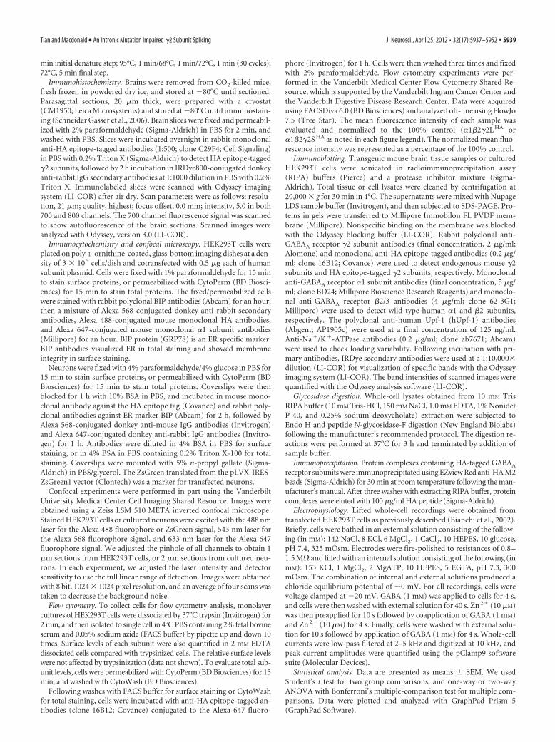
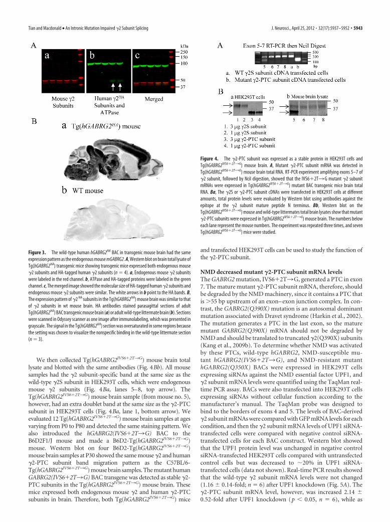
below 50 kDa and weak �2 subunit-specificbands below the nonspecific band (Fig. 3A).The Western blot on adult mouse total braintissue lysate showed that both transgenicmice (Fig. 3Aa, lanes 4, 5) and wild-typelittermates (Fig. 3Aa, lanes 1–3) had en-dogenous mouse �2 subunits, but onlytransgenic Tg(hGABRG2HA) mice had bothendogenous mouse and HA-tagged human�2 subunits (Fig. 3Ab, lanes 4, 5). Themerged image showed that the HA bandmolecular mass was similar to the endoge-nous mouse �2 subunit band (Fig. 3Ac). Werepeated this Western blot four times with15 adult Tg(hGABRG2HA) mouse brainsamples and detected the same HA tag bandin addition to the endogenous mouse �2subunit band. Although the HA-taggedhGABRG2 BAC construct had humanGABRG2 gene genomic sequence andtranscription regulatory elements, it wastranslated to protein in the transgenicmouse brain.
We then collected brain samples fromTg(hGABRG2HA) mice and wild-type lit-termates, cryosectioned them to 20 �msections, stained the sections with HA an-tibodies and IRDye800-conjugated don-key anti-mouse IgG secondary antibodies,and scanned the immunolabeled sectionswith the Odyssey imaging system (Fig.3Ba,Bb). Wild-type mouse brain sectionsonly showed weak fluorescence signal inthe 800 channel (Fig. 3Bb). Its pattern wassimilar to the pattern of its autofluores-cence scanned in the 700 channel, whichdid not receive any antibody labeling(data not shown). The Tg(hGABRG2HA)mouse brain section had enhanced 800channel fluorescence signal primarily in ol-factory bulb, cortex, hippocampus, thala-mus, midbrain, pons, and cerebellum. Theexpression pattern of HA-tagged human �2subunits in the Tg(hGABRG2HA) mousebrain was similar to endogenous mouse �2subunits as reported previously (Fritschy etal., 1994; Fritschy and Mohler, 1995). Thehuman GABRG2 gene promoter inhGABRG2HA BACs and the endogenousmouse GABRG2 gene promoter func-tioned similarly.
The �2-PTC subunit was expressed as astable protein in HEK293T cells andTg(hGABRG2 IVS6�2T3G) mouse brainHaving confirmed the expression patternof hGABRG2 BACs, we next determinedthe effect of the mutation on mRNA splicingin the transgenic mouse brain. We intro-
duced the IVS6�2T3G mutation into the BAC without the HAtag,expressedthemutantBACinC57BL/6-Tg(hGABRG2IVS6�2T3G)RLM mice [Tg(hGABRG2IVS6�2T3G) mice], collected transgenicmouse and wild-type littermate brain total RNAs, and converted
Figure 1. TheGABRG2(IVS6�2T3G)BACtranscriptretained53bpofintron6sequence.A,StructuresofthehumanGABRG2genomicsequence used in this study. The genomic sequence of GABRG2 in the BAC used in this study (RP11-1035I20) and the CMV promoter-drivenGABRG2 BAC are shown. The yellow horizontal lines represent introns and 3�- and 5�-UTRs. The brown horizontal line represents intron 6.The vertical dark blue lines represent exons. The black arrow points to the mutation. The brown arrow at the left end represents the CMVpromoter.Thelengthofeachlineisproportionaltothemolecularsizeoftherepresentedregion(nucleicacidnumber).B,ThemutantmRNAwas cloned into the TOPO cloning vector (top) and human genomic mRNA (bottom) sequences were aligned. The last 91 bp of the mutantexon 6 alignment result including the retained intron 6 sequence are presented. The number on the bottom line shows the position of eachnucleotide in human chromosome 5. The red arrow points to the IVS6�2T3G mutation, and the red line underlines the retained 53 bpintron 6 sequence. C, The structure of the GABRG2(IVS6�2T3G) BAC transcript is shown. The arrows represent exons 1–9. The red linerepresents the retained intron 6 fragment. The yellow lines represent 3�- and 5�-UTRs. The blue lines underline the signal and maturepeptides. The length of each arrow and line is proportional to the molecular size of the represented region (nucleic acid number). D, Thesequence of the mutant intron 6 splice donor site is shown. In the full mutant exon 6 sequence, a “t” in lowercase is shown at the T3Gmutation site. The dotted black box shows the wild-type intron 6 splice site conservative sequences, and the dotted red line marks thewild-type splice site. The solid black box encloses the seven nucleic acids at the mutant donor site sequence, and the solid red line marks thesplice site. The nucleotides in uppercase are exon 6 sequences, and the nucleotides in lowercase are intron sequences.
Tian and Macdonald • An Intronic Mutation Impaired �2 Subunit Splicing J. Neurosci., April 25, 2012 • 32(17):5937–5952 • 5941
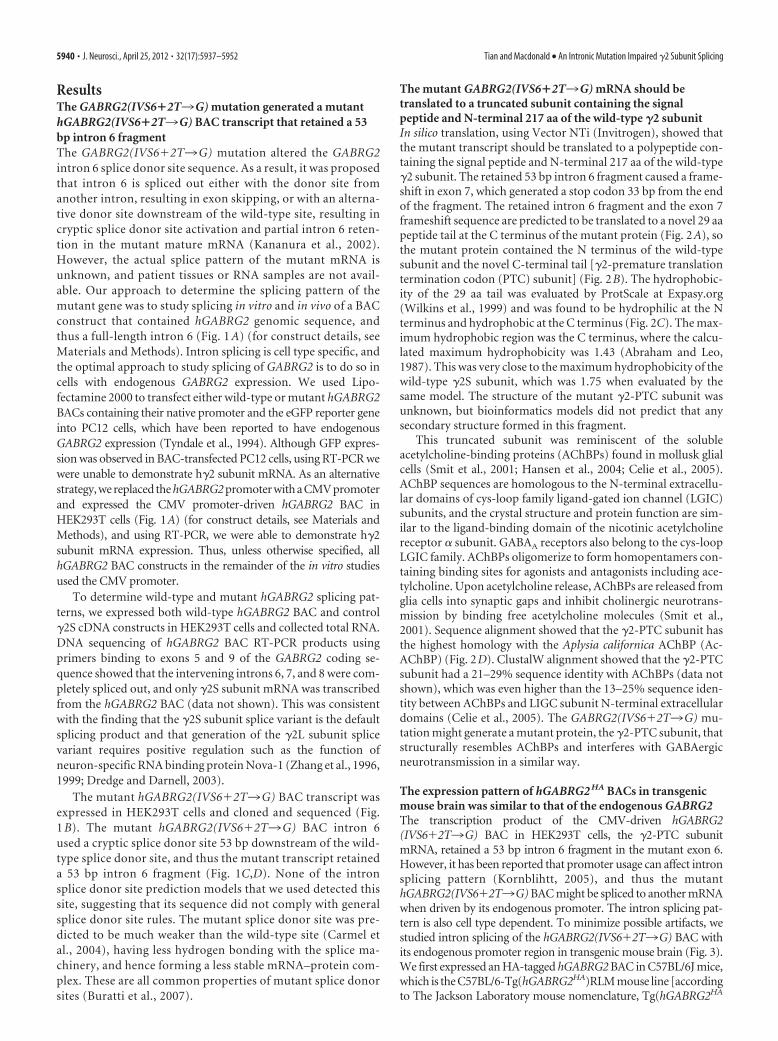
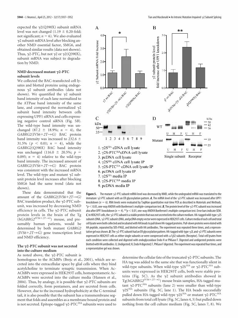
them to total cDNAs. To determine theeffect of the mutation on mRNA splicingpattern in transgenic mice, we performedRT-PCR using primers binding to exon 5–7of the�2 subunit cDNA. The amplified frag-ment from wild-type human or mouse �2subunit cDNA was 320 bp, and the frag-ment amplified from the CMV-driven BACtranscript in HEK293T cell, the �2-PTCsubunit, was 373 bp. The primers ampli-fied only one band from wild-type lit-termate total cDNAs but two bands thatwere almost overlapping from mutantTg(hGABRG2IVS6�2T3G) mouse total cD-NAs (data not shown). The IVS6�2T3Gmutation generated an NciI restriction en-zyme site in the middle of the �2-PTC sub-unit cDNA. NciI should cut the amplifiedmutant exon 5–7 fragment into two frag-ments of 204 and 169 bp, thus allowingmore separation on the gel between the am-plified mouse fragment (320 bp) and the di-gested mutant transgene products (204 and169 bp). We repeated the exon 5–7 RT-PCRin mutant transgenic mouse brain totalRNAs and in wild-type human �2S subunitor �2-PTC subunit cDNA-transfected celltotal RNAs. We then digested the RT-PCRproducts with NciI and separated digestedproducts in ethidium bromide-stained aga-rose gel (Fig. 4A). The human �2S subunitPCR product was undigested and remained�320 bp as expected (Fig. 4 A, lane a), butthe �2-PTC subunit PCR product wasdigested to a broad �200 bp band, consis-tent with two 204 and 169 bp products (Fig.4A, lane b). The Tg(hGABRG2IVS6�2T3G)mouse total RNA RT-PCR product showedtwo bands of �320 and 200 bp (again con-sistent with two 204 and 169 bp products)after digestion (Fig. 4A, lane 5), while theirwild-type littermate RT-PCR fragments hadonly one 320 bp fragment after digestion(Fig. 4A, lanes 6 – 8). Direct DNA se-quencing of the cloned RT-PCR products showed that theTg(hGABRG2IVS6�2T3G) mouse brain had human �2-PTC subunitcDNA identical with the transcription product of CMV-hGABRG2(IVS6�2T3G) BAC in HEK293T cells, as well as endog-enous mouse �2 subunit cDNA. This RT-PCR was repeated in sevenTg(hGABRG2IVS6�2T3G) mouse brain total RNA samples at differ-ent ages (three at P0, four at P35), and the same mutant human BACtranscript was detected in these animals. Thus, the splicing pattern ofthe mutant hGABRG2(IVS6�2T3G) BAC intron 6 in mousebrain was the same as the splicing pattern of the mutant CMV-driven BAC intron 6 in transfected HEK293T cells.
Although mutant GABRG2(IVS6�2T3G) BAC �2 subunitmRNAs were susceptible to degradation by NMD, we still de-tected �2-PTC subunit mRNAs in transfected HEK293T cells andTg(hGABRG2IVS6�2T3G) mouse brain. In silico translation pre-dicted that the �2-PTC subunit retained most of the wild-type �2subunit N terminus. We transfected wild-type �2S subunit cDNAand �2-PTC subunit cDNA in HEK293T cells, separated totalcell lysates using a 4 –12% gradient NuPage Novex Bis-Tris gel
(Invitrogen), and ran Western blots using polyclonal �2 sub-unit antibodies (Fig. 4 Ba). All samples had a faint nonspecificband slightly smaller than 50 kDa and a specific �2 subunitband of wild-type �2 subunit that was �40 kDa (Fig. 4 Ba,lanes 1, 2, top arrow). The �2-PTC subunit-transfected cellshad the same nonspecific band and showed a �2 subunit-specific doublet band smaller than 37 kDa (Fig. 4 Ba, lanes 3, 4,bottom arrow). The faint bottom band in the doublet wasvisible only at the higher �2-PTC subunit amount and wasobvious in 3 �g of cDNA-transfected cells (Fig. 4 Ba, lane 3)but not in the 1 �g of cDNA transfected (Fig. 4 Ba, lane 4). Itwas probably generated by a different pattern of subunit gly-cosylation (Lo et al., 2010). The predicted �2-PTC subunitencodes a �33 kDa protein containing a signal peptide of 4kDa and mature peptide of 28 kDa. The molecular mass of thisprotein band in SDS-PAGE gel was larger than predicted forthe mature peptide, probably because of posttranslationalmodifications. Thus, the �2-PTC subunit was translated as astable protein in HEK293T cells.
Figure 2. The mutant BAC transcript was predicted to encode a truncated protein containing most of the �2 subunit N terminusand a novel hydrophobic C-terminal tail translated from the retained intron 6 fragment and the exon 7 frameshift product. A, Thepredicted sequence of the C-terminal tail of the GABRG2(IVS6�2T3G) BAC transcript (�2-PTC subunit) is shown. The bluebackground shows the retained 53 bp intron 6 fragment, and the red octagon shows the position of the PTC in exon 7. Sequencesof both DNA strands are shown. The predicted amino acids are shown in blue above the DNA sequences. B, The predicted mem-brane topology of the �2-PTC subunit is shown. The gray circles represent the wild-type �2 subunit N terminus 217 aa peptide. Thedark blue circles represent the 29 aa novel C terminus translated from retained intron 6 and exon 7 frameshift product. C, Thehydrophobicity of the novel C-terminal tail translated from retained intron 6 and exon 7 frameshift product was determined basedon an amino acid scale (Abraham and Leo, 1987). The average hydrophobicities of five adjacent amino acids ( y-axis) are plottedagainst the amino acid positions in the peptide (x-axis). D, Peptide sequence alignment by ClustalW2 showing the �2-PTC subunitis homologous to AChBP identified from different species. Ac-AChBP, Aplysia californica; Bt-AChBP, Bulinus truncatusi; Ls-AChBP,Lymnaea stagnalis.
5942 • J. Neurosci., April 25, 2012 • 32(17):5937–5952 Tian and Macdonald • An Intronic Mutation Impaired �2 Subunit Splicing
We then collected Tg(hGABRG2IVS6�2T3G) mouse brain totallysate and blotted with the same antibodies (Fig. 4Bb). All mousesamples had the �2 subunit-specific band at the same size as thewild-type �2S subunit in HEK293T cells, which were endogenousmouse �2 subunits (Fig. 4Ba, lanes 5–8, top arrow). TheTg(hGABRG2IVS6�2T3G) mouse brain sample (from mouse no. 5),however, had an extra doublet band at the same size as the �2-PTCsubunit in HEK293T cells (Fig. 4Ba, lane 1, bottom arrow). Weevaluated 12 Tg(hGABRG2IVS6�2T3G) mouse brain samples at agesvarying from P0 to P80 and detected the same staining pattern. Wealso introduced the hGABRG2(IVS6�2T3G) BAC to theB6D2F1/J mouse and made a B6D2-Tg(hGABRG2IVS6�2T3G)mouse. Western blot on four B6D2-Tg(hGABRG2IVS6�2T3G)mouse brain samples at P30 showed the same mouse �2 and human�2-PTC subunit band migration pattern as the C57BL/6-Tg(hGABRG2IVS6�2T3G) mouse brain samples. The mutant humanGABRG2(IVS6�2T3G) BAC transgene was detected as stable �2-PTC subunits in the Tg(hGABRG2IVS6�2T3G) mouse brain. Thesemice expressed both endogenous mouse �2 and human �2-PTCsubunits in brain. Therefore, both Tg(hGABRG2IVS6�2T3G) mice
and transfected HEK293T cells can be used to study the function ofthe �2-PTC subunit.
NMD decreased mutant �2-PTC subunit mRNA levelsThe GABRG2 mutation, IVS6�2T3G, generated a PTC in exon7. The mature mutant �2-PTC subunit mRNA, therefore, shouldbe degraded by the NMD machinery, since it contains a PTC thatis �55 bp upstream of an exon– exon junction complex. In con-trast, the GABRG2(Q390X) mutation is an autosomal dominantmutation associated with Dravet syndrome (Harkin et al., 2002).The mutation generates a PTC in the last exon, so the maturemutant GABRG2(Q390X) mRNA should not be degraded byNMD and should be translated to truncated �2(Q390X) subunits(Kang et al., 2009b). To determine whether NMD was activatedby these PTCs, wild-type hGABRG2, NMD-susceptible mu-tant hGABRG2(IVS6�2T3G), and NMD-resistant mutanthGABRG2(Q350X) BACs were expressed in HEK293T cellsexpressing siRNAs against the NMD essential factor UPF1, and�2 subunit mRNA levels were quantified using the TaqMan real-time PCR assay. BACs were also transfected into HEK293T cellsexpressing siRNAs without cellular function according to themanufacturer’s manual. The TaqMan probe was designed tobind to the borders of exons 4 and 5. The levels of BAC-derived�2 subunit mRNAs were compared with GFP mRNA levels for eachcondition, and then the �2 subunit mRNA levels of UPF1 siRNA-transfected cells were compared with negative control siRNA-transfected cells for each BAC construct. Western blot showedthat the UPF1 protein level was unchanged in negative controlsiRNA-transfected HEK293T cells compared with untransfectedcontrol cells but was decreased to �20% in UPF1 siRNA-transfected cells (data not shown). Real-time PCR results showedthat the wild-type �2 subunit mRNA levels were not changed(1.16 � 0.14-fold; n 6) after UPF1 knockdown (Fig. 5A). The�2-PTC subunit mRNA level, however, was increased 2.14 �0.52-fold after UPF1 knockdown (p 0.05, n 6), while as
Figure 3. The wild-type human hGABRG2HA BAC in transgenic mouse brain had the sameexpression pattern as the endogenous mouse mGABRG2. A, Western blot on brain total lysate ofTg(hGABRG2HA) transgenic mice showing transgenic mice expressed both endogenous mouse�2 subunits and HA-tagged human �2 subunits (n 4). a, Endogenous mouse �2 subunitswere labeled in the red channel. b, ATPase and HA-tagged proteins were labeled in the greenchannel. c, The merged image showed the molecular size of HA-tagged human �2 subunits andendogenous mouse �2 subunits were similar. The white arrows in b point to the HA bands. B,The expression pattern of �2 HA subunits in the Tg(hGABRG2HA) mouse brain was similar to thatof �2 subunits in wt mouse brain. HA antibodies stained parasagittal sections of adultTg(hGABRG2HA) BAC transgenic mouse brain (a) or adult wild-type littermate brain (b). Sectionswere scanned in Odyssey scanner as one image after immunolabeling, which was presented ingrayscale. The signal in the Tg(hGABRG2HA) section was oversaturated in some regions becausethe setting was chosen to visualize the nonspecific binding in the wild-type littermate section(n 3).
Figure 4. The �2-PTC subunit was expressed as a stable protein in HEK293T cells andTg(hGABRG2IVS6�2T3G) mouse brain. A, Mutant �2-PTC subunit mRNA was detected inTg(hGABRG2IVS6�2T3G) mouse brain total RNA. RT-PCR experiment amplifying exons 5–7 of�2 subunit, followed by NciI digestion, showed that the IVS6�2T3G mutant �2 subunitmRNAs were expressed in Tg(hGABRG2IVS6�2T3G) mutant BAC transgenic mice brain totalRNA. Ba, The �2S or �2-PTC subunit cDNAs were transfected in HEK293T cells at differentamounts, total protein levels were evaluated by Western blot using antibodies against theepitope at the �2 subunit mature peptide N terminus. Bb, Western blot on theTg(hGABRG2IVS6�2T3G) mouse and wild-type littermates total brain lysates show that mutant�2-PTC subunits were expressed in Tg(hGABRG2IVS6�2T3G) mouse brain. The numbers beloweach lane represent the mouse numbers. The experiment was repeated three times, and sevenTg(hGABRG2IVS6�2T3G) mice were studied.
Tian and Macdonald • An Intronic Mutation Impaired �2 Subunit Splicing J. Neurosci., April 25, 2012 • 32(17):5937–5952 • 5943
expected the �2(Q390X) subunit mRNAlevel was not changed (1.19 � 0.20-fold;not significant; n 6). We also evaluated�2 subunit mRNA level after blocking an-other NMD essential factor, SMG6, andobtained similar results (data not shown).Thus, �2-PTC, but not �2 or �2(Q390X),subunit mRNA was subject to degrada-tion by NMD.
NMD decreased mutant �2-PTCsubunit levelsWe collected the BAC-transfected cell ly-sates and blotted proteins using endoge-nous �2 subunit antibodies (data notshown). We quantified the �2 subunitband intensity of each lane normalized tothe ATPase band intensity of the samelane, and compared the normalized �2subunit band intensity between cellsexpressing UPF1 siRNA and cells express-ing negative control siRNA (Fig. 5B).The wild-type band intensity was un-changed (87.2 � 18.9%; n 4), theGABRG2(IVS6�2T3G) BAC proteinband intensity was increased to 232.6 �31.5% (p 0.01; n 4), while theGABRG2(Q390X) BAC band intensitywas unchanged (116.0 � 20.5%; p 0.095; n 4) relative to the wild-typeband intensity. The increased amount ofGABRG2(IVS6�2T3G) BAC proteinwas consistent with the increased mRNAlevel. The wild-type and mutant �2 sub-unit protein level increases after blockingSMG6 had the same trend (data notshown).
These data demonstrated that theamount of the GABRG2(IVS6�2T3G)BAC translation product, the �2-PTC sub-unit, was increased by decreasing NMDefficiency in cells. The �2-PTC subunitprotein levels in the brain of the Tg(hGABRG2IVS6�2T3G) mouse, and pre-sumably human patients, would bedetermined by both mutant GABRG2(IVS6�2T3G) gene transcription leveland NMD efficiency.
The �2-PTC subunit was not secretedinto the culture mediumAs noted above, the �2-PTC subunit ishomologous to the AChBPs (Brejc et al., 2001), which are se-creted into the extracellular space by glial cells where they bindacetylcholine to terminate synaptic transmission. When Ac-AChBPs were expressed in HEK293T cells, homopentameric Ac-AChBPs were secreted into the culture media (Hansen et al.,2004). Thus, by analogy, it is possible that �2-PTC subunits arefolded correctly, form pentamers, and are secreted from cells.However, due to the increased hydrophobicity at the C-terminaltail, it is also possible that the subunit has a transmembrane seg-ment that folds and assembles as a membrane bound protein andis not secreted. Epitope-tagged �2-PTC HA subunits were used to
determine the cellular fate of the truncated �2-PTC subunits. TheHA tag was added to the same site that was functionally silent inwild-type subunits. When wild-type �2S HA or �2-PTC HA sub-units were expressed in HEK293T cells, both were stable pro-teins (Fig. 5C). As the �2 subunit antibodies showed inTg(hGABRG2IVS6�2T3G) mouse brain samples, HA-tagged mu-tant �2-PTC HA subunits (lane 2) were smaller than wild-type�2S HA subunits (Fig. 5C, lane 1). The HA beads successfullypulled down HA-tagged wild-type �2S HA or mutant �2-PTC HA
subunits from total cell lysate (Fig. 5C, lanes 4, 5) but pulled downnothing from the cell culture medium (Fig. 5C, lanes 7, 8). We
Figure 5. The mutant �2-PTC subunit mRNA level was decreased by NMD, while the undegraded mRNA was translated to theimmature �2-PTC subunit with an ER glycosylation pattern. A, The mRNA level of the �2-PTC subunit was increased after UPF1knockdown (n 6). RNA levels were evaluated by TaqMan quantitative real-time PCR as described in Materials and Methods.*p 0.05, one-way ANOVA with Bonferroni’s multiple-comparison test. B, The protein level of the �2-PTC subunit was increasedalso after UPF1 knockdown (n 4). **p 0.01, one-way ANOVA Bonferroni’s multiple-comparison test. Error bars indicate SEM.C, In HEK293T cells, the �2-PTC subunit is a stable protein that was not secreted into the culture medium. HA-tagged wild-type �2Ssubunit cDNA, �2-PTC subunit cDNA, and pcDNA empty vector were expressed in HEK293T cells. Culture media of each cell and totalcell lysate were both collected and incubated with HA beads to pull down HA-tagged proteins. Pull-down proteins were eluted withHA peptide, separated by SDS-PAGE, and blotted with HA antibodies. The experiment was repeated three times, and a represen-tative gel was shown. D, The �2-PTC subunit had an ER glycosylation pattern. HA-tagged wild-type �2L and �2-PTC subunits wereexpressed in HEK293T cells as either single subunits or were coexpressed with �1�2 subunits (Receptor). Total cell lysates fromeach condition were collected and digested with endoglycosidase Endo H or PNGase F. Digested and undigested proteins wereblotted with HA antibodies. U, Undigested; H, Endo H digested; F, PNGase F digested. The experiment was repeated four times, anda representative gel was shown.
5944 • J. Neurosci., April 25, 2012 • 32(17):5937–5952 Tian and Macdonald • An Intronic Mutation Impaired �2 Subunit Splicing
collected �15 ml of culture media from each sample. If the �2-PTC HA subunit was secreted from cells at the same efficiency asAc-AChBP (1 - 3 mg/L) (Hansen et al., 2004), there would be�15– 45 �g of �2-PTC subunit protein in 15 ml of culture media.We used the Odyssey quantitative Western blot system to detectthe �2-PTC HA subunit. According to the manufacturer’s (LI-COR) document, even if the amount of �2-PTC HA subunit was ahundred times less than 15– 45 �g, it should still be sufficient fordetection by our Western blot. Although the �2-PTC subunit ishighly homologous to the secreted Ac-AChBP, �2-PTC HA sub-units were present, but not secreted into the culture medium. InTg(hGABRG2IVS6�2T3G) mouse brain or human patients, themutant allele would be translated to �2-PTC subunits, which arelikely to be also expressed inside of the neurons and not secretedto extrasynaptic spaces.
The �2-PTC subunit attained altered ER-associatedglycosylationWhile not secreted, �2-PTC subunits could still form homo-oligomers or hetero-oligomers that are trafficked to the surfacemembrane. During the process of subunit maturation, immatureN-linked mannose-rich oligosaccharides attached in the ER arereplaced by mature glycans that are attached in the trans-Golgiregion. Wild-type �2L subunits show only low levels of mem-brane trafficking when expressed alone, which increased substan-tially with coexpression of �1 and �2 subunits (Connolly et al.,1999a). To determine whether the �2-PTC subunits had matureglycosylation consistent with surface membrane trafficking, wecompared the glycosylation patterns of �2L and �2-PTC subunitswithout or with cotransfection of �1 and �2 subunits.
Endo H cleaves immature N-linked mannose-rich oligosac-charides attached in the ER but not the mature glycans attachedin the trans-Golgi region. In contrast, PNGase F removes all oli-gosaccharides attached both in the ER and trans-Golgi regions(Maley et al., 1989). When expressed alone, �2L HA subunits onWestern blot ran as a single band that was sensitive to digestion byendoglycosidases Endo H and PNGase F, consistent with primar-ily immature glycosylation and suggesting that �2L HA subunitswere retained in the ER (Fig. 5D, WT subunit). When coex-pressed with �1 and �2 subunits, �2L HA subunits showed anextra band on Western blots that was insensitive to Endo H di-gestion, but was sensitive to PNGase F digestion (Fig. 5D, WTsubunit). With coexpression of �1 and �2 subunits, �2L HA sub-units had mature glycosylation, suggesting processing in theGolgi apparatus and trafficking to the cell membrane.
With expression alone or with coexpression of �1 and �2subunits, �2-PTC HA subunits showed only one band on Westernblot that was sensitive to both Endo H and PNGase F (Fig. 5D,mutant subunit), suggesting that �2-PTC HA subunits were re-tained in the ER and not transported to the Golgi apparatus. Thesize of the digested �2-PTC HA subunit protein band was consis-tent with the predicted size of the mature �2-PTC HA subunitbased on amino acid sequence. This phenomenon is consistentwith the finding that �2-PTC HA subunits were not secreted intothe culture medium, which requires Golgi translocation. Thesedata further suggested that the �2-PTC subunit might not betrafficked to the cell membrane and might instead be retained inthe ER under physiological conditions such as in patients or inTg(hGABRG2IVS6�2T3G) mouse neurons.
The �2-PTC subunits oligomerized with �1 and �2 subunitsGABAA receptor subunit oligomerization is determined by se-quences at the extracellular N-terminal domain (Taylor et al.,
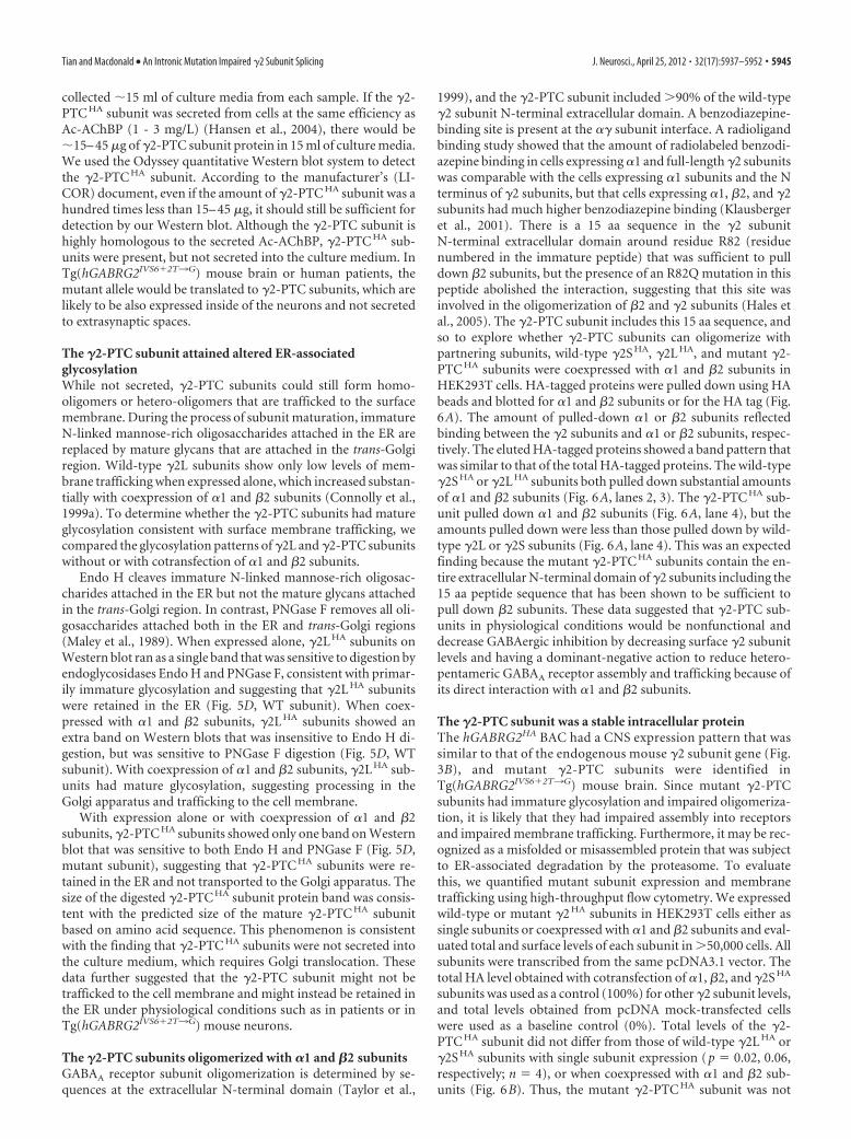
1999), and the �2-PTC subunit included �90% of the wild-type�2 subunit N-terminal extracellular domain. A benzodiazepine-binding site is present at the �� subunit interface. A radioligandbinding study showed that the amount of radiolabeled benzodi-azepine binding in cells expressing �1 and full-length �2 subunitswas comparable with the cells expressing �1 subunits and the Nterminus of �2 subunits, but that cells expressing �1, �2, and �2subunits had much higher benzodiazepine binding (Klausbergeret al., 2001). There is a 15 aa sequence in the �2 subunitN-terminal extracellular domain around residue R82 (residuenumbered in the immature peptide) that was sufficient to pulldown �2 subunits, but the presence of an R82Q mutation in thispeptide abolished the interaction, suggesting that this site wasinvolved in the oligomerization of �2 and �2 subunits (Hales etal., 2005). The �2-PTC subunit includes this 15 aa sequence, andso to explore whether �2-PTC subunits can oligomerize withpartnering subunits, wild-type �2S HA, �2L HA, and mutant �2-PTC HA subunits were coexpressed with �1 and �2 subunits inHEK293T cells. HA-tagged proteins were pulled down using HAbeads and blotted for �1 and �2 subunits or for the HA tag (Fig.6A). The amount of pulled-down �1 or �2 subunits reflectedbinding between the �2 subunits and �1 or �2 subunits, respec-tively. The eluted HA-tagged proteins showed a band pattern thatwas similar to that of the total HA-tagged proteins. The wild-type�2S HA or �2L HA subunits both pulled down substantial amountsof �1 and �2 subunits (Fig. 6A, lanes 2, 3). The �2-PTC HA sub-unit pulled down �1 and �2 subunits (Fig. 6A, lane 4), but theamounts pulled down were less than those pulled down by wild-type �2L or �2S subunits (Fig. 6A, lane 4). This was an expectedfinding because the mutant �2-PTC HA subunits contain the en-tire extracellular N-terminal domain of �2 subunits including the15 aa peptide sequence that has been shown to be sufficient topull down �2 subunits. These data suggested that �2-PTC sub-units in physiological conditions would be nonfunctional anddecrease GABAergic inhibition by decreasing surface �2 subunitlevels and having a dominant-negative action to reduce hetero-pentameric GABAA receptor assembly and trafficking because ofits direct interaction with �1 and �2 subunits.
The �2-PTC subunit was a stable intracellular proteinThe hGABRG2HA BAC had a CNS expression pattern that wassimilar to that of the endogenous mouse �2 subunit gene (Fig.3B), and mutant �2-PTC subunits were identified inTg(hGABRG2IVS6�2T3G) mouse brain. Since mutant �2-PTCsubunits had immature glycosylation and impaired oligomeriza-tion, it is likely that they had impaired assembly into receptorsand impaired membrane trafficking. Furthermore, it may be rec-ognized as a misfolded or misassembled protein that was subjectto ER-associated degradation by the proteasome. To evaluatethis, we quantified mutant subunit expression and membranetrafficking using high-throughput flow cytometry. We expressedwild-type or mutant �2 HA subunits in HEK293T cells either assingle subunits or coexpressed with �1 and �2 subunits and eval-uated total and surface levels of each subunit in �50,000 cells. Allsubunits were transcribed from the same pcDNA3.1 vector. Thetotal HA level obtained with cotransfection of �1, �2, and �2S HA
subunits was used as a control (100%) for other �2 subunit levels,and total levels obtained from pcDNA mock-transfected cellswere used as a baseline control (0%). Total levels of the �2-PTC HA subunit did not differ from those of wild-type �2L HA or�2S HA subunits with single subunit expression (p 0.02, 0.06,respectively; n 4), or when coexpressed with �1 and �2 sub-units (Fig. 6B). Thus, the mutant �2-PTC HA subunit was not
Tian and Macdonald • An Intronic Mutation Impaired �2 Subunit Splicing J. Neurosci., April 25, 2012 • 32(17):5937–5952 • 5945
degraded and was as stable in these cells as wild-type �2Ssubunits.
The �2-PTC subunit had impaired membrane traffickingTo assess surface trafficking of the mutant �2-PTC�� subunit,we used the quantitative technique of flow cytometry without cellpermeabilization. We cotransfected cells using the same subunitcombinations used to assess total cell levels of �1�2�2S HA sub-units by measuring surface HA levels for each subunit (Fig. 6C).
The surface HA level obtained with �1�2�2SHA subunit coexpres-sion was used as a 100% normalization control for �2HA subunitsurface level, and surface HA level obtained with pcDNA mock-transfected cells was used as baseline (0%). The single wild-type�2LHA subunit had a low surface level (2.93 � 0.76%; n 4), whichwas increased substantially (29.71 � 0.88%; p 0.01; n 4) bycoexpression with �1 and �2 subunits. The wild-type �2SHA single-subunit surface level was much higher than �2LHA subunit surfacelevel, probably because the �2SHA single subunits have higher traf-ficking efficiency and lower PKC-dependent endocytosis (Connollyet al., 1999a,b). Its single-subunit surface level (34.08 � 3.80%; n 4) was substantially higher than the �2LHA subunits single-subunitsurface level. Its surface level with �1 and �2 subunit coexpressionwas 100%, also substantially higher than with �1�2�2LHA coexpres-sion (n 4). Compared with �2LHA and �2SHA subunits, the sur-face levels of the �2-PTCHA subunit were substantially smaller withboth expression conditions. The �2-PTCHA single-subunit surfacelevel was low (0.76�0.56%; n4) and did not increase significantlywith �1 and �2 subunit coexpression (2.76 � 0.30%; n 4; notsignificant). The surface levels of �2-PTC�� subunits with or with-out �1 and �2 subunit coexpression were not significantly greaterthan the mock control level (p value: single subunit, �0.05; with �1and �2 subunit coexpression: �0.05; n 4). These results suggestthat, even though the mutant �2-PTC�� subunit oligomerized with�1 and �2 subunits, it was not trafficked to the cell surface.
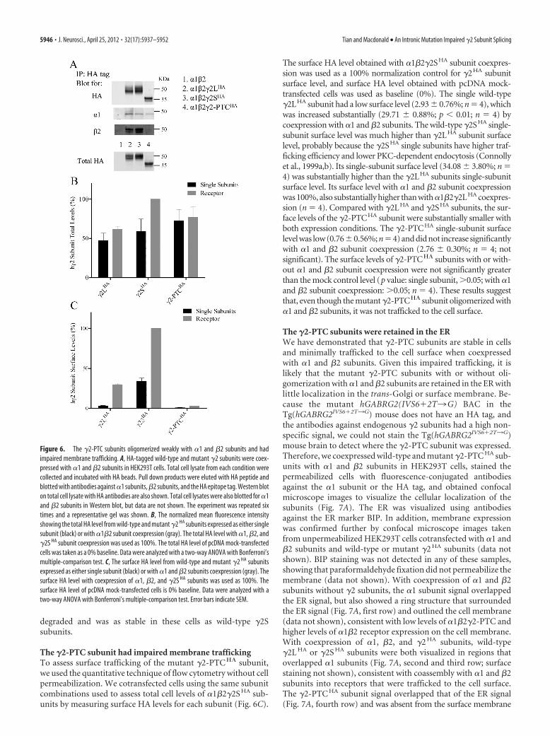
The �2-PTC subunits were retained in the ERWe have demonstrated that �2-PTC subunits are stable in cellsand minimally trafficked to the cell surface when coexpressedwith �1 and �2 subunits. Given this impaired trafficking, it islikely that the mutant �2-PTC subunits with or without oli-gomerization with �1 and �2 subunits are retained in the ER withlittle localization in the trans-Golgi or surface membrane. Be-cause the mutant hGABRG2(IVS6�2T3G) BAC in theTg(hGABRG2IVS6�2T3G) mouse does not have an HA tag, andthe antibodies against endogenous �2 subunits had a high non-specific signal, we could not stain the Tg(hGABRG2IVS6�2T3G)mouse brain to detect where the �2-PTC subunit was expressed.Therefore, we coexpressed wild-type and mutant �2-PTC HA sub-units with �1 and �2 subunits in HEK293T cells, stained thepermeabilized cells with fluorescence-conjugated antibodiesagainst the �1 subunit or the HA tag, and obtained confocalmicroscope images to visualize the cellular localization of thesubunits (Fig. 7A). The ER was visualized using antibodiesagainst the ER marker BIP. In addition, membrane expressionwas confirmed further by confocal microscope images takenfrom unpermeabilized HEK293T cells cotransfected with �1 and�2 subunits and wild-type or mutant �2 HA subunits (data notshown). BIP staining was not detected in any of these samples,showing that paraformaldehyde fixation did not permeabilize themembrane (data not shown). With coexpression of �1 and �2subunits without �2 subunits, the �1 subunit signal overlappedthe ER signal, but also showed a ring structure that surroundedthe ER signal (Fig. 7A, first row) and outlined the cell membrane(data not shown), consistent with low levels of �1�2�2-PTC andhigher levels of �1�2 receptor expression on the cell membrane.With coexpression of �1, �2, and �2 HA subunits, wild-type�2L HA or �2S HA subunits were both visualized in regions thatoverlapped �1 subunits (Fig. 7A, second and third row; surfacestaining not shown), consistent with coassembly with �1 and �2subunits into receptors that were trafficked to the cell surface.The �2-PTC HA subunit signal overlapped that of the ER signal(Fig. 7A, fourth row) and was absent from the surface membrane
Figure 6. The �2-PTC subunits oligomerized weakly with �1 and �2 subunits and hadimpaired membrane trafficking. A, HA-tagged wild-type and mutant �2 subunits were coex-pressed with �1 and �2 subunits in HEK293T cells. Total cell lysate from each condition werecollected and incubated with HA beads. Pull down products were eluted with HA peptide andblotted with antibodies against �1 subunits, �2 subunits, and the HA epitope tag. Western bloton total cell lysate with HA antibodies are also shown. Total cell lysates were also blotted for �1and �2 subunits in Western blot, but data are not shown. The experiment was repeated sixtimes and a representative gel was shown. B, The normalized mean fluorescence intensityshowing the total HA level from wild-type and mutant �2 HA subunits expressed as either singlesubunit (black) or with �1�2 subunit coexpression (gray). The total HA level with �1, �2, and�2S HA subunit coexpression was used as 100%. The total HA level of pcDNA mock-transfectedcells was taken as a 0% baseline. Data were analyzed with a two-way ANOVA with Bonferroni’smultiple-comparison test. C, The surface HA level from wild-type and mutant �2 HA subunitsexpressed as either single subunit (black) or with �1 and �2 subunits coexpression (gray). Thesurface HA level with coexpression of �1, �2, and �2S HA subunits was used as 100%. Thesurface HA level of pcDNA mock-transfected cells is 0% baseline. Data were analyzed with atwo-way ANOVA with Bonferroni’s multiple-comparison test. Error bars indicate SEM.
5946 • J. Neurosci., April 25, 2012 • 32(17):5937–5952 Tian and Macdonald • An Intronic Mutation Impaired �2 Subunit Splicing
(data not shown). The wild-type �1, �2L,and �2S subunits often showed HA signalsin the region that was recognized by Golgi-specific antibodies (data not shown), but the�2-PTCHA subunit was not. Thus, the �2-PTCHA subunit was retained primarily inthe ER, consistent with its background levelson the surface membrane and its absence inthe culture medium.
We then cloned the �2S HA and �2-PTC HA subunit cDNAs into pLVX-�2 HA-IRES-ZsGreen1 vectors expressing �2 HA
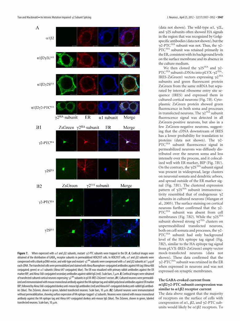
subunits and green fluorescent proteinZsGreen from the same mRNA but sepa-rated by internal ribosome entry site se-quence (IRES) and expressed them incultured cortical neurons (Fig. 7B). Cyto-plasmic ZsGreen protein showed greenfluorescence in both soma and processesin transfected neurons. The �2 HA subunitfluorescence signal was detected in allZsGreen-positive neurons, but also in afew ZsGreen-negative neurons, suggest-ing that the cDNA downstream of IREShas a lower probability for translation toproteins (data not shown). The �2-PTC HA subunit fluorescence signal inpermeabilized neurons was diffusely dis-tributed over the neuron soma and lessintensely over the process, and it colocal-ized well with ER marker, BIP (Fig. 7B1).On the contrary, the �2S HA subunit signalwas present in widespread, large clusterson neuronal somata and dendritic arbors,and spread outside of the ER marker sig-nal (Fig. 7B1). The clustered expressionpattern of �2S HA subunit immunoreac-tivity resembled that of endogenous �2subunits in cultured neurons (Mangan etal., 2005). The surface staining on corticalneurons further confirmed that the �2-PTC HA subunit was absent from cellmembranes (Fig. 7B2). While the �2S HA
subunit showed strong �2 HA clusters onunpermeabilized transfected neurons,both on cell somata and processes, the �2-PTC HA subunit had only backgroundlevel of the HA epitope tag signal (Fig.7B2), similar to the HA epitope tag signalfrom pLVX-IRES-ZsGreen1 empty vectormock-transfected neurons (data notshown). These data confirmed that the�2-PTC HA subunit was retained in the ERwhen expressed in neurons and was notexpressed on synaptic membranes.
The GABA-evoked current from�1�2�2-PTC subunit coexpression wassimilar to �1�2 receptor currentThe data above suggest that the majorityof receptors on the surface of cells withcoexpression of �1, �2, and �2-PTC sub-units would likely be �1�2 receptors. To
Figure 7. When expressed with �1 and �2 subunits, mutant �2-PTC subunits were trapped in the ER. A, Confocal images wereobtained of the distribution of GABAA receptor subunits in permeabilized HEK293T cells. In HEK293T cells, �1 and �2 subunits werecoexpressed with a blank pcDNA vector, and wild-type and mutant �2 HA subunits were coexpressed with �1 and �2 subunits at 1 �g ofeach cDNA. The transfected cells were permeabilized and stained with Alexa fluorophore-conjugated antibodies against HA tag (Alexa 488conjugated; green) or �1 subunits (Alexa 647 conjugated; blue). The ER was visualized with primary rabbit antibodies against the ERmarker BIP, and Alexa 568-conjugated secondary antibodies against rabbit IgG (red). Scale bars, 5�m. B, Confocal images were obtainedof transfected cultured cortical neurons expressing �2 HA subunits in pLVX-IRES-ZsGreen1 vectors. B1, Cultured neurons were permeabil-ized and immunostained with mouse monoclonal antibody against the HA epitope tag and rabbit polyclonal antibodies against ER markerBIP, followed by Alexa 568-conjugated donkey anti-mouse IgG antibodies (red) and Alexa 647-conjugated donkey anti-rabbit IgG antibod-ies (blue). The ZsGreen, shown in green, labeled transfected neurons. Scale bars, 10 �m. B2, Cultured neurons were immunostainedwithout permeabilization, showing surface expression of HA epitope-tagged �2 subunits. Neurons were stained with mouse monoclonalantibody against the HA epitope tag and Alexa 647-conjugated donkey anti-mouse IgG (blue). The ZsGreen, shown in green, labeledtransfected neurons. Scale bars, 10 �m.
Tian and Macdonald • An Intronic Mutation Impaired �2 Subunit Splicing J. Neurosci., April 25, 2012 • 32(17):5937–5952 • 5947
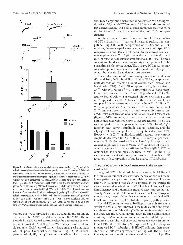
explore this, we coexpressed �1 and �2 subunits and �1 and �2subunits with �2-PTC or �2S subunits in HEK293T cells andrecorded GABA-evoked current evoked by a saturating concen-tration (1 mM) of GABA (Fig. 8A). With coexpression of �1 and�2 subunits, GABA-evoked currents had a small peak amplitudeof �400 pA and very fast desensitization (Fig. 8A). With coex-pression of �1, �2, and �2S subunits, GABA-evoked currents
were much larger and desensitization was slower. With coexpres-sion of �1, �2, and �2-PTC subunits, GABA-evoked currents hadfast desensitization, and a small peak amplitude that was moresimilar to �1�2 receptor currents than �1�2�2S receptorcurrents.
We then recorded from cells coexpressing �1, �2, and �2S or�2-PTC subunits (n 8 cells) and measured peak current am-plitudes (Fig. 8B). With coexpression of �1, �2, and �2-PTCsubunits, the average peak current amplitude was 575.5 pA. Withcoexpression of �1, �2, and �2S subunits, the average peak cur-rent amplitude was 3516.0 pA, and with coexpression of �1 and�2 subunits, the peak current amplitude was 710.4 pA. The peakcurrent amplitudes of these two wild-type receptors fall in thenormal range of reported values. The �1�2�2-PTC receptor peakcurrent amplitude was significantly decreased from �1�2�2S re-ceptors but was similar to that of �1�2 receptors.
The divalent cation Zn 2� is an endogenous neuromodulator(Kay and Toth, 2008). Its ability to inhibit GABAA receptor cur-rents depends on receptor subunit composition (Nagaya andMacdonald, 2001). The �1�2 receptors are highly sensitive toZn 2�, with IC50 values of �0.1–1 �M, while the �1�2�2 recep-tors are very insensitive to Zn 2�, with IC50 values of �200 – 600�M. We bathed cells with an external solution containing 10 �M
Zn 2�, applied 1 mM GABA alone or with 10 �M Zn 2�, and thencompared the peak currents with and without Zn 2� (Fig. 8C).We also applied GABA at the same time interval but withoutZn 2�, and compared the peak currents to quantify current run-down. With coexpression of �1 and �2, �1, �2, and �2S, or �1,�2, and �2-PTC subunits, currents showed minimum peak am-plitude decreases with repetitive GABA applications. The �1�2receptor peak current amplitude decreased 10%, the �1�2�2Sreceptor peak current amplitude did not decrease, and the�1�2�2-PTC receptor peak current amplitude decreased 17%.However, with Zn 2� application, �1�2 receptor peak currentamplitude decreased 83.2%, �1�2�2-PTC receptor peak cur-rent amplitude decreased 85.3%, and �1�2�2S receptor peakcurrent amplitude decreased 9.6%. Zn 2� inhibited all three re-ceptor currents with different efficiencies. The �1�2�2-PTC re-ceptors had the same high sensitivity to Zn 2� as the �1�2receptors consistent with formation primarily of surface �1�2receptors with coexpression of �1, �2, and �2-PTC subunits.
The �2-PTC subunits induced an increase in the ER stressmarker BIPAlthough �2-PTC subunit mRNA was decreased by NMD, andthe translation product was expressed poorly on the cell mem-brane, patients carrying one mutant allele had seizures. The mu-tant �2-PTC subunit was clearly expressed in the transgenicmouse brain and was stable in HEK293T cells and produced hap-loinsufficiency and a dominant-negative effect on receptor as-sembly. Since the �2-PTC subunit was so stable in transgenicmouse brain, the possibility that the mutant subunit had addi-tional functions that might contribute to epilepsy pathogenesis.
The �2-PTC subunits were stable ER proteins with a sequencesimilar to a �2 subunit truncated in the middle of the first trans-membrane domain. While the �2-PTC subunits were stable andnot degraded, the subunit may not have the same conformationas wild-type �2 subunits and could induce the unfolded proteinresponse (UPR). The level of the ER chaperone BIP is an indica-tor of UPR-induced ER stress. We expressed 3 �g of wild-type ormutant �2-PTC HA subunits in HEK293T cells and then evalu-ated cellular BIP levels by Western blot (Fig. 9A). The BIP bandintensity for each condition was normalized to that of pcDNA
Figure 8. GABA-evoked currents recorded from cells coexpressing �1, �2, and �2-PTCsubunits were similar to those obtained with expression of �1and �2 subunits. A, GABAergiccurrents were recorded from coexpressed �1�2, �1�2�2-PTC, and �1�2�2S subunits. Themerged picture showed the relative peak amplitude of currents recorded from �1�2�2-PTCsubunits was much smaller than that from �1�2�2S subunits, but close to those obtainedfrom �1�2 subunits. B, Peak current amplitudes from wild-type and mutant receptors wereplotted. *p 0.05, one-way ANOVA with Bonferroni’s multiple-comparison test. C, The cur-rents recorded from coexpressed �1�2�2-PTC subunits had a Zn 2� sensitivity that was sim-ilar to that of coexpressed �1�2 subunits. Cells expressing �1�2�2S, �1�2�2-PTC, or �1�2subunits were exposed to two 1 mM GABA applications 4 s apart or one 1 mM GABA applicationfollowed by 10 �M Zn 2� washed 4 s and 10 �M Zn 2� with 1 mM GABA application. The peakcurrents ratio of each cell was plotted. **p 0.01, compared with the control conditions(two-way ANOVA with Bonferroni’s multiple-comparison test). Error bars indicate SEM.
5948 • J. Neurosci., April 25, 2012 • 32(17):5937–5952 Tian and Macdonald • An Intronic Mutation Impaired �2 Subunit Splicing
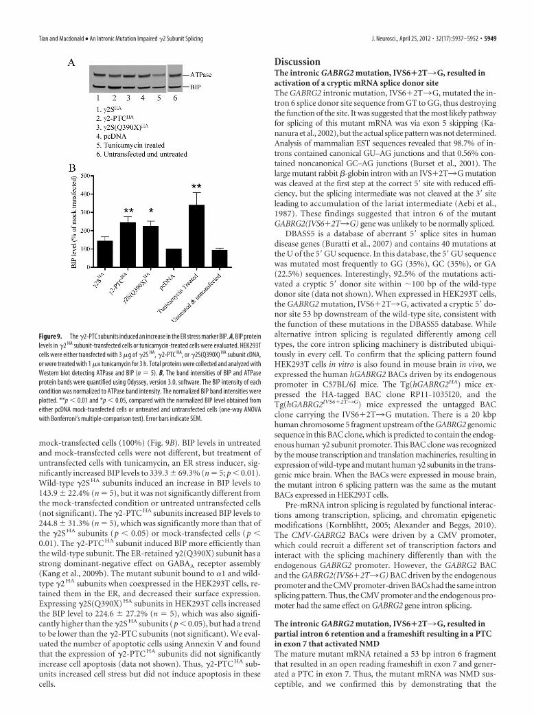
mock-transfected cells (100%) (Fig. 9B). BIP levels in untreatedand mock-transfected cells were not different, but treatment ofuntransfected cells with tunicamycin, an ER stress inducer, sig-nificantly increased BIP levels to 339.3 � 69.3% (n 5; p 0.01).Wild-type �2S HA subunits induced an increase in BIP levels to143.9 � 22.4% (n 5), but it was not significantly different fromthe mock-transfected condition or untreated untransfected cells(not significant). The �2-PTC HA subunits increased BIP levels to244.8 � 31.3% (n 5), which was significantly more than that ofthe �2S HA subunits (p 0.05) or mock-transfected cells (p 0.01). The �2-PTC HA subunit induced BIP more efficiently thanthe wild-type subunit. The ER-retained �2(Q390X) subunit has astrong dominant-negative effect on GABAA receptor assembly(Kang et al., 2009b). The mutant subunit bound to �1 and wild-type �2 HA subunits when coexpressed in the HEK293T cells, re-tained them in the ER, and decreased their surface expression.Expressing �2S(Q390X) HA subunits in HEK293T cells increasedthe BIP level to 224.6 � 27.2% (n 5), which was also signifi-cantly higher than the �2S HA subunits (p 0.05), but had a trendto be lower than the �2-PTC subunits (not significant). We eval-uated the number of apoptotic cells using Annexin V and foundthat the expression of �2-PTC HA subunits did not significantlyincrease cell apoptosis (data not shown). Thus, �2-PTC HA sub-units increased cell stress but did not induce apoptosis in thesecells.
DiscussionThe intronic GABRG2 mutation, IVS6�2T3G, resulted inactivation of a cryptic mRNA splice donor siteThe GABRG2 intronic mutation, IVS6�2T3G, mutated the in-tron 6 splice donor site sequence from GT to GG, thus destroyingthe function of the site. It was suggested that the most likely pathwayfor splicing of this mutant mRNA was via exon 5 skipping (Ka-nanura et al., 2002), but the actual splice pattern was not determined.Analysis of mammalian EST sequences revealed that 98.7% of in-trons contained canonical GU–AG junctions and that 0.56% con-tained noncanonical GC–AG junctions (Burset et al., 2001). Thelarge mutant rabbit �-globin intron with an IVS�2T3G mutationwas cleaved at the first step at the correct 5� site with reduced effi-ciency, but the splicing intermediate was not cleaved at the 3� siteleading to accumulation of the lariat intermediate (Aebi et al.,1987). These findings suggested that intron 6 of the mutantGABRG2(IVS6�2T3G) gene was unlikely to be normally spliced.
DBASS5 is a database of aberrant 5� splice sites in humandisease genes (Buratti et al., 2007) and contains 40 mutations atthe U of the 5� GU sequence. In this database, the 5� GU sequencewas mutated most frequently to GG (35%), GC (35%), or GA(22.5%) sequences. Interestingly, 92.5% of the mutations acti-vated a cryptic 5� donor site within �100 bp of the wild-typedonor site (data not shown). When expressed in HEK293T cells,the GABRG2 mutation, IVS6�2T3G, activated a cryptic 5� do-nor site 53 bp downstream of the wild-type site, consistent withthe function of these mutations in the DBASS5 database. Whilealternative intron splicing is regulated differently among celltypes, the core intron splicing machinery is distributed ubiqui-tously in every cell. To confirm that the splicing pattern foundHEK293T cells in vitro is also found in mouse brain in vivo, weexpressed the human hGABRG2 BACs driven by its endogenouspromoter in C57BL/6J mice. The Tg(hGABRG2HA) mice ex-pressed the HA-tagged BAC clone RP11-1035I20, and theTg(hGABRG2IVS6�2T3G) mice expressed the untagged BACclone carrying the IVS6�2T3G mutation. There is a 20 kbphuman chromosome 5 fragment upstream of the GABRG2 genomicsequence in this BAC clone, which is predicted to contain the endog-enous human �2 subunit promoter. This BAC clone was recognizedby the mouse transcription and translation machineries, resulting inexpression of wild-type and mutant human �2 subunits in the trans-genic mice brain. When the BACs were expressed in mouse brain,the mutant intron 6 splicing pattern was the same as the mutantBACs expressed in HEK293T cells.
Pre-mRNA intron splicing is regulated by functional interac-tions among transcription, splicing, and chromatin epigeneticmodifications (Kornblihtt, 2005; Alexander and Beggs, 2010).The CMV-GABRG2 BACs were driven by a CMV promoter,which could recruit a different set of transcription factors andinteract with the splicing machinery differently than with theendogenous GABRG2 promoter. However, the GABRG2 BACand the GABRG2(IVS6�2T3G) BAC driven by the endogenouspromoter and the CMV promoter-driven BACs had the same intronsplicing pattern. Thus, the CMV promoter and the endogenous pro-moter had the same effect on GABRG2 gene intron splicing.
The intronic GABRG2 mutation, IVS6�2T3G, resulted inpartial intron 6 retention and a frameshift resulting in a PTCin exon 7 that activated NMDThe mature mutant mRNA retained a 53 bp intron 6 fragmentthat resulted in an open reading frameshift in exon 7 and gener-ated a PTC in exon 7. Thus, the mutant mRNA was NMD sus-ceptible, and we confirmed this by demonstrating that the
Figure 9. The �2-PTC subunits induced an increase in the ER stress marker BIP. A, BIP proteinlevels in �2 HA subunit-transfected cells or tunicamycin-treated cells were evaluated. HEK293Tcells were either transfected with 3 �g of �2S HA, �2-PTC HA, or �2S(Q390X) HA subunit cDNA,or were treated with 1 �M tunicamycin for 3 h. Total proteins were collected and analyzed withWestern blot detecting ATPase and BIP (n 5). B, The band intensities of BIP and ATPaseprotein bands were quantified using Odyssey, version 3.0, software. The BIP intensity of eachcondition was normalized to ATPase band intensity. The normalized BIP band intensities wereplotted. **p 0.01 and *p 0.05, compared with the normalized BIP level obtained fromeither pcDNA mock-transfected cells or untreated and untransfected cells (one-way ANOVAwith Bonferroni’s multiple-comparison test). Error bars indicate SEM.
Tian and Macdonald • An Intronic Mutation Impaired �2 Subunit Splicing J. Neurosci., April 25, 2012 • 32(17):5937–5952 • 5949
mutant mRNA was rescued partially by abolishing NMD func-tion. NMD-susceptible mRNAs have lower translational effi-ciency, and protein translated from NMD-susceptible mRNA isoften not stable, probably because such proteins are truncated(Kang et al., 2009a; Zhang et al., 2010). Our study suggested thatthe IVS6�2T3G mutation could significantly decrease mutant�2 subunit mRNA levels due to NMD, suggesting that the diseasemay be, at least in part, caused by GABRG2 haploinsufficiency.
Transcription of the mutant mRNA resulted in production ofa stable truncated protein, the �2-PTC subunitAlthough �2-PTC subunit mRNAs were subject to degradationby NMD, they were not necessarily completely degraded sincedifferent cell types have different NMD efficiency (Linde et al.,2007). For example, we demonstrated that 39.1% of�1(S326fs328X) subunit mRNA survived NMD in HEK293Tcells, 17% survived in HeLa cells, and 24% survived in corticalneuronal cell culture (Kang et al., 2009a). In cell types with lessNMD efficiency than HEK293T cells, the amount of mutant tran-script could be �40%, and mRNA not degraded by NMD couldbe translated to a stable protein. The mutant GABRG2(IVS6�2T3G) mRNA translation product was shown to con-tain the N-terminal 217 aa of the wild-type �2 subunit and anovel 29 aa peptide tail (the �2-PTC subunit) composed of re-tained intron 6 sequence and frameshifted exon 7 sequence at theC terminus of the mutant protein. The �2-PTC subunit is homol-ogous to a �2 subunit truncated in the middle of TM1. The se-quence homology between the first 246 aa of wild-type �2subunit and �2-PTC subunit is 88.2%. Surprisingly, the �2-PTCsubunit was a stable intracellular protein in HEK293T cells,transfected rat cortical neurons, and Tg(hGABRG2IVS6�2T3G)mouse brain, and while the �2-PTC subunit total level was com-parable with wild-type �2S or �2L subunits in HEK293T cells, itssurface level was significantly lower than the wild-type subunits.
The �2-PTC subunit has a structure similar to AChBPs buthas different functionsThe �2-PTC subunit contains the N terminus of the �2 subunit,and the sequence identity between �2-PTC subunit and AChBPswas high (21–29%) (Brejc et al., 2001). Thus, the �2-PTC subunitis homologous to AChBPs. AChBPs form homopentamers inglial cells (Brejc et al., 2001; Hansen et al., 2004; Celie et al., 2005),and the crystal structure of AChBP homopentamers resemblesthe N terminus of assembled cys loop receptors. When wild-type�2 subunits were coexpressed with �1 and �2 subunits inHEK293T cells or adult rat brain, they oligomerized with �1 and�2 subunits to form �1�2�2 heteropentamers, but not �2 homo-pentamers or �1�2 heteropentamers (Angelotti and Macdonald,1993; Klausberger et al., 2001), suggesting that the binding effi-ciency of �2 subunits to �1 and �2 subunits is higher than it isbetween �2 subunits. Given the sequence similarity to AChBPs,the �2-PTC subunit might also be able to oligomerize with �1and �2 subunits and might even be assembled into heteropen-tameric receptors. Our studies showed that �2-PTC subunitsdid oligomerize with �1 and �2 subunits; however, they didnot produce heteropentamers that were secreted or traffickedto the cell membrane. Instead, they were retained in the ERand had a dominant-negative effect on surface trafficking of�1�2�2 receptors. Thus, although �2-PTC subunits and ACh-BPs are highly homologous, these two proteins clearly havevery different functions.
The GABRG2(IVS6�2T3G) mutation could induce epilepsyby both �2 subunit haploinsufficiency and �2-PTC subunitdominant-negative functionsWe demonstrated in vitro in transfected HEK293T cells and invivo in mice that the mutant mRNA was degraded partially byNMD, and that the mRNA that was not degraded was translatedto the stable �2-PTC subunit. The �2-PTC subunit was primarilyretained in the ER and only minimally expressed on cell membranes.GABAA receptors containing �2 subunits are predominately synap-tic receptors that mediate phasic synaptic neurotransmission, andthe �2 subunit is required for synaptic GABAA receptor clustering.The IVS6�2T3G mutation decreased the �2 subunit mRNA leveland generated a mutant protein that was poorly trafficked to the cellmembrane. It should decrease the membrane level of �2 subunit-containing GABAA receptors, decrease the amount of synapticGABAA receptors, and impair inhibitory GABAA receptor currents.Thus, the GABRG2(IVS6�2T3G) mutation could produce epi-lepsy, at least in part, by �2 subunit haploinsufficiency. The �2-PTCsubunit oligomerized with �1 and �2 subunits and had a dominant-negative effect on surface trafficking of �1�2�2 receptors. TheGABRG2(IVS6�2T3G) mutation could also produce with epi-lepsy due to the dominant-negative effects of the �2-PTC subunit.
The GABRG2(IVS6�2T3G) mutation could induce epilepsyalso by inducing chronic ER stressThe �2-PTC subunit is retained in the ER and increased the ERstress marker BIP level significantly higher than wild-type �2Ssubunits. Another GABRG2 epilepsy mutation, the autosomaldominant GABRG2(Q390X) mutation, is associated with Dravetsyndrome (Harkin et al., 2002). The mutant �2(Q390X) subunitis also retained in the ER and not expressed on the cell membrane(Kang et al., 2009b). The �2(Q390X) subunit also increased BIPlevel in HEK293T cells, but to a level slightly less than that theincrease produced by the �2-PTC subunit. Increased BIP levelduring UPR induced ER-stress induces both apoptosis and protec-tive responses such as reduced translation, enhanced ER protein-folding capacity, and clearance of misfolded ER proteins (Lin et al.,2008). These adaptation and apoptosis responses are designed tohelp adaptation to the stress or to remove affected cells, dependingon the nature and severity of the stress (Rutkowski and Kaufman,2007). The fact that �2-PTC subunit-transfected cells did not induceapoptosis suggested that the �2-PTC subunit induced mild, chronicstress in the cell, but the adaptive responses induced by �2-PTCsubunits would affect how cells respond to other stress factors. ERstress responses contribute to the pathogenesis of diseases includingdiabetes mellitus, cancer, and AIDS (Rutkowski and Kaufman,2007). Neurodegenerative diseases such as Alzheimer’s disease andHuntington’s disease are often associated with ER stress responsesinduced by mutant proteins. Thus, ER stress responses may contrib-ute to the pathogenic mechanism of both GABRG2(Q390X) andGABRG2(IVS6�2T3G) mutations.
ReferencesAbraham DJ, Leo AJ (1987) Extension of the fragment method to calculate
amino acid zwitterion and side chain partition coefficients. Proteins2:130 –152.
Aebi M, Hornig H, Weissmann C (1987) 5� cleavage site in eukaryotic pre-mRNA splicing is determined by the overall 5� splice region, not by theconserved 5� GU. Cell 50:237–246.
Alexander R, Beggs JD (2010) Cross talk in transcription, splicing and chro-matin: who makes the first call? Biochem Soc Trans 38:1251–1256.
Angelotti TP, Macdonald RL (1993) Assembly of GABAA receptor subunits:�1�1 and �1�1�2S subunits produce unique ion channels with dissimilarsingle-channel properties. J Neurosci 13:1429 –1440.
5950 • J. Neurosci., April 25, 2012 • 32(17):5937–5952 Tian and Macdonald • An Intronic Mutation Impaired �2 Subunit Splicing
Banker G, Goslin K (1998) Culturing nerve cells. In: Cellular and molecularneuroscience, Ed 2. Cambridge, MA: MIT.
Beghi M, Beghi E, Cornaggia CM, Gobbi G (2006) Idiopathic generalizedepilepsies of adolescence. Epilepsia 47 [Suppl 2]:107–110.
Berkovic SF, Mulley JC, Scheffer IE, Petrou S (2006) Human epilepsies: in-teraction of genetic and acquired factors. Trends Neurosci 29:391–397.
Bianchi MT, Song L, Zhang H, Macdonald RL (2002) Two different mech-anisms of disinhibition produced by GABAA receptor mutations linked toepilepsy in humans. J Neurosci 22:5321–5327.
Brejc K, van Dijk WJ, Klaassen RV, Schuurmans M, van Der Oost J, Smit AB,Sixma TK (2001) Crystal structure of an ACh-binding protein revealsthe ligand-binding domain of nicotinic receptors. Nature 411:269 –276.
Buratti E, Chivers M, Kralovicova J, Romano M, Baralle M, Krainer AR,Vorechovsky I (2007) Aberrant 5� splice sites in human disease genes:mutation pattern, nucleotide structure and comparison of computationaltools that predict their utilization. Nucleic Acids Res 35:4250 – 4263.
Burset M, Seledtsov IA, Solovyev VV (2001) SpliceDB: database of canoni-cal and non-canonical mammalian splice sites. Nucleic Acids Res29:255–259.
Carmel I, Tal S, Vig I, Ast G (2004) Comparative analysis detects dependen-cies among the 5� splice-site positions. RNA 10:828 – 840.
Celie PH, Klaassen RV, van Rossum-Fikkert SE, van Elk R, van Nierop P, SmitAB, Sixma TK (2005) Crystal structure of acetylcholine-binding proteinfrom Bulinus truncatus reveals the conserved structural scaffold and sitesof variation in nicotinic acetylcholine receptors. J Biol Chem280:26457–26466.
Chandler RL, Chandler KJ, McFarland KA, Mortlock DP (2007) Bmp2transcription in osteoblast progenitors is regulated by a distant 3� en-hancer located 156.3 kilobases from the promoter. Mol Cell Biol27:2934 –2951.
Chandra D, Korpi ER, Miralles CP, De Blas AL, Homanics GE (2005)GABAA receptor gamma 2 subunit knockdown mice have enhancedanxiety-like behavior but unaltered hypnotic response to benzodiaz-epines. BMC Neurosci 6:30.
Connolly CN, Krishek BJ, McDonald BJ, Smart TG, Moss SJ (1996) Assem-bly and cell surface expression of heteromeric and homomeric gamma-aminobutyric acid type A receptors. J Biol Chem 271:89 –96.
Connolly CN, Uren JM, Thomas P, Gorrie GH, Gibson A, Smart TG, Moss SJ(1999a) Subcellular localization and endocytosis of homomeric gamma2subunit splice variants of gamma-aminobutyric acid type A receptors.Mol Cell Neurosci 13:259 –271.
Connolly CN, Kittler JT, Thomas P, Uren JM, Brandon NJ, Smart TG, MossSJ (1999b) Cell surface stability of gamma-aminobutyric acid type Areceptors. Dependence on protein kinase C activity and subunit compo-sition. J Biol Chem 274:36565–36572.
Crestani F, Lorez M, Baer K, Essrich C, Benke D, Laurent JP, Belzung C,Fritschy JM, Luscher B, Mohler H (1999) Decreased GABAA-receptorclustering results in enhanced anxiety and a bias for threat cues. NatNeurosci 2:833– 839.
Dredge BK, Darnell RB (2003) Nova regulates GABAA receptor gamma2alternative splicing via a distal downstream UCAU-rich intronic splicingenhancer. Mol Cell Biol 23:4687– 4700.
Essrich C, Lorez M, Benson JA, Fritschy JM, Luscher B (1998) Postsynapticclustering of major GABAA receptor subtypes requires the gamma 2 sub-unit and gephyrin. Nat Neurosci 1:563–571.
Eugene E, Depienne C, Baulac S, Baulac M, Fritschy JM, Le Guern E, Miles R,Poncer JC (2007) GABAA receptor �2 subunit mutations linked to hu-man epileptic syndromes differentially affect phasic and tonic inhibition.J Neurosci 27:14108 –14116.
Farrant M, Nusser Z (2005) Variations on an inhibitory theme: phasic andtonic activation of GABAA receptors. Nat Rev Neurosci 6:215–229.
Fritschy JM, Mohler H (1995) GABAA-receptor heterogeneity in the adultrat brain: differential regional and cellular distribution of seven majorsubunits. J Comp Neurol 359:154 –194.
Fritschy JM, Paysan J, Enna A, Mohler H (1994) Switch in the expression ofrat GABAA-receptor subtypes during postnatal development: an immu-nohistochemical study. J Neurosci 14:5302–5324.
Gallagher MJ, Shen W, Song L, Macdonald RL (2005) Endoplasmic reticu-lum retention and associated degradation of a GABAA receptor epilepsymutation that inserts an aspartate in the M3 transmembrane segment ofthe alpha1 subunit. J Biol Chem 280:37995–38004.
Gunther U, Benson J, Benke D, Fritschy JM, Reyes G, Knoflach F, Crestani
F, Aguzzi A, Arigoni M, Lang Y (1995) Benzodiazepine-insensitivemice generated by targeted disruption of the gamma 2 subunit gene ofgamma-aminobutyric acid type A receptors. Proc Natl Acad Sci U S A92:7749 –7753.
Hales TG, Tang H, Bollan KA, Johnson SJ, King DP, McDonald NA, Cheng A,Connolly CN (2005) The epilepsy mutation, gamma2(R43Q) disrupts ahighly conserved inter-subunit contact site, perturbing the biogenesis ofGABAA receptors. Mol Cell Neurosci 29:120 –127.
Hansen SB, Talley TT, Radic Z, Taylor P (2004) Structural and ligand rec-ognition characteristics of an acetylcholine-binding protein from Aplysiacalifornica. J Biol Chem 279:24197–24202.
Harkin LA, Bowser DN, Dibbens LM, Singh R, Phillips F, Wallace RH, Rich-ards MC, Williams DA, Mulley JC, Berkovic SF, Scheffer IE, Petrou S(2002) Truncation of the GABAA-receptor gamma2 subunit in a familywith generalized epilepsy with febrile seizures plus. Am J Hum Genet70:530 –536.
Kananura C, Haug K, Sander T, Runge U, Gu W, Hallmann K, Rebstock J,Heils A, Steinlein OK (2002) A splice-site mutation in GABRG2 associ-ated with childhood absence epilepsy and febrile convulsions. Arch Neu-rol 59:1137–1141.
Kang JQ, Macdonald RL (2009) Making sense of nonsense GABAA receptormutations associated with genetic epilepsies. Trends Mol Med15:430 – 438.
Kang JQ, Shen W, Macdonald RL (2009a) Two molecular pathways (NMDand ERAD) contribute to a genetic epilepsy associated with the GABAA
receptor GABRA1 PTC mutation, 975delC, S326fs328X. J Neurosci29:2833–2844.
Kang JQ, Shen W, Macdonald RL (2009b) The GABRG2 mutation, Q351X,associated with generalized epilepsy with febrile seizures plus, has bothloss of function and dominant-negative suppression. J Neurosci29:2845–2856.
Kay AR, Toth K (2008) Is zinc a neuromodulator? Sci Signal 1:re3.Klausberger T, Ehya N, Fuchs K, Fuchs T, Ebert V, Sarto I, Sieghart W (2001)
Detection and binding properties of GABAA receptor assembly interme-diates. J Biol Chem 276:16024 –16032.
Kornblihtt AR (2005) Promoter usage and alternative splicing. Curr OpinCell Biol 17:262–268.
Lin JH, Walter P, Yen TS (2008) Endoplasmic reticulum stress in diseasepathogenesis. Annu Rev Pathol 3:399 – 425.
Linde L, Boelz S, Nissim-Rafinia M, Oren YS, Wilschanski M, Yaacov Y,Virgilis D, Neu-Yilik G, Kulozik AE, Kerem E, Kerem B (2007)Nonsense-mediated mRNA decay affects nonsense transcript levels andgoverns response of cystic fibrosis patients to gentamicin. J Clin Invest117:683– 692.
Lo WY, Lagrange AH, Hernandez CC, Harrison R, Dell A, Haslam SM, Shee-han JH, Macdonald RL (2010) Glycosylation of �2 subunits regulatesGABAA receptor biogenesis and channel gating. J Biol Chem285:31348 –31361.
Macdonald RL, Kang JQ, Gallagher MJ, Feng HJ (2006) GABAA receptormutations associated with generalized epilepsies. Adv Pharmacol54:147–169.
Maley F, Trimble RB, Tarentino AL, Plummer TH Jr (1989) Characteriza-tion of glycoproteins and their associated oligosaccharides through theuse of endoglycosidases. Anal Biochem 180:195–204.
Mangan PS, Sun C, Carpenter M, Goodkin HP, Sieghart W, Kapur J (2005)Cultured hippocampal pyramidal neurons express two kinds of GABAA
receptors. Mol Pharmacol 67:775–788.Nagaya N, Macdonald RL (2001) Two gamma2L subunit domains confer
low Zn 2� sensitivity to ternary GABAA receptors. J Physiol 532:17–30.Noebels JL (2003) The biology of epilepsy genes. Annu Rev Neurosci
26:599 – 625.Olsen RW, Sieghart W (2008) International Union of Pharmacology. LXX.
Subtypes of �-aminobutyric acidA receptors: classification on the basis ofsubunit composition, pharmacology, and function. Update. PharmacolRev 60:243–260.
Rutkowski DT, Kaufman RJ (2007) That which does not kill me makes mestronger: adapting to chronic ER stress. Trends Biochem Sci 32:469 – 476.
Schneider Gasser EM, Straub CJ, Panzanelli P, Weinmann O, Sassoe-Pognetto M, Fritschy JM (2006) Immunofluorescence in brain sections:simultaneous detection of presynaptic and postsynaptic proteins in iden-tified neurons. Nat Protoc 1:1887–1897.
Tian and Macdonald • An Intronic Mutation Impaired �2 Subunit Splicing J. Neurosci., April 25, 2012 • 32(17):5937–5952 • 5951
Sieghart W, Sperk G (2002) Subunit composition, distribution and functionof GABAA receptor subtypes. Curr Top Med Chem 2:795– 816.
Sinkkonen ST, Luscher B, Luddens H, Korpi ER (2004) Autoradiographicimaging of altered synaptic alphabetagamma2 and extrasynaptic alpha-beta GABAA receptors in a genetic mouse model of anxiety. NeurochemInt 44:539 –547.
Smit AB, Syed NI, Schaap D, van Minnen J, Klumperman J, Kits KS, LodderH, van der Schors RC, van Elk R, Sorgedrager B, Brejc K, Sixma TK,Geraerts WP (2001) A glia-derived acetylcholine-binding protein thatmodulates synaptic transmission. Nature 411:261–268.
Tan HO, Reid CA, Single FN, Davies PJ, Chiu C, Murphy S, Clarke AL,Dibbens L, Krestel H, Mulley JC, Jones MV, Seeburg PH, Sakmann B,Berkovic SF, Sprengel R, Petrou S (2007) Reduced cortical inhibition ina mouse model of familial childhood absence epilepsy. Proc Natl Acad SciU S A 104:17536 –17541.
Taylor PM, Thomas P, Gorrie GH, Connolly CN, Smart TG, Moss SJ (1999)Identification of amino acid residues within GABAA receptor � subunitsthat mediate both homomeric and heteromeric receptor expression.J Neurosci 19:6360 – 6371.
Tyndale RF, Hales TG, Olsen RW, Tobin AJ (1994) Distinctive patternsof GABAA receptor subunit mRNAs in 13 cell lines. J Neurosci14:5417–5428.
Wade-Martins R, Smith ER, Tyminski E, Chiocca EA, Saeki Y (2001) An
infectious transfer and expression system for genomic DNA loci in hu-man and mouse cells. Nat Biotechnol 19:1067–1070.
Wallace RH, Marini C, Petrou S, Harkin LA, Bowser DN, Panchal RG, Wil-liams DA, Sutherland GR, Mulley JC, Scheffer IE, Berkovic SF (2001)Mutant GABAA receptor gamma2-subunit in childhood absence epilepsyand febrile seizures. Nat Genet 28:49 –52.
Warming S, Costantino N, Court DL, Jenkins NA, Copeland NG (2005)Simple and highly efficient BAC recombineering using galK selection.Nucleic Acids Res 33:e36.
Whiting PJ (2003) GABA-A receptor subtypes in the brain: a paradigm forCNS drug discovery? Drug Discov Today 8:445– 450.
Wilkins MR, Gasteiger E, Bairoch A, Sanchez JC, Williams KL, Appel RD,Hochstrasser DF (1999) Protein identification and analysis tools in theExPASy server. Methods Mol Biol 112:531–552.
Zhang L, Ashiya M, Sherman TG, Grabowski PJ (1996) Essential nucle-otides direct neuron-specific splicing of gamma 2 pre-mRNA. RNA2:682– 698.
Zhang L, Liu W, Grabowski PJ (1999) Coordinate repression of a trio ofneuron-specific splicing events by the splicing regulator PTB. RNA5:117–130.
Zhang Z, Zhou L, Hu L, Zhu Y, Xu H, Liu Y, Chen X, Yi X, Kong X, Hurst LD(2010) Nonsense-mediated decay targets have multiple sequence-relatedfeatures that can inhibit translation. Mol Syst Biol 6:442.
5952 • J. Neurosci., April 25, 2012 • 32(17):5937–5952 Tian and Macdonald • An Intronic Mutation Impaired �2 Subunit Splicing