the impact of an intronic dopamine receptor type-2 single
TRANSCRIPT
Clemson University Clemson University
TigerPrints TigerPrints
All Dissertations Dissertations
December 2019
The Impact of an Intronic Dopamine Receptor Type-2 Single The Impact of an Intronic Dopamine Receptor Type-2 Single
Nucleotide Polymorphism on Growth, Reproduction and Prolactin Nucleotide Polymorphism on Growth, Reproduction and Prolactin
Gene Expression in Beef Bulls Gene Expression in Beef Bulls
Andrea DeCarlo Clemson University, [email protected]
Follow this and additional works at: https://tigerprints.clemson.edu/all_dissertations
Recommended Citation Recommended Citation DeCarlo, Andrea, "The Impact of an Intronic Dopamine Receptor Type-2 Single Nucleotide Polymorphism on Growth, Reproduction and Prolactin Gene Expression in Beef Bulls" (2019). All Dissertations. 2505. https://tigerprints.clemson.edu/all_dissertations/2505
This Dissertation is brought to you for free and open access by the Dissertations at TigerPrints. It has been accepted for inclusion in All Dissertations by an authorized administrator of TigerPrints. For more information, please contact [email protected].
THE IMPACT OF AN INTRONIC DOPAMINE RECEPTOR TYPE-2 SINGLE NUCLEOTIDE POLYMORPHISM ON GROWTH, REPRODUCTION
AND PROLACTIN GENE EXPRESSION IN BEEF BULLS
A Dissertation Presented to
the Graduate School of Clemson University
In Partial Fulfillment of the Requirements for the Degree
Doctor of Philosophy Animal & Veterinary Sciences
by Andrea Nicole DeCarlo
December 2019
Accepted by:
Dr. Scott L. Pratt, Committee Chair Dr. Nathan M. Long Dr. Thomas R. Scott
Dr. William C. Bridges, Jr.
ii
ABSTRACT
Prolactin (PRL) is a ~22 kDa peptide hormone reported to regulate over 300 distinct
biological functions in vertebrates. Tonic inhibition of prolactin (PRL) by dopamine
binding to dopamine receptor-type 2 (DRD2) is well established and changes in PRL serum
concentration resulting in detrimental effects to male reproductive physiology have been
observed. Release and synthesis of PRL may be altered due to a single nucleotide
polymorphism (SNPs) in the DRD2 gene. The objective of these studies were to evaluate
associations of a previously identified DRD2 SNP to PRL serum concentrations, growth
traits, semen quality, and PRL gene expression in beef bulls. Semen quality, growth traits,
and serum PRL concentrations were recorded from bulls exposed to a dopamine agonist
over a 4-year study. Testis and epididymis were collected from bulls in Year 1 at the end
of a 126 d study. Genotyping was performed on genomic DNA from semen samples by
restriction fragment length polymorphism (RFLP) and Taqman custom SNP genotyping
assays. Genotype was not associated with semen quality, serum PRL concentrations, or
growth traits; however, treatment of a dopamine agonist lowered serum PRL
concentrations. Immunohistochemistry revealed the presence of DRD2 in testis,
epididymis and sperm cells. Prolactin protein expression, assessed by western blotting,
indicated presence of PRL protein in only anterior pituitary. Pituitaries were collected from
a local abattoir with selection of male posterior pituitaries performed by duplex PCR and
Southern blotting methods. Genotyping was performed by RFLP for the DRD2 SNP. Slot
blot and densitometry analysis for PRL protein expression was performed on a subset of
male pituitaries (n = 92). No association between the DRD2 SNP and PRL protein

iii
expression in bovine anterior pituitary was observed. Taken together, these data indicate
no DRD2 SNP genotypic effect on growth or semen quality. Further, PRL protein
expression was similar across genotype in the anterior pituitary, the main source of PRL
synthesis and secretion.

iv
DEDICATION I would like to dedicate this dissertation to my other half, my twin, Samantha
DeCarlo. We started every journey in life together. I watched your every move growing
up, wanting to not only look alike, but to also do everything you did. I began college, only
because you did. Through all our accomplishments and all our struggle in school together,
I found an unexpected love of learning. On a journey I only started in pursuit of likeness
to you, I found myself and my passion. It is you that I attribute so much of my interest in
learning and it is you that instilled in me a need to go further. I hope someday Kota will
look back and be so proud and inspired. To my mom and dad, Aggie and Nick DeCarlo.
So much of what I am today is because of you both and all the sacrifices you both had to
make along the way. Your encouragement and support over the years has kept me driven
to make you proud. A special thank you to my mom, who spent countless late nights on
the phone, and countless months traveling and visiting Clemson to support me. To my
husband, Jonathan. Graduate school took so much of our beginning married. You took on
an impossible burden to let me pursue my dreams, putting yourself last. I only hope to get
the chance to give you what you gave me all these years, the opportunity to dream, to
pursue, and to succeed. I look forward to our new beginning, hopeful, as we start this next
chapter of life.
v
ACKNOWLEDGMENTS
I would like to thank my major professor, Dr. Scott Pratt for having the patience to
allow me to make more mistakes than I ever thought possible, to experience it all, good
and bad, so I could truly learn. Thank you to both, Dr. Scott Pratt and Dr. Nathan Long for
being so much more than just mentors during my time here at Clemson. You were both
there to listen and to provide advice when things were tough. I learned more than I ever
thought possible from you both and you challenged me to do more than I ever thought I
was capable of. I hope whatever I endeavor to do next, will make you both proud. Thank
you to my committee members, Dr. Thomas Scott, and Dr. William bridges. So much of
what was planned had to be re-worked and changed over the years with the project and you
were all extremely supportive and helpful in guiding me. Thank you to my undergraduate
students, Sarah Richey and Joe Parrish for your patience as we learned together, your
willingness to learn, and your conscientiousness. To Ralph Ricks for all your help from the
very beginning, on these projects and others, and out on the farm. Your cooperation and
willingness to accommodate all the collections needed and hours on the farm is greatly
appreciated. Thank you to Keelee McCarty for spending months with me to collect
pituitaries for the project. In what should have been the most stressful time of my life, your
friendship and incredible time spent helping me replaced all the struggle with laughs and
good times.
vi
TABLE OF CONTENTS Page
TITLE PAGE ............................................................................................................... i
ABSTRACT ................................................................................................................ ii DEDICATION ........................................................................................................... iv ACKNOWLEDGMENTS ........................................................................................... v LIST OF TABLES ................................................................................................... viii LIST OF FIGURES .................................................................................................... ix CHAPTER 1. LITERATURE REVIEW ........................................................................... 1 Prolactin ............................................................................................... 1 Prolactin Receptor .............................................................................. 17 Dopamine ........................................................................................... 28 Dopamine Receptors .......................................................................... 36 Single Nucleotide Polymorphisms ...................................................... 43 Conclusion ......................................................................................... 48 2. BOVINE DOAMINE TYPE-2 RECEPTOR SNP HAS NO EFFECT ON GROWTH, SEMEN CHARACTERISTICS AND PROLACTIN CONCENTRATIONS IN BEEF BULLS TREATED WITH A DOPAMINE AGONIST ....................................................................... 57 Abstract .............................................................................................. 57 Introduction ........................................................................................ 58 Materials and Methods ....................................................................... 61 Results ............................................................................................... 67 Discussion .......................................................................................... 69 Summary ............................................................................................ 73 3. EVALUATION OF AN INTRONIC BOVINE DRD2 SNP ASSOCIATION TO PROLACTIN PROTEIN EXPRESSION IN BOVINE REPRODUCTIVE AND ANTERIOR PITUITARY TISSUES ................................................................................................. 83
vii
Table of Contents (Continued) Page Abstract .............................................................................................. 83 Introduction ........................................................................................ 84 Materials and Methods ....................................................................... 87 Results ............................................................................................... 96 Discussion .......................................................................................... 97 Summary .......................................................................................... 100 4. OVERALL CONCLUSIONS ................................................................. 107 APPENDICES......................................................................................................... 109 A: Treatment for removal of endogenous peroxidase activity ................. 110 B: Methods for detection of single nucleotide polymorphisms ................ 114 REFERENCES ........................................................................................................ 122
viii
LIST OF TABLES Table Page
2.1 Mean body weight, body condition, and scrotal circumference of bulls grazing ergot alkaloids (E+) or a diet lacking ergot alkaloids (E-) up to 84 days on treatment ........................................................................ 81 2.2 Mean semen characteristics of bulls grazing ergot alkaloids (E+) or a diet lacking ergot alkaloids (E-) up to 84 days on treatment ...................... 82 3.1 Primer sequences, efficiencies, and product sizes used in determining mRNA expression in bovine testis and epididymis by end-point PCR .... 106
ix
LIST OF FIGURES Figure Page
1.1 Dopamine binding to dopamine type-2 receptor coupled with G-inhibitory proteins alpha (µ), Beta (b), and Gamma (g). Inhibition of PRL release and synthesis by hyperpolarization and inhibition of adenylyl cyclase ............................................................ 51
1.2 Dopamine binding to dopamine type-2 receptor coupled with G-inhibitory proteins alpha (µ), Beta (b), and Gamma (g). Inhibition of PRL by inhibition of phospholipase C and inositol phosphates ............. 52 1.3 Prolactin activation of JAK/STAT pathway ............................................. 53 1.4 Comparison of dopamine receptor type-2 and type-1 effects on adenylyl cyclase ....................................................................................... 55 2.1 Immunohistochemical detection and end-point PCR of dopamine receptor tye-2 in testis and epididymis ...................................................... 75 2.2 Treatment effect on prolactin serum concentrations of bulls grazing A forage containing or lacking a dopamine agonist .................................. 77 2.3 Comparison in genotype calls for restriction fragment length polymorphism and the custom Taqman genotyping assays ....................... 78 2.4 A representative sample of bulls analyzed by restriction fragment length polymorphism and Taqman analysis for dopamine receptor type-2 genotype ........................................................................................ 79 3.1 Polymerase chain reactions were performed to assess the presence of PRL and TH in bovine male reproductive tissues. .................................. 101 3.2 Western blotting analysis of prolactin in pituitary but not in testis, epididymis, or ovary. ................................................................... 102 3.3 Messenger RNA expression of prolactin, prolactin receptor, dopamine receptor type-2, and tyrosine hydroxylase in bovine pituitary ................. 103 3.4 Representative southern blots of duplex PCR products, SRY and Beta Actin to assess sex of pituitary sample. ................................................... 104
x
List of Figures (Continued) Figure Page 3.5 Relative PRL protein expression across DRD2 genotype in bovine pituitaries. ............................................................................... 105 A.1 Relative prolactin protein expression across dopamine receptor type-2 in bovine pituitaries. ............................................................................... 112 B.1 Fluorescence resonance energy transfer (FRET) technology (Taqman custom SNP genotyping assay) ................................................ 121
1
CHAPTER ONE
LITERATURE REVIEW
Prolactin
Structure
Prolactin is a member of the GH/PRL gene family (Anthony et al., 1995). Prolactin
cDNA has been characterized across a number of species including cattle, human, rat, and
mouse (Wallis, 1974; Miller et al., 1981; Cooke et al., 1980; Linzer and Talamantes, 1985).
to rat and humans, respectively (Miller et al., 1981). The PRL gene is made up of 5 exons
separated by 4 introns (Miller et al., 1981; Truong et al., 1984). The PRL amino acid
sequence characterized from cDNA for the coding region of the PRL gene, excluding pre-
sequences, share 60.5% identity between cattle and rats and 74% identity between cattle
and humans (Miller et al., 1981). The cDNA characterized for the PRL gene coding region
in cattle pituitary encoded for a protein of ~227 amino acids (~26 kDa) with a 28-nucleotide
long signal peptide. Once the signal peptide is cleaved the mature PRL protein is ~199
amino acids in length with a molecular weight of ~22 kDa (Wallis, 1974; Miller et al.,
1981). The sequence does not possess a traditional AAUAAA poly (A) region; however,
Sasavage and others (1982) reported three poly (A) sites and suggested the possibility of
multiple processing mechanisms through alternative polyadenylation, therefore producing
multiple transcripts from the single gene (Sasavage et al., 1982). Camper and others (1984)
determined PRL gene number by genomic titration of bovine pituitary DNA against a
standard curve of genomic equivalents produced from a PRL cDNA clone. Genomic DNA
2
was blotted to a membrane in increasing concentrations and hybridized with a PRL-specific
32P-labeled probe. Detection of signal was carried out by a scintillation counter. The curve
was linear with 10 µg of pituitary DNA plotted at 1 genomic equivalent indicating a single
copy gene per haploid genome (Camper et al., 1984). According to PRL cDNAs
characterized across mammals all encode for a protein consisting of six conserved cysteine
residues predicted to form 3 disulfide bonds making up a large central loop, and small N-
and C-terminal loops (Wallis, 1974; Cooke et al., 1980; Miller et al., 1981; Linzer and
Talamantes, 1985).
Variants of PRL form by proteolytic cleavage or upon reduction of disulfide bonds.
Posttranslational processing results in cleaved PRL isoforms, 14 and 16 kDa in size
(Freeman et al., 2000). The 16 kDa isoform has been reported in rat pituitary cells.
Cleavage of the large central loop occurs at a Tyr145 to produce the 16 kDa isoform
(Andries et al., 1992) possibly by proteases that are present in the anterior pituitary (DeVito
et al., 1992). Cole and colleagues (1991) purified monkey and baboon PRL identifying a
16 kDa PRL protein. They proposed a cleavage site at Ile133 that would theoretically result
in a 14 kDa fragment. The 14 kDa fragment was confirmed as a posttranslational product
of the native 23 kDa PRL, as RT-PCR and Southern analysis only revealed the 23 kDa
PRL product, indicating the 14 kDa variant would actually be independent from the 16 kDa
fragment. fragment was not a result of alternative splicing (Clapp et al., 1994). A year later
Torner and colleagues (1995) confirmed the 14 kDa variant was the dominant form in rat
hypothalamo-neurohypophyseal system (Torner et al., 1995).
3
Phosphorylation is the main post-translational modification of PRL proteins, with
upwards of 80% of all PRL in the bovine pituitary phosphorylated (Kim and Brooks, 1993).
Phosphorylation of PRL occurs predominantly at serine 90 residue but has also been shown
to occur at two minor sites, ser26 and ser34. In Nb2 cell culture a PRL mixture of both
nonphosphorylated and phosphorylated 125I bovine PRL was set up to compete for binding
with either nonphosphorylated or phosphorylated bovine PRL. Phosphorylated PRL was
not able to compete for binding of the intermediate PRL receptor present in Nb2 cell culture
(Kelly et al., 1992) at concentrations up to 65 nM, indicating phosphorylation produces a
less biologically active form of PRL (Wicks and Brooks, 1995).
Glycosylated PRL (N-linked glycosylation) makes up as little as 15% of PRL in the
pituitary of sheep and humans but upwards of 40% in swine (Sinha, 1992). Prolactin
proteins are glycosylated through attachment of a carbohydrate at asparagine residue 31 in
sheep and humans at a consensus sequence of Asn-X-Ser. Bovine pituitary possesses only
a small portion of glycosylated PRL, approximately 1% due to the lack of a glycosylation
sequence with an Asp in place of the Asn at residue 31, therefore glycosylated PRL is not
prevalent in cattle at as low as 1% in the pituitary (Lewis et al., 1984; Sinha et al., 1995).
Further, a cell-free protein synthesis system was utilized with isolated bovine pituitary
microsomes to identify post-translational modification of bovine and ovine PRL. Protein
extracts from bovine pituitaries did not contain glycosylated forms of PRL (Strickland and
Pierce, 1985). Glycosylated PRL has been shown to elicit greater lactogenic but not
mitogenic activity than non-glycosylated forms in swine (Young et al., 1990) and has been
shown to impact metabolic clearance rate of PRL (Sinha et al., 1991). It is plausible that
4
cattle lack the N-linked glycosylation sites for regulation of PRL function through
conformation, since addition of a carbohydrate moiety would alter conformation and
therefore binding properties. Conformational changes as a result of glycosylation could
play a role in dictating specificity for ligand receptor binding across species. An example
of this species specificity is with human growth hormone receptors, which will only bind
growth hormone from primate origin, but human growth hormone ligand will bind to other
species’ growth hormone receptors (Li et al., 1957). Human glycosylated PRL exhibited
weak competitive binding, comparable to bovine PRL in human kidney cells transfected
with a hybrid human prolactin receptor (PRLR) containing a 5’rat untranslated sequence.
(Lochnan et al., 1995). Therefore, the lack of N-glycosylation consensus sequence for post-
translational modification by glycosylation may be specific for bovine PRL to permit high
affinity binding to bovine PRLR but not the human PRLR.
Larger PRL moieties of PRL can be formed by covalent and non-covalent binding of
PRL monomers with other PRL monomers, binding proteins, and immunoglobulins. A
PRL binding protein in human serum has been observed to bind to human PRL and growth
hormone, producing larger protein aggregates of ~30-60 kDa (Kline and Clevenger, 2001).
Immunoglobulins can also form complexes with PRL (Cavaco et al., 1995). These high
molecular weight forms are often referred to as “big” or “big-big” PRL and can range from
45 to over 100 kDa, respectively (Sinha, 1992). Research on larger PRL moieties has been
extensively studied in human medicine in relation to a condition termed
“macroprolactinemia,” as a form of hyperprolactinemia (Cavaco et al., 1995).
Macroprolactin has been shown to be bioactive in vitro but not in vivo, indicating that larger
5
PRL forms cannot gain access to receptors as they may not be able to pass through the
pituitary capillary bed (Andersen et al., 1982). Big PRL makes up ~15-30% of serum PRL
while big-big forms range from 0-10% in humans (Suh and Frantz, 1974).
Synthesis and secretion of prolactin
The pituitary gland is made up of two main lobes, anterior and posterior. An
intermediate lobe exists in most species, but is not present as a defined structure in humans
(Larkin and Ansorge, 2017). The anterior lobe contains specialized cells called lactotrophs,
that synthesize and secrete of PRL. The pituitary is formed with the expansion of Rathke’s
pouch (Brinkmeier et al., 2009) at ~20 d of gestation in the rat (Fisher et al.,1977) and ~4
wks in humans (Larkin and Ansorge, 2017). In cattle, development of the pituitary must
occur prior to 60 d of gestation, as secretion of GH was observed at ~60 d of gestation
while secretion of PRL was observed at 98 d of gestation (Kineman et al., 1992).
Earlier identification of pituitary cell types within the anterior lobe were named
according to their staining properties by aldehyde-fuchsin which will stain lactotrophs
(acidophils) orange, basophils will stain blue or green and chromophobes will not take up
stain as readily as either acidophils or basophils (Halmi, 1950). With immune-
histochemistry and more advanced microscopy, populations of cell types were named
according to what hormone each produce and secrete as well as their target organs
(Kurosumi, 1968). Corticotropes, thyroropes, somatotropes, gonadotropes and lactotropes
produce and secrete, adrenocorticotropin (ACTH), thyroid stimulating hormone (TSH),
growth hormone (GH), leutinizing hormone (LH), follicle stimulating hormone (FSH), and
PRL, respectively. However, there are exceptions for somatotrophs, with cell morphology
6
able to change under hormonal influence; an example being the change in somatotroph
secretion during pregnancy (Goluboff et al., 1969). In the presence of estrogen,
somatotropes are able to switch secretion from GH to PRL (Stratmann et al., 1974).
Prolactin undergoes packaging in granules prior to release. Granules are formed in the
Golgi apparatus, within the lamellae (Farquhar and Wellings 1957) and range in size from
approximately 200 nm to the largest at around 500 nm cattle (Tesar et al., 1969). Granule
size varies in response to varying physiological status and need. Granules reach maximum
size when storage of hormones is high and they become more localized at the cell
membrane in anticipation for exocytosis. When in a state of higher synthesis, the granules
are localized in the Golgi (Farquhar and Rinehart, 1954). Experiments to assess dynamic
state of lactotrophs in relation to PRL heterogeneity observed that hyperstimulated
lactotrophs bypass packaging and polymerization of PRL when the need for exocytosis
was greater, as in the presence of a secretogogue, whereas interruption of the stimulation
caused lactotrophs to build up secretory granules due to lack of exocytosis (Torres and
Aoki, 1985).
There are four types of granules categorized by size and positioning within the
lactotroph. Studied in rats, granules 100-200 nm in size are considered type I granules,
found within the Golgi. Slightly larger granules, type II, are also within the golgi or
associated with the Golgi but form aggregates with other granules. Type III and Type IV
granules are much larger and more irregular shaped then the smaller Type I round granules.
Type IV granules can get to approximately 900 nm in size (Farquhar, Reid and Daniell,
1978). Through a pulse-chase experiment, in which pituitary cells were “pulsed” with [3H]
7
leucine to label PRL proteins followed by “chased” with unlabeled medium it was
concluded that the path PRL takes through intracellular processing and then onto packaging
within secretory granules is through the endoplasmic reticulum (ER) and then the Golgi in
which immature granules form and then mature granules move out to the cytoplasm for
exocytosis. Localization of granules in the ER occurred immediately after cells were
pulsed. Within 5 minutes all labeled granules were associated with the Golgi. These labels
then showed a progression from Type I all the way to Type IV granules for packaging
within 15 minutes of transport from the Golgi (Farquhar, Reid and Daniell, 1978).
Monomeric PRL was observed localized to organelles while big PRL moieties were
observed aggregated and stored in granules (Torres and Aoki, 1987).
Secretion of PRL has been shown to be dependent on calcium influx (Zorec et al., 1991;
Ratovondrahona et al., 1998). The activation of phospholipase C (PLC) is followed by
hydrolysis of membrane bound phosphatidylinositol 4,5-bisphosphate (PIP2) for formation
of inositol phosphates and diacylglycerol (DAG). Protein kinase C (PKC) is recruited and
activated by DAG. Inositol phosphate 3 binds its receptor in the endoplasmic reticulum
(ER) to increase mobilization of calcium from the ER. Phospholipase 3 phosphorylates
voltage-gated calcium channels to facilitate increased influx of intracellular calcium for
exocytosis of PRL (Fomina and Levitan, 1995).
There is evidence that secretion of PRL varies in response to physiological status.
The most classic example originates from the early pigeon crop assays. Prolactin, facilitates
brooding in pigeons, further an increase in mitotic activity (Lahr and Riddle, 1938) and
differentiation (Dumont, 1965) of crop epithelial cells is observed for the production of
8
crop milk to feed young (Lahr and Riddle, 1938). Heightened serum PRL concentrations
are also observed due to longer photoperiod (Pelletier, 1973; Bourne and Tucker, 1975;
Peters and Tucker, 1978; Kennaway et al., 1983; Griffith and Minton, 1992). For example,
rodents experience a decrease in synthesis and secretion of PRL during testicular
regression, occurring as a result of a shorter photoperiod (Filippa and Mohamed, 2010).
Increased stress has also been shown to increase levels of prolactin (Angelier and Chastel,
2005; Dorshkind and Horseman, 2000).
Other sources of prolactin
Aside from the pituitary, PRL is synthesized in other tissues. Most research on extra-
pituitary sources of PRL is in females, as reviewed extensively by Ben-Jonathan (1996).
Most recently, PRL has been reported in seminal fluid of humans and cattle (Sheth et al.,
1975; Pratt et al., 2014). Further research to determine synthesis of PRL in male
reproductive tissues is needed to uncover the mechanism of impact of PRL on male
reproductive physiology. Prolactin’s presence in seminal fluid implicates involvement in
semen quality for possibly autocrine and paracrine actions of PRL. Further, the possibility
also exists that endocrine effects could be exerted by PRL from the pituitary on male
reproductive tissues such as the testis and accessory glands, impacting semen quality or
fertility. Investigation of the presence of PRLR in these male reproductive tissues, and
further, assays to asses binding of PRLR locally could help to elucidate how PRL exerts
effects on male reproductive physiology.
Regulation
9
Gene expression of PRL is regulated through a proximal promoter region spanning
40 to 250 bp upstream of a canonical TATA box in the proximal region of the PRL gene
(Peers et al., 1990). Extra-pituitary sources of PRL are regulated through another distal
regulatory element 5.8 kilobases upstream of Exon 1a (Berwaer et al., 1994). This distal
promoter controls extra-pituitary prolactin expression in primates, but is not present in
rodents (Christensen et al., 2013). Further modulation occurs by regulatory elements
located within the proximal promoter (Peers et al., 1990), containing pit-1 binding sites, a
transcription factor known to control prolactin transcription (Elsholtz et al., 1991; Lew et
al., 1994). Moreover, two more regulatory elements are located even more distally at
approximately 1750 bp and another at approximately 5000 bp upstream of the promoter
region (Peers et al., 1990).
Pituitary PRL is under tonic inhibition by hypothalamic sources of dopamine (Ben-
Jonathan and Hnasko, 2001). Dopamine suppresses PRL synthesis through binding to the
dopamine Type-2 receptor (DRD2), a G-protein coupled receptor (Baertschi et al., 1992;
Lledo et al., 1992). Binding initiates a conformational change that will prompt an exchange
of GDP for GTP on the alpha subunit of the coupled G-inhibitory protein, resulting in
dissociation of the alpha subunit from the beta and gamma subunits. The dissociation
allows for the subunits to interact with other proteins, receptors, and ion channels to
facilitate inhibition of PRL synthesis and secretion. In the case of interaction with ion
channels, coupling is facilitated by the GTP-binding protein beta and gamma subunits
interaction with gated inward-rectifying potassium channels, increasing potassium
conductance and causing calcium channels to close resulting in decreased intracellular
10
calcium concentration (Pillai et al., 1998). The coupled G-inhibitory protein alpha subunit
can also interact with and inhibit adenylyl cyclase, preventing the conversion of ATP to
second messenger cAMP needed for activation of protein kinase A (PKA) (Ben-Jonathan
and Hnasko, 2001; Lledo et al., 1992; Enjalbert and Bockaert, 1983) (Figure 1.1). Without
activation, PKA remains in a deactivated state and does not phosphorylate cAMP response
element binding proteins (CREB). As a result, CREB is unable to bind to cAMP response
element (CRE) domains and subsequently bind cAMP binding protein. Consequently,
binding of CBP to facilitate activation of pit-1 for stimulation of PRL transcription will not
occur (Kapiloff et al., 1991). The alpha subunit can also interact with and inhibit PLC,
inhibiting the subsequent hydrolysis of PIP2, formation of inositol phosphates and DAG
proteins. Activation of PKC does not occur and Inositol phosphate 3 cannot bind its
receptor in the ER for mobilization of calcium necessary for exocytosis of PRL (Figure
1.2) (Fomina and Levitan, 1995).
Dopamine supply to lactotrophs for regulation of PRL originates from neurons located
in the arcuate nucleus that extend to the median eminence where dopamine is transported
through the portal blood system terminating in the anterior pituitary (Bjorklund et al., 1974;
Fuxe, 1964). The nuclei are classified according to Andén et al. (1966) and are indicated
by A11-A15. The perikarya responsible for dopamine transport, specifically to the anterior
pituitary, are within the A12 region of the arcuate nucleus, called tuberoinfundibular
neurons (TIDA). Axons project into the median eminence where released dopamine is
picked up by longer portal vessels that carry the supply of dopamine to the capillaries of
the hypophysial portal vessels and directly to the anterior pituitary (Fuxe, 1963). Prolactin
11
receptors are located on dopaminergic neurons which allow prolactin to regulate its own
secretion through a short feedback loop. Prolactin is proposed to feedback in this manner
to effect mRNA abundance of TH, and therefore, the production of dopamine (Arbogast
and Voogt, 1991).
Less defined methods of dopaminergic inhibitory effects on PRL are through the
Ras-Raf-MEK-ERK pathway. Suppression of ERK was diminished following mutation of
DRD2 coupled G-inhibitory proteins. Specifically, Gao, but not Gai2 coupled to dopamine
receptors suppresses ERK1 and ERK2 activation (Liu et al., 2002). This is inconsistent
with research in glioma cells, indicating a stimulatory role for dopamine on ERK (Luo et
al., 1997). Differences may be attributed to cell type as the suppression of ERK by
dopamine seems to be unique to pituitary cells. Moreover, abundance of isoform, long or
short DRD2, may differ between cell types. Transgenic mice produced with overexpression
of the short DRD2 isoform in lactotrophs, increases induction of the Ras-Raf-MEK-ERK
pathway and consequently, pituitary hypoplasia and decreased serum PRL concentrations.
Overexpression of DRD2 long isoform does not induce activation of the ERK pathway and
therefore, PRL serum concentrations are higher in DRD2 long isoform overexpressed mice
(Laccarino et al., 2002).
Function in male reproduction
The role of PRL is not well understood in male reproductive physiology. Most of
the research regarding PRL and its effects on male physiology were performed in knockout
mouse models. However, research is conflicting and has not yet provided a clear and
consistent role. Prolactin ablation experiments showed fertile PRL deficient males when
12
they were mated with heterozygous females (Horseman et al., 1997) while experiments
utilizing ablation of the PRLR in mice report none or only a slight decrease in fertility
(Binart et al., 2003; Ormandy C.J. et al., 1997). Moreover, most knockout experiments
were conducted under optimal conditions with few females and adequate feed available,
which does not compare to conditions that would occur naturally or in an agricultural
setting. Further, differences in species exist with PRL not found in rodent testis
(Christensen H.R. et al., 2013).
Consumption of a forage containing a naturally ocurring dopamine agonist, ergot
alkaloids, decreases serum PRL concentrations and negativley impacts fertility in cattle
(Schuenemann at al., 2005). In human males, fluctuations in PRL concentrations have also
been observed to affect male reproductive function with hyperprolactinemia patients
associated with a high instance of infertility (Segal et al., 1976). Findings are contradictory
with the ablation of the PRL gene in mice resulting in no differences in fertility measured
by number of live pups per litter and rate of pregnancy in male PRL knockout mice mated
to normal female mice (Horseman et al., 1997; Stegar et al., 1998). Studies from our
laboratory, performed across three years, utilized fresh extended semen from bulls exposed
to a dopamine agonist or not for artificial insemination to primi- and multiparous cows.
Serum PRL concentrations were determined in the second study by radioimmunoassay.
Results indicated lower serum PRL concentrations due to exposure to a dopamine agonist;
however, pregnancy rates 35 d post-timed artificial insemination in the second study did
not differ in cows artificially inseminated with fresh extended semen from bulls with
decreased PRL serum concentrations. (Burnett et al., 2018).
13
Prolactin has been implicated in a role for stimulation of testicular responsiveness.
The mechanism in which PRL exerts affects are not well understood (Bartke et al., 1977;
Sanford and Baker, 2010). Hamsters treated with a PRL anterior pituitary transplant more
rapidly experience testicular recrudescence in the presence of human chorionic
gonadotropin, while hamsters treated with a dopamine agonist, bromocriptine, displayed
delayed testicular recrudescence in the Spring (Bartke et al., 1980). Effects of PRL could
be exerted by effects on steroidogenesis, as PRL-deficient mice treated with exogenous
ovine PRL or an ectopic homograft exhibit elevated testosterone in response to treatment
of human chorionic gonadotropin in vitro (Bartke et al., 1977). However, changes in
testosterone due to serum PRL concentrations has not been reported across species with
studies indicating no change in testosterone levels in cattle (Smith et al., 1973; Pratt et al.,
2015). Leydig cells do possess high affinity binding sites for PRL (Ka = 8.7 nM) (Barkey
et al., 1987), but in cell culture performed with Percoll gradient isolated rat Leydig cells in
a serum-free medium, a dose-dependent response to treatment of ovine PRL was observed
to exacerbate inhibitory effects of PRL on testosterone secretion in 3 d culture (Barkey et
al., 1987). Testosterone secretion by Leydig cells in response to treatment with human
chorionic gonadotropin was inhibited maximally at 100 ng/mL and minimally at the
highest does of 500 ng/mL. This same inhibition was not observed short term in culture for
4 h. (Barkey et al., 1987).
Effects on steroidogenesis by PRL may also be due to effects on LH receptors.
Evidence for maintenance of LH receptors on Leydig cells by PRL is observed in rams
treated with a dopamine agonist, bromocriptine (Sanford and Baker, 2010). A tendency
14
was observed for 25-30 % decreased LH receptor abundance in testis during testicular
regression in rams treated with a dopamine agonist. In response to treatment with LH, rams
treated with bromocriptine displayed a 20 % decrease in testosterone secreted by the testis
(Sanford and Baker, 2010). If PRL does play a role in augmentation of testosterone, there
is likely more involved than solely maintenance of LH receptors as the effects observed in
rams. The reduction in LH receptors in the testis were observed during testicular regression,
when testosterone is much lower to begin with (Sanford and baker, 2010). In contrast to
rams, LH receptor availability on Leydig cells is increased with treatment of exogenous
PRL in hypophysectomized rats but did not alter binding affinity (Purvis et al., 1979).
Prolactin was observed to act synergistically with LH to stimulate steroidogenesis in the
testis as measured by the dose response of isolated Leydig cells to human chorionic
gonadotropin in rats; however, like in rams, testosterone levels were unaltered by treatment
of exogenous PRL (Purvis et al., 1979). Taken together, findings on PRL role in
steroidogenesis is unclear and warrants further investigation.
Overabundance of PRL, or hyperprolactinemia, is associated with hyperplasia of
the prostate (Wennbo et al., 1997). Moreover, PRL has been implicated in cancer of the
prostate (Mee et al., 1984; Costello and Franklin, 1994) attributed to PRL enhancement of
testosterone levels, as testosterone is the main regulator of the prostate (George and
Peterson, 1988). Prolactin has been shown, in conjunction with testosterone implants in
mice, to increase the level of androgen receptor availability in the prostate, specifically in
the lateral lobe, allowing for increased responsiveness to testosterone (Prins, 1987).
However, later studies in human prostate cancer cells, confirmed both PRL and
15
testosterone function to enhance zinc uptake, with increasing zinc corresponding to
prostate malignancy (Costello et al., 1999). Results in both testis and prostate confirm a
contradictory role for PRL on testosterone levels. However, research findings in the
prostate indicate a synergistic role of both PRL and testosterone in trophic effects (Costello
et al., 1999). However, experiments to elucidate the precise role or roles of PRL on
steroidogenesis is warranted.
Prolactin has been identified in seminal fluid of cattle and humans (Pratt et al.,
2014; Sheth et al., 1975). Prolactin’s role in semen quality is very unclear as there are
differences in findings within the literature. In cattle consuming a forage containing a
dopamine agonist, in which serum PRL concentrations are lowered, semen motility and
morphology are negatively affected (Looper et al., 2009; Pratt et al., 2014). Moreover,
hypoprolactinemia is highly associated with semen quality issues leading to infertility in
human males (Gonzalez et al., 1989). Interestingly, semen from bulls exposed to a
dopamine agonist exhibit decreased semen freezing potential. Post-thaw, semen exhibits
decreased motility as assessed by sperm-quality analysis. Sperm cells from semen collected
from these bulls exhibited an overall lower percentage of normal morphology compared to
those bulls not exposed to a dopamine agonist suggesting a role for PRL in sperm structure
for why motility is so affected post-thaw. Semen utilized to test freezing was above
acceptable sperm morphology percentage (75 %) at the start of freezing and total sperm
number was unchanged post-thaw indicating the effect was not due to concentration or
initial quality pre-freezing (Pratt et al., 2015). Others have reported no change in semen
16
quality as a result of decreased serum PRL concentrations (Schuenemann et al., 2005;
Stowe et al., 2013).
There is evidence that PRL may indirectly play a role in spermatogenesis by
increasing lipids needed for the spermatogenic processes (Gunasekar et al., 1991).
Monkeys, immature and mature were injected with 1mg/kg body weight of either ovine
PRL or bromocriptine (a dopamine agonist) twice a day for 10 days. Total cellular lipids
increased in germ cells, ~15 mg/g to 25 mg/g in mature monkeys and ~12 mg/g to ~20m
mg/g in immature monkeys (Gunasekar et al., 1991). A decrease in mg/g lipids was
observed in Leydig cells of mature monkeys but not in immature monkeys treated with
PRL at 40 mg/g in untreated monkeys compared to 30 mg/g in PRL treated monkeys
(Gunasekar et al., 1991). Prolactin treatment showed the same effect on cholesterol levels
in Leydig cells, with a decrease in cholecterol in Leydig cells of PRL treated mature
monkeys compared to untreated controls (1 mg/g; 5 mg/g). Taken together, age could play
a role in the effect of PRL on lipid accumulation in Leydig cells and suggests that there
could be further effecs on steroidogenesis through Leydig cell production of testosterone
(Gunasekar et al., 1991).
Prolactin’s role in semen quality is further supported by a restriction in maturation
of spermatocytes to spermatids in rats following a decrease in PRL concentration (Nag et
al., 1981). However, exposure to a dopamine agonist had no effect on semen quality in
yearling bulls (Burnett et al., 2018). Further, when bulls were monitored through two full
spermatogenic cycles, bulls grazing a dopaminergic agonist exhibited no differences in
semen quality as assessed by computer assisted semen analysis (CASA), a computer
17
program that utilizes an algorithm to track motility parameters of sperm cells as they move
on recorded short video clips (Stowe et al., 2013). Given all the conflicting findings, at
present, PRL’s role in semen quality has not been clearly defined.
Prolactin Receptor
Structure
The Class 1 cytokine receptor family includes receptors for PRL and growth
hormone (GH) (Goffin and Kelly, 1997). The general structure of PRLR protein consists
of a single chain protein with a single transmembrane domain, a cytoplasmic domain, and
an extracellular region. The receptors bind in a 2:1 ratio, with 2 receptors binding to one
PRL protein (Bole-Feysot et al., 1998). The extracellular domain of PRLR consists of two
regions, the amino-terminal and membrane-proximal regions. The receptors contain two
sets of conserved disulfide-linked cysteines located in the amino-terminal portion (Boutin
et al., 1988, 1989). Within the membrane proximal region, there is a conserved region
assigned the ‘WS motif’ (WSXWS) that facilitates ligand binding (Rozakis-Adcock and
Kelly, 1992). Radiolabeled ligand binding assays performed with a WSXWS motif mutated
PRLR (all five residues mutated with alanine) resulted in decreased binding affinity as
measured by Kd, 0.29 ± 0.04 nM for wild type unmutated control, and 6.76 ± 1.4 nM for
the mutated receptor, (Rozakis-Adcock and Kelly, 1992). Similar findings have been
implicated in other cytokine receptor types finding the motif functions for assistance in
folding of secondary protein structure in erythropoietin receptors (Hilton et al., 1996). A
decrease or complete loss of erythropoietin receptor availability at the cell membrane has
been observed due to reduced migration from the endoplasmic reticulum consequence of
18
mutation of the serine and tryptophan residues in the WS motif effects on secondary
structure (Hilton et al., 1996). Similar results have been reported with mutation of the
tryptophan residues in the same receptor resulting in impaired or abolished ligand
internalization and binding (Quelle et al., 1992). The importance of tryptophan residues
adjacent to the serine residues in the WSXWS motif may be due to polarity of the sequence
and the ability of the tryptophan residues to provide the necessary scaffolding for proper
support of the serine polar residues and therefore proper hydrogen binding to beta sheets
(Quelle et al., 1992). This would agree with observations by Bazan (1990) who proposed
the WSXWS motif was located on a loop connecting the amino-terminal and membrane
proximal regions of the extracellular domain in which proper folding would not be
permitted with substitutions of the outer tryptophan residues do to its location on a hinged
region.
The intracellular portion of PRLR contains two regions, deemed Box 1 and 2, that
are conserved across all isoforms of the PRLR with the exception of Box 2 in some
isoforms of the receptor with shorter cytoplasmic lengths (Goffin et al., 1997). Box 1 is a
proline rich region that has been shown to be essential for association of JAK2 with PRLR
and further, activation (Tanner et al., 1995). Experiments in human kidney fibroblasts
transfected with a pRc/CMV expression vector containing the PRLR cDNA encoding
various mutated prolines within Box 1 indicate that association of Janus Kinase 2 (JAK2)
to the PRLR requires the last proline in Box 1 (residue 250). When this proline is mutated,
JAK2 does not associate with the PRLR and subsequent phosphorylation of signal
transducer and activators of transcription 5 (STAT5) cannot occur for facilitation of PRL
19
transcriptional activity (Pezet et al., 1997). It is not surprising that Box 1 plays such an
important role in PRLR signaling since the sequence is conserved across both short and
long PRLR across species; however, Box 1 alone is not sufficient to permit PRL signal
transduction as two other regions, one between Box 1 and 2 and the other in the carboxyl
terminal have been shown as necessary for transcriptional activity of the b casein promoter,
a promoter who’s activation is dependent on PRL (Lebrun et al., 1995).
The PRLR gene is located on Chromosome 2 in rats, Chromosome 5 in humans
(Arden et al., 1990), Chromosome 13 in mice (Jackson et al., 1988), and Chromosome 20
in cattle (Hayes et al., 1996). Complimentary DNA sequences have been characterized in
a number of species and encode for PRLR proteins of varying cytoplasmic domain length
(Bignon et al., 1997; Anthony et al., 1995; Shirota et al., 1990). Characterization of a
cDNA clone performed in rat liver encoded for a short PRLR protein predicted to be 291
amino acids in length with an average mass of ~40 kDa (Boutin et al., 1988). The cDNA
encoded a predicted transmembrane domain of 25 amino acids in length and the
extracellular domain was ~210 amino acids in length. A ~55 amino acid cytoplasmic
domain was observed for the short PRLR protein in rat liver (Boutin et al.,1988). A longer
PRLR protein was characterized from a cDNA clone derived from rat ovary that encoded
for a PRLR protein with a cytoplasmic domain ~300 bp longer than the previously
identified short PRLR isoform in rat liver with a molecular mass of 80 kDa (Shirota et al.,
1990). An intermediate isoform has been identified in the rat lymphoma Nb2 cell line and
in a human breast cancer cell line (Ali et al., 1991; Kline et al., 1999). The intermediate
isoform lacks ~200 amino acids in the cytoplasmic domain with a molecular mass just shy
20
of the long receptor at 60 kDa in rats (Ali et al., 1991). In humans the intermediate isoform
is homologous to the long PRLR up to nucleotide position 1582 in which a 573-nucleotide
deletion was found, causing a frame shift that resulted in an early stop codon in the carboxyl
terminal (Kline et al., 1999). Similarly, multiple PRLR isoforms have been identified in
mice, however mice possess three short and one long isoform, identified by characterizing
cDNA clones generated from the liver (Davis and Linzer, 1989). In humans, a long PRLR
isoform has also been cloned from the liver (Boutin et al., 1989), an intermediate form
from a breast cancer cell line (Kline et al., 1999), multiple short forms, and a soluble
binding protein (lacking the entire cytoplasmic portion of the receptor) have been identified
(Hu et al., 2001; Trott et al., 2003). The intermediate isoform has similar binding affinity
and signaling properties to long PRLR (Kline et al., 1999).
One long and one short PRLR have been identified in bovine fetal and placental
tissues (Scott et al., 1992; Schuler et al., 1997). The long receptor was characterized from
endometrial cDNA and the preprotein was predicted to be 557 amino acids in length (Scott
et al., 1992). The long bovine PRLR differs in length from human and rats due to an extra
stop codon in the 3’ region encoding the cytoplasmic domain (Scott et al., 1992).
Messenger RNA expression of short variants of the mouse PRLR have been observed to
vary throughout pregnancy in the ovary. One short PRLR variant was differentially
expressed in atretic follicles, corpora lutea and interstitial cells suggesting a role in
follicular atresia (Clarke and Linzer, 1993). Findings for a specialized role of short PRLR
in the mouse placed emphasis on possibilities of these PRLR isoforms in other species.
Shortly after, one short and one long PRLR were isolated and characterized in ovine fetal
21
liver and adult ovaries (Anthony et al., 1995). Characterization of the second sequence
revealed an almost identical cDNA sequence up until nucleotide 420 in which an extra 39
base pairs were inserted. The cDNA encoded for a protein lacking the conserved box 2
sequence due to a truncation in the cytoplasmic domain, encoding for the short ovine PRLR
(Anthony et al., 1995). The primer sequences used to identify PRLR in sheep (Anthony et
al., 1995) were then used for the identification of a short bovine PRLR variant that encoded
a protein similar to that reported in sheep, with a truncated receptor at 227 amino acids in
length containing no Box 2 sequence (Schuler et al., 1997).
In ruminants, the short isoform of the PRLR is produced as a result of an extra 39
bp sequence at residue 420, containing two alternative 3’ stop codons. Ruminants utilize
an intron retention mechanism of alternative splicing that spans a single 5’ or two 3’
alternative sites on the extra 39 bp insert to produce a short or long receptor. Splicing
patterns for PRLR are different in rodents which utilize excision or inclusion of three exons
in the 3’ region to produce long and short PRLR, respectively. However, the end resulting
protein is extremely similar across ruminants and rodents regardless of their splicing
method (Bignon et al., 1997).
Short isoforms of the PRLR serve as compensatory signaling if the loss of long
PRLR occurs. This effect has been demonstrated by restoration of normal mammary gland
development in mice through overexpression of short PRLR in mice heterozygous for the
long PRLR (Binart et al., 2003). However, this is not what is observed in cattle. The long,
but not short PRLR was able to promote gene expression when transfected into a bovine
22
endometrial stromal cell line indicating the specialized functionality of short PRLR in
rodents is not the same in cattle (Schuler et al., 1997).
Differences in functionality may be due to splicing out of tyrosine residues within
the cytoplasmic domain. Shorter PRLR isoforms lack some if not all of these essential
tyrosine residues depending on where splicing occurs (Schuler et al., 1997). In cattle, the
long PRLR is slightly truncated due to an extra stop codon, resulting in the loss of a tyrosine
residue thought to be important for STAT5 docking in rats; however, the slightly truncated
receptor was still able to stimulate transcription (Schuler et al., 1997). The receptor’s ability
to stimulate transcription indicates it may use other tyrosines for STAT docking and
initiation of transcriptional activity. Importance of tyrosine residues have been evaluated
with mutation experiments. Particularly tyrosine 580, results in depletion of STAT5
activation and downstream gene expression. However, tyrosine 479 and 473 allow for
retention of ~20 % activation when tyrosine 580 is mutated. (Pezet et al., 1997).
Explanation of differences across species in ability to initiate signaling through short PRLR
could also be due to heterodimerization. Short PRLR isoforms inhibit long PRLR thought
heterodimerization. When the two receptors were co-expressed in human embryonic
kidney fibroblast cells, activation by phosphorylation of JAK2 and the PRLR does not
occur, therefore transcriptional activation is prevented (Perrot-Applanat et al., 1997). Such
heterodimerization has also been reported in humans (Qazi et al., 2006).
Expression and Function in the male
Prolactin receptor mRNA has been reported in rat interstitial cells, Leydig and
Sertoli cells, and sperm cells (Hondo et al., 1995). In situ hybridization in the rat indicates
23
signal for PRLR in Leydig and endothelial cells within interstitial space in the testis as well
as staining in the epithelial layer of the seminiferous tubules (Hondo et al., 1995). Similar
localization was confirmed in rams and red deer with in situ hybridization revealing signal
for PRLR in the seminiferous tubules, specifically Leydig and developing sperm cells,
interstitial space, and the epithelial layer of the epididymis of the red deer. (Jabbour and
Lincoln, 1999). More recently, PRLR gene expression has been reported in bull and human
testis (Hair et al., 2002; Pratt et al 2014). Immunohistochemistry confirmed localization of
PRLR in Leydig cells and the interstitial space in the seminiferous lumen in the testis in
humans and bulls, respectively (Hair et al., 2002; Pratt et al., 2014).
Prolactin receptor gene expression in the interstitial space and in Leydig cells
suggests a possible role for PRL on steroidogenesis, as Leydig cells produce testosterone.
In vitro studies in the rat testis indicate that circulating serum PRL concentrations impact
LH receptors (Aragona et al., 1977). However, in vivo experiments in hamsters indicate
PRL alone does not modulate steroidogenesis, as other factors such as FSH and LH are
needed to gain full enhancement of steroidogenesis in the testis (Klemcke et al., 1990).
Prolactin receptor expression in developing sperm cells suggests a direct role for
PRL in spermatogenesis. Evidence for effects on germ cells has been reported.
Hypophysectomized rats treated with exogenous PRL display an increase in number of
primary spermatocytes as well as overall number of germ cells (Dombrowicz et al.,1992);
however, sperm concentration does not seem to be altered in patients with
hyperprolactinemia (Merino et al., 1997) but increased serum PRL has been associated
with reduced semen motility (Gonzales et al., 1989). Studies using PRLR ablation in mice
24
indicate that PRL does not play a direct role in spermatogenesis or steroidogenesis, as
histology of the testis was unaffected and there was no change in testosterone levels (Binart
et al., 2003). Effects may differ due to species with rodents possessing possible
compensatory mechanisms.
Mouse PRLR ablation experiments report that the loss of PRLR slightly impacts
male fertility (Ormandy et al., 1997). However, later experiments utilizing the same PRLR
knockout model, found that there was no change in male fertility due to PRLR knockout
(Binart et al., 2003). Male fertility was assessed by mating a male mouse with complete
knockout of the PRLR (PRLR(-/-)) with a heterozygous female PRLR(-/+). Males were
considered fully fertile if they produced pregnancies after the first vaginal plug observed,
and considered partially or completely infertile if they took several times to produce
pregnancies after the observation of a vaginal plug or they never produced pregnancies,
respectively (Binart et al., 2003). Knockout mouse models therefore predict that there is no
association of PRL on overall fertility in males. However, across species this differs with
hyperprolactinemic human males reported as infertile (Segal et al., 1976) and cattle with
decreased serum PRL concentrations displaying detrimental effects to semen motility and
morphology (Looper et al., 2009; Pratt et al., 2014).
Overall, research in PRLR(-/-) models differ in observations within and across
laboratories on the role of PRL in spermatogenesis and steroidogenesis. Results also vary
in in vitro models versus in vivo animal studies on the role of PRL in steroidogenesis. Given
the severity of differences, further exploration of the PRLR and PRL impact on male
reproductive physiology are intriguing.
25
JAK/STAT Pathway
The JAK/STAT pathway is activated by cytokines including PRL (Ihle and Kerr,
1995) (Figure 1.3). Prolactin binds to its receptors possessing two binding sites in which
each single chian protein receptor binds to one of the two sites on the PRL protein.
Dimerization and ligand binding prompt a conformational change to occur in the
cytoplasmic region of the receptor (Ihle and Kerr, 1995; Gertler et al., 1996). The
conformational change activates the the constitutively associated JAK2 to be
transphosphorylated (Rui H. et al., 1992). Activation of JAK2 results in phosphorylation
of tyrosine residues on the PRLR allowing STAT5 molecules to bind to these tyrosine sites
and undergo phosphorylation by JAK2 (Gouilleux et al., 1994; Kisseleva et al., 2002). The
STAT5 molecules then disassociate from the receptor and dimerize, followed by
translocation to the nucleus to effect transcriptional activity by binding to enhancer regions,
such as gamma interferon activated sites (GAS) (Ihle, 1996).
Four different JAKs have been identified, JAK 1, JAK 2, JAK 3, and TYK 2 with
all except JAK 3 expressed ubiquitously (Kisseleva et al., 2002). Janus kinases are 120-
130 kDa proteins, ~1150 amino acids in length and are structurally categorized by 7
domains (JH1-JH7) (Kisseleva et al., 2002). The tyrosine kinase domain (JH1) contains all
the conserved tyrosine residues that are responsible for the conformational change to the
PRLR proceeding phosphorylation (Hubbard, 2018). Phosphorylation of JAK2 occurs in
tyrosine residues Tyr1007 and 1008 (Hubbard, 2018) located in the activation loop to
facilitate activation of JAK2. Phosphorylation of Tyr 813 has been shown to be a main site
26
for phosphorylation to facilitate binding the Src homology 2 (SH2) domain in STAT5 to
JAK2 (Kurzer et al., 2004). The N-terminal domain (JH3-JH7) has been implicated in
binding specificity for cytokine receptors (Zhao et al., 1995) and is the least conserved in
family members of the JAKs (Kisseleva et al., 2002). However, not much else is known
about its function. The pseudokinase domain (JH2) lacks catalytic activity (Velazquez et
al., 1995). Until recently, not much was known about the JH2 domain or its function. The
JH2 domain functions to regulate JAK activation as observed by deletion of the JH2
domain resulting in an increase in tyrosine phosphorylation of both JAK2 and STAT5
(Saharinen et al., 2003). Further, two residues have been implicated in this negative
regulation through mutation of the pseudokinase domain, (Ser 523 and Tyr 570)
(Ungureanu et al., 2011). Phosphorylation of Ser 523 in the JH2 domain is the initial step
of autophosphorylation of JAK2, followed by Tyr 570 (Ungureanu et al., 2011).
There are seven identified STATs, approximately 750-850 amino acids in length at
approximately 95 kDa. All STAT proteins share six conserved domains with the amino-
terminal portion of STATs has been shown to facilitate DNA-binding (Vinkemeier et al.,
1998). Structural characterization studies indicate that the N-terminal domain is made up
of a folded dimerized structure in which the conformation permits binding to DNA
elements (Vinkemeier et al., 1996). The first ~100 amino acids of the amino-terminal
specifically, have also been shown to facilitate translocation of STAT dimers to the nucleus
and after translocation, deactivation (Strehlow and Schindler, 1998). These first 100 amino
acids are highly conserved among the STAT family, however, when chimeric STAT
proteins were produced, the dimer was unable to translocate and deactivate, indicating that
27
the N-terminal domain is involved in more than was originally thought (Horvath et al.,
1995). The DNA binding domain favors a TTC(T/C)N(G/A)GAA GAS sequence
corresponding to three conserved amino acids phenylalanine, glutamine, and glutamic acid
for binding of STAT homodimers to DNA (Soldaini et al., 2000). The specific site of
interaction occurs through the DNA-binding domain, specifically residues 470-474, that
are inserted into the major groove of DNA (Soldaini et al., 2000) The coiled-coil domain
directly proceeding the N-terminal domain, has been shown to facilitate nuclear
translocation of STATs, sequestration to the receptor, as well as dimerization (Zhang et al.,
2000). These functions are all tyrosine phosphorylation dependent and deletions of 5’
coiled coil domain areas result in a loss of these functions due to a loss of phosphorylation
when assessed in STAT3 function (Zhang et al., 2000). The SH2 domain plays a role in
several functions, essential for STAT recruitment to the receptor by docking on
phosphorylated tyrosine residues, dimerization that occurs prior to translocation to the
nucleus, and associations with JAK for subsequent activation (Kisseleva et al., 2002).
Ability of the SH2 domain to facilitate these actions occurs through recognition of specific
sequences. The SH2 domain of STAT5 specifically recognizes Tyr, Leu, Asp, Pro, Thr
motif for PRL receptor signaling (Lebrun et al., 1995).
Ras/Raf/MEK/ERK signaling pathway
Aside from activation of the JAK-STAT pathway, mitogen-activated protein kinase
(MAPK) pathways can also be activated through PRLR signaling. Adaptor proteins, src
homology 2 domain containing protein (Shc), growth factor receptor-bound protein 2
(Grb2), and son of sevenless (SOS) anchor to the phosphorylated tyrosine residue on the
28
PRLR to bridge the JAK-STAT signaling pathway to the Ras/Raf/MEK/ERK cascade (Das
and Vonderhaar, 1996). Son of sevenless is a guanine nucleotide exchange factor that
activates Ras, a small GTPase, through exchange of coupled GDP for GTP. Ras then
phosphorylates Raf, a protein kinase related to an oncogene, followed by phosphorylation
of MEK1/2 and ERK1/2 (extracellular signal regulated kinase). ERK1/2 then translocate
to the nucleus to activate transcription factors such as (E26 transformation-specific) ETS
factors (Bole-Feysot C. et al., 1998; Booth and Gutierrez-Hartman 2015; Clevenger et al.,
1998) (Figure 1.3). A Thr38 residue in ETS-1 is essential for the activation of ETS-1 by
Ras (Yang et al., 1996). Transformation-specific factor E-26 and pit-1, the necessary
homeodomain for the transcriptional activation of the prolactin gene (Ryan and Rosenfeld,
1997), interact on the DNA to increase PRL promoter activity (Wasylyk B. et al., 1998).
Dopamine
Biosynthesis, storage, and exocytosis
Dopamine synthesis occurs in the brain and in the kidneys (Soares Da Silva et al.,
1992; Ben-Jonathan and Hnasko, 2001). In the brain there are four major neuronal systems
dopamine can be produced by; the mesocortical, tuberoinfundibular, mesolimbic, and
nigro-striatal systems. Dopamine in hypophyseal blood was originally detected in rats by
collection of portal blood from female rats in various stages of cyclicity, and in male rats
either castrated or intact; detection of dopamine by radioenzymatic assay (Ben-Jonathan et
al., 1977; Plotsky et al., 1978). The source of dopamine to hypophyseal blood was proposed
to come from tuberoinfundibular (TIDA) neurons, with axons originating in the arcuate
nucleus and terminating in the median eminence, releasing dopamine at the hypophysial
29
capillary bed (Smelik and Van Maanen, 1968). The capillary loops are connected to long
portal vessels that carry released dopamine to the anterior pituitary (Smelik and Van
Maanen, 1968). Tuberohypophyseal (THDA) neurons were later suggested as contributors
of dopamine to the anterior pituitary when experiments utilizing posterior lobectomy
resulted in elevation in serum PRL. The THDA neurons project axons from the
periventricular nucleus to the posterior pituitary to supply dopamine to the anterior
pituitary by transport through the small vascular networks connecting the posterior
pituitary to the anterior lobe (Murai et al.,1986).
Dopamine is in the class of neurotransmitters called catecholamines. Dopamine, as
with all catecholamines, have structurally defining features such as a catechol group,
consisting of a benzol ring with two hydroxyl side chains and a single amine group
(Nagatsu et al., 1964). Like all catecholamines, the synthesis of dopamine starts with
tyrosine (Nagatsu et al., 1964). Tyrosine is readily available in the diet but can be produced
by the hydroxylation of phenylalanine by phenylalanine hydroxylase in the liver (Moss and
Schoenheimer, 1940). In dopaminergic neurons, tyrosine is hydroxylated to produce
dihydroxyphenylalanine (DOPA) by the enzyme tyrosine hydroxylase (TH), with TH
serving as the rate limiting step in dopamine synthesis. Further, DOPA is converted to
dopamine by the enzyme, L-amino acid decarboxylase (Nagatsu et al., 1964).
Tyrosine hydroxylase is structurally unique from other aromatic acid hydroxylases,
as it contains an extra four serine residues in its R-domain (Campbell et al., 1986).
Dopamine exerts negative feedback on TH activity, dependent on the need for
neurotransmitter synthesis (Daubner et al., 2011). Phosphorylation of Ser40 primarily by
30
PKA, results in activation of TH (Lovenberg et al., 1975) due to a conformational change
that decreases affinity of dopamine for TH (Flatmark and Stevens, 1999). Inhibition of TH
activation by dopamine is facilitated by competitive binding of dopamine and TH cofactor,
tetrahydrobiopterin, for binding of the active site in the N-terminal region (Gordon et al.,
2008). Phosphorylation of Ser40 results in a 300-fold reduction in dopamine affinity for
binding TH (Daubner et al., 1992; Ramsey et al., 1998). Deactivation of TH occurs with
dephosphorylation by phosphatase PP2A (Saraf et al., 2010). Prolactin and TH participate
in a short feedback loop for regulation of TH transcriptional activity. Increased signaling
of PRL through the PRLR promotes increased TH mRNA, specifically in the hypothalamus
regions in rats resulting in increased conversion of TH to L-DOPA (Arbogast and Voogt,
1991). Tuberoinfundibular neurons were later discovered to possess PRLR (Arbogast and
Voogt, 1997).
Dopamine is taken up into secretory vesicles by vesicular monoamine transporters
(VMATs). After packaging into secretory vesicles, the vesicles are moved to the synaptic
region for exocytosis (Nirenberg et al., 1996). Monoamine transporter cDNA has been
characterized and is present in bovine adrenal medulla. The cDNA encodes for a protein
517 amino acids in length with a molecular weight of ~56 kDa (Howell et al., 1994).
Vesicular monoamine transporters are found in two isoforms, VMAT 1 and VMAT 2. The
VMAT 2 serves as a specific marker for monoamine containing neurons in the brain
(Nirenberg et al., 1996). Transport and packaging are dependent on an electrochemical
gradient and pH. Hydrogen ions are released, two for each dopamine molecule taken up by
31
the transporter and low acidic pH allows for the packaging of dopamine into storage
vesicles (Njus et al., 1986).
Further regulation of dopamine is through post-translational regulation of VMAT2.
Phosphorylation occurs at both the N-terminal and C-terminal ends of VMAT2, both
eliciting a different response. Phosphorylation at the N-terminal is likely performed by
PKC on serine residues 15 and 18 and is implicated in regulation of efflux away from the
synapse. A reduction of monoamine sequestration to vesicles and efflux from the synapse
in response to treatment with methamphetamine (to permit a high-efflux state) has been
observed (Torres and Ruoho, 2014). N-linked glycosylation occurs on the C-terminal and
between transmembrane 1 and 2 (Yao and Hersh, 2007). Both must be phosphorylated to
permit VMAT2 vesicle packaging (Yao and Hersh, 2007)
Release and Reuptake
Exocytosis of dopamine at the synapse is controlled through a flickering fusion pore
or “kiss and run” exocytosis. Dopamine is released in multiple flickering events per single
exocytotic event. It is the number of flickering events that control overall release of
dopamine as each flicker releases ~25% of vesicle stored dopamine allowing controlled
release of only partial vesicle content for use of vesicles multiple times (Staal et al., 2004).
Trigger of exocytotic events is controlled by calcium influx through sodium and calcium
exchange at the synapse (Taglialatela et al., 1990). Rate of diffusion and distance to binding
target of dopamine have also been observed to influence number of flickers occurring and
how much dopamine is therefore released (Cragg et al., 2004).
32
Dopamine transporters (DATs) participate in reuptake of dopamine that is released
from the synapse but does not bind to its target receptor thereafter (Giros and Caron, 1993).
Dopamine transporter cDNA has been characterized and is present in bovine substantia
nigra. The reported cDNA sequence was 2340 nucleotides in length and encoded for a
predicted protein 693 amino acids in length with an approximate molecular weight of 70
kDa; 80 kDa when glycosylated. The protein consists of 12 transmembrane domains and
three N-linked glycosylation sites located in the larger extracellular loop (Usdin et al.,
1991).
Due to TIDA neuron termination in the median eminence and transportation of
dopamine through perivascular space, TIDA neurons were thought not to form true
synapses and were agreed to lack a functional DAT reuptake system (Demarest and Moore,
1979). Original experiments were performed using radioenzymatic assays to determine the
ratio of total dopamine concentration to dopamine accumulation in the presence or absence
of a reuptake inhibitor. Findings indicated that not much reuptake occurs in the median
eminence where TIDA neurons release dopamine (Demarest and Moore, 1979).
Immunohistochemical experiments allowed for the visualization of DAT transporter
immunoreactivity, observed localized in the median eminence where TIDA neurons release
dopamine (Revay et al., 1996) and further, found in the pituitary stalk by
immunocytochemistry (Demaria et al., 2000). Suppression of PRL gene expression was
observed in the anterior pituitary of rats treated with a DAT blocker for 7 d indicating that
without the reuptake mechanism in the median eminence, increased levels of dopamine
were being transported from the median eminence through portal blood, to the anterior
33
pituitary to suppress PRL expression (Demaria et al., 2000). The DAT transporters have
been localized in both perisynaptic and extra-synaptic regions (Nirenberg et al., 1996).
Extra-synaptic localization of DAT suggests a role in regulating already released
dopamine. Even though TIDA neurons release dopamine as a neurohormone, regulation
by DAT is still possible after dopamine diffuses away from the synaptic region. After
reuptake by DAT, dopamine is degraded through oxidation by monoamine oxidase or
repackaged into a secretory vesicle (Nirenberg et al., 1996).
Dopamine receptors largely regulate DAT function through increasing or
decreasing abundance of DAT on the plasma membrane. Dopamine transporter and DRD2
co-expressed in oocytes, treated with a DRD2 agonist, exhibited upregulated expression of
DAT on the plasma membrane. Treatment with pertussis toxin blocked the effect, further
implicating DRD2 (Mayfield and Zahniser, 2001), as the DRD2 receptor is coupled to G-
inhibitory proteins Gai and Gao (Ben-Jonathan and Hnasko, 2001). Interestingly, DAT is
also able to directly bind to the third intracellular loop of DRD2 resulting in upregulation
of DAT at the plasma membrane (lee et al., 2007).
Dopamine transporter protein expression and availability are regulated by post-
translational modification. The DAT transporter plasma membrane expression has been
observed to be PKC dependent (Grånäs et al., 2003). Truncation of the first 22 residues of
the N-terminal results in a loss of phosphorylation, however, no changes in internalization
(Grånäs et al., 2003). Internalization regulation seems to be mediated on the C-terminal,
specifically residues 587-590 (Boudanova et al., 2008). The predicted mechanism for
regulation of internalization of DAT includes an endocytic “brake.” The proposed brake
34
closely associates with DAT residues 587-590 to decrease binding to an adaptor protein
necessary for internalization. Once PKC is activated the brake dissociates from DAT and
the resulting residues are free to associate with the adaptor protein to facilitate endocytosis
(Boudanova et al., 2008).
Dopamine transporters can be recycled back to the plasma membrane or marked
for lysosomal degradation by ubiquitination (Miranda et al., 2007). Experiments to induce
mutations in ubiquitination proteins such as a protein ubiquitination E3 ligase protein
(Parkin) have been observed to increase degradation of misfolded DAT (Jiang and Feng,
2004). A mechanism for ubiquitination has been proposed with another implicated
ubiquitin, neural precursor cell expressed developmentally down-regulated protein 4
(NEDD4-2) or through PKC mediated phosphorylation (Miranda et al., 2007). After
activation of NEDD4-2 or by phosphorylation, ubiquitinated DAT is transported to a
clathrin-coated pit by binding to Eps15 subsequently could to AP-2 on the clathrin coated
pit. The ubiquitinated DAT is endocytosed and degraded by lysosomes as endosomes
mature. Non-ubiquitinated DAT is recycled back to the plasma membrane through
extensions of multi-vesicular recycling endosomes (Miranda et al., 2007). Glycosylation
of DAT transporters occurs within asparagine binding sites between transmembrane
domains 3 and 4 (Nirenberg et al., 1996). Site directed mutation experiments revealed that
mutation of all three N-linked glycosylation sites of DAT transporters strengthened the
inhibitory action of cocaine on dopamine uptake (Li et al., 2004).
Function in male reproduction
35
Lack of or increased dopamine is associated with many mental health issues such
as schizophrenia, Parkinson’s disease, and depression, all of which have been reviewed
extensively (Meltzer and Stahl, 1976; Lotharius and Brundin, 2002; Dunlop and Nemeroff,
2007). These conditions are treated with dopamine antagonistic and agonistic drugs that
are known to cause a variety of side effects in male patients. Symptoms range in severity
from sexual dysfunction (Segal et al., 1979) to infertility (Segal et al., 1976). These drugs
are classified as neuroleptics and are known to cause hyper- or hypoprolactinemia
depending on whether they are dopamine antagonistic or agonistic drugs, respectively
(Dickson and Glazer, 1999). Specifically, hyperprolactinemia is associated with high
instance of male infertility (Segal et al., 1976) and impotence (Segal et al., 1979).
Hypoprolactinemia has been associated with reduced semen quality (Gonzales et al., 1989)
and decreased libido (Svare et al., 1979).
Cattle consuming a forage containing a dopaminergic agonist experience lowered
serum PRL concentrations and exhibit similar repercussions on reproductive physiology
including decreased cleavage rates of oocytes fertilized with semen from bulls grazing
fescue infected with a dopamine agonist and decreased semen quality and freeze-thaw
survivability (Schuenemann et al., 2005; Pratt et al., 2015). Others report no change in
semen quality (Schuenemann et al., 2005; Stowe et al., 2013). Dopamine type-2 receptors
are present in spermatozoa of cattle, rats, mice, humans (Otth et al., 2007) and boar
(Ramírez et al., 2009). Receptors are also present in mouse Leydig cells (Gonzalez et al.,
2015). The presence of DRD2 receptors in Leydig cells and sperm cells suggests a direct
role for dopamine signaling in steroidogenesis and spermatogenesis. Report of a role for
36
dopamine in capacitation in vitro in pig sperm confirms functionality of dopamine
signaling in male germ cells (Ramírez et al., 2009). Indeed, lower cleavage rates were
observed in oocytes fertilized with semen from bulls grazing fescue infected with a
dopamine agonist (Schuenemann et al., 2005). However, the majority of the literature does
not support a direct effect of dopamine to DRD2 receptors on sperm cells, and instead
research has focused on an indirect mechanism, as most of the work performed has focused
on dopaminergic agonist and antagonists and their effects on the PRL signaling pathway.
Dopamine Receptors
Structure
Dopamine receptors are G-coupled, seven-transmembrane receptors (Missale et al.,
1998). The receptors are classified as either D1-like, including dopamine type-1 (DRD1)
and dopamine type-5 (DRD5) receptors or D2-like, including dopamine type-2, 3, and 4
receptors (DRD2, DRD3, DRD4; respectively). The DRD2 cDNA has been characterized
and identified in human and cow pituitary (Senogles et al.,1988; Grandy et al.,1989), and
in the rat brain (Bunzow et al., 1988). Human, rat, and cattle DRD2 cDNA share over 90%
sequence identity among their amino acid sequence and ~85-90% shared identity in
nucleotide sequence predicted through cDNA characterization (Grandy et al., 1989; Chio
et al.,1990). The cattle DRD2 cDNA sequence is longer than the rat mRNA sequence and
results in a protein product of 444 amino acids (Chio et al.,1990) compared to the rat
sequence at 415 amino acids (Bunzow et al., 1988). Unlike the D1-like receptor genes that
are intronless, the D2-like receptor gene contains 6 introns and 7 exons and encodes for a
predicted protein of ~50 kDa in size (Civelli et al., 1993). Alternative splicing of an extra
37
exon encoding the additional 29 amino acids between introns 4 and 5 occurs in humans to
produce different gene products, designated DRD2L and DRD2S for the long and short
isoforms of the DRD2 receptor, respectively (Dal Toso et al., 1989). Isolation and
characterization of DRD2 cDNA in cattle and rat brain encode for an extra 87 bp insert.
The cDNA sequence was predicated to encode an extra 29 amino acids from this insert,
located in the third intracytoplasmic loop (Chio et al., 1990). The sequence was
homologous to the earlier identified human sequence confirming cattle also undergo
alternative splicing to produce DRD2S and DRD2L (Chio et al., 1990).
The dopamine receptor type-3 cDNA was isolated and characterized in rat brain
(Sokoloff et al., 1990). The DRD3 gene consists of five introns and six exons, 446 amino
acids in length. The amino acid sequences of DRD2 and DRD3 share high identity
among their transmembrane domains at ~75%; however, given the entire amino acid
sequences, the two receptors only share ~50% sequence identity (Sokoloff et al., 1990).
The DRD4 cDNA has been characterized in humans (Van Tol et al., 1991), rats (Sun et
al., 1998), and the mouse (Suzuki et al., 1995). The gene contains five exons and encodes
for a predicted protein of 387 amino acids in length (Van Tol et al., 1991). Like DRD3,
the human DRD4 protein also shares high amino acid sequence identity to DRD2 when
considering transmembrane domains alone; however, DRD4 shares the least homology to
all D2-like receptors overall, when considering the entire amino acid sequence (Van Tol
et al., 1991). Unique to the human DRD4 amino acid sequence are transposable element
repeats that have been identified in the coding region (Lichter et al., 1993). Each variant
is designated DRD4.3, DRD4.6, and DRD4.9 with the end digit corresponding to the
38
number of repeats. Repeats have been found in various locations throughout the DRD4
gene, Specifically within the third cytoplasmic loop. Repeats within DRD4 have been
hypothesized to effect RNA stability and expression (Schoots and Van Tol, 2003) but are
likely not involved in G-protein interactions or enhancement of functional activity for
binding of dopamine agonistic drugs (Asghari et al., 1995; Jovanovic et al., 1999).
Dopamine type-1 receptor has been characterized and is present in human retina
(Dearry et al.,1990), rat striatum (Monsma et al., 1990), and in cattle lung, kidney, and
uterus (Haegeman et al., 1999). The cDNA encoded for a protein 446 amino acids in
length with a molecular weight of 49 kDa. Similarly, DRD5 also has been characterized
in cattle and is present in the lung and kidney, encoding for a protein 477 amino acids in
length. Dopamine type-5 receptor shares 50% amino acid sequence identity with its
counterpart, the DRD1 receptor (Sunahara et al.,1990). A conserved cysteine residue is
located at the end of the D1-like receptor carboxyl terminal and at the beginning of D2-
like receptor carboxyl terminal (Missale et al., 1998). Two conserved cysteines are also
located in the extracellular loops 2 and 3. The cysteine residues have been implicated in
other G-coupled receptor types to form a disulfide bridge for facilitation of ligand
binding (Dohlman et al., 1989). There are two N-glycosylation sites in D1-like receptors
and up to 4 in D2-like receptors (Civelli et al., 1993). Mutation experiments reveal that
receptor activation and signal transduction is dependent on a conserved aspartate residue
80 (Neve et al., 1991). When asp80 was substituted for an adenine or guanine the DRD2
receptor lost its ability to inhibit adenylyl cyclase, as is necessary for the negative
feedback on PRL (Neve et al., 1991).
39
Signaling pathways
Dopamine receptors have been categorized based on their ability to inhibit or
stimulate adenylyl cyclase activity. In general, D1-like receptors stimulate adenylyl cyclase
activity (Monsma et al., 1990). Experiments have demonstrated DRD1 receptor’s ability
to stimulate cAMP production by transfection of COS-7 cell lines with the DRD1 transcript
followed by treatment with DRD1 agonists (Monsma et al., 1990). Further, activation of
adenylyl cyclase was measured by conversion of ATP to cAMP and was associated with
expression of GTP-binding proteins, Golf rather than Gs detected by immunoblotting in the
striatum of rats (Hervé et al., 1993). Dopamine type-1 receptor is bound by dopamine
prompting a conformational change. The conformational change prompts dissociation of
the alpha subunit of the GTP-binding proteins, Gs/olf. Once activated, adenylyl cyclase
initiates the production of cAMP and subsequent activation of PKA. Activated PKA
phosphorylates CREB that binds to CRE domains and subsequently binds cAMP binding
proteins. Consequently, binding of cAMP binding protein facilitates activation of
regulatory elements on the DNA (Nishi et al., 2011).
Dopamine type-2 like receptors work the opposite of D1-like receptors in that they
inhibit adenylyl cyclase (De Camili et al., 1979). Primary examples of its inhibition are
through the actions of dopamine binding DRD2 on lactotrophs for suppression of PRL
synthesis and secretion (Ben-Jonathan and Hnasko, 2001). The inhibition of adenylyl
cyclase is facilitated through coupling of the DRD2 receptor with adenylyl cyclase by GTP-
binding proteins Gai and Gao (Enjalbert et al., 1988). Binding of dopamine to DRD2
induces a conformational change resulting in dissociation of the alpha subunit of the GTP-
40
binding protein trimer. The alpha subunit couples to and inhibits adenylyl cyclase,
preventing activation of PKA, phosphorylation of CREB, and further, downstream
transcriptional activation (Figure 1.4) (Ben-Jonathan and Hnasko, 2001; Lledo et al.,
1992).
D2-like receptors can also mediate inhibition of PRL signaling through inhibition
of the Ras-Raf-MEK-ERK pathway. Dopamine binds to DRD2 and a conformational
change takes place that prompts Gao GTP-binding protein subunit to dissociate from the
G-protein trimer. Coupling of Gao GTP-binding protein subunit inhibits the activation of
ERK1 and ERK2 in GH4ZR7 cells (Liu et al., 2002). The mechanism of action is not
known yet, although it is hypothesized that the mechanism may be through coupling to
adenylyl cyclase or ion channels. Utilizing transgenic mice for either the DRD2 long or
DRD2 short receptor types, induction of the Ras-Raf-MEK-ERK pathway has been
measured in which overexpression of the short isoform resulted in increased
phosphorylation of ERKs and pituitary hypoplasia and reduced serum PRL compared to
the long isoform which decreased phosphorylation of ERKs when overexpressed in the
pituitary and increased PRL (Laccarino et al., 2002). These results indicate that differential
expression of particular DRD2 isoforms adds yet another layer of control over PRL protein
expression and cell proliferation by induction of the Ras-Raf-MEK-ERK pathway (Figure
1.4).
Both D1- and D2-like receptors have the ability to regulate ion channels in order to
elicit effects at the cellular level. Inositol phosphate accumulation has been observed in the
rat striatum (Undie and Friedman, 1990). Slices of rat brain subjected to treatment with 2-
41
[3H]inositol (for incorporation into inositol phosphates) and treatment with DRD1 agonists,
indicated stimulation of inositol phosphates through DRD1 signaling (Undie and
Friedman, 1990). A dose effect was observed with peak inositol phosphate accumulation
at 100µM concentration of a dopamine agonist was required to elicit calcium mobilization
through D1-like receptor signaling with the effect absent at lower doses in rats. Dopamine
like-2 receptors function to inhibit calcium influx through interaction of GTP-binding
proteins with ion gated channels (Lledo et al., 1992). Much of the research on
hyperpolarization claims inhibition of PRL also occurs by coupling of DRD2 to activate
potassium channels (Einhorn, Gregerson, and Oxford, 1991). Coupling is facilitated by the
GTP-binding protein beta and gamma subunits interaction with gated inward-rectifying
potassium channels, increasing potassium conductance and causing calcium channels to
close resulting in decreased intracellular calcium (Pillai et al., 1998). Contrary to D2-like
receptors, D1-like agonists were shown to inhibit activation of potassium channels
(Surmeier and Kital, 1993).
Synthesis and export trafficking
Synthesis and folding of the G-coupled receptor occurs within the endoplasmic
reticulum and the newly synthesized receptor must be transported to the plasma membrane
(Rothman, 1994). The receptors are packaged into vesicles known as coat protein two
(COPII) vesicles, or the coated protein vesicular transport system. The G-coupled receptor
is taken up by the vesicles due to interactions between the receptor and COPII proteins
(Kunduri et al., 2014). The receptors are not yet mature and must make their way from the
endoplasmic reticulum, through the Golgi apparatus and the trans-Golgi network
42
(Rothman, 1994). As they make their way through the Golgi, receptor proteins undergo
post translational modifications until they are sequestered to the plasma membrane. Aside
from transport of the newly synthesized receptor, proteins meant to assist in folding and
post-translational modification travel with the receptor to its destination. These chaperone
proteins often act as quality control for the transported G-coupled receptor. This has been
established between calnexin and a vasopressin receptor in a pulse chase experiment with
mutated and wild type forms of the receptor (Morello et al., 2001). The vasopressin
receptor with a nonsense mutation induced in the proximal portion of the carboxyl tail was
maintained in the endoplasmic reticulum in association with calnexin longer than wild-type
receptors, hypothesized to be unrecognizable by the chaperone proteins needed for
transport to the plasma membrane (Morello et al., 2001). The dopamine receptor exits the
endoplasmic reticulum by interaction of the COPII protein with chaperone protein motifs,
or in the case of the DRD1 receptor, interaction with the DRiP78 protein (Bermak et al.,
2001). In particular, the interaction with DRiP78 protein occurs at the FxxxFxxxF motif
which has been identified in the DRD1 receptor C-terminal (Bermak et al., 2001).
Trafficking mechanisms of DRD2 are not yet fully understood; however, Leucine-rich
repeat kinase 2 (LRRK2) has been associated with decreased trafficking to the membrane,
as DRD2 was localized to the Golgi when LRRK2 was overexpressed in a neuroblastoma
cell line (Rassu et al., 2017).
Once the G-coupled protein receptor exits in the Golgi, export to the plasma
membrane is facilitated by Rab 1 as shown in other G-coupled receptor types (Wu et al.,
2003). Although the mechanism is not fully understood, Rab1 mutation and distribution
43
studies with adrenergic and angiotensin receptors show that mutation of Rab1 results in
much less cell surface receptors (Wu et al., 2003). Internalization of DRD2 receptors at the
membrane is performed for recycling of receptors. In the case of D2 receptors, a GTPase
(dynamin), a Ras related protein (Rab5a) and G-protein coupled receptor kinase 2 (GRK2)
facilitate internalization. Specifically, GRK2 is co-expressed with DRD2 upon
internalization. Dynamin has been hypothesized to phosphorylate DRD2 and close the
vesicles for internalization whereas Rab5a facilitates binding to the clatharin coated pit
where internalization occurs (Iwata et al., 1999). In some cases, degradation is triggered
by lysosomal degradation facilitated by Rab7, as Rab7 was found localized to the cell
membrane of late endosomes by immunofluorescence (Chavrier et al., 1990).
Single Nucleotide Polymorphisms
Single nucleotide polymorphisms within the DRD2 gene
Single nucleotide polymorphisms are defined as single base changes within the
DNA across individuals in a given population. These substitutions must occur > 1 % in any
given population to be considered a SNP and can occur within coding and non-coding
regions of the DNA to alter gene expression as well as protein structure or function (Kwok
and Gu, 1999). The PRL signaling pathway has been implicated in effects on reproductive
efficiency of several species and may provide a route for indirect effects on mammalian
reproduction (Gonzales at al., 1989; Schuenemann et al., 2005). Some of these indirect
effects may be explained by the presence of SNPs reported to be present in the bovine
DRD2 gene (Campbell et al., 2014). Evidence for impact on male physiology due to
dopaminergic SNPs exist.
44
An insertion/deletion SNP occurring in the promoter region of the DRD2 gene at
position 141 across two cytosines in tandem, was highly associated with sexual dysfunction
in men treated with DRD2 antagonist drugs. The three resulting genotypes are
insertion/insertion, insertion/deletion, and deletion/deletion. Men with the cytosine
deletions, whether they are homozygous or heterozygous for cytosine deletion, had overall
lower blood PRL levels (Zhang et al., 2011). The polymorphism is located in the SP1
binding site of DRD2 (Arinami et al., 1997). The DRD2 lacks a TATA promoter sequence
and relies on SP1 binding sites for promoter activity (Minowa et al., 1992; Yajima et al.,
1998). The deletion genotype has been associated with higher density of DRD2 in the
striatum in humans (Jönsson et al., 1999) suggesting this polymorphism may alter promoter
activity and therefore DRD2 gene expression. Subsequently, PRL protein expression may
be altered in deletion genotype patients due to the increased repression of PRL secretion
by elevated DRD2 availability and signaling.
Another polymorphism resulting in a base change between cytosine and thymine in
exon 7 at position 3420 of the human DRD2 gene (encoding the cytoplasmic portion of
DRD2) has been reported to be associated with hyperprolactinemia (Hansen et al., 2005).
The cytoplasmic portion of DRD2 functions in docking of inhibitory G-proteins (Missale
et al., 1998) that facilitate inhibition of PRL (Ben-Jonathan and Hnasko, 2001). The
polymorphism is silent and, therefore, does not change the amino acid sequence. Patients
with the cytosine to thymine substitution resulting in a homozygous genotype were
associated with increased occurrence of hyperprolactinemia (Hansen et al., 2005),
45
suggesting the base change may impact gene expression of the PRL pathway by altering
G-coupled protein interactions.
Recently, a SNP was reported in cattle indicating an association to serum PRL
concentrations in steers grazing a forage containing a dopamine agonist (Campbell et al.,
2014). The DRD2 SNP is located within the third intron at position 404365, on
Chromosome 15, inferring a base substitution between adenine and guanine (Campbell et
al., 2014). Steers grazing the dopaminergic agonist in April and May, but not in June
months, that possessed a genotype homozygous for an ‘A’ allele, were associated with
higher serum PRL concentrations compared to those homozygous for a ‘G’ allele.
Association of decreased PRL in GG steers was also observed to have increased hair coat
scores. Homozygous ‘A’ heifers were also observed to have fewer days to first calf
(Campbell et al., 2014). The same base substitution between adenosine and guanine on the
DRD2 gene tended to be associated with hair coat score but had no effect on calving rates
in Angus crossbred cows (Meyer et al., 2016). Elevated circulating PRL concentrations
have been observed to cause reduced hair coat shedding in the summer months (Porter and
Thompson, 1992); however, serum PRL concentrations were not measured, therefore
associations of genotype to serum PRL concentrations were not explored.
Contrary to DRD2 SNPs in humans, the bovine DRD2 SNP is located within a non-
coding region, and therefore would not affect protein structure/function however, its effects
on PRL release and synthesis could be due to effects on gene expression within the PRL
signaling pathway. An example could be the SNP alters mRNA transcript stability. Other
DRD2 intronic SNPs have been reported to affect transcript abundance, and have also been
46
associated with increased binding of transcriptional repressors that decrease DRD2 mRNA
expression in human striatum (Rogaeva et al., 2007). Conflicting research on male
reproductive function due to exposure to a dopamine agonist warrants further investigation.
Single nucleotide polymorphisms in the DRD2 gene have been associated to serum PRL
concentrations (Campbell et al., 2014) which would provide an indirect mechanism in
which binding of these agonists to DRD2 affect PRL, and therefore male reproductive
function. Contrary to the prior findings, preliminary data from our lab indicate that DRD2
genotype (Campbell et al., 2014) does not impact male physiology, having little or no effect
on semen quality parameters or prolactin serum concentrations in bulls grazing a dopamine
agonist. Investigation of DRD2 genotype association with PRL protein and mRNA
expression would be beneficial to perform directly at the source of PRL production, in the
bovine pituitary to clarify the intronic SNP’s association with decreased serum PRL
concentrations in cattle.
Single nucleotide polymorphisms in genes within the PRL signaling pathway
Aside from SNPs in the DRD2 gene, other variants located within the PRL (Brym
et al., 2005a; Looper et al., 2010), PRLR (Brym et al., 2005b), and STAT5 (Brym et al.,
2004) genes have been reported. These SNPs have been implicated in association to
productivity traits in cattle and may also provide more routes in which PRL synthesis and
release may be affected that may subsequently impact bull reproduction.
A cytosine to thymine substitution at position 1286 of the PRL gene, within the
promoter region was identified in cattle (Looper et al., 2010). Cattle grazing a forage
containing a dopamine agonist possessing a homozygous thymine genotype had reduced
47
calving rates than those heterozygous or homozygous cytosine cows (Looper et al., 2010).
Further, a second SNP was identified in the promoter region, an adenine to guanine
substitution at position 1167. Cow possessing a lower BCS containing a ‘G’ allele had
longer time to calving (Looper et al., 2010). An adenine to guanine substitution in position
8398, in exon 4 of the PRL gene, was associated with milk yield in cows (Brym et al.,
2005a). Dairy cows with a heterozygous genotype had higher milk yields than homozygous
individuals of either ‘A’ or ‘G’ alleles and homozygous ‘G’ cows yielded milk higher in
fat (Brym et al., 2005a). Being located in the promoter region suggests that the SNPs could
impact initiation of transcription directly.
Similar to the intronic DRD2 SNP reported in cattle (Campbell et al., 2014),
intronic SNPs have been reported in the bovine STAT5 gene in intron 9, position 9501,
including an adenine to guanine substitution (Brym et al., 2004). Jersey cows, but now
black and white dairy cows possessing a genotype with an ‘A’ allele, either heterozygous
or homozygous, were associated with higher protein content in milk while cows
homozygous for a ‘G’ allele had overall higher milk yield (Brym et al., 2004). An intronic
SNP has also been reported within the PRLR gene within intron 9 at position 205 (Brym
et al., 2005b). The intronic SNP includes a substitution for adenine to cytosine. Jersey cows
homozygous for a ‘C’ were associated with milk yielding higher protein content (Brym et
al., 2005b). The SNPs located in a non-coding region are not likely to effect protein
structure but could affect gene expression of STAT5. The possibility exists that
downstream effects for activation of transcription by binding of STAT5 dimers to GAS
sequences necessary for the activation of transcription of casein genes may also be effected.
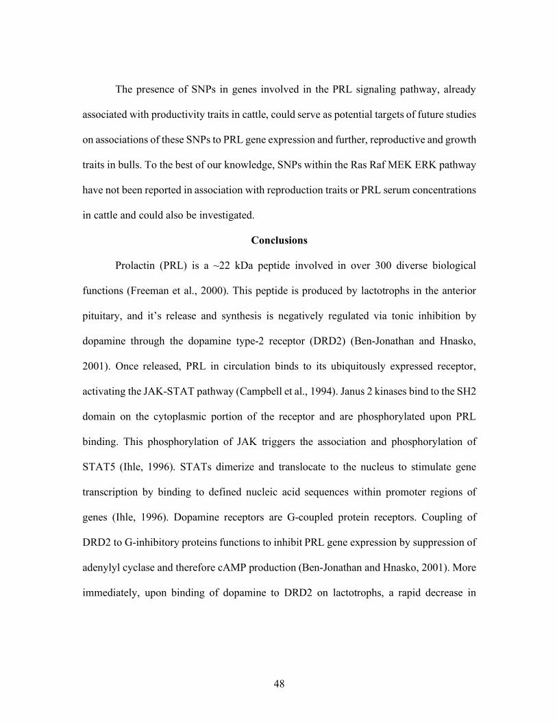
48
The presence of SNPs in genes involved in the PRL signaling pathway, already
associated with productivity traits in cattle, could serve as potential targets of future studies
on associations of these SNPs to PRL gene expression and further, reproductive and growth
traits in bulls. To the best of our knowledge, SNPs within the Ras Raf MEK ERK pathway
have not been reported in association with reproduction traits or PRL serum concentrations
in cattle and could also be investigated.
Conclusions
Prolactin (PRL) is a ~22 kDa peptide involved in over 300 diverse biological
functions (Freeman et al., 2000). This peptide is produced by lactotrophs in the anterior
pituitary, and it’s release and synthesis is negatively regulated via tonic inhibition by
dopamine through the dopamine type-2 receptor (DRD2) (Ben-Jonathan and Hnasko,
2001). Once released, PRL in circulation binds to its ubiquitously expressed receptor,
activating the JAK-STAT pathway (Campbell et al., 1994). Janus 2 kinases bind to the SH2
domain on the cytoplasmic portion of the receptor and are phosphorylated upon PRL
binding. This phosphorylation of JAK triggers the association and phosphorylation of
STAT5 (Ihle, 1996). STATs dimerize and translocate to the nucleus to stimulate gene
transcription by binding to defined nucleic acid sequences within promoter regions of
genes (Ihle, 1996). Dopamine receptors are G-coupled protein receptors. Coupling of
DRD2 to G-inhibitory proteins functions to inhibit PRL gene expression by suppression of
adenylyl cyclase and therefore cAMP production (Ben-Jonathan and Hnasko, 2001). More
immediately, upon binding of dopamine to DRD2 on lactotrophs, a rapid decrease in
49
intracellular calcium occurs suppressing the release of PRL from secretory granules (Ben-
Jonathan and Hnasko, 2001).
Cattle consuming forage containing a naturally occurring dopamine agonist, exhibit
reduced circulating serum concentrations of PRL (Schillo et al., 1987; Strickland et al.,
2011; Pratt et al., 2014). Consumption of dopamine agonists have been reported to decrease
sperm motility and morphology in bulls (Looper et al., 2009; Pratt et al., 2015). Receptors
for dopamine and PRL have been reported to be present in male reproductive tissues (Hair
et al., 2002; Otth et al., 2007; Pratt et al., 2014), therefore, either hormone may have an
effect on male reproduction. Antipsychotic drugs used in human medicine also
antagonistically bind dopamine receptors (Seeman et al., 1975) and patients taking these
dopamine antagonists experience erectile dysfunction as well as increased latency in time
to ejaculate (Segraves, 1989). In humans, hyperprolactinemia is associated with infertility
(Segal et al., 1979) and impotence (Segal et al., 1976) while hypoprolactinemia is
associated with reduced semen motility in male patients (Gonzales et al., 1989). Moreover,
early PRL and PRL receptor ablation experiments in mice resulted in infertile males
(Ormandy et al., 1997). The mechanism in which male reproductive physiology is impacted
could be due to either dopamine through binding of the DRD2 receptor at the level of the
testis and accessory glands or due to lowered serum PRL concentrations.
The PRL signaling pathway has been implicated in effects on reproductive
efficiency of several species and may provide a route for indirect effects on mammalian
reproduction (Flisikowski et al., 2003; Hermanns and Hafez, 1981; Ormandy et al., 1997;
Segal et al., 1976). Some of these effects may be explained by the presence of single
50
nucleotide polymorphisms. The SNPs could impact signaling through DRD2 or PRL
signaling and therefore could affect mammalian reproductive physiology. Evidence for
impact on male physiology due to dopaminergic SNPs exist. A polymorphism in DRD2
exon 7 in which homozygosity is prevalent results in a high association with male infertility
and impotence (Hansen et al., 2005). Further, a cytosine insertion in the DRD2 gene has
been shown to impact PRL secretion as measured by increasing blood serum
concentrations leading to erectile dysfunction (Zhang et al., 2011). A SNP has been
identified that exists within the bovine DRD2 gene, within an intron, consisting of a base
substitution between adenine and guanine (Campbell et al., 2014). Similarly, to humans,
this SNP is associated with decreased serum PRL concentrations in cattle consuming
dopamine agonists (Campbell et al., 2014). Other SNPs within genes involving the PRL
signaling pathway have been reported within PRL (Looper et al., 2010), PRLR (Ormandy
et al., 1997), and STAT5 (Brym et al., 2004) genes. These SNPs have also been implicated
in association to productivity traits in cattle and may also alter PRL synthesis and release
to subsequently impact bull reproductive physiology.
51
Figure 1.1 Dopamine type-2 receptor mediated inhibition of PRL synthesis and
release by Gi/o inhibition of adenylyl cyclase.
Dopamine binds to DRD2 coupled with G-inhibitory proteins alpha (µ), Beta (b),
and Gamma (g). Ligand binding initiates a conformational change of the receptor. An
exchange of GDP for GTP on the alpha subunit of the coupled G-inhibitory protein results
in activation and dissociation of the alpha subunit from the beta and gamma subunits. (1)
GTP-binding protein b and g subunits interact with gated inward-rectifying potassium
channels that stimulate potassium conductance. As a result, calcium channels close
resulting in decreased intracellular calcium and hyperpolarization of the cellular
membrane, decreasing secretion of PRL. (2) Interaction of alpha subunit Gi/o with adenylyl
cyclase suppresses production of cAMP, activation of protein kinase A (PKA), and
therefore, downstream PRL gene expression. Interactions of the alpha subunit are marked
by double-pointed black arrows. Inhibition is marked by red decreasing arrows and
increases in expression or abundance are marked by upward facing green arrows.
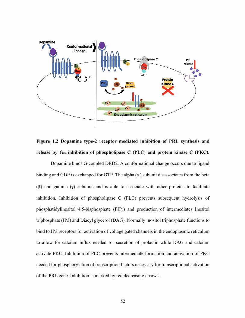
52
Figure 1.2 Dopamine type-2 receptor mediated inhibition of PRL synthesis and
release by Gi/o inhibition of phospholipase C (PLC) and protein kinase C (PKC).
Dopamine binds G-coupled DRD2. A conformational change occurs due to ligand
binding and GDP is exchanged for GTP. The alpha (a) subunit disassociates from the beta
(b) and gamma (g) subunits and is able to associate with other proteins to facilitate
inhibition. Inhibition of phospholipase C (PLC) prevents subsequent hydrolysis of
phosphatidylinositol 4,5-bisphosphate (PIP2) and production of intermediates Inositol
triphosphate (IP3) and Diacyl glycerol (DAG). Normally inositol triphosphate functions to
bind to IP3 receptors for activation of voltage gated channels in the endoplasmic reticulum
to allow for calcium influx needed for secretion of prolactin while DAG and calcium
activate PKC. Inhibition of PLC prevents intermediate formation and activation of PKC
needed for phosphorylation of transcription factors necessary for transcriptional activation
of the PRL gene. Inhibition is marked by red decreasing arrows.
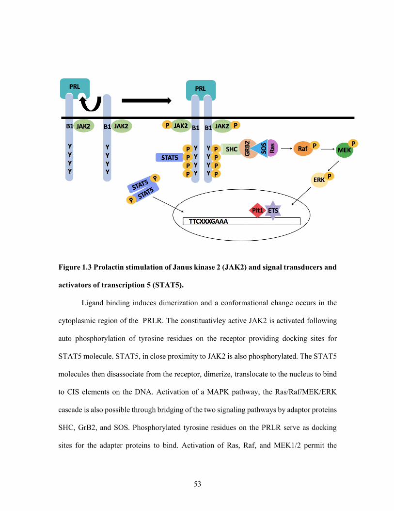
53
Figure 1.3 Prolactin stimulation of Janus kinase 2 (JAK2) and signal transducers and
activators of transcription 5 (STAT5).
Ligand binding induces dimerization and a conformational change occurs in the
cytoplasmic region of the PRLR. The constituativley active JAK2 is activated following
auto phosphorylation of tyrosine residues on the receptor providing docking sites for
STAT5 molecule. STAT5, in close proximity to JAK2 is also phosphorylated. The STAT5
molecules then disassociate from the receptor, dimerize, translocate to the nucleus to bind
to CIS elements on the DNA. Activation of a MAPK pathway, the Ras/Raf/MEK/ERK
cascade is also possible through bridging of the two signaling pathways by adaptor proteins
SHC, GrB2, and SOS. Phosphorylated tyrosine residues on the PRLR serve as docking
sites for the adapter proteins to bind. Activation of Ras, Raf, and MEK1/2 permit the

54
activation of ERK1/2 that translocate to the nucleus to modulate transcription factors such
as ETS factors.
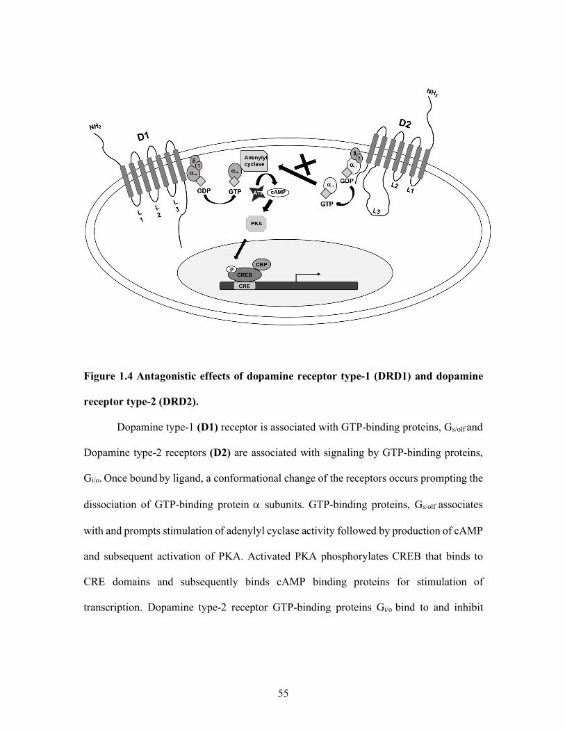
55
Figure 1.4 Antagonistic effects of dopamine receptor type-1 (DRD1) and dopamine
receptor type-2 (DRD2).
Dopamine type-1 (D1) receptor is associated with GTP-binding proteins, Gs/olf and
Dopamine type-2 receptors (D2) are associated with signaling by GTP-binding proteins,
Gi/o. Once bound by ligand, a conformational change of the receptors occurs prompting the
dissociation of GTP-binding protein a subunits. GTP-binding proteins, Gs/olf associates
with and prompts stimulation of adenylyl cyclase activity followed by production of cAMP
and subsequent activation of PKA. Activated PKA phosphorylates CREB that binds to
CRE domains and subsequently binds cAMP binding proteins for stimulation of
transcription. Dopamine type-2 receptor GTP-binding proteins Gi/o bind to and inhibit

56
adenylyl cyclase therefore preventing the signaling cascade for activation of PKA,
phosphorylation of CREB, and further, downstream transcriptional activation.
57
CHAPTER TWO
BOVINE DOPAMINE TYPE-2 RECEPTOR SNP HAS NO EFFECT ON
GROWTH, SEMEN CHARACTERISTICS AND PROLACTIN
CONCENTRATIONS IN BEEF BULLS EXPOSED TO A DOPAMINE AGONIST
Accepted for publication in Animal Reproduction Science June 4, 2019. doi:
10.1016/j.anireprosci.2019.06.005.
Formatting for this chapter may differ due to journal guidelines.
Abstract
A dopamine type-2 receptor (DRD2) SNP, previously found to be correlated with
serum prolactin (PRL) concentrations in cattle, was evaluated for impact on growth traits,
serum prolactin concentration, and semen quality. Over a four-year period, yearling beef
bulls were allowed diets containing or lacking ergot alkaloids (EA). Every 21 or 28 d semen
was collected for semen motility and morphology assessment and blood samples were
collected to measure serum PRL concentrations. In addition, body condition score and
scrotal circumference were evaluated. Serum PRL concentrations were assessed using a
radioimmunoassay. In the first year, all bulls were sacrificed at the end of a 126-day study.
Testicles and epididymis were collected at the end of the study or 60 days after removal
from treatment. Immunohistochemistry was performed on testis, epididymis, and sperm
cells, incubated with or without a primary antibody for DRD2 and counterstained with
DAPI. Isolation of DNA was performed on sperm pellets using DNAzol (Thermo Fisher
Scientific, Waltham, MA, USA) methods. Polymerase chain reaction was performed to
58
amplify the region of the DRD2 gene containing the SNP of interest. The products were
subjected to restriction fragment length polymorphism analysis. Further, all samples were
subjected to genotyping using a custom Taqman genotyping assay (Applied Biosystems,
Foster city, CA, USA). The presence of DRD2 was detected in the testis, epididymis, and
sperm cells. The DRD2 genotype was not associated with semen quality, serum PRL, or
growth traits. Consumption of EA resulted in lesser PRL serum concentrations but had no
effect on values for other variable examined.
Introduction
Prolactin (PRL) is an ~ 22 kDa peptide produced primarily by pituitary lactotrophs
and is regulated by dopamine through tonic inhibition (Ben-Jonathan et al., 2001). Prolactin
is involved in over 300 biological functions across species (Bole-Feysot et al., 1998;
Freeman et al., 2000). Hyperprolactinemia promotes erectile dysfunction as well as
infertility in men which improves through treatment with a dopamine agonist (Segal et al.,
1979). Dopamine is the main regulator of PRL via tonic inhibition (Ben-Jonathan et al.,
2001) and the bovine dopamine type-2 receptor (DRD2) gene possesses several single
nucleotide polymorphisms (SNP), with one SNP reported to be correlated with serum PRL
concentrations (Campbell et al., 2014). This SNP is located within intron 3 and therefore
does not likely affect protein structure or function and currently there is little information
available on the mechanism in which this SNP located in a non-coding portion of the gene
(Campbell et al., 2014) acts. Currently all information is associative and little direct
evaluation of the DRD2 SNP on PRL expression is available as well as little to no
evaluation has been reported on this SNP’s impact on bull growth and reproduction.
59
Dopamine type-2 receptor genotype impact on PRL serum concentrations and hair
coat scores was evaluated in steers consuming or not consuming forage containing ergot
alkaloids (EA) (Campbell et al., 2014). The DRD2 SNP located within intron 3 at position
nt 404365 on Chromosome 15, includes a base substitution between adenine and guanine
at position 534 (Campbell et al., 2014). Dopamine type-2 receptor genotype was shown to
be correlated with serum PRL concentrations, in which homozygous AA steers were
associated with higher serum PRL concentrations compared to GG steers in April and May
but not June months. While the DRD2 SNP (Campbell et al., 2014) is located within a non-
coding region and is not likely involved in protein folding or structure, the SNP may be
involved in altering gene expression within the PRL pathway. Interestingly, the SNP does
not appear to represent an alternate acceptor site to generate alternative splice variants of
the DRD2 mRNA, nor has splice variants been reported for the bovine DRD2. Alternate
splicing of DRD2 does occur to form long and short forms of the receptor to facilitate
coupling of G-proteins and has been reported in the rat and human pituitary and brain (Dal
Toso et al., 1989). Both, long and short forms of DRD2 are able to suppress PRL
transcription (McChesney et al., 1991). However, this alternative splicing of DRD2 has not
yet been reported in cattle. Evidence for effects on the PRL pathway due to dopaminergic
SNPs exists with some continued differences in severity of effects in male physiology.
Homozygosity of the polymorphism located on exon 7 of DRD2, was found to be
associated with hyperprolactinemia in humans (Hansen et al., 2005). Moreover, an
insertion-deletion located within the DRD2 promoter region was associated with sexual
dysfunction and lower serum prolactin concentrations in human male patients (Zhang et
60
al., 2011). However, the aforementioned polymorphisms were not located within a non-
coding region such as the SNP of interest in this study.
Cattle grazing forage producing EA, a naturally occurring dopamine agonist,
possess decreased serum PRL concentrations (Schillo et al., 1988; Strickland et al., 2011).
Ergot alkaloids can bind many neurotransmitter receptors (Larson et al., 1995; Mizinga et
al., 1993; Schoning et al., 2001; Wang et al., 2009) to elicit physiological responses in
cattle that consume them. Ergot alkaloids have been shown to act agonistically with DRD2,
inhibiting the synthesis and secretion of prolactin (Caron et al., 1978; Paterson et al., 1995;
Sibley et al., 1983; Thompson et al., 1993). Ergot alkaloid consumption may negatively
affect semen motility and morphology (Looper et al., 2009; Pratt et al., 2015), as well as
impact bull fertility (Schuenemann et al., 2005). However, there are contradictions between
and within laboratories. Some report no change in semen quality (Schuenemann et al.,
2005; Stowe et al., 2013) or in vivo fertility in PRL knockout mice (Horseman et al., 1997).
It is possible that EA may have an impact on male physiology by binding to DRD2
receptors located in male reproductive tissues. Moreover, DRD2 is localized in the
acrosome and tail regions of spermatazoa in bovine, rats, and mice, and is also present in
spermatocytes and spermtids within the testes of rats. Further, dopamine type-2 receptors
are also present in human spermatazoa, with the exception of the acrosome (Otth et al.,
2007). This is consistent across species with DRD2 receptors present in male reproductive
tissues (Otth et al., 2007; Ramírez et al., 2009; Gonzalez et al., 2015).
Results to date have demonstrated negative effects on growth traits in cattle
exposed to a dopamine agonist. Earlier studies report a clear decrease in average daily gain
61
(ADG) in cattle grazing EA (Hoveland et al., 1984; Crawford et al., 1989) while other
studies report no difference in BW or BCS between treatments of bulls with and without a
dopamine agonist (Stowe et al., 2013). The decreased growth in animals grazing EA may
be attributed to lower concentrations of growth hormone (GH). However, GH levels have
been reported to increase across treatment of EA (Thompson et al., 1987) or remain
unchanged (Paterson et al., 1995).
With much variation in reports on growth and semen characteristics in cattle,
clarification of the role of DRD2 genotypic effects is warranted. The objective of this study
is to assess the presence of DRD2 in bovine testis, epididymis, and sperm and to assess if
the DRD2 SNP (Campbell et al., 2014) has any impact on growth, semen quality, or serum
PRL concentrations in bulls exposed to a naturally occurring dopaminergic agonist, EA.
Materials and Methods
Animals
All animal research was approved by the Clemson University Institutional Animal
Care and Use Committee (AUP #2010-068 and AUP # 2014-60).
Experimental Design and Treatments
Over four years, 2011 (n = 14), 2012 (n = 21), 2014 (n = 25) and 2015 (n = 29),
yearling beef bulls were fed a ration that included or excluded ergot alkaloids. In 2011,
bulls were fed a concentrate ration which contained or lacked tall fescue seeds containing
or lacking EA at 0.8 µg/g dry matter (DM) (Stowe et al., 2013). In all other years bulls
were subjected to grazing forage that produced or lacked EA. Grazing treatments were
performed according to Burnett et al. (2017). Bulls were subjected to electroejaculation
62
every 21 d for all animals in year 2011 with a total grazing period of 126 d. For all other
years, electroejaculation was performed every 28 d with a total of 155 d for 2012 and 168
d for years 2014 and 2015. Breeding soundness exams (BSE) were performed on all bulls
to assess semen motility and semen morphology, scrotal circumference (SC), body
condition (BCS) and structural soundness. Bulls passing the BSE were allotted to treatment
and blocked according to body weight (BW) and BCS. Semen samples collected were
centrifuged, separated into cell and fluid portions, and each individually stored at - 80° C
until used for DNA isolation. Caudal venipuncture was performed to collect blood which
was processed into serum. Blood was allowed to clot overnight at 4° C and serum was
collected by whole blood centrifugation at 2000 x g for 15 min at 4° C and serum was
stored at - 20° C. All bulls were sacrificed at the end of a 126-day study and testicles and
epididymis collected either immediately at the end of the study (group A with 5 and 3 bulls
on E- and E+ diet, respectively) or 60 days after removal from treatment (group B with 3
bulls from each treatment) for year 2011 only. Data for growth, SC and semen quality from
all animals in all years were assessed across periods 2 (0 d) through 5 (84 d).
Radioimmunoassay
Serum PRL concentrations were determined through a previously validated
radioimmunoassay (RIA) performed by the F. Neal Schrick laboratory (Bernard et al.,
1993). The intra-assay coefficient of variation was 9.7 % and the inter-assay coefficient of
variation was 6.0 % (Burnett et al., 2017).
Immunohistochemistry and western Blotting
Formalin-fixed samples were processed and IHC performed as described by
63
Calcatera et al. (2011). Slides were washed in phosphate buffered saline (PBS) 3X, and
incubated in blocking solution of 10 % goat serum/PBS for 15 min. Blocking serum was
removed and sections were incubated with primary antibody for 1 hr at RT. A primary
mouse monoclonal Ab for DRD2 (Sc-5303; Santa Cruz Biotechnology Inc., Dallas, Texas,
USA) was diluted in blocking solution to a concentration of 4 µg/mL. Negative controls
were treated with blocking solution in place of primary antibody. Following incubation in
primary Ab, slides were washed in blocking solution followed by a final 1-hr incubation
using secondary antibody, Alexafluor 594-conjugated goat anti-mouse IgG (A11032;
Invitrogen, Carlsbad, CA, USA), diluted in blocking solution (10 µg/mL). After
incubation, secondary antibody was removed and slides were washed in PBS. Nuclei
staining was done by DAPI (0.33 µg/mL; Sigma, St Louis, MO, USA) and was followed
by PBS wash as above. Slides were mounted with PBS, coverslipped, and immediately
examined.
Protein extracts were produced from cattle testis, epididymis and pituitary tissues
by homogenization in a 1X RIPA buffer (Alfa Aesar, Ward Hill, MA) with addition of
proteinase and phosphatase inhibitors at 10µl/mL (HALT; Thermo Scientific, Waltham,
MA, USA). Concentrations of extracts were obtained using the DC microplate assay (Bio-
Rad; Hercules, CA, USA). Samples were processed by boiling 5 min in a 2X laemeli buffer
with 1/8th volume beta mercaptoethanol. Denatured samples were centrifuged briefly and
then loaded at 45µg total mass per well on a 12 % mini protean TGX precast gel (Bio-Rad;
Hercules, CA, USA). Proteins were blotted onto a 0.2 µm nitrocellulose membrane (Bio-
Rad; Hercules, CA, USA). The membrane was blocked in 2.5 % non-fat dry milk Tris
64
buffered saline with added 0.1 % Tween 20 solution (TBST). Immunodetection was carried
out using primary antibody against DRD2 (B-10) at 1:20 dilution (5303; Santa cruz
biotechnology, Dallas, TX, USA) overnight at 4° C followed by three washes in TBST and
an incubation with a goat anti-mouse IgG HRP conjugated secondary antibody (Sc-2031;
Santa cruz biotechnology, Dallas, TX, USA) for 1 hr at RT. The membrane was washed
again 3X in TBST followed by incubation with a clarity ECL western substrate (Bio-Rad;
Hercules, CA, USA) for 5 min at RT. Chemiluminescent detection was performed in a
Fluor Chem FC2 imager (Alpha Innotech; San Leandro, CA, USA) for 10 min.
RNA isolation and RT-PCR
Total RNA was isolated from testis and epididymis using the mirVana mRNA
isolation kit (Ambion, Austin, Texas, USA). Purity of RNA was assessed by the nanodrop
1000 spectrophotometer (Thermo Fisher Scientific, Wilmington, DE, USA) to obtain
A260:A280 ratio. Quality was assessed using the Agilent 2100 bioanlayzer (Agilent
Technologies Inc, Santa Clara, California, USA) ribonucleic acid integrity number (RIN),
utilizing samples with an 8.0 or greater for analysis. Complimentary DNA was generated
by reverse transcription reaction using Superscript II Frist-Strand kit (Invitrogen, Carlsbad,
Ca, USA). Primers for DRD2 were generated through IDT PrimerQuest Tool (San Jose,
CA, USA) for use in polymerase chain reaction (PCR), specific for bovine DRD2
(NM_174043.2). corresponding to nucleotide positions 510-531 forward primer and 576-
597 reverse primer of the mRNA DRD2 sequence. End-point PCR was performed using
GoTaq green (Promega Madison, WI, USA) and the resulting amplified cDNA was
subjected to slab gel electrophoresis on 1.5 % agarose gels. In addition to size analysis PCR
65
products were ligated into pDrive cloning vectors and used to transform competent E. coli
(Qiagen, Valencia, CA). Transformations were plated and subsequent colonies selected,
propagated, and plasmid DNA isolated and subjected to dideoxysequencing.
DNA isolation
Sperm pellets were suspended in PBS to approximately 1X10⁶ cells, as assessed by
sperm quality analyzer (SQA) (Advanced Agricultural Technologies) or semen samples
were aliquoted to 100µl for use in DNA isolation. Samples were centrifuged at 10,000 x g
for 1 min, supernatant removed, and the sperm pellet washed in 100 µl of 1X PBS. For cell
lysis, 1mL DNAzol (Thermo Fisher Scientific, Waltham, MA, USA) was added to each
100µl semen sample with 1/8th volume of beta mercaptoethanol (BME). Cells were lysed
by gentle pipetting. DNA was precipitated by addition of 500 µl of 100 % ethanol to each
lysed sample. Samples were inverted until spooling of DNA was visible with a white
precipitate. DNA was sedimented by centrifugation at 4,000 x g for 1 min. The supernatant
was discarded and the pellet washed in 80 % ethanol (500 µl), centrifuged at 4,000 x g for
1 min for a total of two washes. After the last wash, the DNA pellet was allowed to dry on
the benchtop for 1 min, then resusupended in 100 µl 8mM NaOH. Samples were stored up
to 1 week at 4° C until complete solubilization of the DNA was achieved. Samples were
subjected to Nanodrop 1000 spectrophotometer (Thermo Fisher Scientific, Waltham, MA,
USA) to determine A260:A280 ratio to assess purity.
Genotyping
Genotypes for each bull were obtained through restriction fragment length
polymorphism (RFLP) analysis (Campbell et al., 2014) and Taqman SNP genotyping
66
assay. In brief, polymerase chain reactions were generated for a previously identified
dopamine SNP region (Campbell et al., 2014). The subsequent products were purified
using the wizard SV gel and PCR clean-up system (PROMEGA, Durham, NC, USA) and
were subjected to slab gel electrophoresis for amplification of the dopamine SNP region
(Campbell et al., 2014) to ensure amplification was successful. Purified products (200 ng)
were digested using a Pfel (Tfil) restriction enzyme (New England Biolabs, Ipswich, MA,
USA), allowed to digest at 37° C for 30 min. Slab gel electrophoresis was performed with
each sample, digested and undigested. Digested samples yielded three genotypes with
different size products; AA (532 and 261bp), AG (793, 532 and 261bp), and GG (793bp).
A custom Taqman SNP genotyping assay (Applied Biosystems, Foster city, CA, USA)
specific to the DRD2 SNP region (Campbell et al., 2014) was performed using 10 ng of
the purified PCR products. Samples were run in 96-well format and run on the CFX Real-
Time System (BIO-RAD Hercules, CA, USA) for a single step at 95° C for 10 min
followed by 40 cycles of denaturation at 92° C for 15 sec and annealing and extension at
60° C for 1 min. Allelic discrimination analysis was performed using CFX Maestro
software (BIO-RAD Hercules, CA, USA) to determine genotype. Sample of known
genotype for each of the three genotypes were run on each plate as positive controls as well
as a no template control (NTC). These results were compared to the RFLP analysis to
ensure efficiency in genotype determination.
Statistical analysis
For serum PRL concentration, BW, BCS and semen characteristics, Analysis of
Variance, followed by pairwise student’s t-tests among LSMeans was used to assess the
67
impact of genotype and EA treatment on semen quality and growth traits. The model for
the Analysis of Variance included fixed effects for treatment, genotype, time on treatment
and their interactions; and year as a random effect. The time on treatment effect was
included in the model since animals in each year were assessed across periods 2 (0 d)
through 5 (84 d). Equality of variances were examined by Levene’s test (P << 0.05). JMP
software (SAS Institute Inc.) was used for all statistical calculations.
Results
DRD2 Expression in bull reproductive tissues
The DRD2 receptor was identified to be present in the bovine testis, epididymis,
and sperm using IHC (Figure 2.1); however, attempts using the same antibody in western
blotting did not yield definitive results (data not shown). To further confirm the presences
of DRD2 gene products, end point (RT-PCR) and dideoxysequencing of the amplified
product (Figure 2.1) were conducted and the expected product size and sequence were
produced. The receptor was present in all samples examined regardless of treatment or bull
genotype.
Genotype frequencies
Genotypic frequency was determined to be 18 % AA, 61 % AG, and 21 % GG
using RFLP and 21 % AA, 59 % AG, 20 % GG by the custom Taqman genotyping methods
across all years. Comparison of RFLP and Taqman DRD2 genotyping methods are shown
(Figure 2.3). Genotypes were identical for all bulls with the exception of 3 animals across
all years in the study. A representative sample is shown for each method RFLP and Taqman
(Figure 2.4).
68
Growth, semen characteristics, and PRL serum concentrations
Prolactin concentrations were not significantly changed across DRD2 genotype but
showed a treatment effect, with higher concentrations of serum prolactin in bulls not
consuming EA compared to bulls consuming EA (144.02 ng/mL ± 49.3; 53.10 ng/mL ±
49.3, respectively (P < 0.05) (Figure 2.2). A year by treatment by genotype random effect
was observed for PRL concentrations at a 37.6 % of total variance (P = 0.02). As expected,
period had a significant effect (P < 0.05) on BCS and BW with animals exhibiting a higher
BCS and BW earlier in treatment. Period also effected scrotal circumference, with bulls at
a lower scrotal circumference (35.4 ± 0.41; P < 0.05) at the start of the study, increasing
up to 84 d (36.3 ± 0.44; P < 0.05) (Table 2.1). A random interaction including year,
treatment, genotype, and period accounted for 9.7 % of the total variance for BCS (P =
0.03) and 0 % for BW and SC (P < 0.0001). There were no significant differences in semen
concentration due to treatment, period, genotype or their interactions. There were no
differences in motility, progressive motility, number of motile sperm cells or number of
progressively motile sperm cells due to treatment, period, genotype or their interactions.
However, period effected velocity with bulls longer on study presenting elevated semen
velocity (P < 0.05). Sperm cell morphology as well as the total number of sperm cells per
ejaculate showed no significant differences. A random effect for cell morphology including
treatment, year, genotype, and period accounted for none of the percent total variance (P =
0.009). Further, no significance was seen for total motile sperm or total progressive motile
sperm cells per ejaculate in relation to genotype, treatment, period or their interactions
(Table 2.2).
69
Discussion
There is contradiction between observations in the body of literature surrounding
the topic of dopamine agonist effects on male reproduction and physiology. The reason for
the variability may be attributed to genetic makeup of animals utilized in the studies. This
study evaluated the presence of DRD2 expression in bovine male reproductive tissues and
assessed genotypic differences in animals for the DRD2 gene in an attempt to determine
any possible differences in bovine male physiology due to a particular DRD2 SNP
(Campbell et al., 2014) on bull serum PRL concentration, growth traits, and semen quality
in bulls grazing a dopamine agonist. Using IHC techniques, the DRD2 receptor was found
to be present in bull testis, epididymis and sperm. This is similar to observations in other
species showing the presence of DRD2 receptor throughout the male reproductive system,
including sperm cells (Gonzalez et al., 2015; Otth et al., 2007; Ramírez et al., 2009). To
further verify the expression of DRD2 receptor in male reproductive tissues, end-point RT-
PCR identified products of the correct size and sequence. The primers utilized in RT-PCR
flank the intronic region the SNP of interest is located. A single product was identified
indicating no splice variants present due to genotype; however, this does not negate the
possibility of alternative spliced forms of DRD2 present in cattle. Alternate splicing of
DRD2 has been reported in rats and humans, resulting in long and short forms of the
receptor; however, both forms are functionally coupled to G-proteins and can suppress
PRL expression (Dal Toso et al., 1989; McChesney et al., 1991).
These data show that EA consumption could impact male reproductive physiology
by binding to DRD2 receptor in the testis and accessory glands could impact reproduction.
70
This binding could alter spermatogenesis, sperm maturation or the makeup of seminal fluid
impacting sperm physiology and subsequent fertility. Further, if the toxin is present in
seminal fluid, sperm physiology could be altered through binding to the DRD2 receptor or
other receptor types which may be present and are capable of binding EA. Dopamine
receptors have been well established in male physiology in both rats and humans.
Dopamine type-2 receptor agonists and antagonists have been heavily studied linking
DRD2 to initiation of ejaculation (Ferrari et al., 1994; Peeters et al., 2008; Stafford et al.,
2006). However, this retrospective study would indicate little if any effect on bovine male
growth and semen quality at time of collection due to EA consumption. In contrast, two
previous studies show an impact of EA consumption on the ability of bovine semen to
survive normal extension and freezing procedures (Burnett et al., 2017; Pratt et al., 2015),
supporting the observation of Gallagher and Senger (1989), showing that incubation of
sperm with dopamine agonists decrease sperm freezing potential.
Genotyping was originally attempted by nested PCR. In brief, end-point PCR was
performed for the F1-R793 DRD2 region (Campbell et al., 2014). The resulting products
were subjected to nested PCR with primers flanking the SNP site (Campbell et al., 2014)
for genotype (AA, AG, GG) on either the forward or reverse primer. The specific forward
and reverse primers were run with primers flanking either the end of the sequence at 793
bp or the beginning of the sequence, 1bp, respectivley. The resulting products should have
amplified only for the specific genotype, but when run with known genotyped animals,
results were inconclusive. The RFLP method was then conducted and the resulting
products were subjected to dideoxysequencing along with subcloned samples of known
71
DRD2 genotype to assess issues with specific amplification of the prior methods. Results
were again inconclusive, with clones of known genotype and resulting products of RFLP
unable to read ‘G’ in all samples sequenced, suggesting the polymerase may be unable to
read through the ‘G’ ‘C’ rich region of this particular site. The custom Taqman geontyping
assay was utilized to compare to RFLP methods. Both produced nearly identical results in
known genotyped animals, with 3 animals differing between methods, accounting for a 3.4
% error rate among calls. These three samples may have differed due to possible
contamination issues, with genotypes all resulting in AA in the taqman genotyping assay.
These samples were limiting and we were unable to genotype them again. An attempt to
isolate DNA from urine samples from these same animals was made but was unsuccessful.
The DNA isolated from urine never resulted in quality template due to contaminants and
sediment in the urine. However, the error in genotypic calls is extremely low and does not
directly impact the overall distribution within the study. This comparison of Taqman
methods to RFLP conclude both methods’ reliability for use in further studies for
genotyping.
Body weight and BCS in relation to period are in agreeance with our lab’s previous
work in which BW and BCS were unaffected by treatment alone, with BCS effected by
day (Stowe et al., 2013). When examining a subset of bulls in 2011, it was found that SC
was decreased at the end of a 126 d study when bulls were consuming a high concentration
of EA; however, considering data across all four years SC were not affected by treatment.
A lack of effect on SC may be due to the duration of the periods analyzed, as this current
study only looked out to 84 days whereas Stowe et al. (2013) extended their analysis to
72
126 days. A change in SC was observed across all bulls due to period, with prolonged time
on study resulting in a higher SC. However, these two studies differ in treatment delivery,
with the current study grazing, with the exception of year 2011, and the previous study by
Stowe et al. (2013) fed a concentrate diet throughout. Moreover, the current observations
on SC are consistent with conclusions by Looper et al. (2009) and Schuenemann et al.
(2005) who also report no change in SC due to EA exposure.
The current observations agree with the consensus of the literature to date in which
animals grazing a dopamine agonist, experience a decrease in circulating serum PRL
concentrations (Porter et al., 1992; Pratt et al., 2015; Schillo et al., 1988). However, the
conclusions on serum PRL concentration are contradictory to findings by Campbell et al.
(2014) who observed differences in serum PRL concentrations according to DRD2
genotype. In the current study, PRL levels remained consistent across DRD2 genotypes.
Campbell et al. (2014) observed a correlation with serum PRL concentrations with GG
genotypes experiencing decrease in PRL and steers with the AA genotype experiencing
higher PRL concentration when grazing E+ in May. However, they did not observe this
same effect consistently across all months that animals were on treatment. Their results for
the month of June, although attributed to heat overriding genotypic advantage, more
closely align with our current findings in which genotype had no effect on PRL
concentrations. Moreover, our study assessed effect of period on cattle grazing E+ and E-
spanning from April to August or February to August. Period was found to have no
significant effect on PRL serum concentrations.
73
Previous data are contradictory on semen quality regarding bull exposure to EA.
Ergot alkaloids have been shown to impact semen quality, with previous studies reporting
decreased semen concentration (Pratt et al., 2015), motility and morphology (Looper et al.,
2009; Pratt et al., 2015) while others have reported no effects (Schuenemann et al., 2005;
Stowe et al., 2013). Our current findings support these later observations, with no effect on
motility, concentration or morphology due to treatment or period. Further, our results
conclude that DRD2 genotype has no effect on semen quality. Interestingly, velocity was
impacted by period, with bulls longer on study exhibiting an increase in semen velocity.
This has been observed in our previous findings for bulls grazing a dopamine agonist in
which velocity was effected by day (Burnett et al., 2017) and is most likely attributed to
increasing bull age and/or seasonal change from winter to spring temperatures. These
results are contradictory to what is seen in hyperprolactinemia patients, in which velocity
is increased with hightened circulating serum PRL concetrations (Panidis et al., 1997).
Summary
Results from this research indicate that DRD2 SNP genotype (Campbell et al.,
2014) does not play a role in male physiology pertaining to semen quality, growth, or
circulating levels of serum PRL. These data are however, in agreement with a treatment
effect on serum PRL concentrations in bulls grazing EA. However, these results are
contradictory to previous findings on this SNP that did find an association of serum PRL
concentrations to SNP genotype in steers (Campbell et al., 2014). The treatment effect
observed with serum PRL concentrations decreased in bulls exposed to a dopamine agonist
indicated that the SNP did not alter structure or function of DRD2. No differences in growth

74
or semen quality due to SNP genotype also indicate that the SNP has no systemic effects.
The SNP is located within a non-coding region of the DRD2 gene, so the current
observations presented here are plausible.
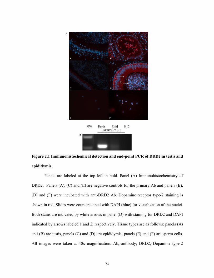
75
Figure 2.1 Immunohistochemical detection and end-point PCR of DRD2 in testis and
epididymis.
Panels are labeled at the top left in bold. Panel (A) Immunohistochemistry of
DRD2: Panels (A), (C) and (E) are negative controls for the primary Ab and panels (B),
(D) and (F) were incubated with anti-DRD2 Ab. Dopamine receptor type-2 staining is
shown in red. Slides were counterstained with DAPI (blue) for visualization of the nuclei.
Both stains are indicated by white arrows in panel (D) with staining for DRD2 and DAPI
indicated by arrows labeled 1 and 2, respectively. Tissue types are as follows: panels (A)
and (B) are testis, panels (C) and (D) are epididymis, panels (E) and (F) are sperm cells.
All images were taken at 40x magnification. Ab, antibody; DRD2, Dopamine type-2

76
receptor. Panel (B) End-point PCR: An 87 bp amplicon for DRD2 mRNA expression is
shown in testis and epididymis. MW, molecular weight ladder; DRD2, Dopamine type-2
receptor.
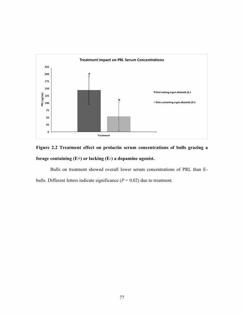
77
Figure 2.2 Treatment effect on prolactin serum concentrations of bulls grazing a
forage containing (E+) or lacking (E-) a dopamine agonist.
Bulls on treatment showed overall lower serum concentrations of PRL than E-
bulls. Different letters indicate significance (P = 0.02) due to treatment.
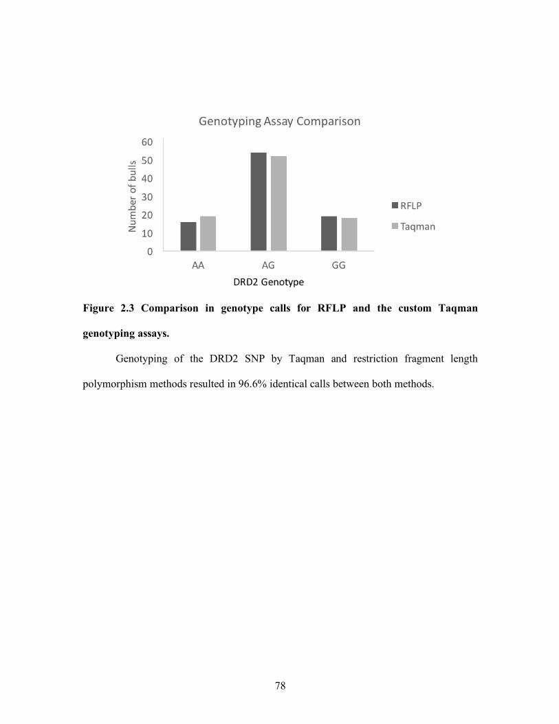
78
Figure 2.3 Comparison in genotype calls for RFLP and the custom Taqman
genotyping assays.
Genotyping of the DRD2 SNP by Taqman and restriction fragment length
polymorphism methods resulted in 96.6% identical calls between both methods.
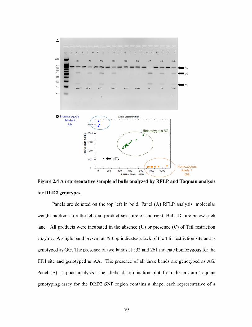
79
Figure 2.4 A representative sample of bulls analyzed by RFLP and Taqman analysis
for DRD2 genotypes.
Panels are denoted on the top left in bold. Panel (A) RFLP analysis: molecular
weight marker is on the left and product sizes are on the right. Bull IDs are below each
lane. All products were incubated in the absence (U) or presence (C) of TfiI restriction
enzyme. A single band present at 793 bp indicates a lack of the TfiI restriction site and is
genotyped as GG. The presence of two bands at 532 and 261 indicate homozygous for the
TFiI site and genotyped as AA. The presence of all three bands are genotyped as AG.
Panel (B) Taqman analysis: The allelic discrimination plot from the custom Taqman
genotyping assay for the DRD2 SNP region contains a shape, each representative of a
80
single animal with each cluster represented by a different color, representing a genotype.
Plots are represented as relative fluorescence units (RFU) for each allele. Allele 1
representing RFU for ‘G’ on the horizontal axis and Allele 2 representing ‘A’ on the
vertical axis. Genotypes are represented as the following: Orange circles, GG; Green
triangles, AG; Blue squares, AA; Black dot, non-template control (NTC).
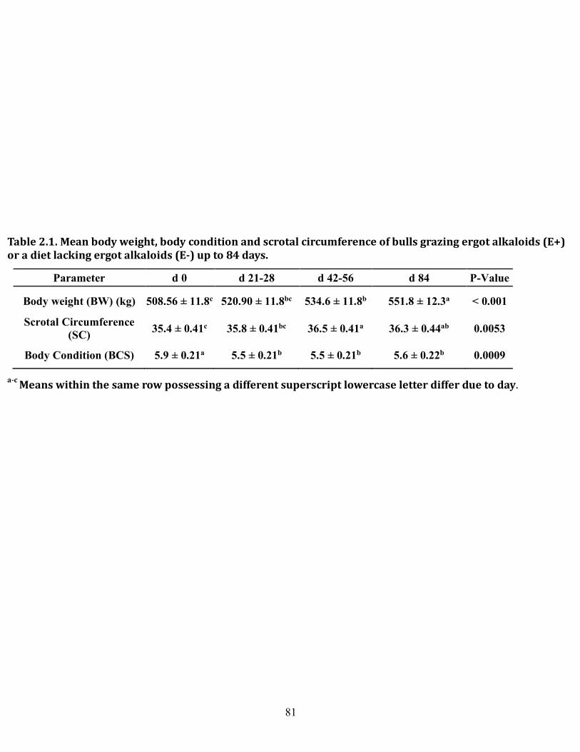
81
Parameter d 0 d 21-28 d 42-56 d 84 P-Value
Body weight (BW) (kg) 508.56 ± 11.8c 520.90 ± 11.8bc 534.6 ± 11.8b 551.8 ± 12.3a < 0.001
Scrotal Circumference (SC) 35.4 ± 0.41c 35.8 ± 0.41bc 36.5 ± 0.41a 36.3 ± 0.44ab 0.0053
Body Condition (BCS) 5.9 ± 0.21a 5.5 ± 0.21b 5.5 ± 0.21b 5.6 ± 0.22b 0.0009
Table2.1.Meanbodyweight,bodyconditionandscrotalcircumferenceofbullsgrazingergotalkaloids(E+)oradietlackingergotalkaloids(E-)upto84days.
a-cMeanswithinthesamerowpossessingadifferentsuperscriptlowercaseletterdifferduetoday.
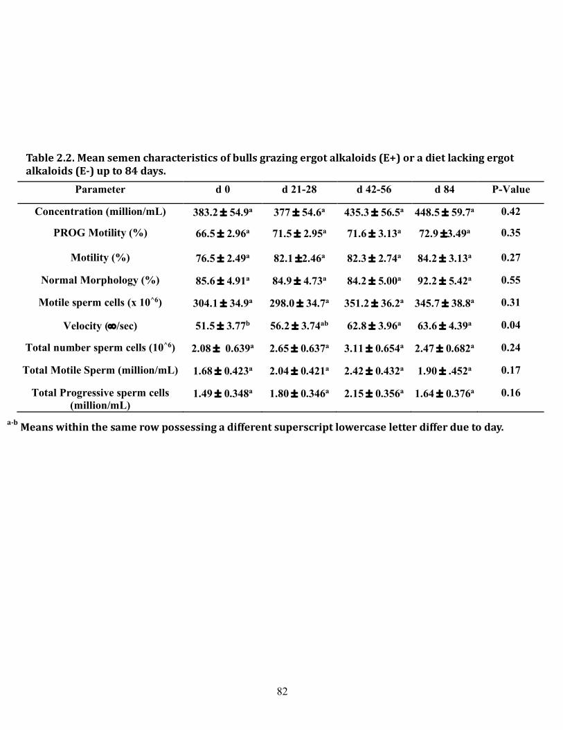
82
Parameter d 0 d 21-28 d 42-56 d 84 P-Value
Concentration (million/mL) 383.2 ± 54.9a 377 ± 54.6a 435.3 ± 56.5a 448.5 ± 59.7a 0.42
PROG Motility (%) 66.5 ± 2.96a 71.5 ± 2.95a 71.6 ± 3.13a 72.9 ±3.49a 0.35
Motility (%) 76.5 ± 2.49a 82.1 ±2.46a 82.3 ± 2.74a 84.2 ± 3.13a 0.27
Normal Morphology (%) 85.6 ± 4.91a 84.9 ± 4.73a 84.2 ± 5.00a 92.2 ± 5.42a 0.55
Motile sperm cells (x 10^6) 304.1 ± 34.9a 298.0 ± 34.7a 351.2 ± 36.2a 345.7 ± 38.8a 0.31
Velocity (¥/sec) 51.5 ± 3.77b 56.2 ± 3.74ab 62.8 ± 3.96a 63.6 ± 4.39a 0.04
Total number sperm cells (10^6) 2.08 ± 0.639a 2.65 ± 0.637a 3.11 ± 0.654a 2.47 ± 0.682a 0.24
Total Motile Sperm (million/mL) 1.68 ± 0.423a 2.04 ± 0.421a 2.42 ± 0.432a 1.90 ± .452a 0.17
Total Progressive sperm cells (million/mL)
1.49 ± 0.348a 1.80 ± 0.346a 2.15 ± 0.356a 1.64 ± 0.376a 0.16
Table2.2.Meansemencharacteristicsofbullsgrazingergotalkaloids(E+)oradietlackingergotalkaloids(E-)upto84days.
a-bMeanswithinthesamerowpossessingadifferentsuperscriptlowercaseletterdifferduetoday.

83
CHAPTER THREE
EVALUATION OF AN INTRONIC BOVINE DRD2 SNP
ASSOCIATION TO PROLACTIN PROTEIN EXPRESSION
IN BOVINE REPRODUCTIVE AND ANTERIOR PITUITARY TISSUES
Abstract
Detrimental effects to male reproductive physiology have been observed due to
changes in prolactin (PRL) serum concentration. Regulation of PRL by dopamine binding
to the dopamine type-2 receptor (DRD2) is well defined and associations between male
physiology and single nucleotide polymorphisms (SNPs) within the DRD2 gene have been
observed. Association of a bovine DRD2 SNP with serum PRL concentrations has been
previously reported in steers. In contrast, the DRD2 SNP was not associated with PRL
serum concentration, growth traits, or semen quality in bulls, warranting further
investigation on the SNP genotypic effects on PRL production and secretion in bulls. The
objective of the current study was to evaluate association of a DRD2 SNP to PRL protein
expression in bull reproductive and anterior pituitary tissues. Testis and epididymis were
collected from bulls grazing a forage containing or lacking a dopamine agonist at the end
of a 126 d study (n = 14) and bovine pituitaries (n = 587) were collected from a local
abattoir. Prolactin protein expression was assessed in all tissues by western blotting;
however, PRL protein was below detection range in reproductive tissues but was present
in pituitary, therefore experiments continued in pituitary. Sex determination of pituitaries
was performed by duplex PCR and Southern blotting methods and genotyping was
84
performed by restriction fragment length polymorphism (RFLP). The DRD2 SNP region
was amplified followed by digestion with a Tfil enzyme, producing 3,2, or 1 band (AG,
AA, GG, respectively). Pituitary genotype population was 17.4% AA, 63% AG, and 19.6%
GG. Protein expression was assessed by slot blot and densitometry analysis, normalized to
GAPDH expression (n = 92). Prolactin protein expression in the pituitary was similar
across genotype (P = 0.23) and agrees with previous observations on this SNP indicating
no genotypic effect on growth or semen quality traits in bulls.
Introduction
Prolactin (PRL) is a 22kDa peptide involved in over 300 diverse biological
functions (Freeman et al., 2000). Prolactin is produced by lactotrophs in the anterior
pituitary, and it’s release and synthesis is negatively regulated via tonic inhibition by
binding of dopamine to the dopamine type-2 receptor (DRD2) (Ben-Jonathan et al., 2001).
Once released, PRL binds to its receptor, activating the Janus Kinase-Signal transducers
and activators of transcription (JAK-STAT) pathway (Campbell et al., 1994).
Phosphorylation of JAK2 triggers the association and phosphorylation of STAT5 (Ihle et
al., 1996). Phosphorylation of STAT1, STAT3, and STAT 5 have been observed in PRL
signaling through PRLR; however, the predominant transcription factor involved is STAT5
(Gouilleux et al., 1994; Lebrun et al., 1995). Transcription factor STATs form heterodimers
and translocate to the nucleus to stimulate gene transcription by binding to defined nucleic
acid sequences within promoter regions of genes (Ihle et al., 1996). The main source of
inhibition of PRL is by suppression of adenylyl cyclase to inhibit PRL production. Cells
treated with a dopamine agonist exhibit reduced PRL transcription which can be rescued
85
by treatment with cyclic AMP (Maurer, 1981). More immediately, upon binding of
dopamine to DRD2 on lactotrophs, a rapid decrease in intracellular calcium occurs
suppressing the release of PRL from secretory granules (Ben-Jonathan et al., 2001).
Cattle consuming dopamine agonists such as ergot alkaloids, which bind to DRD2
receptors, exhibiting reduced circulating serum concentrations of PRL (Schillo et al., 1988;
Strickland et al., 2011; Pratt et al., 2015). Further, decreased semen motility and
morphology (Looper et al., 2009; Pratt et al., 2014) have also been reported in these bulls,
supporting observation in humans where hypoprolactenemia is associated with semen
motility (Gonzales, et al., 1989). In contrast, hyperprolactinemia in human males is
associated with infertility (Segal et al., 1976) and impotence (Segal et al., 1979). Further
supporting effects of PRL on male reproductive physiology, early PRLR ablation
experiments in mice resulted in infertile males (Ormandy et al., 1997). Unfortunately, later
data using knockout models did not support these observations and mechanisms in which
reproductive male physiology is impacted by PRL, whether through a direct or indirect
mechanism, remain unclear.
The PRL signaling pathway has been implicated in effects on reproductive
efficiency of several species and may provide a route for indirect effects on mammalian
reproduction (Segal et al., 1979; Gonzales et al., 1989; Ormandy et al., 1997). Some of
these indirect effects may be explained by the presence of single nucleotide polymorphisms
reported to be present in the bovine DRD2 (Campbell et al., 2014), PRL (Looper et al.,
2010), PRLR (Ormandy et al., 1997), and STAT5 (Brym et al., 2004; Flisikowski et al.,
2003) genes. The SNPs reported for these four genes result in over 2,000 possible genetic
86
combinations that could impact PRL signaling and therefore indirectly affect mammalian
reproductive physiology. Evidence for impact on male physiology due to dopaminergic
SNPs exist. A polymorphism in DRD2 exon 7 in which a cytosine to thymine substitution
at nucleotide 3420 results in homozygous genotype is highly association with
hyperprolactinemia in male patients (Hansen et al., 2005). Further, a cytosine insertion in
the DRD2 gene has been shown to impact PRL secretion as measured by increasing blood
serum concentrations leading to erectile dysfunction (Zhang et al., 2011). Recently in
cattle, a SNP has been identified that exists within the DRD2 gene located on bovine
Chromosome 15 at position 404365, on the third intron. The SNP consists of a base
substitution between adenine and guanine (Campbell et al., 2014). The bovine DRD2 SNP
yields three genotypes when digested with Tfi I, AA (532 and 261bp), AG (793, 532 and
261bp), and GG (793bp) (Campbell et al., 2014). Similarly, to humans, the DRD2 SNP
genotype homozygous ‘GG’ genotype is associated with decreased serum prolactin
concentrations in cattle exposed to a dopamine agonist (Campbell et al., 2014). The bovine
DRD2 SNP is located within a non-coding region, and therefore would not affect protein
structure/function however, its effects on PRL release and synthesis could be due to effects
on gene expression. Contrary to the prior findings, recent research indicates that genotype
does not impact male physiology, having little or no effect on semen quality parameters or
prolactin serum concentrations in bulls grazing a dopamine agonist (DeCarlo et al., 2019).
This research aims to elucidate the role of the DRD2 SNP (Campbell et al., 2014) on PRL
protein expression in the pituitary.
87
Materials and Methods
Treatment and tissue collection
Treatment of the yearling beef bulls in which testis and epididymis tissues were
collected are described by Stowe et al. (2013). In brief, bulls were fed a ration that included
or excluded ergot alkaloids at 0.8µg/g dry matter, then allowed to graze for a total period
of 126 d. Testes and epididymides were collected 60 days after removal from ergot
alkaloids.
Bovine pituitaries (n = 587) were collected at local abattoir, the anterior and
posterior lobes were carefully excised, placed into a cryotube, flash frozen in liquid N2 and
stored at -80 °C until analysis. Bovine ovaries (n = 5) were also collected from the abattoir,
transported on ice to the laboratory, and frozen at -80°C until used for further analysis.
RNA isolation and RT-PCR
Total RNA was isolated from anterior pituitary tissues using the TriZol isolation
methods (Invitrogen, Carlsbad, CA, USA) and isolated from testis and epididymis tissue
using the mirVana mRNA isolation kit (Ambion, Austin, Texas, USA). Samples were
subjected to nanodrop 1000 spectrophotometry (Thermo Fisher Scientific, Waltham, MA,
USA) to assess concentration and purity by A260:A280 ratio. Quality of RNA samples
were confirmed through Agilent 2100 nano chip assay (Agilent Technologies, Santa Clara,
CA, USA) to obtain a RIN value. Only samples that possessed a RIN of ³ 7 were utilized
for analysis.
Complimentary DNA was generated using qScript cDNA Supermix (Quantabio,
Beverly, MA, USA). Primers for PRL (NM_173953.2) flanking nucleotide positions 146-
88
431, TH (NM_173884.2) (an indirect measurement of dopamine synthesis) flanking
nucleotide positions 1263-1381, DRD2 (NM_174043.2) flanking nucleotide positions 510-
597 of the mRNA DRD2 sequence, and PRLR (L02549) flanking nucleotide positions
1107-1216 were generated through IDT primer quest tool (Integrated DNA technologies,
Coralville, IA) for use in polymerase chain reaction (PCR) (Table 3.1). End-point PCR was
performed utilizing the following reaction conditions: denaturation at 95° C for 2 min
followed by 35 cycles at 94° C for 30 sec, annealing 30 sec (PRL, 60° C; DRD2, 52° C;
TH, 52° C; PRLR, 55° C) and 72° C extension for 30 sec followed by 72° C for 5 min and
a 4° C hold. Slab gel electrophoresis was performed and the resulting products were
visualized on a 1.5% agarose gel.
To further validate identity of the products, cDNA from testis and epididymis were
ligated into a pDrive cloning vector overnight at 4° C (Qiagen PCR Plus Cloning kit,
Hilden, Germany). Ligations were used to transform competent E. coli (EZ competent;
Qiagen, Hilden, Germany). Resulting transformations were plated on LB agar plates
containing 100 µg/ml Ampicillin and IPTG/Xgal and allowed to incubate overnight at 37°
C for blue/white screening. Subsequent colonies were selected from each plate for each
respective insert, and were propagated through inoculation of LB broth, incubated
overnight, shaking to aerate at 250 rpm at 37° C. Plasmid DNA was purified using Wizard
mini-prep plus (Promega, Madison, WI), digested with EcoR1 (Promega, Madison, WI,
USA) and products of the expected sizes were subjected to dideoxysequencing.
DNA Isolation
89
Isolations of genomic DNA were performed according to Current Protocols in
Molecular Biology (Wiley and Sons, Inc., 1998). Posterior pituitary lobe tissues (200 mg)
and ovary (50 mg) were homogenized directly in DNA lysis buffer (100 mM NaCl, 10 mM
TrisCl pH 8, 25 mM EDTA pH8, 0.5 % SDS, 0.1 mg/ml proteinase K) (n = 587).
Phenol:chloroform:isoamyl alcohol (25:24:1; pH 8) were added to the homogenized
sample, equal to the volume of the lysate. Samples were vortexed for 1 min and centrifuged
at 1,700 x g for 10 min at RT. The aqueous layer was carefully aspirated and put into a
sterile tube, followed by addition of 0.5 volume of 7.5 M ammonium acetate and 2 vol of
100 % ethanol relative to the aqueous layer volume. Resulting precipitated DNA was
pelleted at 1700 x g for 2 min at RT and DNA pellet saved and the supernatant decanted.
The DNA pellet was washed in 70 % ethanol and allowed to dry for 15 min followed by
resuspension in 100 µl of nuclease free water. Samples were heated at 65° C for 30 min
and then stored at 4° C until further analysis of sex and genotype. Isolated DNA was
evaluated for concentration and nucleic acid to protein ratio (260/280) by
spectrophotometry (Nanodrop 1000; Thermofisher).
Sex Determination
Sex was determined (n = 587) utilizing a duplex PCR method described by Curry
et al. (2008) and Southern blotting techniques. In short, 2 ng of DNA was amplified in a
25 µl PCR reaction (GoTaq Green; Promega, Madison, WI, USA) including 10 µl of Go-
taq, 1 µl of each β-actin primer and 2 µl of each SRY primer. Controls, including a
known male sample (testis) and a known female sample (ovary) were also subjected to
duplex PCR amplification. Single products of β-actin and SRY generated from 2ng of
90
purified plasmid DNA products, verified by dideoxysequencing served as additional
controls on each blot. Reaction conditions consisted of 95° C for 2 min, 94° C for 30 sec,
annealing at 60° C for 30 sec and extension at 72° C for 30 sec for a total of 35 cycles,
followed by 72° C for 5 min and a 4° C hold. Reactions were treated according to Curry
and others (2008). In brief, samples were denatured by addition of NaOH (0.1 vol) and
heated at 37° C for 5 min followed by addition of SSPE (3 M NaCl, 0.2 M NaH2PO4,
0.02 M EDTA) at a final 6X concentration. Reactions were blotted using a dot blot
manifold. Nylon membrane (Biobond-Plus Nylon Membrane; Sigma, St. Louis, MO,
USA) was soaked in nuclease free water for 10 min prior to assembling the blot manifold.
The manifold was set up with the pre-soaked nylon membrane placed on top of two
Whattman filter papers. Each well was washed initially with 300 µl nuclease-free water
followed by blotting of products. After addition of samples to the membrane, the wells
were washed with 300 µl 0.4 M NaOH. The manifold was disassembled and the resulting
blot was rinsed briefly in 2X SSC (20X solution; 0.3 M sodium citrate, 3 M NaCl, pH
7.0) and allowed to dry completely before crosslinking at 120 cm2/j for 1 min (XL-1000
UV crosslinker).
Amplification of single, β-actin and SRY products were generated using 2 ng
genomic DNA (GoTaq Green; Promega, Madison, WI, USA). Reaction conditions are as
described above. Purified products were ligated into pDrive cloning vector (Qiagen PCR
Plus Cloning kit, Hilden, Germany), subcloned, and resulting transformations plated.
Vectors containing the inserts of interest were propagated and plasmids isolated. Inserts
were amplified using end-point PCR for β-actin and SRY (Genewiz, Plainfield, NJ, USA).
91
Resulting PCR products were purified (Wizard SV Gel and PCR Clean-Up System,
Promega, Madison, WI, USA) and subjected to spectrophotometry to assess concentration
and quality by 260/280 nucleic acid to protein ratio. Samples with a ratio of 1.8 - 2.0 were
utilized for generation of probes. Labeling was then carried out by random prime labeling
(North2South Biotin Random Prime DNA Labeling Kit, Thermo Fisher Scientific,
Waltham, MA, USA). Probes were generated using 200 ng of purified PCR products in a
final volume of 24 µl with nuclease-free water followed by addition of 10 µl
heptanucleotide mix. Samples were denatured by boiling for 5 min and quickly placed in a
methanol dry ice bath. The samples were thawed on ice followed by the addition of 10 µl
of 5X dNTP mix, 5 µl of 10X reaction buffer, and 1 µl Klenow fragment for a final volume
of 50 µl. The samples were then incubated at 37° C for 1 hr following inactivation with
addition of 2 µl 500 nM EDTA (pH 8.0). Unincorporated nucleotides were removed by
size exclusion chromatography. Sephadex G50 (1 g) was allowed to swell in approximately
10 mL phosphate buffered saline (PBS) (1X) at RT overnight. A column was produced
using a 1 mL syringe with steel wool packed into the bottom measuring to approximately
0.1 mL on the syringe. The swelled sephadex mixture was loaded into the syringe barrel to
the top and allowed to drip for 1 hr at RT followed by centrifuging at 2000 x g for 1 min
at RT to pack the column. The column was placed into a 15 mL conical tube to catch flow-
through and was initially washed with PBS (100 µl) by slow addition to the top of the
column and centrifugation at 2000 x g for 1 min at RT. The flow through was discarded
and the column was placed into a new sterile tube. The full volume of the labeled probe
was then loaded onto the column followed by centrifugation, 2000 x g for 1 min. The flow-
92
through was saved and the column was washed once more with 100 µl PBS utilizing the
same flow-through tube. The resulting flow through was transferred to a new sterile tube
and subjected to denaturation by boiling for 10 min followed by 5 min incubation on ice.
Pre-hybridization, hybridization, and chemiluminescent detection were performed
using the North2South Chemiluminescent Hybridization and Detection Kit (Thermo Fisher
Scientific, Waltham, MA, USA). Kit contents were allowed to come to RT or incubated at
37° C until no precipitate existed before use. Pre-hybridization of blots were performed in
two separate hybridization tubes in a hybridization oven at 55° C (Problot Hybridization
Oven, Labnet, International) in 10 mL of pre-hybridization buffer for 30 min followed by
addition of probe, either β-actin or SRY, directly to the buffer for each blot (hybridization
buffer). Hybridization was carried out overnight at 55° C, rotating.
The hybridization buffer was discarded and each blot washed 3X in stringency
wash buffer at 55° C for 15 min each wash, rotating. The membranes were then blocked
for 15 min at RT with gentle agitation. Streptavidin was diluted 1:300 in blocking buffer
and then added to the membrane to incubate again for 15 min at RT with gentle agitation.
The solution was then decanted and the blots were incubated in substrate equilibration
buffer for 5 min at RT. The blots were then covered in substrate for 5 min at RT then
exposed to chemiluminescent detection for 2 min (FluorChem FC2, Alpha Innotech, San
Leandro, CA, USA). Confirmation of males was performed by densitometry. Samples were
considered male if they met or exceeded the density of the testis control on each blot.
Samples that came up inconclusive, in which signal density was just below or between the
93
threshold of background versus male control sample, were blotted an additional time to
assure accuracy in calls.
Genotyping
Genotyping of male pituitaries (n = 259) were performed according to the
restriction fragment length polymorphism method described by DeCarlo et al. (2019), with
modifications. In brief, amplification of the DRD2 region flanking a SNP (A534G) was
performed (Campbell et al., 2014) with 10 ng starting template genomic DNA (GoTaq
Green Master Mix, Promega, Madison, WI, USA). Resulting products were digested with
Tfil enzyme overnight at 37° C (ThermoFisher Scientific). Digested products were
visualized on a 1.0 % agarose gel.
SDS-PAGE and western blotting
Protein extracts were produced from testis, epididymis, ovary, and anterior pituitary
tissues (100 mg starting material). Samples were homogenized in an ice-cold lysis buffer
(1X RIPA with 10 µl/mL Protease inhibitor HALT). Protein concentrations were
determined using the DC protein assay with each sample performed in duplicate (Bio-Rad;
Hercules, CA). Samples were stored at -20° C until used for further protein analysis.
Protein extracts of testis, epididymis, ovary, and anterior pituitary were diluted in
molecular grade water and 2X laemmali buffer with addition of 1/8th volume of beta
mercaptoethanol to a total mass of 30 µg and 60 µg, for detection of PRL and GAPDH,
respectively. Samples were denatured for 5 min by boiling and directly chilled on ice for 5
min. Denatured samples were centrifuged at 13,000 x g for 10 min and loaded onto a 12 %
mini protean TGX precast polyacrylamide gel at a total volume of 20 µl per well (Bio-Rad;
94
Hercules, CA). Gel electrophoresis was performed at 75 volts for 1 hr and 15 min and 200
volts for 20 min for GAPDH and PRL, respectively. Proteins were blotted onto a 0.2 µm
nitrocellulose membrane (Bio-Rad, Hercules, CA, USA) at 100 volts for 1 hr. Transfer of
proteins was assessed by staining with Ponceau S for 5 min and the polyacrylamide gel
stained with coimmasse blue overnight. The membrane was allowed to dry overnight at
RT, protected from light.
For the detection of PRL, blotted proteins (30 µg) were blocked in 3.5 % non-fat
dry milk in a tris-buffered saline buffer with 0.1 % Tween added (TBST) for 1 hr at RT
with gentle rocking. The membrane was briefly washed in TBST followed by the addition
of PRL primary antibody diluted to 1:1000 in 1 % non-fat dry milk/ TBST (sc-69874; Santa
Cruz Biotechnology, Dallas, TX, USA) and allowed to incubate overnight at 4° C. The blot
was then washed in TBST 3X, 5 min each wash. Secondary antibody was diluted 1:1000
in TBST (610-703-124; Rockland, Limerick, PA, USA) was added and allowed to incubate
at RT for 1 hr followed by three washes in TBST, 5 min each. The membrane was laid on
an acetate sheet and covered in substrate (Clarity Western ECL Substrate; Bio-Rad
Hercules, CA, USA) for 5 min at RT. The substrate was drained from the blot, covered by
another piece of acetate, and exposed to chemiluminescent detection for 15 min (Alpha
Innotech; San Leandro, CA, USA).
For the detection of GAPDH, blotted proteins (60 µg) were blocked in 5 % non-fat
dry milk in TBST for 1 hr at RT with gentle rocking. The membrane was briefly washed
in TBST followed by the addition of GAPDH primary antibody diluted to 1:1000 in 5 %
BSA/TBST solution (2118; Cell Signaling Technology, Danvers, MA, USA) and allowed
95
to incubate overnight at 4° C. The membrane was then washed in TBST 3X, 5 min each
wash. Secondary antibody was diluted 1:1000 in TBST (7074; Cell Signaling Technology,
Danvers, MA, USA) was added and allowed to incubate at RT for 1 hr followed by three
washes in TBST, 5 min each. Substrate was added to the blot and after 5 min exposed to
chemiluminescent detection for 15 min.
Evaluation of PRL protein expression in bovine pituitary
Linear range of detection was determined by blotting increasing mass of protein by
slot blot manifold followed by exposures at varying times by chemiluminescent detection.
Blotting by slot blot manifold was performed using 6 size No. 1 Whattman filter papers
topped with a 0.2 µm nitrocellulose membrane. The manifold was sealed and each well
washed with 200 µl of nuclease-free water followed by application of vacuum. Anterior
pituitary protein (AA n = 16; AG n = 58; GG n = 18) (10 µg) was then blotted followed by
an additional wash with deionized water. Blots were then stained with Ponceau S (Sigma-
Aldrich, St. Louis, MO, USA) to assess transfer, rinsed with TBST briefly and treated with
hydrogen peroxide (3 %) at 37° C for 10 min with gentle agitation to remove peroxidase
activity. Blots were then placed in non-fat dry milk blocking solution and immune-
detection carried out as described above for GAPDH. Chemiluminescent detection was
performed using an Fluorchem FC2 (alpha innotech, San Leandro, CA, USA) for a 15 min
exposure. Blots were then washed in TBST for 5 min and stripped using two washes of
glycine (400nm, pH 2.5) for 5 min each at RT, followed by two PBS washes and a TBST
wash for 5 min each. Immuno-detection for PRL was then carried out according to the
protocol outlined above. Membranes were again exposed at 15 min for chemiluminescent

96
detection. Total protein on each blot was also evaluated by densitometry utilizing Ponceau
S stain. Inter- and intra-assay coefficients were 7.4 % and 6.9 %, respectively.
Statistical analysis
Densitometry data for PRL protein expression was normalized to a GAPDH
housekeeping protein density as follows. Background subtraction was applied to each
GAPDH and PRL sample densities for all samples in duplicate. Blot normalization factors
were calculated for each blot (highest GAPDH expression value on blot/ GAPDH signal
of each individual sample). The density (after background subtraction) of each sample for
PRL signal was then divided by the blot normalization factor to obtain relative protein
expression. Analysis of Variance was performed for all densitometry data to assess DRD2
genotype impact on pituitary PRL protein expression. The model for the Analysis of
Variance included genotype as a fixed effect. Significance was set at (P < 0.05) and all
statistical analysis was performed using JMP software (SAS Institute Inc.).
Results
Messenger RNA and protein expression
The messages for PRL and TH were present in testis and epididymis (figure 3.1).
Previously published work also confirms the presence of DRD2 in testis and epididymis
(DeCarlo et al., 2019). Further work to assess protein expression of PRL in testis and
epididymis by western blotting was unsuccessful as the protein concentrations may be
below detection range in these tissues. Prolactin (~22kDa) was present in pituitary as
expected and therefore, experiments continued in bovine anterior pituitary (Figure 3.2).
97
Amplifications of PRL, DRD2, PRLR, and TH were performed in anterior pituitary with
the message for all present (Figure 3.3).
Sexing and genotypic distribution
Sexing by PCR and Southern blot revealed 55.9% females and 44.1% males. An
example of dot blots probed with SRY and β-actin are shown in Figure 3.4. Genotyping
was performed by RFLP procedures. Genotypic distribution was determined to be AA
17.4%, AG 63%, and GG 19.6%.
Prolactin protein expression in anterior pituitaries
Prolactin protein expression was evaluated across genotype by slot blot and
immunodetection procedures. Densitometry analysis revealed no differences in PRL
protein expression due to DRD2 genotype (P = 0.23) (Figure 3.5).
Discussion
Initial goals of these experiments were to assess if the message for PRL and its
regulatory system (DRD2 and its rate-limiting enzyme tyrosine hydroxylase) were present
in male reproductive tissues. Our findings indicate that the message for PRL and TH are
present in bovine testis and epididymis. These findings agree with other species in which
PRL has been identified in semen, male accessory glands (Nevalainen et al., 1997; Sanford
and Baker, 2010) and germ cells (Pratt et al. 2014; Sheth et al., 1975). Dopamine type-2
receptor has also been identified in male germ cells (Otth et al., 2007; Ramirez et al., 2009)
as well as in the testes of mice (Gonzalez et al., 2015) and most recently in cattle testes,
epididymides, and germ cells (DeCarlo et al., 2019).
98
We were not able to detect PRL in any testis, epididymis or ovary by western
blotting. Given that the message for PRL is present in testis and epididymis, it is possible
that PRL protein expression was just below the detection range for western blotting. Bovine
anterior pituitary, the main site of production of PRL, did reveal ~22 kDa band as expected.
without possible detection of PRL protein in testis and epididymis, experiments continued
in the pituitary to examine possible genotypic effects of the DRD2 SNP previously
associated with decreased serum PRL concentrations.
Slot blotting of anterior pituitary protein was performed to assess PRL protein
expression across DRD2 SNP genotype. Original experiments to assess antibody
specificity revealed evidence of high peroxidase activity in bovine anterior pituitary
protein. To combat this issue, membranes with slot blotted anterior pituitary protein were
treated with a 3% hydrogen peroxide solution prior to immunodetection procedures
(Figure A.1). Experiments to determine optimal time, temperature, and hydrogen
peroxide concentration to eliminate peroxidase activity were performed (Appendix A).
Sex determination of pituitaries were performed by duplex PCR and Southern
blotting techniques. Original DNA isolations were performed using only 50 mg of posterior
pituitary tissue starting material and a simple phenol chloroform extraction method that
resulted in genomic DNA isolations containing large quantities of RNA. Increased
specificity of Southern blotting utilizing hybridization to combat this issue. Isolations were
then performed with a much higher, 200 mg of tissue for genotyping. The RFLP
genotyping protocol was adapted from the previously used protocol (DeCarlo et al., 2019)
in order to eliminate the need for the PCR purification step prior to digestion. Experiments
99
were performed with known genotyped samples to ensure enzyme digestion accuracy (data
not shown) and an overnight digestion was able to allow for immediate digestion after
amplification of the SNP sequence, without PCR clean-up.
The DRD2 SNP (A534G) is located in a non-coding region and therefore is not
expected to affect protein folding or structure but possibly could affect gene expression
within the PRL pathway. These data indicate no association of DRD2 SNP genotype to
PRL protein expression in the bovine anterior pituitary. Previous studies on this DRD2
SNP showed no changes in serum PRL concentrations observed due to SNP genotype
(DeCarlo et al., 2019). These two observations would indicate that the reported bovine
DRD2 SNP does not affect the production or release of PRL. The genotypic distribution
presented here is nearly identical to previous reported distributions for this DRD2 SNP
(Meyer et al., 2016; DeCarlo et al., 2019) but are contradictory to Campbell et al. (2014)
who observed a genotypic effect on PRL serum concentrations. The same study also
reported a genotypic effect associated with hair coat score, which has been linked to PRL
levels in cattle (Campbell et al., 2014); however, Meyer and others (2016) only observed a
tendency for this association, although they did not directly measure serum PRL
concentrations. Observations from this current study agree with findings by Meyer and
others (2016) in which no change in PRL protein was observed according to genotype.
Further, genotyping methods used in this study have been validated by a second genotyping
method previously (DeCarlo et al., 2019), while no validation was used for Campbell and
others (2014). The DRD2 SNP examined here is located in a non-coding region and
therefore it is not surprising there was no change in PRL protein expression due to
100
genotype. Many SNPs within other genes affecting the PRL signaling pathway exist (Brym
et al., 2005; Looper et al., 2010) and may provide more informative avenues to explore
when assessing SNP genotypic effects on PRL synthesis and secretion.
Summary
In summary, the message for PRL, TH, and DRD2 are present in bovine testis,
epididymis, and pituitary. The message for DRD2 has previously been reported in testis
and epididymis (DeCarlo et al., 2019). The presence of PRL protein was observed in
pituitary protein extracts and was not detectable in testis, epididymis or ovary samples.
These data indicate that PRL protein expression may be lower than what is possible to
detect by western blotting in male reproductive tissues. Experiments continued at the
source of PRL production, the pituitary.
Genotyping ratios were extremely similar to what has previously been reported for
this SNP (Meyer et al., 2016; DeCarlo et al., 2019) and for what is closest to a normal,
random distribution (25:50:25). Further investigation of relative protein expression across
genotype in the pituitary revealed no differences due to DRD2 genotype. These data agree
with previous findings from this laboratory in which this DRD2 SNP had no effect on PRL
serum concentrations and furthermore, no effects on growth, reproduction, and semen
characteristics (DeCarlo et al., 2019). These data are contradictory to what has been
previously reported on this DRD2 SNP, which indicate a genotype association to PRL
serum concentrations (Campbell et al., 2014). This DRD2 SNP is located within an intron
and does not likely affect protein structure but could affect gene expression; however, these
data suggest no association between protein expression to the intronic DRD2 SNP.
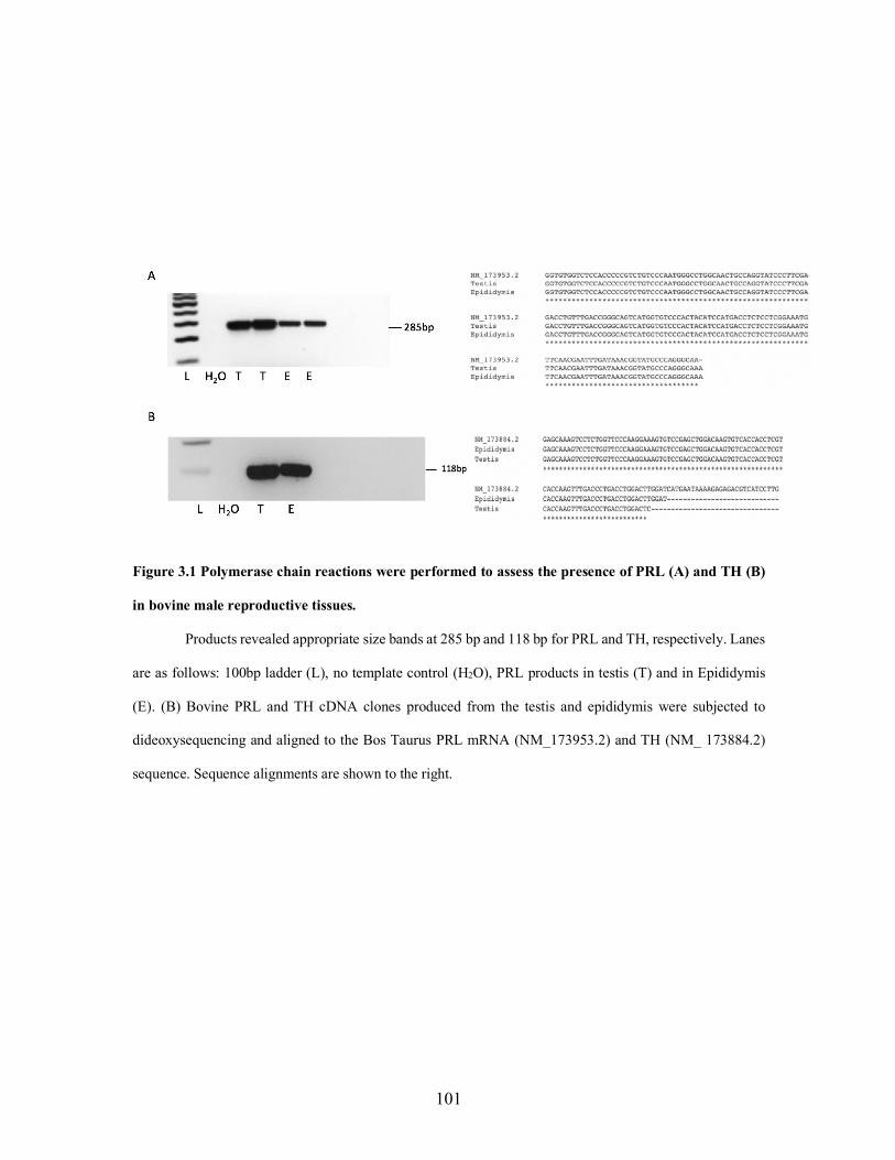
101
Figure 3.1 Polymerase chain reactions were performed to assess the presence of PRL (A) and TH (B)
in bovine male reproductive tissues.
Products revealed appropriate size bands at 285 bp and 118 bp for PRL and TH, respectively. Lanes
are as follows: 100bp ladder (L), no template control (H2O), PRL products in testis (T) and in Epididymis
(E). (B) Bovine PRL and TH cDNA clones produced from the testis and epididymis were subjected to
dideoxysequencing and aligned to the Bos Taurus PRL mRNA (NM_173953.2) and TH (NM_ 173884.2)
sequence. Sequence alignments are shown to the right.
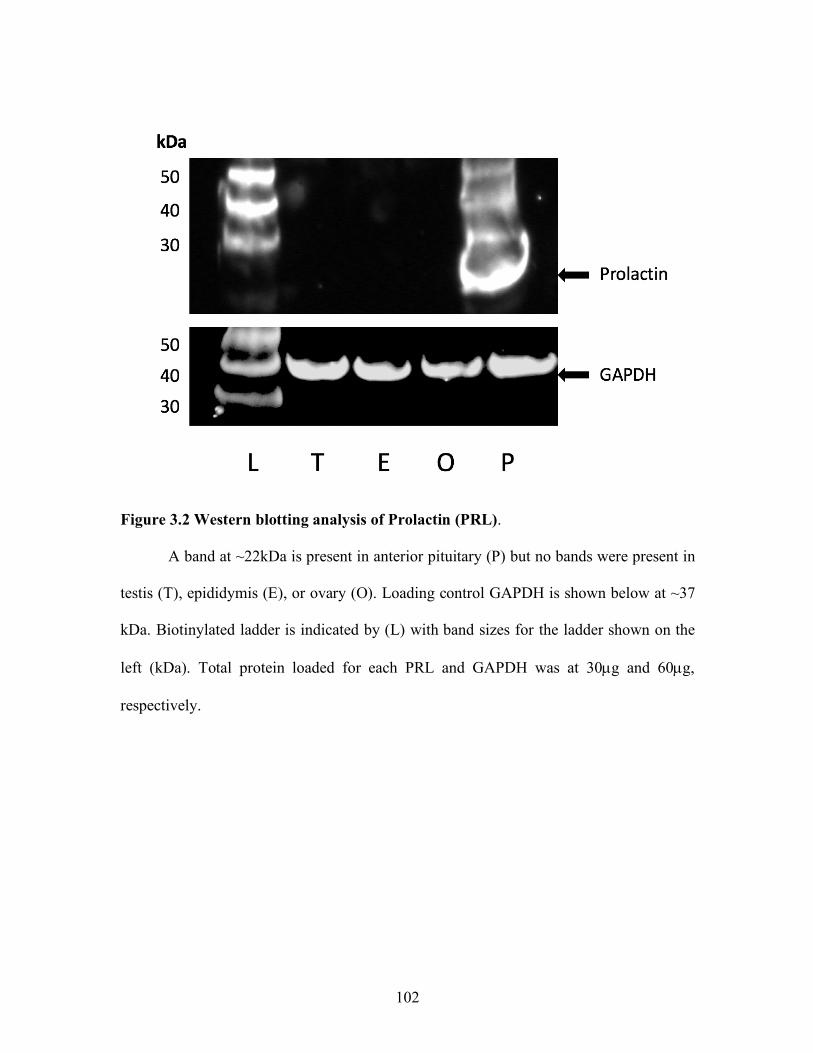
102
Figure 3.2 Western blotting analysis of Prolactin (PRL).
A band at ~22kDa is present in anterior pituitary (P) but no bands were present in
testis (T), epididymis (E), or ovary (O). Loading control GAPDH is shown below at ~37
kDa. Biotinylated ladder is indicated by (L) with band sizes for the ladder shown on the
left (kDa). Total protein loaded for each PRL and GAPDH was at 30µg and 60µg,
respectively.
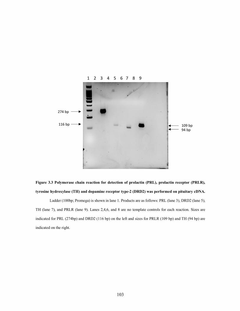
103
Figure 3.3 Polymerase chain reaction for detection of prolactin (PRL), prolactin receptor (PRLR),
tyrosine hydroxylase (TH) and dopamine receptor type-2 (DRD2) was performed on pituitary cDNA.
Ladder (100bp; Promega) is shown in lane 1. Products are as follows: PRL (lane 3), DRD2 (lane 5),
TH (lane 7), and PRLR (lane 9). Lanes 2,4,6, and 8 are no template controls for each reaction. Sizes are
indicated for PRL (274bp) and DRD2 (116 bp) on the left and sizes for PRLR (109 bp) and TH (94 bp) are
indicated on the right.
274 bp
116 bp 109 bp 94 bp
1 2 3 4 5 6 7 8 9
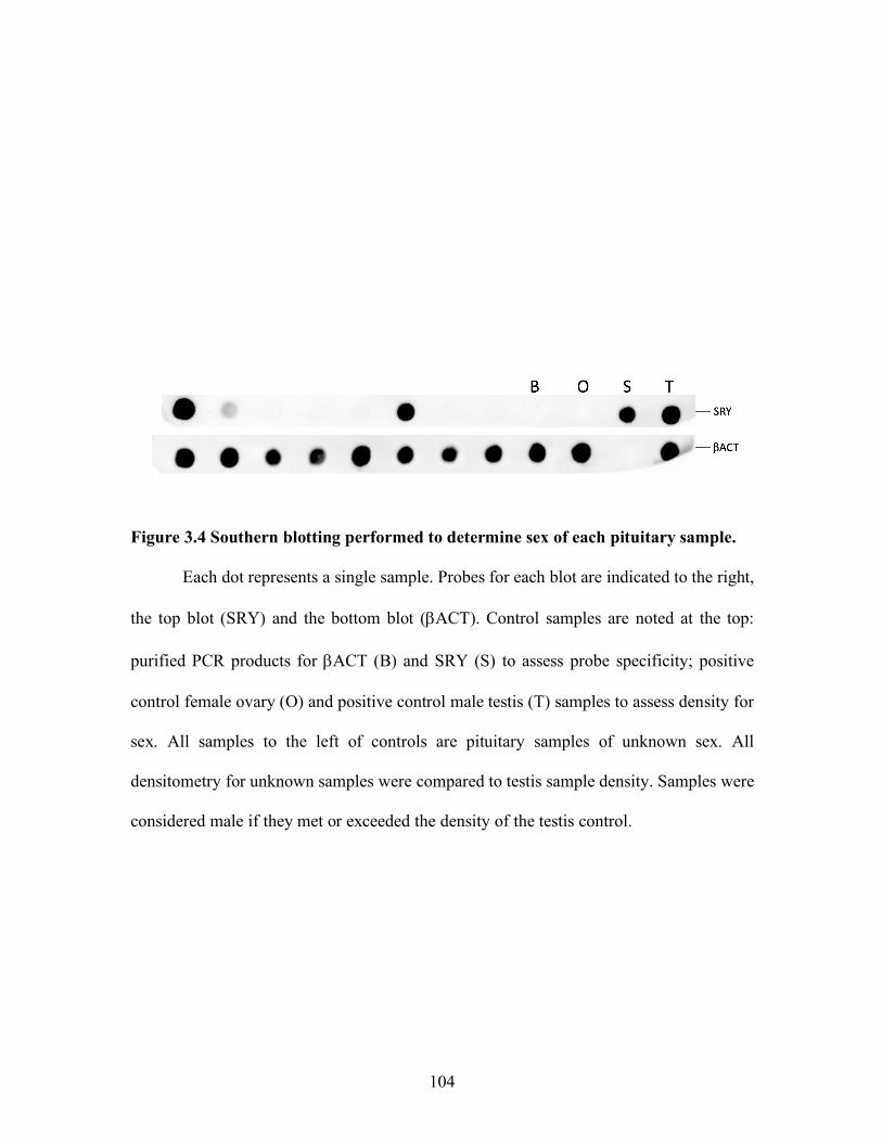
104
Figure 3.4 Southern blotting performed to determine sex of each pituitary sample.
Each dot represents a single sample. Probes for each blot are indicated to the right,
the top blot (SRY) and the bottom blot (bACT). Control samples are noted at the top:
purified PCR products for bACT (B) and SRY (S) to assess probe specificity; positive
control female ovary (O) and positive control male testis (T) samples to assess density for
sex. All samples to the left of controls are pituitary samples of unknown sex. All
densitometry for unknown samples were compared to testis sample density. Samples were
considered male if they met or exceeded the density of the testis control.
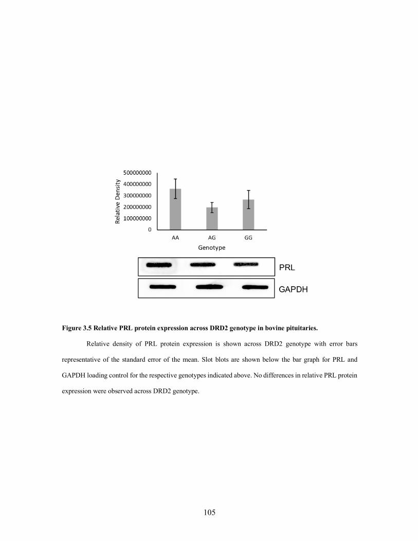
105
Figure 3.5 Relative PRL protein expression across DRD2 genotype in bovine pituitaries.
Relative density of PRL protein expression is shown across DRD2 genotype with error bars
representative of the standard error of the mean. Slot blots are shown below the bar graph for PRL and
GAPDH loading control for the respective genotypes indicated above. No differences in relative PRL protein
expression were observed across DRD2 genotype.
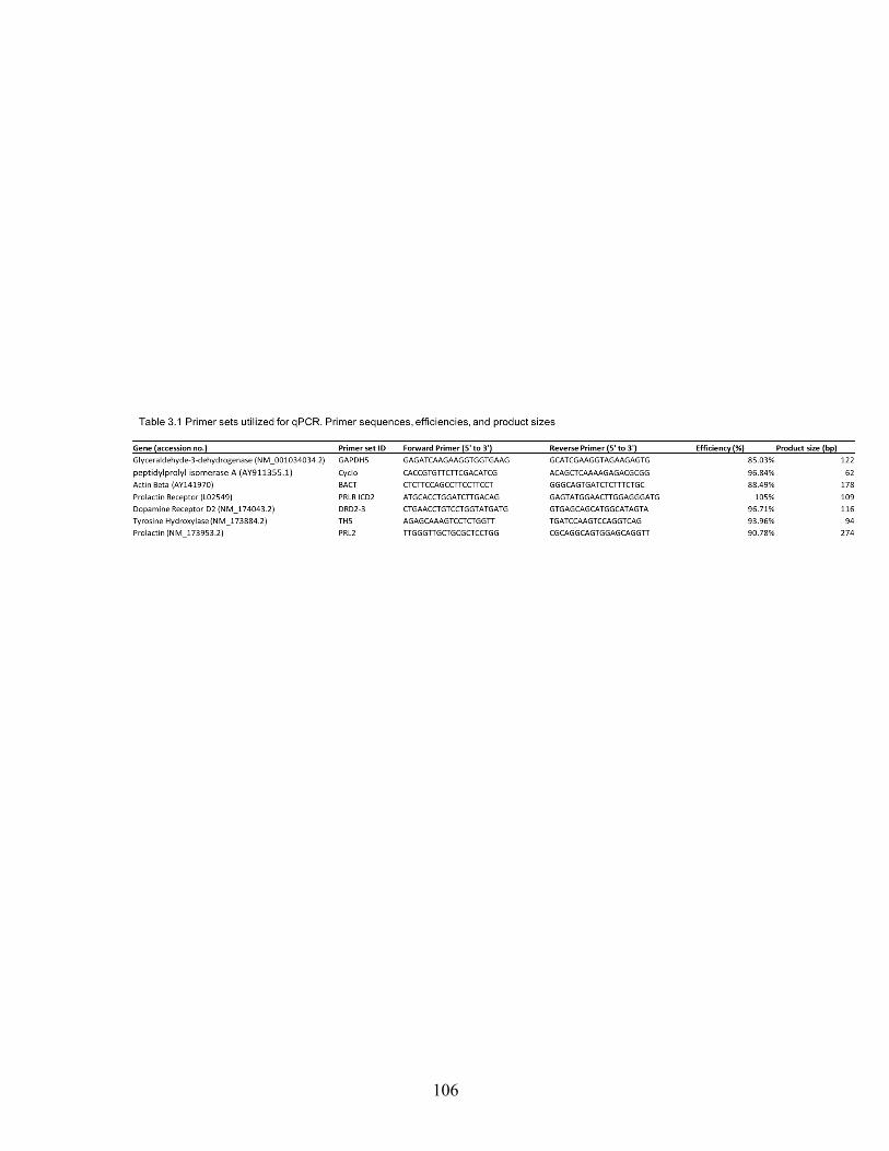
106
107
OVERALL CONCLUSIONS
The goals of these experiments were to evaluate an intronic DRD2 SNP, previously
found to be associated with decreased bovine serum PRL concentrations. Associations
assessed included DRD2 genotype association with growth traits, semen quality, PRL
serum concentrations, mRNA expression in male reproductive tissues, and PRL protein
expression in bovine pituitaries.
Evaluation of DRD2 genotype association with growth traits, semen quality, and
PRL serum concentrations was performed on bulls grazing (E+) or not grazing (E-) a diet
containing a dopamine agonist. The presence of DRD2 was observed in testis and
epididymis by immunohistochemistry and polymerase chain reaction.
Genomic DNA was isolated from semen and used for genotyping by restriction
fragment length polymorphism and TaqMan techniques. Both techniques were almost
identical with genotypic frequencies at 18 % AA, 61 % AG, and 21 % GG using RFLP and
21 % AA, 59 % AG, 20 % GG by the custom Taqman genotyping methods. Results on
association of DRD2 genotype on serum PRL concentrations is contradictory to what has
been previously found, in which there was no association found. However, this is consistent
with a treatment effect in which E+ bulls experience a decrease in PRL serum
concentrations. No differences were observed in growth traits and semen quality
parameters due to genotype.
The message for DRD2, PRL, and TH was present in the testis, epididymis, and
pituitary. Further, DRD2 was also present in sperm cells and the message for PRLR was
also identified in bovine pituitary. Protein expression was evaluated in testis, epididymis,
108
ovary, and pituitary tissues. Western blotting and immunodetection revealed PRL
expression in the pituitary as expected, but we were unable to detect PRL in reproductive
tissues, as they may be below detection range. Experiments continued in the pituitary to
evaluate PRL protein concentrations across genotype.
Bovine pituitaries were collected at a local abattoir. A duplex PCR and southern
blotting technique were utilized to probe for SRY and Beta actin for sexing of samples.
Subsequent male samples were genotyped by restriction fragment length polymorphism
method for the DRD2 SNP. Genotypic frequencies were almost identical to our previous
report at AA 17.4 %, AG 63 %, and GG 19.6 %. Blotting of anterior pituitary protein and
immunodetection for PRL was performed. No differences in PRL protein expression were
observed across genotype. These data indicate that the DRD2 SNP previously found to be
associated with serum PRL concentrations is not associated with PRL protein expression,
directly at the source of PRL synthesis and secretion, the pituitary.
Both studies taken together indicate that we cannot yet negate where effects on
reproduction are originating. The first study indicates decreased serum PRL concentrations
in bulls exposed to a dopamine agonist, indicating that DRD2 is functional. Further,
genotype did not impact PRL serum concentrations indicating that the DRD2 SNP does
not alter structure or function of DRD2. Dopamine receptors, DRD2 located within
reproductive tissues and sperm cells and imply that they can be bound by a dopamine
agonist to elicit effects directly in these tissues to alter processes such as spermatogenesis,
concentration of semen or maturation; however, the possibility also remains that
detrimental effects could also be due to overall decreases in serum PRL concentrations.

109
APPENDICES
110
Appendix A
Treatment for removal of endogenous peroxidase activity
Prior to immunodetection, membranes were treated with hydrogen peroxide to
remove peroxidase activity for all western blot experiments assessing pituitary protein
expression across genotype (Sennepin et al., 2009). To validate the procedure for removal
of endogenous peroxidase activity, bovine pituitary proteins were blotted onto a 0.2 µm
nitrocellulose membrane (Bio-Rad, Hercules, CA, USA) by slot blot manifold in
increasing mass from 1.2 µg to 40 µg. Membranes were cut into individual strips, pre-wet
in TBST for 5 min followed by individual treatment of each strip as follows: (1)
treatment with hydrogen peroxide (3%) for 10 min at 37 °C (2) treatment with hydrogen
peroxide (3%) for 10 min at 37 °C followed by incubation with secondary antibody only
(7074; Cell Signaling Technology, Danvers, MA, USA) to assess cross reactivity (H2O2;
Secondary only) (3) treatment with hydrogen peroxide (3%) for 10 min at 37 °C followed
by GAPDH immunodetection (H2O2; GAPDH) (4) untreated followed by GAPDH
immunodetection (Untreated; GAPDH) (2118; Cell Signaling Technology, Danvers, MA,
USA) (5) Substrate only (Clarity Western ECL Substrate; Bio-Rad Hercules, CA, USA)
(Substrate only). Blots not treated with hydrogen peroxide were incubated in TBST at 37
°C with gentle shaking instead. The hydrogen peroxide or TBST wash were decanted and
the membranes were washed in TBST for 5 min followed by immunodetection
procedures for GAPDH outlined in chapter III materials and methods.
Incubation with only ECL substrate resulted in signal at all pituitary masses,
indicating endogenous peroxidase activity. Without treatment of hydrogen peroxide,
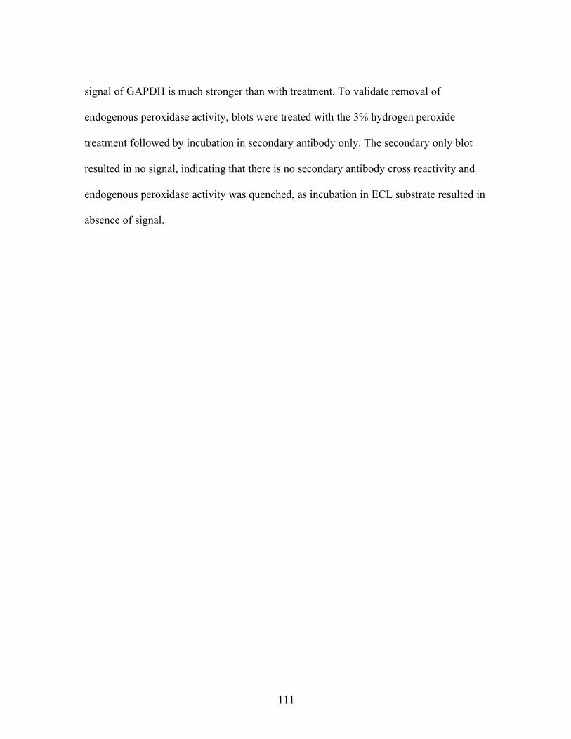
111
signal of GAPDH is much stronger than with treatment. To validate removal of
endogenous peroxidase activity, blots were treated with the 3% hydrogen peroxide
treatment followed by incubation in secondary antibody only. The secondary only blot
resulted in no signal, indicating that there is no secondary antibody cross reactivity and
endogenous peroxidase activity was quenched, as incubation in ECL substrate resulted in
absence of signal.
112
Figure A.1 Treatment of hydrogen peroxide for removal of endogenous peroxidase
activity in bovine anterior pituitary.
Bovine pituitary protein blotted in increasing mass (indicated at the top of the
figure) (B) indicates a blank slot with no protein. Treatments are indicated on the right side
of the figure: (from top to bottom) (1) treatment with hydrogen peroxide (3%) for 10 min
at 37 °C (2) treatment with hydrogen peroxide (3%) for 10 min at 37 °C followed by
incubation with secondary antibody only (7074; Cell Signaling Technology, Danvers, MA,
USA) to assess cross reactivity (H2O2; Secondary only) (3) treatment with hydrogen

113
peroxide (3%) for 10 min at 37 °C followed by GAPDH immunodetection (H2O2; GAPDH)
(4) untreated followed by GAPDH immunodetection (Untreated; GAPDH) (5) Substrate
only.
114
Appendix B
Methods for detection of single nucleotide polymorphisms
Identification of SNPs can be carried out through a variety of methods including,
array-based hybridization, sequencing, and PCR-based methods. Hybridization techniques
are utilized in array technology with two of the most popular platforms called Affymetrix
and Illumina. Affymetrix chips are hybridized with oligonucleotide sequences
complimentary to the SNP region of interest, with fluorescent probes for each possible
allele outcome. Fragments of DNA containing the SNP of interest will bind if they are
similar or an exact match to the hybridized probes on the chip. Signals are obtained from
these bound fragments, in which those that are completely complimentary will bind with
higher affinity and therefore obtain a stronger signal then those that bind with decreased
affinity if they are not completely complimentary. It is also typical to have probes on the
array that do not contain a complementary sequence that matches the SNP site in order to
measure background. (LaFramboise, 2009). Illumina utilizes sequences complimentary to
the adjacent sequence to the SNP of interest and has an extension from that sequence
containing each possible allele outcome. The oligonucleotides, approximately 50 nt in
length, are attached to an Illumina bead. The DNA will then bind to its complimentary
allele and a red or green signal is obtained depending on the allele, with each separate allele
labeled a specific color. Both chip technologies report over 99.5% accuracy in genotyping
(LaFramboise, 2009). Either platform can be performed for targeted genotyping across
many individuals as described above or whole-genome genotyping can be performed to
discover new variations in DNA of individuals across treatments or phenotype. Software
115
such as SNPtest can be used to determine SNPs of interest pertaining to their association
with phenotype (Agler et al., 2019). Association of the mutation to phenotype is not always
based on the smallest P-value, but rather, the abundance in which the mutation occurs in a
large population that shares a given phenotype (Xu and Taylor, 2009).
Next generation sequencing (NGS) is performed through multiple steps and relies
heavily on preparation of a library (Head et al., 2014). Genomic DNA is digested into
fragments and adaptors are added to the 3’ or 5’ ends for recognition by the primers during
sequencing. The library is amplified by PCR and sequencing performed by either
pyrosequencing or sequencing by ligation (SOLiD) (Hurd and Nelson, 2009).
Pyrosequencing is based on complementary strand synthesis through detection of
pyrophosphate release after addition of nucleotides to the DNA. Pyrophosphate will only
be released when the correct base is added onto the template strand and is then converted
into ATP for the conversion of luciferin to oxyluciferin to produce signal. Light intensity
corresponds to how many of that correct base is added in succession (Fakruddin et al.,
2013). Sequencing by ligation is performed through emulsion PCR in which a ssDNA
adaptor is added onto the 3’ end of the sequence that is to be sequenced and all are attached
to a bead. The beaded sequences are subjected to PCR utilizing primers specific for the
adaptor sequence and probes that contain fluorescent dyes at the 5’ end of different colors
corresponding to each base they will recognize. Once the primers bind to the adaptor
sequence, the probe will bind and DNA ligase with enable joining of the probe to the
primer. The fluorescent signal is given off by this joining as it will release the fluorescent
dye that was bound to the probe at the 5’ end. With each round of sequencing, the extension
116
is melted off and the process begins again except with a primer that is 1 less base in length
each time until the entire sequence is finished (Albrecht et al., 2000). Reads from these
methods of sequencing can be used to identify variants between groups of samples. There
are software programs available to assist in variant identification and genotyping of NGS
reads. Microarrays pose issues with SNP genotyping due to nonspecific binding of similar
sequences. Signal to noise ratios also become an issue when multiple sequences reach
either high or low threshold making it difficult to distinguish between groups of those
highly or lowly expressed SNPs. Moreover, NGS allows for SNP detection through
genome wide sequencing without a reference genome, whereas microarrays are limited to
prior knowledge of the genome. Due to preparation of a library and multiples steps for
performance of NGS, the chance of contamination from one step to another is higher (Hurd
and Nelson 2009).
Another form of hybridization detection of SNPs includes a technology called
fluorescence resonance energy transfer (FRET). This technology utilizes fluorescently
labeled primers that correspond to each possible allele. When the DNA sequence is
complimentary to the primer, extension incorporates the labeled probe into the sequence
and fluorescence is emitted. (Sobrino et al., 2005). This FRET technology has been used
in conjunction with the exonuclease activity of the Taq polymerase. Taqman assays for
SNP genotyping require one set of primers that flank the region containing the SNP of
interest. A set of probes, each corresponding to each allele for the SNP of interest and each
of a specific fluorophore color, will then hybridize to the DNA if it is the correct base
match. The fluorophore is attached to the 5’ end and a quencher is attached to the 3’ end.
117
When the both are in close proximity to one another the signal remains quenched; however,
when Taq polymerase reads through and cleaves the 5’ fluorophore, the fluorescent dye is
relieved of its close proximity to the quencher and is able to give off a signal that can be
measured in real time (Schleinitz et al., 2011) (Figure 1.5)
Kompetitive allele specific PCR (KASP) genotyping requires multiple forward
primers with the base at the 5’ end specific for each possible outcome of the SNP and a
universal reverse primer. The forward primers will bind the SNP site but will only be able
to elongate if they bind with the correct complimentary base to the SNP. A probe,
complimentary to the SNP sequence is then incorporated, a separate fluorescent dye for
each SNP outcome, and the sequence is amplified, and with it the signal intensifies and can
be quantified (Ayalew et al., 2019).
RNase H2 enzyme-based amplification (rHAMP) also utilizes allele specific
primers with a 3’ blocking modification consisting of an RNA base. The assay also utilizes
a set of universal primers that flank the SNP region and universal probes. The allele specific
primer will bind to the complimentary SNP sequence and RNase H2 will cleave the RNA
3’ modification to allow for extension by Taq polymerase. A tail sequence is incorporated
in subsequent cycles and a universal probe recognizes the tail sequence to bind, and through
extension, a signal is generated (Ayalew et al., 2019).
Overall, Taqman SNP genotyping allows only up to 6 bp sequences to be genotyped
offering less flexibility in assay design compared to KASP or rHAMP. The Taqman and
KASP techniques lack the modified 3’ end primers that rHAMP technology utilizes to
reduce non-specific binding and unwanted dimerization, therefore, allelic discrimination is
118
more accurate and defined in rHAMP techniques compared to KASP and Taqman (Ayalew
et al., 2019). Taqman is the most expensive of the three genotyping assays at ~$250 for
2,000 reactions compared to KASP and rHAMP at less than $100 for 5,000 reactions
including primers and probes. Master mix is sold separately and costs ~$550 for 2,000
reactions for Taqman assays and ~$1000 for KASP and rHAMP for 10,000 reactions.
Lastly, all assays utilize minimal DNA input with Taqman and KASP at a required 5 ng
DNA, and rHAMP at 20 ng (Ayalew et al., 2019).
Other methods that include PCR are single strand conformation polymorphism
analysis (SSCP) and restriction fragment length polymorphism (RFLP) analysis. The SSCP
analysis can be used to identify polymorphism at any position (Orita et al., 1989; Rajatileka
et al., 2014). Methods include isolating DNA, amplification of the sequence of interest and
denaturation of products in a formamide solution. When the products are denatured, they
will reveal different conformational shapes due to polymorphisms they contain that will
affect how they migrate through a non-denaturing gel. These patterns can then be used to
identify mutations in the DNA (Orita et al., 1989). Cost is relatively low for this method of
genotyping however the use of formamide poses some extra risk in performing this
procedure.
The RFLP analysis takes advantage of a mutation that may or may not abolish a
restriction site. Polymerase chain reaction is used to amplify the region containing the SNP
of interest. The product is purified and digested with the restriction enzyme that will cut at
the restriction site where the SNP is located, resulting in multiple fragments that vary in
size. The band patterns that result are used for SNP genotyping (Maeda et al., 1990).
119
Considerations for this method include sample number and cost, as some of the enzymes
will not tolerate the taq polymerase in the original PCR reaction and cost is incurred for
PCR cleanup for each reaction prior to digestion. Since there are two reactions, PCR and
digestion, there is also concern of contamination from sample to sample.
Sanger sequencing is also a powerful tool to identify SNPs. This method works
through production of fragments that contain dideoxynucleotides at the 3’ end. These
fragments are separated by capillary electrophoresis which will separate each base by
molecular weight. Each is tagged with a different colored fluorescent dye at the 3’ end for
identification by base calls. Each base will be identified by a different color fluorescence
as they pass through capillary electrophoresis (Imelfort et al., 2009). The base calls
resemble peaks of fluorescence that can be used to identify SNPs by varying color and
intensity of the peak. Single nucleotide polymorphisms will show overlap of signal or base
calls for heterozygotes and a single signal for homozygotes. Although Sanger sequencing
was once considered and may still be largely considered the hallmark of this technology,
NGS has largely replaced this technique, as larger data sets can be collected for a fraction
of the price, $0.25 per SNP compared to $2.95 for Sanger sequencing. Aside from price,
platforms such as Illumina are also only able to perform short reads while Sanger
sequencing can perform at over 500 nt (Imelfort et al., 2009).
Overall, when choosing a method for genotyping the main considerations are time,
cost, sample abundance, and abundance of SNPs being studied. The NGS and microarray
options are much more expensive but provide high throughput data allowing for detection
of thousands of SNPs in many samples within a short time frame (Goodwin, McPherson,
120
and McCombie, 2016). The PCR based assays are also capable of processing thousands of
samples as most assays are sold as sets of 2,000-5,000 reactions and are performed in plate
format; however, the region of interest needs to be known prior unlike high throughput
methods that allow for screening of the entire genome for SNPs. The cost for FRET-based
qPCR assays are higher than the cost of SSCP or RFLP; however, the FRET based assays
are much less labor intensive (Chen and Sullivan, 2003). Other considerations include cost
of and access to equipment for high throughput processing and cost of sending samples out
of the laboratory for processing, which can take some time. This extra time and cost can
be worth the vast amount of data generated by high throughput screening for SNPs if the
goal is to identify large quantities of SNPs across a given population or SNPs across the
entire genome.
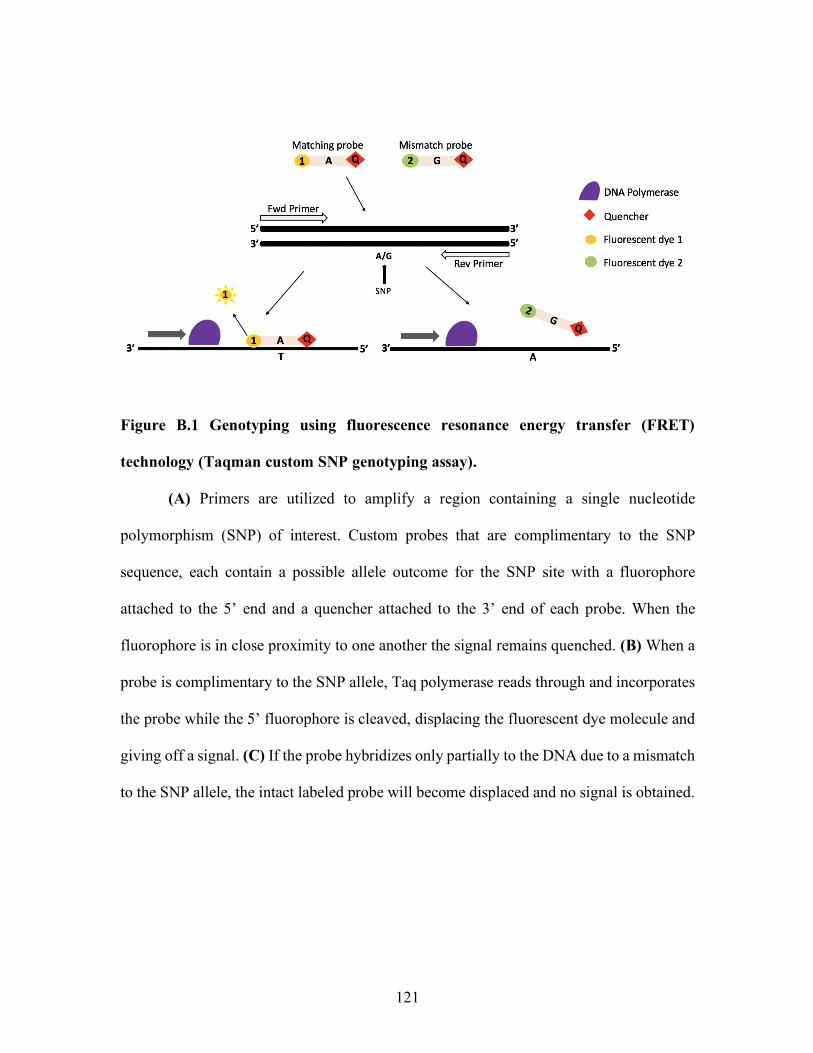
121
Figure B.1 Genotyping using fluorescence resonance energy transfer (FRET)
technology (Taqman custom SNP genotyping assay).
(A) Primers are utilized to amplify a region containing a single nucleotide
polymorphism (SNP) of interest. Custom probes that are complimentary to the SNP
sequence, each contain a possible allele outcome for the SNP site with a fluorophore
attached to the 5’ end and a quencher attached to the 3’ end of each probe. When the
fluorophore is in close proximity to one another the signal remains quenched. (B) When a
probe is complimentary to the SNP allele, Taq polymerase reads through and incorporates
the probe while the 5’ fluorophore is cleaved, displacing the fluorescent dye molecule and
giving off a signal. (C) If the probe hybridizes only partially to the DNA due to a mismatch
to the SNP allele, the intact labeled probe will become displaced and no signal is obtained.
122
REFERENCES
Agler, C., Shungin, D., Zandoná, A., Schmadeke, P., Basts, P., Luo, J., Cantrell, J., Pahel, T. D., Meyer, B. D., Schaffer, J. R., & Schaefer, A. (2019). Protocols, Methods, and Tools for Genome-Wide Association Studies (GWAS) of Dental Traits. Odontogenesis.
Albrecht, G., Brenner, S., Dubridge, R., Lloyd, D., & Palla, M. (2000). Massively parallel signature sequencing by ligation of encoded adaptors. Massively Parallel Signature Sequencing by Ligation of Encoded Adaptors.
Ali, S., Pellegrini, I., & Kelly, P. A. (1991). A prolactin-dependent immune cell line (Nb2) expresses a mutant form of prolactin receptor. Journal of Biological Chemistry, 266(30), 20110–20117.
Andén, N., Dahlström, A., Fuxe, K., Larsson, K., Olson, L., & Ungerstedt, U. (1966). Ascending monoamine neurons to the telencephalon and diencephalon. Physiologica Scandinavica, 67(3–4), 313–326.
Andersen, A. N., Pedersen, H., Djursing, H., & Friesen, H. G. (1982). Bioactivity of prolactin in a woman with an excess of large molecular size prolactin, persistent hyperprolactinemia and spontaneous conception. Fertility and Sterility, 38(5), 625–628. https://doi.org/10.1016/S0015-0282(16)46646-X
Andries, M., Tilemans, D., & Denef, C. (1992). Isolation of cleaved prolactin variants that stimulate DNA synthesis in specific cell types in rat pituitary cell aggregates in culture. Biochemical Journal, 281(2), 393–400. https://doi.org/10.1042/bj2810393
Angelier, F., & Chastel, O. (2009). Stress, prolactin and parental investment in birds: A review. General and Comparative Endocrinology, 163(1–2), 142–148. https://doi.org/10.1016/j.ygcen.2009.03.028
Anthony, R.V., Liang R., Kayl, E. P., & Pratt, S. L. (1995). The growth hormone/prolactin gene family in ruminant placentae. Journal of Reproduction and Fertility Supplement, 49, 83–95.
Aragona, C., Heinz, G., & Henry, G. (1977). Localization of prolactin binding in prostate and testis: the role of serum prolactin concentration on the testicular LH receptor. European Journal of Biochemistry, 84(2), 402–409.
Arbogast, L., & Voogt, J. (1991). Hyperprolactinemia increases and hypoprolactinemia decreases tyrosine hydroxylase messenger ribonucleic acid levels in the arcuate nuclei, but not the substantia nigra or zona incerta. Endocrinology, 128(2), 997–1005.
123
Arbogast, L. A., & Voogt, J. L. (1997). Prolactin (PRL) receptors are colocalized in dopaminergic neurons in fetal hypothalamic cell cultures: Effect of PRL on tyrosine hydroxylase activity. Endocrinology, 138(7), 3016–3023. https://doi.org/10.1210/endo.138.7.5227
Arden, K. C., Boutin, J. M., Djiane, J., Kelly, P. A., & Cavenee, W. K. (1990). The receptors for prolactin and growth hormone are localized in the same region of human chromosome 5. Cytogenetic and Genome Research. https://doi.org/10.1159/000132919
Arinami, T., Gao, M., Hamaguchi, H., & Toru, M. (1997). A functional polymorphism in the promoter region of the dopamine D2 receptor gene is associated with schizophrenia. Human Molecular Genetics, 6(4), 577–582. https://doi.org/10.1093/hmg/6.4.577
Asghari, V., Sanyal, S., Buchwaldt, S., Paterson, A., Jovanovic, V., & Van Tol, H. (1995). Modulation of intracellular cyclic AMP levels by different human dopamine D4 receptor variants. Journal of Neurochemistry, 65(3), 1157–1165.
Ayalew, H., Tsang, P., Chu, C., Wang, J., Liu, S., Chen, C., & Ma, X. (2019). Comparison of TaqMan, KASP and rhAmp SNP genotyping platforms in hexaploid wheat. PloS One, 14(5), e0217222.
Baertschi, A. J., Audigier, Y., Lledo, P. M., Israel, J. M., Bockaert, J., & Vincent, J. D. (1992). Dialysis of lactotropes with antisense oligonucleotides assigns guanine nucleotide binding protein subtypes to their channel effectors. Molecular Endocrinology, 6(12), 2257–2265. https://doi.org/10.1210/mend.6.12.1337149
Bartke, A., Croft, B., & Dalterio, S. (1975). Prolactin restores plasma testosterone levels and stimulates testicular growth in hamsters exposed to short day-length. Endocrinology, 97(6), 1601–1604.
Bartke, A., Bex, F., Dalterio, S., & Goldman, B. D. (1977). Effects of prolactin (prl) on pituitary and testicular function in mice with hereditary prl deficiency. Endocrinology, 101(6), 1760–1766. https://doi.org/10.1210/endo-101-6-1760
Bartke, AGoldman, B., Bex, F., Kelch, R., Smith, M., Dalterio, S., & Doherty, P. (1980). Effects of Prolactin on Testicular Regression and Recrudescence in the Golden Hamster. Endocrinology, 106(1), 167–172.
Barkey, R. J., Weiss-Messer, E., Mandel, S., Gahnem, F., & Amit, T. (1987). Prolactin and antiprolactin receptor antibody inhibit steroidogenesis by purified rat Leydig cells in culture. Molecular and Cellular Endocrinology, 52(1–2), 71–80. https://doi.org/10.1016/0303-7207(87)90098-0
124
Bazan, J. F. (1990). Structural design and molecular evolution of a cytokine receptor superfamily. Proceedings of the National Academy of Sciences of the United States of America, 87(18), 6934–6938. https://doi.org/10.1073/pnas.87.18.6934
Ben-Jonathan, N., Oliver, C., Weiner, H., Mical, R., & Porter, J. (1977). Dopamine in hypophysial portal plasma of the rat during the estrous cycle and throughout pregnancy. Endocrinology, 100(2), 452–458.
Ben-Jonathan, N., Mershon, J. L., Allen, D. L., & Steinmetz, R. W. (1996). Extrapituitary prolactin: Distribution, regulation, functions, and clinical aspects. Endocrine Reviews, 17(6), 639–669. https://doi.org/10.1210/er.17.6.639
Ben-Jonathan, & Nira; Hnasko, R. (2001). Dopamine as a Prolactin (PRL) Inhibitor. Endocrine Reviews, 22(6), 724–763.
Bermak, J. C., Li, M., Bullock, C., & Zhou, Q. Y. (2001). Regulation of transport of the dopamine D1 receptor by a new membrane-associated ER protein. Nature Cell Biology, 3(5), 492–498. https://doi.org/10.1038/35074561
Bernard, J., Chestnut, A., Erickson, B., & Kelly, F. (1933). Effects of prepartum consumption of endophyte-infested tall fescue on serum prolactin and subsequent milk production of Holstein cows. Journal of Dairy Science, 76(7), 1928–1933.
Berwaer, M., Martial, J. A., & Davis, J. R. E. (1994). Characterization of an up-stream promoter directing extrapituitary expression of the human prolactin gene. Molecular Endocrinology, 8(5), 635–642. https://doi.org/10.1210/me.8.5.635
Bignon, C., Binart, N., Ormandy, C., Schuler, L. A., Kelly, P. A., & Djiane, J. (1997). Long and short forms of the ovine prolactin receptor: cDNA cloning and genomic analysis reveal that the two forms arise by different alternative splicing mechanisms in ruminants and in rodents. Journal of Molecular Endocrinology, 19(2), 109–120. https://doi.org/10.1677/jme.0.0190109
Binart, N., Melaine, N., Pineau, C., Kercret, H., Touzalin, A. M., Imbert-Bolloré, P., Kelly, P. A., & Jégou, B. (2003). Male reproductive function is not affected in prolactin receptor-deficient mice. Endocrinology, 144(9), 3779–3782. https://doi.org/10.1210/en.2003-0409
Björklund, A., Moore, R. Y., Nobin, A., & Stenevi, U. (1974). The organization of tubero-hypophyseal and reticulo-infundibular catecholamine neuron systems in the rat brain. Brain Research, 51, 171–191. https://doi.org/10.1016/0006-8993(73)90371-5
125
Bole-Feysot, C., Goffin, V., Edery, M., Binart, N., & Kelly, P. A. (1998). Prolactin (PRL) and its receptor: Actions, signal transduction pathways and phenotypes observed in PRL receptor knockout mice. Endocrine Reviews, 19(3), 225–268. https://doi.org/10.1210/edrv.19.3.0334
Booth, A., & Gutierrez-Hartmann, A. (2015). Signaling pathways regulating pituitary lactotrope homeostasis and tumorigenesis. In Recent Advances in Prolactin Research (pp. 37–59). https://doi.org/10.1007/978-3-319-12114-7
Boudanova, E., Navaroli, D., Stevens, Z., & Melikian, H. (2008). Dopamine transporter endocytic determinants: carboxy terminal residues critical for basal and pkc-stimulated internalization. Molecular and Cellular Neuroscience, 39(2), 211–217. https://doi.org/10.1038/jid.2014.371
Bourne, R., & Tucker, H. (1975). Serum prolactin and LH responses to photoperiod in bull calves. Endocrinology, 97(2), 473–475.
Boutin, J. M., Edery, M., Shirota, M., Jolicoeu, C., Lesueur, L., Ali, S., Djiane, J., & Kelly, P. A. (1989). Identification of a cDNA encoding a long form of prolactin receptor inhuman hepatoma and breastcancer cells. Molecular Endocrinology, 3(9), 1455–1461. https://doi.org/10.1210/mend-3-9-1455
Boutin, J. M., Jolicoeur, C., Okamura, H., Gagnon, J., Edery, M., Shirota, M., Djiane, J., & Kelly, P. A. (1988). Cloning and expression of the rat prolactin receptor, a member of the growth hormone/prolactin receptor gene family. Cell, 53(1), 69–77. https://doi.org/10.1016/0092-8674(88)90488-6
Brym, P., Kamiñski, S., & Rusc, A. (2004). New SSCP polymorphism within bovine STAT5A gene and its associations with milk performance traits in Black-and-White and Jersey cattle. Journal of Applied Genetics, 45(4), 445–452.
Brym, P., Kamiński, S., & Wójcik, E. (2005a). Nucleotide sequence polymorphism within exon 4 of the bovine prolactin gene and its associations with milk performance traits. Journal of Applied Genetics, 46(2), 179–185.
Brym, P., Kamiński, S., & Wójcik, E. (2005b). Polymorphism within the bovine prolactin receptor gene (PRLR). Animal Science Papers and Reports, 23(1), 61–66.
Bunzow, J., Van Tol, H., Grandy, D., Albert, P., Salon, J., Christie, M., Neve, K. A., & Civeli, O. (1988). Cloning and expression of a rat D2 dopamine receptor cDNA. Nature, 336(6201), 783.
126
Burnett, C. R., Bridges, W. C., & Pratt, S. L. (2017). Effects of grazing tall fescue containing ergot alkaloids on bull sperm cryopreservation. Animal Reproduction Science, 181, 24–29. https://doi.org/10.1016/j.anireprosci.2017.03.011
Burnett, C. R., Pratt, S. L., Long, N. M., Sell, G. S., & Schrick, F. N. (2018). Assessment of semen quality and fertility in young growing beef bulls exposed to ergot alkaloids. Theriogenology, 118, 219–224. https://doi.org/10.1016/j.theriogenology.2018.06.007
Calcatera, S. M., Korn, N., Ellis, S., & Pratt, S. L. (2011). Immunohistochemical detection of Argonaute 2 in bovine and porcine tissues. Journal of Histotechnology, 34(2), 74–78. https://doi.org/10.1179/204602311X13007117736023
Campbell, B. T., Kojima, C. J., Cooper, T. A., Bastin, B. C., Wojakiewicz, L., Kallenbach, R. L., Schrick, F. N., & Waller, J. C. (2014). A Single Nucleotide Polymorphism in the Dopamine Receptor D2 Gene May Be Informative for Resistance to Fescue Toxicosis in Angus-Based Cattle. Animal Biotechnology, 25(1), 1–12. https://doi.org/10.1080/10495398.2013.796960
Campbell, D. G., Hardie, D. G., & Vulliet, P. R. (1986). Identification of four phosphorylation sites in the N-terminal region of tyrosine hydroxylase. Journal of Biological Chemistry, 261(23), 10489–10492.
Camper, S. A., Luck, D. N., Yao, Y., Woychik, R. P., Goodwin, R. G., Lyons, R. H., & Rottman, F. M. (1984). Characterization of the Bovine Prolactin Gene. Dna, 3(3), 237–249. https://doi.org/10.1089/dna.1.1984.3.237
Caron, M. G., Beaulieu, M., Raymond, R., Gagné, B., Drouin, J., Lefkowitz, R. J., & Labrie, F. (1978). Receptors in the anterior pituitary gland. The Journal of Biological Chemistry, 253(7), 2244–2253.
Cavaco, B., Leite, V., Santos, M., Arranhado, E., & LG, S. (1995). Some forms of big big prolactin behave as a complex of monomeric prolactin with an immunoglobulin G in patients with macroprolactinemia or prolactinoma. The Journal of Clinical Endocrinology & Metabolism, 80(8), 2342–2346.
Chavrier, P., Parton, R. G., Hauri, H. P., Simons, K., & Zerial, M. (1990). Localization of low molecular weight GTP binding proteins to exocytic and endocytic compartments. Cell, 62(2), 317–329. https://doi.org/10.1016/0092-8674(90)90369-P
127
Chen, X., & Sullivan, P. (2003). Single nucleotide polymorphism genotyping: biochemistry, protocol, cost and throughput. The Pharmacogenomics Journal, 3(2), 77.
Chio, C., Hess, G., Graham, R., & Huff, R. (1990). A second molecular form of D2 dopamine receptor in rat and bovine caudate nucleus. Nature, 343(6255), 266.
Christensen, H. R., Murawsky, M. K., Horseman, N. D., Willson, T. A., & Gregerson, K. A. (2013). Completely humanizing prolactin rescues infertility in prolactin knockout mice and leads to human prolactin expression in extrapituitary mouse tissues. Endocrinology, 154(12), 4777–4789. https://doi.org/10.1210/en.2013-1476
Civelli, O., Bunzow, J., & Grandy, D. (1993). Molecular diversity of the dopamine receptors. Annual Review of Pharmacology and Toxicology, 33(1), 281–307.
Clapp, C., Tomer, L., Gutiirrez-ospina, G., Alcantara, E. V. A., Lopez-gomez, F. J., Nagano, M., Kelly, P. A., Meja, S., Morales, M. A., & de la Escalera, G. (1994). The Prolactin Gene is Expressed in the Hypothalamic-Neurohypophyseal System and the Protein is Processed Into a 14-kDa Fragment with Activity Like 16-kDa prolactin. Proceedings of the National Academy of Sciences, 91(22), 10384–10388.
Clarke, D., & Linzer, D. (1993). Changes in prolactin receptor expression during pregnancy in the mouse ovary. Endocrinologyq, 133(1), 224–232. https://doi.org/10.1210/en.133.1.224
Clevenger, C. V., Freier, D. O., & Kline, J. B. (1998). Prolactin receptor signal transduction in cells of the immune system. Journal of Endocrinology, 157(2), 187–197. https://doi.org/10.1677/joe.0.1570187
Cole, E., Nichols, E., Lauziere, K., Edmunds, T., & McPherson, J. (1991). Characterization of the microheterogeneity of recombinant primate prolactin: implications for posttranslational modifications of the hormone in vivo. Endocrinology, 129(5), 2639–2646.
Cooke, N. E., Coit, D., Weiner, R. I., Baxter, J. D., & Martial, J. A. (1980). Structure of cloned DNA complementary to rat prolactin messenger RNA. Journal of Biological Chemistry, 255(13), 6502–6510.
Costello, L. C., & Franklin, R. B. (1994). Effect of Prolactin on the Prostate. The Prostate, 24(3), 162–166.
128
Costello, L. C., Liu, Y., Zou, J., & Franklin, R. B. (1999). Evidence for a zinc uptake transporter in human prostate cancer cells which is regulated by prolactin and testosterone. Journal of Biological Chemistry, 274(25), 17499–17504. https://doi.org/10.1074/jbc.274.25.17499
Cragg, S. J., & Rice, M. E. (2004). DAncing past the DAT at a DA synapse. Trends in Neurosciences, 27(5), 270–277. https://doi.org/10.1016/j.tins.2004.03.011
Crawford, R., Forwood, J., Belyea, R., & Garner, G. (1989). Relationship between level of endophyte infection and cattle gains on tall fescue. Journal of Production Agriculture, 2(2), 147–151.
Curry, E., Pratt, S. L., Kelley, D. E., Lapin, D. R., & Gibbons, J. R. (2008). Use of a Combined Duplex PCR/Dot Blot Assay for more sensitive genetic characterization. Biochemistry Insights, 1, BCI-S872. https://doi.org/10.4137/bci.s872
Das, R., & Vonderhaar, B. (1996). Involvement of SHC, GRB2, SOS and RAS in prolactin signal transduction in mammary epithelial cells. Oncogene, 13(6), 1139–1145.
Dal Toso, R., Sommer, B., Ewert, M., Herb, A., Pritchett, D. B., Bach, A., Shivers, B. D., & Seeburg, P. H. (1989). The dopamine D2 receptor: two molecular forms generated by alternative splicing. The EMBO Journal, 8(13), 4025–4034. https://doi.org/10.1002/j.1460-2075.1989.tb08585.x
Daubner, S. C., Le, T., & Wang, S. (2011). Tyrosine Hydroxylase and Regulation of Dopamine Synthesis. Arch Biochem Biophys., 508(1), 1–12. https://doi.org/10.1016/j.abb.2010.12.017.
Daubner, S. C., Lauriano, C., Haycock, J. W., & Fitzpatrick, P. F. (1992). Site-directed Mutagenesis of Serine 40 of Rat Tyrosine Hydroxylase. The Journal of Biological Chemistry, 267(16), 12639–12646.
Davis, J. A., & Linzer, D. I. H. (1989). Expression of multiple forms of the prolactin receptor in mouse liver. Molecular Endocrinology, 3(4), 674–680. https://doi.org/10.1210/mend-3-4-674
De Camili, P., Macconi, D., & Spada, A. (1979). Dopamine inhibits adenylate cyclase in human prolactin-secreting pituitary adenomas. Nature, 278(5701), 252–254.
Dearry, A., Gingrich, J., Falardeau, P., Fremeau, J. R., Bates, M., & Caron, M. (1990). Molecular cloning and expression of the gene for a human D1 dopamine receptor. Nature, 347(6288), 72.
129
DeCarlo, A. N., Jennings, K., Pratt, S. L., Burnett, C. R., Long, N. M., Bridges, W. C., & Kojima, C. (2019). Bovine dopamine type-2 receptor SNP has no effect on growth, semen characteristics and prolactin concentrations in beef bulls treated with a dopamine agonist. Animal Reproduction Science, 207(May), 73–82. https://doi.org/10.1016/j.anireprosci.2019.06.005
Demarest, K. T., & Moore, K. E. (1979). Lack of a high affinity transport system for dopamine in the median eminence and posterior pituitary. Brain Research, 171(3), 545–551. https://doi.org/10.1016/0006-8993(79)91060-6
Demaria, J. E., Nagy, G. M., Lerant, A. A., Fekete, M. I. E., Levenson, C. W., & Freeman, M. E. (2000). Dopamine transporters participate in the physiological regulation of prolactin. Endocrinology, 141(1), 366–374. https://doi.org/10.1210/endo.141.1.7281
DeVito, W., Avakian, C., & Stone, S. (1992). Proteolytic modification of prolactin by the female rat brain. Neuroendocrinology, 56(4), 597–603.
Dickson, R. A., & Glazer, W. M. (1999). Neuroleptic-induced hyperprolactinemia. Schizophrenia Research, 35(SUPPL.). https://doi.org/10.1016/S0920-9964(98)00159-5
Dombrowicz, D., Sente, B., Closset, J., & Hennen, G. (1992). Dose-dependent effects of human prolactin on the immature hypophysectomized rat testis. Endocrinology, 130(2), 695–700.
Dorshkind, K., & Horseman, N. D. (2001). Anterior pituitary hormones, stress, and immune system homeostasis. BioEssays, 23(3), 288–294. https://doi.org/10.1002/1521-1878(200103)23:3<288::AID-BIES1039>3.0.CO;2-P
Dumont, J. (1965). Prolactin-induced cytologic changes in the mucosa of the pigeon crop during crop-"milk" formation. Zeitschrift Für Zellforschung Und Mikroskopische Anatomie, 68(6), 755–782.
Dunlop, B. W., & Nemeroff, C. B. (2007). The role of dopamine in the pathophysiology of depression. Archives of General Psychiatry, 64(3), 327–337. https://doi.org/10.1001/archpsyc.64.3.327
Dohlman, H., Caron, M., DeBiasi, A., Frielle, T., & Lefkowitz, R. (1990). Role of extracellular disulfide-bonded cysteines in the ligand binding function of the. beta. 2-adrenergic receptor. Biochemistry, 29(9), 2335–2342.
130
Einhorn, L. C., Gregerson, K. A., & Oxford, G. S. (1991). D2dopamine receptor activation of potassium channels in identified rat lactotrophs: Whole-cell and single-channel recording. Journal of Neuroscience, 11(12), 3727–3737.
Elsholtz, H. P., Lew, a M., Albert, P. R., & Sundmark, V. C. (1991). Inhibitory Control of Prolactin and Pit-1 Gene Promoters by Dopamine. Dueal signaling pathways required for D2 receptor-regulated expression of the prolactin gene. J. Biol. Chem., 266(34), 22919–22925.
Elsworth, J. D., & Roth, R. H. (1997). Dopamine synthesis, uptake, metabolism, and receptors: Relevance to gene therapy of Parkinson’s disease. Experimental Neurology, 144(1), 4–9. https://doi.org/10.1006/exnr.1996.6379
Enjalbert, A., & Bockaert, J. (1983). Pharmacological characterization of the D2 dopamine receptor negatively coupled with adenylate cyclase in rat anterior pituitary. Molecular Pharmacology, 23(3), 576–584. Retrieved from http://www.ncbi.nlm.nih.gov/pubmed/6306429
Enjalbert, A., Musset, F., Chenard, C., Priam, M., Kordon, C., & Heisier, S. (1988). Dopamine inhibits prolactin secretion stimulated by the calcium channel agonist Bay-K-8644 through a pertussis toxin-sensitive G protein in anterior pituitary cells. Endocrinology, 123(1), 406–412.
Fakruddin, Mazumdar, R. M., Chowdhury, A., Hossain, N., Mahajan, S., & Islam, S. (2013). Pyrosequencing-A next generation sequencing technology. World Applied Sciences Journal, 24(12), 1558–1571. https://doi.org/10.5829/idosi.wasj.2013.24.12.2972
Farquhar, M., Reid, J., & Daniell, L. (1978). Intracellular transport and packaging of prolactin: a quantitative electron microscope autoradiographic study of mammotrophs dissociated from rat pituitaries. Endocrinology, 102(1), 296–311.
Farquhar, M. G., & Rinehart, J. F. (1954). Cytologic alterations in the anterior pituitary gland following thyroidectomy: an electron microscopic study. Endocrinology, 55(6), 857–876.
Farquhar, M., & Wellings, S. (1957). Electron microscopic evidence suggesting secretotry granules within the Golgi apparatus. The Journal of Cell Biology, 3(2), 319–322. https://doi.org/10.1042/bst0060705
Ferrari, F., & Giuliani, D. (1994). The selective D2 dopamine receptor antagonist meticlopride counteracts the ejaculation praecox induced by the selective D2 dopamine agonist SND 919 in the rat. Life Sciences, 55(14), 1155–1162.
131
Filippa, V., & Mohamed, F. (2010). Morphological and morphometric changes of pituitary lactotrophs of viscacha (Lagostomus maximus maximus) in relation to reproductive cycle, age, and sex. The Anatomical Record: Advances in Integrative Anatomy and Evolutionary Biology, 293(1), 150–161. https://doi.org/10.1002/ar.21013
Fisher, D., Dussault, J., Sack, J., & Chopra, I. (1976). Ontogenesis of Hypothalamic-Pituitary-Thyroid Function and Metabolism in Man, Sheep, and Rat. Proceedings of the 1976 Laurentian Hormone Conference.
Flatmark, T., & Stevens, R. C. (1999). Structural Insight into the Aromatic Amino Acid Hydroxylases and Their Disease-Related Mutant Forms. Chemical Reviews, 99(8), 2137–2160. https://doi.org/10.1021/cr980450y
Flisikowski, K., & Zwierzchowski, L. (2003). Polymerase chain reaction-heteroduplex (PCR-HD) polymorphism within the bovine STAT5A gene. Journal of Applied Genetics, 44(2), 185–189.
Fomina, A. F., & Levitan, E. S. (1995). Three phases of TRH-induced facilitation of exocytosis by single lactotrophs. Journal of Neuroscience, 15(7 I), 4982–4991. https://doi.org/10.1523/jneurosci.15-07-04982.1995
Freeman, M. E., Kanyicska, L. A., Lerant, A., & Nagy, G. (2000). Prolactin: Structure, Function, and Regulation of Secretion. Physiological Reviews, 80(4), 1523–1631.
Fuxe, D. (1964). Evidence for existence of monoamine-containing neurons in central nervous system: Demonstration of monoamines in the cell bodies of brain stem neurons. Acta Physiol. Scand., 62, 1–55.
Fuxe, K. (1963). Cellular localization of monoamines in the median eminence and the infundibular stem of some mammals. Zeitschrift Für Zellforschung Und Mikroskopische Anatomie, 61(5), 710–724.
Gallagher, G., & Senger, P. (1989). Effect of phenylephrine, ergonovine, oxytocin and norepinephrine as an extender ingredient on viability of bovine spermatozoa. Journal of Animal Science, 67(6), 1573–1576.
George, F., & Peterson, K. (1988). 5α-Dihydrotestosterone formation is necessary for embryogenesis of the rat prostate. Endocrinology, 122(3), 1159–1164.
132
Gertler, A., Grosclaude, J., Strasburger, C. J., Nir, S., & Djiane, J. (1996). Real-time kinetic measurements of the interactions between lactogenic hormones and prolactin-receptor extracellular domains from several species support the model of hormone-induced transient receptor dimerization. Journal of Biological Chemistry, 271(40), 24482–24491. https://doi.org/10.1074/jbc.271.40.24482
Giros, B., & Caron, M. G. (1993). Molecular characterization of the dopamine transporter. Trends in Pharmacological Sciences, 14(2), 43–49. https://doi.org/10.1016/0165-6147(93)90029-J
Goffin, V., & Kelly, P. A. (1997). The Prolactin / Growth Hormone Receptor Family: Structure / Function Relationships. Journal of Mammary Gland Biology and Neoplasia, 2(1), 7–17.
Golander, A., Hurley, T., Barrett, J., Hizi, A., & Handwerger, S. (1978). Prolactin Synthesis by Human Chorion-Decidual Tissue: A Possible Source of Prolactin in the Amniotic Fluid. American Association for the Advancement of Science, 202(4365), 311–313.
Gonzales, G. F., Velasquez, G., & Garcia-Hjarles, M. (1989). Hypoprolactinemia as related to seminal quality and serum testosterone. Journal of Reproductive Systems, 23(3), 259–265. https://doi.org/10.3109/01485018908986849
Gonzalez, C., Gonzalez, B., Matzkin, M., Muniz, J., Cadet, J., Garcia-Rill, E., Urbano, F. J., Vitullo, A. D., & Bisagno, V. (2015). Psychostimulant-induced testicular toxicity in mice: Evidence of cocaine and caffeine effects on the local dopaminergic system. PLoS ONE, 10(11), e0142713. https://doi.org/10.1371/journal.pone.0142713
Goodwin, S., McPherson, J. D., & McCombie, W. R. (2016). Coming of age: Ten years of next-generation sequencing technologies. Nature Reviews Genetics, 17(6), 333–351. https://doi.org/10.1038/nrg.2016.49
Gouilleux, F., Wakao, H., Mundt, M., & Groner, B. (1994). Prolactin induces phosphorylation of Tyr694 of Stat5 (MGF), a prerequisite for DNA binding and induction of transcription. The EMBO Journal, 13(18), 4361–4369. Retrieved from http://www.ncbi.nlm.nih.gov/pubmed/7925280%0Ahttp://www.pubmedcentral.nih.gov/articlerender.fcgi?artid=PMC395363
Grandy, D. K., Littt, M., Allen, L., Bunzow, J. R., Marchionni, M., Makam, H., Reed, L., Magenis, R. E., & Civelli, O. (1989). The Human Dopamine D2 Receptor Gene Is Located on chromosome 11 at q22-q23 and identifies a Taq1 RFLP. American Journal of Human Genetics, 45(5), 778–785.
133
Grånäs, C., Ferrer, J., Loland, C. J., Javitch, J. A., & Gether, U. (2003). N-terminal truncation of the dopamine transporter abolishes phorbol ester- and substance P receptor-stimulated phosphorylation without impairing transporter internalization. Journal of Biological Chemistry, 278(7), 4990–5000. https://doi.org/10.1074/jbc.M205058200
Griffith, M. K., & Minton, J. E. (1992). Effect of Light Intensity on Circadian Profiles and Cortisol in Pigs. Journal of Animal Science, 70(2), 492–498.
Gunasekar, P. G., Kumaran, B., & Govindarajulu, P. (1991). Role of prolactin on Leydig, Sertoli and germ cellular neutral lipids in bonnet monkeys, Macaca radiata. Endocrinologia Japonica, 38(1), 1–8. https://doi.org/10.1507/endocrj1954.38.1
Gordon, S. L., Quinsey, N. S., Dunkley, P. R., & Dickson, P. W. (2008). Tyrosine hydroxylase activity is regulated by two distinct dopamine-binding sites. Journal of Neurochemistry, 106(4), 1614–1623. https://doi.org/10.1111/j.1471-4159.2008.05509.x
Haegeman, A., Williams, J. L., Law, A., Van Zeveren, A., & Peelman, L. J. (2003). Characterization and mapping of bovine dopamine receptors 1 and 5. Animal Genetics, 34(4), 290–293. https://doi.org/10.1046/j.1365-2052.2003.00994.x
Hair, W. M., Gubbay, O., Jabbour, H., & Lincoln, G. (2002). Prolactin receptor expression in human testis and accessory tissues: localization and function. Molecular Human Reproduction, 8(7), 606–611. https://doi.org/10.1093/molehr/8.7.606
Halmi, N. (1950). Two types of basophils in the anterior pituitary of the rat and their respective cytophysiological significance. Endocrinology, 47(4), 289–299.
Hansen, K., Zhang, Y., Colver, R., Tho, S., Plouffe, L., & McDonough, P. (2005). The dopamine receptor D2 genotype is associated with hyperprolactinemia. Fertility and Sterility, 84(3), 711–718. https://doi.org/10.1016/j.fertnstert.2005.03.040
Hayes, H., Le Chalony, C., Goubin, G., Mercier, D., Payen, E., Bignon, C., & Kohno, K. (1996). Localization of ZNF164, ZNF146, GGTA1, SOX2, PRLR and EEF2 on homoeologous cattle, sheep and goat chromosomes by fluorescent in situ hybridization and comparison with the human gene map. Cytogenetic and Genome Research, 72(4), 342–346.
Head, S. R., Kiyomi Komori, H., LaMere, S. A., Whisenant, T., Van Nieuwerburgh, F., Salomon, D. R., & Ordoukhanian, P. (2014). Library construction for next-generation sequencing: Overviews and challenges. BioTechniques, 56(2), 61–77. https://doi.org/10.2144/000114133
134
Hervé, D., Lévi-Strauss, M., Marey-Semper, I., Verney, C., Tassin, J. P., Glowinski, J., & Girault, J. A. (1993). Golf and Gs in rat basal ganglia: possible involvement of Golf in the coupling of dopamine D1 receptor with adenylyl cyclase. The Journal of Neuroscience: The Official Journal of the Society for Neuroscience, 13(5), 2237–2248. Retrieved from http://www.ncbi.nlm.nih.gov/pubmed/8478697
Hilton, D. J., Watowich, S. S., Katz, L., & Lodish, H. F. (1996). Saturation mutagenesis of the WSXWS motif of the erythropoietin receptor. Journal of Biological Chemistry, 271(9), 4699–4708. https://doi.org/10.1074/jbc.271.9.4699
Hondo, E., Kurohmaru, M., Sakai, S., Ogawa, K., & Hayashi, Y. (1995). Prolactin Receptor Expression in Rat Spermatogenic Cells. Biology of Reproduction, 52(6), 1284–1290. https://doi.org/10.1095/biolreprod52.6.1284
Horseman, N. D., Zhao, W., Montecino-Rodriguez, E., Tanaka, M., Nakashima, K., Engle, S. J., Smith, F., Markoff, E., & Dorshkind, K. (1997). Defective mammopoiesis, but normal hematopoiesis, in mice with a targeted disruption of the prolactin gene. The EMBO Journal, 16(23), 6926–6935. https://doi.org/10.1093/emboj/16.23.6926
Horvath, C. M., Wen, Z., & Darnell, J. E. (1995). A STAT protein domain that determines DNA sequence recognition suggests a novel DNA-binding domain. Genes and Development, 9(8), 984–994. https://doi.org/10.1101/gad.9.8.984
Hoveland, C. S. (1993). Importance and economic significance of the Acremonium endophytes to performance of animals and grass plant. Agriculture, Ecosystems and Environment, 44(1–4), 3–12. https://doi.org/10.1016/0167-8809(93)90036-O
Howell, M., Shirvan, A., Stern-Bach, Y., Steiner-Mordoch, S., Strasser, J. E., Dean, G. E., & Schuldiner, S. (1994). Cloning and functional expression of a tetrabenazine sensitive vesicular monoamine transporter from bovine chromaffin granules. FEBS Letters, 338(1), 16–22. https://doi.org/10.1016/0014-5793(94)80108-8
Hu, Z. Z., Meng, J., & Dufau, M. L. (2001). Isolation and Characterization of Two Novel Forms of the Human Prolactin Receptor Generated by Alternative Splicing of a Newly Identified Exon 11. Journal of Biological Chemistry, 276(44), 41086–41094. https://doi.org/10.1074/jbc.M102109200
Hubbard, S. R. (2018). Mechanistic insights into regulation of JAK2 tyrosine kinase. Frontiers in Endocrinology, 8(JAN), 1–7. https://doi.org/10.3389/fendo.2017.00361
Hurd, P. J., & Nelson, C. J. (2009). Advantages of next-generation sequencing versus the microarray in epigenetic research. Briefings in Functional Genomics and Proteomics, 8(3), 174–183. https://doi.org/10.1093/bfgp/elp013
135
Ihle, J. N. (1996). STATs: Signal transducers and activators of transcription. Cell, 84(3), 331–334. https://doi.org/10.1016/S0092-8674(00)81277-5
Ihle, J. N., & Kerr, I. M. (1995). Jaks and Stats in signaling by the cytokine receptor superfamily. Trends in Genetics, 11(2), 69–74. https://doi.org/10.1016/S0168-9525(00)89000-9
Imelfort, M., Duran, C., Batley, J., & Edwards, D. (2009). Discovering genetic polymorphisms in next-generation sequencing data: Review article. Plant Biotechnology Journal, 7(4), 312–317. https://doi.org/10.1111/j.1467-7652.2009.00406.x
Iwasaka, T., Umemura, S., Kakimoto, K., Koizumi, H., & Osamura, Y. R. (2000). Expression of prolactin mRNA in rat mammary gland during pregnancy and lactation. Journal of Histochemistry and Cytochemistry, 48(3), 389–395. https://doi.org/10.1177/002215540004800308
Iwata, K., Ito, K., Fukuzaki, A., Inaki, K., & Haga, T. (1999). Dynamin and rab5 regulate GRK2-dependent internalization of dopamine D2 receptors. European Journal of Biochemistry, 263(2), 596–602.
Jabbour, H. N., & Lincoln, G. A. (1999). Prolactin receptor expression in the testis of the ram: Localization, functional activation and the influence of gonadotrophins. Molecular and Cellular Endocrinology, 148(1–2), 151–161. https://doi.org/10.1016/S0303-7207(98)00220-2
Jiang, H., Jiang, Q., & Feng, J. (2004). Parkin increases dopamine uptake by enhancing the cell surface expression of dopamine transporter. Journal of Biological Chemistry, 279(52), 54380–54386. https://doi.org/10.1074/jbc.M409282200
Jönsson, E. G., Nöthen, M. M., Grünhage, F., Farde, L., Nakashima, Y., Propping, P., & Sedvall, G. C. (1999). Polymorphisms in the dopamine D2 receptor gene and their relationships to striatal dopamine receptor density of healthy volunteers. Molecular Psychiatry, 4(3), 290–296. https://doi.org/10.1038/sj.mp.4000532
Jovanovic, V., Guan, H., & Van, H. (1999). Comparative pharmacological and functional analysis of the human dopamine D4. 2 and D4. 10 receptor variants. Pharmacogenetics, 9(5), 561–568.
Kapiloff, M. S., Farkash, Y., Wegner, M., & Rosenfeld, M. G. (1991). Variable Effects of Phosphorylation of Pit-1 Dictated by the DNA Response Elements. Science, 253(5021), 786–789.
136
Kelly, P., Djiane, J., & Edery, M. (1992). Different forms of the prolactin receptor insights into the mechanism of prolactin action. Trends in Endocrinology & Metabolism, 3(2), 54–59.
Kennaway, D., Sanford, L. M., Godfrey, B., & Friesen, H. G. (1983). Patterns of progesterone, melatonin and prolactin secretion in ewes maintained in four different photoperiods. Journal of Endocrinology, 97(2), 229–242.
Kim, B. G., & Brooks, C. L. (1993). Isolation and characterization of phosphorylated bovine prolactin. Biochemical Journal, 296(1), 41–47. https://doi.org/10.1042/bj2960041.
Kineman, R., Faught, W., & Frawley, L. (1992). The ontogenic and functional relationships between growth hormone-and prolactin-releasing cells during the development of the bovine pituitary. Journal of Endocrinology, 134(1), 91–96.
Kisseleva, T., Bhattacharya, S., Braunstein, J., & Schindler, C. W. (2002). Signaling through the JAK/STAT pathway, recent advances and future challenges. Gene, 285(1–2), 1–24. Retrieved from http://www.ncbi.nlm.nih.gov/pubmed/12039028
Klemcke, H. G., Amador, A. G., & Bartke, A. (1990). Hormonal Regulation of Testicular Prolactin Receptors and Testosterone Synthesis in Golden Hamsters. Biology of Reproduction, 43(1), 162–168. https://doi.org/10.1095/biolreprod43.1.162
Kline, J. B., & Clevenger, C. V. (2001). Identification and Characterization of the Prolactin-binding Protein in Human Serum and Milk. Journal of Biological Chemistry, 276(27), 24760–24766. https://doi.org/10.1074/jbc.M011786200
Kline, J. B., Roehrs, H., & Clevenger, C. V. (1999). Functional characterization of the intermediate isoform of the human prolactin receptor. Journal of Biological Chemistry, 274(50), 35461–35468. https://doi.org/10.1074/jbc.274.50.35461
Kunduri, G., Yuan, C., Parthibane, V., Nyswaner, K. M., Kanwar, R., Nagashima, K., Britt, S. G., Mehta, N., Kotu, V., Porterfield, M., & Tiemeyer, M., (2014). Phosphatidic acid phospholipase A1 mediates ER-Golgi transit of a family of G protein-coupled receptors. Journal of Cell Biology, 206(1), 79–96. https://doi.org/10.1083/jcb.201405020
Kurosumi, K. (1968). Functional classification of cell types of the anterior pituitary gland accomplished by electron microscopy. Archivum Histologicum Japonicum, 29(4), 329–362.
137
Kurzer, J. H., Argetsinger, L. S., Zhou, Y.-J., Kouadio, J.-L. K., O’Shea, J. J., & Carter-Su, C. (2004). Tyrosine 813 Is a Site of JAK2 Autophosphorylation Critical for Activation of JAK2 by SH2-B . Molecular and Cellular Biology, 24(10), 4557–4570. https://doi.org/10.1128/mcb.24.10.4557-4570.2004
Kwok, P. Y., & Gu, Z. (1999). Single nucleotide polymorphism libraries: Why and how are we building them? Molecular Medicine Today, 5(12), 538–543. https://doi.org/10.1016/S1357-4310(99)01601-9
Laccarino, C., Samad, T. A., Mathis, C., Kercret, H., Picetti, R., & Borrelli, E. (2002). Control of lactotrop proliferation by dopamine: Essential role of signaling through D2 receptors and ERKS. Proceedings of the National Academy of Sciences of the United States of America, 99(22), 14530–14535. https://doi.org/10.1073/pnas.222319599
LaFramboise, T. (2009). Single nucleotide polymorphism arrays: a decade of biological, computational and technological advances. Nucleic Acids Research, 37(13), 4481–4193.
Lahr, E., & Riddle, O. (1938). Proliferation of crop-sac epithelium in incubating and in prolactin-injected pigeons studied with the colchicine method. American Journal of Physiology-Legacy Content, 123(3), 614–619.
Larkin, S., & Ansorge, O. (2017). Development And Microscopic Anatomy Of The Pituitary Gland. In Endotext (pp. 2000-). Retrieved from https://www.ncbi.nlm.nih.gov/books/NBK425703/
Larson, B. T., Samford, M. D., Camden, J. M., Piper, E. L., Kerley, M. S., Paterson, J. A., & Turner, J. T. (1995). Ergovaline Binding and Activation of D2 Dopamine Receptors in GH4ZR7 Cells, 73(5), 1396–1400.
Le Provost, F., Leroux, C., Martin, P., Gaye, P., & Djiane, J. (1994). Prolactin gene expression in ovine and caprine mammary galnd. Neuroendocrinology, 60(3), 305–315.
Lebrun, J., Ali, S., Ullrich, A., & Kelly, P. (1995). Proline-rich sequence-mediated Jak2 association to the prolactin receptor is required but not sufficient for signal transduction. Journal of Biological Chemistry, 270(18), 10664–10670.
Lee, F. J. S., Pei, L., Moszczynska, A., Vukusic, B., Fletcher, P. J., & Liu, F. (2007). Dopamine transporter cell surface localization facilitated by a direct interaction with the dopamine D2 receptor. EMBO Journal, 26(8), 2127–2136. https://doi.org/10.1038/sj.emboj.7601656
138
Lew, A., Yao, H., & Elsholtz, H. (1994). G (i) alpha 2-and G (o) alpha-mediated signaling in the Pit-1-dependent inhibition of the prolactin gene promoter. Control of transcription by dopamine D2 receptors. Journal of Biological Chemistry, 269(16), 12007–12013.
Lewis, U. J., Singh, R. N. P., Lewis, L. J., Seavey, B. K., & Sinha, Y. N. (1984). Glycosylated ovine prolactin. Biochemistry, 81(January), 385–389.
Li, C. (1957). Properties of and structural investigations on growth hormones isolated from bovine, monkey and human pituitary glands. Federation Proceedings, 16(3), 775–783.
Li, L. Bin, Chen, N., Ramamoorthy, S., Chi, L., Cui, X. N., Wang, L. C., & Reith, M. E. A. (2004). The role of N-glycosylation in function and surface trafficking of the human dopamine transporter. Journal of Biological Chemistry, 279(20), 21012–21020. https://doi.org/10.1074/jbc.M311972200
Lichter, J. B., Barr, C. L., Kennedy, J. L., Van Tol, H. H. m., Kidd, K. K., & Livak, K. J. (1993). A hypervariable segment in the human dopamine receptor D 4 (DRD4) gene. Human Molecular Genetics, 2(6), 767–773. https://doi.org/10.1093/hmg/2.6.767
Linzer, D. I. H., & Talamantes, F. (1985). Nucleotide sequence of mouse prolactin and growth hormone mRNAs and expression of these mRNAs during pregnancy. Journal of Biological Chemistry, 260(17), 9574–9579.
Liu, J. C., Baker, R. E., Sun, C., Sundmark, V. C., & Elsholtz, H. P. (2002). Activation of G O-coupled dopamine D2 receptors inhibits ERK1/ERK2 in pituitary cells: A key step in the transcriptional suppression of the prolactin gene. Journal of Biological Chemistry, 277(39), 35819–35825. https://doi.org/10.1074/jbc.M202920200
Liu, Y., Costello, L., & Franklin, R. (1996). Prolactin specifically regulates citrate oxidation and m-aconitase of rat prostate epithelial cells. Metabolism, 45(4), 442–449.
Lledo, P. M., Homburger, V., Bockaert, J., & Vincent, J. D. (1992). Differential G protein-mediated coupling of D2 dopamine receptors to K+ and Ca2+ currents in rat anterior pituitary cells. Neuron, 8(3), 455–463. https://doi.org/10.1016/0896-6273(92)90273-G
Lochnan, H. A., Buteau, H., Richards, S., Edery, M., & Kelly, P. A. (1995). Functional activity of the human prolactin receptor and its ligands. Molecular and Cellular Endocrinology, 114(1–2), 91–99. https://doi.org/10.1016/0303-7207(95)03645-N
139
Looper, M L., Black, S. G., Reiter, S. T, Okimoto, R., Johnson, Z. B., Brown, M. A., & Rosenkrans J. C. (2010). Identification of Polymorphisms in the Enhancer Region of the Bovine Prolactin Gene and Association with Profitability Traits of Beef Cattle. The Professional Animal Scientist, 26, 103–108.
Looper, M. L., Rorie, R. W., Person, C. N., Lester, T. D., Hallford, D. M., Aiken, G. E., Roberts, C. A., Rottinghaus, G. E., & Rosenkrans, C. F. (2009). Influence of toxic endophyte-infected fescue on sperm characteristics and endocrine factors of yearling Brahman-influenced bulls. Journal of Animal Science, 87(3), 1184–1191. https://doi.org/10.2527/jas.2008-1258
Lotharius, J., & Brundin, P. (2002). Pathogenesis of parkinson’s disease: Dopamine, vesicles and α-synuclein. Nature Reviews Neuroscience, 3(12), 932–942. https://doi.org/10.1038/nrn983
Lovenberg, W., Bruckwick, E. A., & Hanbauer, I. (1975). ATP, cyclic AMP, and magnesium increase the affinity of rat striatal tyrosine hydroxylase for its cofactor. Proceedings of the National Academy of Sciences of the United States of America, 72(8), 2955–2958. https://doi.org/10.1073/pnas.72.8.2955
Luo, H., Rose, P., Barber, D., Hanratty, W. P., Lee, S., Roberts, T. M., D’Andrea, A. D., & Dearolf, C. R. (1997). Mutation in the Jak kinase JH2 domain hyperactivates Drosophila and mammalian Jak-Stat pathways. Molecular and Cellular Biology, 17(3), 1562–1571. https://doi.org/10.1128/mcb.17.3.1562
Maeda, M., Uryu, N., Murayama, N., Ishii, H., Ota, M., Tsuji, K., & Inoko, H. (1990). A simple and rapid method for HLA-DP genotyping by digestion of PCR-amplified DNA with allele-specific restriction endonucleases. Human Immunology, 27(2), 111–121.
Miranda, M., Dionne, K., Sorkina, T., & Sorkin, A. (2007). Three ubiquitin conjugation sites in the amino terminus of the dopamine transporter mediate protein kinase C–dependent endocytosis of the transporter. Molecular Biology of the Cell, 18(1), 313–323. https://doi.org/10.1091/mbc.E06
Maurer, R. (1981). Transcriptional regulation of the prolactin gene by ergocryptine and cyclic AMP. Nature, 294(5836), 94. https://doi.org/10.1017/CBO9781107415324.004
Mayfield, R., & Zahniser, N. (2001). Dopamine D2 receptor regulation of the dopamine transporter expressed in Xenopus laevis oocytes is voltage-independent. Molecular Pharmacology, 59(1), 113–121.
140
McChesney, R., Sealfon, S., Tsutsumi, M., Dong, K., Roberts, J., & Bancroft, C. (1991). Either isoform of the dopamine D2 receptor can mediate dopaminergic repression of the rat prolactin promoter. Molecular and Cellular Endocrinology, 79(1–3), R1–R7.
Mee, A., Khan, O., & Mashiter, K. (1984). High serum prolactin associated with poor prognosis in carcinoma of the prostate. British Journal of Urology, 56(6), 698–701.
Meltzer, H., & Stahl, S. (1976). The dopamine hypothesis of schizophrenia: a review. Schizophrenia Bulletin, 2(1), 19.
Merino, G., Carranza-Lira, S., Martinez-Chéquer, J. C., Barahona, E., Morán, C., & Bermúdez, J. A. (1997). Hyperprolactinemia in men with asthenozoospermia, oligozoospermia, or azoospermia. Archives of Andrology, 38(3), 201–206. https://doi.org/10.3109/01485019708994878
Meyer, L. R. (2016). Associations of Single Nucleotide Polymorphisms in the Bovine Prolactin, Melatonin Receptor 1A, and Dopamine Receptor D2 Genes with Hair Coat Shedding Scores and Productivity Traits in Beef Cattle.
Miller, W. L., Coit, D., Baxter, J. D., & Martial, J. A. (1981). Cloning of Bovine Prolactin cDNA and Evolutionary Implications of its Sequence. Dna, 1(1), 37–50. https://doi.org/10.1089/dna.1.1981.1.37
Minowa, T., Minowa, M., & Mouradian, M. (1992). Characterization of the 5’flanking region of the human D1A dopamine receptor gene. Biochemistry, 31(36), 8389–8396.
Missale, C., Russel Nash, S., Robinson, S. W., Jaber, M., & Caron, M. G. (1998). Dopamine receptors: From structure to function. Physiological Reviews, 78(1), 189–225. https://doi.org/10.1152/physrev.1998.78.1.189
Mizinga, K., Thompson, F., Steudemann, J., & Edwards, G. (1993). Neural Dopamine D2 Receptors in rats fed endophyte-infected fescue seed. Toxicology, Chemical, 16(3).
Monsma, F. J., Mahan, L. C., Mcvittie, L. D., Gerfen, C. R., & Sibley, D. R. (1990). Molecular cloning and expression of a D1 dopamine receptor linked to adenylyl cyclase activation. Proceedings of the National Academy of Sciences of the United States of America, 87(17), 6723–6727. https://doi.org/10.1073/pnas.87.17.6723
Morello, J., Salahpour, A., Petäjä-Repo, U., Laperrière, A., Lonergan, M., Arthus, M., Nabi, I. R., Bichet, D. G., & Bouvier, M. (2001). Association of calnexin with wild type and mutant AVPR2 that cause nephrogenic diabetes insipidus. Biochemistry, 40(23), 6766–6775.
141
Moss, A. R., & Schoenheimer, R. (1940). The conversion of phenylalanine to tyrosine in normal rats. Journal of Biological Chemistry, 136, 415–429.
Murai, I., & Ben-Jonathan, N. (1986). Chronic posterior pituitary lobectomy: prolonged elevation of plasma prolactin and interruption of cyclicity. Neuroendocrinology, 43(4), 453–458.
Nag, S., Sanyal, S., Ghosh, K., & Biswas, N. (1981). Prolactin suppression and spermatogenic developments in maturing rats. A quantitative study. Hormone Research in Paediatrics, 15(2), 72–77.
Nagatsu, T., Levitt, M., & Udenfriend, S. (1964). Tyrosine hydroxylase the initial step in norepinephrine biosynthesis. The Journal of Biological Chemistry, 239(9), 2910–2917. https://doi.org/10.1097/00005792-196405000-00019
Nevalainen, M. T., Valve, E. M., Ahonen, T., Yagi, A., Paranko, J., & Härkönen, P. L. (1997). Androgen-dependent expression of prolactin in rat prostate epithelium in vivo and in organ culture. FASEB Journal, 11(14), 1297–1307.
Neve, K. A., Cox, B. A., Henningsen, R. A., Spanoyannis, A., & Neve, R. L. (1991). Pivotal role for aspartate-80 in the regulation of dopamine D2 receptor affinity for drugs and inhibition of adenylyl cyclase. Molecular Pharmacology, 39(6), 733–739.
Nishi, A., Kuroiwa, M., & Shuto, T. (2011). Mechanisms for the modulation of dopamine D1 receptor signaling in striatal neurons. Frontiers in Neuroanatomy, 5, 43.
Nirenberg, M. J., Vaughan, R. A., Uhl, G. R., Kuhar, M. J., & Pickel, V. M. (1996). The dopamine transporter is localized to dendritic and axonal plasma membranes of nigrostriatal dopaminergic neurons. Journal of Neuroscience, 16(2), 436–447.
Njus, D., Kelley, P. M., & Harnadek, G. J. (1986). Bioenergetics of secretory vesicles. BBA Reviews On Bioenergetics, 853(3–4), 237–265. https://doi.org/10.1016/0304-4173(87)90003-6
O’Neal, K., Montgomery, D., Truong, T., & Yu-Lee, L. (1992). Prolactin gene expression in human thymocytes. Moelcular and Cellular Endocrinology, 87(1–3), R19–R23.
Orita, M., Iwahana, H., Kanazawa, H., Hayashi, K., & Sekiya, T. (1989). Detection of polymorphisms of human DNA by gel electrophoresis as single-strand conformation polymorphisms. Proceedings of the National Academy of Sciences of the United States of America, 86(8), 2766–2770. https://doi.org/10.1073/pnas.86.8.2766
142
Ormandy, C. J., Camus, A., Barra, J., Damotte, D., Lucas, B., Buteau, H., Binart, N., & Kelly, P. A. (1997). Null mutation of the prolactin receptor gene produces multiple reproductive defects in the mouse. Genes and Development, 11(2), 167–178. https://doi.org/10.1101/gad.11.2.167
Otth, C., Torres, M., Ramírez, A., Fernandez, J. C., Castro, M., Rauch, M. C., Brito, M., Yanez, A. J., Rodríguez-Gil, J. E., Slebe, J. C., & Concha, I. I. (2007). Novel identification of peripheral dopaminergic D2 receptor in male germ cells. Journal of Cellular Biochemistry, 100(1), 141–150. https://doi.org/10.1002/jcb.21037
Panidis, D., Rousso, D., Skiadopoulos, S., Panidou, E., & Mamopoulos, M. (1997). Evaluation of semen parameters in man with hyperprolactinemia induced by metoclopramide. Archives of Andrology, 39(3), 237–242. https://doi.org/10.3109/01485019708987922
Paterson, J., Forcherio, C., Larson, B., Samford, M., & Kerley, M. (1995). The effects of fescue toxicosis on beef-cattle productivity. Journal of Animal Science, 73(3), 889–898.
Peers, B., Voz, M. L., Monget, P., Mathy-Hartert, M., Berwaer, M., Belayew, A., & Martial, J. A. (1990). Regulatory elements controlling pituitary-specific expression of the human prolactin gene. Molecular and Cellular Biology, 10(9), 4690–4700. https://doi.org/10.1128/mcb.10.9.4690
Peeters, M., & Giuliano, F. (2008). Central neurophysiology and dopaminergic control of ejaculation. Neuroscience and Biobehavioral Reviews, 32(3), 438–453. https://doi.org/10.1016/j.neubiorev.2007.07.013
Pellegrini, I., Lebrun, J., Ali, S., & Kelly, P. A. (1992). Expression of Prolactin and Its Receptor in Human Lymphoid Cells. Molecular Endocrinology, 6(7), 1023–1031.
Pelletier, J. (1973). Evidence for photoperiodic control of prolactin release in rams. Reproduction, 35(1), 143–147. https://doi.org/10.1530/jrf.0.0350143
Perrot-Applanat, M., Gualillo, O., Pezet, A., Vincent, V., Edery, M., & Kelly, P. A. (1997). Dominant negative and cooperative effects of mutant forms of prolactin receptor. Molecular Endocrinology, 11(8), 1020–1032. https://doi.org/10.1210/mend.11.8.9954
Peters, R., & Tucker, H. (1978). Prolactin and growth hormone responses to photoperiod in heifers. Endocrinology, 103(1), 229–234.
143
Pezet, A., Ferrag, F., Kelly, P. A., & Edery, M. (1997). Tyrosine docking sites of the rat prolactin receptor required for association and activation of Stat5. Journal of Biological Chemistry, 272(40), 25043–25050. https://doi.org/10.1074/jbc.272.40.25043
Pillai, G., Brown, N., McAllister, G., Milligan, G., & Seabrook, G. (1998). Human D2 and D4 dopamine receptors couple through βγ G-protein subunits to inwardly rectifying K+ channels (GIRK1) in a Xenopus oocyte expression system: selective antagonism by L-741,626 and L-745,870 respectively. Neuropharmacology, 37(8), 983–987.
Plotsky, P. M., Gibbs, D. M., & Neill, J. D. (1978). Liquid chromatographic-electrochemical measurement of dopamine in hypophysial stalk blood of rats. Endocrinology, 102(6), 1887–1894. https://doi.org/10.1210/endo-102-6-1887
Porter, J. K., & Thompson, F. (1992). Effects of Fescue Toxicosis on Reproduction in Livestock. Journal of Animal Science, 70(5), 1594–1603.
Pratt, S. L., Calcatera, S. M., Stowe, H. M., Dimmick, M. A., Schrick, F. N., Duckett, S. K., & Andrae, J. G. (2015). Identification of bovine prolactin in seminal fluid, and expression and localization of the prolactin receptor and prolactin-inducible protein in the testis and epididymis of bulls exposed to ergot alkaloids. Theriogenology, 83(4), 662–669. https://doi.org/10.1016/j.theriogenology.2014.10.031
Pratt, S. L., Calcatera, S. M., Stowe, H. M., Dimmick, M. A., Schrick, F. N., Duckett, S. K., & Andrae, J. G. (2014). Identification of bovine prolactin in seminal fluid, and expression and localization of the prolactin receptor and prolactin-inducible protein in the testis and epididymis of bulls exposed to ergot alkaloids. Theriogenology, 83(4), 662–669. https://doi.org/10.1016/j.theriogenology.2014.10.031
Pratt, S. L., Stowe, H. M., Whitlock, B. K., Strickland, L., Miller, M., Calcatera, S. M., Dimmick, M. D., Aiken, G. E., Schrick, F. N., Long, N. M., & Duckett, S. K. (2015). Bulls grazing Kentucky 31 tall fescue exhibit impaired growth, semen quality, and decreased semen freezing potential. Theriogenology, 83(3), 408–414. https://doi.org/10.1016/j.theriogenology.2014.10.001
Prigent-Tessier, A., Tessier, C., Hirosawa-Takamori, M., Boyer, C., Ferguson-Gottschall, S., & Gibori, G. (1999). Rat decidual prolactin: identification, molecular cloning, and characterization. Journal of Biological Chemistry, 274(53), 37982–37989. https://doi.org/10.1074/jbc.274.53.37982
Prins, G. (1987). Prolactin influence on cytosol and nuclear androgen receptors in the ventral, dorsal, and lateral lobes of the rat prostate. Endocrinology, 120(4), 1457–1464.
144
Purvis, K., Clausen, O. P. F., Olsen, A., Haug, E., & Hansson, V. (1979). Prolactin and leydig cell responsiveness to LH/hCG in the rat. Archives of Andrology, 3(3), 219–230. https://doi.org/10.3109/01485017908988408
Qazi, A., Tsai-Morris, C., & Dufau, M. (2006). heterodimerization of human prolactin receptor variants: inhibitory action of the short forms by heterodimerization. Molecular Endocrinology, 20(8), 1912–1923.
Quelle, D. E., Quelle, F. W., & Wojchowski, D. M. (1992). Mutations in the WSAWSE and cytosolic domains of the erythropoietin receptor affect signal transduction and ligand binding and internalization. Molecular and Cellular Biology, 12(10), 4553–4561. https://doi.org/10.1128/mcb.12.10.4553
Rajatileka, S., Luyt, K., Williams, M., Harding, D., Odd, D., Molnár, E., & Varadi, A. (2014). Detection of three closely located single nucleotide polymorphisms in the EAAT2 promoter: comparison of single-strand conformational polymorphism (SSCP), pyrosequencing and Sanger sequencing. BMC Genetics, 15(1), 80.
Ramírez, A., Castro, M., Angulo, C., Ramió, L., Rivera, M., Torres, M., Rigau, T., Rodríguez-Gil, J. E., & Concha, I. (2009). The presence and function of dopamine type 2 receptors in boar sperm: a possible role for dopamine in viability, capacitation, and modulation of sperm motility. Biology of Reproduction, 80(4), 753–761.
Ratovondrahona, D., Fahmi, M., Fournier, B., Odessa, M. F., Skryma, R., Prevarskaya, N., Djiane, J., & Dufy, B. (1998). Prolactin induces an inward current through voltage-independent Ca2+ channels in Chinese hamster ovary cells stably expressing prolactin receptor. Journal of Molecular Endocrinology, 21(1), 85–95. https://doi.org/10.1677/jme.0.0210085
Rassu, M., Del Giudice, M., Sanna, S., Taymans, J., Morai, M., Brugnoli, A., … Valle, C. (2017). Role of LRRK2 in the regulation of dopamine receptor trafficking. PLoS One, 12(6), e0179082.
Revay, R., Vaughan, R., Grant, S., & Kuhar, M. J. (1996). Dopamine transporter immunohistochemistry in median eminence, amygdala, and other areas of the rat brain. Synapse, 22(2), 93–99. https://doi.org/10.1002/(SICI)1098-2396(199602)22:2<93::AID-SYN1>3.0.CO;2-C
Rogaeva, A., Ou, X. M., Jafar-Nejad, H., Lemonde, S., & Albert, P. R. (2007). Differential repression by Freud-1/CC2D1A at a polymorphic site in the dopamine-D2 receptor gene. Journal of Biological Chemistry, 282(29), 20897–20905. https://doi.org/10.1074/jbc.M610038200
145
Rothman, J. (1994). Mechanisms of intracellular protein transport. Nature, 372 (6501), 55.
Rozakis-Adcock, M., & Kelly, P. (1992). The conserved box 1 motif of cytokine receptors is required for association with JAK kinases. Journal of Biological Chemistry, 267(11), 7428–7433.
Rui, H., Djeu, J. Y., Evans, G. A., Kelly, P. A., & Farrar, W. L. (1992). Prolactin receptor triggering. Evidence for rapid tyrosine kinase activation. Journal of Biological Chemistry, 267(33), 24076–24081.
Ryan, A. K., & Rosenfeld, M. G. (1997). POU domain family values: Flexibility, partnerships, and developmental codes. Genes and Development, 11(10), 1207–1225. https://doi.org/10.1101/gad.11.10.1207
Saharinen, P., & Silvennoinen, O. (2002). The pseudokinase domain is required for suppression of basal activity of Jak2 and Jak3 tyrosine kinases and for cytokine-inducible activation of signal transduction. Journal of Biological Chemistry, 277(49), 47954–47963. https://doi.org/10.1074/jbc.M205156200
Sanford, L. M., & Baker, S. J. (2010). Prolactin regulation of testosterone secretion and testes growth in DLS rams at the onset of seasonal testicular recrudescence. Reproduction, 139(1), 197–207. https://doi.org/10.1530/REP-09-0180
Saraf, A., Oberg, E., & Strack, S. (2010). Molecular determinants for PP2A substrate specificity: charged residues mediate dephosphorylation of tyrosine hydroxylase by the PP2A/B′ regulatory subunit. Saraf, A Oberg, EA Strack, S, 49(5), 986–995. https://doi.org/10.1038/jid.2014.371
Sasavage, N. L., Smitht, M., Gillamt, S., Woychik, R. P., & Rottman, F. M. (1982). Variation in the polyadenylylation site of bovine prolactin mRNA. Proceedings of the National Academy of Sciences, 79(2), 223–227.
Schillo, K., Leshin, L., Boling, J., & Gay, N. (1988). Effects of endophyte-infected fescue on concentrations of prolactin in blood sera and the anterior pituitary and concentrations of dopamine and dopamine metabolites in brains of steers. Journal of Animal Science, 66(3), 713–718.
Schoning, C., Flieger, M., & Pertz, H. H. (2001). Complex interaction of ergovaline with 5-HT2A, 5-HT1B/1D, and alpha1 receptors in isolated arteries of rat and guinea pig, 2202–2209.
146
Schoots, O., & Van Tol, H. H. M. (2003). The human dopamine D4 receptor repeat sequences modulate expression. Pharmacogenomics Journal, 3(6), 343–348. https://doi.org/10.1038/sj.tpj.6500208
Schleinitz, D., DiStefano, J., & Kovacs, P. (2011). Targeted SNP genotyping using the TaqMan® assay. Disease Gene Identification, 77–87.
Schuenemann, G. M., Edwards, J. L., Hopkins, F. M., Rohrbach, N. R., Adair, H. S., Scenna, F. N., Waller, J. C., Oliver, J. W., Saxton, A. M., & Schrick, F. N. (2005). Fertility aspects in yearling beef bulls grazing endophyte-infected tall fescue pastures. Reproduction, Fertility and Development, 17(4), 479–486. https://doi.org/10.1071/RD05005
Schuler, L. A., Nagel, R. J., Gao, J., Horseman, N. D., & Kessler, M. A. (1997). Prolactin receptor heterogeneity in bovine fetal and maternal tissues. Endocrinology, 138(8), 3187–3194. https://doi.org/10.1210/endo.138.8.5292
Scott, P., Kessler, M. A., & Schuler, L. A. (1992). Molecular cloning of the bovine prolactin receptor and distribution of prolactin and growth hormone receptor transcripts in fetal and utero-placental tissues. Molecular and Cellular Endocrinology, 89(1–2), 47–58. https://doi.org/10.1016/0303-7207(92)90210-W
Seeman, P., Chau Wong, M., Tedesco, J., & Wong, K. (1975). Brain receptors for antipsychotic drugs and dopamine: direct binding assays. Proceedings of the National Academy of Sciences of the United States of America, 72(11), 4376–4380. https://doi.org/10.1073/pnas.72.11.4376
Segal, S., Polishuk, W. Z., & Ben David, M. (1976). Hyperprolactinemic male infertility. Fertility and Sterility, 27(12), 1425–1427. https://doi.org/10.1016/S0015-0282(16)42259-4
Segal, S., Yaffe, H., Laufer, N., & Ben-David, M. (1979). Male hyperprolactinemia: Effects on fertility. Fertility and Sterility, 32(5), 556–561. https://doi.org/10.1016/S0015-0282(16)44359-1
Segraves, T. (1989). Effects of psycotropic drugs on human erection and ejaculation. Archives of General Psychiatry, 46(3), 275–284.
Senogles, S. E., Amlaiky, N., Falardeau, P., & Caron, M. G. (1988). Purification and characterization of the D2-dopamine receptor from bovine anterior pituitary. Journal of Biological Chemistry, 263(35), 18996–19002.
147
Sheth, A. R., Mugatwala, P. P., Shah, G. V., & Rao, S. S. (1975). Occurrence of Prolactin in Human Semen. Fertility and Sterility, 26(9), 905–907. https://doi.org/10.1016/s0015-0282(16)41356-7
Shirota, M., Banville, D., Ali, S., Jolicoeur, C., Boutin, J. M., Edery, M., Djiane, J., & Kelly, P. A. (1990). Expression of two forms of prolactin receptor in rat ovary and liver. Molecular Endocrinology, 4(8), 1136–1143. https://doi.org/10.1210/mend-4-8-1136
Sibley, D. R., & Creese, I. (1983). Interactions of ergot alkaloids with anterior pituitary D-2 dopamine receptors. Molecular Pharmacology, 23, 585–593.
Sinha, Y. N. (1995). Structural variants of prolactin: Occurrence and physiological significance. Endocrine Reviews, 16(3), 354–369. https://doi.org/10.1210/edrv-16-3-354
Sinha, Y. (1992). Prolactin Variants. Trends in Endocrinology & Metabolism, 3(3), 100–106.
Sinha, Y. N., DePaolo, L. V., Haro, L. S., Singh, R. N. P., Jacobsen, B. P., Scott, K. E., & Lewis, U. J. (1991). Isolation and biochemical properties of four forms of glycosylated porcine prolactin. Molecular and Cellular Endocrinology, 80(1–3), 203–213. https://doi.org/10.1016/0303-7207(91)90157-N
Smelik, P., & Van Maanen, J. (1968). Pseudopregnancy after Monoamine Depletion in the Median Eminence of the Rat. In Pharmacology of hormonal polypeptides and proteins (pp. 275–277).
Smith, O., Mongkonpunya, K., Hafs, H., Convey, E., & Oxender, W. (1973). Blood serum testosterone after sexual preparation or ejaculation, or after injections of LH or prolactin in bulls. Journal of Animal Science, 37(4), 979–984.
Sobrino, B., Brión, M., & Carracedo, A. (2005). SNPs in forensic genetics: A review on SNP typing methodologies. Forensic Science International, 154(2–3), 181–194. https://doi.org/10.1016/j.forsciint.2004.10.020
Sokoloff, P., Giros, B., Martres, M. P., Bouthenet, M. L., & Schwartz, J. C. (1990). Molecular cloning and characterization of a novel dopamine receptor (D3) as a target for neuroleptics. Nature, 347(6289), 146–151. https://doi.org/10.1038/347146a0
148
Soldaini, E., John, S., Moro, S., Bollenbacher, J., Schindler, U., & Leonard, W. J. (2000). DNA Binding Site Selection of Dimeric and Tetrameric Stat5 Proteins Reveals a Large Repertoire of Divergent Tetrameric Stat5a Binding Sites. Molecular and Cellular Biology, 20(1), 389–401. https://doi.org/10.1128/mcb.20.1.389-401.2000
Staal, R. G. W., Mosharov, E. V., & Sulzer, D. (2004). Dopamine neurons release transmitter via a flickering fusion pore. Nature Neuroscience, 7(4), 341–346. https://doi.org/10.1038/nn1205
Stafford, S. A., & Coote, J. H. (2006). Activation of D 2-like receptors induces sympathetic climactic-like responses in male and female anaesthetised rats. British Journal of Pharmacology, 148(4), 510–516. https://doi.org/10.1038/sj.bjp.0706763
Steger, R. W., Chandrashekar, V., Zhao, W., Bartke, A., & Horseman, N. D. (1998). Neuroendocrine and reproductive functions in male mice with targeted disruption of the prolactin gene. Endocrinology, 139(9), 3691–3695. https://doi.org/10.1210/endo.139.9.6209
Stowe, H. M., Miller, M., Burns, M. G., Calcatera, S. M., Andrae, J. G., Aiken, G. E., Schrick, F. N., Cushing, T., Bridges, W. C., & Pratt, S. L. (2013). Effects of fescue toxicosis on bull growth, semen characteristics, and breeding soundness evaluation. Journal of Animal Science, 91(8), 3686–3692. https://doi.org/10.2527/jas.2012-6078
Stratmann, I., Ezrin, C., & Sellers, E. (1974). Estrogen- induced transformation of somatotrophs into mammotrophs in the rat. Cell and Tissue Research, 152(2), 229–238.
Strehlow, I., & Schindler, C. (1998). Amino-terminal signal transducer and activator of transcription (STAT) domains regulate nuclear translocation and STAT deactivation. Journal of Biological Chemistry, 273(43), 28049–28056. https://doi.org/10.1074/jbc.273.43.28049
Strickland, J. R., Looper, M. L., Matthews, J. C., Rosenkrans, C. F., Flythe, M. D., & Brown, K. R. (2011). Board-invited review: St. Anthony’s Fire in livestock: Causes, mechanisms, and potential solutions. Journal of Animal Science, 89(5), 1603–1626. https://doi.org/10.2527/jas.2010-3478
Strickland, T., & Pierce, J. (1985). Glycosylation of ovine prolactin during cell-free biosynthesis. Endocrinology, 116(4), 1295–1298.
Suh, H., & GF, A. (1974). Size heterogeneity of human prolactin in plasma and pituitary extracts. The Journal of Clinical Endocrinology & Metabolism, 39(5), 928–935.
149
Sun, D., Wilborn, T. W., & Schafer, J. A. (1998). Dopamine D4 receptor isoform mRNA and protein are expressed in the rat cortical collecting duct. American Journal of Physiology - Renal Physiology, 275(5 44-5), 742–751.
Sunahara, R., Niznik, H., Weiner, D., Stormann, T., Brann, M., Kennedy, J., Gelernter, J. E., Rozmahel, R., Yang, Y., Israel, Y., & Seerman, P. (1990). Human dopamine D 1 receptor encoded by an intronless gene on chromosome 5. Nature, 347(6288), 80.
Surmeier, D., & Kitai, S. (1993). D1 and D2 dopamine receptor modulation of sodium and potassium currents in rat neostriatal neurons. In Progress in Brain Research, 99(1), 309–324.
Suzuki, T., Kobayashi, K., & Nagatsu, T. (1995). Genomic structure and tissue distribution of the mouse dopamine D4 receptor. Neuroscience Letters, 199(1), 69–72. https://doi.org/10.1016/0304-3940(95)12021-U
Svare, B., Mason, I., Bartke, A., Doherty, P., & Drive, F. C. (1979). Hyperprolactinemia Suppresses Behavior in Male Rats and Mice of Massachusetts PRL-producing, 529–535.
Taglialatela, M., Canzoniero, L., Cragoe, E., Di Renzo, G., & Annunziato, L. (1990). Na(+)-Ca2+ exchange activity in central nerve endings. II. Relationship between pharmacological blockade by amiloride analogues and dopamine release from tuberoinfundibular hypothalamic neurons. Molecular Pharmacology, 38(3), 393–400.
Tanner, W., Chen, W., Young, R., Longmore, D., & Shaw, A. (1995). The conserved box 1 motif of cytokine receptors is required for association with JAK kinases. Journal of Biological Chemistry, 270(12), 6523–6530.
Truong, A. T., Duez, C., Belayew, A., Renard, A., Pictet, R., Bell, G. I., & Martial, J. A. (1984). Isolation and characterization of the human prolactin gene. The EMBO Journal, 3(2), 429–437. https://doi.org/10.1002/j.1460-2075.1984.tb01824.x
Tesar, J. T., Koenig, H., & Hughes, C. (1969). Hormone storage granules in the beef anterior pituitary. Isolation, ultrastructure, and some biochemical properties. The Journal of Cell Biology, 40(1), 225–235. https://doi.org/10.1083/jcb.40.1.225
Thompson, F. N., & Stuedemann, J. A. (1993). Pathophysiology of fescue toxicosis. Agriculture, Ecosystems and Environment, 44(1–4), 263–281. https://doi.org/10.1016/0167-8809(93)90050-Y
150
Thompsonm, F. N., Stuedemann, J. A., Sartin, J. L., Belesky, D., & Devine, O. J. (1987). Selected Hormonal Changes with Summer Fescue Toxicosis. Journal of Animal Science, 65(3), 727–733. https://doi.org/10.2527/jas1987.653727x
Torner, L., Mejía, S., López-Gómez, F., Quintanar, A., Martínez de la Escalera, G., & Clapp, C. (1995). A 14-kilodalton prolactin-like fragment is secreted by the hypothalamo-neurohypophyseal system of the rat. Endocrinology, 136(12), 5454–5460.
Trott, J. F., Hovey, R. C., Koduri, S., & Vonderhaar, B. K. (2003). Alternative splicing to exon 11 of human prolactin receptor gene results in multiple isoforms including a secreted prolactin-binding protein. Journal of Molecular Endocrinology, 30(1), 31–47. https://doi.org/10.1677/jme.0.0300031
Torres, A., & Aoki, A. (1985). Subcellular compartmentation of prolactin in rat lactotrophs. The Journal of Endocrinology, 105(2), 219–225.
Torres, A., & Aoki, A. (1987). Release of big and small molecular forms of prolactin: dependence upon dynamic state of the lactotroph. Journal of Endocrinology, 114(2), 213-NP.
Torres, B., & Ruoho, A. (2014). N-terminus regulation of VMAT2 mediates methamphetamine stimulated efflux. Neuroscience, 259, 194–202. https://doi.org/10.1016/j.cortex.2009.08.003.Predictive
Undie, H., S, A., & Friedman, E. (1990). Stimulation of a Dopamine Enhances Inositol Phosphates Formation in Rat Brain1. The Journal of Pharmacology and Experimental Theraputics, 253(3), 987–992.
Ungureanu, D., Wu, J., Pekkala, T., Niranjan, Y., Young, C., Jensen, O. N., … Silvennoinen, O. (2011). The pseudokinase domain of JAK2 is a dual-specificity protein kinase that negatively regulates cytokine signaling. Nat Struct Mol Biol, 18(9), 971–976. https://doi.org/10.1016/j.physbeh.2017.03.040
Usdin, T. B., Mezey, E., Chen, C., Brownstein, M. J., & Hoffman, B. J. (1991). Cloning of the cocaine-sensitive bovine dopamine transporter. Proceedings of the National Academy of Sciences of the United States of America, 88(24), 11168–11171. https://doi.org/10.1073/pnas.88.24.11168
Van Tol, H., Bunzow, J., Guan, H., Sunahara, R., Seeman, P., Niznik, H., & Civelli, O. (1991). Cloning of the gene for a human dopamine D4 receptor with high affinity for the antipsychotic clozapine. Nature, 350(6319), 610.
151
Velazquez, L., Mogensen, K. E., Barbieri, G., Fellous, M., Uze, G., & Pellegrini, S. (1995). Distinct domains of the protein tyrosine kinase tyk2 required for binding of interferon-α/β and for signal transduction. Journal of Biological Chemistry, 270(7), 3327–3334. https://doi.org/10.1074/jbc.270.7.3327
Vinkemeier, U., Moarefi, I., JE, D., & Kuriyan, J. (1998). Structure of the Amino-Terminal Protein Interaction Domain of STAT-4. Science, 279(5353), 1048–1052.
Wallis, M. (1974). The primary structure of bovine prolactin. FEBS Letters, 44(2), 205–208.
Walters, C., Daly, D., Chapitis, J., Kuslis, S., Prior, J., Kusmik, W., & Riddick, D. (1983). Human myometrium: a new potential source of prolactin. American Journal of Obstetrics and Gynecology, 147(6), 639–644.
Wang, H., Looper, M. L., Johnson, Z. B., Rorie, R. W., & Rosenkrans, C. F. (2009). Involvement of Signaling Pathways in Bovine Sperm Motility, and Effect of Ergot Alkaloids. In Vitro Cellular & Developmental Biology-Animal, 45(8), 483–489. https://doi.org/10.1007/s
Wasylyk, B., Hagman, J., & Gutierrez-Hartmann, A. (1998). Ets transcription factors: Nuclear effectors of the Ras-MAP-kinase signaling pathway. Trends in Biochemical Sciences, 23(6), 213–216. https://doi.org/10.1016/S0968-0004(98)01211-0
Wennbo, H., Kindblom, J., Ísaksson, O. G. P., & Törnell, J. (1997). Transgenic mice overexpressing the prolactin gene develop dramatic enlargement of the prostate gland. Endocrinology, 138(10), 4410–4415. https://doi.org/10.1210/endo.138.10.5461
Wicks, J. R., & Brooks, C. L. (1995). Biological activity of phosphorylated and dephosphorylated bovine prolactin. Molecular and Cellular Endocrinology, 112(2), 223–229. https://doi.org/10.1016/0303-7207(95)03604-6
Wu, G., Zhao, G., & He, Y. (2003). Distinct pathways for the trafficking of angiotensin II and adrenergic receptors from the endoplasmic reticulum to the cell surface: Rab1-independent transport of a G protein-coupled receptor. Journal of Biological Chemistry, 278(47), 47062–47069. https://doi.org/10.1074/jbc.M305707200
Xu, Z., & Taylor, J. (2009). SNPinfo: integrating GWAS and candidate gene information into functional SNP selection for genetic association studies. Nucleic Acids Research, 37(suppl_2), W600–W605.
152
Yang, B. S., Hauser, C. A., Henkel, G., Colman, M. S., Van Beveren, C., Stacey, K. J., Hume, D. A., Maki, R. A., & Ostrowski, M. C. (1996). Ras-mediated phosphorylation of a conserved threonine residue enhances the transactivation activities of c-Ets1 and c-Ets2. Molecular and Cellular Biology, 16(2), 538–547. https://doi.org/10.1128/mcb.16.2.538
Yao, J., & Hersh, L. B. (2007). The vesicular monoamine transporter 2 contains trafficking signals in both its N-glycosylation and C-terminal domains. Journal of Neurochemistry, 100(5), 1387–1396. https://doi.org/10.1111/j.1471-4159.2006.04326.x
Yajima, S., Lee, S., Minowas, T., & Mouradian, M. (1998). Sp Family Transcription Factors Regulate Expression of Rat D2 Dopamine Receptor Gene. DNA and Cell Biology, 17(5), 471–479.
Young, K. H., Buhi, W. C., Horseman, N., Davis, J., Kraeling, R., Linzer, D., & Bazer, F. W. (1990). Biological activities of glycosylated and nonglycosylated porcine prolactin. Molecular and Cellular Endocrinology, 71(2), 155–162. https://doi.org/10.1016/0303-7207(90)90252-4
Zhang, T., Kee, W. H., Seow, K. T., Fung, W., & Cao, X. (2000). The Coiled-Coil Domain of Stat3 Is Essential for Its SH2 Domain-Mediated Receptor Binding and Subsequent Activation Induced by Epidermal Growth Factor and Interleukin-6. Molecular and Cellular Biology, 20(19), 7132–7139. https://doi.org/10.1128/mcb.20.19.7132-7139.2000
Zhang, X. R., Zhang, Z. J., Zhu, R. X., Gui, Y., Jenkins, T. A., & Reynolds, G. P. (2011). Sexual dysfunction in male schizophrenia: influence of antipsychotic drugs, prolactin and polymorphisms of the dopamine D2 receptor genes. Pharmacogenomics, 12(8), 1127–1136.
Zorec, R., Sikdar, S. K., & Mason, W. T. (1991). Increased cytosolic calcium stimulates exocytosis in bovine lactotrophs: Direct evidence from changes in membrane capacitance. Journal of General Physiology, 97(3), 473–497. https://doi.org/10.1085/jgp.97.3.473