the effects of lead and copper on the cellular architecture and metabolism of the red alga...
TRANSCRIPT

The Effects of Lead and Copper on the CellularArchitecture and Metabolism of the RedAlga Gracilaria domingensis
Claudiane Gouveia,1,† Marianne Kreusch,2,† Éder C. Schmidt,1,3,*,† Marthiellen R. de L. Felix,1
Luz K.P. Osorio,1 Debora T. Pereira,2 Rodrigo dos Santos,1 Luciane C. Ouriques,1,3
Roberta de Paula Martins,4 Alexandra Latini,4 Fernanda Ramlov,5 Tiago José G. Carvalho,5
Fungyi Chow,6 Marcelo Maraschin,5 and Zenilda L. Bouzon1,3,7
1Plant Cell Biology Laboratory, Department of Cell Biology, Embryology and Genetics,
Federal University of Santa Catarina, 88049-900, CP 476, Florianópolis, SC, Brazil2Scientific Initiation-PIBIC-CNPq, Department of Cell Biology, Embryology and Genetics,
Federal University of Santa Catarina, 88049-900, CP 476, Florianópolis, SC, Brazil3Post-Graduate Program in Cell Biology and Development, Department of Cell Biology,
Embryology and Genetics, Federal University of Santa Catarina, 88049-900, CP 476, Florianópolis, SC, Brazil4Laboratório de Bioenergética e Estresse Oxidativo, Department of Biochemistry,
Federal University of Santa Catarina, 88049-900, CP 476, Florianópolis, SC, Brazil5Plant Morphogenesis and Biochemistry Laboratory, Federal University of Santa Catarina, 88049-900,
CP 476, Florianópolis, SC, Brazil6Institute of Bioscience, University of São Paulo, CEP 05508-090, São Paulo, SP, Brazil7Central Laboratory of Electron Microscopy, Federal University of Santa Catarina, 88049-900,
CP 476, Florianópolis, SC, Brazil
Abstract: The effect of lead and copper on apical segments of Gracilaria domingensis was examined. Over aperiod of 7 days, the segments were cultivated with concentrations of 5 and 10 ppm under laboratoryconditions. The samples were processed for light, confocal, and electron microscopy, as well as histochemistry,to evaluate growth rates, mitochondrial activity, protein levels, chlorophyll a, phycobiliproteins, and carot-enoids. After 7 days of exposure to lead and copper, growth rates were slower than control, and biomass losswas observed on copper-treated plants. Ultrastructural damage was primarily observed in the internalorganization of chloroplasts and cell wall thickness. X-ray microanalysis detected lead in the cell wall, whilecopper was detected in both the cytoplasm and cell wall. Moreover, lead and copper exposure led tophotodamage of photosynthetic pigments and, consequently, changes in photosynthesis. However, proteincontent and glutathione reductase activity decreased only in the copper treatments. In both treatments,decreased mitochondrial NADH dehydrogenase activity was observed. Taken together, the present studydemonstrates that ~1! heavy metals such as lead and copper negatively affect various morphological,physiological, and biochemical processes in G. domingensis and ~2! copper is more toxic than lead in G.
domingensis.
Key words: Gracilaria domingensis, heavy metal, morphology, photosynthetic pigments, biochemical response
INTRODUCTION
Over the past few years, increasing human population andindustrial development have led to an increase of contami-nants in aquatic systems ~Rocchetta et al., 2007!. Accord-ingly, studies reporting the effects of heavy metals onaquatic organisms are currently attracting more attention,particularly those focusing on industrial and urban pollu-tion. The contamination of coastal waters with trace metalsthrough sewage and other anthropogenic sources has be-come a severe problem ~Mamboya et al., 1999!. Heavymetals, such as lead, copper, cadmium, zinc, and nickel, areamong the most common pollutants found in both indus-
trial and urban effluents ~Sheng et al., 2004!. In lowconcentrations, some heavy metals ~Cu, Zn, Ni, and Mn! areessential trace elements for photosynthetic organisms; how-ever, in higher concentrations, these metals cause severetoxic effects ~Hu et al., 1996!. Heavy metals affect allbiological organisms, especially those in the aquatic ecosys-tem. Several studies have shown such effects as decreasedmacroalgal growth rates ~Mamboya et al., 1999!, photosyn-thetic pigments ~Rocchetta et al., 2007; Bouzon et al., 2012!,and photosynthetic efficiency ~Mamboya et al., 1999; Bou-zon et al., 2012! but increased total proteins and lipidcontents ~Rocchetta et al., 2007!. Some reports have shownchanges in the chloroplast ultrastructure of the red algaeAudouinella savina ~F.S. Collins!Woelkerling ~Talarico, 2002!,Ceramium ciliatum ~J. Ellis! Ducluzeau ~Diannelidis &Delivopoulos, 1997!, Gracilaria domingensis ~Kützing! Sonderex Dickie ~Santos et al., 2012!, and Hypnea musciformis
Received September 3, 2012; accepted January 30, 2013*Corresponding author. E-mail: [email protected]†Claudiane Gouveia, Marianne Kreusch, and Éder C. Schmidt should be consideredas first authors.
Microsc. Microanal. 19, 513–524, 2013doi:10.1017/S1431927613000317 Microscopy AND
Microanalysis© MICROSCOPY SOCIETY OF AMERICA 2013

~Wulfen! Lamouroux ~Bouzon et al., 2012!; the green algaeDunaliella minuta Lerche ~Visviki & Rachlin, 1992! andEnteromorpha flexuosa ~Wulfen! J.Agardh ~Andrade et al.,2004!; the photosynthetic euglenoid Euglena gracilis Klebs~Rocchetta et al., 2007!; and the brown alga Padina gymno-
spora ~Kützing! Sonder ~Andrade et al., 2002!.The gradual increase in the discharge of heavy metals
and other pollutants into the environment directly exposesmarine organisms to different levels of toxicity, affectingdevelopment and decreasing both growth and biodiversity~Torres et al., 2008!. Heavy metals in high concentrationsare nonbiodegradable pollutants ~Mallick & Rai, 2001!.
Lead ~Pb! is a major environmental contaminant thatarises from human, agricultural, and industrial activities,e.g., mining, burning of coal, effluents from storage batterymanufacture, automobile exhaust, metal plating and finish-ing operations, fertilizers, pesticides, and additives in pig-ments and gasoline ~Eick et al., 1999!. This metal is not anessential element for photosynthesis in an organism, but itcan be easily absorbed and accumulated in different parts ofthe organism ~Sharma & Dubey, 2005!.
On the other hand, copper ~Cu! is an essential micro-nutrient for plant growth and development. This metal is astructural element in regulatory proteins and participates inphotosynthetic electron transport, mitochondrial respira-tion, oxidative stress, cell wall metabolism, transcription,protein trafficking, and hormone signaling ~Yruela, 2005!.However, in excess, it can inhibit growth and impair impor-tant cellular processes, such as photosynthetic electrontransport, photosynthesis, and respiration. The membranetransport systems seem to be Cu targets, playing a centralrole in toxicity processes ~Yruela, 2005!. Various sources ofcopper, including industrial and domestic wastes, agricul-tural practices, copper marine drainage, copper-based pes-ticides, and antifouling paints, have led to a clear increase inCu concentrations in aquatic environments ~Callow & Cal-low, 2002!.
Gracilaria is a red macroalga ~Rhodophyta, Gracilari-aceae! extensively used in industry based on its productionof secondary metabolites ~Lee et al., 1999!, and the genus isresponsible for about 60% of the world’s agar production~Tseng, 2001!. The agar producer species G. domingensis isimportant in the human diet ~fresh food!, and Japan occu-pies a prominent place in the consumer world market~Guimarães et al., 1999!. This species is distributed alongthe Brazilian coastline from Ceara State to Santa CatarinaState ~Schmidt et al., 2010a!. It occurs within the upperintertidal zone to the subtidal zone.
Despite the ecological and economic importance of thered macroalga G. domingensis, the effects of copper and leadexposure on its biochemical and ultrastructural traits havenot been studied. Thus, in this study, we investigated the in
vitro effect of heavy metal on this species. To accomplishthis, we investigated ~1! changes in cellular architecture,ultrastructure, and cytochemistry between the control sam-ples and lead and copper-treated in relation to the sensitiv-ity of G. domingensis to heavy metals and ~2! differences in
the content of photosynthetic pigments, protein levels, anti-oxidant activity, and mitochondrial function after exposureto lead and copper.
MATERIALS AND METHODS
Algal Material
Individuals of G. domingensis were collected at Ponta dasCanas Beach ~27823'34''S and 48826'11''W!, Florianopolis-SC, Brazil, in July 2011, from the intertidal rocky shore andimmediately transported at ambient temperature in darkcontainers to the Macroalgae Laboratory, Federal Universityof Santa Catarina, Florianopolis, Santa Catarina, Brazil~LAMAR-UFSC!.
Macroepiphytes from macroalgal samples were meticu-lously eliminated by cleaning with a brush and filteredseawater. Apical portions were maintained in culture me-dium with filtered seawater plus von Stosch enrichmentsolution at half strength ~VSES/2; Edwards, 1970! and culti-vated under laboratory-controlled conditions during 14 days~experimental acclimation period! before experimental treat-ment with lead and copper.
Culture Conditions
The apical thallus portions were selected ~62.0 g! from theacclimated G. domingensis plants and cultivated for 7 daysin Erlenmeyer flasks containing 500 mL of natural sterilizedseawater, 34 practical salinity units ~p.s.u.!, and enrichedwith VSES/2 ~without ethylene diamine tetraacetic acid!.Culture room conditions were 248C, continuous aeration,80 mmol photons m�2{s�1 @fluorescent lamps, Philips C-5Super 84 16W/840, Brazil; LI-COR light meter 250 ~LI-COR Bioscience, Lincoln, NE, USA!# , and 12 h photocycle~starting at 8 h!.
Experimental treatments were carried out with a con-trol and copper and lead treatments, adding CuCl2 or PbCl2~individually! at different concentrations of 5 or 10 ppm tothe culture medium following the description of Tononet al. ~2011! for Gracilaria tenuistipitata, Gracilaria birdiae,and G. domingensis. Eight replicates were made for eachexperimental group.
Growth Rates
Growth rates ~GRs! for treatment groups and control werecalculated using the following equation: GRs @% day�1# �
@~Wt /Wi ! � 1# * 100/t, where Wi � initial wet mass, Wt �
wet mass after 7 days, and t � internal time in days ~Penni-man et al., 1986!.
Light Microscope
Samples, approximately 5 mm in length, were fixed in 2.5%paraformaldehyde in 0.1 M ~pH 7.2! phosphate buffer over-night ~Schmidt et al., 2009!. The samples were subsequentlydehydrated in increasing series of ethanol aqueous solu-tions. After dehydration, the samples were infiltrated withHistoresin ~Leica Historesin, Heidelberg, Germany!. Micro-tome sections ~Leica RM 2135, Heidelberg, Germany! of
514 Claudiane Gouveia et al.

5 mm in length were stained with different cytochemicaltechniques. Five samples of each treatment were sectioned,and 10 cross sections per glass slide were utilized for cyto-chemical staining ~six glass slides per cytochemical test!. Toevaluate mucilage layer and cell wall thickness ~mm!, 10cortical and subcortical cells were measured on six differentglass slides ~n � 60! stained with TB-O. The samples wereinvestigated with an Epifluorescent ~Olympus BX 41, Tokyo,Japan! microscope equipped with Image Q Capture Pro 5.1software ~QImaging Corporation, Austin, TX, USA!.
Cytochemical Staining
Light microscope ~LM! sections were stained with PeriodicAcid-Schiff ~PAS! to identify neutral polysaccharides ~Schmidtet al., 2010a! and TB-O 0.5%, pH 3.0 ~Merck, Darmstadt,Germany! to identify acid polysaccharides through a meta-chromatic reaction ~Schmidt et al., 2010a!. Controls con-sisted of applying solutions to sections without the stainingcomponent ~e.g., omission of periodic acid application inthe PAS reaction!.
Confocal Laser Scanning Microscopy
Live cells of G. domingensis samples were investigated by alaser scanning confocal microscope ~Leica TCS SP-5, Wetz-lar, Germany!. A Leica HCX PLAPO lambda 63�/1.4–0.6oil immersion objective was fitted on the inverted fluores-cent microscope ~Schmidt et al., 2012b!. Chloroplast auto-fluorescence was observed at 488 nm laser wavelengthexcitation with emission spectrum from 639 to 701 nm~Hepler & Gunning, 1998!. The LAS-AF Lite software ~Leica!was used for processing the confocal images.
Transmission Electron Microscope
For observation under the transmission electron micro-scope ~TEM!, samples approximately 5 mm in length werefixed with 2.5% glutaraldehyde in 0.1 M sodium cacodylatebuffer ~pH 7.2! plus 0.2 M sucrose overnight. The materialwas post-fixed with 1% osmium tetroxide for 4 h, de-hydrated in a graded acetone series, and embedded inSpurr’s resin. Thin sections were stained with aqueous ura-nyl acetate followed by lead citrate. Four replicates weremade for each experimental group; two samples per replica-tion were then examined under TEM JEM 1011 ~JEOL Ltd.,Tokyo, Japan, at 80 kV!. Similarities based on the compari-son of individual treatments with replicates ~five grids pertreatment! suggested that the ultrastructural analyses werereliable.
Scanning Electron Microscope
The samples were fixed for scanning electron microscope~SEM! observations using procedures identical to thoseused for TEM. For SEM imaging, the samples were de-hydrated with ethanolic series, dried on critical point EM-CPD-030 ~Leica, Heidelberg, Germany!, and then sputter-coated with gold prior to examination ~Schmidt et al.,2012a!. The samples were examined under SEM JSM 6390LV ~JEOL Ltd. at 10 kV!. Lead and copper were analyzed in
the cell wall and cryofracture section for cytoplasm usingSEM ~NORAN System 7, Thermo Scientific, Rockford, IL,USA! coupled to an energy dispersive X-ray spectrometer~SEM-EDX!, without post-fixing the samples in osmiumtetroxide or gold coating.
Pigments Analysis
The content of photosynthetic pigments ~chlorophyll a andphycobiliproteins! of G. domingensis was analyzed for thetreatment group and control. Immediately after collection,the samples ~fresh weight! were frozen by immersion inliquid nitrogen and kept at �408C until ready for use. Allpigments were extracted in quadruplicate as previously re-ported ~Schmidt et al., 2010b!.
Chlorophyll a
Chlorophyll a ~Chl a! was extracted from approximately 1 gof tissue in 3 mL of dimethylsulfoxide ~DMSO, Merck,Darmstadt, Germany! at 408C, for 30 min, using a glasstissue homogenizer ~Hiscox & Israelstam, 1979; Schmidtet al., 2010c!. Pigments were quantified spectrophotometri-cally, according to Wellburn ~1994!.
Phycobiliproteins
About 1 g of algae material was ground to a powder withliquid nitrogen and extracted at 48C in darkness in 0.1 Mphosphate buffer, pH 6.4. The homogenates were centri-fuged at 2,000 g for 20 min. Phycobiliprotein levels @allophy-cocyanin ~APC!, phycocyanin ~PC!, and phycoerythrin ~PE!#were determined by ultraviolet-visible ~UV-vis! spectropho-tometry, and calculations were performed using the equa-tions of Kursar et al. ~1983!.
Carotenoid Analyses
Carotenoids were extracted from samples ~1.0 g fresh mass,n � 4! using hexane: acetone ~1:1, v/v! containing 100 mg L�1
tert-butyl hydroxytoluene ~BHT!. Solutions were filteredthrough a cellulose membrane to remove particles, and theorganosolvent extract was evaporated under N2 flux. Theresidue was dissolved in hexane ~3 mL!. Prior to chromato-graphic analysis, 10% KOH in methanol ~100 mL/mL!was added to 1 mL of the organosolvent extract to obtaincomplete carotenoid saponification, which allowed betteridentification of compounds by high performance liquidchromatography ~HPLC!. This solution was incubated ~3 hin the dark at room temperature!, followed by washing withdistilled-deionized water ~three times!. The de-esterifiedextract was collected, concentrated under an N2 flux, andresolubilized in hexane: acetone: BHT ~100 mL! for furtherchromatographic analysis, as previously described ~Kuhnenet al., 2009!. A concentrated sample ~10 mL, n � 3! wasinjected onto the liquid chromatograph ~Shimadzu LC-10A,São Paulo, Brazil! equipped with a C18 reverse-phase col-umn ~Vydac 218TP54; 250 mm � 4.6 mm �, 5 mm, 308C!,protected by a 5 mm C18 reverse-phase guard column~Vydac 218GK54! and a UV-vis detector ~450 nm!. Elutionwas performed with MeOH: CH3CN ~90: 10, v/v! at a flow
Effects of Lead and Copper in the G. domingensis 515

rate of 1 mL{min�1. Carotenoid identification ~b-caroteneand lutein! was performed using retention times andco-chromatography of standard compounds ~Sigma-Aldrich, St. Louis, MO, USA!, as well as by analogy withother reports of carotenoid analysis by RP-HPLC-UV-visunder similar conditions ~Scott & Eldridge, 2005; Hulshofet al., 2007!. Carotenoid quantification was based on stan-dard curves, employing the lutein standard curve ~0.5–45 mg mL�1 ; y � 7,044x; r2
� 0.999! for lutein xanthophyllquantification and the b-carotene standard curve ~0.01–12 mg mL�1 ; y � 1,019x; r2
� 0.998!. The results werecomputed as means of triplicates and expressed as mean6standard deviation ~SD!.
Biochemical Analyses
Glutathione reductase, NADH dehydrogenase activities, andprotein content were assessed in the samples. To accomplishthis, samples from the control and treated G. domingensis
groups were homogenized in 20 mM phosphate buffer,pH 7.4, and centrifuged at 1,000 � g for 10 min at 4 8C. Thesupernatant was separated and used for assessing glutathi-one reductase activity and protein content.
Glutathione Reductase Assay
Glutathione reductase ~GR! activity was determined by themethod described by Carlberg and Mannervik ~1985!. Therate of GSSG reduction was indirectly determined throughmonitoring the NADPH disappearance at 340 nm. Resultsare expressed as mg/mg protein.
Sample Preparations for Measuring the NADHDehydrogenase Activity
Samples from the control and treated G. domingensis groupswere homogenized in 10 volumes of 50 mM phosphatebuffer, pH 7.4, containing 0.3 M sucrose, 5 mM 3-morpho-linopropanesulfonic acid ~MOPS!, 1 mM EGTA, and 0.1%bovine serum albumin ~BSA!. The homogenates were cen-trifuged at 1,000 � g for 10 min at 48C; the pellet was thendiscarded, and the supernatants were used for measuringNADH dehydrogenase activity ~Schmidt et al., 2012c!.
Determination of NADH Dehydrogenase Activity
NADH dehydrogenase activity was assessed in supernatantsby the rate of NADH-dependent ferricyanide reduction at420 nm ~1 mM�1 cm�1!, as previously described in Cassi-ana and Radi ~1996!. This method was slightly modified, asdetailed in a previous report by Latini et al. ~2005!. Enzymeactivity was calculated as nmol/minute/mg protein.
Protein Determination
The amount of protein in the samples was determinedaccording to Lowry et al. ~1951!, using BSA as the standard.
Data Analysis
Data were analyzed by unifactorial ANOVA and Tukey’sa posteriori test using the Statistica software ~Release 6.0!,considering p � 0.05. Statistical comparisons were per-formed to evaluate the variations in growth rates, concentra-tion of photosynthetic pigments, and biochemical responseunder lead and copper treatments.
RESULTS
Growth Rates
After 7 days of cultivation, control, lead- and copper-treatedG. domingensis samples were compared, and a significantdifference in GRs ~ p , 0.02! was observed. Control plantsshowed the highest GRs of 3.4% day�1 and increasingformation of lateral pigmented branches at the end of theexperiment ~Fig. 1a!. Plants exposed to lead or coppertreated at 5 or 10 ppm showed lower GRs than control~Table 1!, and biomass loss was observed in copper-treatedplants. Relative reduction of lateral branches and depigmen-tation were observed in lead-treated plants ~Figs. 1b, 1c! andcopper-treated algae ~Figs. 1d, 1e!.
Observations under LM and Cytochemistry
Control cells of G. domingensis stained with TB-O showedmetachromatic reaction in the cell walls, indicating thepresence of acidic polysaccharides, such as agar ~Fig. 2a!.Similar to control, transversal section of lead- ~Figs. 2b, 2c!or copper-treated ~Figs. 2d, 2e! plants at 5 and 10 ppm alsorevealed metachromatic reaction. Changes in mucilage andcell wall thickness are summarized in Table 2.
Control samples stained with PAS exhibited a strongreaction in the cell wall and mucilage layer, indicating thepresence of cellulosic compounds and neutral polysaccha-rides ~Fig. 2f!. Positive reaction for neutral polysaccharideswas detected in the cytoplasm with a notable presence offloridean starch grains, the particular reserve substance ofred algae ~Fig. 2f!. Cortical cells exhibited an abundance ofstarch grains, which decreased in number toward the sub-cortical cells ~Fig. 2f!. Lead-treated cortical cells seemed topresent amounts of starch grains similar to those of controlplants, but subcortical cells showed higher density ~Figs. 2g,2h!. The increasingly thick mucilaginous layer in plantsexposed to lead can be seen in Figures 2g and 2h. On theother hand, plants treated with copper ~5 ppm! showed a
Table 1. Growth Rates ~GRs! of G. domingensis after 7 days of Exposure to Lead and Copper Treatments ~n � 4,6 SD!*
Control 5 ppm of Pb 10 ppm of Pb 5 ppm of Cu 10 ppm of Cu
GRs % day�1 3.46 1.5a 2.46 2.0b 2.36 1.0b�4.06 1.5c
�4.86 1.0c
*Superscript letters indicate significant differences according to Tukey’s test ~ p � 0.05!.
516 Claudiane Gouveia et al.

reduction of floridean starch grains ~Fig. 2i!. Interestingly,plants treated with copper at 10 ppm ~Fig. 2j! showed fewfloridean starch grains in either cortical or subcortical cells,as well as mucilage reduction ~Fig. 2j!.
Confocal Laser Scanning Microscopy
Confocal microscopy of G. domingensis under control treat-ment showed cortical cells with a large quantity of chloro-plasts and high autofluorescence intensity ~Fig. 3a!. Both
metal treatments led to reduction of autofluorescence~Figs. 3b–3e! compared to the control, but a more dramaticeffect was detected under copper treatment ~Figs. 3d, 3e! thanlead ~Fig. 3b, 3c!. Chloroplast morphology was altered byheavy metal treatments, suggesting a susceptible toxic effect.
Scanning Electron Microscope
Under SEM, the surface topography of control G. domingen-
sis showed regular undulations of cortical cell walls, indicat-
Figure 1. Morphological structure of G. domingensis after 7 days of exposure to lead and copper. ~a! Control treatmentshowing natural red color and large quantity of dichotomies ~arrows!. ~b, c! Lead treatment at 5 and 10 ppm,respectively, showing reduction of dichotomies ~arrows! and thallus bleaching ~arrowheads!. ~d, e! Copper treatment at5 and 10 ppm, respectively, showing extreme thallus bleaching ~arrowheads! and delicate tip portions ~arrows!.
Figure 2. Light microscopy of thallus transversal sections of G. domingensis after 7 days of exposure to lead and coppertreatments. Sections stained with TB-O. ~a! Metachromatic reaction of control cell wall of cortical cell ~CC! andsubcortical cell ~SC!. ~b, c! Lead-treated and ~d, e! copper-treated plants with metachromatic reaction in the cell walls~CW! of cortical and subcortical cells. Sections stained with PAS. ~f! Positive PAS reaction evidencing floridean starchgrains ~S! in the cells, cell wall and mucilage ~arrow! in control plants. ~g, h! Lead-treated plants with large quantity ofstarch grains in cortical and subcortical cells. Note the increasing mucilage thickness ~arrow!. ~i! 5 ppm copper-treatedplant with decreasing floridean starch grains in the cortical and subcortical cells. ~j! 10 ppm copper-treated plant withdramatic reduction of floridean starch grains in cortical and subcortical cells.
Effects of Lead and Copper in the G. domingensis 517

ing a rough surface aspect ~Fig. 4a!. In contrast, plantstreated with lead ~Figs. 4b, 4c! and copper ~Figs. 4d, 4e!showed a gradual decrease in rough surface, apparently as aresult of heavy metal absorption. This result indicates thatplants exposed to lead and copper probably undergo changesin the mucilage that coats the thallus.
The results of X-ray micronanalysis of G. domingensis
can be considered qualitative. X-ray microanalysis of con-
trol cell wall surface ~Table 3! revealed the presence of peaksof elements, e.g., calcium, carbon, iron, magnesium, oxygenphosphorus, and sulfur, but lead and copper were notdetected on the surface of the control cell wall ~Table 3!.Lead and copper were found in the cell wall surface of G.
domingensis treated with 5 and 10 ppm.X-ray microanalysis of control cytoplasm did not reveal
the presence of lead. However, copper was detected in the
Table 2. Thickness Measurements ~mm! of Mucilage and Cell Wall of G. domingensis Following TB-O Staining ofCortical and Subcortical Cells after 7 days of Exposure to Different Concentrations of Lead and Copper ~n � 60 cells,6 SD!.*
Control 5 ppm of Pb 10 ppm of Pb 5 ppm of Cu 10 ppm of Cu
Mucilage 3.56 1.0d 2.06 1.0e 12.06 2.0a 6.06 1.0c 8.06 3.0b
Cell wall ~cortical cell! 12.06 3.0c 14.06 3.0b 19.06 5.0a 14.06 3.0b 13.56 1.8b
Cell wall ~subcortical cell! 3.56 1.0c 5.06 1.0b 14.06 2.0a 6.06 2.5b 5.56 2.0b
*Superscript letters indicate significant differences according to Tukey’s test ~ p � 0.05!.
Figure 3. Confocal microscopy of thallus transversal sections of G. domingensis after 7 days of exposure to lead andcopper treatments. ~a! Control samples showing large amount of chloroplasts ~C! and high autofluorescence.~b, c! Lead-treated plants showing chloroplast morphology and moderate autofluorescence. ~d, e! Copper-treated plantsshowing low autofluorescence.
Figure 4. Scanning electron microscopy images of G. domingensis after 7 days of exposure to lead and coppertreatments. ~a! Detail of control surface topography of cortical cell walls showing a shriveled aspect. ~b–e! Topographyof lead- and copper-treated plants showing a rough and irregular surface ~arrows! after heavy metal treatment.
518 Claudiane Gouveia et al.
cytoplasm of G. domingensis for both concentrations usedin the study ~see Table 4!.
Transmission Electron Microscope
When observed by transmission electron microscopy, G.
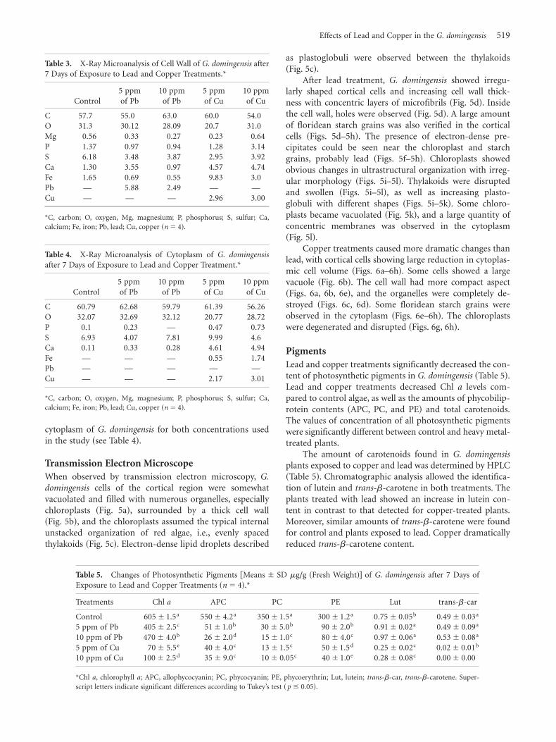
domingensis cells of the cortical region were somewhatvacuolated and filled with numerous organelles, especiallychloroplasts ~Fig. 5a!, surrounded by a thick cell wall~Fig. 5b!, and the chloroplasts assumed the typical internalunstacked organization of red algae, i.e., evenly spacedthylakoids ~Fig. 5c!. Electron-dense lipid droplets described
as plastoglobuli were observed between the thylakoids~Fig. 5c!.
After lead treatment, G. domingensis showed irregu-larly shaped cortical cells and increasing cell wall thick-ness with concentric layers of microfibrils ~Fig. 5d!. Insidethe cell wall, holes were observed ~Fig. 5d!. A large amountof floridean starch grains was also verified in the corticalcells ~Figs. 5d–5h!. The presence of electron-dense pre-cipitates could be seen near the chloroplast and starchgrains, probably lead ~Figs. 5f–5h!. Chloroplasts showedobvious changes in ultrastructural organization with irreg-ular morphology ~Figs. 5i–5l!. Thylakoids were disruptedand swollen ~Figs. 5i–5l!, as well as increasing plasto-globuli with different shapes ~Figs. 5i–5k!. Some chloro-plasts became vacuolated ~Fig. 5k!, and a large quantity ofconcentric membranes was observed in the cytoplasm~Fig. 5l!.
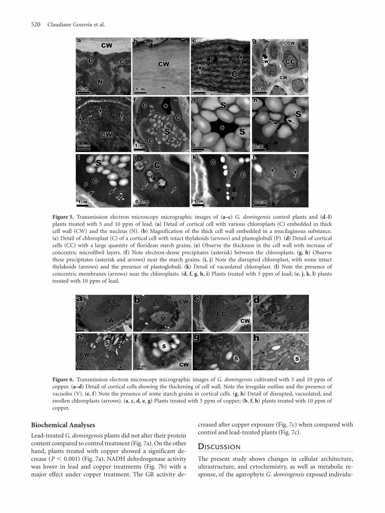
Copper treatments caused more dramatic changes thanlead, with cortical cells showing large reduction in cytoplas-mic cell volume ~Figs. 6a–6h!. Some cells showed a largevacuole ~Fig. 6b!. The cell wall had more compact aspect~Figs. 6a, 6b, 6e!, and the organelles were completely de-stroyed ~Figs. 6c, 6d!. Some floridean starch grains wereobserved in the cytoplasm ~Figs. 6e–6h!. The chloroplastswere degenerated and disrupted ~Figs. 6g, 6h!.
Pigments
Lead and copper treatments significantly decreased the con-tent of photosynthetic pigments in G. domingensis ~Table 5!.Lead and copper treatments decreased Chl a levels com-pared to control algae, as well as the amounts of phycobilip-rotein contents ~APC, PC, and PE! and total carotenoids.The values of concentration of all photosynthetic pigmentswere significantly different between control and heavy metal-treated plants.
The amount of carotenoids found in G. domingensis
plants exposed to copper and lead was determined by HPLC~Table 5!. Chromatographic analysis allowed the identifica-tion of lutein and trans-b-carotene in both treatments. Theplants treated with lead showed an increase in lutein con-tent in contrast to that detected for copper-treated plants.Moreover, similar amounts of trans-b-carotene were foundfor control and plants exposed to lead. Copper dramaticallyreduced trans-b-carotene content.
Table 3. X-Ray Microanalysis of Cell Wall of G. domingensis after7 Days of Exposure to Lead and Copper Treatments.*
Control5 ppmof Pb
10 ppmof Pb
5 ppmof Cu
10 ppmof Cu
C 57.7 55.0 63.0 60.0 54.0O 31.3 30.12 28.09 20.7 31.0Mg 0.56 0.33 0.27 0.23 0.64P 1.37 0.97 0.94 1.28 3.14S 6.18 3.48 3.87 2.95 3.92Ca 1.30 3.55 0.97 4.57 4.74Fe 1.65 0.69 0.55 9.83 3.0Pb — 5.88 2.49 — —Cu — — — 2.96 3.00
*C, carbon; O, oxygen, Mg, magnesium; P, phosphorus; S, sulfur; Ca,calcium; Fe, iron; Pb, lead; Cu, copper ~n � 4!.
Table 4. X-Ray Microanalysis of Cytoplasm of G. domingensis
after 7 Days of Exposure to Lead and Copper Treatment.*
Control5 ppmof Pb
10 ppmof Pb
5 ppmof Cu
10 ppmof Cu
C 60.79 62.68 59.79 61.39 56.26O 32.07 32.69 32.12 20.77 28.72P 0.1 0.23 — 0.47 0.73S 6.93 4.07 7.81 9.99 4.6Ca 0.11 0.33 0.28 4.61 4.94Fe — — — 0.55 1.74Pb — — — — —Cu — — — 2.17 3.01
*C, carbon; O, oxygen, Mg, magnesium; P, phosphorus; S, sulfur; Ca,calcium; Fe, iron; Pb, lead; Cu, copper ~n � 4!.
Table 5. Changes of Photosynthetic Pigments @Means 6 SD mg/g ~Fresh Weight!# of G. domingensis after 7 Days ofExposure to Lead and Copper Treatments ~n � 4!.*
Treatments Chl a APC PC PE Lut trans-b-car
Control 6056 1.5a 5506 4.2a 3506 1.5a 3006 1.2a 0.756 0.05b 0.496 0.03a
5 ppm of Pb 4056 2.5c 516 1.0b 306 5.0b 906 2.0b 0.916 0.02a 0.496 0.09a
10 ppm of Pb 4706 4.0b 266 2.0d 156 1.0c 806 4.0c 0.976 0.06a 0.536 0.08a
5 ppm of Cu 706 5.5e 406 4.0c 136 1.5c 506 1.5d 0.256 0.02c 0.026 0.01b
10 ppm of Cu 1006 2.5d 356 9.0c 106 0.05c 406 1.0e 0.286 0.08c 0.006 0.00
*Chl a, chlorophyll a; APC, allophycocyanin; PC, phycocyanin; PE, phycoerythrin; Lut, lutein; trans-b-car, trans-b-carotene. Super-script letters indicate significant differences according to Tukey’s test ~ p � 0.05!.
Effects of Lead and Copper in the G. domingensis 519
Biochemical Analyses
Lead-treated G. domingensis plants did not alter their proteincontent compared to control treatment ~Fig. 7a!. On the otherhand, plants treated with copper showed a significant de-crease ~P , 0.001! ~Fig. 7a!. NADH dehydrogenase activitywas lower in lead and copper treatments ~Fig. 7b! with amajor effect under copper treatment. The GR activity de-
creased after copper exposure ~Fig. 7c! when compared withcontrol and lead-treated plants ~Fig. 7c!.
D ISCUSSION
The present study shows changes in cellular architecture,ultrastructure, and cytochemistry, as well as metabolic re-sponse, of the agarophyte G. domingensis exposed individu-
Figure 5. Transmission electron microscopy micrographic images of ~a–c! G. domingensis control plants and ~d–l!
plants treated with 5 and 10 ppm of lead. ~a! Detail of cortical cell with various chloroplasts ~C! embedded in thickcell wall ~CW! and the nucleus ~N!. ~b! Magnification of the thick cell wall embedded in a mucilaginous substance.~c! Detail of chloroplast ~C! of a cortical cell with intact thylakoids ~arrows! and plastoglobuli ~P!. ~d! Detail of corticalcells ~CC! with a large quantity of floridean starch grains. ~e! Observe the thickness in the cell wall with increase ofconcentric microfibril layers. ~f! Note electron-dense precipitates ~asterisk! between the chloroplasts. ~g, h! Observethese precipitates ~asterisk and arrows! near the starch grains. ~i, j! Note the disrupted chloroplast, with some intactthylakoids ~arrows! and the presence of plastoglobuli. ~k! Detail of vacuolated chloroplast. ~l! Note the presence ofconcentric membranes ~arrows! near the chloroplasts. ~d, f, g, h, i! Plants treated with 5 ppm of lead; ~e, j, k, l! plantstreated with 10 ppm of lead.
Figure 6. Transmission electron microscopy micrographic images of G. domingensis cultivated with 5 and 10 ppm ofcopper. ~a–d! Detail of cortical cells showing the thickening of cell wall. Note the irregular outline and the presence ofvacuoles ~V!. ~e, f! Note the presence of some starch grains in cortical cells. ~g, h! Detail of disrupted, vacuolated, andswollen chloroplasts ~arrows!. ~a, c, d, e, g! Plants treated with 5 ppm of copper; ~b, f, h! plants treated with 10 ppm ofcopper.
520 Claudiane Gouveia et al.

ally to 5 and 10 ppm of lead or copper. G. domingensis
treated with copper showed a decrease in GRs, togetherwith a reduction in algal biomass and, hence, negativegrowth rates, indicating that copper stress is a key factorlimiting growth. However, while plants treated with leadshowed a slower rate of growth than control, no reductionin algal biomass was evident. Ologuin et al. ~2009! observeddifferent results in Gracilaria coronopifolia and Gracilaria
eucheumoides exposed to lead. Nonsignificant differences inGRs were observed in G. coronopifolia, while GRs in G.
eucheumoides were inhibited. These results clearly indicatethat the toxicity of lead is species dependent.
On the other hand, the bleaching and partial necrosisof the apical segments of G. domingensis during 7 days ofcultivation under copper and lead could have resulted fromchanges in metabolism and DNA damage by the formation
of pyrimidine dimers ~Bouzon et al., 2012; Santos et al.,2012!. Tissue deformation and necrosis were also observedin the carragenophyte H. musciformis after exposure tocadmium during 7 days ~Bouzon et al., 2012! and G. domin-
gensis exposed to UV radiation-B during 21 days ~Schmidtet al., 2010a!.
According to Visviki and Rachlin ~1992!, copper is anessential element for the photosynthetic electron transportsystem and a component or cofactor of several enzymes.The algae may actively exclude, or sequester, the copper tominimize toxic impact. The decrease in GRs observed in theG. domingensis samples studied may be related to the use ofenergy as an activation mechanism for the adaptation orrepair of damage induced by high copper stress.
The cell wall of the control and heavy metal-treatedplants of G. domingensis reacted positively to TB-O. This oc-curred because the cell wall contains sulfated polysaccha-rides in the molecular structure of agar. The metachromaticgranulations were observed inside the cortical and subcorti-cal cells of plants treated with lead and copper. Especially invacuoles, these granulations appeared as reserves of complexpolysaccharides and could be sites of accumulation that wassubsequently incorporated into the cell wall and the muci-lage. Overall, this finding indicates an increase in cell wallthickness, which was observed in cells after treatment withTB-O of G. domingensis exposed to lead and copper.
When analyzed under a TEM, the cell wall of G. domin-
gensis control showed a microfibrillar texture with mi-crofibrils structured in concentric layers with differentdegrees of compression. Observations of the cell wall of plantsexposed to lead and copper showed an increase in that struc-tural component. The increase in the thickness of the cellwall of G. domingensis exposed to lead and copper can beinterpreted as a defense mechanism. The presence of precip-itates in cytoplasm of G. domingensis exposed to lead ~seeFigs. 5f, 5g! could be associated with a metal-immobilizingfunction to minimize lead stress.
The analysis of the cell walls of treated plants by energydispersive spectrometer made it possible to detect the pres-ence of lead and copper, indicating the metal accumulationand metal binding in these structures. An increase in thepresence of Ca and Fe was verified on the cell wall surface ofplants treated with Cu. On the other hand, Ca was reducedin plants treated with 10 ppm of Pb. This change in cell wallsurface composition with increasing concentration of Caand Fe may be associated with a barrier against the presenceof copper. The sulfated polysaccharides that compose thecell wall, such as agar and carrageenan, are responsible formost of the metal accumulation and metal binding ~Bouzonet al., 2012!. In the case of agar, metal binding is associatedwith the degree of sulfatation ~Diannelidis & Delivopoulos,1997; Bouzon et al., 2012; Santos et al., 2012!. According theSharma and Dubey ~2005!, mucilage binding restricts metaluptake and establishes an important barrier protecting thecells. The retention of Pb was observed in the cell wall andmucilage upon the binding of Pb to ion-exchangeable siteson the cell wall and extracellular precipitation, mainly in the
Figure 7. Biochemical responses of G. domingensis after 7 days ofexposure to lead and copper treatments. ~a! Protein content of G.
domingensis. ~b! Relative changes of mitochondrial NADH de-hydrogenase activity of G. domingensis. ~c! Glutathione reductaseassay activity of G. domingensis. Means6 SD ~n � 4!. The asteriskindicates the statistical differences.
Effects of Lead and Copper in the G. domingensis 521

form of Pb carbonate deposited in the cell wall. The pres-ence of carboxyl and sulfated groups in the cell walls of G.
domingensis restricts the transportation of Pb via apoplast.On the other hand, Yruela ~2005! describes many coppertransporters already characterized in plant cells, such asthose in the cell membrane ~COPT1!, cytosol ~copper chap-erones! and chloroplast ~COPT3, PAA1, PAA2, and CpCCS!, mitochondria ~COX17!, and Golgi body ~RAN1, chap-erones and COPT5!.
Cellular metabolism of G. domingensis exposed to leadand copper was modified. As a consequence, cell wall thick-ening was produced in both treatments, and this, in turn,resulted in copper-induced reduction in the synthesis offloridean starch grains. The decrease in floridean starch grainsobserved through TEM and LM in plants exposed to coppermay be related to a change in the route of biosynthesis ofstarch enzymes of the Calvin cycle, possibly by activating thedegradation pathways. However, Schmidt et al. ~2010a! ob-served one increase in floridean starch grains in G. domingen-
sis after exposure to UV radiation-B, and Bouzon et al. ~2012!observed a decrease in starch grains in H. musciformis aftertreatment with different concentrations of cadmium.
In red algae, the thylakoids that are not associated witheach other are free in the chloroplasts. The chloroplasts ofG. domingensis control showed a structure typical of redalgae, having one peripheral thylakoid surrounded by paral-lel thylakoids. The number of parallel thylakoids is variable,and this number mainly depends on the spatial location ofthe cell in the algae. In contrast, the chloroplasts of G.
domingensis exposed to lead showed structural changes,including disrupted and swollen thylakoids.
However, the plants treated with copper showed moredramatic chloroplast alteration. Similar results were observedwith the red macroalgae Ceramium ciliatum exposed to cop-per, where the chloroplast appeared with disrupted thyla-koids and an increase in plastoglobuli volume ~Diannelidis &Delivopoulos, 1997! and the green algae Enteromorpha flexu-
osa ~Andrade et al., 2004!. Finally, when analyzed by TEM, G.
domingensis exposed to lead revealed an increase in the num-ber of plastoglobuli in the chloroplast. This increase in thenumber of lipids can be considered as a change in metabo-lism, as observed in G. domingensis treated with lead.
In the present study, Chl a contents of G. domingensis
showed more resistance upon exposure to lead in compari-son to copper, which showed a reduction in concentrationby 85%. These results can be associated with changes inchloroplast organization, i.e., a drastic reduction in autofluo-rescence, as shown by confocal microscopy. Other investiga-tions of red macroalgae have shown an increase in Chl a
contents after copper exposure, including, for example, G.
tenuistipitata ~Pinto et al., 2011!. However, in Gracilaria
lemaneiformis, Xia et al. ~2004! observed that Chl a contentwas not altered after exposure to copper.
The amounts of phycobiliproteins decreased in G.
domingensis treated with lead and copper. The phycobilip-roteins are located in the phycobilisomes outside the chlo-roplast thylakoids. In red algae, PE is used during the
acclimation process; therefore, it is located more externallyin the phycobilisomes ~Talarico, 1996!. Our results demon-strated that phycobiliprotein levels, including APC, PC, andPE, decreased in G. domingensis after lead and coppertreatments. These molecules absorb solar energy, transfer-ring it to the reaction center of photosystem II, where Chl a
is excited by the flow of electrons ~Gantt, 1981!. Accordingto Xia et al. ~2004!, a high concentration of copper alteredphycobilisome structure, and these changes resulted in adecline of absorbed light energy, thus inhibiting photosyn-thesis. We found a decrease in phycobiliprotein levels,similar to the findings of Xia et al. ~2004!, who studied thered macroalgae G. lemaneiformis cultivated with copperduring 4 days. This indicates that lead and copper stronglyinhibited the accumulation of phycobiliproteins.
Carotenoid contents in G. domingensis were also alteredby lead and copper treatment. The contents of carotenoids,mainly lutein, remarkably increased in lead-treated G. domin-
gensis plants, suggesting that this metal induces the accumu-lation of those pigments, along with the decrease in Chl a
and phycobiliprotein contents. This result may be inter-preted as a protective acclimation mechanism to prevent thetoxic effects of lead. Collén et al. ~2003! and Pinto et al.~2011! observed similar results in G. tenuistipitata exposedto copper and cadmium, with the increase of lutein andb-carotene. In contrast, copper-treated G. domingensis plantsdecreased their carotenoid contents by 78%, and trans-b-carotene was not detected in copper-treated samples.
According to Collén et al. ~2003!, heavy metal in ex-posed algae induces the production of reactive oxygen spe-cies ~ROS!. The ROS induce changes in several molecules,such as lipids, proteins, and nucleic acid. In G. tenuistipitata
treated with cadmium and copper, one increase in theamount of saturated and monounsaturated fatty acids ~Pintoet al., 2011!. However, in the same study, a reduction of thelevels of polyunsaturated fatty acids was observed. As astrategy to prevent the effects of ROS, photosynthetic organ-isms produce antioxidant defenses, such as flavonoids, toco-pherols, carotenoids, and enzymes. Collén et al. ~2003!observed that treatment of G. tenuistipitata with copper ledto an increase in the enzymatic activity of catalase, ascor-bate peroxidase, and superoxide dismutase, while treatmentwith cadmium only increased catalase activity.
The significant reduction in protein content observedin copper-treated G. domingensis, when compared with con-trol and lead-treated plants, indicates that copper inhibitsprotein metabolism. Similar results were observed by Collénet al. ~2003! in G. tenuistipitata treated with cadmium andcopper. They proposed that the decreased content of pro-tein could have been caused by toxic effects to protein oramino acid synthesis, as well as nitrate reduction. In ourstudy, treatment with both copper and lead blocked mito-chondrial NADH dehydrogenase activity, including that ofcomplex I. Under these conditions, complex I of the respi-ratory chain has been reported as one of the major sites ofROS generation in mitochondria under physiological condi-tions ~Boveris et al., 1976; Chen et al., 2003!, and an overt
522 Claudiane Gouveia et al.

ROS production is observed when this redox center isblocked ~Turrens, 1997; Turrens & Boveris, 1980!. Further-more, ROS formed at the NADH dehydrogenase site ofcomplex I are released into the mitochondrial matrix ~Chenet al., 2003!, thereby eliciting oxidative damage to mitochon-drial enzymes, including the complexes of the respiratorychain, enzymes of the Krebs cycle, and several other sensi-tive proteins, as well as mtDNA ~Bandy & Davison, 1990;Zhang et al., 1990; Hausladen & Fridovich, 1994!.
However, our study found that cellular antioxidantpotential could be characterized by a significant increase innonenzymatic antioxidant defenses, such as the increase inlutein content in the presence of high lead concentration,while antioxidant enzymatic activities were essentially un-changed by copper treatment.
In summary, the present study demonstrates that leadand copper negatively affect various morphological, physio-logical, and biochemical processes in G. domingensis. Thisbecame obvious after exposure of plant samples to concen-trations of 5 and 10 ppm under laboratory conditions overa 7-day experimental period, resulting in reduction in growthrates, ultrastructural damage observed primarily in the in-ternal organization of chloroplasts, increased cell wall thick-ness and plastoglobuli. Based on the relative toxicity profileof the two heavy metals studied in G. domingensis, copper ismore toxic than lead.
ACKNOWLEDGMENTS
The authors would like to acknowledge the staff of theCentral Laboratory of Electron Microscopy ~LCME!, Fed-eral University of Santa Catarina, Florianopolis, Santa Cata-rina, Brazil, for the use of their TEM. This study wassupported in part by the Coordenação de Aperfeiçoamentode Pessoal de Nível Superior ~CAPES, Brazil!, ConselhoNacional de Desenvolvimento Científico e Tecnológico~CNPq, Brazil!, and Fundação de Apoio à Pesquisa Cienti-fica e Tecnológica do Estado de Santa Catarina ~FAPESC!.The authors are grateful to CAPES for providing a scholar-ship to C.G. and CNPq-PIBIC for providing a scholarshipto M.K.. É.C.S. holds a postdoctoral fellowship from CAPES-PNPD-Institucional. Z.L.B., M.M., and A.L. are CNPq fel-lows. F.C. is a FAPESP and CNPq fellow. This study is partof the Master’s thesis of the first author.
REFERENCES
Andrade, L.R., Farina, M. & Amado Filho, G.M. ~2002!. Role ofPadina gymnospora ~Dictyotales, Phaeophyceae! cell walls incadmium accumulation. Phycologia 41, 39–48.
Andrade, L.R., Farina, M. & Amado Filho, G.M. ~2004!. Effectsof copper on Enteromorpha flexuosa ~Chlorophyta! in vitro.
Ecotoxicol Environ Saf 58, 117–125.Bandy, B. & Davison, A.J. ~1990!. Mitochondrial mutations may
increase oxidative stress: Implications for carcinogenesis andaging? Free Radic Biol Med 8, 523–539.
Bouzon, Z.L., Ferreira, E.C., Santos, R., Scherner, F., Horta,
P.A., Maraschin, M. & Schmidt, E.C. ~2012!. Influences ofcadmium on fine structure and metabolism of Hypnea musci-
formis ~Rhodophyta, Gigartinales! cultivated in vitro. Proto-
plasma 249, 637–650.Boveris, A., Cadenas, E. & Stoppani, A.O. ~1976!. Role of ubiq-
uinone in the mitochondrial generation of hydrogen peroxide.Biochem J 156, 435–444.
Callow, M.E. & Callow, J. ~2002!. Marine biofouling: A stickingproblem. Biologist 49, 1–5.
Carlberg, I. & Mannervik, B. ~1985!. Glutathione reductase.Methods Enzimol 113, 484–490.
Cassiana, A. & Radi, R. ~1996!. Differential inhibitory action ofnitric oxide and peroxynitrite on mitochondrial electron trans-mit. Arch Biochem 328, 309–316.
Chen, Q., Vazquez, E.J., Moghaddas, S., Hoppel, C.L. & Lesnef-
sky, E.J. ~2003!. Production of reactive oxygen species by mito-chondria: Central role of complex III. J Biol Chem 278,36027–36031.
Collén, J., Pinto, E., Pedersén, M. & Colepicolo, P. ~2003!.Induction of oxidative stress in the red macroalgae Gracilaria
tenuistipitata by polluant metals. Arch Environ Contam Toxicol
45, 337–342.Diannelidis, B. & Delivopoulos, S.G. ~1997!. The effects of zinc,
copper and cadmium on the fine structure of Ceramium ciliatum
~Rhodophyceae, Ceramiales!. Mar Environ Res 44~2!, 127–134.Edwards, P. ~1970!. Illustrated guide to the seaweeds and sea
grasses in the vicinity of Porto Aransas. Texas Contrib Mar Sci
15, 1–228.Eick, M.J., Peak, J.D., Brady, P.V. & Pesek, J.D. ~1999!. Kinetics of
lead adsorption and desorption on goethite: Residence timeeffect. J Soil Sci 164, 28–39.
Gantt, E. ~1981!. Phycobilisomes. Annu Rev Plant Physiol 32,327–347.
Guimarães, G.M., Plastino, E.M. & Oliveira, E.C. ~1999!.Life history reproduction and growth of Gracilaria domingen-
sis ~Gracilariales, Rhodophyta! from Brasil. Bot Mar 42, 481–486.
Hausladen, A. & Fridovich, I. ~1994!. Superoxide and peroxy-nitrite inactivate aconitases, but nitric oxide does not. J Biol
Chem 269, 29405–29408.Hepler, P.K. & Gunning, B.E.S. ~1998!. Confocal fluorescence
microscopy of plant cells. Protoplasma 201, 121–157.Hiscox, J.D. & Israelstam, G.F. ~1979!. A method for the extrac-
tion of chlorophyll from leaf tissue without maceration. Can J
Bot 57, 1332–1334.Hu, S., Tang, C.H. & Wu, M. ~1996!. Cadmium accumulation by
several seaweeds. Sci Total Environ 187, 65–71.Hulshof, P.J.M., Kosmeijer-Schuil, T., West, C.E. & Hollman,
P.C.H. ~2007!. Quick screening of maize kernels for provitaminA content. J Food Comp Anal 20, 655–661.
Kuhnen, S., Lemos, P.M.M., Campestrini, L.H., Ogliari, J.B.,
Dias, P.F. & Maraschin, M. ~2009!. Antiangiogenic propertiesof carotenoids: A potential role of maize as functional food. J
Funct Foods 1, 284–290.Kursar, T.A., van Der Meer, J. & Alverte, R.S. ~1983!. Light-
harvesting system of the red alga Gracilaria tikvahiae. II. Phyco-bilisome characteristics of pigment mutants. Plant Physiol 73,361–369.
Latini, A., Rodriguez, M., Borba Rosa, R., Scussiato, K., Leip-
nitz, G., Reir de Assis, D., da Costa Ferreira, G., Funchal,
C., Jacques-Silva, MC., Buzin, L., Giugliani, R., Cassiana,
A., Radi, R. & Wajner, M. ~2005!. 3-Hydroxyglutaric acidmoderately impairs energy metabolism in brain of young rats.Neuroscience 135, 111–120.
Effects of Lead and Copper in the G. domingensis 523

Lee, T.M., Chang, Y.C. & Lin, Y.H. ~1999!. Differences in physio-logical responses between winter and summer Gracilaria tenuis-
tipitata ~Gigartinales, Rhodophyta! to varying temperature. Bot
Bull Acad Sin 40, 93–100.Lowry, O.H., Rosebough, N.G. & Farr, A.L. ~1951!. Protein
measurement with the folin phenol reagent. J Biol Chem 193,265–275.
Mallick, N. & Rai, L.C. ~2001!. Physiological responses of non-vascular plants to heavy metals. In Physiology and Biochemistry
of Metal Toxicity and Tolerance in Plants, Prasad, M.N.V. &Strzalka, K. ~Eds.!, pp. 111–147. Dordrecht, The Netherlands:Kluwer Publishers.
Mamboya, F.A., Pratap, H.B., Mtolera, M. & Bjork, M. ~1999!.The effect of copper on the daily growth rate and photosyn-thetic efficiency of the brown macroalgae Padina boergesenii. InProceedings of the Conference on Advances on Marine Sciences in
Tanzania, Richmond, M.D. & Francis, J. ~Eds.!, pp. 185–192.Ologuin, M.M., Roa, E.C. & Uy, W.H. ~2009!. In vitro lead
accumulation in Gracilaria coronopifolia and Gracilaria eucheu-
moides. J Environ Aquatic Res 1, 87–98.Penniman, C.A., Mathieson, A.C. & Penniman, C.E. ~1986!.
Reproductive phenology and growth of Gracilaria tikvahiae
McLachlan ~Gigartinales, Rhodophyta! in the Great Bay Estu-ary New Hampshire. Bot Mar 29, 147–154.
Pinto, E., Carvalho, A.P., Cardozo, K.H.M., Malcata, F.X., dos
Anjos, F.M. & Colepicolo, P. ~2011!. Effects of heavy metalsand light levels on the biosynthesis of carotenoids and fattyacids in the macroalgae Gracilaria tenuistipitata ~var. liui Zhang& Xia!. Rev Bras Farmacogn 21, 349–354.
Rocchetta, I., Leonardi, P.I., Amado Filho, G., Molina, M.C.R.
& Conforti, V. ~2007!. Ultrastructure and X-ray microanalysisof Euglena gracilis ~Euglenophyta! under chromium stress.Phycologia 46, 300–306.
Santos, R., Schmidt, E.C., Paula, M.R., Latini, A., Horta, P.A.,
Maraschin, M. & Bouzon, Z.L. ~2012!. Effects of cadmiumon growth, photosynthetic pigments, photosynthetic perfor-mance, biochemical parameters and structure of chloroplastsin the agarophyte Gracilaria domingensis ~Rhodophyta, Gracilari-ales!. Am J Plant Sci 3, 1077–1084.
Schmidt, E.C., dos Santos, R. & Horta, P.A. ~2010a!. Effects ofUVB radiation on the agarophyte Gracilaria domingensis ~Rhodo-phyta, Gracilariales!: Changes in cell organization, growth andphotosynthetic performance. Micron 41, 919–930.
Schmidt, E.C., Maraschin, M. & Bouzon, Z.L. ~2010b!. Effectsof UVB radiation on the carragenophyte Kappaphycus alvarezii
~Rhodophyta, Gigartinales!: Changes in ultrastructure, growth,and photosynthetic pigments. Hydrobiologia 649, 171–182.
Schmidt, E.C., Nunes, B.G., Maraschin, M. & Bouzon, Z.L.
~2010c!. Effect of ultraviolet-B radiation on growth, photosyn-thetic pigments, and cell biology of Kappaphycus alvarezii
~Rhodophyta, Gigartinales!macroalgae brown strain. Photosyn-
thetica 48, 161–172.Schmidt, E.C., Pereira, B., Pontes, C.L.M., Santos, R., Scherner,
F., Horta, P.A., Paula, M.R., Latini, A., Maraschin, M. &Bouzon, Z.L. ~2012a!. Alterations in architecture and metabo-lism induced by ultraviolet radiation-B in the carragenophyteChondracanthus teedei ~Rhodophyta, Gigartinales!. Protoplasma
249, 353–367.Schmidt, E.C., Pereira, B., Santos, R., Gouveia, C., Costa,
G.B., Faria, G.S.M., Scherner, F., Horta, P.A., Paula, M.R.,
Latini, A., Ramlov, F., Maraschin, M. & Bouzon, Z.L. ~2012b!.Responses of the macroalgae Hypnea musciformis after in vitro
exposure to UV-B. Aquatic Bot 100, 8–17.Schmidt, E.C., Santos, R., Faveri, C., Horta, P.A., Paula, M.R.,
Latini, A., Ramlov, F., Maraschin, M. & Bouzon, Z.L. ~2012c!.Response of the agarophyte Gelidium floridanum after in vitro
exposure to ultraviolet radiation B: Changes in ultrastructure,pigments, and antioxidant systems. J Appl Phycol 24, 1341–1352.
Schmidt, E.C., Scariot, L.A., Rover, T. & Bouzon, Z.L. ~2009!.Changes in ultrastructure and histochemistry of two red mac-roalgae strains of Kappaphycus alvarezii ~Rhodophyta, Gigarti-nales!, as a consequence of ultraviolet B radiation exposure.Micron 40, 860–869.
Scott, C.E. & Eldridge, A.L. ~2005!. Comparison of carotenoidcontent in fresh, frozen and canned corn. J Food Comp Anal 18,551–559.
Sharma, P. & Dubey, R.S. ~2005!. Lead toxicity in plants. Braz J
Plant Physiol 17, 35–52.Sheng, P.X., Ting, Y., Chen, J.P. & Hong, L. ~2004!. Sorption of
lead, copper, cadmium, zinc and nickel by marine algal bio-mass: Characterization of biosorptive capacity and investiga-tion of mechanisms. J Colloid Interface Sci 275, 131–141.
Talarico, L. ~1996!. Phycobiliproteins and phycobilisomes in redalgae: Adaptative responses to light. Sci Mar 60, 205–222.
Talarico, L. ~2002!. Fine structure and X-ray microanalysis of ared macrophyte cultured under cadmium stress. Environ Pollut
120, 813–821.Tonon, A.P., Oliveira, M.C., Soriano, E.M. & Colepicolo, P.
~2011!. Absorption of metals and characterization of chemicalelements present in three species of Gracilaria ~Gracilariaceae!Greville: A genus of economical importance. Rev Bras Farma-
cogn 21, 355–360.Torres, M.A., Barros, M.P., Campos, S.C.G., Pinto, E., Raja-
mani, S., Sayre, R.T. & Colepicolo, P. ~2008!. Biochemicalbiomarkers in algae and marine pollution: A review. Ecotoxicol
Environ Saf 71, 1–15.Tseng, C.K. ~2001!. Algal biotechnology industries and research
activities in China. J Appl Phycol 13, 375–380.Turrens, J.F. ~1997!. Superoxide production by the mitochondri-
alrespiratory chain. Biosci Rep 17, 3–8.Turrens, J.F. & Boveris, A. ~1980!. Generation of superoxide
anion by the NADH dehydrogenase of bovine heart mitochon-dria. Biochem J 191, 421–427.
Visviki, I. & Rachlin, J.W. ~1992!. Ultrastructural changes inDunaliella minuta following acute and chronic exposure tocopper and cadmium. Arch Environ Contam Toxicol 23, 420–425.
Wellburn, A.R. ~1994!. The spectral determination of chlorophylla and chlorophyll b, as well as total carotenoids, using varioussolvents with spectrophotometers of different resolution. J Plant
Physiol 144, 307–313.Xia, J.R., Li, Y.J., Lu, J. & Chen, B. ~2004!. Effects of copper and
cadmium on growth, photosynthesis, and pigment content inGracilaria lemaneiformis. Bull Environ Contam Toxicol 73,979–986.
Yruela, I. ~2005!. Copper in plants. Braz J Plant Physiol 17,145–146.
Zhang, Y., Marcillat, O., Giulivi, C., Ernster, L. & Davies,
K.J. ~1990!. The oxidative inactivation of mitochondrial elec-tron transport chain components and ATPase. J Biol Chem 265,16330–16336.
524 Claudiane Gouveia et al.