the effect of intravitreal bevacizumab and ranibizumab on cutaneous tensile strength during wound...
TRANSCRIPT
© 2013 Christoforidis et al, publisher and licensee Dove Medical Press Ltd. This is an Open Access article which permits unrestricted noncommercial use, provided the original work is properly cited.
Clinical Ophthalmology 2013:7 185–191
Clinical Ophthalmology
The effect of intravitreal bevacizumab and ranibizumab on cutaneous tensile strength during wound healing
John B Christoforidis1
Jillian Wang2
Angela Jiang2
James Willard5
Cedric Pratt2
Mahmoud Abdel-Rasoul3
Sashwati Roy4
Heather Powell5
1Department of Ophthalmology and Vision Science, College of Medicine, The University of Arizona, Tucson, AZ, USA; 2Department of Ophthalmology, College of Medicine, The Ohio State University, Columbus, OH, USA; 3Center for Biostatistics, The Ohio State University, Columbus, OH, USA; 4Center Surgery, The Ohio State University, Columbus, OH, USA; 5Department of Materials Science, College of Engineering, The Ohio State University, Columbus, OH, USA
Correspondence: John B Christoforidis Retina Division, Department of Ophthalmology, University of Arizona Medical Center, 655 N Alvernon Way, Suite 108, Tucson, AZ 85711, USA Tel +1 520 321 3677 Fax +1 520 321 3665 Email [email protected]
Purpose: To investigate the effect of intravitreal bevacizumab and ranibizumab on wound tension
and by histopathology during cutaneous wound healing in a rabbit model and to compare this
effect to placebo intravitreal saline controls 1 and 2 weeks following intravitreal injection.
Methods: A total of 120 New Zealand white rabbits were randomly assigned to one of three
treatment groups each consisting of 40 rabbits. Each group received intravitreal injections of
bevacizumab, ranibizumab, or normal saline. Immediately afterwards, each rabbit underwent
four 6 mm full-thickness dermatologic punch biopsies. Twenty rabbits from each agent group
underwent wound harvesting on day 7 or day 14. The skin samples were stained for CD34
for vascular endothelial cells on day 7, and maximal wound tensile load was measured on
days 7 and 14. Quantitative assessment of mean neovascularization (MNV) scores was obtained
from 10 contiguous biopsy margin 400× fields of CD34-stained sections by two independent
observers.
Results: Wound tension reading means (N) with standard error and adjusted P-values on day 7
were: saline placebos, 7.46 ± 0.87; bevacizumab, 4.50 ± 0.88 (P = 0.041); and ranibizumab,
4.67 ± 0.84 (P = 0.025). On day 14 these were: saline placebos, 7.34 ± 0.55; bevacizumab,
6.05 ± 0.54 (P = 0.18); and ranibizumab 7.99 ± 0.54 (P = 0.40). MNV scores in CD34 stained
sections were: saline controls, 18.31 ± 0.43; bevacizumab, 11.02 ± 0.45 (P , 0.0001); and
ranibizumab, 13.55 ± 0.43 (P , 0.0001). The interobserver correlation coefficient was 0.928.
Conclusion: At day 7, both anti–vascular endothelial growth factor (anti-VEGF) agents had
significantly suppressed MNV scores and exerted a significant reduction of cutaneous wound
tensile strength compared with saline controls. At day 14, neither agent produced a significant
effect on tensile wound strength. Since angiogenesis is an integral component of the proliferative
phase of wound healing, we encourage clinicians to be aware of their patients’ recent surgical
history during intravitreal anti-VEGF therapy and to consider refraining from their use during
the perioperative period.
Keywords: wound healing, tensile strength, bevacizumab, ranibizumab
IntroductionBevacizumab and ranibizumab are two therapeutic agents that suppress all isoforms
of vascular endothelial growth factor (VEGF). They are currently the most commonly
used agents for the treatment of the exudative form of age-related macular degeneration
(AMD) and macular edema due to central and branch retinal vein occlusion. Both
agents are used on an off-label basis for the treatment of non-AMD choroidal
neovascularization and diabetic macular edema in the United States.
Wound healing complications following systemic use of bevacizumab are well
known and include wound dehiscence and hemothorax after thoracotomy and port
Dovepress
submit your manuscript | www.dovepress.com
Dovepress 185
O R i g i N A L R E S E A R C H
open access to scientific and medical research
Open Access Full Text Article
http://dx.doi.org/10.2147/OPTH.S40537

Clinical Ophthalmology 2013:7
placement, surgical site bleeding, incisional hernias, erosions,
ecchymoses, and infection.1–5 These adverse events are
especially prevalent in patients over the age of 60 years.6
As a result of these, several reports have recommended
discontinuation of systemic bevacizumab 4–6 weeks prior
to surgery and recommencement 4 weeks after surgery.6–10
Despite these findings, there has been little investigation
into the effects of wound healing after intravitreal use of
bevacizumab and ranibizumab. This is partly because of the
large number of variables associated with wound healing
(eg, differing incision size and depth, time of wounding in
relation to intravitreal injection). Therefore, an animal model
in which the number of these variables is minimized becomes
a reasonable option to study the effects of intravitreal anti-
VEGF agents on cutaneous wound healing.
A recent histopathologic report demonstrated that both of
these intravitreally placed agents suppressed neovasculariza-
tion of cutaneous wound margins by quantitative assessment
of CD34 immunostaining 1 week following treatment com-
pared with untreated controls. There was a nonsignificant
inhibitory trend for both bevacizumab and ranibizumab
(P = 0.013 and 0.071 respectively, alpha , 0.008). At
2 weeks, there was no demonstrable effect for either agent
(P = 0.471 and 0.297 respectively), presumably since the
wounds had entered the maturation phase of healing when
the number of wound blood vessels is decreased.11
To the authors’ knowledge, there are no reports on the
functional outcomes of cutaneous wound healing follow-
ing intravitreal anti-VEGF therapy. Measurement of wound
tensile strength is an established method for evaluating
the biomechanical properties of wound repair, specifically
collagen function.14–17 The primary purpose of this study was
to investigate the effect of intravitreally placed bevacizumab
and ranibizumab on wound tensile strength during cutaneous
wound healing in a rabbit model at 1 and 2 weeks and to com-
pare these effects to intravitreal saline controls. Since there was
an inhibitory trend at week one in our previous report, we also
evaluated neovascularization of cutaneous wound margins by
quantitative assessment of CD34 immunostaining 1 week fol-
lowing treatment compared with untreated controls.
Methodsintravitreal injection and cutaneous biopsyA total of 120 New Zealand white rabbits (Myrtle’s Rabbitry,
Thompsons Station, TN, USA) weighing 3.8–3.9 kg was used
in this study. All treatments were conducted in agreement with
the principles of laboratory animal care (National Institutes
of Health publication No 85-23, revised 1985), the OPRR
Public Health Service Policy on the Humane Care and Use
of Laboratory Animals (revised 1986) and the US Animal
Welfare Act. All experimental protocols were approved, and
the procedures followed were in accordance with the ethical
standards of The Institutional Animal Care and Use Commit-
tee at The Ohio State University. Prior to general anesthesia,
each rabbit was administered 0.3 mg/kg of meloxicam orally.
Anesthesia was induced with 10 mg/mL acepromazine sub-
cutaneously and maintained with 3%–5% isoflurane. Electric
clippers were used to shave the dorsal skin of the rabbit,
and this area was then cleansed with 10% povidone-iodine
and 70% alcohol. A standard 6 mm dermatologic punch
biopsy (Sklar Tru Punch; Sklar Instruments, West Chester,
PA, USA) was used to create two full-thickness cutaneous
wounds with removal of the skin core. These were created
approximately 3 cm lateral to each side of the cervicothoracic
vertebral column in the mid-dorsum area. The rabbits were
randomly assigned to one of three groups, each consisting of
40 rabbits. Immediately following the biopsies, each rabbit
received an intravitreal injection in the left eye consisting of
0.05 mL of 1.25 mg/0.05 mL bevacizumab, 0.5 mg/0.05 mL
ranibizumab or 0.05 mL of normal saline.
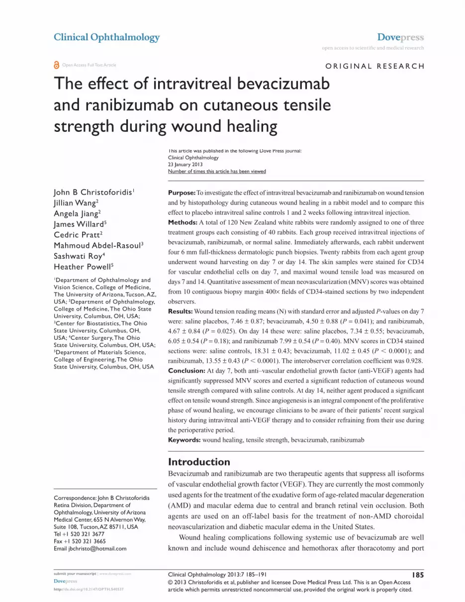
Wound biomechanicsTo assess the strength of the healing wounds, skin biopsies
(30 mm × 80 mm) were harvested from the flanks of the
bevacizumab-, ranibizumab-, and saline-treated rabbits 7 or
14 days post wounding (Figure 1A). Only biopsy sites with
clearly visible central core defects were harvested. Following
wound harvesting, the anesthetized rabbits were euthanized
by intravenous injection of 3 mL of saturated potassium
chloride. Each rectangular-shaped skin biopsy was stored
in a 50 mL polypropylene conical tube containing normal
saline solution and placed in ice for 2–4 hours until cut into
a dog bone–shaped specimen it was (gauge length of 2 mm
and gauge width of 4 mm) for tensile testing (Figure 1B).
The harvested skin was placed in a position such that the
biopsied site was in the center of the gauge length and
width (Figure 1B). The muscle fascia was removed from
each dog bone specimen with a scalpel. Each specimen was
then mounted into the grips of a TestResources mechanical
tester model 1000R12 (TestResources, Shakopee, MN, USA)
with a 50 lbf load cell (Figure 1C). All skin samples were
strained at a rate of 2 mm/s until failure and maximum load
was recorded in newtons (N). Specimens that failed at the
wound site due to the weight of the bottom half of the dog
bone were assigned a strength of zero N.
submit your manuscript | www.dovepress.com
Dovepress
Dovepress
186
Christoforidis et al
Clinical Ophthalmology 2013:7
HistopathologyOn day 7, two 6 mm biopsy sites were harvested from the
dorsal skin of the anesthetized rabbit, with a standard 12 mm
dermatologic punch biopsy (Accuderm, Ft Lauderdale, FL,
USA) that included the entire previous biopsy site along with
surrounding normal skin. Only biopsy sites with clearly vis-
ible central core defects were harvested. Immediately after
harvesting, skin samples were placed into conical tubes con-
taining 10% neutral-buffered formalin. After 48 hours, the
formalin was exchanged with 70% ethanol and the biopsied
skin was routinely processed, embedded in paraffin wax, and
sectioned at 5 µm. Serial paraffin sections placed on poly-L-
lysine slides were immunohistoc hemically stained for CD34
as described in a previous report.11 Ten contiguous 400× fields
at the biopsy site wound margins of CD34-stained sections
were photographed using a six-headed Olympus BX51
light microscope with attached Altra 20 digital camera and
MicroSuite software (B&B Microscopes Limited, Pittsburgh,
PA, USA). The photomicrographs were assorted randomly,
and two independent observers who were blinded to the
experimental groups recorded the numbers of CD34-stained
endothelial cell clusters for each slide. The observer score
means for each photomicrograph and the mean neovascular-
ization (MNV) scores representing all of the 10 photomicro-
graphs for each subject were calculated and evaluated.
Statistical analysisA linear mixed model with random intercepts was fit to com-
pare the difference in max load between treatment groups
and the control group. The mixed model adjusts for the pos-
sible correlation of repeated measurements within subjects.
Holm’s step-down method for multiple comparisons was
used to adjust for multiple hypothesis tests.
A linear mixed model was fit to compare the difference
in mean scores between all three treatment groups (avastin,
lucentis, and saline). The mixed model adjusts for the possible
correlation of repeated measurements of specimens within
wounds and wounds within rabbits by treating wounds within
rabbits and specimens within wounds as random effects.
Tukey’s method for pair-wise comparisons was used to
adjust for multiple hypothesis tests. Interobserver correlation
coefficient was used to assess agreement among raters.
ResultsWound biomechanicsFollowing the procedures, the rabbits were monitored daily.
At the time of wound harvesting all of the rabbits had clearly
evident cutaneous wound without signs of infection. The
treated eyes were absolutely quiet without signs of discharge,
redness, or irritation at the time of harvesting.
All of the subjects had at least one specimen harvested for
tensile strength evaluation. However, eight biopsy sites on
day 7 (five bevacizumab and three saline controls) from eight
separate rabbits and one biopsy site from on day 14 (saline
control) did not have clearly visible central core defects and
these were not harvested.
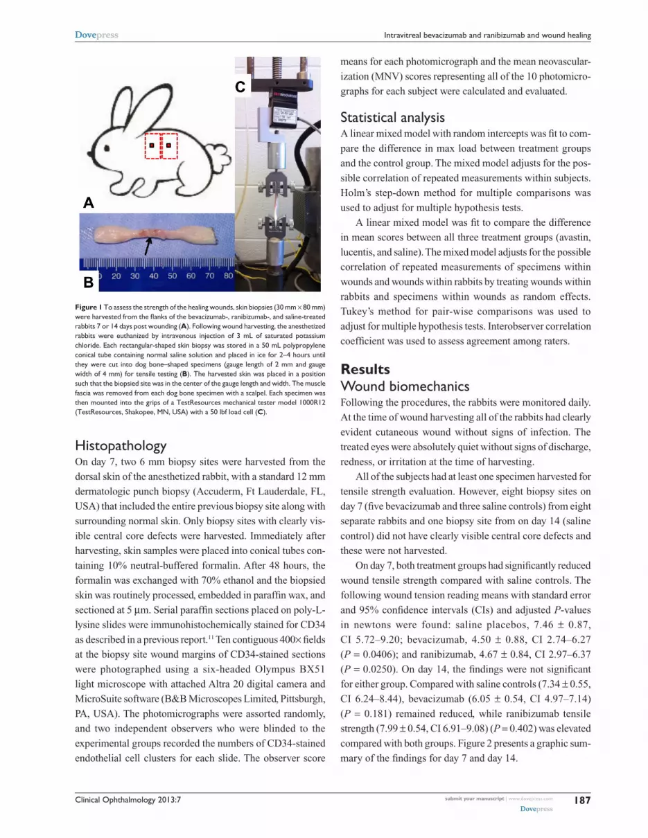
On day 7, both treatment groups had significantly reduced
wound tensile strength compared with saline controls. The
following wound tension reading means with standard error
and 95% confidence intervals (CIs) and adjusted P-values
in newtons were found: saline placebos, 7.46 ± 0.87,
CI 5.72–9.20; bevacizumab, 4.50 ± 0.88, CI 2.74–6.27
(P = 0.0406); and ranibizumab, 4.67 ± 0.84, CI 2.97–6.37
(P = 0.0250). On day 14, the findings were not significant
for either group. Compared with saline controls (7.34 ± 0.55,
CI 6.24–8.44), bevacizumab (6.05 ± 0.54, CI 4.97–7.14)
(P = 0.181) remained reduced, while ranibizumab tensile
strength (7.99 ± 0.54, CI 6.91–9.08) (P = 0.402) was elevated
compared with both groups. Figure 2 presents a graphic sum-
mary of the findings for day 7 and day 14.
Figure 1 To assess the strength of the healing wounds, skin biopsies (30 mm × 80 mm) were harvested from the flanks of the bevacizumab-, ranibizumab-, and saline-treated rabbits 7 or 14 days post wounding (A). Following wound harvesting, the anesthetized rabbits were euthanized by intravenous injection of 3 mL of saturated potassium chloride. Each rectangular-shaped skin biopsy was stored in a 50 mL polypropylene conical tube containing normal saline solution and placed in ice for 2–4 hours until they were cut into dog bone–shaped specimens (gauge length of 2 mm and gauge width of 4 mm) for tensile testing (B). The harvested skin was placed in a position such that the biopsied site was in the center of the gauge length and width. The muscle fascia was removed from each dog bone specimen with a scalpel. Each specimen was then mounted into the grips of a TestResources mechanical tester model 1000R12 (TestResources, Shakopee, MN, USA) with a 50 lbf load cell (C).
submit your manuscript | www.dovepress.com
Dovepress
Dovepress
187
intravitreal bevacizumab and ranibizumab and wound healing

Clinical Ophthalmology 2013:7
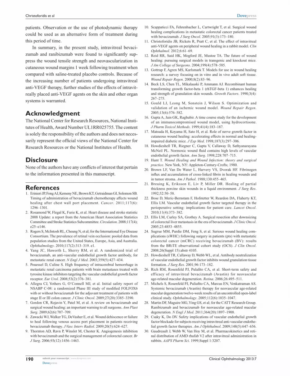
HistopathologyAt the time of wound harvesting on day 7, 115/120 wound
sites were clearly visible with central core defects and were
harvested. All of the subjects had at least one biopsied wound
harvested for histopathologic evaluation. Five biopsy sites
(three bevacizumab, one ranibizumab, and one saline control)
from five separate rabbits did not have clearly visible central
core defects and were not harvested. On light microscopy,
all of the wound margin borders were clearly visible and
were scored. MNV scores were calculated from all of the
400× CD34-stained rabbit skin sections (Figure 3).
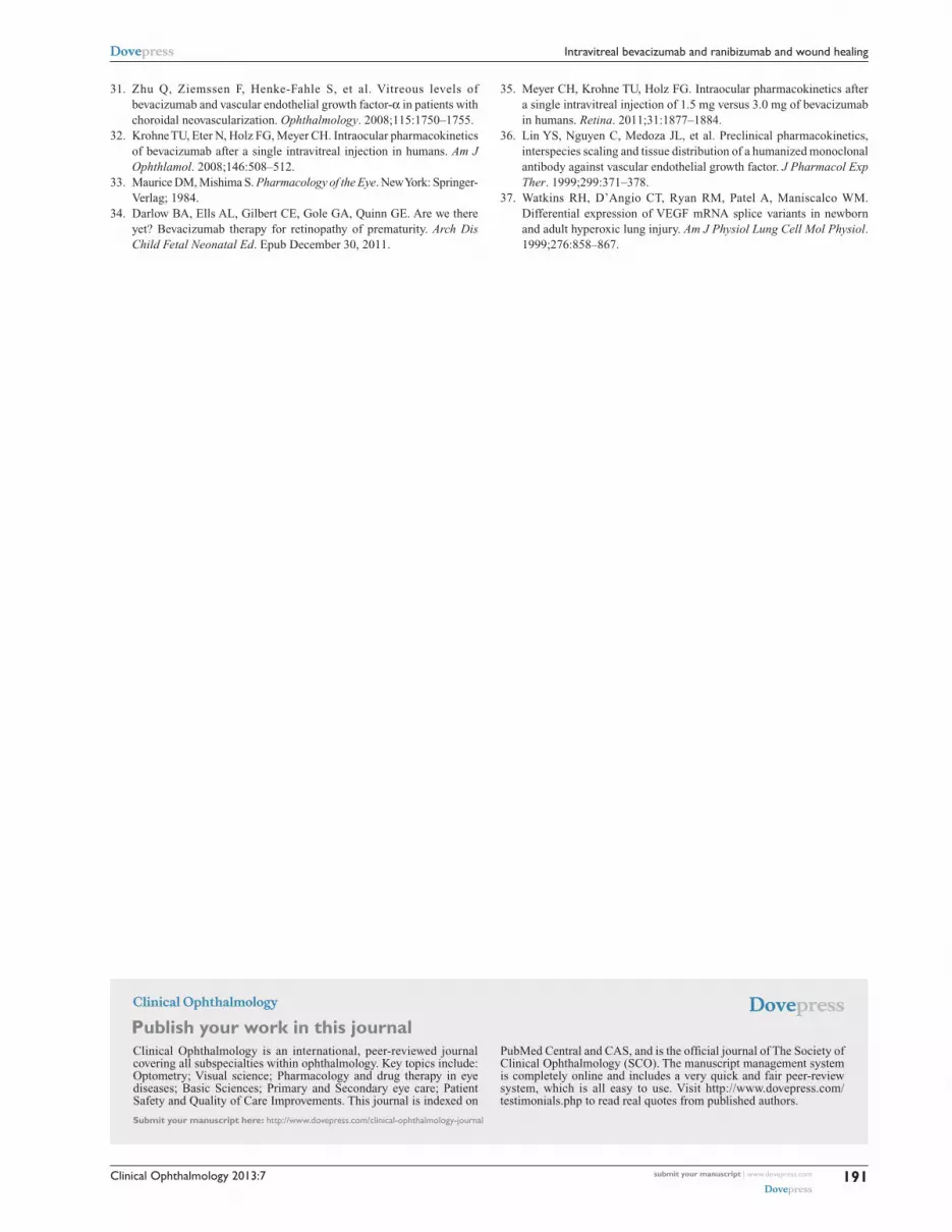
Both of the treatment groups were found to significantly
inhibit the number of vessels compared with the placebo
group (shown as mean with standard deviation and 95% CI).
Compared with saline controls (18.31 ± 0.43, CI 17.45–19.16),
bevacizumab (11.02 ± 0.45, CI 10.13–11.91) (P , 0.0001)
inhibited neovascularization in cutaneous wound margins. The
mean MNV for ranibizumab was higher than bevacizumab
and was significantly higher than saline control (13.55 ± 0.43,
CI 12.70–14.41; P , 0.0001) (Figure 4). The interobserver
correlation coefficient was 0.921, indicating high interpreta-
tion agreement between raters (P , 0.0001). Score 1 and
score 2 were highly correlated, with a correlation coefficient
of 0.928. The mean of score 1 and score 2 was used as the
outcome variable to compare the treatment effects.
DiscussionIn this study, both intravitreal bevacizumab and ranibizumab
were found to exert significant inhibition on cutaneous
wound tensile strength and neovascularization in cutaneous
wound margins at day 7 compared with saline controls. At
day 14, neither agent produced a significant effect on tensile
wound strength. This work demonstrates that intravitreally
placed bevacizumab and ranibizumab suppresses cutane-
ous wound tensile strength during the proliferative phase of
wound healing in a rabbit model.
The proliferation phase during wound healing usually
occurs between days 4 and 14.18 Angiogenesis suppression
at this phase disturbs the integrity of wound restoration.19–21
This effect on cutaneous wound healing is the likely mecha-
nism by which systemic bevacizumab adversely affects
wound healing as previously described in several clinical
studies.6,8–10,22–24 Following the proliferative phase of wound
healing is the maturation phase, when the number of blood
vessels in the wound decreases.
The suppressed histologic wound changes on day 7
translated to functional cutaneous changes as demonstrated
14
Wound tension maximal load (N)averages with confidence intervals
12
10
8
6
4
2
0Bevac BevacRanib
Day 7
Max
imal
load
(n
ewto
ns)
Day 14Saline
P < 0.041 P < 0.025
P < 0.181
P < 0.402
Ranib Saline
–4.50 –4.67
–6.05
–7.99–7.34–7.46
Figure 2 Wound tension maximal load means with 95% confidence intervals and treatment group P-values for each group at days 7 and 14.Note: The adjusted P-values are in relation to saline control at each time point.Abbreviations: Bevac, bevacizumab-treated group; Ranib, ranibizumab-treated group; Saline, saline-treated group.
Figure 3 Representative CD34 histological figures of cutaneous wounds 1 week after wounding. At 20×, wound margins are demonstrated (A, C and E). At 400×, note increased numbers of endothelial cells counts in the placebo (about 17) (B) versus bevacizumab (about 11) (D) and ranibizumab (about 12) (F).
submit your manuscript | www.dovepress.com
Dovepress
Dovepress
188
Christoforidis et al

Clinical Ophthalmology 2013:7
by wound tension measurements. On day 14, differences in
wound tensile strength between groups were not significant,
and the number of vessels was decreased compared with
those on day 7. The findings in this study are consistent
with a recent report describing an inhibitory trend for MNV
scores on day 7 for bevacizumab (P = 0.013) and ranibi-
zumab (P = 0.071) compared with pegaptanib (P = 0.378)
and untreated controls.11 In that report, the central cores were
not removed and the average MNV scores were lower for all
groups than in the present study.
In addition to the effects on wound healing, low rates of
hypertension and arterial thromboembolic events including
angina pectoris, myocardial infarction, transient ischemic
attack, and cerebrovascular accident have been associated with
both bevacizumab and ranibizumab.25–28 Because of the small
incidences of these events reported in these studies, they do
not have enough statistical power to detect low rates of these
events.29 At this time, the mechanism of serum availability
of bevacizumab and ranibizumab after intravitreal injection
is speculative. It may gain access to the systemic circulation
either anteriorly through the trabecular meshwork or posteri-
orly through the retina and the choroidal circulation. The effects
of bevacizumab and ranibizumab on wound healing following
intravitreal injections are yet to be specifically examined in a
major clinical trial involving these agents.
There were several limitations in this study. The use of a
rabbit model has several inherent constraints. The vitreous
volume in rabbits is approximately one-third that of humans
(4.5 mL versus 1.5 mL), while the serum compartment is
significantly smaller than that of humans. Clinical doses
of intravitreal anti-VEGF agents in a rabbit model increase
the systemic exposure of these agents in comparison with
humans. The clearance half-lives of bevacizumab and
ranibizumab have been found to be longer in adult humans
than in rabbits, possibly because the larger vitreous volume
results in lowered agent concentrations and increased diffu-
sion time prior to clearance from the eye.30–33 On the other
hand, the rabbit vitreous volume is closer in size to that of a
retinopathy of a prematurity eye (1.6–2.5 mL)34,35 and may
be used to more accurately simulate the intravitreal pharma-
cokinetic properties of these agents in this group of patients.
Furthermore, rabbit retinas contain a tapetum lucidum, and
it is unclear how this affects agent absorption. Nevertheless,
the rabbit model is an accepted and valuable experimental
model for many human ocular and systemic conditions that
otherwise could not be feasibly studied.
Each rabbit had at least one biopsied wound harvested
for tensile and histopathologic evaluation. Eight wounds
from day 7, and 1 wound from day 14 had incomplete
central core defects and were excluded from the tensiometry
data measurements. With incomplete core removal from
the wound, normal skin tissue overgrowth occurs and dog
bone wound tensiometry is measured high. This is a known
limitation with tensile strength measurements in immature
wounds and was an important reason that two wounds were
created per subject. Similarly, five wound biopsies for
histopathology were not harvested because of incomplete
central core defects. Incomplete core defects result in a
decreased inflammatory response and lowered endothelial
cell proliferation. In a previous report where central cores
were not removed, MNV scores for bevacizumab were
7.41 (versus 10.91), ranibizumab 8.71 (versus 13.51), and
11.51 (versus 18.37) for the controls. Lastly, we were,
unfortunately, not able to directly measure ranibizumab and
bevacizumab in the blood because the methodology was not
available at our institution or any of our referral labs.
Rabbit VEGF has been found to bind to bevacizumab and
is estimated to be 94% homologous with human counterparts
of VEGF 121, 165, and 189 isoforms.36,37 Nevertheless,
because of interspecies differences, it is somewhat
presumptuous to extrapolate our findings in this rabbit model
to clinical situations with humans. Clinicians should be aware
of the potential for delayed wound healing, to be cognizant of
their patients’ recent surgical history and/or the presence of
any open wounds, particularly in retinopathy of prematurity
20
P < 0.0001
P < 0.0001
Bevac
NV
mea
ns
Ranib
Day 7 – NV means with95% confidence intervals
Saline
18
16
14
12
10
8
6
4
2
0
–11.0
–13.6
–18.3
Figure 4 CD34 endothelial cell cluster per high power field means with 95% confidence intervals and treatment group P-values for each group at day 7.Abbreviations: NV, neovascularization; Bevac, bevacizumab-treated group; Ranib, ranibizumab-treated group; Saline, saline-treated group.
submit your manuscript | www.dovepress.com
Dovepress
Dovepress
189
intravitreal bevacizumab and ranibizumab and wound healing
Clinical Ophthalmology 2013:7
patients. Observation or the use of photodynamic therapy
could be used as an alternative form of treatment during
this period of time.
In summary, in the present study, intravitreal bevaci-
zumab and ranibizumab were found to significantly sup-
press the wound tensile strength and neovascularization in
cutaneous wound margins 1 week following treatment when
compared with saline-treated placebo controls. Because of
the increasing number of patients undergoing intravitreal
anti-VEGF therapy, further studies of the effects of intravit-
really placed anti-VEGF agents on the skin and other organ
systems is warranted.
AcknowledgmentThe National Center for Research Resources, National Insti-
tutes of Health, Award Number UL1RR025755. The content
is solely the responsibility of the authors and does not neces-
sarily represent the official views of the National Center for
Research Resources or the National Institutes of Health.
DisclosureNone of the authors have any conflicts of interest that pertain
to the information presented in this manuscript.
References1. Erinieri JP, Fong AJ, Kemeny NE, Brown KT, Getraidman GI, Solomon SB.
Timing of administration of bevacizumab chemotherapy affects wound healing after chest wall port placement. Cancer. 2011;117(6): 1296–1301.
2. Rosamond W, Flegal K, Furie K, et al. Heart disease and stroke statistic 2008 Update: a report from the American Heart Association Statistics Committee and Stroke Statistics Subcommittee. Circulation. 2008;117(4); e25–e146.
3. Rogers S, McIntosh RL, Cheung N, et al; for the International Eye Disease Consortium. The prevalence of retinal vein occlusion: pooled data from population studies from the United States, Europe, Asia, and Australia. Ophthalmology. 2010;117(2):313–319. e1.
4. Yang JC, Haworth L, Sherry RM, et al. A randomized trial of bevacizumab, an anti-vascular endothelial growth factor antibody, for metastatic renal cancer. N Engl J Med. 2003;359(5):427–434.
5. Pouessel D, Culine S. High frequency of intracerebral hemorrhage in metastatic renal carcinoma patients with brain metastases treated with tyrosine kinase inhibitors targeting the vascular endothelial growth factor receptor. Eur Urol. 2008;53(2):376–381.
6. Allegra CJ, Yothers G, O’Connell MJ, et al. Initial safety report of NSABP C-08: a randomized Phase III study of modified FOLFOX6 with or without bevacizumab for the adjuvant treatment of patients with stage II or III colon cancer. J Clinic Oncol. 2009;27(20):3385–3390.
7. Gordon CR, Rojavin Y, Patel M, et al. A review on bevacizumab and surgical wound healing: an important warning to all surgeons. Ann Plast Surg. 2009;62(6):707–709.
8. Zawacki WJ, Walker TG, DeVasher E, et al. Wound dehiscence or failure to heal following venous access port placement in patients receiving bevacizumab therapy. J Vasc Interv Radiol. 2009;20(5):624–627.
9. Thornton AD, Ravn P, Winslet M, Chester K. Angiogenesis inhibition with bevacizumab and the surgical management of colorectal cancer. Br J Surg. 2006;93(12):1456–1463.
10. Scappaticci FA, Fehrenbacher L, Cartwright T, et al. Surgical wound healing complications in metastatic colorectal cancer patients treated with bevacizumab. J Surg Oncol. 2005;91(3):173–180.
11. Christoforidis JB, Rickets R, Pratt C, et al. The effect of intravitreal anti-VEGF agents on peripheral wound healing in a rabbit model. Clin Ophthalmol. 2012;6:61–69.
12. Reid RR, Said HK, Mogford JE, Mustoe TA. The future of wound healing: pursuing surgical models in transgenic and knockout mice. J Am College of Surgeons. 2004;199(4):578–585.
13. Gottrup F, Agren MS, Karlsmark T. Models for use in wound healing research: a survey focusing on in vitro and in vivo adult soft tissue. Wound Repair Regen. 2000;8(2):83–96.
14. Beck LS, Chen TL, Mikalauski P, Ammann AJ. Recombinant human transforming growth factor-beta 1 (rhTGF-beta 1) enhances healing and strength of granulation skin wounds. Growth Factors. 1990;3(4): 267–275.
15. Gould LJ, Leong M, Sonstein J, Wilson S. Optimization and validation of an ischemic wound model. Wound Repair Regen. 2005;13(6):576–582.
16. Gupta A, Jain GK, Raghubir. A time course study for the development of an immunocompromised wound model, using hydrocortisone. J Pharm Toxicol Methods. 1999;41(4):183–187.
17. Matsuda H, Koyama H, Sato H, et al. Role of nerve growth factor in cutaneous wound healing: accelerating effects in normal and healing-impaired diabetic mice. J Exp Med. 1998;187(3):297–306.
18. Howdieshell TR, Riegner C, Gupta V, Callaway D, Sathyanarayana McNeil PL. Normoxic wound fluid contains high levels of vascular endothelial growth factor. Ann Surg. 1998;228:707–715.
19. Hunt T. Wound Healing and Wound Infection: theory and surgical practice. New York, NY: Appleton-Century-Crofts; 1980.
20. Brown LF, Van De Water L, Harvery VS, Dvorak HF. Fibrinogen influx and accumulation of cross-linked fibrin in healing wounds and in tumor stroma. Am J Pathol. 1988;130:455–465.
21. Breuing K, Eriksson E, Liv P, Miller DR. Healing of partial thickness porcine skin wounds in a liquid environment. J Surg Res. 1992;52:50–58.
22. Bose D, Meric-Bernstam F, Hofstetter W, Reardon DA, Flaherty KT, Ellis LM. Vascular endothelial growth factor targeted therapy in the perioperative setting: implications for patient care. Lancet Oncol. 2010;11(4):373–382.
23. Ellis LM, Curley SA, Grothey A. Surgical resection after downsizing of colorectal liver metastasis in the era of bevacizumab. J Clinic Oncol. 2005;23:4853–4855.
24. Sugrue MM, Purdie DM, Feng S, et al. Serious wound healing com-plications (sWHC) following surgery in patients (pts) with metastatic colorectal cancer (mCRC) receiving bevacizumab (BV): results from the BRiTE observational cohort study (OCS). J Clin Oncol. 2008;26(Suppl 15):abstr 4105.
25. Howdieshell TR, Callaway D, Webb WL, et al. Antibody neutralization of vascular endothelial growth factor inhibits wound granulation tissue formation. J Surg Res. 2001;96:173–182.
26. Rich RM, Rosenfeld PJ, Pulafito CA, et al. Short-term safety and eff icacy of intravitreal bevacizumab (Avastin) for neovascular age-related macular degeneration. Retina. 2006;26:495–511.
27. Michels S, Rosenfeld PJ, Puliafito CA, Marcus EN, Venkatraman AS. Systemic bevacizumab (Avastin) therapy for neovascular age-related macular degeneration twelve-week results of an uncontrolled open-label clinical study. Ophthalmology. 2005;112(6):1035–1047.
28. Martin DF, Maguire MG, Ying GS, et al; for the CATT Research Group. Ranibizumab and bevacizumab for neovascular age-related macular degeneration. N Engl J Med. 2011;364(20):1897–1908.
29. Csaky K, Do DV. Safety implications of vascular endothelial growth factor blockade for subjects receiving intravitreal anti-vascular endothe-lial growth factor therapies. Am J Ophthalmol. 2009;148(5):647–656.
30. Gaudreault J, Webb W, Van Hoy M, et al. Pharmacokinetics and reti-nal distribution of AMD rhufab V2 after intravitreal administration in rabbits. AAPS Pharm Sci. 1999;Suppl 1:3207.
submit your manuscript | www.dovepress.com
Dovepress
Dovepress
190
Christoforidis et al
Clinical Ophthalmology
Publish your work in this journal
Submit your manuscript here: http://www.dovepress.com/clinical-ophthalmology-journal
Clinical Ophthalmology is an international, peer-reviewed journal covering all subspecialties within ophthalmology. Key topics include: Optometry; Visual science; Pharmacology and drug therapy in eye diseases; Basic Sciences; Primary and Secondary eye care; Patient Safety and Quality of Care Improvements. This journal is indexed on
PubMed Central and CAS, and is the official journal of The Society of Clinical Ophthalmology (SCO). The manuscript management system is completely online and includes a very quick and fair peer-review system, which is all easy to use. Visit http://www.dovepress.com/ testimonials.php to read real quotes from published authors.
Clinical Ophthalmology 2013:7
31. Zhu Q, Ziemssen F, Henke-Fahle S, et al. Vitreous levels of bevacizumab and vascular endothelial growth factor-α in patients with choroidal neovascularization. Ophthalmology. 2008;115:1750–1755.
32. Krohne TU, Eter N, Holz FG, Meyer CH. Intraocular pharmacokinetics of bevacizumab after a single intravitreal injection in humans. Am J Ophthlamol. 2008;146:508–512.
33. Maurice DM, Mishima S. Pharmacology of the Eye. New York: Springer- Verlag; 1984.
34. Darlow BA, Ells AL, Gilbert CE, Gole GA, Quinn GE. Are we there yet? Bevacizumab therapy for retinopathy of prematurity. Arch Dis Child Fetal Neonatal Ed. Epub December 30, 2011.
35. Meyer CH, Krohne TU, Holz FG. Intraocular pharmacokinetics after a single intravitreal injection of 1.5 mg versus 3.0 mg of bevacizumab in humans. Retina. 2011;31:1877–1884.
36. Lin YS, Nguyen C, Medoza JL, et al. Preclinical pharmacokinetics, interspecies scaling and tissue distribution of a humanized monoclonal antibody against vascular endothelial growth factor. J Pharmacol Exp Ther. 1999;299:371–378.
37. Watkins RH, D’Angio CT, Ryan RM, Patel A, Maniscalco WM. Differential expression of VEGF mRNA splice variants in newborn and adult hyperoxic lung injury. Am J Physiol Lung Cell Mol Physiol. 1999;276:858–867.
submit your manuscript | www.dovepress.com
Dovepress
Dovepress
Dovepress
191
intravitreal bevacizumab and ranibizumab and wound healing