the crosslinking and antimicrobial properties of tunichrome
TRANSCRIPT
The crosslinking and antimicrobial properties of tunichrome
Mingmei Cai a, Manickam Sugumaran b, William E. Robinson a,⁎a University of Massachusetts Boston, Environmental, Earth and Ocean Sciences Department, 100 Morrissey Boulevard, Boston, MA 02125-3393, USAb University of Massachusetts Boston, Department of Biology, 100 Morrissey Boulevard, Boston, MA 02125-3393, USA
A B S T R A C TA R T I C L E I N F O
Article history:Received 28 March 2008Received in revised form 2 June 2008Accepted 7 June 2008Available online 12 June 2008
Keywords:Antimicrobial activityCrosslinkingDehydrodopaNatural productQuinone methideTunicatesTunichromeTunic formation
Tunichromes are small peptides containing one or more dehydrodopa derived units that have been identifiedin the blood cells of at least eleven species of tunicates. Incubation of tunichromes isolated from Ascidia nigrahemocytes (or model dopa-containing compounds) under oxidative conditions with either lysozyme,cytochrome c or ovalbumin resulted in a time-dependent polymerization of these test proteins to dimers,trimers, tetramers and potentially to other oligomers. These results indicate that the oxidation products oftunichromes possess inherent crosslinking properties. Hence it is possible that tunichromes participate intunic production by forming adducts and crosslinks with structural proteins and/or carbohydrate polymers,similar to the well-understood process of insect cuticle hardening. Since such crosslinking potentials couldalso be beneficial for defense reactions against invading microorganisms, antibacterial activity oftunichromes was tested using both a radial diffusion assay and the Microtox® test. Tunichromes exhibitedantimicrobial activity against gram-negative bacteria Escherichia coli and Photobacterium phosphorium.However, they did not show any antimicrobial activity against the gram-positive bacteria Staphylococcusaureus at the concentrations tested. We propose that the crosslinking and antimicrobial functions are bothbased on the reactivity of dehydrodopa units present in the tunichromes, and their subsequent ability toform highly reactive quinone methides.
© 2008 Elsevier Inc. All rights reserved.
1. Introduction
Tunicates (also called ascidians or simply sea squirts; subphylumTunicata, Class Ascidiacea) are sessile marine filter-feeding inverte-brates that exhibit features characteristic of the vertebrates, includinggill slits, larval notocord and larval dorsal nerve cord, yet areconsidered to be a highly divergent offshoot from the other membersof the Chordata (Zeng and Swalla, 2005). Several species of tunicatescontain various, low molecular weight, dopa- (=3,4 dihydroxyphenyl-alanine-) or topa- (=3,4,5-trihydroxyphenylalanine-) containing pep-tides (or modified peptides) in their blood cells or other body tissuesincluding styelins from Styela clava (Taylor et al., 2000), plicatamidefrom Styela plicata (Tincu et al., 2000), halocyamines from Halo-cynthia roretzi (Azumi et al., 1990a), lamellarins from Didemnumchartaceum (Lindquist et al., 1988), ferreascidin from Pyura stolonifera(Dorsett et al., 1987) and tunichromes from eleven species of tunicates
(Macara et al., 1979; Bruening et al., 1985, 1986; Oltz et al., 1988; Bayeret al., 1992; Parry et al., 1992; Tincu and Taylor, 2002).
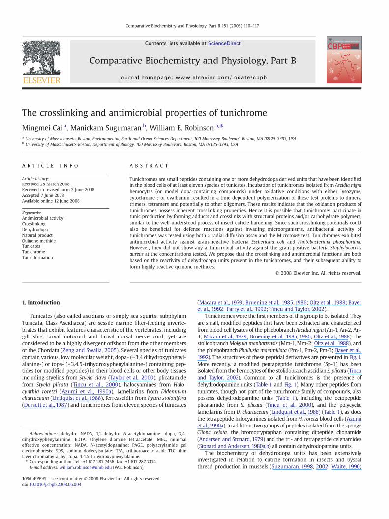
Tunichromeswere the firstmembers of this group to be isolated. Theyare small, modified peptides that have been extracted and characterizedfrom blood cell lysates of the phlebobranch Ascidia nigra (An-1, An-2, An-3; Macara et al., 1979; Bruening et al., 1985, 1986; Oltz et al., 1988), thestolidobranchMolgula manhattensis (Mm-1, Mm-2; Oltz et al., 1988), andthe phlebobranch Phallusia mammillata (Pm-1, Pm-2, Pm-3; Bayer et al.,1992). The structures of these peptidal derivatives are presented in Fig. 1.More recently, a modified pentapeptide tunichrome (Sp-1) has beenisolated from the hemocytes of the stolidobranch ascidian S. plicata (Tincuand Taylor, 2002). Common to all tunichromes is the presence ofdehydrodopamine units (Table 1 and Fig. 1). Many other peptides fromtunicates, though not part of the tunichrome family of compounds, alsopossess dehydrodopamine units (Table 1), including the octopeptideplicatamide from S. plicata (Tincu et al., 2000), and the polycycliclamellarins from D. chartaceum (Lindquist et al., 1988) (Table 1), as doesthe tetrapeptide halocyamines isolated from H. roretzi blood cells (Azumiet al.,1990a). In addition, two groups of peptides isolated from the spongeCliona celata, the bromotryptophan containing dipeptide clionamide(Andersen and Stonard, 1979) and the tri- and tetrapeptide celenamides(Stonard and Andersen, 1980a,b) all contain dehydrodopamine units.
The biochemistry of dehydrodopa units has been extensivelyinvestigated in relation to cuticle formation in insects and byssalthread production in mussels (Sugumaran, 1998, 2002; Waite, 1990;
Comparative Biochemistry and Physiology, Part B 151 (2008) 110–117
Abbreviations: dehydro NADA, 1,2-dehydro N-acetyldopamine; dopa, 3,4-dihydroxyphenylalanine; EDTA, ethylene diamine tetraacetate; MEC, minimaleffective concentration; NADA, N-acetyldopamine; PAGE, polyacrylamide gelelectrophoresis; SDS, sodium dodecylsulfate; TFA, trifluoroacetic acid; TLC, thinlayer chromatography; topa, 3,4,5-trihydroxyphenylalanine.⁎ Corresponding author. Tel.: +1 617 287 7456; fax: +1 617 287 7474.
E-mail address: [email protected] (W.E. Robinson).
1096-4959/$ – see front matter © 2008 Elsevier Inc. All rights reserved.doi:10.1016/j.cbpb.2008.06.004
Contents lists available at ScienceDirect
Comparative Biochemistry and Physiology, Part B
j ourna l homepage: www.e lsev ie r.com/ locate /cbpb
Rzepecki and Waite, 1991; Sugumaran and Ricketts, 1995; Burzio andWaite, 2001). In general, dopa units are initially converted into thecorresponding quinones by the action of phenoloxidases. Thequinones generated are further transformed either enzymatically ornonenzymatically to dehydrodopa units. Subsequent nonenzymaticoxidation of the dehydrodopa units directly generates highly reactivequinone methide imine amide (Sugumaran, 2000) that can formcrosslinks and adducts necessary for hardening the substratum(Sugumaran, 1998, 2002; Waite, 1990).
In light of theses findings, it is highly likely that the multipledehydrodopamineunits of tunichromeswould alsopossess the ability toform crosslinks and therefore aid tunic formation. It has been knownthat the lysis of the blood cells of P. mammillata leads to the productionof a greenish black fluid, called Henze solution, that subsequently yieldsa dark, sometimes fibrous precipitate (Nette et al., 2000; Ciancio et al.,2004). Furthermore, Robinson et al. (1986) found that tunic of 46-h-oldAscidia callosa larvae reared from dechorionated neurulae was eithermarkedly reduced in thickness or absent altogether. The epidermis wasfragile and cuticular fins failed to develop. Dechorionated neurulaetreated with tunichrome showed an enhancement in tunic formationand rudimentary fin development (Robinson et al., 1986), suggestingthat tunichrome plays a role in tunic formation. A number ofinvestigators have subsequently proposed that tunichrome is involvedin the hardening of the outer cuticle of the tunic, the tunic itself, or thecrosslinking of tunic fibers (Robinson et al., 1986, 1996; Waite, 1990;Taylor et al., 1995, 1997a,b).
Some of the tunichrome-related compounds that have been isolatedfrom tunicate blood cells and possess dehydrodopa units exhibitantimicrobial properties, including styelins, clavanins, halocyamine andplicatamide (Azumi et al., 1990a,b; Lee et al., 1997a,b; Taylor et al., 2000;Tincu et al., 2003; Lehrer, 2003; Lehrer et al., 2001, 2003; Tincu and Taylor,2004). Whether tunichrome itself exhibits in vitro antimicrobial proper-ties, or whether any of these dopa-containing peptides possess in vivoantimicrobial activity, has yet to be determined. Since the crosslinkingpotential of dehydrodopa compounds could be beneficial in defensereactions against microorganisms, we therefore hypothesize that tuni-chromes would exhibit both crosslinking and antimicrobial activity. Toassess suchpossible functions,we examined the crosslinking properties oftunichromes (and some of its analogs) by measuring the degree ofpolymerization of three test proteins (lysozyme, cytochrome c andovalbumin). We also studied the antibacterial properties of tunichromes(and its analogs) with Microtox® and two-stage radial diffusion assays.
2. Materials and methods
2.1. Materials
A. nigra (Savigny) were purchased from Biomarine Technology(Key West, FL, USA). Waters Sep-Pak Vac C18 cartridges wereprocurred from Millipore (Billerica, MA, USA). Mushroom tyrosinase(E.C. 1.14.18.1.), trifluoracetic acid (TFA), urea, EDTA, sodium phos-phate, lysozyme, cytochrome c, FeCl3, SDS, glycerol, Tris, β-mercap-toethanol, bromophenol blue, Coomassie brilliant blue R, glycine-glycine-tyrosine, N-acetlydopamine (=NADA), N-acetly tyrosineethyl ester, dopa methyl ester and routine laboratory reagents wereprovided by Sigma-Aldrich Chemicals (St. Louis, MO, USA). High-performance Thin Layer Chromatography plates were obtained fromVWR International Ltd (Bridgeport, NJ, USA). Acetonitrile, acetic acid,and methanol were supplied by Fisher Chemical (Pittsburgh, PA,USA). Protein markers were purchased from Biorad (Hercules, CA,USA). S. aureus (ATCC 33591) and E. coli strain ML-35p were obtainedfrom American Type Culture Collection (Manassas, VA, USA). Trypti-case soy broth and agar powder were provided by Becton, Bickinsonand Company (Sparks, MD, USA). Microtox® reagent, diluent, andreconstitution solution were purchased from Strategic Diagnostics(Newark, DE, USA). N-acetyl dopa, N-acetyl dopa methyl ester, N-acetyldehydrodopa methyl ester, 3,4-dihydroxyphenyl glycine, N-acetyl
Fig. 1. Structures of some simple tunichromes.
Table 1Examples of low molecular weight dehydrodopamine-containing peptide derivativesisolated from tunicates
Compound Structure
Tunichrome An-1 topa-dehydrotopa-dehydrotopamineTunichrome An-2 dopa-dehydrotopa-dehydrotopamineTunichrome An-3 dopa-dehydrotopa-dehydrodopamineTunichrome Pm-1 topa-topa-dehydrotopamineTunichrome Pm-2 dopa-topa-dehydrotopamineTunichrome Pm-3 topa-topa-dehydrodopamineTunichrome Mm-1 gly-dehydrodopa-dehydrodopamineTunichrome Mm-2 leu-dehydrodopa-dehydrodopamineTunichrome Sp-1 dopa-dopa-gly-pro-dehydrodopaminePlicatamide phe-phe-his-leu-his-phe-his-dehydrodopamineLamellarins (A-H) polycyclic compounds having dehydrodopamines
111M. Cai et al. / Comparative Biochemistry and Physiology, Part B 151 (2008) 110–117
dehydrodopa, ketocatechol, N-acetyl artererone, and N-acetyl dehydro-dopamine (=dehydro NADA) were synthesized in our laboratory (Daliand Sugumaran,1988; Sugumaran and Ricketts,1995; Sugumaran et al.,1996).
2.2. Methods
2.2.1. Isolation and identification of tunichromesBlood was obtained from healthy A. nigra by cutting through the
tunic of the ventral base of the body, exposing the body cavity. Thebright yellow/green blood was drained into 50 mL centrifuge tubes onice and subsequently centrifuged for 20 min at 800 g and 4 °C. Theplasmawas decanted anddiscarded. Tunichromewas isolated fromtheblood cell pellet following the method of Taylor et al. (1995). In brief,the blood cell pellet was extracted with 5% acetic acid containing 8 Murea and 0.1M EDTA. Acetic acid urea-EDTA extracts were subjected tosolid phase extraction onWaters Sep-Pak Vac C18 cartridges (6mL; 1 gof packing material) and eluted with 60% aqueous solution ofacetonitrile containing 0.09% trifluoracetic acid (TFA) after a 20 mL0.1% aqueous TFA wash. Eluent was lyophilized, resuspended in 0.1%TFA, and separated by Reverse Phase Thin Layer Chromatography(10 cm!10 cm plates), using 50% acetonitrile (containing 0.09% TFA) asthe mobile phase. The tunichrome band was identified by itsdistinctive pumpkin-colored fluorescence under long wavelength UVlight (364 nm; Robinson et al., 1996). The TLC plates were stained with2% FeCl3 in order to identify phenol groups (Wagner et al.,1983). All themajor bands were scraped off the TLC plates and eluted from the silicagelwith 60% aqueous solution of acetonitrile containing 0.09% TFA, andevaporated to dryness to collect the purified products (Oltz et al.,1988;Robinson et al., 1996). Subsamples of the dried products wereredissolved in 0.1% TFA and the UV absorbance-spectra weredetermined on a Perkin-Elmer 552A spectrophotometer.
2.2.2. Polymerization studiesThe participation of tunichrome and two model compounds (NADA
and dehydro NADA) in polymerization reactions was investigated usingthree test proteins, lysozyme (Mr 14,300), cytochrome c (Mr 12,300) andovalbumin (Mr 44,300), under oxidative conditions.Mushroom tyrosinasewas used to generate the oxidative derivatives of tunichrome that may beinvolved in polymerization (Sugumaran et al., 1987). The progress of thepolymerization reactionwas easily monitored by sodium dodecylsulfate-polyacrylamide gel electrophoresis (SDS-PAGE).
A standard reaction mixture (500 μL) containing 1 mg of testprotein (e.g. lysozyme, cytochrome c or ovalbumin), 50 μg mushroomtyrosinase, and 500 nmol of catecholic compound (tunichromes ormodel compounds), in 0.01 M sodium phosphate buffer, pH 7.0, wereincubated in an Eppendorf tube at 30 °C for 2 h. The reaction wasusually initiated by the addition of substrate and stopped by theaddition of 500 μL of denaturing buffer (0.125 M Tris-HCl; 20%glycerol; 4% SDS; 10% β-mercaptoethanol and 0.025% bromophenolblue). After the reaction was stopped, the tubes were heated in aboiling water bath for 3 min, and the samples were subject toelectrophoresis on either a 10% SDS-PAGE gel (3.5% stacking gel; 10%running gel) or a 4%–15% gradient SDS-PAGE gel. Protein bands werestained with 0.2% Coomassie brilliant blue R. The size of thepolymerized products was determined by comparison of theirmobilities with those of standard protein molecular weight markers.All polymerization experiments were repeated three times.
2.2.3. Microtox® assay and antimicrobial assaysTunichromes, and 12 model compounds (N-acetyldopa, N-acetyl
dehydrodopa, dopa methylester, N-acetyldopa ester, N-acetyl tyrosineethyl ester, N-acetyl dehydrodopa methyl ester, 3,4-dihydroxyphenylglycine, N-acetyldopamine (=NADA), 1,2-dehydro N-acetyldopamine(=dehydro NADA), glycine–glycine–tyrosine, ketocatechol, and N-acetyl artererone) were tested for antibacterial properties using
Microtox® (Strategic Diagnostics, Newark, DE, USA), a rapid, routinebacterial toxicity test system that measures the reduction in lightoutput by the chemiluminescent bacteria, Photobacterium phosphor-ium (gram-negative) upon exposure to antimicrobial compounds.Tunichrome and all twelve model compounds were tested on 3separate occasions using standard procedures (Azur Environmental,1995). By using various dilutions of the tested compounds (0.1 mMto1 mM), dose-response was quantified as EC50 (i.e. the effectiveconcentration causing a 50% decrease in light output). EPA aquatictoxicity criteria were used to classify the toxicity of tunichromes andthe other model compounds (EPA, 2002): highly toxic if EC50b1 mM;moderately toxic if 1 mMbEC50b10 mM; slightly toxic if 10 mMbEC50b100 mM.
Tunichromes andmodel compoundswere also tested against two testmicroorganisms, S. aureus (gram-positive) and E. coli (gram-negative),using a standard two-stage radial diffusion assay (Steinberg and Lehrer,1997). Organisms were grown to mid-logarithmic phase at 37 °C intrypticase soy broth. After they were washed with 10 mM phosphatebuffer (pH 7.4), approximately 4!106 bacterial colony-forming units werespread onto the underlay gel mixture (1% agarose, 10 mM sodiumphosphate buffer, pH 7.4, and 0.3 mg/mL trypticase soy broth powder).Samplewells, 6mm in diameter and 1.2mmdeep, were punched into theunderlay gel and filled with 10 μL of test sample (0.1 mM to 2 mM). Afterthe plates were incubated for 3 h at 37 °C, a nutrient-rich overlay gel(60 mg/mL trypticase soy broth, 1% agarose cooled to 40 °C) was pouredonto the plates and allowed to harden. Plateswere incubated overnight toallow surviving organisms to form microcolonies. The diameters ofcompletely clear zones were measured to the nearest 0.5 mm andexpressed inunits (1unit=1mm), afterfirst subtracting thewell diameter.The relationship between zone diameter and the log10 of the peptideconcentration was found to be linear. Slopes of the regression lines fortunichrome, dopa methyl ester and N-acetyl dehydrodopa methyl esterwere compared using Student's t-test (Zar, 1984). P≤0.05 was accepted asbeing significant. Because these regressions were linear, the x-intercept ofthe least mean squares regression line was considered to represent theminimal effective concentration (MEC), according to the convention usedin similar studies on antimicrobial compounds in tunicates (Lee et al.,1997a,b; Taylor et al., 2000; Tincu et al., 2000, 2003). All experimentswererepeated three times.
3. Results
3.1. Tunichrome isolation and identification
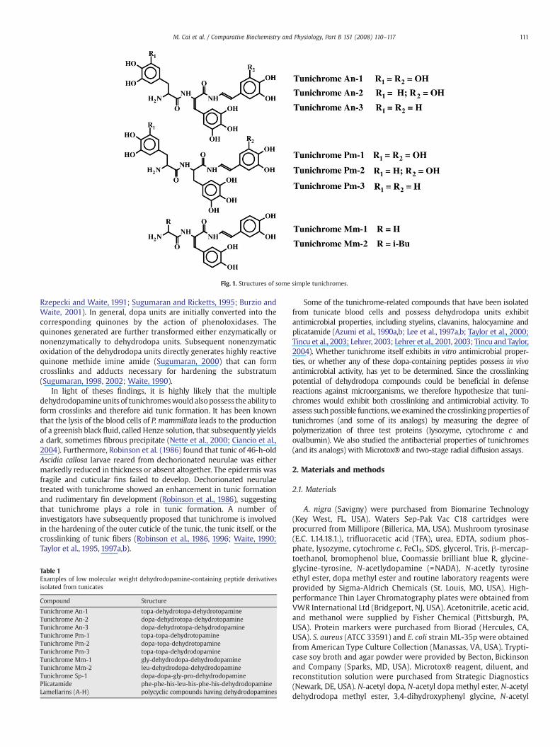
The blood cell extracts (5% acetic acid) were bright yellow/green.After they were subjected to solid phase extraction on Waters Sep-PakVac C18 cartridges, a light yellow eluent was obtained. Final tunichromeseparation by reverse phase TLC revealed over ten differently coloredbands, five of which were major bands (Fig. 2), with the remainder tooweak or variable to clearly differentiate. The five major bands consistedof two yellow bands (Rf=0.15 and 0.20), a bright orange band (Rf=0.6), ablack band (Rf=0.26) and a pumpkin orange band (Rf=0.37) (Fig. 2).Staining with FeCl3 revealed that the pumpkin-orange band was theonly band with phenolic characteristics.
All major bands were scraped off, eluted with 60% acetonitrile, andchecked for UV absorbance. Spectral studies revealed that only thepumpkin-orange band exhibited a UV absorbance spectrum char-acteristic of tunichrome An-1 (Oltz et al., 1988). This band exhibited abroad absorbance peak at 340 nm and a tiny shoulder peak at 300 nm(Fig. 2). The previously published spectrum for An-1 contained a340 nm peak that was narrower and higher than our purified product,probably indicating that our product contained a mixture oftunichrome An-1, An-2 and An-3. The identities of the remainingTLC bands are unknown. Approximately 10 mg of purified tuni-chromes were obtained from the whole blood extracts of 75 medium-sized A. nigra.
112 M. Cai et al. / Comparative Biochemistry and Physiology, Part B 151 (2008) 110–117
3.2. Polymerization of test proteins and either model compounds ortunichromes
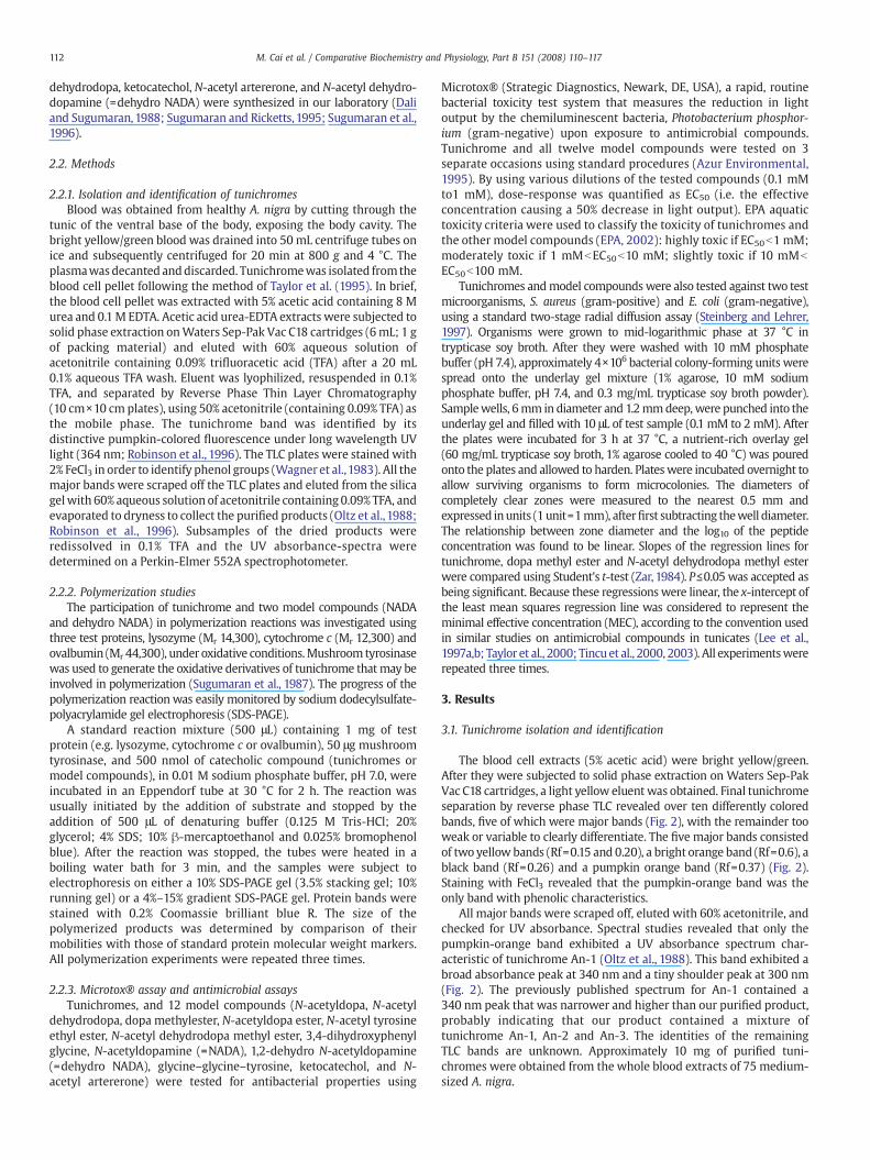
Initial studies with two model compounds, NADA and dehydroNADA, demonstrated that these compounds were capable of cross-linking all three of the test proteins (Fig. 3 depicts lysozyme, a typicalexample). No polymerization was evident at zero time for any of thethree proteins. In the case of lysozyme, dimers (~25 kDa) formed after5 min of incubation with both model compounds. As incubation timewas increased, the concentrations of dimers increased and the trimer(~40 kDa) and tetramer (~60 kDa) gradually appeared. The degree ofpolymerization increased with increasing incubation time. A similarpattern of dimer, trimer and tetramer formationwas observedwhen themodel compoundswere incubatedwith cytochrome c (data not shown).However, in the polymerization of ovalbumin, only the monomer anddimer could be observed after 1 hour incubation with either NADA ordehydro NADA (data not shown).
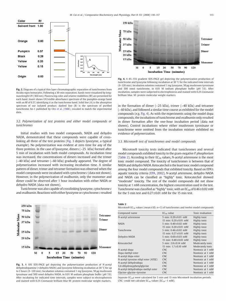
Tunichromewasalso capable of crosslinking lysozyme, cytochrome candovalbumin.Reactionswitheither lysozymeor cytochromec resulted
in the formation of dimer (~25 kDa), trimer (~40 kDa) and tetramer(~60 kDa), and followed a similar time course as exhibited for themodelcompounds (e.g. Fig. 4). As with the experiments using the model dopacompounds, the incubations of tunichromeandovalbuminonly resultedin dimer formation after the one-hour incubation period (data notshown). Control incubations where either mushroom tyrosinase ortunichrome were omitted from the incubation mixture exhibited noevidence of polymerization.
3.3. Microtox® test of tunichromes and model compounds
Microtox® toxicity tests indicated that tunichromes and severalmodel compounds exhibited toxicity to the gram-negative P. phosphorium(Table 2). According to their EC50 values, N-acetyl arterenone is the mosttoxic model compound. The toxicity of tunichromes is between that ofNADAanddehydroNADA.Ketocatechol is the least toxicmodel compoundamong the four model compounds that exhibited toxicity. Based on EPA'saquatic toxicity criteria (EPA, 2002), N-acetyl arterenone, dehydro NADAand NADA can be classified as “highly” toxic. Ketocatechol showed“moderate” toxicity. The rest of the model compounds did not showtoxicity at 1mMconcentration, the highest concentration used in the test.Tunichromewasclassifiedas “highly” toxic,withanEC50of0.46±0.02mMfor the 5 min test and 0.27±0.01 mM for the 15 min test.
Fig. 2. Diagram of a typical thin layer chromatographic separation of tunichromes fromAscidia nigra hemocytes. Following a 30-min separation, bands were visualized by long-wavelength UV (364 nm). Fluorescing color and relativemobilities (Rf) are presented foreach band. Insert shows UV/visible absorbance spectrum of the pumpkin-orange bandwith an Rf of 0.37, identifying it as the tunichrome band. Solid line (A) is the absorptionspectrum of our isolated product; dashed line (B) is the spectrum of purifiedtunichrome An-1 published by Oltz et al. (1988), rescaled to match the experimentaldata.
Fig. 3. A 10% SDS-PAGE gel depicting the polymerization production of N-acetyldehydrodopamine (=dehydro NADA) and lysozyme following incubation at 30 °C for upto 2 hours (0–120 min). Incubation solution contained 1 mg lysozyme, 50 μg mushroomtyrosinase and 500 nmol dehydro NADA, in 0.01 M sodium phosphate buffer (pH 7.0).After incubating for indicated time intervals, samples were subject to electrophoresisand stained with 0.2% Coomassie brilliant blue M: protein molecular weight markers.
Fig. 4. A 4%–15% gradient SDS-PAGE gel depicting the polymerization production oftunichrome and lysozyme following incubation at 30 °C for the indicated time intervals(0–120min). Incubation solution contained 1mg lysozyme, 50 μgmushroom tyrosinaseand 500 nmol tunichrome, in 0.01 M sodium phosphate buffer (pH 7.0). Afterincubation, samples were subjected to electrophoresis and stained with 0.2% Coomassiebrilliant blue. M: protein molecular weight markers.
Table 2Microtox® EC50 values (mean±SD, n=3) of tunichromes and twelve model compounds
Compound name EC50 value Toxic evaluation
N-acetyl arterenone 5 min: 0.20±0.01 mM Highly toxic15 min: 0.20±0.01 mM Highly toxic
NADA 5 min: 0.40±0.02 mM Highly toxic15 min: 0.20±0.01 mM Highly toxic
Tunichrome 5 min: 0.46±0.02 mM Highly toxic15 min: 0.27±0.01 mM Highly toxic
Dehydro-NADA 5 min: 0.60±0.03 mM Highly toxic15 min: 0.40±0.02 mM Highly toxic
Ketocatechol 5 min: 2.8±0.14 mM Moderately toxic15 min: 1.7±0.10 mM Moderately toxic
N-acetyl dopa CNC Nontoxic at 1 mMDopa methyl ester CNC Nontoxic at 1 mMN-acetyl dopa ester CNC Nontoxic at 1 mMN-acetyl tyrosine ethyl ester (ATEE) CNC Nontoxic at 1 mMN-acetyl dehydrodopa CNC Nontoxic at 1 mM3,4-dihydroxyphenylglycine CNC Nontoxic at 1 mMN-acetyl dehydrodopa methyl ester CNC Nontoxic at 1 mMGlycine–glycine–tyrosine CNC Nontoxic at 1 mM
Separate EC50s were calculated for 5 min and 15 min Microtox® incubation periods.CNC: could not calculate EC50 values (EC50N1 mM).
113M. Cai et al. / Comparative Biochemistry and Physiology, Part B 151 (2008) 110–117
3.4. Two-stage radial diffusion assay results of tunichromes and modelcompounds
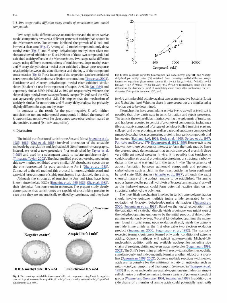
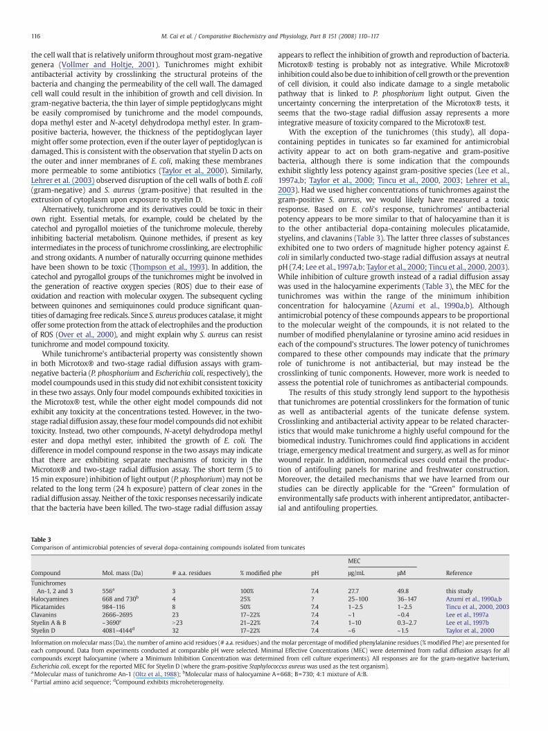
Two-stage radial diffusion assays on tunichrome and the other twelvemodel compounds revealed a different pattern of toxicity than shown inthe Microtox® tests. Tunichrome inhibited the growth of E. coli andformed a clear zone (Fig. 5). Among all 12 model compounds, only dopamethyl ester (Fig. 5) and N-acetyl-dehydrodopa methyl ester (data notshown) showed inhibition on E. coli.Neither of these two compounds hadexhibited toxicity effects in theMicrotox® test. Two-stage radial diffusionassays using different concentrations of tunichromes, dopa methyl esterand N-acetyl dehydrodopa methyl ester exhibited a linear dose-responserelationship between the zone diameter and the log10 of the compoundconcentration (Fig. 6). The x-intercept of the regression can be consideredto represent theMEC. (minimal effective concentration; Tincu et al., 2003).Tunichrome and N-acetyl dehydrodopa methyl ester exhibited similarslopes (Student's t-test for comparison of slopes; PN0.05; Zar, 1984) andapparently similar MECs (49.8 μM vs 49.9 μM respectively), whereas theslope of dopamethyl esterwas significantly steeper (Pb0.05) and theMECwas apparently greater (61.1 μM). This implies that the mechanism oftoxicity is similar for tunichrome andN-acetyl dehydrodopa, but probablyslightly different for dopa methyl ester.
In contrast to the result for the gram-negative E. coli, neithertunichromes nor any other model compounds inhibited the growth ofS. aureus (data not shown). No clear zones were observed compared tothe positive control (0.1 mM ampicillin).
4. Discussion
The initial purification of tunichromeAns andMms (Bruening et al.,1985, 1986; Oltz et al., 1988) involved protection of the unstablemolecule byacetylation and Sephadex LH-20 column chromatography.Instead, we used a new procedure first established by Taylor et al.(1995) and used in a subsequent study to isolate tunichrome Sp-1(Tincu and Taylor, 2002). The final purified product we obtained usingthis new method exhibited a very similar UV absorbance spectrum tothe one represented for pure tunichrome An-1 (Oltz et al., 1988).Compared to the oldmethod, this protocol ismore straightforward andcan yield large amounts of stable tunichrome in a relatively short time.
Although the structures of tunichrome Ans and Mms have beenknown since the late 1980's (Brueninget al.,1985,1986;Oltz et al.,1988),their biological functions remain unknown. The present study clearlydemonstrates that tunichromes are capable of crosslinking proteins invitro once they are enzymatically oxidized by tyrosinase, and they have
in vitro antimicrobial activity against two gram-negative bacteria (E. coliand P. phosphorium).Whether these in vitro properties aremanifested invivo has yet to be determined.
If tunichromes have crosslinking activity in vivo aswell as in vitro, it ispossible that they participate in tunic formation and repair processes.The tunic is the extracellularmatrix covering the epidermis of tunicates,and has been reported to consist of a variety of compounds, including afibrous matrix composed of a type of cellulose (called tunicin), elastins,collagen and other proteins, as well as a ground substance composed ofmucopolysaccharide, glycoproteins, proteins, inorganic compounds andhemocytes (Hall and Saxl, 1961; Deck et al., 1966; De Leo et al., 1977;Patricolo andDeLeo,1979; Robinson et al.,1983,1996). However, it is notknown how these compounds interact to form the tunic matrix. Sincethe present study demonstrates that tunichrome could crosslink threevery different model proteins in vitro, it is possible that tunichromecould crosslink structural proteins, glycoproteins, or structural carbohy-drates in the same way and form the tunic in vivo. The occurrence ofadduct formation between quinonoid compounds and polymericcarbohydrates such as chitin in the insect cuticle has been confirmedby solid state NMR studies (Schaefer et al., 1987), although the exactchemical nature of the adduct remains to be established. The aminogroups (generated by partial hydrolysis ofN-acetylamino sugars) aswellas the hydroxyl groups could form potential reactive sites on thestructural carbohydrate polymers.
The most likely mechanism involved in tunichrome polymerizationshould involve quinone methide imine amide generated by theoxidation of N-acetyl dehydrodopamine derivatives (Sugumaran,2000; Sugumaran et al., 1992). Based on the logical expectation thatthe oxidation of a catechol directly yields a quinone, one might expectthe dehydrodopamine quinone to be the initial product of dehydrodo-pamine oxidation. However,N-acetyl 1,2-dehydrodopamine, themono-mer found in tunichrome, upon oxidation directly yields the quinonemethide imine amide as the first observable two electron oxidationproduct (Sugumaran, 2000; Sugumaran et al., 1992). The normallyexpected isomeric quinone is formed only under conditions of extremeacidity. Quinone methides will exhibit non-enzymatic Michael-1,6-nucleophilic addition with any available nucleophiles including sidechains of proteins, chitin and even water molecules (Sugumaran, 1998,2002). The Shiff's base imine amidewill react with another nucleophile,simultaneously and independently forming another adduct or a cross-link (Sugumaran, 1998, 2002). Quinone methide reactions with nucleicacids are responsible for the antitumor activity of antibiotics such asmitomycin C, adriamycin and daunomycin (reviewed inThompson et al.,1993). If no other molecules are available, quinonemethides can simplyself-dimerize or self-oligomerize to form a variety of polymeric productgroups (Wagner andGrompper,1974; Sugumaran,1998). In addition, theside chains of a number of amino acids could potentially react with
Fig. 5. The two-stage radial diffusion assay of different compounds using E. coli. A: negativecontrol; B:positive control=ampicillin (0.1mM); C: dopamethyl ester (0.5mM);D:purifiedtunichromes (0.5 mM).
Fig. 6. Dose response curve for tunichromes (▲), dopa methyl ester (■) and N-acetyldehydrodopa methyl ester (○) obtained from two-stage radial diffusion assays.Regression equations (least mean squares fit): y=2.3 log10(x) −9.1, r2=0.962; y=3.8log10(x) −15.7, r2=0.893; y=2.5 log10(x) −9.7, r2=0.876 respectively. Toxic units aredefined as the diameters (mm) of completely clear zones after subtracting the welldiameter. Data points are mean±SD, n=3.
114 M. Cai et al. / Comparative Biochemistry and Physiology, Part B 151 (2008) 110–117
quinone methides and form adducts (Sugumaran, 1998). The hydroxylgroup of the chitin polymer can also react with quinone methide underverymild conditions,making themvery attractive candidates for adductformation and crosslinking.
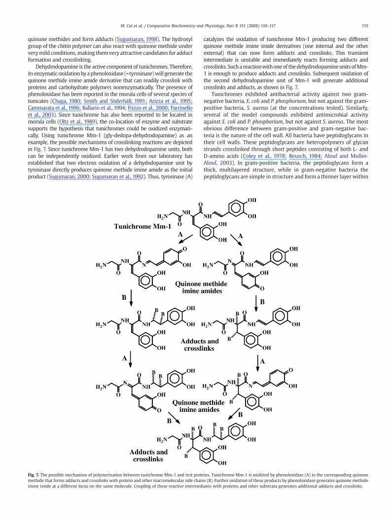
Dehydrodopamine is the active component of tunichromes. Therefore,its enzymatic oxidationbyaphenoloxidase (=tyrosinase)will generate thequinone methide imine amide derivative that can readily crosslink withproteins and carbohydrate polymers nonenzymatically. The presence ofphenoloxidase has been reported in the morula cells of several species oftunicates (Chaga, 1980; Smith and Söderhäll, 1991; Arizza et al., 1995;Cammarata et al., 1996; Ballarin et al., 1994; Frizzo et al., 2000; Parrinelloet al., 2003). Since tunichrome has also been reported to be located inmorula cells (Oltz et al., 1989), the co-location of enzyme and substratesupports the hypothesis that tunichromes could be oxidized enzymati-cally. Using tunichrome Mm-1 (gly-dedopa-dehydrodopamine) as anexample, the possible mechanisms of crosslinking reactions are depictedin Fig. 7. Since tunichrome Mm-1 has two dehydrodopamine units, bothcan be independently oxidized. Earlier work from our laboratory hasestablished that two electron oxidation of a dehydodopamine unit bytyrosinase directly produces quinone methide imine amide as the initialproduct (Sugumaran, 2000; Sugumaran et al., 1992). Thus, tyrosinase (A)
catalyzes the oxidation of tunichrome Mm-1 producing two differentquinone methide imine imide derivatives (one internal and the otherexternal) that can now form adducts and crosslinks. This transientintermediate is unstable and immediately reacts forming adducts andcrosslinks. Sucha reactionwithoneof thedehydrodopamineunits ofMm-1 is enough to produce adducts and crosslinks. Subsequent oxidation ofthe second dehydrodopamine unit of Mm-1 will generate additionalcrosslinks and adducts, as shown in Fig. 7.
Tunichromes exhibited antibacterial activity against two gram-negative bacteria, E. coli and P. phosphorium, but not against the gram-positive bacteria, S. aureus (at the concentrations tested). Similarly,several of the model compounds exhibited antimicrobial activityagainst E. coli and P. phosphorium, but not against S. aureus. The mostobvious difference between gram-positive and gram-negative bac-teria is the nature of the cell wall. All bacteria have peptidoglycans intheir cell walls. These peptidoglycans are heteropolymers of glycanstrands crosslinked through short peptides consisting of both L- andD-amino acids (Coley et al., 1978; Reusch, 1984; Alouf and Muller-Alouf, 2003). In gram-positive bacteria, the peptidoglycans form athick, multilayered structure, while in gram-negative bacteria thepeptidoglycans are simple in structure and form a thinner layer within
Fig. 7. The possible mechanism of polymerization between tunichrome Mm-1 and test proteins. Tunichrome Mm-1 is oxidized by phenoloxidase (A) to the corresponding quinonemethide that forms adducts and crosslinks with protein and other macromolecular side chains (B). Further oxidation of these products by phenoloxidase generates quinone methideimine imide at a different locus on the same molecule. Coupling of these reactive intermediates with proteins and other substrata generates additional adducts and crosslinks.
115M. Cai et al. / Comparative Biochemistry and Physiology, Part B 151 (2008) 110–117
the cell wall that is relatively uniform throughout most gram-negativegenera (Vollmer and Holtje, 2001). Tunichromes might exhibitantibacterial activity by crosslinking the structural proteins of thebacteria and changing the permeability of the cell wall. The damagedcell wall could result in the inhibition of growth and cell division. Ingram-negative bacteria, the thin layer of simple peptidoglycans mightbe easily compromised by tunichrome and the model compounds,dopa methyl ester and N-acetyl dehydrodopa methyl ester. In gram-positive bacteria, however, the thickness of the peptidoglycan layermight offer some protection, even if the outer layer of peptidoglycan isdamaged. This is consistent with the observation that styelin D acts onthe outer and inner membranes of E. coli, making these membranesmore permeable to some antibiotics (Taylor et al., 2000). Similarly,Lehrer et al. (2003) observed disruption of the cell walls of both E. coli(gram-negative) and S. aureus (gram-positive) that resulted in theextrusion of cytoplasm upon exposure to styelin D.
Alternatively, tunichrome and its derivatives could be toxic in theirown right. Essential metals, for example, could be chelated by thecatechol and pyrogallol moieties of the tunichrome molecule, therebyinhibiting bacterial metabolism. Quinone methides, if present as keyintermediates in the process of tunichrome crosslinking, are electrophilicand strong oxidants. A number of naturally occurring quinone methideshave been shown to be toxic (Thompson et al., 1993). In addition, thecatechol and pyrogallol groups of the tunichromes might be involved inthe generation of reactive oxygen species (ROS) due to their ease ofoxidation and reaction with molecular oxygen. The subsequent cyclingbetween quinones and semiquinones could produce significant quan-tities of damaging free redicals. Since S. aureusproduces catalase, itmightoffer some protection from the attack of electrophiles and the productionof ROS (Over et al., 2000), and might explain why S. aureus can resisttunichrome and model compound toxicity.
While tunichrome's antibacterial property was consistently shownin both Microtox® and two-stage radial diffusion assays with gram-negative bacteria (P. phosphorium and Escherichia coli, respectively), themodel coumpounds used in this study did not exhibit consistent toxicityin these two assays. Only four model compounds exhibited toxicities inthe Microtox® test, while the other eight model compounds did notexhibit any toxicity at the concentrations tested. However, in the two-stage radial diffusion assay, these fourmodel compounds did not exhibittoxicity. Instead, two other compounds, N-acetyl dehydrodopa methylester and dopa methyl ester, inhibited the growth of E. coli. Thedifference inmodel compound response in the two assays may indicatethat there are exhibiting separate mechanisms of toxicity in theMicrotox® and two-stage radial diffusion assay. The short term (5 to15min exposure) inhibition of light output (P. phosphorium) may not berelated to the long term (24 h exposure) pattern of clear zones in theradial diffusion assay. Neither of the toxic responses necessarily indicatethat the bacteria have been killed. The two-stage radial diffusion assay
appears to reflect the inhibition of growth and reproduction of bacteria.Microtox® testing is probably not as integrative. While Microtox®inhibition could alsobedue to inhibition of cell growthor thepreventionof cell division, it could also indicate damage to a single metabolicpathway that is linked to P. phosphorium light output. Given theuncertainty concerning the interpretation of the Microtox® tests, itseems that the two-stage radial diffusion assay represents a moreintegrative measure of toxicity compared to the Microtox® test.
With the exception of the tunichromes (this study), all dopa-containing peptides in tunicates so far examined for antimicrobialactivity appear to act on both gram-negative and gram-positivebacteria, although there is some indication that the compoundsexhibit slightly less potency against gram-positive species (Lee et al.,1997a,b; Taylor et al., 2000; Tincu et al., 2000, 2003; Lehrer et al.,2003). Had we used higher concentrations of tunichromes against thegram-positive S. aureus, we would likely have measured a toxicresponse. Based on E. coli's response, tunichromes' antibacterialpotency appears to be more similar to that of halocyamine than it isto the other antibacterial dopa-containing molecules plicatamide,styelins, and clavanins (Table 3). The latter three classes of substancesexhibited one to two orders of magnitude higher potency against E.coli in similarly conducted two-stage radial diffusion assays at neutralpH (7.4; Lee et al., 1997a,b; Taylor et al., 2000; Tincu et al., 2000, 2003).While inhibition of culture growth instead of a radial diffusion assaywas used in the halocyamine experiments (Table 3), the MEC for thetunichromes was within the range of the minimum inhibitionconcentration for halocyamine (Azumi et al., 1990a,b). Althoughantimicrobial potency of these compounds appears to be proportionalto the molecular weight of the compounds, it is not related to thenumber of modified phenylalanine or tyrosine amino acid residues ineach of the compound's structures. The lower potency of tunichromescompared to these other compounds may indicate that the primaryrole of tunichrome is not antibacterial, but may instead be thecrosslinking of tunic components. However, more work is needed toassess the potential role of tunichromes as antibacterial compounds.
The results of this study strongly lend support to the hypothesisthat tunichromes are potential crosslinkers for the formation of tunicas well as antibacterial agents of the tunicate defense system.Crosslinking and antibacterial activity appear to be related character-istics that would make tunichrome a highly useful compound for thebiomedical industry. Tunichromes could find applications in accidenttriage, emergency medical treatment and surgery, as well as for minorwound repair. In addition, nonmedical uses could entail the produc-tion of antifouling panels for marine and freshwater construction.Moreover, the detailed mechanisms that we have learned from ourstudies can be directly applicable for the “Green” formulation ofenvironmentally safe products with inherent antipredator, antibacter-ial and antifouling properties.
Table 3Comparison of antimicrobial potencies of several dopa-containing compounds isolated from tunicates
MEC
Compound Mol. mass (Da) # a.a. residues % modified phe pH μg/mL μM Reference
TunichromesAn-1, 2 and 3 556a 3 100% 7.4 27.7 49.8 this study
Halocyamines 668 and 730b 4 25% ? 25–100 36–147 Azumi et al., 1990a,bPlicatamides 984–116 8 50% 7.4 1–2.5 1–2.5 Tincu et al., 2000, 2003Clavanins 2666–2695 23 17–22% 7.4 ~1 ~0.4 Lee et al., 1997aStyelin A & B ~3690c N23 21–22% 7.4 1–10 0.3–2.7 Lee et al., 1997bStyelin D 4081–4144d 32 17–22% 7.4 ~6 ~1.5 Taylor et al., 2000
Information onmolecular mass (Da), the number of amino acid residues (# a.a. residues) and the molar percentage of modified phenylalanine residues (%modified Phe) are presented foreach compound. Data from experiments conducted at comparable pH were selected. Minimal Effective Concentrations (MEC) were determined from radial diffusion assays for allcompounds except halocyamine (where a Minimum Inhibition Concentration was determined from cell culture experiments). All responses are for the gram-negative bacterium,Escherichia coli, except for the reported MEC for Styelin D (where the gram-positive Staphylococcus aureus was used as the test organism).aMolecular mass of tunichrome An-1 (Oltz et al., 1988); bMolecular mass of halocyamine A=668; B=730; 4:1 mixture of A:B.c Partial amino acid sequence; dCompound exhibits microheterogeneity.
116 M. Cai et al. / Comparative Biochemistry and Physiology, Part B 151 (2008) 110–117
Acknowledgement
Research was funded by the Massachusetts Institute of TechnologySea Grant Program, under grant number RB-56 subaward 5710001422.
References
Alouf, J.E., Muller-Alouf, H., 2003. Staphylococcal and streptococcal superantigens:Molecular, biological and clinical aspects. Int. J. Med. Microbiol. 292, 429–440.
Andersen, R.J., Stonard, R.J., 1979. Clionamide, a major metabolite of the sponge Clionacelata Grant. Can J. Chem. 57, 2325–2328.
Arizza, V., Cammarata, M., Tomasino, M.C., Parrinello, N., 1995. Phenoloxidasecharacterization in vacuolar hemocytes from the solitary ascidian Styela plicata. J.Invert. Pathol. 66, 297–302.
Azumi, K., Yokosawa, H., Ishii, S., 1990a. Halocyamines: Novel antibacterial tetrapeptide-like structures isolated from the hemocytes of the solitary ascidian Halocynthiaroretzi. Biochemistry 29, 159–165.
Azumi, K., Yoshimizu, M., Suzuki, S., Ezura, Y., Yokosawa, H., 1990b. Inhibitory effect ofhalocyamine, an antimicrobial substance from ascidian hemocytes, on the growthof fish viruses and marine bacteria. Experientia 46, 1066–1068.
Azur Environmental,1995.Microtox® Acute Toxicity Basic Test ProceduresManual. 63 pp.Ballarin, L., Cima, F., Sabbadin, A., 1994. Phenoloxidase in the colonial ascidian Botryllus
schlosseri (Urochordata: Ascidiacea). Anim. Biol. 3, 41–48.Bayer, E., Schiefer, G., Waidelich, D., Scippa, S., De Vincentiis, M., 1992. Structure of the
tunichrome of tunicates and its role in concentration of vanadium. Angew. Chem.Inter. Ed. Engl. 31, 52–54.
Bruening, R.C., Oltz, E.M., Furukawa, J., Nakanishi, K., 1985. Isolation and structure oftunichrome B-1, a reducing blood pigment from the tunicate Ascidia nigra L. J. Am.Chem. Soc. 107, 5298–5300.
Bruening,R.C.,Oltz, E.M., Furukawa, J.,Nakanishi,K.,Kustin,K.,1986. Isolationof tunichromeB-1, a reducing blood pigment of the sea squirt, Ascidia nigra. J. Nat. Prod. 49, 193–204.
Burzio, L.A., Waite, J.H., 2001. Reactivity of peptidyl-tyrosine to hydroxylation and cross-linking. Protein Sci. 10, 735–740.
Cammarata, M., Arizza, V., Vazzana, M., Parrinello, N., 1996. Prophenoloxidase activatingsystem in tunicate hemolymph. Ital. J. Zool. 63, 345–351.
Chaga, O.Y., 1980. Ortho-diphenoloxidase system of ascidians. Tsitologiya 22, 619–625.Ciancio, A., Scippa, S., Nette, G., De Vincentiis, M., 2004. Analysis of the Henze
precipitate from the blood cells of the ascidian Phallusia mammillata. Naturwis-senschaften 91, 366–370.
Coley, J., Tarelli, E., Archibald, A.R., Baddiley, J., 1978. The linkage between teichoic acidand peptidoglycan in bacterial cell walls. FEBS Lett. 88, 1–9.
Dali, H., Sugumaran, M., 1988. An improved synthesis of 1,2-dehydro-N-acetyldopa-mine. Org. Prep. Proc. Intl. 20, 191–195.
Deck, J.D., Hay, E.D., Revel, J.P., 1966. Fine structure and origin of the tunic of Perophoraviridis. J. Morph. 120, 267–280.
De Leo, G., Patricolo, E., Lunetta, G.D'A.,1977. Studies on the fibrous components of the testof Ciona intestinalis Linnaeus. I. Cellulose-like polysaccharide. Acta Zool. 58, 135–141.
Dorsett, L.C., Hawkins, C.J., Grice, J.A., Lavin, M.F., Merefield, P.M., Parry, D.L., Ross, I.L.,1987. Ferreascidin: A highly aromatic protein containing 3,4-dihydroxypphenyla-lanine from the blood cells of a stolidobranch ascidian. Biochemistry 26,8078–8082.
EPA (Environmental Protection Agency), 2002. Federal Pollution Control Act. 33 UnitedStates Code, Sections 101–607 [as amended through Public Law 107–303,November 2002. http://www.epa.gov/region5/water/pdf/ecwa.pdf.
Frizzo, A., Guidolin, L., Ballarin, L., Baldan, B., Sabbadin, A., 2000. Immunolocation ofphenyloxidase in vacuoles of the compound ascidian Botryllus schlosseri morulacells. Ital. J. Zool. 67, 273–276.
Hall, D.A., Saxl, H., 1961. Studies of human and tunicate cellulose and of their relation toreticulin. Proc. Roy. Soc. 155B, 202–217.
Lee, I.H., Cho, Y., Lehrer, R.I., 1997a. Effects of pH and salinity on the antimicrobialproperties of clavanins. Infect. Immun. 65, 2898–2903.
Lee, I.H., Cho, Y., Lehrer, R.I., 1997b. Styelins, broad-spectrum antimicrobial peptidesfrom the solitary tunicate, Styela clava. Comp. Biochem. Physiol. B 118, 515–521.
Lehrer, R.I., 2003. Plicatamide, an antimicrobial octapeptide from Styela plicatahemocytes. J. Biol. Chem. 278, 13546–13553.
Lehrer, R.I., Lee, I.H., Menzel, L., Waring, A., Zhao, C., 2001. Clavanins and styelins, alpha-helicalantimicrobial peptides from thehemocytes ofStyela clava. Adv. Exp.Med. Biol. 484, 71–76.
Lehrer, R.I., Tincu, J.A., Taylor, S.W., Menzel, L.P., Waring, A.J., 2003. Natural peptide antibioticsfrom tunicates: Structures, functions and potential uses. Integr. Comp. Biol. 43, 313–322.
Lindquist, N., Fenical, W., Van Duyne, G.D., Clardy, J., 1988. New alkaloids of thelamellarin class from the marine ascidian Didemnum charteceum (Sluiter, 1909). J.Org. Chem. 53, 4570–4574.
Macara, I.G., McLeod, G.C., Kustin, K., 1979. Isolation, properties and structural studieson a compound from tunicate blood cells that may be involved in vanadiumaccumulation. Biochem. J. 181, 457–465.
Nette, G., Scippa, S., De Vincentiis, M., 2000. Origin of the Henze solution/precipitatefrom morula cells of the blood of the ascidian Phallusia mammillata. Naturwis-senschaften 87, 220–224.
Oltz, E.M., Bruening, R.C., Smith, M.J., Kustin, K., Nakanishi, K., 1988. The tunichromes. Aclass of reducing blood pigments from sea squirts: Isolation, structures andvanadium chemistry. J. Amer. Chem. Soc. 110, 6162–6172.
Oltz, E.M., Pollack, S., Delohery, T., Smith, M.J., Ojika, M., Lee, S., Kustin, K., Nakanishi, K.,1989. Distribution of tunichrome and vanadium in sea squirt blood cells sorted byflow cytometry. Experientia 45, 186–190.
Over, U., Tuc, Y., Soyletir, G., 2000. Catalase-negative Staphylococcus aureus: A rareisolate of human infection. Clin. Microbiol. Infect. 6, 681–682.
Parrinello, N., Arizza, V., Chinnici, C., Parrinello, D., Cammarata, M., 2003. Phenolox-idases in ascidian hemocytes: Characterization of the pro-phenoloxidase activatingsystem. Comp. Biochem. Physiol. B 135, 583–591.
Parry, D.L., Brand, S.G., Kustin, K., 1992. Distribution of tunichromes in the Ascidiacea.Bull. Mar. Sci. 50, 302–306.
Patricolo, E., De Leo, G., 1979. Studies on the fibrous components of the test of Cionaintestinalis Linnaeus. II. Collagen–elastin-like protein. Acta Zool. 60, 259–269.
Reusch, V.M., 1984. Lipopolymers, isoprenoids, and the assembly of the gram-positivecell wall. Crit. Rev. Microbiol. 11, 129–155.
Robinson, W.E., Kustin, K., McLeod, G.C., 1983. Incorporation of [14C]glucose into thetunic of the ascidian, Ciona intestinalis (Linnaeus). J. Exp. Zool. 225, 187–195.
Robinson, W.E., Kustin, K., Cloney, R.A., 1986. The influence of tunichrome and otherreducing compounds on tunic and fin formation in embryonic Ascidia callosaStimpson. J. Exp. Zool. 237, 63–72.
Robinson,W.E., Kustin, K.,Matulef, K., Parry, D.L., He, X.Y., Ryan, E.K.,1996. C14 andH3-tyrosineincorporation into Ascidia ceratodes tunichrome in vivo. Comp. Biochem. Physiol. B. 115,475–481.
Rzepecki, L.M., Waite, J.H., 1991. α,β-dehydro-3,4-dihydroxyphenylalanine derivatives:Rate and mechanism of formation. Arch. Biochem. Biophys. 285, 27–36.
Schaefer, J., Kramer, K.J., Garbow, J.R., Jacob, G.S., Stejskal, E.O., Hopkins, T.L., Speirs, R.D.,1987. Aromatic cross-links in insect cuticle: Detection by solid-state 13C and 15NNMR. Science 235, 1200–1204.
Smith, V.J., Söderhäll, K., 1991. A comparison of phenoloxidase activity in the blood ofmarine invertebrates. Dev. Comp. Immunol. 15, 251–261.
Steinberg, D.A., Lehrer, R.I., 1997. Designer assays for antimicrobial peptides. Disputingthe “one-size-fits-all” theory. Methods Mol. Biol. 78, 169–186.
Stonard, R.J., Andersen, R.J., 1980a. Celenamides A and B linear peptide alkaloids fromthe sponge Cliona celala. J. Org. Chem. 45, 3687–3691.
Stonard, R.J., Andersen, R.J.,1980b. Linear peptide alkaloids from the sponge Cliona celata(Grant). Celenamides C and D. Can J. Chem. 58, 2121–2126.
Sugumaran, M., 1998. Unified mechanism for sclerotization of insect cuticle. Adv. InsectPhysiol. 27, 229–334.
Sugumaran, M., 2000. Oxidation chemistry of 1,2-dehydro-N-acetyldopamines: Directevidence for the formation of 1,2-dehydro-N-acetyldopamine quinone. Arch.Biochem. Biophys. 378, 404–410.
Sugumaran, M., 2002. Comparative biochemistry of eumelanogenesis and theprotective roles of phenoloxidase and melanin in insects. Pigment Cell Res. 15, 2–9.
Sugumaran, M., Ricketts, D., 1995. Model sclerotization studies. 3. Cuticular enzymecatalyzed oxidation of peptidyl model tyrosine and dopa derivatives. Arch. InsectBiochem. Physiol. 28, 17–32.
Sugumaran, M., Hennigan, B., O'Brien, J., 1987. Tyrosinase catalized protein polymerizationas an in vitromodel for sclerotization of insect cuticle. Arch. Insect Biochem. Physiol. 6,9–25.
Sugumaran, M., Semensi, V., Kalyanaraman, B., Bruce, J.M., Land, E.J., 1992. Evidence for theformation of a quinone methide during the oxidation of the insect cuticular sclerotizingprecursor, 1,2-dehydro-N-acetyldopamine. J. Biol. Chem. 267, 10355–10361.
Sugumaran, M., Tan, S., Sun, H.L., 1996. Tyrosinase catalyzed oxidation of 3,4-dihydroxyphenylglycine. Arch. Biochem. Biophys. 329, 175–180.
Taylor, S.W., Ross,M.M.,Waite, H.J.,1995.Novel 3,4-di- and 3,4,5-trihydroxyphenylalanine-containing polypeptides from the blood cells of the ascidians Ascidia ceratodes andMolgula manhattensis. Arch. Biochem. Biophys. 324, 228–240.
Taylor, S.W., Kammerer, B., Bayer, E., 1997a. New perspectives in the chemistry andbiochemistry of the tunichromes and related compounds. Chem. Rev. 97, 333–346.
Taylor, S.W., Kammerer, B., Nicholson, G.J., Pusecker, K., Walk, T., Bayer, E., Scippa, S., DeVincentiis, M., 1997b. Morulin Pm: A modified polypeptide containing TOPA and 6-bromotryptophan from the morula cells of the ascidian, Phallusia mammillata. Arch.Biochem. Biophys. 348, 278–288.
Taylor, S.W., Craig, A.F., Fischer, W.H., Park, M., Lehrer, R.I., 2000. Styelin D, anextensively modified antimicrobial peptide from ascidian hemocytes. J. Biol. Chem.275, 38417–38426.
Thompson, D.C., Thompson, J.A., Sugumaran, M., Moldeus, P., 1993. Biological andtoxicological consequences of quinone methide formation. Chem. Biol. Inter. 86,129–162.
Tincu, J.A., Taylor, S.W., 2002. Tunichrome Sp-1: New pentapeptide tunichrome fromthe hemocytes of Styela plicata. J. Nat. Prod. 65, 377–378.
Tincu, J.A., Taylor, S.W., 2004. Antimicrobial peptides from marine invertebrates.Antimicrob. Agents Chemother. 48, 3645–3654.
Tincu, J.A., Craig, A.G., Taylor, S.W., 2000. Plicatamide: A lead to the biosynthetic originsof the tunichromes. Biochem. Biophys. Res. Commun. 270, 421–424.
Tincu, J.A., Menzel, L.P., Azimov, R., Sands, J., Hong, T.,Waring, A.J., Taylor, S.W., Lehrer, R.I.,2003. Plicatamide, an antimicrobial octapeptide from Sytela plicata hemocytes. J.Biol. Chem. 278, 13546–13553.
Vollmer, W., Holtje, J.V., 2001. Morphogenesis of Escherichia coli. Curr. Opin. Microbiol.4, 625–633.
Wagner, H.U., Grompper, R., 1974. The Chemistry of Quinonoid Compounds. Wiley, NewYork.
Wagner, H., Bladt, S., Zagainski, E.M., 1983. Plant Drug Analysis. Springer-Verlag, Berlin.Waite, H.J., 1990. The phylogeny and chemical diversity of quinone tanned glues and
varnishes. Comp. Biochem. Physiol. B 97, 19–29.Zar, J.H., 1984. Statistical Analysis, 2nd Ed. Prentice Hall, Englewood Cliffs, N.J.Zeng, L., Swalla, B.J., 2005. Molecular phylogeny of the protochordates: Chordate
evolution. Can. J. Zool. 83, 24–33.
117M. Cai et al. / Comparative Biochemistry and Physiology, Part B 151 (2008) 110–117