(2014) 15655–15662 copper substituted hydroxyapatite and fluorapatite: synthesis, characterization...
TRANSCRIPT
CERAMICSINTERNATIONAL
Available online at www.sciencedirect.com
http://dx.doi.org/0272-8842/& 20
nCorrespondinE-mail addre
(2014) 15655–15662
Ceramics International 40 www.elsevier.com/locate/ceramintCopper substituted hydroxyapatite and fluorapatite: Synthesis,characterization and antimicrobial properties
Sumathi Shanmugamn, Buvaneswari Gopal
Materials Chemistry Division, School of Advanced Sciences, VIT University, Vellore 632 014, Tamilnadu, India
Received 18 June 2014; received in revised form 17 July 2014; accepted 17 July 2014Available online 24 July 2014
Abstract
Copper substituted hydroxyapatite (Ca10�xCux(PO4)6(OH)2 (x¼0.05–2.0), and fluorapatite Ca10�xCux(PO4)6(F)2 (x¼0.05–2.0) weresynthesized by co-precipitation method. The synthesized compounds were characterized by powder X-ray diffraction, Fourier transform infraredspectroscopy, scanning electron microscopy, energy dispersive X-ray spectroscopy and thermo gravimetric analysis. The antimicrobial activity ofthe synthesized compounds were tested against the organisms Escherichia coli (Gram negative bacteria), Staphylococcus aureus (Gram positivebacteria) and Candida albicans (yeast) quantitatively by spread plate method. 100% reduction in the number of colonies were observed withCa10�xCux(PO4)6(F)2 (x¼0.15–0.5) against E. coli. This material can be considered as good antibacterial agent for bone implantation.& 2014 Elsevier Ltd and Techna Group S.r.l. All rights reserved.
Keywords: Antimicrobial agent; Hydroxyapatite; Fluorapatite; Copper substitution; Candida albicans
1. Introduction
Hydroxyapatite (HAP) is a biocompatible and bioactivematerial. Because of its structural similarity with the bonemineral it is widely investigated as the bone implant material[1–3]. The composition of bone is approximately 75–25%HAP by weight and 65–35% HAP by volume [4]. The naturalbone is non-stoichiometric, poorly crystalline and containscarbonate ions [5]. Stoichiometric HAP (Ca10(PO4)6(OH)2) hasa calcium-to-phosphate ratio of 1.67 [6]. Because of theflexibility in the structure of the hydroxyapatite, the variousions such as silver, zinc, bismuth, copper, strontium, silicateand carbonate are substituted in hydroxyapatite to improve theproperties such as antibacterial property, mechanical strengthand solubility for bone implantation [7–10].
The problem with the bone implant material is infectionafter the surgery [11]. These infections become serious if it isnot treated properly and leads to revision of surgeries. In orderto avoid such infections the biomedical device with good
10.1016/j.ceramint.2014.07.08614 Elsevier Ltd and Techna Group S.r.l. All rights reserved.
g author.ss: [email protected] (S. Shanmugam).
antimicrobial property is significant. In the recent years,inorganic antimicrobial agents have gained the attention ofthe researchers because of its stability and safety [12]. Mostlythese inorganic antimicrobial agents are copper, silver and zincions [9,13]. The antimicrobial activity of silver, copper, zincand cerium substituted hydroxyapatite compounds are found tobe higher than unsubstituted hydroxyapatite [9,14,15].Silver, copper and zinc ion co-doped with β-tricalcium
phosphate (TCP) synthesized by conventional high tempera-ture solid state method is reported for its antibacterial activity,it is observed that AgZn-TCP and AgCu-TCP exhibited higheractivity than Ag-TCP [16]. The effect of divalent cationsubstitution such as Pb2þ , Cu2þ , Mg2þ , Co2þ and Zn2þ
on the biological behavior of hydroxyapatite is studied byLima et al. [17]. Yang et al. stated that the silver ion andcopper ion have excellent antibacterial activity than othermetallic ions [18]. The antibacterial activity of water bornepolyurethane doped copper (II) substituted hydroxyapatitenanocomposites against Escherichia coli and Staphylococcusaureus studied by Zhao et al. An enhanced antibacterialactivity is observed by this nanocomposite [19]. Kim et al.studied the antibacterial effect of the composed metal ion
S. Shanmugam, B. Gopal / Ceramics International 40 (2014) 15655–1566215656
(silver, copper and zinc) doped hydroxyapatite synthesized bywet chemical method. An improved activity is observed withsilver doped hydroxyaptite compared with other metal ions[20]. Literature reveals that researchers focused on new apatitebiomaterials with good antibacterial property in terms ofsubstitution [13], or composites [21], or thin films [22].
Copper in small quantities are important for variousactivities in the living organisms. Copper fluorapatite isreported as the catalyst in many organic transformations suchas N-arylation of heterocycles with bromo and iodoarenes [23]and N-arylation of Imidazoles [24]. But copper substitutedfluorapatite is not studied for its antibacterial activity. Anti-bacterial activity of fluoride and silver co-doped hydroxyapa-tite against E. coli and fluorine substituted hydroxyapatiteagainst S. mutans are reported recently [25,26].
In this paper the synthesis of Ca10�xCux(PO4)6(OH)2(x=0.05–2.0) and Ca10�xCux(PO4)6(F)2 (x=0.05–2.0) by co-precipitation method, characterization by powder XRD andFTIR, comparison of in vitro antimicrobial study against theorganisms E. coli, S. aureus and Candida albicans by spreadplate method and effect of copper content on the antimicrobialactivity are discussed.
2. Materials and methods
2.1. Materials
Calcium carbonate (Aldrich, 99.9%), Ammonium dihydro-gen orthophosphate (S.D. Fine, 97%), Ammonium fluoride(Thomas Baker), Cupric nitrate (S.D. Fine 98%).
2.2. Methods
Ca10�xCux(PO4)6Z2 (x=0.05–2 and Z=F or OH) wereprepared by co-precipitation method [27]. Diammoniumhydrogen phosphate (7.92 g) was dissolved in 250 ml ofdouble distilled water and pH of the solution was maintained(�12) by addition of ammonium hydroxide (in the case offluorapatite, 1 g of ammonium fluoride was used as thefluoride source along with phosphate solution). The phosphatesolution was added under constant stirring into 150 ml solutioncontaining stoichiometric quantities of calcium and copperions. The suspension was refluxed for 4 h. The obtainedprecipitates were filtered, washed with distilled water, driedovernight at 80 1C and calcined in air at 700 1C for 30 min.
2.3. Characterization
The compound is characterized by Powder X ray diffraction(PANalytical make) with CuKα radiation in the 2 Theta range10–701. The lattice parameters are calculated by least squaremethod. Fourier Transform Infrared Spectra (FTIR) is recordedin the range 4000–400 cm�1 using Thermonicolet Avator 330USA model. Scanning Electron microscopic image and Energydispersive X-ray diffraction analysis (SEM–EDAX) is obtainedwith scanning electron microscope Hitachi 3000H. Thermalbehavior of the compounds is studied by Thermogravimetric
analysis (TGA) using SDT Q600 instrument in nitrogen atmo-sphere at a heating rate of 20 1C/min.
2.4. Antimicrobial activity test
The antimicrobial activity was evaluated by spread platemethod. The following bacteria and yeast were used in thisstudy: E. coli, S. aureus and C. albicans among which E.coli is gram negative bacteria, S. aureus is gram positivebacteria and C. albicans is fungi. The inocula of allmicroorganisms were prepared from fresh overnight brothcultures that were incubated at 37 1C. The resulting brothcultures were used for the test. 4 ml of phosphate buffersolution of pH 7 and 1 ml of bacterial or yeast suspension(1� 106 cells/ml) was added to the 100 mg of the sterilizedsample. The suspensions were incubated at 37 1C for 24 hwith shaking at 250 rpm. 100 ml of the suspension afterserial dilution was then inoculated on nutrient agar plates(for bacteria) and sabourated dextrose agar plates (for yeast).These plates were incubated for 24 h and 48 h (for bacteriaand yeast respectively) and the number of colony formingunits (cfus) were counted and photographed. The number ofcolony forming units represents the survival number ofbacteria and yeast. It is inversely proportional to theantibacterial activity of the sample [10].Antimicrobial ratio (K) of the samples was calculated using
the formula [28]K ¼ ðA�BÞ
A � 100A¼no. of colonies in the control (cfu/ml),B¼no. of colonies in the sample (cfu/ml).
2.5. Ion release test
In vitro solubility of the compounds was studied in thephosphate buffered medium of pH 7.4 in a thermostaticincubator at 37 1C. Phosphate buffered medium (PBS) wasprepared using 2.38 g of Na2HPO4, 0.19 g of KH2PO4 and8 g of NaCl/L of water. A pellet of 12 mm diameter and3 mm thickness was prepared from the synthesized com-pounds and sintered at 1100 1C/12 h. Sintered pellet wascrushed and sieved to get 100 mesh size particles. 250 mg of100 mesh size particles was immersed in 250 ml of thebuffered solution (1 mg/1 ml). 10 ml of the aliquots weretaken at an interval of 24 h and replaced with fresh buffersolution. Ionic concentration in the aliquots was estimated byICP–OES analysis [10].
3. Results and discussion
3.1. X-ray diffraction analysis
Based on the powder X-ray diffraction analysis (Fig. 1),it is noted that pure apatite phase (JCPDS #82-0740) isobtained up to x¼0.5 in the case of copper substitutedhydroxyapatite Ca10�xCux(PO4)6(OH)2 (x¼0.05–2.0).With increase in copper content (from 0.5 to 2.0) theformation of the secondary phase (β-tri calcium phosphate)is noticed. The unit cell dimensions are given in Table 1.

10 20 30 40 50 60 70
(302
)(1
13)
(203
)
(004
)(4
02)
(410
)(3
21)
(312
)(2
13)
(222
)
(310
)
(301
)(3
00)
(112
)(2
11)
(210
)(1
02)
(002
)(1
11)
(200
)
(100
)
x = 0.2
x = 0.15
x = 0.1
x = 0.05
Inte
nsity
(a.u
)
x = 0.5
x = 0.25
x = 1.0
x = 2.0
x = 1.5
Fig. 1. Powder XRD patterns of Ca10�xCux(PO4)6 (OH)2 (x¼0.05–2.0) (* -β-TCP).
Table 1Lattice parameters of copper substituted hydroxyapatite phases.
Compound a (Ǻ) c (Ǻ) V (Ǻ3)
Ca10(PO4)6(OH)2 (86–0740) 9.420 6.880 528.8Ca9.95Cu0.05(PO4)6(OH)2 9.328(4) 6.844(3) 515.6Ca9.9Cu0.1(PO4)6(OH)2 9.358(9) 6.837(9) 518.7Ca9.85Cu0.15(PO4)6(OH)2 9.333(0) 6.843(3) 516.2Ca9.8Cu0.2(PO4)6(OH)2 9.353(2) 6.842(7) 518.4Ca9.75Cu0.25(PO4)6(OH)2 9.337(8) 6.842(7) 516.7Ca9.5Cu0.5(PO4)6(OH)2 9.397(0) 6.872(7) 525.6
10 20 30 40 50 60 70
(241
)(2
40)
(332
)(3
23)
(502
)
(313
)
(322
)(0
04)
(402
)(3
21)
(213
)(132
)(2
22)(2
03)
(113
)(3
02)(1
30)
(202
)(3
00)(2
11)
(210
)(1
02)(002
)
(111
)(2
00)
(100
)
x = 0.05In
tens
ity (a
.u)
2θ(o)
x = 0.2
x = 0.15
x = 0.1
x = 0.25
x = 0.5***
x = 1.5
x = 1.0
x = 2.0
Fig. 2. Powder XRD patterns of Ca10�xCux(PO4)6(F2) (x¼0.05–2.0) (* -β-TCP).
Table 2Lattice parameters of substituted fluorapatite phases.
Compound a (Ǻ) c (Ǻ) V (Ǻ3)
Ca10(PO4)6F2 (76–0558) 9.366 6.883 523.03Ca9.95Cu0.05(PO4)6(F2) 9.307(3) 6.866(7) 515.1Ca9.9Cu0.1(PO4)6(F2) 9.355(4) 6.877(3) 521.3Ca9.85Cu0.15(PO4)6(F2) 9.235(4) 6.848(4) 505.9Ca9.8Cu0.2(PO4)6(F2) 9.261(5) 6.874(3) 510.7Ca9.75Cu0.25(PO4)6(F2) 9.333(3) 6.842(3) 516.2Ca9.5Cu0.5(PO4)6(F2) 9.352(4) 6.877(5) 521.1
S. Shanmugam, B. Gopal / Ceramics International 40 (2014) 15655–15662 15657
A slight decrease in the lattice parameters ‘a’ and ‘c’ isobserved. This could be attributed to the lower ionic radiusof copper (Cu2þ (0.72 Å) compared to calcium (Ca2þ
(0.99 Å) [9,29].Fig. 2 shows the X-ray diffraction pattern of copper
substituted fluorapatite Ca10�xCux(PO4)6(F)2 (x¼0.05–2.0).All the reflections are indexed based on Ca5(PO4)3F (FAP)(JCPDSa76–0558) pattern. The pattern indicates that thecompounds formed are highly crystalline. Similar to coppersubstituted hydroxyapatite, when x value is increased from 0.5to 2.0, a secondary phase β-tricalcium phosphate is formed.The lattice parameters of the substituted fluorapatite phases aregiven in Table 2. The unit cell parameters a and c arecompared with parent fluorapatite Ca5(PO4)3F. The replace-ment of bigger calcium by smaller copper ion decreases boththe lattice parameters a and c.
3.2. Infrared spectroscopic analysis
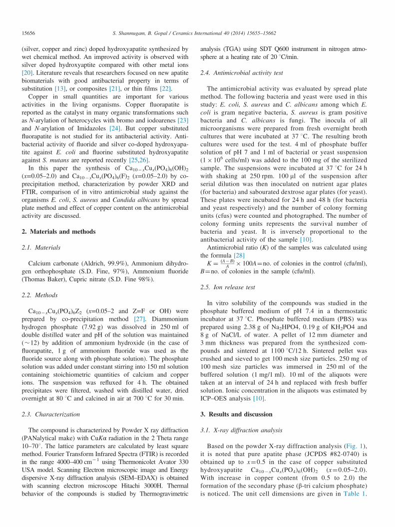
The Infrared spectrum of copper substituted hydroxyapatiteis given in Fig. 3. The peaks at 3570 cm�1 and 644 cm�1 areattributed to the stretching and bending modes of –OH group[30,31]. The peaks in the range 970–1031 cm�1 represents thePO4
3- symmetric and asymmetric vibrations. The bendingvibrational modes of PO4
3- are observed in the region 599–567 cm�1. The infrared spectrum of Ca10�xCux(PO4)6F2(x=0.05–0.5) is shown in Fig. 4. The spectra display thecharacteristic vibrational modes corresponding to the apatiticphosphate group. Stretching (947–1037 cm�1) and bendingvibrations (571–611 cm�1) of P–O bond are not influencedmuch by the substitution of copper ions in the fluorapatite. Inaddition to the characteristic peaks, carbonate absorption bandsare noticed around 870 cm�1 and 1400 cm�1 in copper
4000 3500 3000 2500 2000 1500 1000 500
x = 0.05
Wave number (cm-1)
x = 0.156
759
964
4970
1101
1037
3570
% T
rans
mitt
ance
x = 0.15
x = 0.2
x = 0.5
x = 0.25
Fig. 3. FT-IR spectrum of Ca10�xCux(PO4)6(OH)2(x¼0.05–0.5).
4000 3500 3000 2500 2000 1500 1000 500Wave number (cm-1)
962
1115 56
961
3
1039
x = 0.1
x = 0.05
% T
rans
mitt
ance
x = 0.15
x = 0.2
x = 0.5
x = 0.25
Fig. 4. FT-IR spectrum of Ca10�xCux(PO4)6(F2)(x¼0.05–0.5).
S. Shanmugam, B. Gopal / Ceramics International 40 (2014) 15655–1566215658
substituted hydroxyapatite and fluorapatite (x¼0.05–0.25).But the intensity of these bands is very weak.
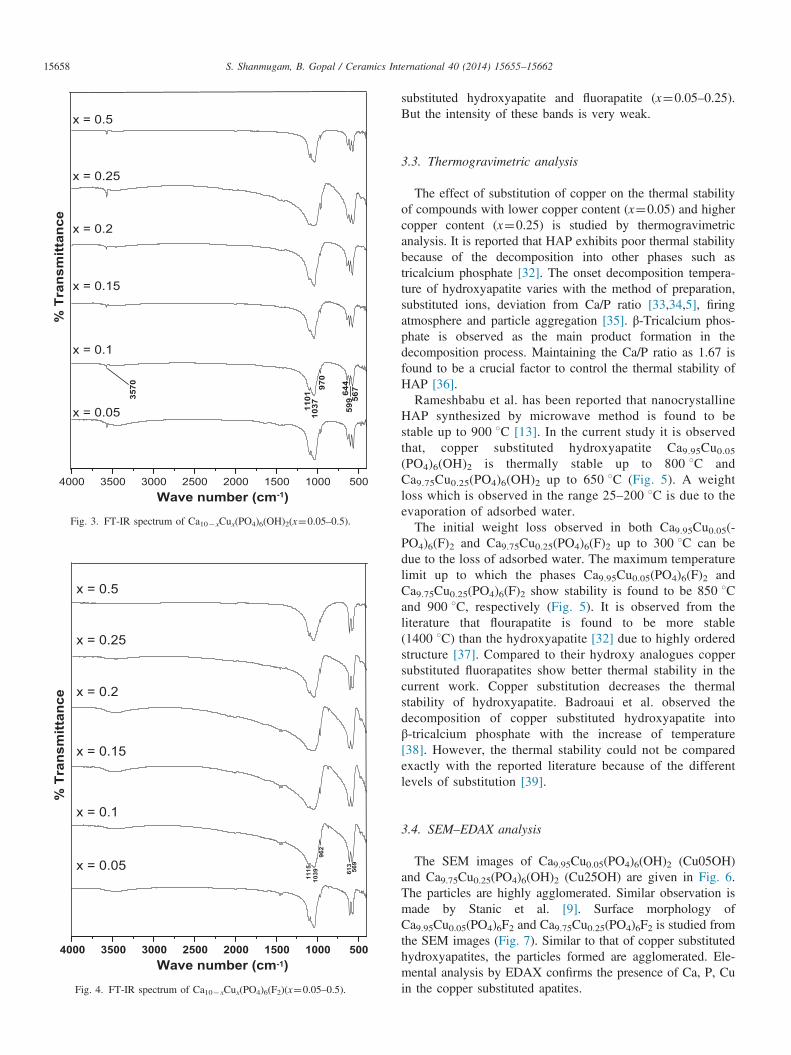
3.3. Thermogravimetric analysis
The effect of substitution of copper on the thermal stabilityof compounds with lower copper content (x¼0.05) and highercopper content (x¼0.25) is studied by thermogravimetricanalysis. It is reported that HAP exhibits poor thermal stabilitybecause of the decomposition into other phases such astricalcium phosphate [32]. The onset decomposition tempera-ture of hydroxyapatite varies with the method of preparation,substituted ions, deviation from Ca/P ratio [33,34,5], firingatmosphere and particle aggregation [35]. β-Tricalcium phos-phate is observed as the main product formation in thedecomposition process. Maintaining the Ca/P ratio as 1.67 isfound to be a crucial factor to control the thermal stability ofHAP [36].Rameshbabu et al. has been reported that nanocrystalline
HAP synthesized by microwave method is found to bestable up to 900 1C [13]. In the current study it is observedthat, copper substituted hydroxyapatite Ca9.95Cu0.05(PO4)6(OH)2 is thermally stable up to 800 1C andCa9.75Cu0.25(PO4)6(OH)2 up to 650 1C (Fig. 5). A weightloss which is observed in the range 25–200 1C is due to theevaporation of adsorbed water.The initial weight loss observed in both Ca9.95Cu0.05(-
PO4)6(F)2 and Ca9.75Cu0.25(PO4)6(F)2 up to 300 1C can bedue to the loss of adsorbed water. The maximum temperaturelimit up to which the phases Ca9.95Cu0.05(PO4)6(F)2 andCa9.75Cu0.25(PO4)6(F)2 show stability is found to be 850 1Cand 900 1C, respectively (Fig. 5). It is observed from theliterature that flourapatite is found to be more stable(1400 1C) than the hydroxyapatite [32] due to highly orderedstructure [37]. Compared to their hydroxy analogues coppersubstituted fluorapatites show better thermal stability in thecurrent work. Copper substitution decreases the thermalstability of hydroxyapatite. Badroaui et al. observed thedecomposition of copper substituted hydroxyapatite intoβ-tricalcium phosphate with the increase of temperature[38]. However, the thermal stability could not be comparedexactly with the reported literature because of the differentlevels of substitution [39].
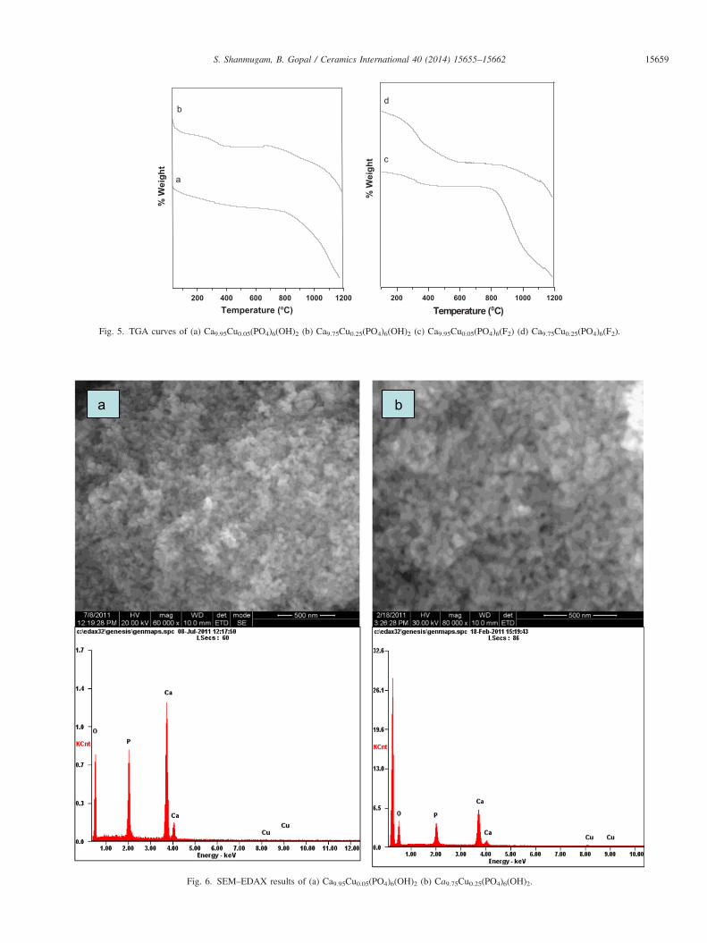
3.4. SEM–EDAX analysis
The SEM images of Ca9.95Cu0.05(PO4)6(OH)2 (Cu05OH)and Ca9.75Cu0.25(PO4)6(OH)2 (Cu25OH) are given in Fig. 6.The particles are highly agglomerated. Similar observation ismade by Stanic et al. [9]. Surface morphology ofCa9.95Cu0.05(PO4)6F2 and Ca9.75Cu0.25(PO4)6F2 is studied fromthe SEM images (Fig. 7). Similar to that of copper substitutedhydroxyapatites, the particles formed are agglomerated. Ele-mental analysis by EDAX confirms the presence of Ca, P, Cuin the copper substituted apatites.
a
Temperature (oC)
% W
eigh
t
b
200 400 600 800 1000 1200 200 400 600 800 1000 1200
Temperature (0C)
% W
eigh
t
d
c
Fig. 5. TGA curves of (a) Ca9.95Cu0.05(PO4)6(OH)2 (b) Ca9.75Cu0.25(PO4)6(OH)2 (c) Ca9.95Cu0.05(PO4)6(F2) (d) Ca9.75Cu0.25(PO4)6(F2).
Fig. 6. SEM–EDAX results of (a) Ca9.95Cu0.05(PO4)6(OH)2 (b) Ca9.75Cu0.25(PO4)6(OH)2.
S. Shanmugam, B. Gopal / Ceramics International 40 (2014) 15655–15662 15659
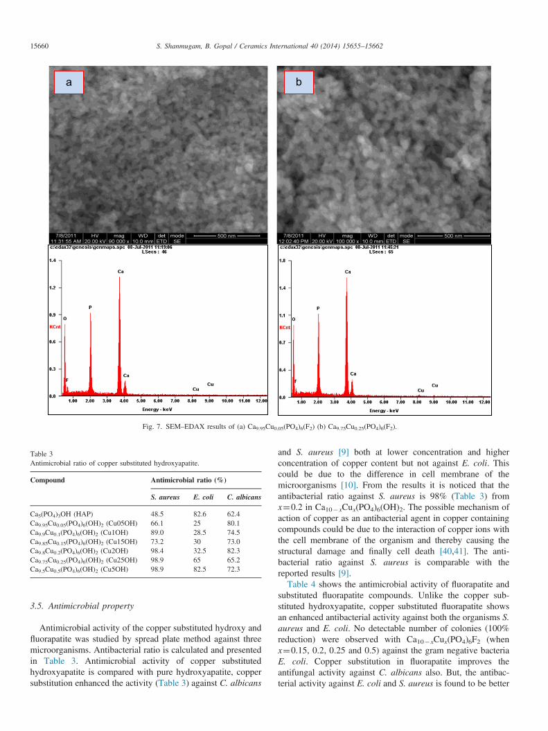
Fig. 7. SEM–EDAX results of (a) Ca9.95Cu0.05(PO4)6(F2) (b) Ca9.75Cu0.25(PO4)6(F2).
Table 3Antimicrobial ratio of copper substituted hydroxyapatite.
Compound Antimicrobial ratio (%)
S. aureus E. coli C. albicans
Ca5(PO4)3OH (HAP) 48.5 82.6 62.4Ca9.95Cu0.05(PO4)6(OH)2 (Cu05OH) 66.1 25 80.1Ca9.9Cu0.1(PO4)6(OH)2 (Cu1OH) 89.0 28.5 74.5Ca9.85Cu0.15(PO4)6(OH)2 (Cu15OH) 73.2 30 73.0Ca9.8Cu0.2(PO4)6(OH)2 (Cu2OH) 98.4 32.5 82.3Ca9.75Cu0.25(PO4)6(OH)2 (Cu25OH) 98.9 65 65.2Ca9.5Cu0.5(PO4)6(OH)2 (Cu5OH) 98.9 82.5 72.3
S. Shanmugam, B. Gopal / Ceramics International 40 (2014) 15655–1566215660
3.5. Antimicrobial property
Antimicrobial activity of the copper substituted hydroxy andfluorapatite was studied by spread plate method against threemicroorganisms. Antibacterial ratio is calculated and presentedin Table 3. Antimicrobial activity of copper substitutedhydroxyapatite is compared with pure hydroxyapatite, coppersubstitution enhanced the activity (Table 3) against C. albicans
and S. aureus [9] both at lower concentration and higherconcentration of copper content but not against E. coli. Thiscould be due to the difference in cell membrane of themicroorganisms [10]. From the results it is noticed that theantibacterial ratio against S. aureus is 98% (Table 3) fromx¼0.2 in Ca10�xCux(PO4)6(OH)2. The possible mechanism ofaction of copper as an antibacterial agent in copper containingcompounds could be due to the interaction of copper ions withthe cell membrane of the organism and thereby causing thestructural damage and finally cell death [40,41]. The anti-bacterial ratio against S. aureus is comparable with thereported results [9].Table 4 shows the antimicrobial activity of fluorapatite and
substituted fluorapatite compounds. Unlike the copper sub-stituted hydroxyapatite, copper substituted fluorapatite showsan enhanced antibacterial activity against both the organisms S.aureus and E. coli. No detectable number of colonies (100%reduction) were observed with Ca10�xCux(PO4)6F2 (whenx¼0.15, 0.2, 0.25 and 0.5) against the gram negative bacteriaE. coli. Copper substitution in fluorapatite improves theantifungal activity against C. albicans also. But, the antibac-terial activity against E. coli and S. aureus is found to be better
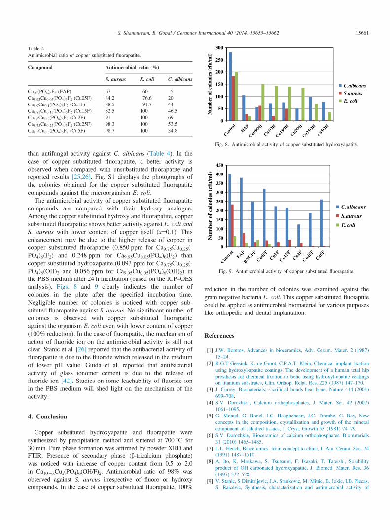
Table 4Antimicrobial ratio of copper substituted fluorapatite.
Compound Antimicrobial ratio (%)
S. aureus E. coli C. albicans
Ca10(PO4)6F2 (FAP) 67 60 5Ca9.95Cu0.05(PO4)6F2 (Cu05F) 84.2 76.6 20Ca9.9Cu0.1(PO4)6F2 (Cu1F) 88.5 91.7 44Ca9.85Cu0.15(PO4)6F2 (Cu15F) 82.5 100 46.5Ca9.8Cu0.2(PO4)6F2 (Cu2F) 91 100 69Ca9.75Cu0.25(PO4)6F2 (Cu25F) 98.3 100 53.5Ca9.5Cu0.5(PO4)6F2 (Cu5F) 98.7 100 34.8
0
50
100
150
200
250
300
Num
ber
of c
olon
ies
(cfu
/ml)
C.albicans
S.aureus
E. coli
Fig. 8. Antimicrobial activity of copper substituted hydroxyapatite.
0
50
100
150
200
250
300
350
400
450
Num
ber
of c
olon
ies
(cfu
/ml)
C.albicans
S.aureus
E.coli
Fig. 9. Antimicrobial activity of copper substituted fluorapatite.
S. Shanmugam, B. Gopal / Ceramics International 40 (2014) 15655–15662 15661
than antifungal activity against C. albicans (Table 4). In thecase of copper substituted fluorapatite, a better activity isobserved when compared with unsubstituted fluorapatite andreported results [25,26]. Fig. S1 displays the photographs ofthe colonies obtained for the copper substituted fluorapatitecompounds against the microorganism E. coli.
The antimicrobial activity of copper substituted fluorapatitecompounds are compared with their hydroxy analogue.Among the copper substituted hydroxy and fluorapatite, coppersubstituted fluorapatite shows better activity against E. coli andS. aureus with lower content of copper itself (x=0.1). Thisenhancement may be due to the higher release of copper incopper substituted fluorapatite (0.850 ppm for Ca9.75Cu0.25(-PO4)6(F2) and 0.248 ppm for Ca9.95Cu0.05(PO4)6(F2) thancopper substituted hydroxapatite (0.093 ppm for Ca9.75Cu0.25(-PO4)6(OH)2 and 0.056 ppm for Ca9.95Cu0.05(PO4)6(OH)2) inthe PBS medium after 24 h incubation (based on the ICP–OESanalysis). Figs. 8 and 9 clearly indicates the number ofcolonies in the plate after the specified incubation time.Negligible number of colonies is noticed with copper sub-stituted fluorapatite against S. aureus. No significant number ofcolonies is observed with copper substituted fluorapatiteagainst the organism E. coli even with lower content of copper(100% reduction). In the case of fluorapatite, the mechanism ofaction of fluoride ion on the antimicrobial activity is still notclear. Stanic et al. [26] reported that the antibacterial activity offluorapatite is due to the fluoride which released in the mediumof lower pH value. Guida et al. reported that antibacterialactivity of glass ionomer cement is due to the release offluoride ion [42]. Studies on ionic leachability of fluoride ionin the PBS medium will shed light on the mechanism of theactivity.
4. Conclusion
Copper substituted hydroxyapatite and fluorapatite weresynthesized by precipitation method and sintered at 700 1C for30 min. Pure phase formation was affirmed by powder XRD andFTIR. Presence of secondary phase (β-tricalcium phosphate)was noticed with increase of copper content from 0.5 to 2.0in Ca10�xCux(PO4)6(OH/F)2. Antimicrobial ratio of 98% wasobserved against S. aureus irrespective of fluoro or hydroxycompounds. In the case of copper substituted fluorapatite, 100%
reduction in the number of colonies was examined against thegram negative bacteria E. coli. This copper substituted fluoraptitecould be applied as antimicrobial biomaterial for various purposeslike orthopedic and dental implantation.
References
[1] J.W. Boretos, Advances in bioceramics, Adv. Ceram. Mater. 2 (1987)15–24.
[2] R.G.T Geesink, K. de Groot, C.P.A.T. Klein, Chemical implant fixationusing hydroxyl-apatite coatings. The development of a human total hipprosthesis for chemical fixation to bone using hydroxyl-apatite coatingson titanium substrates, Clin. Orthop. Relat. Res. 225 (1987) 147–170.
[3] J. Currey, Biomaterials: sacrificial bonds heal bone, Nature 414 (2001)699–708.
[4] S.V. Dorozhkin, Calcium orthophosphates, J. Mater. Sci. 42 (2007)1061–1095.
[5] G. Montel, G. Bonel, J.C. Heughebaert, J.C. Trombe, C. Rey, Newconcepts in the composition, crystallization and growth of the mineralcomponent of calcified tissues, J. Cryst. Growth 53 (1981) 74–79.
[6] S.V. Dorozhkin, Bioceramics of calcium orthophosphates, Biomaterials31 (2010) 1465–1485.
[7] L.L. Hench, Bioceramics: from concept to clinic, J. Am. Ceram. Soc. 74(1991) 1487–1510.
[8] A. Ito, K. Maekawa, S. Tsutsumi, F. Ikazaki, T. Tateishi, Solubilityproduct of OH carbonated hydroxyapatite, J. Biomed. Mater. Res. 36(1997) 522–528.
[9] V. Stanic, S Dimitrijevic, J.A. Stankovic, M. Mitric, B. Jokic, I.B. Plecas,S. Raicevic, Synthesis, characterization and antimicrobial activity of
S. Shanmugam, B. Gopal / Ceramics International 40 (2014) 15655–1566215662
copper and zinc-doped hydroxyapatite nanopowders, Appl. Surf. Sci. 256(2010) 6083–6089.
[10] Sumathi Shanmugam, Buvaneswari Gopal, Antimicrobial and cytotoxi-city evaluation of aliovalent substituted hydroxyapatite, Appl. Surf. Sci.303 (2014) 277–281.
[11] O.M. Lidwell, E.J. Lowbury, W. Whyte, R. Blowers, S.J. Stanley,D. Lowe, Infection and sepsis after operations for total hip or knee-joint replacement: influence of ultraclean air, prophylactic antibiotics andother factors, J. Hyg. 93 (1984) 505–529.
[12] H. Korai, Current situation and future of inorganic antimicrobial agent, J.Inorg. Mater. Jpn. 6 (1999) 428–436.
[13] N. Rameshbabu, T.S. Sampathkumar, T.G. Prabhakar, V.S. Sastry, K.V.G.K. Murty, K. Prasad Rao, Antibacterial nanosized silver substitutedhydroxyapatite: synthesis and characterization, J. Biomed. Mater. Res.Part A 80A (2007) 581–591.
[14] Y. Lin, Z. Yang, J. Cheng, Preparation, characterization and antibacterialpropertyof cerium substituted hydroxyapatite nanoparticles, J. RareEarths 25 (2007) 452–456.
[15] B. Singh, A.K. Dubey, S. Kumar, N. Saha, B. Basu, R. Gupta, In vitrobiocompatibility and antimicrobial activity of wet chemically preparedCa10�xAgx(PO4)6(OH)2 (0.0rxr0.5) hydroxyapatites, Mater. Sci. Eng.C 31 (2011) 1320–1329.
[16] N. Matsumoto, K. Sato, K. Yoshida, K. Hashimoto, Y. Toda, Preparationand characterization of beta-tricalcium phosphate co-doped with mono-valent and divalent antibacterial metal ions, Acta Biomater. 5 (2009)3157–3164.
[17] I.R. Lima, G.G. Alves, C.A. Soriano, A.P. Campaneli, T.H. Gasparoto,E.S. Ramos, L.A. Sena, A.M. Rossi, J.M. Granjeir, Understanding theimpact of divalent cationsubstitution on hydroxyapatite: an in vitromultiparametric study on biocom-patibility, J. Biomed. Mater. Res. PartA 98A (2011) 351–358.
[18] H. Yang, B. Xiao, K.W. Xu, Synthesis and characterization of Ag/Cu/HAPwith platelet morphology, J. Mater. Sci.—Mater. Med. 20 (2009) 785–792.
[19] C.X. Zhao, W.D. Zheng, A.P. Mai, X.M. Huang, Y.S. Ouyang, Synthesisand characterization of waterborne polyurethane/Cu(II)-loaded hydroxya-patite nanocomposites with antibacterial activity, J. Nanosci. Nanotech-nol. 11 (2011) 6779–6787.
[20] T.N. Kim, Q.L. Feng, J.O. Kim, J. Wu, H. Wang, G.C. Chen, F.Z. Cui,Antimicrobial effects of metal ions (Agþ , Cu2þ , Zn2þ ) in hydro-xyapatite, J. Mater. Sci.—Mater. Med. 9 (1998) 129–134.
[21] S. Rakmae, Y. Ruksakulpiwat, W. Sutapun, N. Suppakarn, Physicalproperties and cytotoxicity of surface-modified bovine bone-basedhydroxyapatite/poly(lactic acid) composites, J. Compos. Mater. 45 (2011)1259–1269.
[22] Q.L. Feng, T.N. Kim, J. Wu, E.S. Park, J.O. Kim, D.Y. Lim, F.Z. Cui,Antibacterial effects of Ag-HAp thin films on alumina substrates, ThinSolid Films 335 (1998) 214–219.
[23] M.L. Kantam, G.T. Venkanna, Ch. Sridhar, K.B. Shiva Kumar, Copperfluorapatite catalyzed N-arylation of heterocycles with bromo andiodoarenes, Tetrahedron Lett. 47 (2006) 3897–3899.
[24] M.L. Kantam, G.T. Venkanna, C. Sridhar, B. Sreedhar, B.M. Choudary,An efficient base free N-arylation of imidazoles and amines witharylboronic acids using copper exchanged fluorapatite, J. Org. Chem.71 (2006) 9522–9524.
[25] M. Turkoza, A. Atillab, Z. Evis, Silver and fluoride doped hydroxyapa-tites: Investigation microstructure, mechanical and antibacterial proper-ties, Ceram. Int. 39 (2013) 8925–8931.
[26] V. Stanic, S. Dimitrijevic, D.G. Antonovic, B.M. Jokic, S.P. Zeca, S.T. Tanaskovic, S. Raicevi, Synthesis of fluorine substituted hydroxyapa-tite nanopowders and application of the central composite design fordetermination of its antimicrobial effects, Appl. Surf. Sci. 290 (2014)346–352.
[27] H. El Badaoui, F. Bazi, R. Tahir, H.B. Lazrek, S. Sebti, Synthesis of3,4dihydropyrimidin-2-ones catalysed by fluorapatite doped with metalhalides, Catal. Commun. 6 (2005) 455–458.
[28] Y. Chen, X. Zheng, Y. Xie, C. Ding, H. Ruan, C. Fan, Anti-bacterial andcytotoxic properties of plasma sprayed silver-containing HA coatings, J.Mater. Sci.—Mater. Med. 19 (2008) 3603–3609.
[29] M. Misono, W.K. Hall, Oxidation-reduction properties of copper-andnickel-substituted hydroxyapatites, J. Phys. Chem. 77 (1973) 791–800.
[30] B.O. Fowler, Infrared studies of apatites. I. Vibrational assignments forcalcium, strontium, and barium hydroxyapatites utilizing isotopic sub-stitution, Inorg. Chem. 13 (1974) 194–207.
[31] A. Rapacz-Kmita, A. Slosarczyk, Z. Paszkiewicz, C. Paluszkiewicz,Phase stability of hydroxyapatite-zirconia (HAP/ZrO2) composites forbone replacement, J. Mol. Struct. 704 (2004) 333–340.
[32] Y. Chen, X. Miao, Thermal and chemical stability of fluorohydroxyapa-tite ceramics with different fluorine contents, Biomaterials 26 (2005)1205–1210.
[33] Y.R. Cai, R.K. Tang, Calcium phosphate nanoparticles in biomineraliza-tion and biomaterials, J. Mater. Chem. 18 (2008) 3775–3787.
[34] C. Combes, C. Rey, Amorphous calcium phosphates: synthesis, proper-ties and uses in biomaterials, Acta Biomater. 6 (2010) 3362–3378.
[35] A. Tampieri, G. Celotti, S. Sprio, C. Mingazzini, Characteristics ofsynthetic hydroxyapatite and attempts to improve their thermal stability,Mater. Chem. Phy. 64 (2000) 54–61.
[36] A.S. Posner, A. Perloff, A.F. Diorio, Refinement of the hydroxyapatitestructure, Acta Crystallogr., Sect. A: Found. Crystallogr. 11 (1958)308–309.
[37] A. Bianco, I. Cacciotti, M. Lombardi, L. Montanaro, E. Bemporad,M. Sebastini, F-substituted hydroxyapatite nanopowders: thermal stabi-lity, sintering behaviour and mechanical properties, Ceram. Int. 36 (2010)313–322.
[38] B. Badraoui, M. Othmani, H. Bachoua, Structural modification andthermal stability on hydroxyapatites containing copper in substitution,Ann. Chim. Sci. Mat. 33 (4) (2008) 329–337.
[39] K. Tonsuaadu, K.A. Gross, L. Pluduma, M. Veiderma, A review on thethermal stability of calcium apatites, J. Therm. Anal. Calorim. 110 (2012)647–659.
[40] W.L. Du, Y.L. Xu, Z.R. Xu, C.L. Fan, Preparation, characterization andantibacterial properties against E. coli K88 of chitosan nanoparticleloaded copper ions, Nanotechnology 19 (2008) 085707.
[41] L. Nan, Y. Liu, M. Lu, K. Yang, Study on antibacterial mechanism ofcopper bearing austenitic antibacterial stainless steel by atomic forcemicroscopy, J. Mater. Sci. Mater. Med. 19 (2008) 3057–3062.
[42] A. Guida, M.R. Towler, J.G. Wall, R.G. Hill, S. Eramo, Preliminary workon the antibacterial effect of strontium in glass ionomer cements, J.Mater. Sci. Lett. 22 (2003) 1401–1403.