amidine- and amidoxime-substituted heterocycles - mdpi
TRANSCRIPT
molecules
Article
Amidine- and Amidoxime-Substituted Heterocycles: Synthesis,Antiproliferative Evaluations and DNA Binding
Silvija Maracic 1 , Petra Grbcic 2 , Suresh Shammugam 3, Marijana Radic Stojkovic 3,* , Krešimir Pavelic 4 ,Mirela Sedic 5 , Sandra Kraljevic Pavelic 6 and Silvana Raic-Malic 1,*
�����������������
Citation: Maracic, S.; Grbcic, P.;
Shammugam, S.; Radic Stojkovic, M.;
Pavelic, K.; Sedic, M.; Kraljevic
Pavelic, S.; Raic-Malic, S. Amidine-
and Amidoxime-Substituted
Heterocycles: Synthesis,
Antiproliferative Evaluations and
DNA Binding. Molecules 2021, 26,
7060. https://doi.org/10.3390/
molecules26227060
Academic Editors: Steven Fletcher
and Roberto Romeo
Received: 27 October 2021
Accepted: 19 November 2021
Published: 22 November 2021
Publisher’s Note: MDPI stays neutral
with regard to jurisdictional claims in
published maps and institutional affil-
iations.
Copyright: © 2021 by the authors.
Licensee MDPI, Basel, Switzerland.
This article is an open access article
distributed under the terms and
conditions of the Creative Commons
Attribution (CC BY) license (https://
creativecommons.org/licenses/by/
4.0/).
1 Department of Organic Chemistry, Faculty of Chemical Engineering and Technology, University of Zagreb,Marulicev trg 19, HR-10000 Zagreb, Croatia; [email protected]
2 Department of Biotechnology, University of Rijeka, Ulica Radmile Matejcic 2, HR-51000 Rijeka, Croatia;[email protected]
3 Division of Organic Chemistry and Biochemistry, Laboratory for Biomolecular Interactions and Spectroscopy,Ruder Boškovic Institute, Bijenicka 54, HR-10000 Zagreb, Croatia; [email protected]
4 Faculty of Medicine, Juraj Dobrila University of Pula, HR-52100 Pula, Croatia; [email protected] Centre for Applied Bioanthropology, Institute for Anthropological Research, Ljudevita Gaja 32, HR-10000
Zagreb, Croatia; [email protected] Faculty of Health Studies, University of Rijeka, Ulica Viktora Cara Emina 5, HR-51000 Rijeka, Croatia;
[email protected]* Correspondence: [email protected] (M.R.S.); [email protected] (S.R.-M.); Tel.: +385-1-4571220 (M.R.S.);
+385-1-4597213 (S.R.-M.)
Abstract: The novel 1,2,3-triazolyl-appended N- and O-heterocycles containing amidine 4–11 andamidoxime 12–22 moiety were prepared and evaluated for their antiproliferative activities in vitro.Among the series of amidine-substituted heterocycles, aromatic diamidine 5 and coumarine amidine11 had the most potent growth-inhibitory effect on cervical carcinoma (HeLa), hepatocellular carci-noma (HepG2) and colorectal adenocarcinoma (SW620), with IC50 values in the nM range. Althoughcompound 5 was toxic to non-tumor HFF cells, compound 11 showed certain selectivity. From theamidoxime series, quinoline amidoximes 18 and 20 showed antiproliferative effects on lung adeno-carcinoma (A549), HeLa and SW620 cells emphasizing compound 20 that exhibited no cytostaticeffect on normal HFF fibroblasts. Results of CD titrations and thermal melting experiments indicatedthat compounds 5 and 10 most likely bind inside the minor groove of AT-DNA and intercalate intoAU-RNA. Compounds 6, 9 and 11 bind to AT-DNA with mixed binding mode, most probably minorgroove binding accompanied with aggregate binding along the DNA backbone.
Keywords: amidine; amidoxime; antiproliferative activity; DNA binding; fluorescence; CD spec-troscopy; thermal denaturation
1. Introduction
Nitrogen- and oxygen-containing heterocyclic compounds may be found in pharma-ceuticals, natural products, dyes, organic materials, and particularly in biologically activemolecules [1,2]. Natural and synthetic N- and O-heterocycles have been discovered aspromising anti-cancer agents used in clinic or clinical evaluations, suggesting their promi-nent place in anti-cancer drugs development. Although the design of bioactive moleculesis primarily focused on novel N-heterocycles, due to their ease of synthesis and possi-ble mimicking of physiological molecules, O-heterocycles have provided equally potentmolecules with a lesser risk of toxicity [2,3]. These compounds were found to act via vari-ous targets such as protein kinases, histone deacetylases, topoisomerase I and II, carbonicanhydrase, aromatase, sulfatase, vascular endothelial growth factor (VEGF), and epider-mal growth factor (EGF) receptors, monocarboxylate transporters, DNA polynucleotide,tubulin/microtubule system [1,4–14].
Molecules 2021, 26, 7060. https://doi.org/10.3390/molecules26227060 https://www.mdpi.com/journal/molecules

Molecules 2021, 26, 7060 2 of 22
Applying the molecular hybridization approach for the synthesis of new moleculeswhere 1,2,3-triazole ring acts as pharmacophore or linker leads to libraries of compoundswith significant anticancer activity [15]. Thus, indole scaffold, found to be widely dis-tributed in natural products and bioactive molecules, was linked to 1,2,3-triazole andyielded new hybrids with potent anticancer activity [11]. For example, in order to ob-tain histone deacetylases (HDAC) inhibitors with lower side effects, indole-derived N-hydroxyarylamide I with the triazole ring as a linker was obtained and showed the positiveeffects of 5-methoxy group and the one-carbon-bridge between triazole and the indole ringon antiproliferative activity (Figure 1) [16].
Molecules 2021, 26, 7060 2 of 22
Applying the molecular hybridization approach for the synthesis of new molecules
where 1,2,3‐triazole ring acts as pharmacophore or linker leads to libraries of compounds
with significant anticancer activity [15]. Thus, indole scaffold, found to be widely distrib‐
uted in natural products and bioactive molecules, was linked to 1,2,3‐triazole and yielded
new hybrids with potent anticancer activity [11]. For example, in order to obtain histone
deacetylases (HDAC) inhibitors with lower side effects, indole‐derived N‐hydroxyaryla‐
mide I with the triazole ring as a linker was obtained and showed the positive effects of
5‐methoxy group and the one‐carbon‐bridge between triazole and the indole ring on an‐
tiproliferative activity (Figure 1) [16].
I IIIII
N
H3CO
Cl
N
N N
N
IV
N
ON
NN
O
OO
O
V
N
O
O
NN
N
NN
N
F3C
CF3
VI
N
N N
N
O
NO2
ClNH
NN
N OCH3
H3CO
NH
OH
O
O
N
H3CO
ON
O
F HN
O
NNN
Cl
Figure 1. Triazolyl‐appended N‐heterocycles indole and quinoline with anticancer activities.
Bis‐1,2,3‐triazole–indole hybrids were further synthesized and evaluated for their in
vitro and in vivo anticancer activity, among which compound II caused DNA cleavage
and displayed significant activity against breast cancer (MCF‐7), cervical carcinoma
(HeLa) and embryonic kidney (HEK 293) cell lines, as compared with cisplatin [17]. In‐
dole–1,2,3‐triazole–chalcone III acted via noncovalent intercalative mode of binding to
DNA, causing antiproliferative activity on human cervical cancer (SiHa) and colorectal
epithelial carcinoma (SW620) cancer cell lines with no cytotoxicity in human embryonic
kidney cells (HEK293) at the similar concentration [18]. Another triazolyl‐appended N‐
heterocycles that exhibited anticancer activities are quinoline‐based 1,2,3‐triazole hybrids.
New quinoline–1,2,3‐triazole–dihydroquinoline derivatives were synthesized [19] and
displayed cytotoxicity against lung adenocarcinoma (A549) and breast cancer (MCF7)
cells, emphasizing the compound IV (Figure 1), that showed antiproliferative activity
against A549 cells comparable to that of the reference drug doxorubicin. 8‐Hydroxyquin‐
oline derivative V with sugar moiety linked via 1,2,3‐triazole ring exhibited potent anti‐
proliferative activity and high selectivity toward ovarian cancer (OVCAR‐03) cells, with
higher activity than the doxorubicin [20]. The 1,2,3‐triazole‐4‐carboxamide VI was identi‐
fied as a multitargeted receptor tyrosine kinase inhibitor with a more potent growth‐inhi‐
bition effect on human colon adenocarcinoma (HT‐29) cells than foretinib [21].
Hybridization of coumarin moiety with other anticancer pharmacophores was found
to provide novel anticancer candidates with low toxicity, high specificity and excellent
efficacy against drug‐susceptible and drug‐resistant cancers [3,13,22]. It was observed that
substituent, length and position of alkyl spacer had a profound effect on the anticancer
potency. 1,2,3‐Triazole‐containing novobiocin analogues with triazole ring at C‐3 of cou‐
marin were investigated [23] and showed that coumarin–1,2,3‐triazole–indole VII (Figure
2) exhibited potent antiproliferative activity against two breast cancer cell lines (SKBr‐3
Figure 1. Triazolyl-appended N-heterocycles indole and quinoline with anticancer activities.
Bis-1,2,3-triazole–indole hybrids were further synthesized and evaluated for theirin vitro and in vivo anticancer activity, among which compound II caused DNA cleav-age and displayed significant activity against breast cancer (MCF-7), cervical carcinoma(HeLa) and embryonic kidney (HEK 293) cell lines, as compared with cisplatin [17]. Indole–1,2,3-triazole–chalcone III acted via noncovalent intercalative mode of binding to DNA,causing antiproliferative activity on human cervical cancer (SiHa) and colorectal epithelialcarcinoma (SW620) cancer cell lines with no cytotoxicity in human embryonic kidney cells(HEK293) at the similar concentration [18]. Another triazolyl-appended N-heterocycles thatexhibited anticancer activities are quinoline-based 1,2,3-triazole hybrids. New quinoline–1,2,3-triazole–dihydroquinoline derivatives were synthesized [19] and displayed cytotox-icity against lung adenocarcinoma (A549) and breast cancer (MCF7) cells, emphasizingthe compound IV (Figure 1), that showed antiproliferative activity against A549 cellscomparable to that of the reference drug doxorubicin. 8-Hydroxyquinoline derivative Vwith sugar moiety linked via 1,2,3-triazole ring exhibited potent antiproliferative activityand high selectivity toward ovarian cancer (OVCAR-03) cells, with higher activity than thedoxorubicin [20]. The 1,2,3-triazole-4-carboxamide VI was identified as a multitargetedreceptor tyrosine kinase inhibitor with a more potent growth-inhibition effect on humancolon adenocarcinoma (HT-29) cells than foretinib [21].
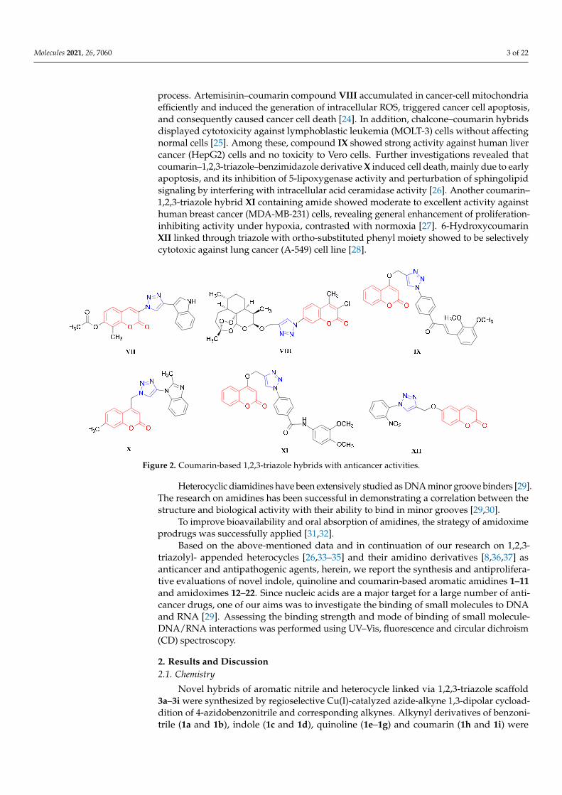
Hybridization of coumarin moiety with other anticancer pharmacophores was foundto provide novel anticancer candidates with low toxicity, high specificity and excellentefficacy against drug-susceptible and drug-resistant cancers [3,13,22]. It was observed thatsubstituent, length and position of alkyl spacer had a profound effect on the anticancer po-tency. 1,2,3-Triazole-containing novobiocin analogues with triazole ring at C-3 of coumarinwere investigated [23] and showed that coumarin–1,2,3-triazole–indole VII (Figure 2) ex-hibited potent antiproliferative activity against two breast cancer cell lines (SKBr-3 andMCF-7), which was directly related to inhibition of the Hsp90-mediated protein folding
Molecules 2021, 26, 7060 3 of 22
process. Artemisinin–coumarin compound VIII accumulated in cancer-cell mitochondriaefficiently and induced the generation of intracellular ROS, triggered cancer cell apoptosis,and consequently caused cancer cell death [24]. In addition, chalcone–coumarin hybridsdisplayed cytotoxicity against lymphoblastic leukemia (MOLT-3) cells without affectingnormal cells [25]. Among these, compound IX showed strong activity against human livercancer (HepG2) cells and no toxicity to Vero cells. Further investigations revealed thatcoumarin–1,2,3-triazole–benzimidazole derivative X induced cell death, mainly due to earlyapoptosis, and its inhibition of 5-lipoxygenase activity and perturbation of sphingolipidsignaling by interfering with intracellular acid ceramidase activity [26]. Another coumarin–1,2,3-triazole hybrid XI containing amide showed moderate to excellent activity againsthuman breast cancer (MDA-MB-231) cells, revealing general enhancement of proliferation-inhibiting activity under hypoxia, contrasted with normoxia [27]. 6-HydroxycoumarinXII linked through triazole with ortho-substituted phenyl moiety showed to be selectivelycytotoxic against lung cancer (A-549) cell line [28].
Molecules 2021, 26, 7060 3 of 22
and MCF‐7), which was directly related to inhibition of the Hsp90‐mediated protein fold‐
ing process. Artemisinin–coumarin compound VIII accumulated in cancer‐cell mitochon‐
dria efficiently and induced the generation of intracellular ROS, triggered cancer cell
apoptosis, and consequently caused cancer cell death [24]. In addition, chalcone–couma‐
rin hybrids displayed cytotoxicity against lymphoblastic leukemia (MOLT‐3) cells with‐
out affecting normal cells [25]. Among these, compound IX showed strong activity against
human liver cancer (HepG2) cells and no toxicity to Vero cells. Further investigations re‐
vealed that coumarin–1,2,3‐triazole–benzimidazole derivative X induced cell death,
mainly due to early apoptosis, and its inhibition of 5‐lipoxygenase activity and perturba‐
tion of sphingolipid signaling by interfering with intracellular acid ceramidase activity
[26]. Another coumarin–1,2,3‐triazole hybrid XI containing amide showed moderate to
excellent activity against human breast cancer (MDA‐MB‐231) cells, revealing general en‐
hancement of proliferation‐inhibiting activity under hypoxia, contrasted with normoxia
[27]. 6‐Hydroxycoumarin XII linked through triazole with ortho‐substituted phenyl moi‐
ety showed to be selectively cytotoxic against lung cancer (A‐549) cell line [28].
Figure 2. Coumarin‐based 1,2,3‐triazole hybrids with anticancer activities.
Heterocyclic diamidines have been extensively studied as DNA minor groove bind‐
ers [29]. The research on amidines has been successful in demonstrating a correlation be‐
tween the structure and biological activity with their ability to bind in minor grooves
[29,30].
To improve bioavailability and oral absorption of amidines, the strategy of amidox‐
ime prodrugs was successfully applied [31,32].
Based on the above‐mentioned data and in continuation of our research on 1,2,3‐tri‐
azolyl‐ appended heterocycles [26,33–35] and their amidino derivatives [8,36,37] as anti‐
cancer and antipathogenic agents, herein, we report the synthesis and antiproliferative
evaluations of novel indole, quinoline and coumarin‐based aromatic amidines 1–11 and
amidoximes 12–22. Since nucleic acids are a major target for a large number of anticancer
drugs, one of our aims was to investigate the binding of small molecules to DNA and
RNA [29]. Assessing the binding strength and mode of binding of small molecule‐
DNA/RNA interactions was performed using UV–Vis, fluorescence and circular dichro‐
ism (CD) spectroscopy.
2. Results and Discussion
2.1. Chemistry
Novel hybrids of aromatic nitrile and heterocycle linked via 1,2,3‐triazole scaffold
3a–3i were synthesized by regioselective Cu(I)‐catalyzed azide‐alkyne 1,3‐dipolar
Figure 2. Coumarin-based 1,2,3-triazole hybrids with anticancer activities.
Heterocyclic diamidines have been extensively studied as DNA minor groove binders [29].The research on amidines has been successful in demonstrating a correlation between thestructure and biological activity with their ability to bind in minor grooves [29,30].
To improve bioavailability and oral absorption of amidines, the strategy of amidoximeprodrugs was successfully applied [31,32].
Based on the above-mentioned data and in continuation of our research on 1,2,3-triazolyl- appended heterocycles [26,33–35] and their amidino derivatives [8,36,37] asanticancer and antipathogenic agents, herein, we report the synthesis and antiprolifera-tive evaluations of novel indole, quinoline and coumarin-based aromatic amidines 1–11and amidoximes 12–22. Since nucleic acids are a major target for a large number of anti-cancer drugs, one of our aims was to investigate the binding of small molecules to DNAand RNA [29]. Assessing the binding strength and mode of binding of small molecule-DNA/RNA interactions was performed using UV–Vis, fluorescence and circular dichroism(CD) spectroscopy.
2. Results and Discussion2.1. Chemistry
Novel hybrids of aromatic nitrile and heterocycle linked via 1,2,3-triazole scaffold3a–3i were synthesized by regioselective Cu(I)-catalyzed azide-alkyne 1,3-dipolar cycload-dition of 4-azidobenzonitrile and corresponding alkynes. Alkynyl derivatives of benzoni-trile (1a and 1b), indole (1c and 1d), quinoline (1e–1g) and coumarin (1h and 1i) were

Molecules 2021, 26, 7060 4 of 22
synthesized by alkylation with propargyl bromide in the presence of a base (Scheme 1),while azido derivative 2 was prepared by diazotation reaction according to the well-knownprocedure [38]. To study the influence of the type of linker between heterocycles on theirantiproliferative activity, 1,2,3-triazolyl-appended aryl and quinoline analogues containingnitrogen- (3a and 3e) and oxygen-containing (3b, 3f and 3g) aliphatic linkers were prepared.Coumarin was attached to aryl-triazole unit at C-4 (3h) and C-7 (3i) positions.
Molecules 2021, 26, 7060 4 of 22
cycloaddition of 4‐azidobenzonitrile and corresponding alkynes. Alkynyl derivatives of
benzonitrile (1a and 1b), indole (1c and 1d), quinoline (1e–1g) and coumarin (1h and 1i)
were synthesized by alkylation with propargyl bromide in the presence of a base (Scheme
1), while azido derivative 2 was prepared by diazotation reaction according to the well‐
known procedure [38]. To study the influence of the type of linker between heterocycles
on their antiproliferative activity, 1,2,3‐triazolyl‐appended aryl and quinoline analogues
containing nitrogen‐ (3a and 3e) and oxygen‐containing (3b, 3f and 3g) aliphatic linkers
were prepared. Coumarin was attached to aryl‐triazole unit at C‐4 (3h) and C‐7 (3i) posi‐
tions.
Scheme 1. Synthesis of cyano‐substituted 1,2,3‐triazole derivatives. Reagents and conditions: (i) propargylation of hy‐
droxyl group: propargyl bromide, acetone, K2CO3, reflux, 24 h; propargylation of amino group: propargyl bromide, DMF,
K2CO3, 80 °C, 24 h; (ii) methanol, Cu(OAc)2, r.t., 24 h.
Nitrile derivatives 3a–3i were subsequently used as precursors for the synthesis of
amidine‐ (4–11) and amidoxime‐substituted (12–22) heterocycles (Schemes 2 and 3). Am‐
idines were synthesized according to the Pinner method [39]. Treatment of corresponding
cyano derivative 3a–3h with dry gaseous HCl in absolute ethanol following by the reac‐
tion of the resulting imidate salt with ethylenediamine afforded targeted 2‐imidazolinyl
substituted derivatives 4–11. Conversely, the reaction of nitriles 3a–3i with hydroxyla‐
mine and triethylamine resulted in the desired amidoxime derivatives 12–22. In the syn‐
thesis of amidoximes both mono‐ 12, 14, 16 and 19 and their bis‐amidoxime analogues 13,
15, 17 and 20 were obtained (Scheme 2).
Scheme 2. Synthesis of amidine‐ (4–9) and amidoxime‐substituted (12–20) 1,2,3‐triazolyl heterocycle derivatives. Reagents
and conditions: (i) 1. HCl(g), abs. ethanol; 2. ethylenediamine (EDA), abs. ethanol, reflux, 24 h; 3. HCl(g), abs. ethanol; (ii)
methanol:DMF = 2:1, Et3N, NH2OH⸱HCl, 100 °C, 6 h.
Scheme 1. Synthesis of cyano-substituted 1,2,3-triazole derivatives. Reagents and conditions: (i) propargylation of hydroxylgroup: propargyl bromide, acetone, K2CO3, reflux, 24 h; propargylation of amino group: propargyl bromide, DMF, K2CO3,80 ◦C, 24 h; (ii) methanol, Cu(OAc)2, r.t., 24 h.
Nitrile derivatives 3a–3i were subsequently used as precursors for the synthesisof amidine- (4–11) and amidoxime-substituted (12–22) heterocycles (Schemes 2 and 3).Amidines were synthesized according to the Pinner method [39]. Treatment of correspond-ing cyano derivative 3a–3h with dry gaseous HCl in absolute ethanol following by thereaction of the resulting imidate salt with ethylenediamine afforded targeted 2-imidazolinylsubstituted derivatives 4–11. Conversely, the reaction of nitriles 3a–3i with hydroxylamineand triethylamine resulted in the desired amidoxime derivatives 12–22. In the synthesisof amidoximes both mono- 12, 14, 16 and 19 and their bis-amidoxime analogues 13, 15, 17and 20 were obtained (Scheme 2).
Molecules 2021, 26, 7060 4 of 22
cycloaddition of 4‐azidobenzonitrile and corresponding alkynes. Alkynyl derivatives of
benzonitrile (1a and 1b), indole (1c and 1d), quinoline (1e–1g) and coumarin (1h and 1i)
were synthesized by alkylation with propargyl bromide in the presence of a base (Scheme
1), while azido derivative 2 was prepared by diazotation reaction according to the well‐
known procedure [38]. To study the influence of the type of linker between heterocycles
on their antiproliferative activity, 1,2,3‐triazolyl‐appended aryl and quinoline analogues
containing nitrogen‐ (3a and 3e) and oxygen‐containing (3b, 3f and 3g) aliphatic linkers
were prepared. Coumarin was attached to aryl‐triazole unit at C‐4 (3h) and C‐7 (3i) posi‐
tions.
Scheme 1. Synthesis of cyano‐substituted 1,2,3‐triazole derivatives. Reagents and conditions: (i) propargylation of hy‐
droxyl group: propargyl bromide, acetone, K2CO3, reflux, 24 h; propargylation of amino group: propargyl bromide, DMF,
K2CO3, 80 °C, 24 h; (ii) methanol, Cu(OAc)2, r.t., 24 h.
Nitrile derivatives 3a–3i were subsequently used as precursors for the synthesis of
amidine‐ (4–11) and amidoxime‐substituted (12–22) heterocycles (Schemes 2 and 3). Am‐
idines were synthesized according to the Pinner method [39]. Treatment of corresponding
cyano derivative 3a–3h with dry gaseous HCl in absolute ethanol following by the reac‐
tion of the resulting imidate salt with ethylenediamine afforded targeted 2‐imidazolinyl
substituted derivatives 4–11. Conversely, the reaction of nitriles 3a–3i with hydroxyla‐
mine and triethylamine resulted in the desired amidoxime derivatives 12–22. In the syn‐
thesis of amidoximes both mono‐ 12, 14, 16 and 19 and their bis‐amidoxime analogues 13,
15, 17 and 20 were obtained (Scheme 2).
Scheme 2. Synthesis of amidine‐ (4–9) and amidoxime‐substituted (12–20) 1,2,3‐triazolyl heterocycle derivatives. Reagents
and conditions: (i) 1. HCl(g), abs. ethanol; 2. ethylenediamine (EDA), abs. ethanol, reflux, 24 h; 3. HCl(g), abs. ethanol; (ii)
methanol:DMF = 2:1, Et3N, NH2OH⸱HCl, 100 °C, 6 h.
Scheme 2. Synthesis of amidine- (4–9) and amidoxime-substituted (12–20) 1,2,3-triazolyl heterocycle derivatives. Reagentsand conditions: (i) 1. HCl(g), abs. ethanol; 2. ethylenediamine (EDA), abs. ethanol, reflux, 24 h; 3. HCl(g), abs. ethanol; (ii)methanol:DMF = 2:1, Et3N, NH2OH·HCl, 100 ◦C, 6 h.

Molecules 2021, 26, 7060 5 of 22Molecules 2021, 26, 7060 5 of 22
Scheme 3. Synthesis of amidino‐ (10 and 11) and amidoxime‐substituted (12 and 22) 1,2,3‐triazolyl
coumarin derivatives. Reagents and conditions: (i) 1. HCl(g), abs. ethanol; 2. EDA, abs. ethanol, re‐
flux, 24 h; 3. HCl(g), abs. ethanol; (ii) methanol:DMF = 2:1, Et3N, NH2OH⸱HCl, 100 °C, 6 h.
2.2. Biological Evaluation of Aromatic Amidines and Amidoximes
2.2.1. Evaluation of Antiproliferative Activity
Antiproliferative evaluations of amidine‐ and amidoxime‐substituted heterocycles
were performed on human tumor cell lines including cervical carcinoma (HeLa), colorec‐
tal adenocarcinoma, metastatic (SW620), lung adenocarcinoma (A549) and hepatocellular
carcinoma (HepG2) and non‐tumor HFF (human foreskin fibroblasts) cells (Table 1). 5‐
Fluorouracil (5‐FU) was used as the reference drug.
Table 1. Antiproliferative activity of amidino‐ 4–11 and amidoxime‐substituted 12–22 1,2,3‐triazole
derivatives.
4–22
Compd R R′ IC50 a (μM)
A549 HeLa HepG2 SW620 HFF
4
46.68 41.23 48.87 61.87 NA
5 ONH
H+Cl-N
15.67 0.80 0.64 0.22 <0.01
6
49.87 13.54 2.37 18.02 <0.01
7
>100 >100 69.07 49.77 NA
8
29.13 23.75 4.84 35.55 11.66
9
25.22 10.27 9.07 2.69 <0.01
10
7.86 1.90 3.55 1.75 <0.01
Scheme 3. Synthesis of amidino- (10 and 11) and amidoxime-substituted (12 and 22) 1,2,3-triazolyl coumarin derivatives.Reagents and conditions: (i) 1. HCl(g), abs. ethanol; 2. EDA, abs. ethanol, reflux, 24 h; 3. HCl(g), abs. ethanol; (ii)methanol:DMF = 2:1, Et3N, NH2OH·HCl, 100 ◦C, 6 h.
2.2. Biological Evaluation of Aromatic Amidines and Amidoximes2.2.1. Evaluation of Antiproliferative Activity
Antiproliferative evaluations of amidine- and amidoxime-substituted heterocycleswere performed on human tumor cell lines including cervical carcinoma (HeLa), colorectaladenocarcinoma, metastatic (SW620), lung adenocarcinoma (A549) and hepatocellularcarcinoma (HepG2) and non-tumor HFF (human foreskin fibroblasts) cells (Table 1). 5-Fluorouracil (5-FU) was used as the reference drug.
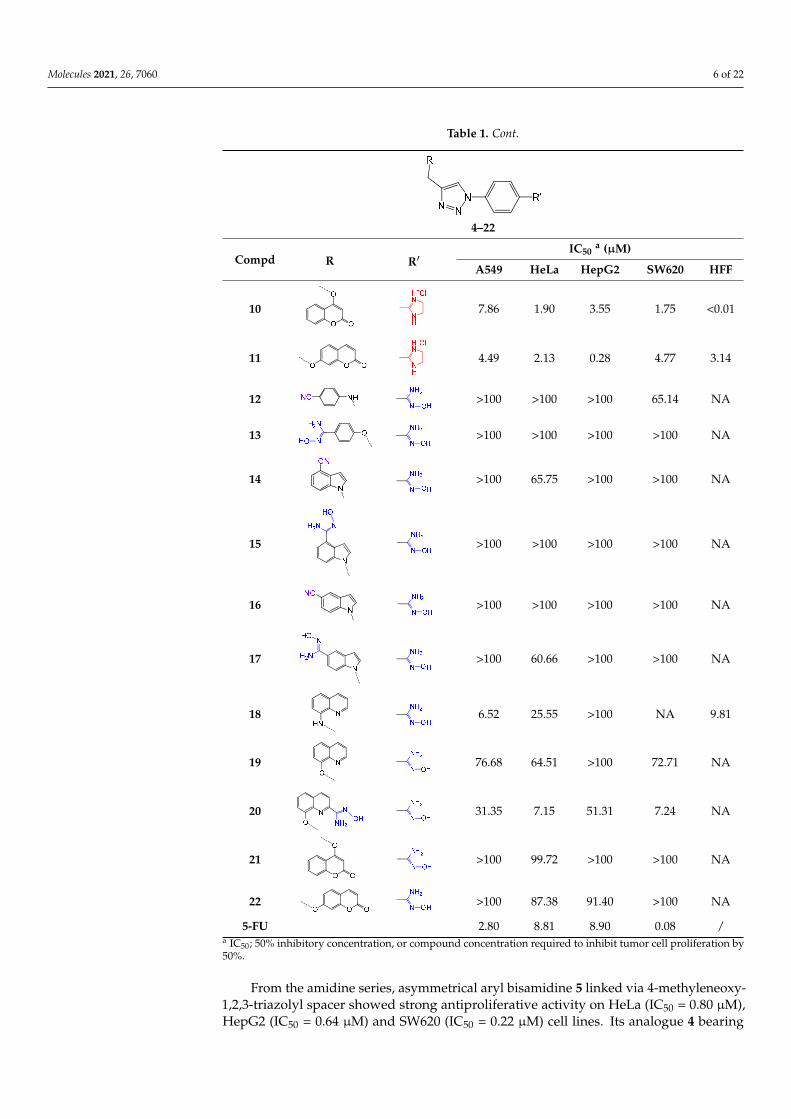
Table 1. Antiproliferative activity of amidino- 4–11 and amidoxime-substituted 12–22 1,2,3-triazolederivatives.
Molecules 2021, 26, 7060 5 of 22
Scheme 3. Synthesis of amidino‐ (10 and 11) and amidoxime‐substituted (12 and 22) 1,2,3‐triazolyl
coumarin derivatives. Reagents and conditions: (i) 1. HCl(g), abs. ethanol; 2. EDA, abs. ethanol, re‐
flux, 24 h; 3. HCl(g), abs. ethanol; (ii) methanol:DMF = 2:1, Et3N, NH2OH⸱HCl, 100 °C, 6 h.
2.2. Biological Evaluation of Aromatic Amidines and Amidoximes
2.2.1. Evaluation of Antiproliferative Activity
Antiproliferative evaluations of amidine‐ and amidoxime‐substituted heterocycles
were performed on human tumor cell lines including cervical carcinoma (HeLa), colorec‐
tal adenocarcinoma, metastatic (SW620), lung adenocarcinoma (A549) and hepatocellular
carcinoma (HepG2) and non‐tumor HFF (human foreskin fibroblasts) cells (Table 1). 5‐
Fluorouracil (5‐FU) was used as the reference drug.
Table 1. Antiproliferative activity of amidino‐ 4–11 and amidoxime‐substituted 12–22 1,2,3‐triazole
derivatives.
4–22
Compd R R′ IC50 a (μM)
A549 HeLa HepG2 SW620 HFF
4
46.68 41.23 48.87 61.87 NA
5 ONH
H+Cl-N
15.67 0.80 0.64 0.22 <0.01
6
49.87 13.54 2.37 18.02 <0.01
7
>100 >100 69.07 49.77 NA
8
29.13 23.75 4.84 35.55 11.66
9
25.22 10.27 9.07 2.69 <0.01
10
7.86 1.90 3.55 1.75 <0.01
4–22
Compd R R′IC50
a (µM)
A549 HeLa HepG2 SW620 HFF
4
Molecules 2021, 26, 7060 5 of 22
Scheme 3. Synthesis of amidino‐ (10 and 11) and amidoxime‐substituted (12 and 22) 1,2,3‐triazolyl
coumarin derivatives. Reagents and conditions: (i) 1. HCl(g), abs. ethanol; 2. EDA, abs. ethanol, re‐
flux, 24 h; 3. HCl(g), abs. ethanol; (ii) methanol:DMF = 2:1, Et3N, NH2OH⸱HCl, 100 °C, 6 h.
2.2. Biological Evaluation of Aromatic Amidines and Amidoximes
2.2.1. Evaluation of Antiproliferative Activity
Antiproliferative evaluations of amidine‐ and amidoxime‐substituted heterocycles
were performed on human tumor cell lines including cervical carcinoma (HeLa), colorec‐
tal adenocarcinoma, metastatic (SW620), lung adenocarcinoma (A549) and hepatocellular
carcinoma (HepG2) and non‐tumor HFF (human foreskin fibroblasts) cells (Table 1). 5‐
Fluorouracil (5‐FU) was used as the reference drug.
Table 1. Antiproliferative activity of amidino‐ 4–11 and amidoxime‐substituted 12–22 1,2,3‐triazole
derivatives.
4–22
Compd R R′ IC50 a (μM)
A549 HeLa HepG2 SW620 HFF
4
46.68 41.23 48.87 61.87 NA
5 ONH
H+Cl-N
15.67 0.80 0.64 0.22 <0.01
6
49.87 13.54 2.37 18.02 <0.01
7
>100 >100 69.07 49.77 NA
8
29.13 23.75 4.84 35.55 11.66
9
25.22 10.27 9.07 2.69 <0.01
10
7.86 1.90 3.55 1.75 <0.01
46.68 41.23 48.87 61.87 NA
5
Molecules 2021, 26, 7060 5 of 22
Scheme 3. Synthesis of amidino‐ (10 and 11) and amidoxime‐substituted (12 and 22) 1,2,3‐triazolyl
coumarin derivatives. Reagents and conditions: (i) 1. HCl(g), abs. ethanol; 2. EDA, abs. ethanol, re‐
flux, 24 h; 3. HCl(g), abs. ethanol; (ii) methanol:DMF = 2:1, Et3N, NH2OH⸱HCl, 100 °C, 6 h.
2.2. Biological Evaluation of Aromatic Amidines and Amidoximes
2.2.1. Evaluation of Antiproliferative Activity
Antiproliferative evaluations of amidine‐ and amidoxime‐substituted heterocycles
were performed on human tumor cell lines including cervical carcinoma (HeLa), colorec‐
tal adenocarcinoma, metastatic (SW620), lung adenocarcinoma (A549) and hepatocellular
carcinoma (HepG2) and non‐tumor HFF (human foreskin fibroblasts) cells (Table 1). 5‐
Fluorouracil (5‐FU) was used as the reference drug.
Table 1. Antiproliferative activity of amidino‐ 4–11 and amidoxime‐substituted 12–22 1,2,3‐triazole
derivatives.
4–22
Compd R R′ IC50 a (μM)
A549 HeLa HepG2 SW620 HFF
4
46.68 41.23 48.87 61.87 NA
5 ONH
H+Cl-N
15.67 0.80 0.64 0.22 <0.01
6
49.87 13.54 2.37 18.02 <0.01
7
>100 >100 69.07 49.77 NA
8
29.13 23.75 4.84 35.55 11.66
9
25.22 10.27 9.07 2.69 <0.01
10
7.86 1.90 3.55 1.75 <0.01
15.67 0.80 0.64 0.22 <0.01
6
Molecules 2021, 26, 7060 5 of 22
Scheme 3. Synthesis of amidino‐ (10 and 11) and amidoxime‐substituted (12 and 22) 1,2,3‐triazolyl
coumarin derivatives. Reagents and conditions: (i) 1. HCl(g), abs. ethanol; 2. EDA, abs. ethanol, re‐
flux, 24 h; 3. HCl(g), abs. ethanol; (ii) methanol:DMF = 2:1, Et3N, NH2OH⸱HCl, 100 °C, 6 h.
2.2. Biological Evaluation of Aromatic Amidines and Amidoximes
2.2.1. Evaluation of Antiproliferative Activity
Antiproliferative evaluations of amidine‐ and amidoxime‐substituted heterocycles
were performed on human tumor cell lines including cervical carcinoma (HeLa), colorec‐
tal adenocarcinoma, metastatic (SW620), lung adenocarcinoma (A549) and hepatocellular
carcinoma (HepG2) and non‐tumor HFF (human foreskin fibroblasts) cells (Table 1). 5‐
Fluorouracil (5‐FU) was used as the reference drug.
Table 1. Antiproliferative activity of amidino‐ 4–11 and amidoxime‐substituted 12–22 1,2,3‐triazole
derivatives.
4–22
Compd R R′ IC50 a (μM)
A549 HeLa HepG2 SW620 HFF
4
46.68 41.23 48.87 61.87 NA
5 ONH
H+Cl-N
15.67 0.80 0.64 0.22 <0.01
6
49.87 13.54 2.37 18.02 <0.01
7
>100 >100 69.07 49.77 NA
8
29.13 23.75 4.84 35.55 11.66
9
25.22 10.27 9.07 2.69 <0.01
10
7.86 1.90 3.55 1.75 <0.01
49.87 13.54 2.37 18.02 <0.01
7
Molecules 2021, 26, 7060 5 of 22
Scheme 3. Synthesis of amidino‐ (10 and 11) and amidoxime‐substituted (12 and 22) 1,2,3‐triazolyl
coumarin derivatives. Reagents and conditions: (i) 1. HCl(g), abs. ethanol; 2. EDA, abs. ethanol, re‐
flux, 24 h; 3. HCl(g), abs. ethanol; (ii) methanol:DMF = 2:1, Et3N, NH2OH⸱HCl, 100 °C, 6 h.
2.2. Biological Evaluation of Aromatic Amidines and Amidoximes
2.2.1. Evaluation of Antiproliferative Activity
Antiproliferative evaluations of amidine‐ and amidoxime‐substituted heterocycles
were performed on human tumor cell lines including cervical carcinoma (HeLa), colorec‐
tal adenocarcinoma, metastatic (SW620), lung adenocarcinoma (A549) and hepatocellular
carcinoma (HepG2) and non‐tumor HFF (human foreskin fibroblasts) cells (Table 1). 5‐
Fluorouracil (5‐FU) was used as the reference drug.
Table 1. Antiproliferative activity of amidino‐ 4–11 and amidoxime‐substituted 12–22 1,2,3‐triazole
derivatives.
4–22
Compd R R′ IC50 a (μM)
A549 HeLa HepG2 SW620 HFF
4
46.68 41.23 48.87 61.87 NA
5 ONH
H+Cl-N
15.67 0.80 0.64 0.22 <0.01
6
49.87 13.54 2.37 18.02 <0.01
7
>100 >100 69.07 49.77 NA
8
29.13 23.75 4.84 35.55 11.66
9
25.22 10.27 9.07 2.69 <0.01
10
7.86 1.90 3.55 1.75 <0.01
>100 >100 69.07 49.77 NA
8
Molecules 2021, 26, 7060 5 of 22
Scheme 3. Synthesis of amidino‐ (10 and 11) and amidoxime‐substituted (12 and 22) 1,2,3‐triazolyl
coumarin derivatives. Reagents and conditions: (i) 1. HCl(g), abs. ethanol; 2. EDA, abs. ethanol, re‐
flux, 24 h; 3. HCl(g), abs. ethanol; (ii) methanol:DMF = 2:1, Et3N, NH2OH⸱HCl, 100 °C, 6 h.
2.2. Biological Evaluation of Aromatic Amidines and Amidoximes
2.2.1. Evaluation of Antiproliferative Activity
Antiproliferative evaluations of amidine‐ and amidoxime‐substituted heterocycles
were performed on human tumor cell lines including cervical carcinoma (HeLa), colorec‐
tal adenocarcinoma, metastatic (SW620), lung adenocarcinoma (A549) and hepatocellular
carcinoma (HepG2) and non‐tumor HFF (human foreskin fibroblasts) cells (Table 1). 5‐
Fluorouracil (5‐FU) was used as the reference drug.
Table 1. Antiproliferative activity of amidino‐ 4–11 and amidoxime‐substituted 12–22 1,2,3‐triazole
derivatives.
4–22
Compd R R′ IC50 a (μM)
A549 HeLa HepG2 SW620 HFF
4
46.68 41.23 48.87 61.87 NA
5 ONH
H+Cl-N
15.67 0.80 0.64 0.22 <0.01
6
49.87 13.54 2.37 18.02 <0.01
7
>100 >100 69.07 49.77 NA
8
29.13 23.75 4.84 35.55 11.66
9
25.22 10.27 9.07 2.69 <0.01
10
7.86 1.90 3.55 1.75 <0.01
29.13 23.75 4.84 35.55 11.66
9
Molecules 2021, 26, 7060 5 of 22
Scheme 3. Synthesis of amidino‐ (10 and 11) and amidoxime‐substituted (12 and 22) 1,2,3‐triazolyl
coumarin derivatives. Reagents and conditions: (i) 1. HCl(g), abs. ethanol; 2. EDA, abs. ethanol, re‐
flux, 24 h; 3. HCl(g), abs. ethanol; (ii) methanol:DMF = 2:1, Et3N, NH2OH⸱HCl, 100 °C, 6 h.
2.2. Biological Evaluation of Aromatic Amidines and Amidoximes
2.2.1. Evaluation of Antiproliferative Activity
Antiproliferative evaluations of amidine‐ and amidoxime‐substituted heterocycles
were performed on human tumor cell lines including cervical carcinoma (HeLa), colorec‐
tal adenocarcinoma, metastatic (SW620), lung adenocarcinoma (A549) and hepatocellular
carcinoma (HepG2) and non‐tumor HFF (human foreskin fibroblasts) cells (Table 1). 5‐
Fluorouracil (5‐FU) was used as the reference drug.
Table 1. Antiproliferative activity of amidino‐ 4–11 and amidoxime‐substituted 12–22 1,2,3‐triazole
derivatives.
4–22
Compd R R′ IC50 a (μM)
A549 HeLa HepG2 SW620 HFF
4
46.68 41.23 48.87 61.87 NA
5 ONH
H+Cl-N
15.67 0.80 0.64 0.22 <0.01
6
49.87 13.54 2.37 18.02 <0.01
7
>100 >100 69.07 49.77 NA
8
29.13 23.75 4.84 35.55 11.66
9
25.22 10.27 9.07 2.69 <0.01
10
7.86 1.90 3.55 1.75 <0.01
25.22 10.27 9.07 2.69 <0.01
Molecules 2021, 26, 7060 6 of 22
Table 1. Cont.
Molecules 2021, 26, 7060 5 of 22
Scheme 3. Synthesis of amidino‐ (10 and 11) and amidoxime‐substituted (12 and 22) 1,2,3‐triazolyl
coumarin derivatives. Reagents and conditions: (i) 1. HCl(g), abs. ethanol; 2. EDA, abs. ethanol, re‐
flux, 24 h; 3. HCl(g), abs. ethanol; (ii) methanol:DMF = 2:1, Et3N, NH2OH⸱HCl, 100 °C, 6 h.
2.2. Biological Evaluation of Aromatic Amidines and Amidoximes
2.2.1. Evaluation of Antiproliferative Activity
Antiproliferative evaluations of amidine‐ and amidoxime‐substituted heterocycles
were performed on human tumor cell lines including cervical carcinoma (HeLa), colorec‐
tal adenocarcinoma, metastatic (SW620), lung adenocarcinoma (A549) and hepatocellular
carcinoma (HepG2) and non‐tumor HFF (human foreskin fibroblasts) cells (Table 1). 5‐
Fluorouracil (5‐FU) was used as the reference drug.
Table 1. Antiproliferative activity of amidino‐ 4–11 and amidoxime‐substituted 12–22 1,2,3‐triazole
derivatives.
4–22
Compd R R′ IC50 a (μM)
A549 HeLa HepG2 SW620 HFF
4
46.68 41.23 48.87 61.87 NA
5 ONH
H+Cl-N
15.67 0.80 0.64 0.22 <0.01
6
49.87 13.54 2.37 18.02 <0.01
7
>100 >100 69.07 49.77 NA
8
29.13 23.75 4.84 35.55 11.66
9
25.22 10.27 9.07 2.69 <0.01
10
7.86 1.90 3.55 1.75 <0.01
4–22
Compd R R′IC50
a (µM)
A549 HeLa HepG2 SW620 HFF
10
Molecules 2021, 26, 7060 5 of 22
Scheme 3. Synthesis of amidino‐ (10 and 11) and amidoxime‐substituted (12 and 22) 1,2,3‐triazolyl
coumarin derivatives. Reagents and conditions: (i) 1. HCl(g), abs. ethanol; 2. EDA, abs. ethanol, re‐
flux, 24 h; 3. HCl(g), abs. ethanol; (ii) methanol:DMF = 2:1, Et3N, NH2OH⸱HCl, 100 °C, 6 h.
2.2. Biological Evaluation of Aromatic Amidines and Amidoximes
2.2.1. Evaluation of Antiproliferative Activity
Antiproliferative evaluations of amidine‐ and amidoxime‐substituted heterocycles
were performed on human tumor cell lines including cervical carcinoma (HeLa), colorec‐
tal adenocarcinoma, metastatic (SW620), lung adenocarcinoma (A549) and hepatocellular
carcinoma (HepG2) and non‐tumor HFF (human foreskin fibroblasts) cells (Table 1). 5‐
Fluorouracil (5‐FU) was used as the reference drug.
Table 1. Antiproliferative activity of amidino‐ 4–11 and amidoxime‐substituted 12–22 1,2,3‐triazole
derivatives.
4–22
Compd R R′ IC50 a (μM)
A549 HeLa HepG2 SW620 HFF
4
46.68 41.23 48.87 61.87 NA
5 ONH
H+Cl-N
15.67 0.80 0.64 0.22 <0.01
6
49.87 13.54 2.37 18.02 <0.01
7
>100 >100 69.07 49.77 NA
8
29.13 23.75 4.84 35.55 11.66
9
25.22 10.27 9.07 2.69 <0.01
10
7.86 1.90 3.55 1.75 <0.01
Molecules 2021, 26, 7060 5 of 22
Scheme 3. Synthesis of amidino‐ (10 and 11) and amidoxime‐substituted (12 and 22) 1,2,3‐triazolyl
coumarin derivatives. Reagents and conditions: (i) 1. HCl(g), abs. ethanol; 2. EDA, abs. ethanol, re‐
flux, 24 h; 3. HCl(g), abs. ethanol; (ii) methanol:DMF = 2:1, Et3N, NH2OH⸱HCl, 100 °C, 6 h.
2.2. Biological Evaluation of Aromatic Amidines and Amidoximes
2.2.1. Evaluation of Antiproliferative Activity
Antiproliferative evaluations of amidine‐ and amidoxime‐substituted heterocycles
were performed on human tumor cell lines including cervical carcinoma (HeLa), colorec‐
tal adenocarcinoma, metastatic (SW620), lung adenocarcinoma (A549) and hepatocellular
carcinoma (HepG2) and non‐tumor HFF (human foreskin fibroblasts) cells (Table 1). 5‐
Fluorouracil (5‐FU) was used as the reference drug.
Table 1. Antiproliferative activity of amidino‐ 4–11 and amidoxime‐substituted 12–22 1,2,3‐triazole
derivatives.
4–22
Compd R R′ IC50 a (μM)
A549 HeLa HepG2 SW620 HFF
4
46.68 41.23 48.87 61.87 NA
5 ONH
H+Cl-N
15.67 0.80 0.64 0.22 <0.01
6
49.87 13.54 2.37 18.02 <0.01
7
>100 >100 69.07 49.77 NA
8
29.13 23.75 4.84 35.55 11.66
9
25.22 10.27 9.07 2.69 <0.01
10
7.86 1.90 3.55 1.75 <0.01 7.86 1.90 3.55 1.75 <0.01
11
Molecules 2021, 26, 7060 6 of 22
11
4.49 2.13 0.28 4.77 3.14
12 >100 >100 >100 65.14 NA
13
>100 >100 >100 >100 NA
14
>100 65.75 >100 >100 NA
15
>100 >100 >100 >100 NA
16
>100 >100 >100 >100 NA
17
>100 60.66 >100 >100 NA
18
6.52 25.55 >100 NA 9.81
19
76.68 64.51 >100 72.71 NA
20
31.35 7.15 51.31 7.24 NA
21
>100 99.72 >100 >100 NA
22
>100 87.38 91.40 >100 NA
5‐FU 2.80 8.81 8.90 0.08 /
a IC50; 50% inhibitory concentration, or compound concentration required to inhibit tumor cell
proliferation by 50%.
From the amidine series, asymmetrical aryl bisamidine 5 linked via 4‐methyleneoxy‐
1,2,3‐triazolyl spacer showed strong antiproliferative activity on HeLa (IC50 = 0.80 μM),
HepG2 (IC50 = 0.64 μM) and SW620 (IC50 = 0.22 μM) cell lines. Its analogue 4 bearing 4‐
aminomethylene‐1,2,3‐triazolyl spacer showed significantly reduced activity (Figure 3).
Similarly, quinoline amidine 9 with 8‐methyleneoxy‐1,2,3‐triazolyl unit exhibited 13‐fold
stronger inhibitory effects to tumor cells compared to its structural analogue 8. The only
exception was the growth‐inhibition effect on HEPG2 cells that was 2‐fold increased by 8
relative to its congener 9. Conversely, aminomethylene units in amidines 4 and 8 contrib‐
uted to their selectivity, showing less toxicity to normal HFF fibroblasts with selectivity
index (SI) of 2 compared to amidines 5 and 9, with methyleneoxy spacer, which showed
high cytotoxicity to normal fibroblasts. Antiproliferative evaluations of indole diamidines
showed that imidazoline 4‐substituted indole 6 was favorable for the activity, particularly
on HEPG2 cells (IC50 = 2.37 μM). Conversely, placement of the imidazoline in the 5‐posi‐
tion reduced the cytostatic activity of compound 7. Moreover, both 4‐ and 7‐substituted
4.49 2.13 0.28 4.77 3.14
12
Molecules 2021, 26, 7060 6 of 22
11
4.49 2.13 0.28 4.77 3.14
12 >100 >100 >100 65.14 NA
13
>100 >100 >100 >100 NA
14
>100 65.75 >100 >100 NA
15
>100 >100 >100 >100 NA
16
>100 >100 >100 >100 NA
17
>100 60.66 >100 >100 NA
18
6.52 25.55 >100 NA 9.81
19
76.68 64.51 >100 72.71 NA
20
31.35 7.15 51.31 7.24 NA
21
>100 99.72 >100 >100 NA
22
>100 87.38 91.40 >100 NA
5‐FU 2.80 8.81 8.90 0.08 /
a IC50; 50% inhibitory concentration, or compound concentration required to inhibit tumor cell
proliferation by 50%.
From the amidine series, asymmetrical aryl bisamidine 5 linked via 4‐methyleneoxy‐
1,2,3‐triazolyl spacer showed strong antiproliferative activity on HeLa (IC50 = 0.80 μM),
HepG2 (IC50 = 0.64 μM) and SW620 (IC50 = 0.22 μM) cell lines. Its analogue 4 bearing 4‐
aminomethylene‐1,2,3‐triazolyl spacer showed significantly reduced activity (Figure 3).
Similarly, quinoline amidine 9 with 8‐methyleneoxy‐1,2,3‐triazolyl unit exhibited 13‐fold
stronger inhibitory effects to tumor cells compared to its structural analogue 8. The only
exception was the growth‐inhibition effect on HEPG2 cells that was 2‐fold increased by 8
relative to its congener 9. Conversely, aminomethylene units in amidines 4 and 8 contrib‐
uted to their selectivity, showing less toxicity to normal HFF fibroblasts with selectivity
index (SI) of 2 compared to amidines 5 and 9, with methyleneoxy spacer, which showed
high cytotoxicity to normal fibroblasts. Antiproliferative evaluations of indole diamidines
showed that imidazoline 4‐substituted indole 6 was favorable for the activity, particularly
on HEPG2 cells (IC50 = 2.37 μM). Conversely, placement of the imidazoline in the 5‐posi‐
tion reduced the cytostatic activity of compound 7. Moreover, both 4‐ and 7‐substituted
>100 >100 >100 65.14 NA
13
Molecules 2021, 26, 7060 6 of 22
11
4.49 2.13 0.28 4.77 3.14
12 >100 >100 >100 65.14 NA
13
>100 >100 >100 >100 NA
14
>100 65.75 >100 >100 NA
15
>100 >100 >100 >100 NA
16
>100 >100 >100 >100 NA
17
>100 60.66 >100 >100 NA
18
6.52 25.55 >100 NA 9.81
19
76.68 64.51 >100 72.71 NA
20
31.35 7.15 51.31 7.24 NA
21
>100 99.72 >100 >100 NA
22
>100 87.38 91.40 >100 NA
5‐FU 2.80 8.81 8.90 0.08 /
a IC50; 50% inhibitory concentration, or compound concentration required to inhibit tumor cell
proliferation by 50%.
From the amidine series, asymmetrical aryl bisamidine 5 linked via 4‐methyleneoxy‐
1,2,3‐triazolyl spacer showed strong antiproliferative activity on HeLa (IC50 = 0.80 μM),
HepG2 (IC50 = 0.64 μM) and SW620 (IC50 = 0.22 μM) cell lines. Its analogue 4 bearing 4‐
aminomethylene‐1,2,3‐triazolyl spacer showed significantly reduced activity (Figure 3).
Similarly, quinoline amidine 9 with 8‐methyleneoxy‐1,2,3‐triazolyl unit exhibited 13‐fold
stronger inhibitory effects to tumor cells compared to its structural analogue 8. The only
exception was the growth‐inhibition effect on HEPG2 cells that was 2‐fold increased by 8
relative to its congener 9. Conversely, aminomethylene units in amidines 4 and 8 contrib‐
uted to their selectivity, showing less toxicity to normal HFF fibroblasts with selectivity
index (SI) of 2 compared to amidines 5 and 9, with methyleneoxy spacer, which showed
high cytotoxicity to normal fibroblasts. Antiproliferative evaluations of indole diamidines
showed that imidazoline 4‐substituted indole 6 was favorable for the activity, particularly
on HEPG2 cells (IC50 = 2.37 μM). Conversely, placement of the imidazoline in the 5‐posi‐
tion reduced the cytostatic activity of compound 7. Moreover, both 4‐ and 7‐substituted
>100 >100 >100 >100 NA
14
Molecules 2021, 26, 7060 6 of 22
11
4.49 2.13 0.28 4.77 3.14
12 >100 >100 >100 65.14 NA
13
>100 >100 >100 >100 NA
14
>100 65.75 >100 >100 NA
15
>100 >100 >100 >100 NA
16
>100 >100 >100 >100 NA
17
>100 60.66 >100 >100 NA
18
6.52 25.55 >100 NA 9.81
19
76.68 64.51 >100 72.71 NA
20
31.35 7.15 51.31 7.24 NA
21
>100 99.72 >100 >100 NA
22
>100 87.38 91.40 >100 NA
5‐FU 2.80 8.81 8.90 0.08 /
a IC50; 50% inhibitory concentration, or compound concentration required to inhibit tumor cell
proliferation by 50%.
From the amidine series, asymmetrical aryl bisamidine 5 linked via 4‐methyleneoxy‐
1,2,3‐triazolyl spacer showed strong antiproliferative activity on HeLa (IC50 = 0.80 μM),
HepG2 (IC50 = 0.64 μM) and SW620 (IC50 = 0.22 μM) cell lines. Its analogue 4 bearing 4‐
aminomethylene‐1,2,3‐triazolyl spacer showed significantly reduced activity (Figure 3).
Similarly, quinoline amidine 9 with 8‐methyleneoxy‐1,2,3‐triazolyl unit exhibited 13‐fold
stronger inhibitory effects to tumor cells compared to its structural analogue 8. The only
exception was the growth‐inhibition effect on HEPG2 cells that was 2‐fold increased by 8
relative to its congener 9. Conversely, aminomethylene units in amidines 4 and 8 contrib‐
uted to their selectivity, showing less toxicity to normal HFF fibroblasts with selectivity
index (SI) of 2 compared to amidines 5 and 9, with methyleneoxy spacer, which showed
high cytotoxicity to normal fibroblasts. Antiproliferative evaluations of indole diamidines
showed that imidazoline 4‐substituted indole 6 was favorable for the activity, particularly
on HEPG2 cells (IC50 = 2.37 μM). Conversely, placement of the imidazoline in the 5‐posi‐
tion reduced the cytostatic activity of compound 7. Moreover, both 4‐ and 7‐substituted
>100 65.75 >100 >100 NA
15
Molecules 2021, 26, 7060 6 of 22
11
4.49 2.13 0.28 4.77 3.14
12 >100 >100 >100 65.14 NA
13
>100 >100 >100 >100 NA
14
>100 65.75 >100 >100 NA
15
>100 >100 >100 >100 NA
16
>100 >100 >100 >100 NA
17
>100 60.66 >100 >100 NA
18
6.52 25.55 >100 NA 9.81
19
76.68 64.51 >100 72.71 NA
20
31.35 7.15 51.31 7.24 NA
21
>100 99.72 >100 >100 NA
22
>100 87.38 91.40 >100 NA
5‐FU 2.80 8.81 8.90 0.08 /
a IC50; 50% inhibitory concentration, or compound concentration required to inhibit tumor cell
proliferation by 50%.
From the amidine series, asymmetrical aryl bisamidine 5 linked via 4‐methyleneoxy‐
1,2,3‐triazolyl spacer showed strong antiproliferative activity on HeLa (IC50 = 0.80 μM),
HepG2 (IC50 = 0.64 μM) and SW620 (IC50 = 0.22 μM) cell lines. Its analogue 4 bearing 4‐
aminomethylene‐1,2,3‐triazolyl spacer showed significantly reduced activity (Figure 3).
Similarly, quinoline amidine 9 with 8‐methyleneoxy‐1,2,3‐triazolyl unit exhibited 13‐fold
stronger inhibitory effects to tumor cells compared to its structural analogue 8. The only
exception was the growth‐inhibition effect on HEPG2 cells that was 2‐fold increased by 8
relative to its congener 9. Conversely, aminomethylene units in amidines 4 and 8 contrib‐
uted to their selectivity, showing less toxicity to normal HFF fibroblasts with selectivity
index (SI) of 2 compared to amidines 5 and 9, with methyleneoxy spacer, which showed
high cytotoxicity to normal fibroblasts. Antiproliferative evaluations of indole diamidines
showed that imidazoline 4‐substituted indole 6 was favorable for the activity, particularly
on HEPG2 cells (IC50 = 2.37 μM). Conversely, placement of the imidazoline in the 5‐posi‐
tion reduced the cytostatic activity of compound 7. Moreover, both 4‐ and 7‐substituted
>100 >100 >100 >100 NA
16
Molecules 2021, 26, 7060 6 of 22
11
4.49 2.13 0.28 4.77 3.14
12 >100 >100 >100 65.14 NA
13
>100 >100 >100 >100 NA
14
>100 65.75 >100 >100 NA
15
>100 >100 >100 >100 NA
16
>100 >100 >100 >100 NA
17
>100 60.66 >100 >100 NA
18
6.52 25.55 >100 NA 9.81
19
76.68 64.51 >100 72.71 NA
20
31.35 7.15 51.31 7.24 NA
21
>100 99.72 >100 >100 NA
22
>100 87.38 91.40 >100 NA
5‐FU 2.80 8.81 8.90 0.08 /
a IC50; 50% inhibitory concentration, or compound concentration required to inhibit tumor cell
proliferation by 50%.
From the amidine series, asymmetrical aryl bisamidine 5 linked via 4‐methyleneoxy‐
1,2,3‐triazolyl spacer showed strong antiproliferative activity on HeLa (IC50 = 0.80 μM),
HepG2 (IC50 = 0.64 μM) and SW620 (IC50 = 0.22 μM) cell lines. Its analogue 4 bearing 4‐
aminomethylene‐1,2,3‐triazolyl spacer showed significantly reduced activity (Figure 3).
Similarly, quinoline amidine 9 with 8‐methyleneoxy‐1,2,3‐triazolyl unit exhibited 13‐fold
stronger inhibitory effects to tumor cells compared to its structural analogue 8. The only
exception was the growth‐inhibition effect on HEPG2 cells that was 2‐fold increased by 8
relative to its congener 9. Conversely, aminomethylene units in amidines 4 and 8 contrib‐
uted to their selectivity, showing less toxicity to normal HFF fibroblasts with selectivity
index (SI) of 2 compared to amidines 5 and 9, with methyleneoxy spacer, which showed
high cytotoxicity to normal fibroblasts. Antiproliferative evaluations of indole diamidines
showed that imidazoline 4‐substituted indole 6 was favorable for the activity, particularly
on HEPG2 cells (IC50 = 2.37 μM). Conversely, placement of the imidazoline in the 5‐posi‐
tion reduced the cytostatic activity of compound 7. Moreover, both 4‐ and 7‐substituted
>100 >100 >100 >100 NA
17
Molecules 2021, 26, 7060 6 of 22
11
4.49 2.13 0.28 4.77 3.14
12 >100 >100 >100 65.14 NA
13
>100 >100 >100 >100 NA
14
>100 65.75 >100 >100 NA
15
>100 >100 >100 >100 NA
16
>100 >100 >100 >100 NA
17
>100 60.66 >100 >100 NA
18
6.52 25.55 >100 NA 9.81
19
76.68 64.51 >100 72.71 NA
20
31.35 7.15 51.31 7.24 NA
21
>100 99.72 >100 >100 NA
22
>100 87.38 91.40 >100 NA
5‐FU 2.80 8.81 8.90 0.08 /
a IC50; 50% inhibitory concentration, or compound concentration required to inhibit tumor cell
proliferation by 50%.
From the amidine series, asymmetrical aryl bisamidine 5 linked via 4‐methyleneoxy‐
1,2,3‐triazolyl spacer showed strong antiproliferative activity on HeLa (IC50 = 0.80 μM),
HepG2 (IC50 = 0.64 μM) and SW620 (IC50 = 0.22 μM) cell lines. Its analogue 4 bearing 4‐
aminomethylene‐1,2,3‐triazolyl spacer showed significantly reduced activity (Figure 3).
Similarly, quinoline amidine 9 with 8‐methyleneoxy‐1,2,3‐triazolyl unit exhibited 13‐fold
stronger inhibitory effects to tumor cells compared to its structural analogue 8. The only
exception was the growth‐inhibition effect on HEPG2 cells that was 2‐fold increased by 8
relative to its congener 9. Conversely, aminomethylene units in amidines 4 and 8 contrib‐
uted to their selectivity, showing less toxicity to normal HFF fibroblasts with selectivity
index (SI) of 2 compared to amidines 5 and 9, with methyleneoxy spacer, which showed
high cytotoxicity to normal fibroblasts. Antiproliferative evaluations of indole diamidines
showed that imidazoline 4‐substituted indole 6 was favorable for the activity, particularly
on HEPG2 cells (IC50 = 2.37 μM). Conversely, placement of the imidazoline in the 5‐posi‐
tion reduced the cytostatic activity of compound 7. Moreover, both 4‐ and 7‐substituted
>100 60.66 >100 >100 NA
18
Molecules 2021, 26, 7060 6 of 22
11
4.49 2.13 0.28 4.77 3.14
12 >100 >100 >100 65.14 NA
13
>100 >100 >100 >100 NA
14
>100 65.75 >100 >100 NA
15
>100 >100 >100 >100 NA
16
>100 >100 >100 >100 NA
17
>100 60.66 >100 >100 NA
18
6.52 25.55 >100 NA 9.81
19
76.68 64.51 >100 72.71 NA
20
31.35 7.15 51.31 7.24 NA
21
>100 99.72 >100 >100 NA
22
>100 87.38 91.40 >100 NA
5‐FU 2.80 8.81 8.90 0.08 /
a IC50; 50% inhibitory concentration, or compound concentration required to inhibit tumor cell
proliferation by 50%.
From the amidine series, asymmetrical aryl bisamidine 5 linked via 4‐methyleneoxy‐
1,2,3‐triazolyl spacer showed strong antiproliferative activity on HeLa (IC50 = 0.80 μM),
HepG2 (IC50 = 0.64 μM) and SW620 (IC50 = 0.22 μM) cell lines. Its analogue 4 bearing 4‐
aminomethylene‐1,2,3‐triazolyl spacer showed significantly reduced activity (Figure 3).
Similarly, quinoline amidine 9 with 8‐methyleneoxy‐1,2,3‐triazolyl unit exhibited 13‐fold
stronger inhibitory effects to tumor cells compared to its structural analogue 8. The only
exception was the growth‐inhibition effect on HEPG2 cells that was 2‐fold increased by 8
relative to its congener 9. Conversely, aminomethylene units in amidines 4 and 8 contrib‐
uted to their selectivity, showing less toxicity to normal HFF fibroblasts with selectivity
index (SI) of 2 compared to amidines 5 and 9, with methyleneoxy spacer, which showed
high cytotoxicity to normal fibroblasts. Antiproliferative evaluations of indole diamidines
showed that imidazoline 4‐substituted indole 6 was favorable for the activity, particularly
on HEPG2 cells (IC50 = 2.37 μM). Conversely, placement of the imidazoline in the 5‐posi‐
tion reduced the cytostatic activity of compound 7. Moreover, both 4‐ and 7‐substituted
6.52 25.55 >100 NA 9.81
19
Molecules 2021, 26, 7060 6 of 22
11
4.49 2.13 0.28 4.77 3.14
12 >100 >100 >100 65.14 NA
13
>100 >100 >100 >100 NA
14
>100 65.75 >100 >100 NA
15
>100 >100 >100 >100 NA
16
>100 >100 >100 >100 NA
17
>100 60.66 >100 >100 NA
18
6.52 25.55 >100 NA 9.81
19
76.68 64.51 >100 72.71 NA
20
31.35 7.15 51.31 7.24 NA
21
>100 99.72 >100 >100 NA
22
>100 87.38 91.40 >100 NA
5‐FU 2.80 8.81 8.90 0.08 /
a IC50; 50% inhibitory concentration, or compound concentration required to inhibit tumor cell
proliferation by 50%.
From the amidine series, asymmetrical aryl bisamidine 5 linked via 4‐methyleneoxy‐
1,2,3‐triazolyl spacer showed strong antiproliferative activity on HeLa (IC50 = 0.80 μM),
HepG2 (IC50 = 0.64 μM) and SW620 (IC50 = 0.22 μM) cell lines. Its analogue 4 bearing 4‐
aminomethylene‐1,2,3‐triazolyl spacer showed significantly reduced activity (Figure 3).
Similarly, quinoline amidine 9 with 8‐methyleneoxy‐1,2,3‐triazolyl unit exhibited 13‐fold
stronger inhibitory effects to tumor cells compared to its structural analogue 8. The only
exception was the growth‐inhibition effect on HEPG2 cells that was 2‐fold increased by 8
relative to its congener 9. Conversely, aminomethylene units in amidines 4 and 8 contrib‐
uted to their selectivity, showing less toxicity to normal HFF fibroblasts with selectivity
index (SI) of 2 compared to amidines 5 and 9, with methyleneoxy spacer, which showed
high cytotoxicity to normal fibroblasts. Antiproliferative evaluations of indole diamidines
showed that imidazoline 4‐substituted indole 6 was favorable for the activity, particularly
on HEPG2 cells (IC50 = 2.37 μM). Conversely, placement of the imidazoline in the 5‐posi‐
tion reduced the cytostatic activity of compound 7. Moreover, both 4‐ and 7‐substituted
76.68 64.51 >100 72.71 NA
20
Molecules 2021, 26, 7060 6 of 22
11
4.49 2.13 0.28 4.77 3.14
12 >100 >100 >100 65.14 NA
13
>100 >100 >100 >100 NA
14
>100 65.75 >100 >100 NA
15
>100 >100 >100 >100 NA
16
>100 >100 >100 >100 NA
17
>100 60.66 >100 >100 NA
18
6.52 25.55 >100 NA 9.81
19
76.68 64.51 >100 72.71 NA
20
31.35 7.15 51.31 7.24 NA
21
>100 99.72 >100 >100 NA
22
>100 87.38 91.40 >100 NA
5‐FU 2.80 8.81 8.90 0.08 /
a IC50; 50% inhibitory concentration, or compound concentration required to inhibit tumor cell
proliferation by 50%.
From the amidine series, asymmetrical aryl bisamidine 5 linked via 4‐methyleneoxy‐
1,2,3‐triazolyl spacer showed strong antiproliferative activity on HeLa (IC50 = 0.80 μM),
HepG2 (IC50 = 0.64 μM) and SW620 (IC50 = 0.22 μM) cell lines. Its analogue 4 bearing 4‐
aminomethylene‐1,2,3‐triazolyl spacer showed significantly reduced activity (Figure 3).
Similarly, quinoline amidine 9 with 8‐methyleneoxy‐1,2,3‐triazolyl unit exhibited 13‐fold
stronger inhibitory effects to tumor cells compared to its structural analogue 8. The only
exception was the growth‐inhibition effect on HEPG2 cells that was 2‐fold increased by 8
relative to its congener 9. Conversely, aminomethylene units in amidines 4 and 8 contrib‐
uted to their selectivity, showing less toxicity to normal HFF fibroblasts with selectivity
index (SI) of 2 compared to amidines 5 and 9, with methyleneoxy spacer, which showed
high cytotoxicity to normal fibroblasts. Antiproliferative evaluations of indole diamidines
showed that imidazoline 4‐substituted indole 6 was favorable for the activity, particularly
on HEPG2 cells (IC50 = 2.37 μM). Conversely, placement of the imidazoline in the 5‐posi‐
tion reduced the cytostatic activity of compound 7. Moreover, both 4‐ and 7‐substituted
31.35 7.15 51.31 7.24 NA
21
Molecules 2021, 26, 7060 6 of 22
11
4.49 2.13 0.28 4.77 3.14
12 >100 >100 >100 65.14 NA
13
>100 >100 >100 >100 NA
14
>100 65.75 >100 >100 NA
15
>100 >100 >100 >100 NA
16
>100 >100 >100 >100 NA
17
>100 60.66 >100 >100 NA
18
6.52 25.55 >100 NA 9.81
19
76.68 64.51 >100 72.71 NA
20
31.35 7.15 51.31 7.24 NA
21
>100 99.72 >100 >100 NA
22
>100 87.38 91.40 >100 NA
5‐FU 2.80 8.81 8.90 0.08 /
a IC50; 50% inhibitory concentration, or compound concentration required to inhibit tumor cell
proliferation by 50%.
From the amidine series, asymmetrical aryl bisamidine 5 linked via 4‐methyleneoxy‐
1,2,3‐triazolyl spacer showed strong antiproliferative activity on HeLa (IC50 = 0.80 μM),
HepG2 (IC50 = 0.64 μM) and SW620 (IC50 = 0.22 μM) cell lines. Its analogue 4 bearing 4‐
aminomethylene‐1,2,3‐triazolyl spacer showed significantly reduced activity (Figure 3).
Similarly, quinoline amidine 9 with 8‐methyleneoxy‐1,2,3‐triazolyl unit exhibited 13‐fold
stronger inhibitory effects to tumor cells compared to its structural analogue 8. The only
exception was the growth‐inhibition effect on HEPG2 cells that was 2‐fold increased by 8
relative to its congener 9. Conversely, aminomethylene units in amidines 4 and 8 contrib‐
uted to their selectivity, showing less toxicity to normal HFF fibroblasts with selectivity
index (SI) of 2 compared to amidines 5 and 9, with methyleneoxy spacer, which showed
high cytotoxicity to normal fibroblasts. Antiproliferative evaluations of indole diamidines
showed that imidazoline 4‐substituted indole 6 was favorable for the activity, particularly
on HEPG2 cells (IC50 = 2.37 μM). Conversely, placement of the imidazoline in the 5‐posi‐
tion reduced the cytostatic activity of compound 7. Moreover, both 4‐ and 7‐substituted
>100 99.72 >100 >100 NA
22
Molecules 2021, 26, 7060 6 of 22
11
4.49 2.13 0.28 4.77 3.14
12 >100 >100 >100 65.14 NA
13
>100 >100 >100 >100 NA
14
>100 65.75 >100 >100 NA
15
>100 >100 >100 >100 NA
16
>100 >100 >100 >100 NA
17
>100 60.66 >100 >100 NA
18
6.52 25.55 >100 NA 9.81
19
76.68 64.51 >100 72.71 NA
20
31.35 7.15 51.31 7.24 NA
21
>100 99.72 >100 >100 NA
22
>100 87.38 91.40 >100 NA
5‐FU 2.80 8.81 8.90 0.08 /
a IC50; 50% inhibitory concentration, or compound concentration required to inhibit tumor cell
proliferation by 50%.
From the amidine series, asymmetrical aryl bisamidine 5 linked via 4‐methyleneoxy‐
1,2,3‐triazolyl spacer showed strong antiproliferative activity on HeLa (IC50 = 0.80 μM),
HepG2 (IC50 = 0.64 μM) and SW620 (IC50 = 0.22 μM) cell lines. Its analogue 4 bearing 4‐
aminomethylene‐1,2,3‐triazolyl spacer showed significantly reduced activity (Figure 3).
Similarly, quinoline amidine 9 with 8‐methyleneoxy‐1,2,3‐triazolyl unit exhibited 13‐fold
stronger inhibitory effects to tumor cells compared to its structural analogue 8. The only
exception was the growth‐inhibition effect on HEPG2 cells that was 2‐fold increased by 8
relative to its congener 9. Conversely, aminomethylene units in amidines 4 and 8 contrib‐
uted to their selectivity, showing less toxicity to normal HFF fibroblasts with selectivity
index (SI) of 2 compared to amidines 5 and 9, with methyleneoxy spacer, which showed
high cytotoxicity to normal fibroblasts. Antiproliferative evaluations of indole diamidines
showed that imidazoline 4‐substituted indole 6 was favorable for the activity, particularly
on HEPG2 cells (IC50 = 2.37 μM). Conversely, placement of the imidazoline in the 5‐posi‐
tion reduced the cytostatic activity of compound 7. Moreover, both 4‐ and 7‐substituted
>100 87.38 91.40 >100 NA
5-FU 2.80 8.81 8.90 0.08 /a IC50; 50% inhibitory concentration, or compound concentration required to inhibit tumor cell proliferation by50%.
From the amidine series, asymmetrical aryl bisamidine 5 linked via 4-methyleneoxy-1,2,3-triazolyl spacer showed strong antiproliferative activity on HeLa (IC50 = 0.80 µM),HepG2 (IC50 = 0.64 µM) and SW620 (IC50 = 0.22 µM) cell lines. Its analogue 4 bearing
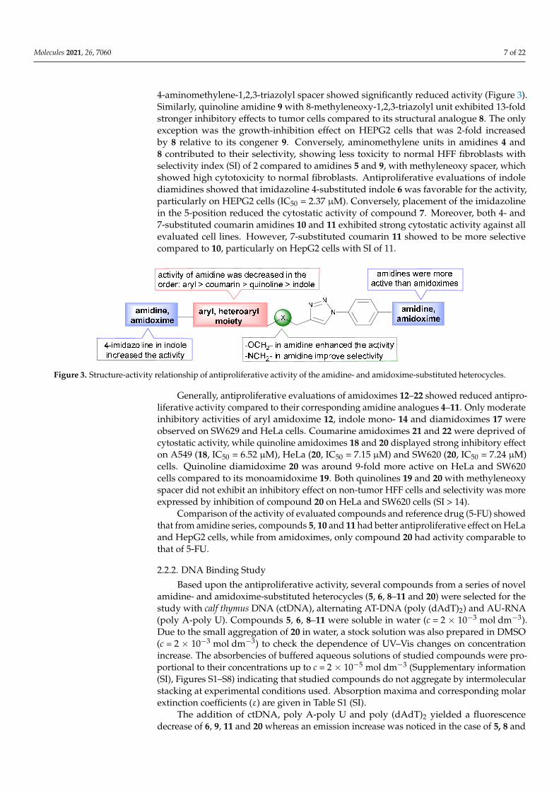
Molecules 2021, 26, 7060 7 of 22
4-aminomethylene-1,2,3-triazolyl spacer showed significantly reduced activity (Figure 3).Similarly, quinoline amidine 9 with 8-methyleneoxy-1,2,3-triazolyl unit exhibited 13-foldstronger inhibitory effects to tumor cells compared to its structural analogue 8. The onlyexception was the growth-inhibition effect on HEPG2 cells that was 2-fold increasedby 8 relative to its congener 9. Conversely, aminomethylene units in amidines 4 and8 contributed to their selectivity, showing less toxicity to normal HFF fibroblasts withselectivity index (SI) of 2 compared to amidines 5 and 9, with methyleneoxy spacer, whichshowed high cytotoxicity to normal fibroblasts. Antiproliferative evaluations of indolediamidines showed that imidazoline 4-substituted indole 6 was favorable for the activity,particularly on HEPG2 cells (IC50 = 2.37 µM). Conversely, placement of the imidazolinein the 5-position reduced the cytostatic activity of compound 7. Moreover, both 4- and7-substituted coumarin amidines 10 and 11 exhibited strong cytostatic activity against allevaluated cell lines. However, 7-substituted coumarin 11 showed to be more selectivecompared to 10, particularly on HepG2 cells with SI of 11.
Molecules 2021, 26, 7060 7 of 22
coumarin amidines 10 and 11 exhibited strong cytostatic activity against all evaluated cell
lines. However, 7‐substituted coumarin 11 showed to be more selective compared to 10,
particularly on HepG2 cells with SI of 11.
Figure 3. Structure‐activity relationship of antiproliferative activity of the amidine‐ and amidoxime‐substituted heterocycles.
Generally, antiproliferative evaluations of amidoximes 12–22 showed reduced anti‐
proliferative activity compared to their corresponding amidine analogues 4–11. Only
moderate inhibitory activities of aryl amidoxime 12, indole mono‐ 14 and diamidoximes
17 were observed on SW629 and HeLa cells. Coumarine amidoximes 21 and 22 were de‐
prived of cytostatic activity, while quinoline amidoximes 18 and 20 displayed strong in‐
hibitory effect on A549 (18, IC50 = 6.52 μM), HeLa (20, IC50 = 7.15 μM) and SW620 (20, IC50
= 7.24 μM) cells. Quinoline diamidoxime 20 was around 9‐fold more active on HeLa and
SW620 cells compared to its monoamidoxime 19. Both quinolines 19 and 20 with meth‐
yleneoxy spacer did not exhibit an inhibitory effect on non‐tumor HFF cells and selectivity
was more expressed by inhibition of compound 20 on HeLa and SW620 cells (SI > 14).
Comparison of the activity of evaluated compounds and reference drug (5‐FU)
showed that from amidine series, compounds 5, 10 and 11 had better antiproliferative
effect on HeLa and HepG2 cells, while from amidoximes, only compound 20 had activity
comparable to that of 5‐FU.
2.2.2. DNA Binding Study
Based upon the antiproliferative activity, several compounds from a series of novel
amidine‐ and amidoxime‐substituted heterocycles (5, 6, 811 and 20) were selected for the
study with calf thymus DNA (ctDNA), alternating AT‐DNA (poly (dAdT)2) and AU‐RNA
(poly A‐poly U). Compounds 5, 6, 811 were soluble in water (c = 2 10−3 mol dm−3). Due
to the small aggregation of 20 in water, a stock solution was also prepared in DMSO (c =
2 10−3 mol dm−3) to check the dependence of UV–Vis changes on concentration increase.
The absorbencies of buffered aqueous solutions of studied compounds were proportional
to their concentrations up to c = 2 10−5 mol dm−3 (Supplementary information (SI), Figures
S1–S8) indicating that studied compounds do not aggregate by intermolecular stacking at
experimental conditions used. Absorption maxima and corresponding molar extinction
coefficients (ε) are given in Table S1 (SI).
The addition of ctDNA, poly A‐poly U and poly (dAdT)2 yielded a fluorescence de‐
crease of 6, 9, 11 and 20 whereas an emission increase was noticed in the case of 5, 8 and
10 with all studied polynucleotides (Table 2, Figure 4). The fluorescence changes of stud‐
ied compounds were not dependent on the structure of polynucleotide added.
Figure 3. Structure-activity relationship of antiproliferative activity of the amidine- and amidoxime-substituted heterocycles.
Generally, antiproliferative evaluations of amidoximes 12–22 showed reduced antipro-liferative activity compared to their corresponding amidine analogues 4–11. Only moderateinhibitory activities of aryl amidoxime 12, indole mono- 14 and diamidoximes 17 wereobserved on SW629 and HeLa cells. Coumarine amidoximes 21 and 22 were deprived ofcytostatic activity, while quinoline amidoximes 18 and 20 displayed strong inhibitory effecton A549 (18, IC50 = 6.52 µM), HeLa (20, IC50 = 7.15 µM) and SW620 (20, IC50 = 7.24 µM)cells. Quinoline diamidoxime 20 was around 9-fold more active on HeLa and SW620cells compared to its monoamidoxime 19. Both quinolines 19 and 20 with methyleneoxyspacer did not exhibit an inhibitory effect on non-tumor HFF cells and selectivity was moreexpressed by inhibition of compound 20 on HeLa and SW620 cells (SI > 14).
Comparison of the activity of evaluated compounds and reference drug (5-FU) showedthat from amidine series, compounds 5, 10 and 11 had better antiproliferative effect on HeLaand HepG2 cells, while from amidoximes, only compound 20 had activity comparable tothat of 5-FU.
2.2.2. DNA Binding Study
Based upon the antiproliferative activity, several compounds from a series of novelamidine- and amidoxime-substituted heterocycles (5, 6, 8–11 and 20) were selected for thestudy with calf thymus DNA (ctDNA), alternating AT-DNA (poly (dAdT)2) and AU-RNA(poly A-poly U). Compounds 5, 6, 8–11 were soluble in water (c = 2 × 10⁻³ mol dm−3).Due to the small aggregation of 20 in water, a stock solution was also prepared in DMSO(c = 2 × 10⁻³ mol dm−3) to check the dependence of UV–Vis changes on concentrationincrease. The absorbencies of buffered aqueous solutions of studied compounds were pro-portional to their concentrations up to c = 2 × 10⁻⁵ mol dm−3 (Supplementary information(SI), Figures S1–S8) indicating that studied compounds do not aggregate by intermolecularstacking at experimental conditions used. Absorption maxima and corresponding molarextinction coefficients (ε) are given in Table S1 (SI).
The addition of ctDNA, poly A-poly U and poly (dAdT)2 yielded a fluorescencedecrease of 6, 9, 11 and 20 whereas an emission increase was noticed in the case of 5, 8 and
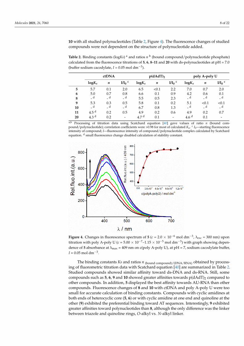
Molecules 2021, 26, 7060 8 of 22
10 with all studied polynucleotides (Table 2, Figure 4). The fluorescence changes of studiedcompounds were not dependent on the structure of polynucleotide added.
Table 2. Binding constants (logKs) a and ratios n b (bound compound/polynucleotide phosphate)calculated from the fluorescence titrations of 5, 6, 8–11 and 20 with ds-polynucleotides at pH = 7.0(buffer sodium cacodylate, I = 0.05 mol dm−3).
ctDNA p(dAdT)2 poly A-poly U
logKs n I/I0c logKs n I/I0
c logKs n I/I0c
5 5.7 0.1 2.0 6.5 <0.1 2.2 7.0 0.7 2.06 5.0 0.7 0.8 6.6 0.1 0.9 4.2 0.6 0.18 - d - d - d 5.5 0.5 2.3 - d - d - d
9 5.3 0.3 0.5 5.8 0.1 0.2 5.1 <0.1 <0.110 - d - d - d 6.7 0.8 1.3 - d - d - d
11 4.5 d 0.2 0.5 4.9 0.2 0.6 4.9 0.2 0.720 4.3 d 0.2 - 4.7 d 0.1 - 4.6 d 0.1 -
a,b Processing of titration data using Scatchard equation [40] gave values of ratio n (bound com-pound/polynucleotide); correlation coefficients were >0.98 for most of calculated Ka. c I0—starting fluorescenceintensity of compound; I—fluorescence intensity of compound/polynucleotide complex calculated by Scatchardequation. d small fluorescence change disabled calculation of stability constant.
Molecules 2021, 26, 7060 8 of 22
Figure 4. Changes in fluorescence spectrum of 5 (c = 2.0 10−6 mol dm−3, λexc = 300 nm) upon titration
with poly A‐poly U (c = 5.00 10−7–1.15 10−5 mol dm−3) with graph showing dependence of 5 ab‐
sorbance at λmax = 409 nm on c(poly A‐poly U), at pH = 7, sodium cacodylate buffer, I = 0.05 mol
dm−3.
Table 2. Binding constants (logKs) a and ratios n b (bound compound/polynucleotide phosphate)
calculated from the fluorescence titrations of 5, 6, 8–11 and 20 with ds‐polynucleotides at pH = 7.0
(buffer sodium cacodylate, I = 0.05 mol dm−3).
ctDNA p(dAdT)2 poly A‐poly U
logKs n I/I0 c logKs n I/I0 c logKs n I/I0 c
5 5.7 0.1 2.0 6.5 <0.1 2.2 7.0 0.7 2.0
6 5.0 0.7 0.8 6.6 0.1 0.9 4.2 0.6 0.1
8 ‐ d ‐ d ‐ d 5.5 0.5 2.3 ‐ d ‐ d ‐ d
9 5.3 0.3 0.5 5.8 0.1 0.2 5.1 <0.1 <0.1
10 ‐ d ‐ d ‐ d 6.7 0.8 1.3 ‐ d ‐ d ‐ d
11 4.5 d 0.2 0.5 4.9 0.2 0.6 4.9 0.2 0.7
20 4.3 d 0.2 ‐ 4.7 d 0.1 ‐ 4.6 d 0.1 ‐ a,b Processing of titration data using Scatchard equation [40] gave values of ratio n (bound com‐
pound/polynucleotide); correlation coefficients were >0.98 for most of calculated Ka. c I0—starting
fluorescence intensity of compound; I—fluorescence intensity of compound/polynucleotide com‐
plex calculated by Scatchard equation. d small fluorescence change disabled calculation of stability
constant.
The binding constants Ks and ratios n (bound compound)/(DNA/RNA) obtained by processing of
fluorometric titration data with Scatchard equation [40] are summarized in Table 2. Stud‐
ied compounds showed similar affinity toward ds‐DNA and ds‐RNA. Still, some com‐
pounds such as 5, 6, 9 and 10 showed greater affinities towards p(dAdT)2 compared to
other compounds. In addition, 5 displayed the best affinity towards AU‐RNA than other
compounds. Fluorescence changes of 8 and 10 with ctDNA and poly A‐poly U were too
small for accurate calculation of binding constants. Compounds with cyclic amidines at
both ends of heterocyclic core (5, 6) or with cyclic amidine at one end and quinoline at the
other (9) exhibited the preferential binding toward AT sequences. Interestingly, 9 exhib‐
ited greater affinities toward polynucleotides than 8, although the only difference was the
linker between triazole and quinoline rings, O‐alkyl vs. N‐alkyl linker.
Compounds 9–11 and especially 5 and 6 showed moderate to big stabilization effect
of AT‐DNA (Table S2, Figure S41). In comparison to AT‐DNA, stabilization of ctDNA by
Figure 4. Changes in fluorescence spectrum of 5 (c = 2.0 × 10⁻⁶ mol dm−3, λexc = 300 nm) upontitration with poly A-poly U (c = 5.00 × 10⁻⁷–1.15 × 10⁻⁵ mol dm−3) with graph showing depen-dence of 5 absorbance at λmax = 409 nm on c(poly A-poly U), at pH = 7, sodium cacodylate buffer,I = 0.05 mol dm−3.
The binding constants Ks and ratios n (bound compound)/(DNA/RNA) obtained by process-ing of fluorometric titration data with Scatchard equation [40] are summarized in Table 2.Studied compounds showed similar affinity toward ds-DNA and ds-RNA. Still, somecompounds such as 5, 6, 9 and 10 showed greater affinities towards p(dAdT)2 compared toother compounds. In addition, 5 displayed the best affinity towards AU-RNA than othercompounds. Fluorescence changes of 8 and 10 with ctDNA and poly A-poly U were toosmall for accurate calculation of binding constants. Compounds with cyclic amidines atboth ends of heterocyclic core (5, 6) or with cyclic amidine at one end and quinoline at theother (9) exhibited the preferential binding toward AT sequences. Interestingly, 9 exhibitedgreater affinities toward polynucleotides than 8, although the only difference was the linkerbetween triazole and quinoline rings, O-alkyl vs. N-alkyl linker.

Molecules 2021, 26, 7060 9 of 22
Compounds 9–11 and especially 5 and 6 showed moderate to big stabilization effectof AT-DNA (Table S2, Figure S41). In comparison to AT-DNA, stabilization of ctDNAby 5 and 6 was weaker as a consequence of the mixed base pair composition of ctDNA(58% AT, 42% GC). Other compounds showed either small or no stabilization effect ofpolynucleotides. Only a derivative possessing two imidazole moieties at the end of theheteroaromatic structure, 5 stabilized both DNA and RNA.
CD spectroscopy is a highly sensitive method for gaining insight into the conforma-tional changes in the secondary structure of polynucleotides induced by small moleculebinding [41]. In addition, it can provide information on the binding mode for small achiralmolecules possessing UV–Vis spectra above 300 nm.
Generally, the addition of compounds caused either a small decrease or a smallincrease of CD intensity of DNA and RNA polynucleotides at their maximal values (ctDNAat 275 nm, AT-DNA and AU-RNA at 260 nm). Still, significantly induced CD (ICD) bandsappeared in titrations of 5, 6 and 9–11 with AT-DNA (Figure 5, SI). Addition of AT-DNA to6, 9 and 11 induced positive ICD signals in the region from 270–303 nm at ratios, r < 0.2which could be ascribed to minor groove binding [42]. However, positive and negativesignals positioned at 320 and 380 nm appeared at ratios of r higher than 0.2, suggestingadditional binding mode—formation and binding of dimers or higher-order aggregatesalong the polynucleotide backbone. In titrations with AT-DNA, compounds 5 and 10caused strong positive ICD bands centered around 276 and 287 nm, respectively which isconsistent with minor groove binding (Figure 5, SI). In titrations with AU-RNA with 10and 11, weak negative ICD signals visible around 300 nm are indicative of intercalativebinding mode where the ligands are oriented “parallel” to the long axis of adjacent basepairs [42]. Compound 5 caused an appearance of similar ICD signals (strong positive ICDsignals around 284 nm) with ctDNA as in titration with AT-DNA and additionally, positiveand negative signals positioned at 320 and 390 nm were indicative of binding of aggregatesalong the polynucleotide backbone or most probably inside the groove.
Molecules 2021, 26, 7060 9 of 22
5 and 6 was weaker as a consequence of the mixed base pair composition of ctDNA (58%
AT, 42% GC). Other compounds showed either small or no stabilization effect of polynu‐
cleotides. Only a derivative possessing two imidazole moieties at the end of the heteroar‐
omatic structure, 5 stabilized both DNA and RNA.
CD spectroscopy is a highly sensitive method for gaining insight into the conforma‐
tional changes in the secondary structure of polynucleotides induced by small molecule
binding [41]. In addition, it can provide information on the binding mode for small achiral
molecules possessing UV–Vis spectra above 300 nm.
Generally, the addition of compounds caused either a small decrease or a small in‐
crease of CD intensity of DNA and RNA polynucleotides at their maximal values (ctDNA
at 275 nm, AT‐DNA and AU‐RNA at 260 nm). Still, significantly induced CD (ICD) bands
appeared in titrations of 5, 6 and 9–11 with AT‐DNA (Figure 5, SI). Addition of AT‐DNA
to 6, 9 and 11 induced positive ICD signals in the region from 270–303 nm at ratios, r < 0.2
which could be ascribed to minor groove binding[42]. However, positive and negative
signals positioned at 320 and 380 nm appeared at ratios of r higher than 0.2, suggesting
additional binding mode—formation and binding of dimers or higher‐order aggregates
along the polynucleotide backbone. In titrations with AT‐DNA, compounds 5 and 10
caused strong positive ICD bands centered around 276 and 287 nm, respectively which is
consistent with minor groove binding (Figure 5, SI). In titrations with AU‐RNA with 10
and 11, weak negative ICD signals visible around 300 nm are indicative of intercalative
binding mode where the ligands are oriented “parallel” to the long axis of adjacent base
pairs [42]. Compound 5 caused an appearance of similar ICD signals (strong positive ICD
signals around 284 nm) with ctDNA as in titration with AT‐DNA and additionally, posi‐
tive and negative signals positioned at 320 and 390 nm were indicative of binding of ag‐
gregates along the polynucleotide backbone or most probably inside the groove.
(a) (b)
Figure 5. CD titration of poly (dAdT)2 (c = 3.0 × 10⁻⁵ mol dm−3) with (a) 11 and (b) 5 at molar ratios r = compound/polynu‐
cleotide (pH = 7.0, buffer sodium cacodylate, I = 0.05 mol dm−3).
In titrations of polynucleotides with 8 and 20, there were no significantly induced CD
signals possibly due to the formation of aggregates such as in titration of 20 with ctDNA
or a lack of any dominant orientation of 8 and 20 molecules with respect to the polynucle‐
otide chiral axis.
Figure 5. CD titration of poly (dAdT)2 (c = 3.0 × 10⁻⁵ mol dm−3) with (a) 11 and (b) 5 at molar ratios r = com-pound/polynucleotide (pH = 7.0, buffer sodium cacodylate, I = 0.05 mol dm−3).
In titrations of polynucleotides with 8 and 20, there were no significantly induced CDsignals possibly due to the formation of aggregates such as in titration of 20 with ctDNA ora lack of any dominant orientation of 8 and 20 molecules with respect to the polynucleotidechiral axis.
Molecules 2021, 26, 7060 10 of 22
3. Experimental Part3.1. Materials and Methods
All solvents were purified following recommended drying agents and/or distilledover 3 Å molecular sieves. For monitoring the progress of a reaction and for comparisonpurposes, thin layer chromatography (TLC) was performed on pre-coated Merck (Darm-stadt, Germany) silica gel 60F-254 plates using an appropriate solvent system and thespots were detected under UV light (254 nm). For column chromatography silica gel(Fluka, Buchs, Switzerland, 0.063–0.2 mm) was employed, glass column was slurry-packedunder gravity. Melting points (uncorrected) were determined with Kofler micro hot-stage(Reichert, Wien, Austria). The IR spectra were recorded on PerkinElmer Spectrum Onespectrophotometer (Boston, MA, USA) with KBr disks. Elemental composition analysesof all novel compounds were within the 0.4% of the calculated values. 1H and 13C NMRspectra were acquired on a Bruker 300 and 600 MHz NMR spectrometer (Bruker Biospin,Rheinstetten, Germany). All data were recorded in DMSO-d6 at 298 K. Chemical shifts werereferenced to the residual solvent signal of DMSO-d6 at δ 2.50 ppm for 1H and δ 39.50 ppmfor 13C. Individual resonances were assigned on the basis of their chemical shifts, signalintensities, multiplicity of resonances and H–H coupling constants. 1H and 13C NMRspectra of compounds 4–22 are provided in Supporting Information (Figures S43–S61).
3.2. Synthetic Procedures
Compounds 4-azidobenzonitrile (2) [39] and alkynyl derivatives of benzonitrile (1aand 1b) [43,44], quinoline (1e and 1f) [45,46] and coumarin (1h and 1i) [47] were synthesizedaccording to the known procedure.
3.3. Preparation of Alkynyl Derivatives 1c, 1d and 1g3.3.1. 4-Cyano-1-((prop-2-ynyl)amino)-1H-indole (1c)
4-Cyanoindole (500 mg; 3.52 mmol) was dissolved in DMF (10 mL); K2CO3 (1.2 equiv.)was added and reaction mixture was heated at 80 ◦C. After 30 min, propargyl bromide (1.2equiv.) was added. Reaction mixture was stirred about 24 h. Solvent was removed underreduced pressure and residue purified by column chromatography with dichloromethaneas eluens. Compound 1c was isolated as white powder (403 mg; 64%; m.p. = 137–140 ◦C).1H NMR (300 MHz, DMSO-d6) (δ/ppm): 7.92 (d, J = 8.3 Hz, 1H, H-5), 7.72 (d, J = 3.2 Hz,1H, H-2), 7.60 (dd, J = 7.4, 0.7 Hz, 1H, H-7), 7.39–7.31 (m, 1H, H-6), 6.63 (dd, J = 3.2, 0.8 Hz,1H, H-3), 5.21 (d, J = 2.5 Hz, 2H, CH2), 3.46 (t, J = 2.5 Hz, 1H, CH). 13C NMR (151 MHz,DMSO-d6) (δ/ppm): 135.13 (C-7a), 131.70 (C-2), 129.10 (C-3a), 125.06 (C-5), 121.37 (C-6),118.36 (CN), 115.59 (C-7), 101.67 C-4), 99.59 (C-3), 78.63 (C-2′), 76.04 (C-3′), 35.45 (CH2-1′).
3.3.2. 5-Cyano-1-((prop-2-ynyl)amino)-1H-indole (1d)
5-Cyanoindole (1.00 g; 7.03 mmol) was dissolved in DMF (20 mL), K2CO3 (1.2 equivwas added and reaction mixture was heated at 80 ◦C. After 30 min propargyl bromide (1.2equiv.) was added. Reaction mixture was stirred about 24 h. Solvent was removed underreduced pressure and residue purified by column chromatography with dichloromethaneas eluens. Compound 1d was isolated as pink powder (900 mg; 71%, m.p. = 107–109 ◦C).1H NMR (300 MHz, DMSO-d6) (δ/ppm): 5.19 (d, J = 2.5 Hz, 2H, CH2), 3.44 (t, J = 2,4 Hz,1H, CH), 7.54 (dd, J = 8.5, 1.4 Hz, 1H, H-6), 6.64 (d, J = 3.0 Hz, 1H, H-3), 7.62 (d, J = 3.2 Hz,1H, H-2), 7.72 (d, J = 8.5, 1H, H-7), 8.12 (d, J = 0.8, 1H, H-4). 13C NMR (75 MHz, DMSO-d6)(δ/ppm): 137.02 (C-7a), 131.03 (C-2), 128.07 (C-3a), 126.15 (C-4), 124.11 (C-6), 120.48 (CN),111.40 (C-7), 102.42 (C-3), 101.64 (C-5), 78.64 (C-2′), 76.01 (C-3′), 35.36 (CH2-1′).
3.3.3. 2-Cyano-8-(prop-2-ynyloxy)-quinoline (1g)
To a solution of 8-hydroxy-2-carbonitrile (500 mg; 2.94 mmol) in acetone (18 mL),K2CO3 (6 equiv.) was added and reaction mixture was heated under reflux. After 30 min,propargyl bromide (0.4 mL; 2.94 mmol) was added. Reaction mixture was stirred andheated under reflux for about 24 h. Solvent was removed under reduced pressure and
Molecules 2021, 26, 7060 11 of 22
residue purified by column chromatography with petroleum ether:ethyl acetate = 6:4 aseluens. Compound 1g was isolated as white powder (498 mg; 81%, m.p. = 144–147 ◦C). 1HNMR (600 MHz, DMSO-d6) (δ/ppm): 8.62 (d, J = 8.5 Hz, 1H, H-4), 8.05 (d, J = 8.4 Hz, 1H,H-3), 7.75 (t, J = 8.0 Hz, 1H, H-6), 7.69 (d, J = 7.5 Hz, 1H, H-5), 7.44 (d, J = 7.7 Hz, 1H, H-7),5.10 (d, J = 2.3 Hz, 2H, CH2), 3.65 (t, J = 2.3 Hz, 1H, CH). 13C NMR (75 MHz, DMSO-d6)(δ/ppm): 152.62 (C-8), 139.53 (C-4a), 138.09 (C-4), 131.56 (C-8a), 130.00 (C-6), 129.77 (C-2),124.32 (C-3), 120.28 (C-5), 117.72 (CN), 111.64 (C-7), 78.90 (C-3′), 78.68 (C-2′), 56.22 (C-1′).
3.4. General Procedure for Synthesis of Cyano-Substituted 1,2,3-Triazole Derivatives 3a–3i
4-Azidobenzonitrile (2) (1 equiv.) and corresponding alkyne 1a–1i (1 equiv.) weredissolved in methanol, followed by addition of Cu(OAc)2 (0.05 equiv.). Reaction mixturewas stirred for 24 h at room temperature. Solvent was removed under reduced pressureand residue purified by column chromatography with dichloromethane as eluent.
3.4.1. 4-((1-(4-Cyanophenyl)-1H-1,2,3-triazole-4-yl)methylamino)benzonitrile (3a)
Compound 3a was prepared according to general procedure from 4-azidobenzonitrile(396 mg; 2.75 mmol) and compound 1a (428 mg; 2.75 mmol) in methanol (30 mL). Afterpurification by column chromatography with dichloromethane:methanol = 100:1 as eluens,compound 3a was isolated as white powder (306.7 mg; 37%, m.p. = 247–248 ◦C). 1H NMR(300 MHz, DMSO-d6) (δ/ppm): 8.90 (s, 1H, CH-triazole), 8.12 (q, J = 9.0 Hz, 4H, Ph), 7.47 (d,J = 8.8 Hz, 2H, Ph), 7.25 (t, J = 5.6 Hz, 1H, NH), 6.75 (d, J = 8.8 Hz, 2H, Ph), 4,48 (d, J = 5.7 Hz,2H, CH2). 13C NMR (75 MHz, DMSO-d6) (δ/ppm): 151.68 (Cq-Ph), 146.29 (Cq-Ph), 139.48(Cq-triazole), 134.30 (C-Ph), 133.36 (C-Ph), 121.53 (CH-triazole), 120.46 (CN), 120.34 (C-Ph),118.09 (CN), 112.19 (C-Ph), 110.97 (Cq-Ph), 96.36 (Cq-Ph), 37.64 (CH2).
3.4.2. 4-((1-(4-Cyanophenyl)-1H-1,2,3-triazol-4-yl)methyloxy)benzonitrile (3b)
Compound 3b was prepared according to general procedure from 4-azidobenzonitrile(250 mg; 1.74 mmol) and compound 1b (273 mg; 1.74 mmol) in methanol (20 mL). Com-pound 3b was isolated as white powder (418 mg; 80%, m.p. = 207–210 ◦C). 1H NMR(600 MHz, DMSO-d6) (δ/ppm): 9.13 (s, 1H, H-triazole), 8.14 (dd, J = 29.0, 8.8 Hz, 4H, Ph),7.81 (d, J = 8.9 Hz, 2H, Ph), 7.26 (d, J = 8.9 Hz, 2H, Ph), 5.38 (s, 1H, CH2). 13C NMR (75 MHz,DMSO-d6) (δ/ppm): 161.36 (C-Ph), 143.66 (C-Ph), 139.44 (Cq-triazole), 134.36 (C-Ph), 134.31(CH-triazole), 123.47 (C-Ph), 120.69 (C-Ph), 119.09 (CN), 118.11 (CN), 115.91 (C-Ph), 111.27(C-Ph), 103.39 (C-Ph), 61.32 (CH2).
3.4.3. 4-Cyano-1-((1-(4-cyanophenyl)-1H-1,2,3-triazol-4-yl)methylamino)-1H-indole (3c)
Compound 3c was prepared according to general procedure from 4-azidobenzonitrile(240 mg; 1.67 mmol) and compound 1c (300 mg; 1.67 mmol) in methanol (20 mL). Com-pound 3c was isolated as white powder (308 mg; 57%, m.p. = 200–203 ◦C). 1H NMR(300 MHz, DMSO-d6) (δ/ppm): 8.95 (s, 1H, H-triazole), 8.13–8.05 (m, 4H, H-5), 8.02 (d,J = 8.3 Hz, 1H, H-5), 7.80 (d, J = 3.2 Hz, 1H, H-2), 7.57 (d, J = 7.3 Hz, 1H, H-7), 7.36–7.27 (m,1H, H-6), 6.65 (d, J = 2.7 Hz, 1H, H-3), 5.70 (s, 2H, CH2). 13C NMR (75 MHz, DMSO-d6)(δ/ppm): 144.65 (C-Ph), 139.36 (Cq-triazole), 135.31(C-7a), 134.23 (C-Ph), 132.14 (C-2),129.08 (C-3a), 124.90 (C-5), 122.03 (CH-triazole), 121.28 (C-6), 120.54 (C-Ph), 118.46 (CN),118.04 (CN), 115.73 (C-7), 111.10 (C-Ph), 101.54 (C-Ph), 99.53 (C-3), 40.93 (CH2).
3.4.4. 5-Cyano-1-((1-(4-cyanophenyl)-1H-1,2,3-triazol-4-yl)methylamino)-1H-indole (3d)
Compound 3d was prepared according to general procedure from 4-azidobenzonitrile(250 mg; 1.74 mmol) and compound 1d (313 mg; 1.74 mmol) in methanol (20 mL). Com-pound 3d was isolated as white powder (513 mg; 91%, m.p. = 215–217 ◦C). 1H NMR(300 MHz, DMSO-d6) (δ/ppm): 8.96 (s, 1H, H-triazole), 8.22–8.04 (m, 5H, Ph, H-4), 7.83 (d,J = 8.6 Hz, 1H, H-7), 7.70 (d, J = 3.2 Hz, 1H, H-2), 7.51 (dd, J = 8.6, 1.5 Hz, 1H, H-6), 6.65(d, J = 3.2 Hz, 1H, H-3), 5.67 (s, 2H, CH2). 13C NMR (75 MHz, DMSO-d6) (δ/ppm): 144.58(C-Ph), 139.36 (Cq-triazole), 137.20 (C-7a), 134.23 (C-Ph), 131.44 (C-2), 128.03 (C-3a), 126.05
Molecules 2021, 26, 7060 12 of 22
(C-4), 124.00 (C-6), 122.05 (CH-triazole), 120.54 (C-Ph), 118.03 (CN), 111.51 (C-7), 111.11(C-Ph), 102.37 (C-3), 101.43 (C-5), 40.83 (CH2).
3.4.5. 8-((1-(4-Cyanophenyl)-1H-1,2,3-triazol-4-yl)methylamino)quinoline (3e)
Compound 3e was prepared according to general procedure from 4-azidobenzonitrile(209 mg; 1.45 mmol) and compound 1e (316 mg; 1.74 mmol) in methanol (20 mL). Afterpurification by column chromatography with dichloromethane:methanol = 100:1 as eluens,compound 3e was isolated as yellow crystalline powder (269 mg; 60%, m.p. = 196–198 ◦C).1H NMR (300 MHz, DMSO-d6) (δ/ppm): 8.90 (s, 1H, H-triazole), 8.76 (dd, J = 4.2, 1.6 Hz,1H, H-2), 8.22 (dd, J = 8.3, 1.6 Hz, 1H, H-4), 8.10 (dd, J = 24.2, 8.9 Hz, 4H), 7.51 (dd, J = 8.3,4.2 Hz, 1H, H-3), 7.36 (t, J = 7.9 Hz, 1H, H-6), 7.10 (d, J = 8.2 Hz, 1H, H-7), 7.05 (t, J = 6.0 Hz,1H, NH), 6.80 (d, J = 7.6 Hz, 1H, H-5), 4.68 (d, J = 6.0 Hz, 2H, CH2). 13C NMR (151 MHz,DMSO-d6) (δ/ppm): 147.05 (C-2), 143.89 (C-8), 139.55 (Cq-triazole), 137.58 (C-4a), 135.95(C-4), 134.22 (C-Ph), 128.23 (C-8a), 127.67 (C-6), 121.75 (C-3), 121.30 (CH-triazole), 120.37(C-Ph), 118.09 (CN), 113.91 (C-5), 110.87 (C-Ph), 104.89 (C-7), 38.28 (CH2).
3.4.6. 8-((1-(4-Cyanophenyl)-1H-1,2,3-triazol-4-yl)methyloxy)quinoline (3f)
Compound 3f was prepared according to general procedure from 4-azidobenzonitrile(353 mg; 2.45 mmol) and compound 1f (538 mg; 2.94 mmol) in methanol (15 mL). Com-pound 3f was isolated as white powder (655 mg; 82%, m.p. = 158–162 ◦C). 1H NMR(300 MHz, DMSO-d6) (δ/ppm): 9.19 (s, 1H, H-triazole), 8.84 (dd, J = 4.1, 1.7 Hz, 1H, H-2),8.33 (dd, J = 8.3, 1.7 Hz, 1H, H-7), 8.16 (dd, J = 23.9, 8.9 Hz, 4H, Ph), 7.65–7.39 (m, 4H, H-3,H-5, H-6, H-7), 5,47 (s, 2H, CH2). 13C NMR (75 MHz, DMSO-d6) (δ/ppm): 153.71 (C-8),149.04 (C-2), 144.26 (C-Ph), 139.73 (C-4a), 139.47 (Cq-triazole), 135.82 (C-4), 134.31 (C-Ph),129.09 (C-8a), 126.73 (C-6), 123.47 (CH-triazole), 121.90 (C-3), 120.59 (C-Ph), 120.30 (C-5),118.08 (CN), 111.16 (C-Ph), 110.31 (C-7), 61.67 (CH2).
3.4.7. 2-Cyano-8-((1-(4-cyanophenyl)-1H-1,2,3-triazol-4-yl)methyloxy)quinoline (3g)
Compound 3g was prepared according to general procedure from 4-azidobenzonitrile(187 mg; 1.30 mmol) and compound 1g (270 mg; 1.30 mmol) in methanol (20 mL). Com-pound 3g was isolated as white powder (260 mg; 57%, m.p. = 263–265 ◦C). 1H NMR(300 MHz, DMSO-d6) (δ/ppm): 9,21 (s, 1H, H-triazole), 8.62 (d, J = 8.5 Hz, 1H, H-4), 8.15(dd, J = 21.8, 8.9 Hz, 4H, Ph), 8.05 (d, J = 8.5 Hz, 1H, H-3), 7.60–7.80 (m, 3H, H-5, H-5, H-7),5,54 (s, 2H, CH2). 13C NMR (75 MHz, DMSO-d6) (δ/ppm): 153.47 (C-8), 143.74 (C-Ph),139.56 (C-4a), 139.45 (Cq-triazole), 138.07 (C-4), 134.27 (C-Ph), 131.43 (C-8a), 130.21 (C-6),129.84 (C-2), 124.33 (C-3), 123.73 (CH-triazole), 120.66 (C-Ph), 120.07 (C-5), 118.06 (CN),117.74 (CN), 111.49 (C-7), 111.18 (Ph), 61.66 (CH2).
3.4.8. 4-((1-(4-Cyanophenyl)-1H-1,2,3-triazol-4-yl)methyloxy)coumarin (3h)
Compound 3h was prepared according to general procedure from 4-azidobenzonitrile(158 mg; 1.10 mmol) and compound 1h (220 mg; 1.10 mmol) in methanol (20 mL). Com-pound 3h was isolated as white powder (245 mg; 71%, m.p. = 274–276 ◦C). 1H NMR(600 MHz, DMSO-d6) (δ/ppm): 9.23 (s, 1H, H-triazole), 8.17 (dd, J = 43.7, 8.4 Hz, 4H, Ph),7.84 (d, J = 7.8 Hz, 1H, H-5), 7.67 (t, J = 7.7 Hz, 1H, H-7), 7.42 (d, J = 8.3 Hz, 1H, H-8), 7.36(t, J = 7.6 Hz, 1H, H-6), 6,21 (s, 1H, H-3), 5.55 (s, 2H, CH2). 13C NMR (75 MHz, DMSO-d6)(δ/ppm): 164.28 (C-4), 161.50 (C-2), 152.77 (C-8a), 142.80 (C-Ph), 139.40 (Cq-triazole), 134.32(C-Ph), 132.88 (C-7), 124.22 (C-6), 123.58 (CH-triazole), 123.02 (C-5), 120.70 (C-Ph), 118.07(CN), 116.47 (C-8), 115.01 (C-4a), 111.29 (C-Ph), 91.53 (C-3), 62.71 (CH2).
3.4.9. 7-((1-(4-Cyanophenyl)-1H-1,2,3-triazol-4-yl)methyloxy)coumarin (3i)
Compound 3i was prepared according to general procedure from 4-azidobenzonitrile(187 mg; 1.30 mmol) and compound 1i (260 mg; 1.30 mmol) in methanol (20 mL). Com-pound 3i was isolated as white powder (187 mg; 41.8%, m.p. = 228–229 ◦C). 1H NMR(300 MHz, DMSO-d6) (δ/ppm): 9.16 (s, 1H, H-triazole), 8.14 (td, J = 8.9, 2.0 Hz, 4H, Ph), 8.01
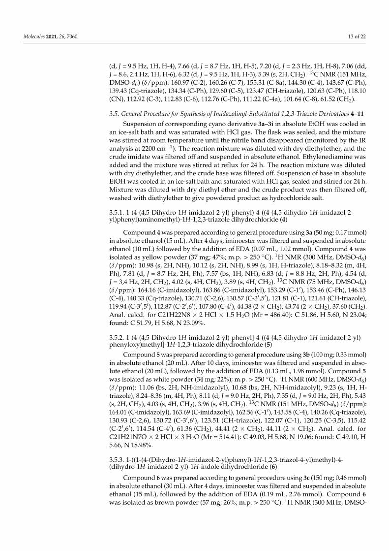
Molecules 2021, 26, 7060 13 of 22
(d, J = 9.5 Hz, 1H, H-4), 7.66 (d, J = 8.7 Hz, 1H, H-5), 7.20 (d, J = 2.3 Hz, 1H, H-8), 7.06 (dd,J = 8.6, 2.4 Hz, 1H, H-6), 6.32 (d, J = 9.5 Hz, 1H, H-3), 5.39 (s, 2H, CH2). 13C NMR (151 MHz,DMSO-d6) (δ/ppm): 160.97 (C-2), 160.26 (C-7), 155.31 (C-8a), 144.30 (C-4), 143.67 (C-Ph),139.43 (Cq-triazole), 134.34 (C-Ph), 129.60 (C-5), 123.47 (CH-triazole), 120.63 (C-Ph), 118.10(CN), 112.92 (C-3), 112.83 (C-6), 112.76 (C-Ph), 111.22 (C-4a), 101.64 (C-8), 61.52 (CH2).
3.5. General Procedure for Synthesis of Imidazolinyl-Substituted 1,2,3-Triazole Derivatives 4–11
Suspension of corresponding cyano derivative 3a–3i in absolute EtOH was cooled inan ice-salt bath and was saturated with HCl gas. The flask was sealed, and the mixturewas stirred at room temperature until the nitrile band disappeared (monitored by the IRanalysis at 2200 cm−1). The reaction mixture was diluted with dry diethylether, and thecrude imidate was filtered off and suspended in absolute ethanol. Ethylenediamine wasadded and the mixture was stirred at reflux for 24 h. The reaction mixture was dilutedwith dry diethylether, and the crude base was filtered off. Suspension of base in absoluteEtOH was cooled in an ice-salt bath and saturated with HCl gas, sealed and stirred for 24 h.Mixture was diluted with dry diethyl ether and the crude product was then filtered off,washed with diethylether to give powdered product as hydrochloride salt.
3.5.1. 1-(4-(4,5-Dihydro-1H-imidazol-2-yl)-phenyl)-4-((4-(4,5-dihydro-1H-imidazol-2-yl)phenyl)aminomethyl)-1H-1,2,3-triazole dihydrochloride (4)
Compound 4 was prepared according to general procedure using 3a (50 mg; 0.17 mmol)in absolute ethanol (15 mL). After 4 days, iminoester was filtered and suspended in absoluteethanol (10 mL) followed by the addition of EDA (0.07 mL, 1.02 mmol). Compound 4 wasisolated as yellow powder (37 mg; 47%; m.p. > 250 ◦C). 1H NMR (300 MHz, DMSO-d6)(δ/ppm): 10.98 (s, 2H, NH), 10.12 (s, 2H, NH), 8.99 (s, 1H, H-triazole), 8.18–8.32 (m, 4H,Ph), 7.81 (d, J = 8.7 Hz, 2H, Ph), 7.57 (bs, 1H, NH), 6.83 (d, J = 8.8 Hz, 2H, Ph), 4.54 (d,J = 3,4 Hz, 2H, CH2), 4.02 (s, 4H, CH2), 3.89 (s, 4H, CH2). 13C NMR (75 MHz, DMSO-d6)(δ/ppm): 164.16 (C-imidazolyl), 163.86 (C-imidazolyl), 153.29 (C-1′), 153.46 (C-Ph), 146.13(C-4), 140.33 (Cq-triazole), 130.71 (C-2,6), 130.57 (C-3′,5′), 121.81 (C-1), 121.61 (CH-triazole),119.94 (C-3′,5′), 112.87 (C-2′,6′), 107.80 (C-4′), 44.38 (2 × CH2), 43.74 (2 × CH2), 37.60 (CH2).Anal. calcd. for C21H22N8 × 2 HCl × 1.5 H2O (Mr = 486.40): C 51.86, H 5.60, N 23.04;found: C 51.79, H 5.68, N 23.09%.
3.5.2. 1-(4-(4,5-Dihydro-1H-imidazol-2-yl)-phenyl]-4-((4-(4,5-dihydro-1H-imidazol-2-yl)phenyloxy)methyl]-1H-1,2,3-triazole dihydrochloride (5)
Compound 5 was prepared according to general procedure using 3b (100 mg; 0.33 mmol)in absolute ethanol (20 mL). After 10 days, iminoester was filtered and suspended in abso-lute ethanol (20 mL), followed by the addition of EDA (0.13 mL, 1.98 mmol). Compound 5was isolated as white powder (34 mg; 22%); m.p. > 250 ◦C). 1H NMR (600 MHz, DMSO-d6)(δ/ppm): 11.06 (bs, 2H, NH-imidazolyl), 10.68 (bs, 2H, NH-imidazolyl), 9.23 (s, 1H, H-triazole), 8.24–8.36 (m, 4H, Ph), 8.11 (d, J = 9.0 Hz, 2H, Ph), 7.35 (d, J = 9.0 Hz, 2H, Ph), 5.43(s, 2H, CH2), 4.03 (s, 4H, CH2), 3.96 (s, 4H, CH2). 13C NMR (151 MHz, DMSO-d6) (δ/ppm):164.01 (C-imidazolyl), 163.69 (C-imidazolyl), 162.56 (C-1′), 143.58 (C-4), 140.26 (Cq-triazole),130.93 (C-2,6), 130.72 (C-3′,6′), 123.51 (CH-triazole), 122.07 (C-1), 120.25 (C-3,5), 115.42(C-2′,6′), 114.54 (C-4′), 61.36 (CH2), 44.41 (2 × CH2), 44.11 (2 × CH2). Anal. calcd. forC21H21N7O × 2 HCl × 3 H2O (Mr = 514.41): C 49.03, H 5.68, N 19.06; found: C 49.10, H5.66, N 18.98%.
3.5.3. 1-((1-(4-(Dihydro-1H-imidazol-2-yl)phenyl)-1H-1,2,3-triazol-4-yl)methyl)-4-(dihydro-1H-imidazol-2-yl)-1H-indole dihydrochloride (6)
Compound 6 was prepared according to general procedure using 3c (150 mg; 0.46 mmol)in absolute ethanol (30 mL). After 4 days, iminoester was filtered and suspended in absoluteethanol (15 mL), followed by the addition of EDA (0.19 mL, 2.76 mmol). Compound 6was isolated as brown powder (57 mg; 26%; m.p. > 250 ◦C). 1H NMR (300 MHz, DMSO-
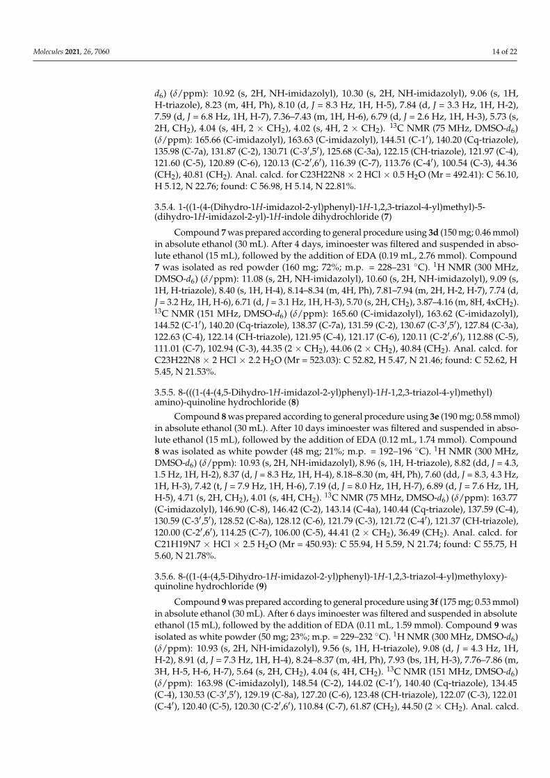
Molecules 2021, 26, 7060 14 of 22
d6) (δ/ppm): 10.92 (s, 2H, NH-imidazolyl), 10.30 (s, 2H, NH-imidazolyl), 9.06 (s, 1H,H-triazole), 8.23 (m, 4H, Ph), 8.10 (d, J = 8.3 Hz, 1H, H-5), 7.84 (d, J = 3.3 Hz, 1H, H-2),7.59 (d, J = 6.8 Hz, 1H, H-7), 7.36–7.43 (m, 1H, H-6), 6.79 (d, J = 2.6 Hz, 1H, H-3), 5.73 (s,2H, CH2), 4.04 (s, 4H, 2 × CH2), 4.02 (s, 4H, 2 × CH2). 13C NMR (75 MHz, DMSO-d6)(δ/ppm): 165.66 (C-imidazolyl), 163.63 (C-imidazolyl), 144.51 (C-1′), 140.20 (Cq-triazole),135.98 (C-7a), 131.87 (C-2), 130.71 (C-3′,5′), 125.68 (C-3a), 122.15 (CH-triazole), 121.97 (C-4),121.60 (C-5), 120.89 (C-6), 120.13 (C-2′,6′), 116.39 (C-7), 113.76 (C-4′), 100.54 (C-3), 44.36(CH2), 40.81 (CH2). Anal. calcd. for C23H22N8 × 2 HCl × 0.5 H2O (Mr = 492.41): C 56.10,H 5.12, N 22.76; found: C 56.98, H 5.14, N 22.81%.
3.5.4. 1-((1-(4-(Dihydro-1H-imidazol-2-yl)phenyl)-1H-1,2,3-triazol-4-yl)methyl)-5-(dihydro-1H-imidazol-2-yl)-1H-indole dihydrochloride (7)
Compound 7 was prepared according to general procedure using 3d (150 mg; 0.46 mmol)in absolute ethanol (30 mL). After 4 days, iminoester was filtered and suspended in abso-lute ethanol (15 mL), followed by the addition of EDA (0.19 mL, 2.76 mmol). Compound7 was isolated as red powder (160 mg; 72%; m.p. = 228–231 ◦C). 1H NMR (300 MHz,DMSO-d6) (δ/ppm): 11.08 (s, 2H, NH-imidazolyl), 10.60 (s, 2H, NH-imidazolyl), 9.09 (s,1H, H-triazole), 8.40 (s, 1H, H-4), 8.14–8.34 (m, 4H, Ph), 7.81–7.94 (m, 2H, H-2, H-7), 7.74 (d,J = 3.2 Hz, 1H, H-6), 6.71 (d, J = 3.1 Hz, 1H, H-3), 5.70 (s, 2H, CH2), 3.87–4.16 (m, 8H, 4xCH2).13C NMR (151 MHz, DMSO-d6) (δ/ppm): 165.60 (C-imidazolyl), 163.62 (C-imidazolyl),144.52 (C-1′), 140.20 (Cq-triazole), 138.37 (C-7a), 131.59 (C-2), 130.67 (C-3′,5′), 127.84 (C-3a),122.63 (C-4), 122.14 (CH-triazole), 121.95 (C-4), 121.17 (C-6), 120.11 (C-2′,6′), 112.88 (C-5),111.01 (C-7), 102.94 (C-3), 44.35 (2 × CH2), 44.06 (2 × CH2), 40.84 (CH2). Anal. calcd. forC23H22N8 × 2 HCl × 2.2 H2O (Mr = 523.03): C 52.82, H 5.47, N 21.46; found: C 52.62, H5.45, N 21.53%.
3.5.5. 8-(((1-(4-(4,5-Dihydro-1H-imidazol-2-yl)phenyl)-1H-1,2,3-triazol-4-yl)methyl)amino)-quinoline hydrochloride (8)
Compound 8 was prepared according to general procedure using 3e (190 mg; 0.58 mmol)in absolute ethanol (30 mL). After 10 days iminoester was filtered and suspended in abso-lute ethanol (15 mL), followed by the addition of EDA (0.12 mL, 1.74 mmol). Compound8 was isolated as white powder (48 mg; 21%; m.p. = 192–196 ◦C). 1H NMR (300 MHz,DMSO-d6) (δ/ppm): 10.93 (s, 2H, NH-imidazolyl), 8.96 (s, 1H, H-triazole), 8.82 (dd, J = 4.3,1.5 Hz, 1H, H-2), 8.37 (d, J = 8.3 Hz, 1H, H-4), 8.18–8.30 (m, 4H, Ph), 7.60 (dd, J = 8.3, 4.3 Hz,1H, H-3), 7.42 (t, J = 7.9 Hz, 1H, H-6), 7.19 (d, J = 8.0 Hz, 1H, H-7), 6.89 (d, J = 7.6 Hz, 1H,H-5), 4.71 (s, 2H, CH2), 4.01 (s, 4H, CH2). 13C NMR (75 MHz, DMSO-d6) (δ/ppm): 163.77(C-imidazolyl), 146.90 (C-8), 146.42 (C-2), 143.14 (C-4a), 140.44 (Cq-triazole), 137.59 (C-4),130.59 (C-3′,5′), 128.52 (C-8a), 128.12 (C-6), 121.79 (C-3), 121.72 (C-4′), 121.37 (CH-triazole),120.00 (C-2′,6′), 114.25 (C-7), 106.00 (C-5), 44.41 (2 × CH2), 36.49 (CH2). Anal. calcd. forC21H19N7 × HCl × 2.5 H2O (Mr = 450.93): C 55.94, H 5.59, N 21.74; found: C 55.75, H5.60, N 21.78%.
3.5.6. 8-((1-(4-(4,5-Dihydro-1H-imidazol-2-yl)phenyl)-1H-1,2,3-triazol-4-yl)methyloxy)-quinoline hydrochloride (9)
Compound 9 was prepared according to general procedure using 3f (175 mg; 0.53 mmol)in absolute ethanol (30 mL). After 6 days iminoester was filtered and suspended in absoluteethanol (15 mL), followed by the addition of EDA (0.11 mL, 1.59 mmol). Compound 9 wasisolated as white powder (50 mg; 23%; m.p. = 229–232 ◦C). 1H NMR (300 MHz, DMSO-d6)(δ/ppm): 10.93 (s, 2H, NH-imidazolyl), 9.56 (s, 1H, H-triazole), 9.08 (d, J = 4.3 Hz, 1H,H-2), 8.91 (d, J = 7.3 Hz, 1H, H-4), 8.24–8.37 (m, 4H, Ph), 7.93 (bs, 1H, H-3), 7.76–7.86 (m,3H, H-5, H-6, H-7), 5.64 (s, 2H, CH2), 4.04 (s, 4H, CH2). 13C NMR (151 MHz, DMSO-d6)(δ/ppm): 163.98 (C-imidazolyl), 148.54 (C-2), 144.02 (C-1′), 140.40 (Cq-triazole), 134.45(C-4), 130.53 (C-3′,5′), 129.19 (C-8a), 127.20 (C-6), 123.48 (CH-triazole), 122.07 (C-3), 122.01(C-4′), 120.40 (C-5), 120.30 (C-2′,6′), 110.84 (C-7), 61.87 (CH2), 44.50 (2 × CH2). Anal. calcd.
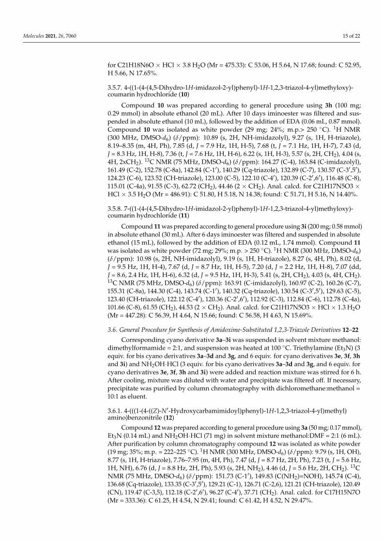
Molecules 2021, 26, 7060 15 of 22
for C21H18N6O × HCl × 3.8 H2O (Mr = 475.33): C 53.06, H 5.64, N 17.68; found: C 52.95,H 5.66, N 17.65%.
3.5.7. 4-((1-(4-(4,5-Dihydro-1H-imidazol-2-yl)phenyl)-1H-1,2,3-triazol-4-yl)methyloxy)-coumarin hydrochloride (10)
Compound 10 was prepared according to general procedure using 3h (100 mg;0.29 mmol) in absolute ethanol (20 mL). After 10 days iminoester was filtered and sus-pended in absolute ethanol (10 mL), followed by the addition of EDA (0.06 mL, 0.87 mmol).Compound 10 was isolated as white powder (29 mg; 24%; m.p.> 250 ◦C). 1H NMR(300 MHz, DMSO-d6) (δ/ppm): 10.89 (s, 2H, NH-imidazolyl), 9.27 (s, 1H, H-triazole),8.19–8.35 (m, 4H, Ph), 7.85 (d, J = 7.9 Hz, 1H, H-5), 7.68 (t, J = 7.1 Hz, 1H, H-7), 7.43 (d,J = 8.3 Hz, 1H, H-8), 7.36 (t, J = 7.6 Hz, 1H, H-6), 6.22 (s, 1H, H-3), 5.57 (s, 2H, CH2), 4.04 (s,4H, 2xCH2). 13C NMR (75 MHz, DMSO-d6) (δ/ppm): 164.27 (C-4), 163.84 (C-imidazolyl),161.49 (C-2), 152.78 (C-8a), 142.84 (C-1′), 140.29 (Cq-triazole), 132.89 (C-7), 130.57 (C-3′,5′),124.23 (C-6), 123.52 (CH-triazole), 123.00 (C-5), 122.10 (C-4′), 120.39 (C-2′,6′), 116.48 (C-8),115.01 (C-4a), 91.55 (C-3), 62.72 (CH2), 44.46 (2 × CH2). Anal. calcd. for C21H17N5O3 ×HCl × 3.5 H2O (Mr = 486.91): C 51.80, H 5.18, N 14.38; found: C 51.71, H 5.16, N 14.40%.
3.5.8. 7-((1-(4-(4,5-Dihydro-1H-imidazol-2-yl)phenyl)-1H-1,2,3-triazol-4-yl)methyloxy)-coumarin hydrochloride (11)
Compound 11 was prepared according to general procedure using 3i (200 mg; 0.58 mmol)in absolute ethanol (30 mL). After 6 days iminoester was filtered and suspended in absoluteethanol (15 mL), followed by the addition of EDA (0.12 mL, 1.74 mmol). Compound 11was isolated as white powder (72 mg; 29%; m.p. > 250 ◦C). 1H NMR (300 MHz, DMSO-d6)(δ/ppm): 10.98 (s, 2H, NH-imidazolyl), 9.19 (s, 1H, H-triazole), 8.27 (s, 4H, Ph), 8.02 (d,J = 9.5 Hz, 1H, H-4), 7.67 (d, J = 8.7 Hz, 1H, H-5), 7.20 (d, J = 2.2 Hz, 1H, H-8), 7.07 (dd,J = 8.6, 2.4 Hz, 1H, H-6), 6.32 (d, J = 9.5 Hz, 1H, H-3), 5.41 (s, 2H, CH2), 4.03 (s, 4H, CH2).13C NMR (75 MHz, DMSO-d6) (δ/ppm): 163.91 (C-imidazolyl), 160.97 (C-2), 160.26 (C-7),155.31 (C-8a), 144.30 (C-4), 143.74 (C-1′), 140.32 (Cq-triazole), 130.54 (C-3′,5′), 129.63 (C-5),123.40 (CH-triazole), 122.12 (C-4′), 120.36 (C-2′,6′), 112.92 (C-3), 112.84 (C-6), 112.78 (C-4a),101.66 (C-8), 61.55 (CH2), 44.53 (2 × CH2). Anal. calcd. for C21H17N5O3 × HCl × 1.3 H2O(Mr = 447.28): C 56.39, H 4.64, N 15.66; found: C 56.58, H 4.63, N 15.69%.
3.6. General Procedure for Synthesis of Amidoxime-Substituted 1,2,3-Triazole Derivatives 12–22
Corresponding cyano derivative 3a–3i was suspended in solvent mixture methanol:dimethylformamide = 2:1, and suspension was heated at 100 ◦C. Triethylamine (Et3N) (3equiv. for bis cyano derivatives 3a–3d and 3g, and 6 equiv. for cyano derivatives 3e, 3f, 3hand 3i) and NH2OH·HCl (3 equiv. for bis cyano derivatives 3a–3d and 3g, and 6 equiv. forcyano derivatives 3e, 3f, 3h and 3i) were added and reaction mixture was stirred for 6 h.After cooling, mixture was diluted with water and precipitate was filtered off. If necessary,precipitate was purified by column chromatography with dichloromethane:methanol =10:1 as eluent.
3.6.1. 4-(((1-(4-((Z)-N′-Hydroxycarbamimidoyl)phenyl)-1H-1,2,3-triazol-4-yl)methyl)amino)benzonitrile (12)
Compound 12 was prepared according to general procedure using 3a (50 mg; 0.17 mmol),Et3N (0.14 mL) and NH2OH·HCl (71 mg) in solvent mixture methanol:DMF = 2:1 (6 mL).After purification by column chromatography compound 12 was isolated as white powder(19 mg; 35%; m.p. = 222–225 ◦C). 1H NMR (300 MHz, DMSO-d6) (δ/ppm): 9.79 (s, 1H, OH),8.77 (s, 1H, H-triazole), 7.76–7.95 (m, 4H, Ph), 7.47 (d, J = 8.7 Hz, 2H, Ph), 7.23 (t, J = 5.6 Hz,1H, NH), 6.76 (d, J = 8.8 Hz, 2H, Ph), 5.93 (s, 2H, NH2), 4.46 (d, J = 5.6 Hz, 2H, CH2). 13CNMR (75 MHz, DMSO-d6) (δ/ppm): 151.73 (C-1′), 149.83 (C(NH2)=NOH), 145.74 (C-4),136.68 (Cq-triazole), 133.35 (C-3′,5′), 129.21 (C-1), 126.71 (C-2,6), 121.21 (CH-triazole), 120.49(CN), 119.47 (C-3,5), 112.18 (C-2′,6′), 96.27 (C-4′), 37.71 (CH2). Anal. calcd. for C17H15N7O(Mr = 333.36): C 61.25, H 4.54, N 29.41; found: C 61.42, H 4.52, N 29.47%.
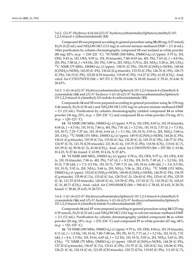
Molecules 2021, 26, 7060 16 of 22
3.6.2. (Z)-N′-Hydroxy-4-(4-((4-((Z)-N′-hydroxycarbamimidoyl)phenoxy)methyl)-1H-1,2,3-triazol-1-yl)benzimidamide (13)
Compound 13 was prepared according to general procedure using 3b (80 mg; 0.27 mmol),Et3N (0.22 mL) and NH2OH·HCl (113 mg) in solvent mixture methanol:DMF = 2:1 (6 mL).After purification by column chromatography compound 13 was isolated as white powder(80 mg; 82%; m.p. = 218–221 ◦C). 1H NMR (300 MHz, DMSO-d6) (δ/ppm): 9.75 (s, 1H,OH), 9.43 (s, 1H, OH), 8.95 (s, 1H, H-triazole), 7.80–8.03 (m, 4H, Ph), 7.63 (d, J = 8.8 Hz,2H, Ph), 7.08 (d, J = 8.8 Hz, 2H, Ph), 5.89 (s, 2H, NH2), 5.70 (s, 2H, NH2), 5.28 (s, 2H, CH2).13C NMR (75 MHz, DMSO-d6) (δ/ppm): 158.51 (C-Ph), 150.55 (C(NH2)=NOH), 149.84(C(NH2)=NOH), 143.83 (C-Ph), 136.62 (Cq-triazole), 133.52 (C-Ph), 126.76 (C-Ph), 126.73(C-Ph), 126.15 (C-Ph), 122.82 (CH-triazole), 119.69 (C-Ph), 114.27 (C-Ph), 61.02 (CH2). Anal.calcd. for C17H17N7O3 (Mr = 367.37): C 55.58, H 4.66, N 26.69; found: C 55.41, H 4.66, N26.63%.
3.6.3. 1-((1-(4-((Z)-N′-Hydroxycarbamimidoyl)phenyl)-1H-1,2,3-triazol-4-yl)methyl)-4-cyanoindole (14) and (Z)-N′-hydroxy-1-((1-(4-((Z)-N′-hydroxycarbamimidoyl)phenyl)-1H-1,2,3-triazol-4-yl)methyl)-1H-indole-4-carboximidamide (15)
Compounds 14 and 15 were prepared according to general procedure using 3c (150 mg;0.46 mmol), Et3N (0.38 mL) and NH2OH·HCl (192 mg) in solvent mixture methanol:DMF= 2:1 (12 mL). Purification by column chromatography yielded compound 14 as whitepowder (36 mg; 22%; m.p. = 229–232 ◦C) and compound 15 as white powder (74 mg; 41%;m.p. = 120–123 ◦C).
14: 1H NMR (300 MHz, DMSO-d6) (δ/ppm): 9.79 (s, 1H, OH), 8.83 (s, 1H, H-triazole),8.04 (d, J = 8.3 Hz, 1H, H-5), 7.86 (s, 4H, Ph), 7.81 (d, J = 3.2 Hz, 1H, H-2), 7.57 (d, J = 7.3 Hz,1H, H-7), 7.25–7.37 (m, 1H, H-6), 6.64 (d, J = 3.1 Hz, 1H, H-3), 5.93 (s, 2H, NH2), 5.68 (s,2H, CH2). 13C NMR (151 MHz, DMSO-d6) (δ/ppm): 149.93 (C(NH2)=NOH), 144.26 (C-Ph),136.61 (Cq-triazole), 135.39 (C-7a), 133.56 (C-3a), 132.20 (C-2), 129.15 (C-Ph), 126.77 (C-Ph),124.97 (C-5), 121.76 (CH-triazole), 121.36 (C-6), 119.75 (C-Ph), 118.56 (CN), 115.81 (C-7),101.59 (C-4), 99.56 (C-3), 41.04 (CH2). Anal. calcd. for C19H15N7O (Mr = 357.38): C 63.86,H 4.23, N 27.44; found: C 63.89, H 4.24, N 27.42%.
15: 1H NMR (600 MHz, DMSO-d6) (δ/ppm): 9.78 (s, 1H, OH), 9.57 (s, 1H, OH), 8.82(s, 1H, H-triazole), 7.86 (s, 4H, Ph), 7.67 (d, J = 8.2 Hz, 1H, H-5), 7.49 (d, J = 3,2 Hz, 1H,H-2), 7.30 (dd, J = 7.3, 0.5 Hz, 1H, H-7), 7.09–7.21 (m, 1H, H-6), 6.88 (dd, J = 3.0, 0.5 Hz,1H, H-3), 5.92 (s, 2H, NH2), 5.68 (s, 2H, NH2), 5.56 (s, 2H, CH2). 13C NMR (151 MHz,DMSO-d6) (δ/ppm): 152.02 (C(NH2)=NOH), 149.80 (C(NH2)=NOH), 144.59 (C-Ph), 136.55(Cq-triazole), 135.89 (C-7a), 133.42 (C-3a), 128.70 (C-2), 126.65 (C-Ph), 125.61 (C-Ph), 125.55(C-4), 121.53 (CH-triazole), 120.69 (C-6), 119.59 (C-Ph), 117.65 (C-7), 110.78 (C-5), 102.63(C-3), 40.71 (CH2). Anal. calcd. for C19H18N8O2 (Mr = 390.41): C 58.45, H 4.65, N 28.70;found: C 58.40, H 4.65, N 28.72%.
3.6.4. 1-((1-(4-((Z)-N′-Hydroxycarbamimidoyl)phenyl)-1H-1,2,3-triazol-4-yl)methyl)-5-cyanoindole (16) and (Z)-N′-hydroxy-1-((1-(4-((Z)-N′-hydroxycarbamimidoyl)phenyl)-1H-1,2,3-triazol-4-yl)methyl)-indole-5-carboximidamide (17)
Compounds 16 and 17 were prepared according to general procedure using 3d (125 mg;0.39 mmol), Et3N (0.32 mL) and NH2OH·HCl (163 mg) in solvent mixture methanol:DMF= 2:1 (12 mL). Purification by column chromatography yielded compound 16 as whitepowder (26 mg; 18%; m.p. = 232–234 ◦C) and compound 17 as white powder (67 mg; 44%;m.p. = 152–155 ◦C).
16: 1H NMR (300 MHz, DMSO-d6) (δ/ppm): 9.79 (s, 1H, OH), 8.84 (s, 1H, H-triazole),8.11 (d, J = 1.0 Hz, 1H, H-4), 7.80–7.88 (m, 5H, Ph, H-7), 7.71 (d, J = 3.2 Hz, 1H, H-2), 7.51(dd, J = 8.6, 1.5 Hz, 1H, H-6), 6,65 (d, J = 3.2 Hz, 1H, H-3), 5,92 (s, 2H, NH2), 5,65 (s, 2H,CH2). 13C NMR (75 MHz, DMSO-d6) (δ/ppm): 150.03 (C(NH2)=NOH), 144.28 (C-Ph),137.32 (Cq-triazole), 136.67 (C-7a), 133.61 (C-Ph), 131.57 (C-2), 128.18 (C-3a), 126.86 (C-Ph),126.21 (C-4), 124.14 (C-6), 121.85 (CH-triazole), 120.72 (CN), 119.83 (C-Ph), 111.65 (C-7),
Molecules 2021, 26, 7060 17 of 22
102.51 (C-3), 101.50 (C-5), 40.98 (CH2). Anal. calcd. for C19H15N7O (Mr = 357.38): C 63.86,H 4.23, N 27.44; found: C 63.79, H 4.24, N 27.51%.
17: 1H NMR (600 MHz, DMSO-d6) (δ/ppm): 9.78 (s, 1H, OH), 9.39 (s, 1H, OH), 8.83 (s,1H, H-triazole), 7.71–7.95 (m, 5H), 7.60 (d, J = 8.7 Hz, 1H, 2H), 7.43–7.54 (m, 2H, H-4, H-6),6.38–6.55 (m, 1H, H-3), 5.91 (s, 2H, NH2), 5.72 (s, 2H, NH2), 5.56 (s, 2H, CH2). 13C NMR(75 MHz, DMSO-d6) (δ/ppm): 151.91 (C(NH2)=NOH), 149.81 (C(NH2)=NOH), 144.59(C-Ph), 136.57 (Cq-triazole), 135.90 (C-7a), 133.45 (C-Ph), 129.31 (C-2), 127.77 (C-3a), 126.68(C-Ph), 124.78 (C-5), 121.58 (CH-triazole), 119.61 (C-Ph), 119.26 (C-4), 117.77 (C-6), 109.68(C-7), 101.71 (C-3), 40.78 (CH2). Anal. calcd. for C19H18N8O2 (Mr = 390.41): C 58.45, H4.65, N 28.70; found: C 58.51, H 4.66, N 28.61%.
3.6.5. 8-(((1-(4-((Z)-N′-Hydroxycarbamimidoyl)phenyl)-1H-1,2,3-triazol-4-yl)methyl)amino)quinoline (18)
Compound 18 was prepared according to general procedure using 3e (80 mg; 0.25 mmol),Et3N (0.10 mL) and NH2OH·HCl (51 mg) in solvent mixture methanol:DMF = 2:1 (9 mL).After purification by column chromatography, compound 18 was isolated as white powder(35 mg; 40%; m.p. = 181–183 ◦C). 1H NMR (300 MHz, DMSO-d6) (δ/ppm): 9.78 (s, 1H,OH), 8.77 (s, 1H, H-triazole), 8.22 (dd, J = 8.3, 1.6 Hz, 1H, H-2), 7.80–7.97 (m, 5H, Ph, H-4),7.51 (dd, J = 8.3, 4.2 Hz, 1H, H-3), 7.37 (t, J = 7.9 Hz, 1H, H-6), 7.11 (d, J = 8.1 Hz, 1H,H-5), 7.02 (t, J = 5.8 Hz, 1H, NH), 6.82 (d, J = 7.0 Hz, 1H, H-7), 5.92 (s, 2H, NH2), 4.67 (d,J = 6.0 Hz, 2H, CH2). 13C NMR (75 MHz, DMSO-d6) (δ/ppm): 150.00 (C(NH2)=NOH),147.19 (C-2), 146.62 (C-8), 144.01 (C-1′), 137.66 (Cq-triazole), 136.84 (C-4a), 136.07 (C-4),133.36 (C-1′), 128.33 (C-8a), 127.80 (C-6), 126.80 (C-3′,5′), 121.88 (C-3), 121.09 (CH-triazole),119.63 (C-2′,6′), 114.00 (C-5), 105.02 (C-7), 35.90 (CH2). Anal. calcd. for C19H17N7O (Mr =359.39): C 63.50, H 4.77, N 27.28; found: C 63.45, H 4.76, N 27.27%.
3.6.6. 8-((1-(4-((Z)-N′-Hydroxycarbamimidoyl)phenyl)-1H-1,2,3-triazol-4-yl)methoxy)quinoline (19)
Compound 19 was prepared according to general procedure using 3f (90 mg; 0.28 mmol),Et3N (0.12 mL) and NH2OH·HCl (58 mg) in solvent mixture methanol: DMF = 2:1 (9 mL).Compound 19 was isolated as white powder (78 mg; 79%; m.p. = 230–232 ◦C). 1H NMR(600 MHz, DMSO-d6) (δ/ppm): 9.81 (s, 1H, OH), 9.06 (s, 1H, H-triazole), 8.84 (dd, J = 4.1,1.7 Hz, 1H, H-2), 8.33 (dd, J = 8.3, 1.6 Hz, 1H, H-4), 7.88–7.99 (m, 4H, Ph), 7.50–7.59 (m, 3H,H-3, H-5, H-6), 7.46 (dd, J = 6.3, 2.7 Hz, 1H, H-7), 5.95 (s, 2H, NH2), 5.46 (s, 2H, CH2). 13CNMR (151 MHz, DMSO-d6) (δ/ppm): 153.78 (C-8), 149.84 (C(NH2)=NOH), 149.02 (C-2),143.80 (C-Ph), 139.74 (Cq-triazole), 136.65 (C-4a), 135.81 (C-4), 133.54 (C-Ph), 129.21 (C-6),129.08 (C-8a), 126.74 (C-Ph), 123.13 (CH-triazole), 121.88 (C-3), 120.21 (C-5), 119.71 (C-Ph),110.24 (C-7), 61.74 (CH2). Anal. calcd. for C19H16N6O2 (Mr = 360.38): C 63.33, H 4.48, N23.32; found: C 63.21, H 4.49, N 23.33%.
3.6.7. (Z)-N′-Hydroxy-8-((1-(4-((Z)-N′-hydroxycarbamimidoyl)phenyl)-1H-1,2,3-triazol-4-yl)methoxy)quinoline-2-carboximidamide (20)
Compound 20 was prepared according to general procedure using 3g (80 mg; 0.23 mmol),Et3N (0.19 mL) and NH2OH·HCl (95 mg) in solvent mixture methanol: DMF = 2:1 (9 mL).Compound 20 was isolated as white powder (84 mg; 87%; m.p. = 238–241 ◦C). 1H NMR(600 MHz, DMSO-d6) (δ/ppm): 10.17 (s, 1H, OH), 9.81 (s, 1H, OH), 9.07 (s, 1H, H-triazol),8.31 (d, J = 8.7 Hz, 1H), 7.99 (d, J = 8.6 Hz, 1H), 7.89–7.98 (m, 4H, Ph), 7.48–7.60 (m, 3H),5.80–6.23 (m, 4H, 2 × NH2), 5,53 (s, 2H, CH2). 13C NMR (75 MHz, DMSO-d6) (δ/ppm):153.58 (C-8), 149.85 (C(NH2)=NOH), 149.57 (C(NH2)=NOH), 148.58 (C-2), 144.07 (C-Ph),138.30 (C-triazol), 136.65 (C-4a), 136.27 (C-4), 133.49 (C-Ph), 129.16 (C-8a), 127.18 (C-6),126.74 (C-Ph), 122.90 (CH-triazol), 120.30 (C-3), 119.67 (C-Ph), 117.71 (C-5), 111.82 (C-7),62.39 (CH2). Anal. calcd. for C20H18N8O3 (Mr = 418.42): C 57.41, H 4.34, N 26.78; found:C 57.50, H 4.34, N 26.74%.
Molecules 2021, 26, 7060 18 of 22
3.6.8. 4-((1-(4-((Z)-N′-Hydroxycarbamimidoyl)phenyl)-1H-1,2,3-triazol-4-yl)methoxy)coumarin (21)
Compound 21 was prepared according to general procedure using 3h (100 mg;0.29 mmol), Et3N (0.12 mL) and NH2OH·HCl (60 mg) in solvent mixture methanol:DMF= 2:1 (9 mL). Compound 21 was isolated as white powder (75 mg; 69%; m.p. = 235–237◦C). 1H NMR (300 MHz, DMSO-d6) (δ/ppm): 9.81 (s, 1H, OH), 9.11 (s, 1H, H-triazole),7.94 (q, J = 8.9 Hz, 4H, Ph), 7,85 (d, J = 7,8 Hz, 1H, H-5), 7,61–7,72 (m, 1H, H-7), 7,42 (d,J = 8,2 Hz, 1H, H-8), 7.35 (t, J = 7.6 Hz, 1H, H-6), 6.21 (s, 1H, H-3), 5.96 (s, 2H, NH2), 5.53(s, 2H, CH2). 13C NMR (151 MHz, DMSO-d6) (δ/ppm): 164.32 (C-4), 161.50 (C-2), 152.76(C-8a), 149.79 (C(NH2)=NOH), 142.33 (C-triazole), 136.54 (C-Ph), 133.63 (C-Ph), 132.84(C-7), 126.71 (C-Ph), 124.210 (C-6), 123.26 (CH-triazole), 123.04 (C-5), 119.78 (C-Ph), 116.43(C-8), 115.02 (C-4a), 91.46 (C-3), 62.81 (CH2). Anal. calcd. for C19H15N5O4 (Mr = 377.36):C 60.48, H 4.01, N 18.56; found: C 60.39, H 4.01, N 18.59%.
3.6.9. 7-((1-(4-((Z)-N′-Hydroxycarbamimidoyl)phenyl)-1H-1,2,3-triazol-4-yl)methoxy)coumarin (22)
Compound 22 was prepared according to general procedure using 3i (100 mg; 0.29 mmol),Et3N (0.12 mL) and NH2OH·HCl (60 mg) in solvent mixture methanol: DMF = 2:1 (9 mL).Compound 22 was isolated as white powder (68 mg; 62%; m.p. = 232–235 ◦C). 1H NMR(300 MHz, DMSO-d6) (δ/ppm): 9.81 (s, 1H, OH), 9.03 (s, 1H, H-triazole), 8.01 (d, J = 9.5 Hz,1H, H-4), 7.91 (q, J = 9.0 Hz, 4H, Ph), 7.67 (d, J = 8.7 Hz, 1H, H-5), 7.21 (d, J = 2.4 Hz, 1H,H-8), 7.07 (dd, J = 8.6, 2.4 Hz, 1H, H-6), 6.32 (d, J = 9.5 Hz, 1H, H-3), 5.95 (s, 2H, NH2), 5.37(s, 3H, CH2). 13C NMR (151 MHz, DMSO-d6) (δ/ppm): 161.04 (C-2), 160.25 (C-7), 155.31(C-8a), 149.86 (C(NH2)=NOH), 144.29 (C-4), 143.24 (C-Ph), 136.60 (Cq-triazole), 133.59(C-Ph), 129.58 (C-5), 126.76 (C-Ph), 123.09 (CH-triazole), 119.77 (C-Ph), 112.92 (C-3), 112.78(C-6), 112.72 (C-4a), 101.66 (C-8), 61.62 (CH2). Anal. calcd. for C19H15N5O4 (Mr = 377.36):C 60.48, H 4.01, N 18.56; found: C 60.54, H 4.01, N 18.50%.
3.7. Cell Culturing
The cell lines HeLa (cervical carcinoma), SW620 (colorectal adenocarcinoma, metastatic),A549 (lung adenocarcinoma), HepG2 (hepatocellular carcinoma) and HFF (human foreskinfibroblasts) were cultured as monolayers and maintained in Dulbecco’s modified Eaglemedium (DMEM), supplemented with 10% fetal bovine serum (FBS), 2mM L-glutamine,100 U/mL penicillin and 100 µg/mL streptomycin in a humidified atmosphere with 5%CO2 at 37 ◦C.
3.8. Proliferation Assays
Cells were seeded onto 96-well microtiter plates at a seeding density of 3000 cells/wellfor carcinoma cell lines, and 5000 cells/well for normal human fibroblasts. The next day,cells were treated with test agents at five different concentrations (0.01–100 µM) and furtherincubated for 72 h. DMSO (solvent) was tested for potential cytotoxic effect but it did notexceed 0.1%. Following 72 h incubation, the MTT assay was performed and measuredabsorbances were transformed into percentage of cell growth as described previously [48].Results were obtained from three independent experiments. IC50 values were calculatedusing linear regression analysis and results were statistically analyzed by ANOVA, Tukeypost hoc test (p < 0.05).
3.9. DNA Binding Study
Compound solutions were used for measurements in aqueous buffer (pH = 7, sodiumcacodylate buffer, I = 0.05 mol dm−3). Polynucleotides were purchased as noted: polyA-poly U, poly (dAdT)2 and calf thymus (ct)DNA (Sigma-Aldrich). Polynucleotides weredissolved in Na-cacodylate buffer, I = 0.05 mol dm−3, pH = 7. The calf thymus ctDNAwas additionally sonicated and filtered through a 0.45 mm filter [49]. Polynucleotideconcentration was determined spectroscopically [50,51] as the concentration of phosphates.

Molecules 2021, 26, 7060 19 of 22
3.9.1. UV–Vis Measurements
UV–Vis spectra were recorded on a Varian Cary 100 Bio spectrophotometer using1cm path quartz cuvettes. Calibration experiments were performed at 25 ◦C and pH = 7(I = 0.05 mol dm−3, sodium cacodylate buffer). Thermal melting curves for DNA andtheir complexes with studied compounds were determined as previously described byfollowing the absorption change at 260 nm as a function of temperature. The absorbanceof the ligands was subtracted from every curve and the absorbance scale was normalized.Tm values are the midpoints of the transition curves determined from the maximum ofthe first derivative and checked graphically by the tangent method. The ∆Tm values werecalculated by subtracting the Tm of the free nucleic acid from the Tm of the complex. Every∆Tm value reported here was the average of at least two measurements. The error in ∆Tmis ±0.5 ◦C.
3.9.2. Fluorometric Measurements
Fluorescence spectra were recorded on a Varian Cary Eclipse spectrophotometerat 25 ◦C using appropriate 1cm path quartz cuvettes. Fluorometric experiments wereperformed at pH = 7 (I = 0.05 mol dm−3, sodium cacodylate buffer) by adding portionsof polynucleotide solution into the solution of the studied compound. In fluorometricexperiments, an excitation wavelength of λexc ≥ 300 nm was used to avoid the innerfilter effect caused due to increasing absorbance of the polynucleotide. Emissions weredetermined in the range λem = 300–600 nm. Values for Ks obtained by processing titrationdata using the Scatchard equation [40], in most titrations have satisfactory correlationcoefficients (≥0.98).
3.9.3. CD Measurements
CD spectra were recorded on a JASCO J815 spectrophotometer in 1cm path quartzcuvettes. CD parameters: range = 500–220 nm, data pitch = 2, standard sensitivity, scanningspeed = 200 nm/min, accumulation = 3–5. CD experiments were performed at 25 ◦C andpH = 7 (I = 0.05 mol dm−3, sodium cacodylate buffer) by adding portions of compoundstock solution into the polynucleotide solution.
4. Conclusions
The novel 1,2,3-triazolyl-appended indole, quinoline and coumarin heterocycles con-taining aryl imidazoline 4–11 and amidoxime 12–22 moiety substituted at the variousposition of heterocycle or connected via nitrogen- and oxygen-containing linkers weresynthesized with the aim to assess the influence of performed structural modification onantiproliferative activity.
Antiproliferative evaluations on hepatocellular carcinoma (HepG2), showed thatasymmetrical aromatic diamidine 5 and coumarin amidine 11 were the most potent com-pounds, with IC50 values in the nM range. However, 5 was also highly cytotoxic to normalHFF fibroblasts, while 11 exhibited less toxicity to non-tumor cells with a selectivity index(SI) of 11. Quinoline amidine 8 with -NCH2- spacer exhibited marked and selective effectsagainst HepG2. Its analogue 9 with -OCH2- unit significantly inhibited HFF fibroblasts.From the amidoxime series, only quinoline amidoximes 18 and 20 showed a strong antipro-liferative effect on A549, HeLa and SW620. Bis-amidoxime moiety in 20 contributed toits better activity compared to mono-amidoxime analogue 19. Based on the results of CDtitrations and thermal melting experiments, it can be concluded that 5 and 10 most likelybind inside the minor groove of AT-DNA and intercalate into AU-RNA. 6, 9 and 11 bind toAT-DNA with mixed binding mode, most probably minor groove binding accompaniedwith aggregate binding along the DNA backbone.
Supplementary Materials: The following are available online. UV–VIS spectra, fluorescence spectra,1H and 13C NMR spectra.

Molecules 2021, 26, 7060 20 of 22
Author Contributions: Conceptualization, S.R.-M. and M.R.S.; Methodology, S.M., P.G. and S.S.;Investigation, S.M., P.G. and S.S.; Writing—Original Draft Preparation, S.R.-M., S.M. and M.R.S.;Writing—Review and Editing, S.M., P.G., S.S., M.R.S., K.P., M.S., S.K.P. and S.R.-M.; Visualization, S.M.and S.R.-M.; Supervision, M.R.S., K.P., M.S., S.K.P. and S.R.-M.; Project Administration, S.R.-M. andM.R.S.; Funding Acquisition, S.R.-M. and M.R.S. All authors have read and agreed to the publishedversion of the manuscript.
Funding: This research was funded by Croatian Science Foundation, grant numbers IP-2018-01-4682and IP-2018-01-4694.
Data Availability Statement: Data is contained within the article and Supplementary Materials.
Conflicts of Interest: The authors declare no conflict of interest.
References1. Akhtar, J.; Khan, A.A.; Ali, Z.; Haider, R.; Shahar Yar, M. Structure-activity relationship (SAR) study and design strategies
of nitrogen-containing heterocyclic moieties for their anticancer activities. Eur. J. Med. Chem. 2017, 125, 143–189. [CrossRef][PubMed]
2. Singh, P.K.; Silakari, O. The Current Status of O-Heterocycles: A Synthetic and Medicinal Overview. ChemMedChem 2018, 13,1071–1087. [CrossRef] [PubMed]
3. Zhang, L.; Xu, Z. Coumarin-containing hybrids and their anticancer activities. Eur. J. Med. Chem. 2019, 181, 111587. [CrossRef][PubMed]
4. Singla, P.; Luxami, V.; Paul, K. Benzimidazole-biologically attractive scaffold for protein kinase inhibitors. RSC Adv. 2014, 4,12422–12440. [CrossRef]
5. Carneiro, A.; Matos, M.J.; Uriarte, E.; Santana, L. Trending topics on coumarin and its derivatives in 2020. Molecules 2021, 26, 501.[CrossRef] [PubMed]
6. Dhuguru, J.; Skouta, R. Role of Indole Scaffolds as Pharmacophores in the Development of Anti-Lung Cancer Agents. Molecules2020, 25, 1615. [CrossRef]
7. Mešcic Macan, A.; Perin, N.; Jakopec, S.; Mioc, M.; Stojkovic, M.R.; Kralj, M.; Hranjec, M.; Raic-Malic, S. Synthesis, antiproliferativeactivity and DNA/RNA-binding properties of mono- and bis-(1,2,3-triazolyl)-appended benzimidazo[1,2-a]quinoline derivatives.Eur. J. Med. Chem. 2020, 185, 111845. [CrossRef] [PubMed]
8. Bistrovic, A.; Krstulovic, L.; Harej, A.; Grbcic, P.; Sedic, M.; Koštrun, S.; Pavelic, S.K.; Bajic, M.; Raic-Malic, S. Design, synthesisand biological evaluation of novel benzimidazole amidines as potent multi-target inhibitors for the treatment of non-small celllung cancer. Eur. J. Med. Chem. 2018, 143, 1616–1634. [CrossRef] [PubMed]
9. Bistrovic, A.; Harej, A.; Grbcic, P.; Sedic, M.; Pavelic, S.K.; Cetina, M.; Raic-Malic, S. Synthesis and anti-proliferative effects ofmono-and bis-purinomimetics targeting Kinases. Int. J. Mol. Sci. 2017, 18, 2292. [CrossRef]
10. Martorana, A.; la Monica, G.; Lauria, A. Quinoline-based molecules targeting c-Met, EGF, and VEGF receptors and the proteinsinvolved in related carcinogenic pathways. Molecules 2020, 25, 4279. [CrossRef]
11. Wan, Y.; Li, Y.; Yan, C.; Yan, M.; Tang, Z. Indole: A privileged scaffold for the design of anti-cancer agents. Eur. J. Med. Chem. 2019,183, 111691. [CrossRef] [PubMed]
12. Dadashpour, S.; Emami, S. Indole in the target-based design of anticancer agents: A versatile scaffold with diverse mechanisms.Eur. J. Med. Chem. 2018, 150, 9–29. [CrossRef] [PubMed]
13. Al-Warhi, T.; Sabt, A.; Elkaeed, E.B.; Eldehna, W.M. Recent advancements of coumarin-based anticancer agents: An up-to-datereview. Bioorg. Chem. 2020, 103, 104163. [CrossRef] [PubMed]
14. Fotopoulos, I.; Hadjipavlou-Litina, D. Hybrids of Coumarin Derivatives as Potent and Multifunctional Bioactive Agents: AReview. Med. Chem. 2019, 16, 272–306. [CrossRef] [PubMed]
15. Xu, Z.; Zhao, S.J.; Liu, Y. 1,2,3-Triazole-containing hybrids as potential anticancer agents: Current developments, actionmechanisms and structure-activity relationships. Eur. J. Med. Chem. 2019, 183, 111700. [CrossRef] [PubMed]
16. Cai, M.; Hu, J.; Tian, J.L.; Yan, H.; Zheng, C.G.; Hu, W. Le Novel hybrids from N-hydroxyarylamide and indole ring through clickchemistry as histone deacetylase inhibitors with potent antitumor activities. Chin. Chem. Lett. 2015, 26, 675–680. [CrossRef]
17. Narsimha, S.; Satheesh Kumar, N.; Kumara Swamy, B.; Vasudeva Reddy, N.; Althaf Hussain, S.K.; Srinivasa Rao, M. Indole-2-carboxylic acid derived mono and bis 1,4-disubstituted 1,2,3-triazoles: Synthesis, characterization and evaluation of anticancer,antibacterial, and DNA-cleavage activities. Bioorg. Med. Chem. Lett. 2016, 26, 1639–1644. [CrossRef]
18. Aneja, B.; Arif, R.; Perwez, A.; Napoleon, J.V.; Hasan, P.; Rizvi, M.M.A.; Azam, A.; Rahisuddin; Abid, M. N-Substituted1,2,3-Triazolyl-Appended Indole-Chalcone Hybrids as Potential DNA Intercalators Endowed with Antioxidant and AnticancerProperties. ChemistrySelect 2018, 3, 2638–2645. [CrossRef]
19. Praveena, K.S.S.; Shivaji Ramarao, E.V.V.; Murthy, N.Y.S.; Akkenapally, S.; Ganesh Kumar, C.; Kapavarapu, R.; Pal, S. Design ofnew hybrid template by linking quinoline, triazole and dihydroquinoline pharmacophoric groups: A greener approach to novelpolyazaheterocycles as cytotoxic agents. Bioorg. Med. Chem. Lett. 2015, 25, 1057–1063. [CrossRef]
Molecules 2021, 26, 7060 21 of 22
20. De O Freitas, L.B.; Borgati, T.F.; De Freitas, R.P.; Ruiz, A.L.T.G.; Marchetti, G.M.; De Carvalho, J.E.; Da Cunha, E.F.F.; Ramalho,T.C.; Alves, R.B. Synthesis and antiproliferative activity of 8-hydroxyquinoline derivatives containing a 1,2,3-triazole moiety. Eur.J. Med. Chem. 2014, 84, 595–604. [CrossRef] [PubMed]
21. Liu, M.; Hou, Y.; Yin, W.; Zhou, S.; Qian, P.; Guo, Z.; Xu, L.; Zhao, Y. Discovery of a novel 6,7-disubstituted-4-(2-fluorophenoxy)quinolines bearing 1,2,3-triazole-4-carboxamide moiety as potent c-Met kinase inhibitors. Eur. J. Med.Chem. 2016, 119, 96–108. [CrossRef] [PubMed]
22. Akkol, E.K.; Genç, Y.; Karpuz, B.; Sobarzo-Sánchez, E.; Capasso, R. Coumarins and coumarin-related compounds in pharma-cotherapy of cancer. Cancers 2020, 12, 1959. [CrossRef]
23. Peterson, L.B.; Blagg, B.S.J. Click chemistry to probe Hsp90: Synthesis and evaluation of a series of triazole-containing novobiocinanalogues. Bioorg. Med. Chem. Lett. 2010, 20, 3957–3960. [CrossRef] [PubMed]
24. Yu, H.; Hou, Z.; Tian, Y.; Mou, Y.; Guo, C. Design, synthesis, cytotoxicity and mechanism of novel dihydroartemisinin-coumarinhybrids as potential anti-cancer agents. Eur. J. Med. Chem. 2018, 151, 434–449. [CrossRef] [PubMed]
25. Pingaew, R.; Saekee, A.; Mandi, P.; Nantasenamat, C.; Prachayasittikul, S.; Ruchirawat, S.; Prachayasittikul, V. Synthesis, biologicalevaluation and molecular docking of novel chalcone-coumarin hybrids as anticancer and antimalarial agents. Eur. J. Med. Chem.2014, 85, 65–76. [CrossRef] [PubMed]
26. Kraljevic, T.G.; Harej, A.; Sedic, M.; Pavelic, S.K.; Stepanic, V.; Drenjancevic, D.; Talapko, J.; Raic-Malic, S. Synthesis, in vitroanticancer and antibacterial activities and in silico studies of new 4-substituted 1,2,3-triazole–coumarin hybrids. Eur. J. Med.Chem. 2016, 124, 794–808. [CrossRef] [PubMed]
27. An, R.; Hou, Z.; Li, J.T.; Yu, H.N.; Mou, Y.H.; Guo, C. Design, synthesis and biological evaluation of novel 4-substituted coumarinderivatives as antitumor agents. Molecules 2018, 23, 2281. [CrossRef] [PubMed]
28. Shakeel-u-Rehman; Masood-ur-Rahman; Tripathi, V.K.; Singh, J.; Ara, T.; Koul, S.; Farooq, S.; Kaul, A. Synthesis and biologicalevaluation of novel isoxazoles and triazoles linked 6-hydroxycoumarin as potent cytotoxic agents. Bioorg. Med. Chem. Lett. 2014,24, 4243–4246. [CrossRef] [PubMed]
29. Demeunynck, M.; Bailly, C.; Wilson, W.D. Small Molecule DNA and RNA Binders: From Synthesis to Nucleic Acid Complexes;Wiley-VCH Verlag GmbH & Co. KGaA: Weinheim, Germany, 2002.
30. Munde, M.; Kumar, A.; Peixoto, P.; Depauw, S.; Ismail, M.A.; Farahat, A.A.; Paul, A.; Say, M.V.; David-Cordonnier, M.H.; Boykin,D.W.; et al. The unusual monomer recognition of guanine-containing mixed sequence DNA by a dithiophene heterocyclicdiamidine. Biochemistry 2014, 53, 1218–1227. [CrossRef] [PubMed]
31. Reeh, C.; Wundt, J.; Clement, B. N,N′-dihydroxyamidines: A new prodrug principle to improve the oral bioavailability ofamidines. J. Med. Chem. 2007, 50, 6730–6734. [CrossRef] [PubMed]
32. Berger, O.; Ortial, S.; Wein, S.; Denoyelle, S.; Bressolle, F.; Durand, T.; Escale, R.; Vial, H.J.; Vo-Hoang, Y. Evaluation of amidoximederivatives as prodrug candidates of potent bis-cationic antimalarials. Bioorg. Med. Chem. Lett. 2019, 29, 2203–2207. [CrossRef][PubMed]
33. Maracic, S.; Lapic, J.; Djakovic, S.; Opacak-Bernardi, T.; Glavaš-Obrovac, L.; Vrcek, V.; Raic-Malic, S. Quinoline and ferroceneconjugates: Synthesis, computational study and biological evaluations. Appl. Organomet. Chem. 2019, 33, e4628. [CrossRef]
34. Bistrovic, A.; Stipanicev, N.; Opacak-Bernardi, T.; Jukic, M.; Martinez, S.; Glavaš-Obrovac, L.; Raic-Malic, S. Synthesis of 4-aryl-1,2,3-triazolyl appended natural coumarin-related compounds with antiproliferative and radical scavenging activities andintracellular ROS production modification. New J. Chem. 2017, 41, 7531–7543. [CrossRef]
35. Maracic, S.; Kraljevic, T.G.; Paljetak, H.C.; Peric, M.; Matijašic, M.; Verbanac, D.; Cetina, M.; Raic-Malic, S. 1,2,3-Triazolepharmacophore-based benzofused nitrogen/sulfur heterocycles with potential anti-Moraxella catarrhalis activity. Bioorg. Med.Chem. 2015, 23, 7448–7463. [CrossRef] [PubMed]
36. Popov, A.B.; Krstulovic, L.; Koštrun, S.; Jelic, D.; Bokulic, A.; Stojkovic, M.R.; Zonjic, I.; Taylor, M.C.; Kelly, J.M.; Bajic, M.; et al.Design, synthesis, antitrypanosomal activity, DNA/RNA binding and in vitro ADME profiling of novel imidazoline-substituted2-arylbenzimidazoles. Eur. J. Med. Chem. 2020, 207, 112802. [CrossRef] [PubMed]
37. Popov, A.B.; Stolic, I.; Krstulovic, L.; Taylor, M.C.; Kelly, J.M.; Tomic, S.; Tumir, L.; Bajic, M.; Raic-Malic, S. Novel symmetricbis-benzimidazoles: Synthesis, DNA/RNA binding and antitrypanosomal activity. Eur. J. Med. Chem. 2019, 173, 63–75. [CrossRef]
38. Barral, K.; Moorhouse, A.D.; Moses, J.E. Efficient Conversion of Aromatic Amines into Azides: A One-Pot Synthesis of TriazoleLinkages. Org. Lett. 2007, 9, 1809–1811. [CrossRef]
39. Hranjec, M.; Starcevic, K.; Zamola, B.; Mutak, S.; Derek, M.; Karminski-Zamola, G. New amidino-benzimidazolyl derivatives oftylosin and desmycosin. J. Antibiot. 2002, 55, 308–314. [CrossRef]
40. Scatchard, G. The attractions of proteins for small molecules and ions. Ann. N. Y. Acad. Sci. 1949, 51, 660–672. [CrossRef]41. Eriksson, M.; Nordén, B. Linear and circular dichroism of drug-nucleic acid complexes. Methods Enzymol. 2001, 340, 68–98.42. Šmidlehner, T.; Piantanida, I.; Pescitelli, G. Polarization spectroscopy methods in the determination of interactions of small
molecules with nucleic acids—Tutorial. Beilstein J. Org. Chem. 2018, 14, 84–105. [CrossRef] [PubMed]43. Sakai, N.; Hori, H.; Ogiwara, Y. Copper(II)-Catalyzed [4+1] Annulation of Propargylamines with N,O-Acetals: Entry to the
Synthesis of Polysubstituted Pyrrole Derivatives. Eur. J. Org. Chem. 2015, 2015, 1905–1909. [CrossRef]44. Ferroni, C.; Pepe, A.; Kim, Y.S.; Lee, S.; Guerrini, A.; Parenti, M.D.; Tesei, A.; Zamagni, A.; Cortesi, M.; Zaffaroni, N.; et al.
1,4-Substituted Triazoles as Nonsteroidal Anti-Androgens for Prostate Cancer Treatment. J. Med. Chem. 2017, 60, 3082–3093.[CrossRef] [PubMed]

Molecules 2021, 26, 7060 22 of 22
45. Wang, L.; Jin, J.; Zhao, L.; Shen, H.; Shen, C.; Zhang, P. Synthesis of C-glycosyl triazolyl quinoline-based fluorescent sensors forthe detection of mercury ions. Carbohydr. Res. 2016, 433, 41–46. [CrossRef] [PubMed]
46. Maity, D.; Govindaraju, T. A differentially selective sensor with fluorescence turn-on response to Zn2+ and dual-mode ratiometricresponse to Al3+ in aqueous media. Chem. Commun. (Camb.) 2012, 48, 1039–1041. [CrossRef] [PubMed]
47. Yu, B.; Qi, P.-P.; Shi, X.-J.; Huang, R.; Guo, H.; Zheng, Y.-C.; Yu, D.-Q.; Liu, H.-M. Efficient synthesis of new antiproliferativesteroidal hybrids using the molecular hybridization approach. Eur. J. Med. Chem. 2016, 117, 241–255. [CrossRef]
48. Gazivoda, T.; Raic-Malic, S.; Krištafor, V.; Makuc, D.; Plavec, J.; Bratulic, S.; Kraljevic-Pavelic, S.; Pavelic, K.; Naesens, L.; Andrei,G.; et al. Synthesis, cytostatic and anti-HIV evaluations of the new unsaturated acyclic C-5 pyrimidine nucleoside analogues.Bioorg. Med. Chem. 2008, 16, 5624–5634. [CrossRef]
49. Chaires, J.B.; Dattagupta, N.; Crothers, D.M. Studies on Interaction of Anthracycline Antibiotics and Deoxyribonucleic Acid:Equilibrium Binding Studies on Interaction of Daunomycin with Deoxyribonucleic Acid. Biochemistry 1982, 21, 3933–3940.[CrossRef] [PubMed]
50. Bresloff, J.L.; Crothers, D.M. Equilibrium Studies of Ethidium-Polynucleotide Interactions. Biochemistry 1981, 20, 3547–3553.[CrossRef]
51. Chalikian, T.V.; Völker, J.; Plum, G.E.; Breslauer, K.J. A more unified picture for the thermodynamics of nucleic acid duplexmelting: A characterization by calorimetric and volumetric techniques. Proc. Natl. Acad. Sci. USA 1999, 96, 7853–7858. [CrossRef][PubMed]