the biosynthesis of n-arachidonoyl dopamine (nada), a putative endocannabinoid and endovanilloid,...
TRANSCRIPT

ARTICLE IN PRESS
Prostaglandins, Leukotrienes and Essential Fatty Acids ] (]]]]) ]]]–]]]
Contents lists available at ScienceDirect
Prostaglandins, Leukotrienes andEssential Fatty Acids
0952-32
doi:10.1
Abbre
tyrosine
AM404,
acid dec
tyrosine
channel
tetrahyd
oxidase
tograph
spectrom$This
(DA1301
Bloomin
Stiftung� Corr
Kinsey I
1101 E.
fax: +18
E-m
hbbrads1 Cu2 D
Pleasendo
journal homepage: www.elsevier.com/locate/plefa
The biosynthesis of N-arachidonoyl dopamine (NADA), a putativeendocannabinoid and endovanilloid, via conjugation ofarachidonic acid with dopamine$
Sherry Shu-Jung Hu a, Heather B. Bradshaw a,b,�, Valery M. Benton a, Jay Shih-Chieh Chen a,Susan M. Huang c, Alberto Minassi d,1, Tiziana Bisogno d, Kim Masuda e, Bo Tan a,Robert Roskoski Jr.f, Benjamin F. Cravatt e, Vincenzo Di Marzo d, J. Michael Walker a,2
a Department of Psychological and Brain Sciences and the Gill Center for Biomolecular Science, Indiana University, Bloomington, IN 47405, USAb The Kinsey Institute for Research in Sex, Gender and Reproduction, Indiana University, 1101 E. 10th Street, Bloomington, IN 47405, USAc Department of Biological Chemistry, Johns Hopkins University, Baltimore, MD 21205, USAd Endocannabinoid Research Group, Institute of Biomolecular Chemistry, National Research Council, Pozzuoli, Napoli, Italye Departments of Cell Biology and Chemistry, the Scripps Research Institute, La Jolla, CA 92037, USAf Department of Biochemistry and Molecular Biology, Louisiana State University Medical Center, New Orleans, LA 70119, USA
a r t i c l e i n f o
Article history:
Received 22 February 2009
Accepted 22 May 2009
Keywords:
NADA
Dopamine
TH
FAAH
Biosynthesis
78/$ - see front matter Published by Elsevier
016/j.plefa.2009.05.026
viations: NADA, N-arachidonoyl dopamine;
; NA-DOPA, N-arachidonoyl-L-DOPA; NAGly, N
N-arachidonoyl phenolamine; TH, tyrosine h
arboxylase; 6-OHDA, 6-hydroxydopamine; A
methyl ester; TRPV1, transient receptor pote
s; CB1 receptors, cannabinoid receptor type 1
robiopterin; COMT, catechol-O-methyltransfe
; ATP, adenosine 50-triphosphate; HPLC, high-
y; LC/MS/MS, liquid chromatography/triple q
etry; MRM, multiple reaction monitoring
study was supported by grants from Nation
2 to J.M.W.), the Gill Center for Biomolecula
gton, the Lilly Foundation Inc., Indianapolis,
(to V.D.M.).
esponding author at: Department of Psycholo
nstitute for Research in Sex, Gender and Repr
10th Street, Bloomington, IN 47405, USA. Tel.
12 855 4691.
ail address:
[email protected] (H.B. Bradshaw).
rrent address: 3DISCAFF, Universit�a del Piem
eceased.
e cite this article as: S.S.-J. Hu, et avanilloid, via..., Prostaglandins Leuko
a b s t r a c t
N-arachidonoyl dopamine (NADA) is an endogenous ligand that activates the cannabinoid type 1
receptor and the transient receptor potential vanilloid type 1 channel. Two potential biosynthetic
pathways for NADA have been proposed, though no conclusive evidence exists for either. The first is the
direct conjugation of arachidonic acid with dopamine and the other is via metabolism of a putative N-
arachidonoyl tyrosine (NA-tyrosine). In the present study we investigated these biosynthetic
mechanisms and report that NADA synthesis requires TH in dopaminergic terminals; however, NA-
tyrosine, which we identify here as an endogenous lipid, is not an intermediate. We show that NADA
biosynthesis primarily occurs through an enzyme-mediated conjugation of arachidonic acid with
dopamine. While this conjugation likely involves a complex of enzymes, our data suggest a direct
involvement of fatty acid amide hydrolase in NADA biosynthesis either as a rate-limiting enzyme that
liberates arachidonic acid from AEA, or as a conjugation enzyme, or both.
Published by Elsevier Ltd.
Ltd.
NA-tyrosine, N-arachidonoyl
-arachidonoyl glycine;
ydroxylase; AADC, L-amino
MPT, DL-a-methyl-para-
ntial vanilloid type 1
; DTT, dithiothreitol; BH4,
rase; MAO, monoamine
performance liquid chroma-
uadrupole tandem mass
al Institutes on Drug Abuse
r Science, Indiana University,
IN, and by the Volkswagen-
gical and Brain Sciences, The
oduction, Indiana University,
: +1812 856 1559;
onte Orientale, Novara, Italy.
l., The biosynthesis of N-artrienes Essent. Fatty Acids
1. Introduction
N-arachidonoyl dopamine (NADA) was recently identified as anendogenous capsaicin-like compound that displays nanomolarpotency for cannabinoid type 1 receptors (CB1) and transientreceptor potential vanilloid type 1 (TRPV1) channels. It is found inbrain areas with a high density of TRPV1 (i.e., the striatum,hippocampus, cerebellum, and dorsal root ganglia (DRG)) [1–3],suggesting it may be an endogenous ligand for this channel.
In DRG neurons, NADA increased intracellular Ca2+ throughactivation of CB1 receptors and depolarized membrane potentialvia TRPV1 channels [1,4]. NADA also increased TRPV1-mediatedrelease of substance P and calcitonin gene-related peptide (CGRP)in rat dorsal spinal cord slices [1]. Furthermore, NADA enhancedTRPV1-mediated paired-pulse depression by increasing GABAer-gic transmission in rat hippocampal slices [1]. NADA also initiatedvasorelaxant effects via the activation of TRPV1 and CB1 receptorsin small mesenteric vessels [5]. Finally, NADA leads to aCB1-mediated reduction of GABAergic and glutamatergic trans-mission, and to TRPV1-mediated stimulation of glutamatergictransmission, onto dopaminergic neurons in rat substantia nigraslices [6].
achidonoyl dopamine (NADA), a putative endocannabinoid and(2009), doi:10.1016/j.plefa.2009.05.026

ARTICLE IN PRESS
S.S.-J. Hu et al. / Prostaglandins, Leukotrienes and Essential Fatty Acids ] (]]]]) ]]]–]]]2
In addition to the in vitro effects described above, NADA exertsopposing effects on pain modulation via the activation of CB1 andTRPV1, respectively. In contrast to CB1-mediated analgesiaobserved when administrated intraperitoneally (i.p.) in mice(10 mg/kg), NADA induced TRPV1-mediated thermal hyperalgesia(EC50 ¼ 1.5mg/50ml ¼ 68.2mM) when injected intradermally(i.pl.) into the rat hindpaw [1,3]. Correspondingly, Huang andWalker [7] identified the neural correlates of behavioral thermalhyperalgesia in anesthetized rats. They found that when admini-strated intradermally (i.pl.) into the receptive fields of dorsal hornnociceptive neurons of the ipsilateral hindpaw, NADA increasedboth spontaneous and heat-evoked activity in spinal nociceptiveneurons. NADA-induced neural hypersensitivity was dose-dependent (EC50 ¼ 1.55mg/50ml ¼ 68.2mM) and TRPV1-dependent,but CB1-independent. However, other researchers found thatNADA inhibited both CB1-mediated innocuous mechanicallyevoked responses of dorsal horn neurons and TRPV1-mediatednoxious-evoked responses of dorsal horn neurons, when injectedintradermally (i.pl.) into the receptive fields of neurons [4]. Thesefindings suggest complex effects of NADA on sensory neurons,presumably due to the differential expression of CB1 receptors andTRPV1 channels on the primary afferent fibers and/or differentmolecular mechanisms for different stimulus modalities [4].
As a putative endocannabinoid and endovanilloid, NADA may playa role in pain and inflammation through the activation of CB1 andTRPV1. Therefore, it is important to elucidate the pathways of itsbiosynthesis. There are several potential routes for NADA biosynth-esis, based on previous knowledge of the formation of dopamine or ofthe occurrence of phospholipid aminolysis. One possibility is thatNADA may be synthesized from a putative NA-tyrosine that could beconverted to NA-L-DOPA and NADA by the same enzymes that converttyrosine to dopamine; another possible mechanism is through theconjugation of arachidonic acid with dopamine (see Fig. 1). Huang etal. [1] observed that a compound with chromatographic propertiesidentical to NADA could be biosynthesized in rat brain homogenatesincubated with radiolabeled arachidonic acid and dopamine or
Fig. 1. Two potential pathways for NADA biosynthesis. Pathway A involves the conju
hypothesized to be metabolized sequentially by TH and AADC to form NA-DOPA and N
Please cite this article as: S.S.-J. Hu, et al., The biosynthesis of N-arendovanilloid, via..., Prostaglandins Leukotrienes Essent. Fatty Acids
tyrosine. In addition, the enzymatic formation of the radiolabeledNADA-like compound from tyrosine was reduced by inhibition of TH,which is evidence in support of both this and the aforementionedroute [1]. In either of the above cases, the conjugation of dopamine ortyrosine and arachidonic acid might occur via direct condensation orvia the previous formation of arachidonoyl-CoA, which would bemore reactive to the nucleophilic attack by the NH2 group of the twoamines. Finally, NADA might be formed in principle also via thedopamine-induced aminolysis of the arachidonoyl–glycerol esterbond in phospholipids [8], or else its synthesis might use asbiosynthetic precursor a preferential pool of arachidonic acidesterified in phospholipids rather than free or CoA-activatedarachidonic acid. NADA is rapidly taken up by a putativeendocannabinoid transporter in C6 glioma cells and is hydrolyzed(albeit significantly more slowly than AEA) by fatty acid amidehydrolase (FAAH) to arachidonic acid and dopamine [1]. Conversely,NADA was shown to be a good substrate for O-catecholamine-methyltransferase in this experiment [1]. To date it is not knownwhether manipulation of FAAH activity affects NADA levels. RecentlyFAAH was reported to participate in the synthesis of N-arachidonoylphenolamine (AM404) by conjugation of arachidonic acid toexogenously administered acetaminophen [9] and, therefore, apotential participant in an arachidonic acid and dopamineconjugation pathway.
The aim of this study was to examine these proposedbiosynthetic pathways for NADA using both in vivo and in vitro
assays and to investigate the involvement of several enzymes,including FAAH in regulating NADA production. We demonstratethat NA-tyrosine is formed endogenously in rat brain; however, itdoes not appear to be a precursor for NADA in the striatum.Additionally, in vitro experiments suggest that arachidonoyl CoAand arachidonic acid esterified to phospholipids are not directprecursors to NADA. However, we do show through both in vitro
and in vivo assays that the primary biosynthetic pathway is viaconjugation of arachidonic acid to dopamine through a FAAH-dependent pathway. FAAH is likely acting as part of a complex of
gation of dopamine with arachidonic acid (AA). Pathway B involves NA-tyrosine,
ADA, respectively.
achidonoyl dopamine (NADA), a putative endocannabinoid and(2009), doi:10.1016/j.plefa.2009.05.026

ARTICLE IN PRESS
S.S.-J. Hu et al. / Prostaglandins, Leukotrienes and Essential Fatty Acids ] (]]]]) ]]]–]]] 3
mitochondrial enzymes that are involved in the biosynthesis andmetabolism of the larger family of N-acyl amides.
2. Materials and methods
2.1. Animals
Male Sprague-Dawley rats (Charles River, Boston, MA) weigh-ing between 300 and 400 g served as experimental subjects. Allanimals were housed in groups prior to surgery, and singlythereafter, under controlled lighting conditions (lights on from 7AM to 7 PM) and at room temperature. Food and water wereavailable ad libitum. Additionally, 10 mice were used. Five wild-type and five FAAH knockout mice used in this study werelittermates from the thirteenth generation offspring from inter-crosses of 129SvJ-C57BL/6 FAAH (7) mice [10]. All protocols wereapproved by the Brown University, Indiana University, and ScrippsResearch Institutional Animal Care and Use Committee.
2.2. Chemicals
All chemicals were obtained from Sigma-Aldrich (St. Louis,MO) unless otherwise stated. [2H4]dopamine was obtained fromCambridge Isotope Laboratories (Andover, MA). Dithiothreitol(DTT) was obtained from Promega (Madison, WI). URB597 wasobtained from BioMol International (Plymouth Meeting, PA).[2,5,6-3H]dopamine was obtained from Amersham Biosciences(Pittsburgh, PA). [1-13C]Arachidonoyl-CoA was obtained fromAmerican Radiolabeled Chemicals, Inc. (St. Louis, MO). TH waspurified from rat PC12 cells as described [11]. Tetrahydrobiopterin(BH4) was provided by Dr. Robert Roskoski, Jr. NA-tyrosine,[2H8]NA-tyrosine, [2H8]NA-L-DOPA, [2H8]NADA were synthesizedfrom the corresponding amines and arachidonoylchloride or[2H8]arachidonoylchloride as described [2,3,12].
2.3. Partial purification of dopamine and NADA in rat brain
Striata were homogenized in 2 ml of ice-cold methanol bysonication with a Brinkmann sonicator (Kinematica GmbH,Switzerland) for 1 min and centrifuged at 31,000g at 4 1C for20 min. Supernatants were removed and 2 ml of 1 M Tris buffersolution containing 0.1% glutathione and 0.1% semicarbazidehydrochloride (adjusted to pH 8.5 with 1 M HCl) was added tomethanol extract. Silica-based 500 mg phenylboronic acid solid-phase cartridges (PBA, Varian, Harbor City, CA) were conditionedwith 1 ml methanol, 1 ml water, and 1 ml saturated NaHCO3 buffersolution (pH 9). The extract was loaded on PBA cartridges andpassed through by gentle low-pressure aspiration. After washingwith 1 ml water and 2 ml methanol, dopamine was eluted fromcartridges with 0.5, 1, and 1 ml of 5 M formic acid/methanol (1:5,v/v). 1ml of each elution was then used for LC/MS/MS analysis.Dopamine was chromatographed by an isocratic method with amobile phase containing 5% methanol, 0.05% acetic acid, and1 mM ammonium acetate. After the LC/MS/MS analysis fordopamine, 2.7 ml water was added to the original 2.5 ml eluent.C18-SD (4 mm/1 ml) high-performance extraction disk cartridges(3 M Bioanalytical Technologies, St. Paul, MN) were conditionedwith 150ml methanol and 300ml water. The samples were loadedon the disk cartridges (1 ml) and washed with 300ml 5% methanol.NADA was eluted with 100ml methanol and 10ml of each elutionwas used for LC/MS/MS analysis. NADA was chromatographed bygradient elution: mobile phase A: 20% methanol, 0.5% acetic acid,1 mM ammonium acetate; mobile phase B: 100% methanol, 0.5%
Please cite this article as: S.S.-J. Hu, et al., The biosynthesis of N-arendovanilloid, via..., Prostaglandins Leukotrienes Essent. Fatty Acids
acetic acid, 1 mM ammonium acetate; 25% B to 100% B 5.50 min,followed by 1 min re-equilibration with 25% B.
Analysis of all extracts was performed with an AppliedBiosystems/MDS Sciex (Foster City, CA) API 3000 triple quadru-pole mass spectrometer (LC/MS/MS). Dopamine samples wereinjected onto a 100-mm Aquasil C18 reversed phase HPLC column(2.1-mm internal diameter, 0.200 ml/min) (Thermo ElectronCorporation, San Jose, CA) and NADA samples were injected ontoa 50-mm Zorbax Eclipse XDB-C18 reversed phase HPLC column(2.1-mm internal diameter, 0.200 ml/min) (Agilent TechnologiesInc., New Castle, DE). Levels were analyzed in multiple reactionmonitoring mode (MRM) on the LC/MS/MS system. In MRM mode,detection is based on the fragmentation of the precursor ion (MH+
or MH�) to yield a prominent product ion. For dopamine,MH+
¼ m/z 154.1, product ions ¼ m/z 91.2, 119.0, and 65.1. ForNADA, MH� ¼ m/z 438.4, product ion ¼ m/z 123.1.
2.4. Partial purification of NA-tyrosine in rat brain
Using six fresh male rat brains, lipid extractions wereperformed as previously described [1,10]. Levels of NA-tyrosinewere analyzed in MRM mode on the LC/MS/MS system with amethod optimized with the synthetic standard that generated aparent ion of NA-tyrosine, MH� ¼ m/z 466.3 and productions ¼ m/z 118.9, 162.9, and 179.9. The gradient used to eluteNA-tyrosine was the same as that used for NADA. NA-tyrosinestandards were added to separate brain extracts to obtainpercentage recoveries.
2.5. Nano-HPLC quadrupole TOF analysis for NA-tyrosine
Exact mass measurements and structural characteristics of NA-tyrosine from rat brain extracts were made with a quadrupoletime-of-flight (qTOF) LC/MS/MS mass spectrometer as previouslydescribed [13]. One difference in the methodologies was that theESI voltage was �3000 V to generate a negatively charged NA-tyrosine molecular ion, whereas the previous report identifiedN-palmitoyl glycine with a positive voltage.
2.6. Quantification of L-DOPA and NA-L-DOPA
Quantitative analysis of all extracts was performed with aShimadzu gradient HPLC system and an Applied Biosystems/MDSSciex API 3000 triple quadrupole mass spectrometer (LC/MS/MS)(Foster City, CA). L-DOPA samples were injected onto an AquasilC18 reversed phase HPLC column (100 mm�2.1 mm i.d.,0.200 ml/min, Thermo Electron Corporation, San Jose, CA) andNA-L-DOPA samples were injected onto a 50 mm Zorbax EclipseXDB-C18 reversed phase HPLC column (2.1 mm i.d., 0.200 ml/min)(Agilent Technologies Inc., New Castle, DE). Levels were deter-mined in MRM mode on the LC/MS/MS system. For L-DOPA,MH� ¼ m/z 195.9, product ions ¼ m/z 135.1, 121.9, and 108.9. ForNA-L-DOPA, MH� ¼ m/z 482.4, product ion ¼ m/z 195.9. Themobile phase used to elute L-DOPA was the same as that usedfor dopamine, and the gradient used to elute NA-L-DOPA was thesame as that used for NADA. Percentage recoveries for L-DOPA andNA-L-DOPA samples were determined by separate assay solutionsprocessed in the presence of L-DOPA and NA-L-DOPA standards.
2.7. 6-Hydroxydopamine (6-OHDA) lesions
Rats (n ¼ 5) were initially injected with desipramine (15 mg/kg, i.p.) to protect noradrenergic terminals. Under deep nembutalanesthesia (45 mg/kg, i.p.) rats received a unilateral 6-OHDAlesion (total 8mg in 4ml of 0.008% ascorbic acid kept on ice) to the
achidonoyl dopamine (NADA), a putative endocannabinoid and(2009), doi:10.1016/j.plefa.2009.05.026

ARTICLE IN PRESS
S.S.-J. Hu et al. / Prostaglandins, Leukotrienes and Essential Fatty Acids ] (]]]]) ]]]–]]]4
left medial forebrain bundle over an 8-min period (coordinatesfrom bregma: anteroposterior, �3.0 mm; mediolateral, �1.4 mm;dorsoventral, �9.5 mm from skull). The syringe was left in placefor 1 min before removal. Seven days after surgery, both left(lesioned side) and right (control side) striata were removed from6-OHDA lesioned animals and were processed for LC/MS/MSanalysis as described above for analysis of dopamine and NADA.
2.8. TH inhibition
Rats (n ¼ 4 at each time point) were injected intraperitoneallythe TH inhibitor, DL-a-methyltyrosine methyl ester HCl (AMPT)(115 mg/kg) [14] and sacrificed at 90, 180, 270, 360, and 450 min.Both the ipsilateral and contralateral striata were quicklydissected and frozen on dry ice (stored thereafter at �80 1C forno more than 2 weeks). Control animals were sacrificedimmediately (time ¼ 0) after injection of distilled water (vehicle).The striata were processed as described above for analysis ofdopamine and NADA. In addition, rat brains (n ¼ 8) obtained at270 min after AMPT injection were processed according to the NA-tyrosine extraction procedure for LC/MS/MS analysis.
2.9. Synthesis of L-DOPA or NA-L-DOPA in cell-free TH assays
L-Tyrosine (1mM) or [2H8]NA-tyrosine (1mM) was incubatedseparately with 16.5mg TH at 37 1C for 10 min in 100ml of 50 mMPIPES, pH 6.0, which also includes 1 mM BH4, 50mg/ml catalase,and 5 mM DTT. The reaction was terminated with 300ml 100%methanol (final volume 400ml). Buffer or boiled enzyme condi-tions were also tested. After termination of the reaction, the NA-L-DOPA assay solution (400ml) was used for LC/MS/MS analysis asdescribed above. The L-DOPA assay solution was evaporated in aSpeedVac (Savant Instruments, Halbrook, NY) and reconstituted in400ml 0.1% formic acid for LC/MS/MS analysis according to thequantification method of L-DOPA.
2.10. Synthesis of [2H8]NADA following injection of [2H8]NA-L-DOPA
in rat striatum
All rats (n ¼ 5) were anesthetized with ketamine/xylazine (73/8.8 mg/kg, i.p.), and [2H8] NA-L-DOPA was injected (total 1 nmol in1ml of 1:1:18 ethanol/emulphor/saline on ice) into the leftstriatum over a 1-min period (coordinates from bregma: ante-roposterior, +1.0 mm; mediolateral, �2.5 mm; dorsoventral,�6.2 mm from skull). After injection, the syringe was left in placefor 1 min further before removal. The animals were then injected0.1 ml antisedan (an a2-adrenergic antagonist) intramuscularly topromote recovery from sedation. One hour after surgery, both left(experimental side) and right (control side) striata were removedand processed according to the NADA extraction procedure forfurther LC/MS/MS analysis as described above.
2.11. Biosynthesis of NADA in rat brain membrane assays
[2H8]arachidonic acid (20, 50, or 100mM) with [2H4]dopamine(20, 50, or 100mM) was incubated with rat P2 membranes(3 volumes brain weight total, 0.5 ml 50 mM Tris–HCl, 1 mMEDTA, pH 7.4, also 0.17 mM ascorbic acid, 6.7 mM ATP, 0.1 mMpargyline, and 1mM RO 41-0960) at 37 1C for 25 min. Buffer, boiledmembrane, and non-incubated conditions were also tested.Following incubation, the preparation was extracted and analyzedby the procedure for NADA described above. Rat second pellet (P2)brain membranes were prepared as described by Huang et al. [15].
Please cite this article as: S.S.-J. Hu, et al., The biosynthesis of N-arendovanilloid, via..., Prostaglandins Leukotrienes Essent. Fatty Acids
2.12. Experiments with arachidonoyl-CoA in homogenates
For these experiments rat striatal, midbrain, or hippocampalslices were homogenized in Tris–HCl buffer (50 mM, pH 7.4)containing 5 pmol of [2H8]-NADA. The debris was eliminated bycentrifugation at 1000g at 4 1C for 15 min, and the homogenatewas incubated with either (1) arachidonoyl-CoA (5, 15, 25,50mM)+[2,5,6-3H]dopamine (1mCi, 5.8 Ci/mmol)+50mM dopa-mine in the presence or absence of ascorbic acid (100mM); or(2) [1-13C]arachidonoyl-CoA (0.1mCi, 55 mCi/mmol)+50mM ara-chidonoyl-CoA+50 mM dopamine in the presence or absence ofascorbic acid (100mM), or thimerosal (50mM, Sigma) or CaCl2
(1 mM). Either 200 or 500mg proteins were used in eachexperiment. The incubations were carried out for either 10 or30 min at 37 1C. Homogenates that had been boiled for 5 min wereused as negative controls. After blocking the incubation on ice, thehomogenates were extracted three times with CHCl3:CH3OH (2:1,v:v) and the organic phase lyophilized under a flow of nitrogen.The extracts were then analyzed by reverse-phase HPLC on aSupelco C18 column (150 mm�4.6 mm, particle size 5mm) usingCH3OH:H2O:CH3OH (80:20:0.1, v:v:v) as the mobile phase at aflow rate of 1 ml/min. Under this condition [2H8]NADA was elutedafter 17 min. The radioactivity present in each HPLC fraction wasmeasured with a scintillation beta-counter, or else the fractionseluting between 15 and 19 min were pooled, lyophilized, andanalyzed by HPLC coupled to atmospheric pressure chemicalionization mass spectrometry as described previously [6]. Thelower limit of detection for NADA using this method was 0.5 pmoland was sufficient in previous studies to detect NADA followinghigh potassium stimulation of midbrain slices [6].
2.13. Experiments with radiolabeled arachidonic acid incorporated
into the plasma membrane
Confluent rat C6 glioma cells were incubated overnight with[5,6,8,9,11,12,14,15-3H]arachidonic acid (0.1mCi/ml of medium,208 Ci/mmol). After washing with medium three times, cells wereeither: (1) homogenated in Tris–HCl buffer (50 mM, pH 7.4), andthe homogenate incubated with 100mM dopamine for 0, 5, 30,and 60 min at 37 1C, or (2) directly incubated with 100mMdopamine for 0, 5, 30, and 60 min at 37 1C either in the presenceor in the absence of ionomycin (4mM). In either case, homo-genates or cells were extracted three times with CHCl3:CH3OH(2:1, v:v), the extract spiked with 5 pmol [2H8]NADA and theorganic phase lyophilized under a flow of nitrogen. The extractswere then analyzed by reverse-phase HPLC as described above,and the radioactivity present in the HPLC fractions eluting asynthetic standard of 2-arachidonoyl glycerol (2-AG) (retentiontime 14 min) or measured with a scintillation beta-counter.
2.14. Effects of FAAH inhibition on striatal levels of AEA and NADA
Animals were injected with either URB597 (0.3 mg/kg, in 1%DMSO, i.p.) or vehicle. After 2 h animals were decapitated andstriata were dissected and flash-frozen in liquid nitrogen,extracted, purified, and analyzed as described above.
2.15. Analysis of striatal extracts from FAAH knockout
and wild-type mice
FAAH knockout and wild-type mice were sacrificed when theywere 6 weeks old. The striata were dissected and stored at �80 1Cuntil used. Lipid extraction, partial purification, and quantitationwere performed with methods identical to those described above.
achidonoyl dopamine (NADA), a putative endocannabinoid and(2009), doi:10.1016/j.plefa.2009.05.026

ARTICLE IN PRESS
0
20
40
60
80
100
120
140
Lesioned Side(n = 5)
Stri
atal
Dop
amin
e (n
mol
/g)
∗∗∗
0
2
4
6
8
10
12
Stri
atal
NA
DA
(pm
ol/g
)
∗
Normal Side(n = 5)
Lesioned Side(n = 5)
Normal Side(n = 5)
S.S.-J. Hu et al. / Prostaglandins, Leukotrienes and Essential Fatty Acids ] (]]]]) ]]]–]]] 5
2.16. Recombinant FAAH cell-free assay
To determine the rates of hydrolysis a solution of ethanol andcompound (400mM, 10ml) was added to a solution of recombinantFAAH protein (10ml, 1.3mg/ml in 20 mM HEPES (pH 7.8), 150 mMNaCl, 10% glycerol, 1% Triton X-100) in buffer (Tris/EDTA, 380ml,pH 9) at room temperature. A 40ml aliquot of the reaction mixturewas taken at appropriate time points and quenched with 1 ml ofMeOH. To control for loss of AEA and NADA to the sides of thetube and into micelles in the aqueous buffer, equal numbers ofcontrols were run at the same time without FAAH. One microliterof the quenched solution from each (FAAH incubations andcontrols) was analyzed by LC/MS/MS mass spectrometry asdiscussed above. Hydrolysis rates were determined by the averagevalues of the analyte measured from the FAAH incubationssubtracted from the average values of the controls at each timepoint.
2.17. Data analysis
The quantitation of analytes was made via the Analyst software(Applied Biosystems-MDS Sciex; Framingham MA) quantitationwizard that quantifies the amount of analyte in femtomoles (fmol)per injection in an unknown sample based on the regression fit ofknown values from synthetic standards. Each of these compar-isons was statistically analyzed using SPSS software (Chicago, IL).Statistical differences were determined using one-way analysis ofvariance (ANOVA). The effects of 6-OHDA on the striatal NADAlevels were analyzed by nonparametric Wilcoxon signed-rankstests because the NADA levels on the lesioned side dropped belowour detection limit (o1 fmol). The time-dependent effects ofAMPT injection on striatal dopamine and NADA levels were firstanalyzed by one-way ANOVA and then the levels of striataldopamine and NADA at each time point were compared withthose of controls (time ¼ 0 min), respectively, by using Dunnettpost-hoc comparison. Data are presented as mean7SE of themeans where po0.05 was considered statistically significant.
Fig. 2. Effect of nigrostriatal dopaminergic terminals lesion by 6-OHDA (8mg/4ml)
on levels of dopamine and NADA in rat striata (n ¼ 5). (A) 85% depletion of
dopamine was found in the lesioned striata (mean dopamine level on lesioned
side ¼ 16 nmol/g of striatum vs. mean dopamine level on untreated side ¼ 121
nmol/g of striatum, ***po0.0005). (B) The levels of NADA in the lesioned striata
fell below our detection limit (o1 fmol) (mean NADA level on lesioned sideo1
fmol/g striatum vs. mean NADA level on untreated side ¼ 1894 fmol/g striatum,
*po0.05, nonparametric Wilcoxon signed-ranks tests).
3. Results
3.1. NADA biosynthesis in striatum requires dopaminergic terminals
and TH
NADA is most abundant in the striatum, which is the brainregion with the greatest dopamine content; therefore weexamined whether striatal NADA levels are dopamine-dependent.6-OHDA (8mg/4ml) was injected into the left medial forebrainbundle to damage dopaminergic terminals and observed an 85%decrease in dopamine in the lesioned striatum (Fig. 2A).Correspondingly, the levels of NADA in the lesioned striatum fellbelow our detection limit (o1 fmol) (Fig. 2B). This resultsuggested that NADA biosynthesis in the striatum requiresdopaminergic terminals. However, because 6-OHDA lesions alsodeplete striatal TH, we next examined the role of TH, the rate-limiting enzyme in dopamine synthesis, in NADA biosynthesis.
To test whether NADA biosynthesis requires TH, rats wereinjected with the TH inhibitor AMPT (115 mg/kg, i.p.), and striatawere subsequently removed at 90, 180, 270, 360, and 450 minintervals. The control animals were injected with vehicle (distilledwater) and immediately sacrificed. LC/MS/MS analysis revealed atime-dependent decrease in striatal levels of both dopamine,which fell to 44% of control and NADA, which fell to 27% of control,360 min after AMPT injection supporting the involvement of TH inNADA biosynthesis (Fig. 3A and B).
Please cite this article as: S.S.-J. Hu, et al., The biosynthesis of N-arendovanilloid, via..., Prostaglandins Leukotrienes Essent. Fatty Acids
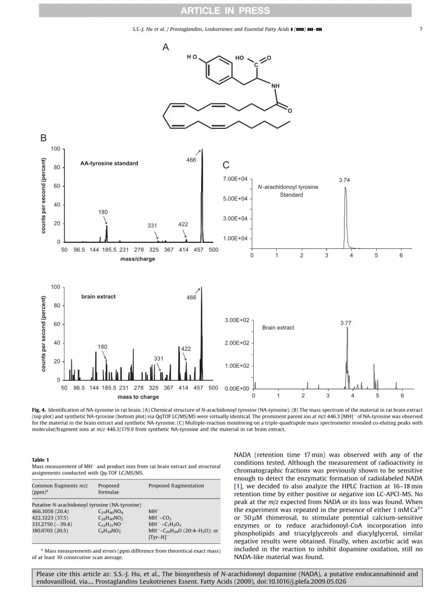
3.2. Identification of endogenous NA-tyrosine in rat brain
N-arachidonoyl-tyrosine (NA-tyrosine) is a potential NADAprecursor; therefore, we examined the possibility that it isproduced in the brain. Identification of NA-tyrosine (Fig. 4A)was accomplished using a partially purified brain lipid extractthat was analyzed by nano-LC-quadrupole/time-of-flight massspectrometry (nano-HPLC/QqTOF MS) and by chromatographicmatching using LC/MS/MS. Nano-HPLC/QqTOF MS isolated a lipidcompound within the brain extract with an exact mass of466.3058, which is within 20 parts per million of the expectedmass of NA-tyrosine (466.3038; Table 1). Additionally, thefragmentation pattern of the 466.3058 mass was the same asthat produced by the synthetic standard (Fig. 4B), which isconsidered a mass fingerprint. Likewise, with LC/MS/MS using atechnique that filters for the parent ion (466 amu) and pairs itwith a predicted fragment (180 amu) a chromatographic matchwas revealed of a compound in the brain extract that matchedwith the NA-tyrosine standard (Fig. 4C). Taken together, these
achidonoyl dopamine (NADA), a putative endocannabinoid and(2009), doi:10.1016/j.plefa.2009.05.026

ARTICLE IN PRESS
0
10
20
30
40
50
60
70
80
0Time (min) (n = 4 at each time point)
Stri
atal
Dop
amin
e (n
mol
/g)
∗∗∗
∗∗∗∗∗∗
∗∗∗
∗∗
0
2
4
6
8
10
12
Time (min) (n = 4 at each time point)
Stri
atal
NA
DA
(pm
ol/g
)
∗∗ ∗
90 180 270 360 450
0 90 180 270 360 450
Fig. 3. The TH inhibitor AMPT decreases dopamine and NADA levels in rat striata in a time-dependent fashion. (A) A time-dependent decrease in striatal levels of dopamine
was observed when compared to control (time ¼ 0) (**po0.01 at 90 min; ***po0.0005 at 180, 270, 360, and 450 min). (B) A time-dependent decrease in striatal levels of
NADA was also observed when compared to control (time ¼ 0) (*po0.05 at 270, 360, and 450 min) (n ¼ 4 at each time point).
S.S.-J. Hu et al. / Prostaglandins, Leukotrienes and Essential Fatty Acids ] (]]]]) ]]]–]]]6
data support the hypothesis that NA-tyrosine is produced in thebrain.
3.3. Examination of NA-tyrosine as a precursor of striatal NADA
With the evidence that NA-tyrosine is present in the brain, weexamined the possibility that NADA biosynthesis occurs viametabolism of NA-tyrosine to NA-L-DOPA by TH, which wouldthen be metabolized to NADA by L-amino acid decarboxylase(AADC) (Fig. 1). NADA levels were significantly decreased 270 minafter the TH inhibitor AMPT injection (Fig. 3B); therefore, weexamined whether NA-tyrosine accumulated after TH inhibition.However, no significant increase was found, suggesting thatNA-tyrosine may not be a substrate for TH in vivo. In orderto substantiate this interpretation, we tested whether THcould convert NA-tyrosine to NA-L-DOPA in vitro. We incubatedL-tyrosine (1mM) or [2H8]NA-tyrosine (1mM) with TH (16.5mg)and examined the production of L-DOPA and [2H8]NA-L-DOPA byLC/MS/MS. The results indicated that 24.44% of L-tyrosine wasconverted to L-DOPA (24.44 pmol yield), whereas only 0.23% of[2H8]NA-tyrosine was converted to [2H8]NA-L-DOPA (2.28 pmolyield), indicating that NA-tyrosine is a poor substrate for TH.
Please cite this article as: S.S.-J. Hu, et al., The biosynthesis of N-arendovanilloid, via..., Prostaglandins Leukotrienes Essent. Fatty Acids
Neither L-DOPA nor [2H8]NA-L-DOPA was detected when bufferonly or boiled enzymes were incubated with the respectiveprecursors.
To test the hypothesis that NA-L-DOPA is a direct precursor toNADA, we measured the amount of [2H8]NADA formed followingthe injection of [2H8]NA-L-DOPA in the rat striatum. There were nodetectable levels of [2H8]NADA in either injected or non-injectedstriata, which implied that NA-L-DOPA is not converted to NADA inthe striatum. Together, these data indicated that it is unlikely thatNADA biosynthesis occurs via metabolism of NA-tyrosine or NA-L-DOPA.
3.4. No detectable NADA is formed via arachidonoyl-CoA and
dopamine incubation with brain membranes
It is likely that NADA is formed via conjugation of arachido-noyl-CoA and dopamine. However, when homogenates of striatal,midbrain, or hippocampal rat slices were incubated with 50mM of[14C]arachidonoyl-CoA and 50mM dopamine, or with variousconcentrations (5–50mM) of arachidonoyl-CoA and 50mM of[3H]dopamine, and the incubated homogenate extracted andsubmitted to HPLC analysis, no radiolabeled peak co-eluting with
achidonoyl dopamine (NADA), a putative endocannabinoid and(2009), doi:10.1016/j.plefa.2009.05.026
ARTICLE IN PRESS
H O
NH
CHO O
O
0
20
40
60
80
100
50 96.5 144 185.5 231 278 325 367 414 457 500
coun
ts p
er s
econ
d (p
erce
nt)
mass to charge
180
331
422
466brain extract
0
20
40
60
80
100
50 96.5 144 185.5 231 278 325 367 414 457 500
coun
ts p
er s
econ
d (p
erce
nt)
mass/charge
180
331 422
466AA-tyrosine standard
1.00E+04
3.00E+04
5.00E+04
7.00E+04
0
3.74N -arachidonoyl tyrosine
Standard
1 2 3 4 5 6
0.00E+00
1.00E+02
2.00E+02
3.00E+02 3.77Brain extract
0 1 2 3 4 5 6
Fig. 4. Identification of NA-tyrosine in rat brain. (A) Chemical structure of N-arachidonoyl tyrosine (NA-tyrosine). (B) The mass spectrum of the material in rat brain extract
(top plot) and synthetic NA-tyrosine (bottom plot) via QqTOF LC/MS/MS were virtually identical. The prominent parent ion at m/z 446.3 [MH]� of NA-tyrosine was observed
for the material in the brain extract and synthetic NA-tyrosine. (C) Multiple-reaction monitoring on a triple-quadrupole mass spectrometer revealed co-eluting peaks with
molecular/fragment ions at m/z 446.3/179.9 from synthetic NA-tyrosine and the material in rat brain extract.
Table 1Mass measurement of MH� and product ions from rat brain extract and structural
assignments conducted with Qq-TOF LC/MS/MS.
Common fragments m/z
(ppm)a
Proposed
formulae
Proposed fragmentation
Putative N-arachidonoyl tyrosine (NA-tyrosine)
466.3058 (20.4) C29H40NO4 MH�
422.3223 (37.5) C28H40NO2� MH�–CO2
331.2750 (�39.4) C22H37NO� MH� –C7H3O3
180.0703 (20.5) C9H10NO3� MH�–C20H30O (20:4–H2O); or
[Tyr–H]�
a Mass measurements and errors (ppm difference from theoretical exact mass)
of at least 10 consecutive scan average.
S.S.-J. Hu et al. / Prostaglandins, Leukotrienes and Essential Fatty Acids ] (]]]]) ]]]–]]] 7
Please cite this article as: S.S.-J. Hu, et al., The biosynthesis of N-arendovanilloid, via..., Prostaglandins Leukotrienes Essent. Fatty Acids
NADA (retention time 17 min) was observed with any of theconditions tested. Although the measurement of radioactivity inchromatographic fractions was previously shown to be sensitiveenough to detect the enzymatic formation of radiolabeled NADA[1], we decided to also analyze the HPLC fraction at 16–18 minretention time by either positive or negative ion LC-APCI-MS. Nopeak at the m/z expected from NADA or its loss was found. Whenthe experiment was repeated in the presence of either 1 mM Ca2+
or 50mM thimerosal, to stimulate potential calcium-sensitiveenzymes or to reduce arachidonoyl-CoA incorporation intophospholipids and triacylglycerols and diacylglycerol, similarnegative results were obtained. Finally, when ascorbic acid wasincluded in the reaction to inhibit dopamine oxidation, still noNADA-like material was found.
achidonoyl dopamine (NADA), a putative endocannabinoid and(2009), doi:10.1016/j.plefa.2009.05.026

ARTICLE IN PRESS
S.S.-J. Hu et al. / Prostaglandins, Leukotrienes and Essential Fatty Acids ] (]]]]) ]]]–]]]8
3.5. No detectable NADA is formed via arachidonic acid esterified to
phospholipids incubated with C6 glioma cells
Rat C6 glioma cells were incubated overnight with [3H]arachi-donic acid in order to allow its incorporation into membranephospholipids. They were then incubated at 37 1C for increasingperiods of time (0–60 min) with 100mM dopamine, either in thepresence or in the absence of ionomycin (4mM) to stimulate Ca2+
influx into the cells. Under these conditions no formation oftritiated NADA-like components in cell homogenates was ob-served using HPLC analysis. Under similar conditions, [3H]2-arachidonoyl glycerol ([3H]2-AG) was formed following stimula-tion with 4mM ionomycin (data not shown). Analogous negativeresults were obtained when whole-cell homogenates fromradiolabeled C6 cells were incubated at 37 1C for increasingperiods of time (0–60 min) with 100mM dopamine.
0
5
10
15
20
25
30
35
pmol
es/g
ram
tiss
ue Vehicle (n = 14)
URB597 (n = 5)
∗
∗
3.6. NADA biosynthesis occurs via conjugation of arachidonic acid
with dopamine in rat brain membranes
Previously, the hypothesis that NADA could be formed byconjugation of arachidonic acid with dopamine was examinedusing radiolabeled precursors and TLC characterization of theenzymatic products [9]. In the current study, the possibility thatNADA biosynthesis occurs through conjugation of arachidonicacid with dopamine was investigated quantitatively by incubatingrat brain membranes with 20, 50, and 100mM [2H8]arachidonicacid and 20, 50, and 100mM [2H4]dopamine at 37 1C for 25 min,respectively. Here we found that when rat brain membranes wereincubated with [2H8]arachidonic acid and [2H4]dopamine, deut-erated NADA was produced and detectable at masses correspond-ing to molecular ions ranging from [2H8]NADA to [2H12]NADA (thevarying degree of deuterium incorporation is expected due toproton exchange with the aqueous buffer and the tissue). Theproduction of [2H12]- and [2H11]NADA molecular ions wasconcentration-dependent with higher concentrations of precur-sors, leading to higher concentrations of product (Fig. 5). Anenzymatic mechanism is likely as deuterated NADA was notdetected when the substrates were incubated with buffer alone or
0
10
20
30
40
50
20
[2H8]Arachidonic Acid (μM)
[2 H12
]NA
DA
(fm
ol)
100 μM50 μM20 μM
Boiled membrane or Buffer only
[2H4]Dopamine:
10050
Fig. 5. Biosynthesis of [2H12]NADA by incubation of [2H8]arachidonic acid and
[2H4]dopamine with rat brain membrane homogenates. Rat brain membranes
were incubated with 20, 50, and 100mM [2H8]arachidonic acid and 20, 50, and
100mM [2H4]dopamine, respectively, in Tris–HCl buffer at 37 1C for 25 min (n ¼ 9
for each condition). The production of [2H12]NADA was concentration-dependent
with higher concentrations of precursors leading to higher concentrations of
product. Deuterated NADA was not detected when buffer or boiled membranes
were incubated with the precursors or when membranes were incubated without
the precursors. Values are mean7SEM from three independent experiments
performed in triplicate.
Please cite this article as: S.S.-J. Hu, et al., The biosynthesis of N-arendovanilloid, via..., Prostaglandins Leukotrienes Essent. Fatty Acids
with boiled membranes (Fig. 5). These results indicate that NADAcan be synthesized through the enzymatic conjugation ofarachidonic acid and dopamine via an enzymatic process.
3.7. Endogenous level of striatal NADA after URB597 injections in
rats and in FAAH knockout and wild-type mice
To determine whether FAAH modulates endogenous NADAlevels, we examined AEA and NADA levels in rats treated withURB597 or vehicle and in FAAH knockout and wild-type mice. Thelevels of AEA in rat striatum significantly increased 2 h aftersystemic injection of the FAAH inhibitor URB597 (0.3 mg/kg)compared to vehicle controls (Fig. 6A). In contrast, striatal levels ofNADA significantly decreased (Fig. 6A). The same pattern wasobserved in the striatal levels of AEA and NADA in FAAH knockoutand wild-type mice: AEA levels were significantly higher, whereasNADA levels were significantly lower in striata from FAAHknockout mice (Fig. 6B).
AEA
0
10
20
30
40
50
60
70
80
90
100
pmol
es/g
ram
tiss
ue
Wild-type (n = 5)
Knockout (n = 5)
∗
∗
NADA
AEA NADA
Fig. 6. Pharmacological and genetic inhibition of FAAH increases striatal AEA and
decreases striatal NADA. (A) Levels of AEA and NADA in rat striata 2 h after vehicle
(n ¼ 14) or 0.3 mg/kg URB597 (n ¼ 5) (*po0.05). (B) Levels of AEA and NADA in
mouse striata of wild-type (n ¼ 5) or FAAH knockout (n ¼ 5) (*po0.05).
achidonoyl dopamine (NADA), a putative endocannabinoid and(2009), doi:10.1016/j.plefa.2009.05.026

ARTICLE IN PRESS
S.S.-J. Hu et al. / Prostaglandins, Leukotrienes and Essential Fatty Acids ] (]]]]) ]]]–]]] 9
3.8. NADA is a poor substrate for recombinant FAAH
The evidence that NADA levels were significantly decreasedwith FAAH inhibition and in FAAH knockout mice led us to test thehypothesis that (1) FAAH is acting as a biosynthetic enzyme withAEA and dopamine as precursors and (2) NADA is a poor substratefor FAAH. If NADA is a high-affinity substrate for FAAH producingrobust levels of hydrolysis, then it is unlikely to play a role in itsbiosynthesis. Conversely, if AEA were converted to NADA duringhydrolysis in the presence of dopamine, then FAAH would in factbe a candidate enzyme for NADA biosynthesis. Our results showthat when recombinant FAAH was incubated with AEA anddopamine, AEA was measured via HPLC/MS/MS and was shownto be rapidly hydrolyzed as expected (Fig. 7A). At 3 min�80% of allAEA was hydrolyzed and by �10 min over 95%. At 30 min therewas almost no detectable AEA present in the solution. Conversely,
FAAH Hydrolysis of A
0 3 6 9 120
20
40
60
80
100
Time
% H
ydro
lysi
s
5
10
15
20
NA
DA
pro
duce
d (p
mol
s)
Time0 3 6 9 12
Fig. 7. Hydrolysis and biosynthesis of NADA via recombinant FAAH. (A) Comparisons o
incubations of AEA, dopamine, and recombinant FAAH.
Please cite this article as: S.S.-J. Hu, et al., The biosynthesis of N-arendovanilloid, via..., Prostaglandins Leukotrienes Essent. Fatty Acids
at 3 min less than 10% of NADA was hydrolyzed and at 30 minthere was still over 50% of NADA still left in the solution.Surprisingly, trace amounts of NADA were produced in a time-dependent manner when AEA and dopamine were incubated withFAAH. Small amounts of NADA were detected throughout theincubation during the first 15 min and at 30 min, when there wasno detectable AEA (therefore, an abundance of liberatedarachidonic acid), and up to �3% of NADA production wasmeasured (mole for mole compared to a theoretical 100% AEA).To control for any carry-over or MS contamination of NADA, whichhas a history of carry-over on columns and in tubing (unpublishedobservations), we used new columns and tubing and performedthe recombinant FAAH incubations with deuterium-labeled NADA([H8] NADA; Fig. 7A). Therefore, the NADA that was measured viaHPLC/MS/MS after incubation of AEA and dopamine withrecombinant FAAH could not be carry-over contaminants.
EA & [2H8]-NADA
15 18 21 24 27 30
[2H8]-NADA
AEA
[Minutes]
[Minutes]15 18 21 24 27 30
f hydrolysis rates of AEA and [2H8]-NADA. (B) De novo production of NADA from
achidonoyl dopamine (NADA), a putative endocannabinoid and(2009), doi:10.1016/j.plefa.2009.05.026

ARTICLE IN PRESS
S.S.-J. Hu et al. / Prostaglandins, Leukotrienes and Essential Fatty Acids ] (]]]]) ]]]–]]]10
4. Discussion
In the present study we investigated several biosyntheticmechanisms for NADA and report that NADA synthesis requiresTH in dopaminergic terminals and occurs through an enzyme-mediated conjugation of arachidonic acid with dopamine and notthrough the endogenous NA-tyrosine. One enzyme involved in thebiosynthetic process is fatty acid amide hydrolase, the blockade ofwhich significantly decreases in vivo the production of NADA.
Lesion studies of the striatum supported the hypothesis thatNADA is synthesized in dopaminergic terminals where TH islocated. One potential implication of the significant decrease inNADA production measured after striatal lesions is that Km of theconjugation reaction for dopamine is high, so that small decreasesin dopamine concentration may lead to large decreases in NADA.However, this has not been examined in vitro.
We originally proposed two potential pathways for NADAbiosynthesis: one mediated by metabolism of the putativeprecursor NA-tyrosine and the other via direct conjugation ofarachidonic acid with dopamine [1]. However, based on the lesionstudies presented here, it is hard to distinguish which of these twopathways is dominant in NADA biosynthesis since the essentialmolecular component for both pathways, TH, was depleted underconditions of dopaminergic terminal destruction or TH inhibition.To elucidate the primary pathway in NADA biosynthesis, wedetermined that NA-tyrosine is a naturally occurring moleculein the rat, a conclusion based on mass spectrometric analysis(LC/MS/MS and nano-HPLC/QqTOF), showing that the exactmasses, column retention times, and fragmentation patterns ofthe constituent found in rat brain were virtually identical to thoseof NA-tyrosine. Although our results support the existence ofendogenous NA-tyrosine, which could be a precursor for NADAbiosynthesis via TH, our data did not support the hypothesis thatNA-tyrosine is a precursor for NADA biosynthesis given thefollowing: (1) NA-tyrosine did not accumulate after TH inhibition,(2) incubating deuterium-labeled NA-tyrosine with purifiedrecombinant TH failed to produce deuterated NA-L-DOPA, and(3) the injection of deuterium-labeled NA-L-DOPA into striatumfailed to produce deuterated NADA. These results suggest thatNA-tyrosine is a poor substrate for TH in vivo and in vitro and thatthe presence of NA-tyrosine in rat brain might have little to dowith NADA biosynthesis. Rather, it is possible that endogenousNA-tyrosine is an endogenous signaling molecule with a separatefunction to NADA and/or a mediator for biosynthesis of otherN-arachidonoyl amino acids, such as the previously identifiedconjugates of arachidonic acid with glycine, alanine, g-amino-butyric acid, and serine [15,16]. However, it is important to keep inmind that the possibility of NA-tyrosine conversion into NA-L-DOPA by enzymes other than TH in vivo cannot be ruled out by theabove results. It is also possible, though unlikely, that the failedproduction of NADA from NA-L-DOPA injection may due tocompartmentalization. A faster degradation of exogenous NA-L-DOPA than its uptake into cells and then conversion into NADAmay also explain the negative findings in the current study.
Confirmed here is the hypothesis that NADA is synthesized viaconjugation of arachidonic acid and dopamine through anenzyme-dependent process. Whether or not intermediates areformed as part of this process remains uncertain. Although little isknown about the enzyme(s) involved in NADA biosynthesis,Huang et al. [1] proposed a possible mechanism similar to theformation of N-arachidonoyl glycine (NAGly) [10]. The enzyme,arachidonoyl-CoA synthetase, which belongs to an enzymaticfamily of acyl-CoA synthetase, converts arachidonic acid intoarachidonoyl-CoA in the brain, platelets, and aorta [17–19].A second family of enzymes, the acyl-coenzyme A:glycineN-acyltransferases, may conjugate glycine to various aliphatic
Please cite this article as: S.S.-J. Hu, et al., The biosynthesis of N-arendovanilloid, via..., Prostaglandins Leukotrienes Essent. Fatty Acids
and aromatic acyl-CoAs [20–25]. Here, we tested the possible roleof arachidonoyl-CoA in the direct formation of NADA and wereunable to produce NADA under several experimental conditions.Likewise, we did not observe the formation of any NADA-likeHPLC component from arachidonic acid incorporated into phos-pholipids when using either intact cell or cell-free conditions. It ispossible that the bioavailability of these intermediates is an issueand that the biosynthetic process is such that there is a rate-limiting step that initiates the production of NADA and in theabsence of this step NADA would not be formed even with anexcess of substrate.
A recent report showed that FAAH can participate in the in vivo
synthesis of AM404 by conjugation of arachidonic acid toexogenously administered acetaminophen [9]. We examinedwhether striatal levels of NADA were changed after pharmacolo-gical or genetic inhibition of FAAH. That the normal NADA levelsin rat striatum could be so dramatically decreased with FAAHinhibition and also that FAAH knockout mice have significantlyless NADA than wide-type mice were convincing evidence thatFAAH was in some way involved in the biosynthesis of NADA.Accordingly, FAAH mRNA is moderately expressed in striatum andFAAH activity is detectable in rat striatum [26]. Also, an increasedlevel of striatal AEA and a decreased activity of FAAH wereobserved after destruction of dopaminergic terminals by 6-OHDA[27]. These data support the possibility that FAAH has thepotential to be involved in the direct conjugation of dopaminewith arachidonic acid liberated from AEA, and is the rate-limitingenzyme for NADA biosynthesis. Alternately, the decreased levelsof striatal NADA by FAAH inhibition or depletion may result fromelevation of AEA concentrations in the striatum. Maccarrone et al.[28] reported that elevation of AEA concentrations in the striatumby FAAH inhibition or depletion reduced the levels, metabolism,and physiological effects of 2-AG. It is possible that elevatedstriatal AEA has modulatory effects on metabolism of NADA,thereby reducing its levels in the striatum, and future studies willaddress this issue.
Early studies showed that AEA can be synthesized byconjugation of arachidonic acid and ethanolamine and thisreaction might be catalyzed by FAAH acting in reverse [29,30].However, this pathway was later shown to be unlikely given thatAEA is an excellent substrate for FAAH (also confirmed here); thusthe rate of AEA hydrolysis by FAAH was much greater than AEAsynthesis from the conjugation of arachidonic acid and ethano-lamine. Later research found that there are at least three potentialpathways involved in AEA biosynthesis that includes several novelenzymes such as N-acyl phosphatidylethanolamine phospholi-pase D (NAPE-PLD), glycerophosphodiesterase (GDE1), a/b hydro-lase 4 (ABHD4), or an unknown phosphatase [31–35]. To date,there are no known N-acyl phosphatidyldopamines, so theseanalogous pathways are highly unlikely.
The possibility that FAAH is a candidate for an enzymecomplex involved in NADA biosynthesis is in further agreementwith the observation that NADA is a poor substrate for FAAH [1].We confirmed this observation here and showed that the rate ofhydrolysis of NADA by FAAH is dramatically lower than that ofAEA (7-fold at 3 min and 5-fold at 30 min). When AEA wasincubated with FAAH in the presence of dopamine, NADA wassynthesized in trace amounts. Our HPLC/MS/MS method fordetection of NADA is �500 atamoles and, therefore, the mostsensitive published to date. In fact, previous methods used in thisstudy for our earlier assays would not have detected these traceamounts. The largest amount of production of NADA (�3%) was at30 min when there were only trace amounts of AEA left in thesolution, so that the substrates available were arachidonic acidand dopamine. Given that FAAH is an integral membrane proteinand that recombinant FAAH is in an unbound state in vitro, it is
achidonoyl dopamine (NADA), a putative endocannabinoid and(2009), doi:10.1016/j.plefa.2009.05.026

ARTICLE IN PRESS
S.S.-J. Hu et al. / Prostaglandins, Leukotrienes and Essential Fatty Acids ] (]]]]) ]]]–]]] 11
unlikely to act in exactly the same manner as the bound state in
vivo. It is difficult to determine whether the synthesis of NADA in
vitro represents the result of an energetically favorable reaction orthe result of FAAH acting as a biosynthetic enzyme at a sub-optimal level given that its conformation is potentially differentfrom that in the membrane-bound form. Future studies willaddress this issue.
In conclusion, we report here in vitro and in vivo evidence thatNADA biosynthesis in rat brain depends on TH in dopaminergicterminals and that this endocannabinoid/endovanilloid can bemade from the conjugation of dopamine with arachidonic acid,but that it is likely not formed via NA-tyrosine and NA-L-DOPAintermediates. Our data suggest a direct involvement of FAAH inNADA biosynthesis either as a rate-limiting enzyme that liberatesarachidonic acid from AEA, or as a conjugation enzyme, or both.Further investigations are needed to elucidate the exact moleculardetails of this enzymatic process.
Acknowledgements
This work is dedicated to Dr. J. Michael Walker (1950–2008)whose love of science was an inspiration to us all. We are gratefulfor Dr. Ken Mackie’s comments on the manuscript. We thankDr. Filomena Fezza (University of Rome ‘‘Tor Vergata’’, Italy) andDr. Mauro Maccarrone (University of Teramo, Italy) for theirvaluable help in the initial phase of this study and Jocelyn F. Kreyfor the technical assistance.
References
[1] S.M. Huang, T. Bisogno, M. Trevisani, A. Al-Hayani, L. De Petrocellis, F. Fezza,M. Tognetto, T.J. Petros, J.F. Krey, C.J. Chu, J.D. Miller, S.N. Davies, P. Geppetti,J.M. Walker, V. Di Marzo, An endogenous capsaicin-like substance with highpotency at recombinant and native vanilloid VR1 receptors, Proc. Natl. Acad.Sci. USA 99 (2002) 8400–8405.
[2] V. Bezuglov, M. Bobrov, N. Gretskaya, A. Gonchar, G. Zinchenko, D. Melck, T.Bisogno, V. Di Marzo, D. Kuklev, J.C. Rossi, J.P. Vidal, T. Durand, M.Yu. Bobrov,N.M. Gretskaya, V.V. Bezuglov, L. De Petrocellis, Synthesis and biologicalevaluation of novel amides of polyunsaturated fatty acids with dopamine,Bioorg. Med. Chem. Lett. 11 (2001) 447–449.
[3] T. Bisogno, D. Melck, M.Yu. Bobrov, N.M. Gretskaya, V.V. Bezuglov, L. DePetrocellis, V. Di Marzo, N-acyl-dopamines: novel synthetic CB(1) cannabi-noid-receptor ligands and inhibitors of anandamide inactivation withcannabimimetic activity in vitro and in vivo, Biochem. J. 351 (2000) 817–824.
[4] D.R. Sagar, P.A. Smith, P.J. Millns, D. Smart, D.A. Kendall, V. Chapman, TRPV1and CB(1) receptor-mediated effects of the endovanilloid/endocannabinoid N-arachidonoyl-dopamine on primary afferent fibre and spinal cord neuronalresponses in the rat, Eur. J. Neurosci. 20 (2004) 175–184.
[5] S.E. O’Sullivan, D.A. Kendall, M.D. Randall, Vascular effects of delta 9-tetrahydrocannabinol (THC), anandamide and N-arachidonoyldopamine(NADA) in the rat isolated aorta, Eur. J. Pharmacol. 507 (2005) 211–221.
[6] S. Marinelli, V. Di Marzo, F. Florenzano, F. Fezza, M.T. Viscomi, M. van der Stelt,G. Bernardi, M. Molinari, M. Maccarrone, N.B. Mercuri, N-Arachidonoyl-dopamine tunes synaptic transmission onto dopaminergic neurons byactivating both cannabinoid and vanilloid receptors, Neuropsychophar 32(2007) 298–308.
[7] S.M. Huang, J.M. Walker, Enhancement of spontaneous and heat-evokedactivity in spinal nociceptive neurons by the endovanilloid/endocannabinoidN-arachidonoyldopamine (NADA), J. Neurophysiol. 95 (2006) 1207–1212.
[8] H. Pajouhesh, A.J. Hancock, Synthesis of cyclopentano-N-methylphosphati-dylethanolamines: aminolysis during the use of methylamine, J. Lipid Res. 25(1984) 310–312.
[9] E.D. Hogestatt, B.A. Jonsson, A. Ermund, D.A. Andersson, H. Bjork, J.P.Alexander, B.F. Cravatt, A.I. Basbaum, P.M. Zygmunt, Conversion of acetami-nophen to the bioactive N-acylphenolamine AM404 via fatty acid amidehydrolase-dependent arachidonic acid conjugation in the nervous system,J. Biol. Chem. 280 (2005) 31405–31412.
[10] B.F. Cravatt, K. Demarest, M.P. Patricelli, M.H. Bracey, D.K. Giang, B.R. Martin,A.H. Lichtman, Supersensitivity to anandamide and enhanced endogenouscannabinoid signaling in mice lacking fatty acid amide hydrolase, Proc. Natl.Acad. Sci. USA 98 (2001) 9371–9376.
[11] L.G. Gahn, R. Roskoski Jr., Tyrosine hydroxylase purification from rat PC12cells, Protein Expression Purif. 2 (1991) 10–14.
Please cite this article as: S.S.-J. Hu, et al., The biosynthesis of N-arendovanilloid, via..., Prostaglandins Leukotrienes Essent. Fatty Acids
[12] M. Grazia Cascio, A. Minassi, A. Ligresti, G. Appendino, S. Burstein, V. DiMarzo, A structure–activity relationship study on N-arachidonoyl-aminoacids as possible endogenous inhibitors of fatty acid amide hydrolase,Biochem. Biophys. Res. Commun. 314 (2004) 192–196.
[13] N. Rimmerman, H.B. Bradshaw, H.V. Hughes, J.S. Chen, S.S. Hu, D. McHugh, E.Vefring, J.A. Jahnsen, E.L. Thompson, K. Masuda, B.F. Cravatt, S. Burstein, M.R.Vasko, A.L. Prieto, D.K. O’Dell, J.M. Walker, N-Palmitoyl glycine, a novelendogenous lipid that acts as a modulator of calcium influx and nitric oxideproduction in sensory neurons, Mol. Pharmacol. 74 (2008) 213–224.
[14] M.D. Schechter, P.G. Cook, Dopaminergic mediation of the interoceptive cueproduced by d-amphetamine in rats, Psychopharmacologia 42 (1975)185–193.
[15] S.M. Huang, T. Bisogno, T.J. Petros, S.Y. Chang, P.A. Zavitsanos, R.E. Zipkin, R.Sivakumar, A. Coop, D.Y. Maeda, L. De Petrocellis, S. Burstein, V. Di Marzo, J.M.Walker, Identification of a new class of molecules, the arachidonyl aminoacids, and characterization of one member that inhibits pain, J. Biol. Chem.276 (2001) 42639–42644.
[16] G. Milman, Y. Maor, S. Abu-Lafi, M. Horowitz, R. Gallily, S. Batkai, F.M. Mo, L.Offertaler, P. Pacher, G. Kunos, R. Mechoulam, N-arachidonoyl L-serine, anendocannabinoid-like brain constituent with vasodilatory properties, Proc.Natl. Acad. Sci. USA 103 (2006) 2428–2433.
[17] T.S. Reddy, N.G. Bazan, Kinetic properties of arachidonoyl-coenzyme Asynthetase in rat brain microsomes, Arch. Biochem. Biophys. 226 (1983)125–133.
[18] D.B. Wilson, S.M. Prescott, P.W. Majerus, Discovery of an arachidonoylcoenzyme A synthetase in human platelets, J. Biol. Chem. 257 (1982)3510–3515.
[19] N. Morisaki, Y. Saito, A. Kumagai, Synthesis and metabolism of arachidonyl-and eicosapentaenoyl-CoA in rat aorta, Biochim. Biophys. Acta 752 (1983)301–306.
[20] D. Schachter, J.V. Taggart, Benzoyl coenzyme A and hippurate synthesis,J. Biol. Chem. 203 (1953) 925–934.
[21] D. Schachter, J.V. Taggart, Glycine N-acylase: purification and properties,J. Biol. Chem. 208 (1954) 263–275.
[22] D.L. Nandi, S.V. Lucas, L.T. Webster, Benzoyl-coenzyme A:glycine N-acyl-transferase and phenylacetyl-coenzyme A:glycine N-acyltransferase frombovine liver mitochondria. Purification and characterization, J. Biol. Chem.254 (1979) 7230–7237.
[23] S. K½lvraa, N. Gregersen, Acyl-CoA:glycine N-acyltransferase: organellelocalization and affinity toward straight- and branched-chained acyl-CoAesters in rat liver, Biochem. Med. Metab. Biol. 36 (1986) 98–105.
[24] N. Gregersen, S. K½lvraa, P.B. Mortensen, Acyl-CoA: glycine N-acyltransferase:in vitro studies on the glycine conjugation of straight- and branched-chainedacyl-CoA esters in human liver, Biochem. Med. Metab. Biol. 35 (1986)210–218.
[25] Y.R. Mawal, I.A. Qureshi, Purification to homogeneity of mitochondrial acylCoA:glycine N-acyltransferase from human liver, Biochem. Biophys. Res.Commun. 205 (1994) 1373–1379.
[26] E.A. Thomas, B.F. Cravatt, P.E. Danielson, N.B. Gilula, J.G. Sutcliffe, Fatty acidamide hydrolase, the degradative enzyme for anandamide and oleamide, hasselective distribution in neurons within the rat central nervous system,J. Neurosci. Res. 50 (1997) 1047–1052.
[27] P. Gubellini, B. Picconi, M. Bari, N. Battista, P. Calabresi, D. Centonze, G.Bernardi, A. Finazzi-Agr �o, M. Maccarrone, Experimental parkinsonism altersendocannabinoid degradation: implications for striatal glutamatergic trans-mission, J. Neurosci. 22 (2002) 6900–6907.
[28] M. Maccarrone, S. Rossi, M. Bari, V. De Chiara, F. Fezza, A. Musella, V. Gasperi,C. Prosperetti, G. Bernardi, A. Finazzi-Agr �o, B.F. Cravatt, D. Centonze,Anandamide inhibits metabolism and physiological actions of 2-arachido-noylglycerol in the striatum, Nat. Neurosci. 11 (2008) 152–159.
[29] W.A. Devane, J. Axelrod, Enzymatic synthesis of anandamide, an endogenousligand for the cannabinoid receptor, by brain membranes, Proc. Natl. Acad.Sci. USA 91 (1994) 6698–6701.
[30] G. Arreaza, W.A. Devane, R.L. Omeir, G. Sajnani, J. Kunz, B.F. Cravatt, D.G.Deutsch, The cloned rat hydrolytic enzyme responsible for the breakdown ofanandamide also catalyzes its formation via the condensation of arachidonicacid and ethanolamine, Neurosci. Lett. 234 (1997) 59–62.
[31] K. Ahn, M.K. McKinney, B.F. Cravatt, Enzymatic pathways that regulateendocannabinoid signaling in the nervous system, Chem. Rev. 108 (2008)1687–1707.
[32] Y. Okamoto, J. Morishita, K. Tsuboi, T. Tonai, N. Ueda, Molecular characteriza-tion of a phospholipase D generating anandamide, its congeners, J. Biol.Chem. 279 (2004) 5298–5305.
[33] D. Leung, A. Saghatelian, G.M. Simon, B.F. Cravatt, Inactivation of N-acylphosphatidylethanolamine phospholipase D reveals multiple mechanismsfor the biosynthesis of endocannabinoids, Biochemistry 45 (2006)4720–4726.
[34] G.M. Simon, B.F. Cravatt, Endocannabinoid biosynthesis proceeding throughglycerophospho-N-acyl ethanolamine and a role for alpha/beta-hydrolase 4 inthis pathway, J. Biol. Chem. 281 (2006) 26465–26472.
[35] G.M. Simon, B.F. Cravatt, Anandamide biosynthesis catalyzed by thephosphodiesterase GDE1 and detection of glycerophospho-N-acyl ethanola-mine precursors in mouse brain, J. Biol. Chem. 283 (2008) 9341–9349.
achidonoyl dopamine (NADA), a putative endocannabinoid and(2009), doi:10.1016/j.plefa.2009.05.026