n-arachidonoyl-dopamine tunes synaptic transmission onto dopaminergic neurons by activating both...
TRANSCRIPT

N-Arachidonoyl-Dopamine Tunes Synaptic Transmission ontoDopaminergic Neurons by Activating both Cannabinoidand Vanilloid Receptors
Silvia Marinelli*,1 Vincenzo Di Marzo2, Fulvio Florenzano3, Filomena Fezza4, Maria Teresa Viscomi3,Mario van der Stelt2, Giorgio Bernardi1,5, Marco Molinari3, Mauro Maccarrone4,6,7 and Nicola B Mercuri1,5,7
1Experimental Neurology, IRCCS Fondazione Santa Lucia, Rome, Italy; 2Endocannabinoid Research Group, Istituto di Chimica Biomolecolare,
Consiglio Nazionale delle Ricerche, Pozzuoli, Naples, Italy; 3Experimental Neurorehabilitation Labs, IRCCS Santa Lucia Foundation, Rome, Italy;4Department of Biomedical Sciences, University of Teramo, Teramo, Italy; 5Dipartimento di Neuroscienze, Universita di Tor Vergata, Rome, Italy;6IRCCS C. Mondino, Mondino-Tor Vergata Center for Experimental Neurobiology, Rome, Italy
In the present study, we used electrophysiological, biochemical, and confocal microscopy techniques, to investigate the functional role of
transient receptor potential vanilloid type 1 (TRPV1) and cannabinoid type 1 receptors (CB1-R) in the substantia nigra pars compacta
(SNpc) and their stimulation by the endocannabinoid N-arachidonoyl-dopamine (NADA). Liquid chromatography–mass spectrometry
analyses revealed that a NADA-like compound is produced in substantia nigra slices, in conditions of hyperactivity. Moreover, the
functional role of both TRPV1 and CB1-R in modulating synaptic transmission in this area was suggested by confocal microscopy data,
showing TRPV1 and CB1-R immunoreactivity in punctate structures, probably representing synaptic contacts on cell bodies of the SNpc.
In patch-clamp recordings from dopamine (DA) neurons of the SNpc, we found that NADA increases or reduces glutamatergic
transmission onto DA neurons by activating TRPV1 and CB1 receptors, respectively, whereas it decreases GABAergic transmission via
CB1 stimulation. Facilitation of glutamate release through TRPV1 was blocked in the presence of a selective blocker of the putative
endocannabinoid membrane transporter (EMT), indicating that NADA needs to be taken up by cells to interact with this receptor. In line
with these data, biochemical results demonstrated that NADA selectively acted at CB1-R when its re-uptake was blocked. Altogether
these data demonstrate a significant role exerted by the endocannabinoid/endovanilloid NADA in the regulation of synaptic transmission
to DA neurons of the SNpc. Moreover, they highlight a key function of the EMT transporter in promoting the stimulation of TRPV1 or
CB1-R, thus favoring facilitation or inhibition of glutamate synaptic release.
Neuropsychopharmacology (2007) 32, 298–308. doi:10.1038/sj.npp.1301118; published online 7 June 2006
Keywords: NADA; TRPV1; cannabinoid receptors; patch-clamp recordings; confocal microscopy; endocannabinoid transporter
���������������������������������������������������������
INTRODUCTION
N-arachidonoyl-dopamine (NADA) and anandamide (AEA)(Al-Hayani et al, 2001; Di Marzo et al, 2001; Huang et al,2002b; Marinelli et al, 2003; Sagar et al, 2004) are endo-genous lipids of the central nervous system (CNS), acting onboth ionotropic transient receptor potential vanilloid type 1(TRPV1; for a review see Caterina and Julius, 2001)and metabotropic cannabinoid type 1 receptors (CB1-R;reviewed by Howlett, 2002). We have previously shown a
functional role of TRPV1 in the ventral midbrain (Marinelliet al, 2003, 2005) and other studies have demonstrated apresynaptic modulation of neurotransmission by CB1 in thesame area (Szabo et al, 2002a; Melis et al, 2004). Comparedwith AEA, NADA is more potent at TRPV1 and less potentbut more efficacious at CB1-R (Bisogno et al, 2000;Bezuglov et al, 2001; Huang et al, 2002b).
In dorsal root ganglia (DRG) neurons, NADA depolarizesthe membrane potential and increases intracellular calciumthrough TRPV1 and CB1-R stimulation, whereas it increasessubstance P and calcitonin gene-related peptide (CGRP)release via TRPV1 in slices of rat dorsal spinal cord (Huanget al, 2002b; Sagar et al, 2004). In the hippocampus, NADAenhances paired-pulse depression through an increase ofGABAergic transmission (Huang et al, 2002b). Besides thesein vitro effects, NADA induces thermal hyperalgesia,stimulates spontaneous and heat-evoked activity in spinalnociceptive neurons (Huang and Walker, 2006), and exerts
Online publication: 11 May 2006 at http://www.acnp.org/citations/Npp051106050678/default.pdf
Received 14 November 2005; revised 27 April 2006; accepted 28 April2006
*Correspondence: Current address: Dr S Marinelli, European BrainResearch Institute (EBRI), Via del Fosso di Fiorano, 64 Rome, Italy, Tel:+ 39 06501703127, Fax: + 39 06501703315, E-mail: [email protected] senior authors.
Neuropsychopharmacology (2007) 32, 298–308& 2007 Nature Publishing Group All rights reserved 0893-133X/07 $30.00
www.neuropsychopharmacology.org
a contractile action on smooth muscles by acting on TRPV1(Huang et al, 2002b; Harrison et al, 2003; Price et al, 2004),while it causes analgesia, hypothermia, and catalepsy byactivation of CB1-R (Bisogno et al, 2000). Finally, NADAexerts vasorelaxant effects by acting on both CB1-R andTRPV1 in small mesenteric vessels (O’Sullivan et al, 2004).Together, these studies indicate that NADA can behave aseither an endovanilloid or endocannabinoid, depending onwhether it interacts with TRPV1 or CB1-R, respectively.
Anatomical studies have found an immunocytochemicaldistribution of CB1-R (Julian et al, 2003; Tsou et al, 1998;Pettit et al, 1998) and TRPV1 (Mezey et al, 2000; Szabo et al,2002b; Toth et al, 2005) in the same brain nuclei. Theexpression of both receptors was not confined only toneurons, but has been reported also in astrocytes for TRPV1and in glial cells for CB1-R (Doyle et al, 2002; Toth et al,2005; Pazos et al, 2005). In addition, there is evidence of ahigh degree of colocalization of CB1-R and TRPV1 in DRGneurons, enriched mesencephalic cultures, hippocampus,and cerebellum (Ahluwalia et al, 2000, 2002; Kim et al, 2005;Cristino et al, 2006). However, there have been no reportson the colocalization of both receptors at synaptic level.
Thus, the aim of the present study is to analyze the anato-mical distribution of TRPV1 and CB1-R in slices of substantianigra by confocal microscopy and the effects of NADA ontothese receptors, by electrophysiological recordings.
Being an endogenous chemical signal, NADA signalingcan be regulated by acting on both its membrane transportsystem and metabolism (Di Marzo et al, 1994; Beltramoet al, 1997; Cravatt et al, 2001; Fegley et al, 2004). Inparticular, NADA is rapidly taken up by C6 glioma cells viathe putative endocannabinoid transporter (EMT), althoughit is only slowly hydrolyzed by fatty acid amide hydrolase todopamine (DA) and arachidonic acid (Huang et al, 2002b).
Therefore, in this study, we also attempted to separatevanilloid and cannabinoid effects evoked by NADA byblocking the activity of EMT in slices of substantia nigrapars compacta (SNpc) using electrophysiological andbiochemical techniques.
METHODS
Slice Preparation and Patch-Clamp Technique
Wistar rats (12–24 days old) were anesthetized with halo-thane and killed by decapitation. All experiments followedinternational guidelines on the ethical use of animalsfrom the European Communities Council Directive of 24November 1986 (86/609/EEC). The brain was rapidlyremoved from the skull and horizontal midbrain slices(300 mm) were cut in cold artificial cerebrospinal fluid(ACSF) using a vibratome and left to recover at 331C for atleast 1 h. Slices were placed in a recording chamber andsubmerged in a continuously flowing (3 ml/min, 341C)ACSF. ACSF composition was (in mM): NaCl 126; KCl 2.5;MgCl2 1.2; CaCl2 2.4; NaH2PO4 1.2; NaHCO3 19; glucose 10.
Neurons were visualized using infrared Nomarski opticson an upright microscope (Olympus BX50WI). Patch-clamprecordings were obtained using glass electrodes (3–4 MO)filled with (in mM): K-methylsulfate 150; MgCl2 2; EGTA0.1; HEPES 10; MgATP 2 mM; Na3GTP 0.3 for spontaneousexcitatory postsynaptic current (sEPSC), whereas for the
recordings of spontaneous GABAergic postsynaptic current(sIPSC) K-methylsulphate was substituted with KCl 140.
Whole-cell patch-clamp (�60 mV holding potential)recordings were obtained using an Axopatch 1D (AxonInstruments) from 54 DA neurons of the SNpc. All recordedneurons were located rostrally, with respect to the medialterminal nucleus of the accessory optic tract. DA neuronswere identified both visually and on the basis of theirelectrophysiological properties, by the presence of a promi-nent time-dependent Ih, in response to hyperpolarizingvoltage steps (Mercuri et al, 1996). sEPSCs and sIPSCs werefiltered at 1 kHz, digitized at 10 kHz, and recorded oncomputer using the pClamp9 software (Axon Instruments).Series resistance (8–15 MO) was not compensated, in orderto maintain the highest possible signal–to–noise ratio andneurons in which series resistance varied after drugsapplication by more than 10%, were rejected.
Glutamatergic and GABAergic synaptic events wereisolated by recording in the presence of the GABAA receptorantagonist picrotoxin (100 mM) and the ionotropic gluta-mate receptor antagonists CNQX (10 mM) and AP5 (50 mM).
Statistical Analyses
Spontaneous events were detected and analyzed withClampfit9 software (Axon Instruments), using a specifictemplate waveform and an appropriate threshold (3.5–5) foreach experiment. Each event was also visually inspected toprevent noise disturbance of the analysis.
In the figures, the cumulative interevent plots, obtainedfor each cell in control and after a specific treatment, werecompared using the Kolmogorov–Smirnov (KS) test. Thenumerical data in the text are given as percentage7SEM ofcontrol (set to 100%) or as mean7SEM and comparedusing the Student’s t-test or the w2 test.
Each slice received only a single exposure to NADA.
Drugs
For the electrophysiological experiments, drugs were bathapplied at the following final concentrations: picrotoxin(100 mM), CNQX (10 mM), AP5 (50 mM), NADA (10 mM),AM281 (500 nM), iodoresiniferatoxin (IRTX, 300 nM), (9Z)-N-(1-(R-4-Hydroxbenzyl)-2-hydroxyethyl)-9-octadecenamide(OMDM-1, 1 mM). NADA and OMDM-1 were synthesized atthe Endocannabinoid Research Group (Pozzuoli, Italy).CNQX, AP5, AM281, WIN55,212-2 (WIN), and IRTX werepurchased from Tocris Cookson (Bristol, UK).
NADA, AM281, and OMDM-1 were dissolved in ethanol.IRTX and WIN were dissolved in dimethyl sulfoxide(DMSO). The final concentration of DMSO and EtOH wasless than 0.05%.
Immunohistochemistry
Three adult male Wistar rats (body weight 200–250 g;Harlan, San Pietro al Natisone, UD, Italy) were perfusedtranscardially with 250 ml of saline followed by 250 ml of 4%paraformaldehyde in phosphate buffer (PB 0.1 M; pH 7.4),under general anesthesia induced by intraperitoneal injec-tions of sodium pentobarbital (60 mg/kg). Each brain wasremoved from the skull, postfixed in the same fixative for
TRPV1 and CB1 control neurotransmission in the SNpcS Marinelli et al
299
Neuropsychopharmacology

2 h and then transferred to 30% sucrose in PB at 41C until itsank. Transverse sections, 40mm thick, were cut at the levelof the substantia nigra using a freezing microtome. Doubleimmunofluorescence was carried out on free-floating sec-tions incubated overnight in a mixture of the followingantibodies: rabbit anti-TRPV1 N-terminus (1 : 100; Immuno-logical Sciences) and mouse anti-tyrosine hydroxylase (TH;1 : 500; Sigma) a marker for dopaminergic neurons; rabbitanti-TRPV1 and mouse antiglial fibrillary protein (GFAP;1 : 400: Sigma), a marker for astrocytes; rabbit anti-TRPV1and goat anti-CB1 N-terminus (1 : 100; Santa Cruz;). Allantibody solutions were prepared in PB and 0.3% Triton X-100 and each incubation step was followed by three washes inPB. After incubation with the cocktail of primary antibodies,the sections were incubated for 2 h at room temperature in amixture of secondary immunoglobulin G antibodies includ-ing Cy3-conjugated donkey antirabbit and Cy2-conjugateddonkey antimouse, or Cy2-conjugated donkey antigoat(1 : 100; Jackson Immunoresearch Laboratories, West Grove,PA). Sections were then mounted on gelatin-coated slides, airdried, and cover slipped with GEL/MOUNT (Biomeda; FosterCity, CA). Images were acquired through a confocal laserscanning microscope (Zeiss, LSM 510) equipped with anargon laser emitting at 488 nm and a helium/neon laseremitting at 543 nm. Plates were generated adjusting thecontrast and brightness of digital images (Corel Draw, 9).
Analysis of NADA in Midbrain Slices
Slices identical to those used for patch-clamp experimentswere obtained by dissection and treated with either vehicleor 15 mM KCl for 15–18 min. After the stimulation, eachslice was frozen in liquid nitrogen and stored at �801C untillipid extraction. The latter was carried out with 5 v of amixture of Tris-HCl 50 mM, pH 7.0, methanol, and chloro-form (1:1:2, by volume) containing 50 pmol of d8-NADA.After homogenization, the aqueous phase was extracted twomore times with an equal volume of chloroform, and thethree organic phases were pooled and dried under a gentleflow of argon. The extract from each slice was directlyanalyzed by liquid chromatography coupled to electrosprayionization mass spectrometric analysis (LC–ESI–MS). AsNADA is extremely unstable and easily oxidized, theanalysis had to be carried out the same day of theextraction. LC–ESI–MS was carried out using a Shimadzuhigh-performance liquid chromatography (HPLC) appara-tus (LC-10ADVP) fitted with a Phenomenex reverse phasecolumn (5 mm, 150� 4.5 mm2), eluted as described pre-viously (Marsicano et al, 2002). The HPLC was coupled to aShimadzu (LCMS-2010) quadruple MS via a Shimadzu ESIinterface, the ESI source operating at 2501C and 1.7 keV andthe quadruple monitoring positive ions. The analysis wascarried out using the select ion monitoring (SIM) mode toimprove sensitivity. Quantification was carried out byisotope dilution, that is, by comparing the areas of thepeaks corresponding to native NADA with those of a knownamount (10 pmol) of deuterated-NADA (NADA with 8-deuterium instead of hydrogen atoms on the fatty acylmoiety, d8-NADA) as an internal standard. Selected ionswere set at m/z¼ 440 (quasimolecular ion, M + 1) and 441(M + 2) for NADA and at m/z¼ 448 (quasimolecular ion,M + 1) and 449 (M + 2) for d8-NADA, and the amount of
endogenous NADA in stimulated tissue were quantified bycalculating the ratio of the areas of the peaks eluting after17.3 min and corresponding to native and d8-NADA, with asensitivity threshold of 0.570.1 pmol, and a reproducibilityof 89.573.5% (means7SE, n¼ 10). The peak area ratiosbetween the quasi molecular ions of d8-NADA (10 pmol)and d0 NADA varied in a linear way with respect to thepeak areas of the quasimolecular ion of d0 NADA in the0.5–500 pmol range of concentrations of the latter.
[3H]NADA Cellular Uptake
The [3H]NADA was synthesized by reacting [3H]-DA(20.50 Ci/mmol, from Perkin–Elmer Life Sciences, Boston,MA), diluted with unlabeled DA and an excess ofarachidonoyl chloride in anhydrous dichlormethane(Huang et al, 2002b). The mixture was left at 41C for15 min, then the reaction mixture was purified by thin layerchromatography (TLC) developed in hexane:ethyl acetate:acetic acid (50/50/1, by volume). The final specific activityof [3H]NADA was 1 mCi/mmol.
The uptake of [3H]NADA by SN slices was performed asdescribed recently for the uptake of [3H]AEA (Maccarroneet al, 2003; Fezza et al, 2006). Slices (three for each assay)were incubated for 15 min with 25 mM [3H]NADA, thenthey were washed three times in 2 ml phosphate-bufferedsaline (PBS) containing 1% bovine serum albumin and werefinally resuspended in 200 ml PBS. Membrane lipids werethen extracted, resuspended in 0.5 ml methanol, mixed with3.5 ml Sigma–Fluor liquid scintillation cocktail for nonaqu-eous samples (Sigma), and radioactivity was measured in aLKB1214 Rackbeta scintillation counter (Amersham Phar-macia Biotech, Uppsala, Sweden). To discriminate non-carrier-mediated from carrier-mediated transport throughcell membranes, [3H]NADA uptake at 371C was comparedwith that at 41C (Maccarrone et al, 2003).
Biochemical data reported in this paper are the mean(7SD) of three independent determinations, each in dupli-cate. Statistical analysis was performed by the nonpara-metric Mann–Whitney U-test, elaborating experimentaldata by means of the InStat 4 program (GraphPAD Softwarefor Science).
RESULTS
A NADA-Like Lipid is Produced Upon High K +
Stimulation of Rat Midbrain Slices
When analyzed by means of ESI–LC–MS, lipid extracts fromrat midbrain slices did not exhibit any detectable amount ofNADA. However, when the slices had been stimulated with15 mM KCl, a small peak with the same retention time andmolecular weight as NADA was detected. When quantifiedby isotope dilution with synthetic d8-NADA, this compoundamounted to 3.370.4 pmol/g wet weight tissue (mean7SD,n¼ 5).
NADA Increases or Decreases Glutamatergic SynapticTransmission Via TRPV1 or CB1-R, Respectively
As TRPV1 and CB1-R modulate the excitability of DAneurons and NADA activates both receptors (Bisogno et al,
TRPV1 and CB1 control neurotransmission in the SNpcS Marinelli et al
300
Neuropsychopharmacology

2000; Mezey et al, 2000; Moldrich and Wenger, 2000; Huanget al, 2002b; Szabo et al, 2002b; Julian et al, 2003; Marinelliet al, 2003, 2005; Melis et al, 2004), we performedexperiments to assess the effect of NADA on glutamatergicsynaptic transmission.
In the presence of the GABAA antagonist picrotoxin(100 mM), the sEPSCs were blocked by the non-NMDAreceptor antagonist, CNQX (10 mM, n¼ 5, Figure 1a).Superfusion of NADA (1 mM) increased sEPSCs frequencyby 16.2078.19% of control (n¼ 5, po0.05, Figure 1e),whereas it did not significantly affect the amplitude(8.0271.12%; n¼ 5, p¼ 0.47). At higher concentrations,NADA showed neither a CB1- nor a TRPV1-mediated effect.
In fact, NADA (10 mM) did not affect the frequency andthe amplitude of sEPSCs (in control, 6.6171.33 Hzand 13.8870.92 pA, during NADA, 5.9471.93 Hz and15.3771.39 pA, n¼ 7). However, when NADA (10 mM) wasapplied together with the CB1 antagonist AM281 (500 nM),it significantly increased the frequency of sEPSCs by61.91712.91% of control (in control 3.5570.85 Hz andwith NADA 5.8470.8 Hz; n¼ 5, po0.05, Figure 1b and e)without affecting their amplitude (13.8279.10%, 12.7876.39 and 13.5977.26 pA, in control and NADA, respec-tively, n¼ 5, p¼ 0.22). Similarly, NADA (3 mM) togetherwith AM281 (500 nM) enhanced the rate of sEPSCs by27.16 + 8.92% (n¼ 5, po0.05; Figure 1d), whereas it did
Figure 1 Effects of NADA on sEPSCs of SNpc DA neurons. Recordings were performed in the presence of picrotoxin (100 mM). (a) Bath perfusion ofthe selective AMPA receptor antagonist, CNQX (10 mM), completely abolished sEPSCs (right panel). (b) In the presence of AM281 (500 nM), bathperfusion of NADA (10 mM) for 3–5 min increased the frequency of sEPSCs. The top and bottom panels show trace records obtained before and duringNADA, respectively. On the right, the cumulative probability distributions of interevent intervals from the neuron recorded on the left, in control (AM281,solid line), and during NADA (NADA + AM281, dotted line) are significantly different (po0.02, KS test). (c) In the presence of IRTX (300 nM), bathperfusion of NADA (10 mM) reduced the frequency of sEPSCs. The top and bottom panels show trace records obtained before and during NADA,respectively. On the right, the cumulative probability distributions of interevent intervals from the neuron recorded on the left, in control (IRTX, solid line),and during NADA (NADA + IRTX, dotted line) are significantly different (po0.02, KS test). (d) In the presence of both IRTX and AM281, NADA did notmodify the frequency of sEPSCs. The top and bottom panels show trace records, before and during NADA perfusion. On the right, the cumulativeprobability distributions of interevent intervals of the neuron recorded on the left, in the presence of IRTX and AM281 (solid line), and during NADA( + AM281 and IRTX, dotted line) did not vary significantly (p¼ 0.22, KS test). (e) Summary histogram of the effect of NADA (1 mM) (% of control7SEM,n¼ 5), in the presence of the TRPV1 antagonist IRTX (% of control7SEM, n¼ 12); NADA (3 mM) (% of control7SEM, n¼ 5); NADA (10 mM) (% ofcontrol7SEM, n¼ 7), in the presence of the CB1-R antagonist, AM281 (% of control7SEM, n¼ 5), the IRTX 300 nM (% of control7SEM, n¼ 6), and ofboth IRTX and AM281 (% of control7SEM, n¼ 6). (**po0.02, *po0.05, Student’s t-test paired).
TRPV1 and CB1 control neurotransmission in the SNpcS Marinelli et al
301
Neuropsychopharmacology

not significantly affect their amplitude (11.65 + 0.97%,p¼ 0.085).
To test whether low doses of NADA are able to activateCB1-R as well, NADA (1 mM) was added to the bathperfusion in the presence of the TRPV1 antagonist, IRTX.Superfusion of IRTX (300 nM) decreased per se thefrequency of sEPSCs compared to the control (from 10.6473.4 to 8.65¼ 2.75 Hz, n¼ 12, po0.05) without affecting theiramplitude (11.5470.57–10.5070.52 pA, n¼ 12, p¼ 0.09).Simultaneous application of NADA (1 mM) further reducedthe rate of sEPSCs (by 28.4177.22% of control in IRTX,n¼ 12, po0.02, Figure 1e) but did not change their ampli-tude (by 1.1273.19%, n¼ 12, p¼ 0.67). Thus, the cannabi-noid-mediated effects by NADA (1 mM) are masked by itseffects on TRPV1.
These results indicate that NADA activates TRPV1, but athigher concentrations (3–10 mM) this effect is masked byNADA activation of CB1-R. However, NADA (1 mM) is ableto activate CB1-R as well, when the vanilloid receptors areblocked.
Similarly, higher doses of NADA (10 mM) in the presenceof IRTX, significantly reduced the frequency of sEPSCs by58.68711.20% (n¼ 6, po0.02; Figure 1c and e) withoutaffecting their amplitude (6.1376.01%, p¼ 0.49). Likewise,the synthetic CB1 agonist WIN55, 212-2 (WIN, 1 mM, thedose attaining maximal effect) caused a significant reduc-tion of sEPSC frequency (38.4278.85%, from 6.7672.07to 4.0471.24 Hz, n¼ 9, po0.02), but it did not change
the sEPSC amplitude (14.1870.60 pA in control and 13.8170.63 pA during WIN perfusion, n¼ 9, p¼ 0.32). This indi-cates that when TRPV1 channels are blocked by IRTX,NADA (10 mM) reduces glutamatergic neurotransmission tothe same extent of WIN (Figure 2b, (2¼ 0.62, p¼ 0.834).
Excitatory and inhibitory effects of NADA were due to theactivation of TRPV1 and CB1 receptors, respectively,because in the presence of both IRTX and AM281, NADAneither modified the frequency nor the amplitude of sEPSCs(5.2876.09 and 1.1277.44%, p¼ 0.51 and 0.29, respec-tively, n¼ 6; Figure 1d and e).
NADA Disinhibits Dopamine Neurons by ActivatingCB1
In a previous study (Marinelli et al, 2003), we have demon-strated that TRPV1 activation does not modulate theGABAergic transmission on DA neurons of the substantianigra. In contrast, stimulation of CB1-R reduces both GABAand glutamate release (Szabo et al, 2002a; Wallmichrath andSzabo, 2002a, b; Melis et al, 2004) onto these cells.
In the following experiments, we investigated the effectsof NADA on spontaneous GABAergic postsynaptic currents.NADA (10 mM) significantly reduced the sIPSC frequencyby 32.06711% (before drug, 4.7671 Hz; after drug 3.4870.9 Hz, n¼ 6, po0.05; Figure 2a and b) without changingthe sIPSC amplitude (control 20.0373.8 pA; during drug19.275.7 pA, n¼ 6, p¼ 0.16). Similar data were obtained
Figure 2 NADA decreases the frequency of sIPSCs via CB1 activation. All experiments are performed in the presence of CNQX (10 mM) and AP5(50 mM). (a) Top and bottom panels show trace records obtained from a DA neuron before and during 10 mM NADA application. On the right, thecumulative curves of interevent intervals of the same neuron on the left in control (solid line) and in the presence of NADA (dotted line) are significantlydifferent (po0.01, KS test). NADA shifts the cumulative distribution curve towards the right. (b) Summary of the effect of NADA on sIPSCs frequency(n¼ 6, *po0.05, Student’s t-test paired). (c) In the presence of AM281 (500 nM), NADA (10 mM) did not modify the frequency of sIPSCs as shown by thetrace records and cumulative curve distribution (p¼ 0.82, KS test). (d) Summary of NADA effects on GABAergic transmission in the presence of AM281(p¼ 0.57, n¼ 5, Student’s t-test paired).
TRPV1 and CB1 control neurotransmission in the SNpcS Marinelli et al
302
Neuropsychopharmacology

using the synthetic CB1-R agonist WIN (1 mM), whichreduced sIPSC rate by 34.5675.36% (n¼ 7, po0.05) but didnot significantly affect their mean amplitude (13.7776.01%;n¼ 7, p¼ 0.0.9; data not shown).
In the presence of the CB1-R antagonist AM 281(500 nM), NADA neither modified the rate nor the ampli-tude of sIPSCs (from 9.7972.26 to 9.2372.58 Hz, n¼ 5,p¼ 0.54 and from 29.3273.26 to 27.9073.21 pA, n¼ 5,p¼ 0.095, respectively; Figure 2c and d). These data demon-strate that NADA, similarly to WIN (w2¼ 0.07, p¼ 0.79),disinhibits DA neurons via CB1 stimulation.
Blockade of EMT Causes a Switch to the CB1-MediatedEffect of NADA
In the next set of experiments, we investigated whetherNADA is taken up by cells via the putative endocannabinoidtransporter, EMT (Hillard and Jarrahian, 2003), andwhether EMT controls the neuromodulatory actions ofNADA via TRPV1 or CB1-R. Agents able to block the EMThave been shown to facilitate endocannabinoids actionat CB1 and to reduce their TRPV1-mediated effects (DePetrocellis et al, 2001); thus, we used a novel and selectiveinhibitor of endocannabinoid cellular uptake, OMDM-1(Ortar et al, 2003).
Bath application of OMDM-1 (1 mM) diminished on itsown the frequency of sEPSCs from 7.5371.9 to 5.1670.9 Hz(n¼ 6, po0.05; Figure 3a) without altering sEPSC ampli-tude (13.5671.93 pA in control, 12.672.02, n¼ 6, p¼ 0.09).Subsequent coapplication of NADA (1 mM) further reducedthe rate of sEPSCs to 3.0670.74 Hz (n¼ 6, po0.02;Figure 3a and b) without changing the sEPSC amplitude(to 11.2971.3 pA, n¼ 6, p¼ 0.1). As shown in Figure 3b, theCB1-R antagonist AM281 partially counteracted the inhibi-tory effects of NADA on sEPSCs frequency (from 3.0670.74to 4.2670.33 Hz, n¼ 6, p¼ 0.52). These results indicatethat exogenous NADA is transported inside the cells byEMT and pharmacological blockade of this process preventsTRPV1 activation, allowing for NADA to exert uniquely aCB1-R-mediated inhibitory effect.
To test whether the above effects could be ascribed toendogenous agonists as well, we recorded sEPSCs in thepresence of OMDM-1 while blocking TRPV1 with IRTX.In these conditions, the EMT blocker did not significantlydecrease both sEPSC rates (from 4.1270.93 to 3.5270.6 Hz,n¼ 5, p¼ 0.2 Figure 3c and d) and amplitudes (from11.0970.62 to 9.5370.37 pA, n¼ 5, p¼ 0.18).
In contrast to the effects on TRPV1, CB1-R-mediatedresponses were not dependent on EMT activity. This wassuggested by experiments on sIPSCs, which are known to beinsensitive to TRPV1 stimulation in substantia nigra(Marinelli et al, 2003). In the presence of CNQX, bath perfu-sion of OMDM-1 (1mM), did not significantly affect bothsIPSC frequency (from 7.8371.41 to 8.0171.5 Hz; n¼ 4,p¼ 0.56, not shown) and amplitude (from 23.6773.47 to20.0171.92 pA, n¼ 4, p¼ 0.19).
Transport of [3H]NADA by SN Slices
In order to give biochemical background to the patch-clamprecordings, we sought to measure the ability of SN slices totake up [3H]NADA. In keeping with the electrophysiological
data, we found that SN slices transported 25 mM [3H]NADAat a rate of 25.273.0 pmol/min/mg protein at 371C,compared to a negligible rate of 4.071.0 pmol/min per mgprotein at 41C (po0.01 vs 371C; n¼ 3). In addition, theuptake of 25 mM [3H]NADA at 371C was minimized by 1 mMOMDM-1, that reduced it down to 7.171.0 pmol/min/mgprotein (po0.01 vs control; n¼ 3). Taken together, thesedata suggest that SN slices transported [3H]NADA througha carrier, and that the NADA transporter was at least similarto the EMT.
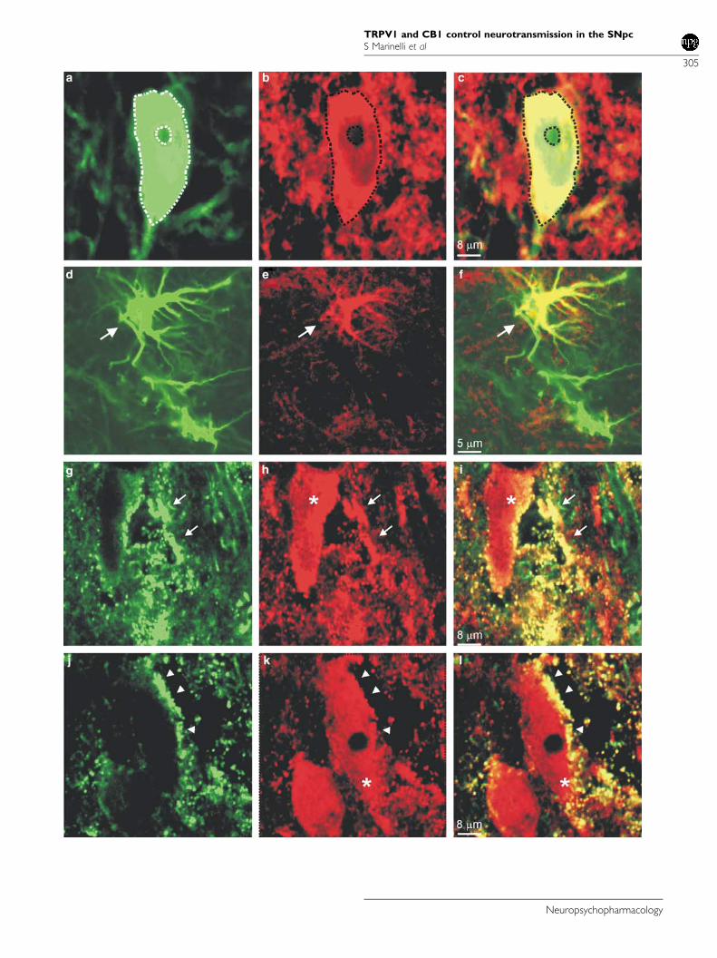
Double Immunofluorescence
Confocal microscopy showed that TRPV1 immunoreactivitywas present in cell bodies, dendrites, and terminals as welldiffusely in the neurophil (Figure 4a–f). In the cell bodies,TRPV1 was present in both the nuclear and cytoplasmaticcompartments leaving the nucleoli unstained. Preliminaryimmunofluorescence experiments performed on juvenilerats used for electrophysiological recordings showed thesame results of the adult rats. Double immunofluorescencefor TRPV1 and TH showed that many TH-positive neuronspresented TRPV1 immunoreactivity and that some TRPV1-positive cells were TH negative. (Figure 4a–c). No quanti-fication was attempted. TRPV1-positive TH-negative cellbodies were scattered and some of them presented themorphological appearance of astrocytes. As TRPV1-positiveastrocytes have been found in other brain areas, a doublelabeling between TRPV1 and an astrocyte marker wasperformed. This hypothesis was confirmed by TRPV1 andGFAP double immunofluorescence that showed colocaliza-tion of GFAP and TRPV1 immunoreactivities (Figure 4d–f).TRPV1 immunoreactivity was found in astrocytic cellbodies as well in processes throughout the tissue. Onlysome astrocytes were TRPV1 positive, indicating thepresence of a subpopulation of TRPV1-expressing astro-cytes. CB1-R immunofluorescence was found in cell proces-ses, small diameter fibers, and puncta. No clear cell labelingwas observed. Often CB1-R-positive processes and punctawere in close apposition with cellular profiles (Figure 4g–l).Double immunofluorescence for CB1-R and TRPV1 showedthat many of CB1-R-positive processes and puncta were alsoTRPV1 positive. These double-labeled structures tended tosurround neuronal bodies appearing either TRPV1 positiveor negative (Figure 4g–l).
DISCUSSION
The results of the present study show a complex patternof effects mediated by NADA on DA neurons in theSNpc, via activation of TRPV1 and CB1-R. In particular, ouranatomical and physiological data indicate the presenceof both receptors on the same presynaptic structuresand suggest the presence of CB1-R on glutamatergicand GABAergic terminals, while TRPV1 are localized onglutamatergic terminals only.
Confocal analyses demonstrate a colocalization of CB1-Rand TRPV1 in punctate structures, possibly synaptic struc-tures, in close contact with SNpc DA neurons. However,these analyses do not allow a definite characterization of thelabeled structures as presynaptic elements. Nevertheless,
TRPV1 and CB1 control neurotransmission in the SNpcS Marinelli et al
303
Neuropsychopharmacology

patch-clamp results indicating functional interactionsbetween TRPV1 and CB1-R, particularly on putative gluta-matergic terminals, strongly support the anatomical colo-calization data. Moreover, previous studies sustain thissuggestion indicating that excitatory afferents to SNpc origi-
nating from cortex, subthalamic nucleus, and amygdaloidcomplex, express mRNA for both TRPV1 and CB1-R(Mailleux and Vanderhaeghen, 1992; Matsuda et al, 1993;Mezey et al, 2000; Moldrich and Wenger, 2000; Romeroet al, 2002).
Figure 3 NADA exerts a CB1-mediated effect when the EMT is blocked. (a) Trace records obtained from a DA neuron in control, with OMDM-1(1mM), NADA (1 mM), and after washout with the CB1-R antagonist, AM281 (500 nM). On the right, cumulative curve distributions of the same neuron onthe left, in control (solid line), OMDM-1 (dashed line), during NADA superfusion (dotted line), and during washout with AM 281(dashed dotted line). Notethat OMDM-1 reduced per se the frequency of sEPSCs (po0.01 KS-test). NADA further shifted the distribution curve towards the right (po0.01 KS test).(b) Summary histogram of NADA on sEPSCs in the presence of OMDM-1. Both OMDM-1 and NADA (in OMDM-1) significantly reduced the frequency ofsEPSCs (mean7SEM, n¼ 6, *po0.05 and po0.02, respectively, Student’s t-test paired). AM 281 partially reversed the reduction caused by NADA whenthe EMT was blocked. (c) Sample of traces from a DA neuron in control, with IRTX (300 nM), with IRTX + OMDM-1 (1 mM), with IRTX + OMDM-1, andNADA (10 mM). On the right, the cumulative probability of interevent intervals from the same neuron on the left. IRTX significantly reduced the frequencyof sEPSCs (KS-test po0.002) while bath coapplication with OMDM-1 did not (KS-test p¼ 0.30). Conversely, NADA still reduced sEPSCs frequency (KS-test po0.002). All these compounds did not affect sEPSCs amplitude. (d) Summary histogram of the effect of OMDM-1 on sEPSCs in the presence of IRTX.IRTX significantly reduced the frequency of sEPSCs (mean7SEM, n¼ 5, *po0.05, Student’s t-test paired) whereas OMDM-1 did not further reducesignificantly the rate of sEPSCs when coapplied with IRTX (mean7SEM, n¼ 5, *p¼ 0.2, Student’s t-test paired).
Figure 4 Laser confocal images of double immunofluorescence for TRPV1 and TH (a–c), for TRPV1 and GFAP (d–f), and for TRPV1 and CB1 (g–l) in thesubstantia nigra pars compacta (SNpc). (a–c) Dopaminergic neuron positive for TRPV1. (a) TH immunoreactivity (green), (b) TRPV1 immunoreactivity(red), (c) merge. Note TRPV1 positive structures in close apposition with the neuronal membrane (outline). (d–f) Astrocyte positive for TRPV1. (d) GFAPimmunoreactivity (green), (e) TRPV1 immunoreactivity (red), (f) merge. (g–l) Processes (arrow heads) and puncta (arrows) expressing both TRPV1 andCB1 in contact with TRPV1-positive neuronal cell bodies (asterisks). (g and j) CB1 immunoreactivity (green), (h and k) TRPV1 immunoreactivity (red),(i and l) merge.
TRPV1 and CB1 control neurotransmission in the SNpcS Marinelli et al
304
Neuropsychopharmacology
TRPV1 and CB1 control neurotransmission in the SNpcS Marinelli et al
305
Neuropsychopharmacology

Confocal microscopy analyses also suggest that CB1-Rexpression is only presynaptic, whereas TRPV1 can be bothpre- and postsynaptic as previously reported in rat spinalcord (Valtschanoff et al, 2001). The functional role ofpostsynaptic TRPV1 still needs to be identified, as ourelectrophysiological observations only refer to the presynapticfunction of this receptor (see below). TRPV1 immuno-reactivity is present in dopaminergic cell bodies and dendritesas well as in astrocytes. The presence of TRPV1 in the neuro-nal bodies and dendrites has been already shown in differentareas of the brain (Mezey et al, 2000; Toth et al, 2005). So far,the TRPV1 expression has been demonstrated in astro-cytes and pericytes in different areas of the CNS (Doyle et al,2002; Toth et al, 2005). Interestingly, the localization ofTRPV1 on both neurons and glia suggest the hypothesis of afunctional neuron–glia crosstalk sustained through thisreceptor. The functional role of TRPV1 expression in glialcells is intriguing and will be addressed in future experiments.
NADA is an endogenous molecule present in several brainareas (Huang et al, 2002b). In control midbrain slices,we could not detect significant levels of NADA, probablybecause it was below the detection limit of the assay(o0.5 pmol). Indeed, after stimulation by high potassium, acomponent with the characteristics of NADA (retentiontime and molecular mass) was detected. This could beexplained by the fact that NADA is mainly synthesized inareas not included within our slice preparation, so thatonly terminals from those areas can release this mediatorfollowing depolarization with high potassium. A possiblecandidate could be the striatum, which has been shown tocontain high levels of NADA (Huang et al, 2002b). Prelimi-nary data indicate that, indeed, NADA is produced in thestriatum from the condensation of DA and arachidonicacid, and not from the transformation of N-arachidonoyl-tyrosine (Michael J Walker, personal communication and VDi Marzo, unpublished observations). However, the enzymecatalyzing the formation of NADA has not been identifiedyet, nor it is known if, as possibly suggested by the presentdata, it is a Ca2 + -sensitive protein as in the case of AEA-biosynthesizing enzymes.
By using patch-clamp recordings, we found that exogen-ously applied NADA (10 mM)-enhanced or reduced gluta-matergic neurotransmission by a presynaptic mechanism,when coapplied with the CB1-R or TRPV1 antagonist,AM281 and IRTX, respectively. Remarkably, exogenouslyapplied NADA could facilitate glutamate release via TRPV1even in the absence of the CB1-R antagonist, when testedat low doses (1mM). This is in agreement with a reportedhigher effect of NADA at TRPV1 than at CB1-R (Bisognoet al, 2000; Huang et al, 2002b). It is worth to note thatNADA, when applied at a high dose (10 mM) and in theabsence of either CB1-R or TRPV1 antagonists, did notsignificantly modify glutamate transmission to the DAneurons. The lack of effects might be due to the simul-taneous activation of CB1-R and TRPV1, giving rise toopposing actions. When compared to our previous experi-ments (Marinelli et al, 2003), and in line with other in vitrostudies (Huang et al, 2002b; Harrison et al, 2003; Priceet al, 2004), the TRPV1-mediated increase of glutamatergictransmission by NADA or by the exogenous TRPV1 agonistcapsaicin, was significantly higher than that induced byAEA. In fact, AEA did not produce any significant facili-
tation of glutamate release unless coapplied with theadenylate cyclase activator forskolin (Marinelli et al, 2003).
The presynaptic distribution of CB1-R in the SNpc herereported is in line with the presynaptic effects on glutamateand GABA release by NADA and its synthetic cannabinoidagonist WIN. This is in agreement with earlier studiesreporting a presynaptic modulation of neurotransmissionby CB1-R in several brain regions, especially in the ventralmidbrain (Szabo et al, 2000; Gerdeman and Lovinger,2001; Huang et al, 2001, 2002a; Azad et al, 2003; Melis et al,2004).
Similarly to what has been previously observed for AEA(De Petrocellis et al, 2001), the vanilloid and cannabinoideffects of NADA shown in this study appear to depend onthe cellular distribution of CB1-R and TRPV1 binding sitesin the extracellular and intracellular milieu, respectively.The binding site for capsaicin and AEA on TRPV1 hasbeen found in a cytosolic domain of this channel (Jordtand Julius, 2002); therefore, these compounds mustcross the plasma membrane in order to stimulate TRPV1(De Petrocellis et al, 2001). With regard to NADA, thiscompound is rapidly taken up by C6 glioma cells via afacilitated diffusion process similar to that of AEA (Huanget al, 2002b). We now show that, when this transportprocess is blocked by the selective EMT inhibitor OMDM-1,the effects of NADA at TRPV1 are abolished, whereas thosethrough CB1-R are favored. Thus, it appears that NADA,like AEA, must be taken up by cells in order to interact withthe intracellular binding site of TRPV1. Such phenomenonhas recently been reported also for TRPV1-mediated actionsby NADA and AEA in trigeminal neurons (Price et al, 2005).Although the exact binding site of NADA on TRPV1 has notbeen identified yet, its chemical similarity with AEA mightsuggest that NADA uses the same intracellular domainimplicated in AEA binding to this channel. Unfortunately,to date, there is no molecular evidence for the expression ofEMT. However, selective endocannabinoid uptake inhi-bitors, such as OMDM-1, are useful pharmacological toolsto study its functional role. Even though such tools donot allow to establish the exact localization of this putativeprotein at the pre- or post-synaptic level, functionalevidence suggests the existence of a postsynaptic transpor-ter mechanism in striatal neurons (Ronesi et al, 2004).
Interestingly, we have shown that OMDM-1 reduced perse the frequency of sEPSCs, whereas it did not affectGABAergic synapses, as they are not under the control ofTRPV1 (Marinelli et al, 2003). Inhibition of glutamatergictransmission by NADA in the presence of OMDM-1 was notcompletely reverted by the CB1-R antagonist AM 281(Figure 3b), indicating that this effect was not due just toan increased action onto CB1-R by endogenous compounds,but also to inhibition of a tonic facilitatory control throughTRPV1. This hypothesis is confirmed by the lack of theeffects of OMDM-1 on the glutamatergic transmission whenthe TRPV1 are blocked and supported by our previousfinding of a tonic facilitation of glutamate release by TRPV1(Marinelli et al, 2003). Thus, these results may be seen asfurther evidence that, at least in our experimental condi-tions, the excitatory inputs onto DA neurons are underTRPV1 tonic control. With regard to CB1-R, they have beenshown to exert a tonic inhibition at GABAergic terminalonto the DA neurons (Yanovsky et al, 2003), while at
TRPV1 and CB1 control neurotransmission in the SNpcS Marinelli et al
306
Neuropsychopharmacology

glutamatergic synapses their effect becomes evident onlyupon DA neurons depolarization (Melis et al, 2004).
In conclusion, NADA can exert opposing actions on DAneurons via activation of TRPV1 or CB1-R: an excitatoryeffect, exerted by increasing glutamatergic (throughTRPV1) and decreasing GABAergic transmission (throughCB1-R), and an inhibitory one, occurring through adecrease of glutamatergic transmission (through CB1-Ronly).
It might be speculated that activation of either TRPV1 orCB1-R through NADA may be dependent on the functionalstate of the dopamine system. As NADA levels stronglydepend on those of dopamine (JM Walker, personalcommunication to VDM), it is possible that, whereaslow levels of NADA preferentially activate the vanilloidreceptors with subsequent glutamate release, a progressiveincrease in neuronal excitation is expected to lead to higherlevels of this compound, and thus to retrograde activa-tion of CB1-R and of its subsequent inhibitory effects onglutamatergic signaling.
ACKNOWLEDGEMENTS
We thank Dr N Berretta, for the helpful discussion and forreading the manuscript.
REFERENCES
Ahluwalia J, Urban L, Bevan S, Capogna M, Nagy I (2002).Cannabinoid 1 receptors are expressed by nerve growth factor-and glial cell-derived neurotrophic factor-responsive primarysensory neurons. Neuroscience 110: 747–753.
Ahluwalia J, Urban L, Capogna M, Bevan S, Nagy I (2000).Cannabinoid 1 receptors are expressed in nociceptive primarysensory neurons. Neuroscience 100: 685–688.
Al-Hayani A, Wease KN, Ross RA, Pertwee RG, Davies SN (2001).The endogenous cannabinoid anandamide activates vanilloidreceptors in the rat hippocampal slice. Neuropharmacology 41:1000–1005.
Azad SC, Eder M, Marsicano G, Lutz B, Zieglgansberger W,Rammes G (2003). Activation of the cannabinoid receptor type 1decreases glutamatergic and GABAergic synaptic transmissionin the lateral amygdala of the mouse. Learn Mem 10: 116–128.
Beltramo M, Stella N, Calignano A, Lin SY, Makriyannis A,Piomelli D (1997). Functional role of high-affinity anandamidetransport, as revealed by selective inhibition. Science 277:1094–1097.
Bezuglov V, Bobrov M, Gretskaya N, Gonchar A, Zinchenko G,Melck D et al (2001). Synthesis and biological evaluation ofnovel amides of polyunsaturated fatty acids with dopamine.Bioorg Med Chem Lett 11: 447–449.
Bisogno T, Melck D, Bobrov M, Gretskaya NM, Bezuglov VV, DePetrocellis L et al (2000). N-acyl-dopamines: novel syntheticCB(1) cannabinoid-receptor ligands and inhibitors of ananda-mide inactivation with cannabimimetic activity in vitro andin vivo. Biochem J 351(Part 3): 817–824.
Caterina MJ, Julius D (2001). The vanilloid receptor: a moleculargateway to the pain pathway. Annu Rev Neurosci 24: 487–517.
Cravatt BF, Demarest K, Patricelli MP, Bracey MH, Giang DK,Martin BR et al (2001). Supersensitivity to anandamide andenhanced endogenous cannabinoid signaling in mice lackingfatty acid amide hydrolase. Proc Natl Acad Sci USA 98:9371–9376.
Cristino L, de Petrocellis L, Pryce G, Baker D, Guglielmotti V,Di Marzo V (2006). Immunohistochemical localization of
cannabinoid type 1 and vanilloid transient receptor potentialvanilloid type 1 receptors in the mouse brain. Neuroscience 139:1405–1415.
De Petrocellis L, Bisogno T, Maccarrone M, Davis JB, Finazzi-AgroA, Di Marzo V (2001). The activity of anandamide at vanilloidVR1 receptors requires facilitated transport across the cellmembrane and is limited by intracellular metabolism. J BiolChem 276: 12856–12863.
Di Marzo V, Bisogno T, De Petrocellis L (2001). Anandamide: somelike it hot. Trends Pharmacol Sci 22: 346–349.
Di Marzo V, Fontana A, Cadas H, Schinelli S, Cimino G, SchwartzJC et al (1994). Formation and inactivation of endogenouscannabinoid anandamide in central neurons. Nature 372:686–691.
Doyle MW, Bailey TW, Jin YH, Andresen MC (2002). Vanilloidreceptors presynaptically modulate cranial visceral afferentsynaptic transmission in nucleus tractus solitarius. J Neurosci22: 8222–8229.
Fegley D, Kathuria S, Mercier R, Li C, Goutopoulos A, MakriyannisA et al (2004). Anandamide transport is independent offatty-acid amide hydrolase activity and is blocked by thehydrolysis-resistant inhibitor AM1172. Proc Natl Acad Sci USA101: 8756–8761.
Fezza F, Battista N, Bari M, Maccarrone M (2006). Methods toassay anandamide hydrolysis and transport in synaptosomes.Methods Mol Med 123: 163–168.
Gerdeman G, Lovinger DM (2001). CB1 cannabinoid receptorinhibits synaptic release of glutamate in rat dorsolateralstriatum. J Neurophysiol 85: 468–471.
Harrison S, De Petrocellis L, Trevisani M, Benvenuti F, Bifulco M,Geppetti P et al (2003). Capsaicin-like effects of N-arachidonoyl-dopamine in the isolated guinea pig bronchi and urinarybladder. Eur J Pharmacol 475: 107–114.
Hillard CJ, Jarrahian A (2003). Cellular accumulation of anand-amide: consensus and controversy. Br J Pharmacol 140: 802–808.
Howlett AC (2002). The cannabinoid receptors. ProstaglandinsOther Lipid Mediat 68–69: 619–631.
Huang CC, Chen YL, Lo SW, Hsu KS (2002a). Activation of cAMP-dependent protein kinase suppresses the presynaptic cannabi-noid inhibition of glutamatergic transmission at corticostriatalsynapses. Mol Pharmacol 61: 578–585.
Huang CC, Lo SW, Hsu KS (2001). Presynaptic mechanismsunderlying cannabinoid inhibition of excitatory synaptic trans-mission in rat striatal neurons. J Physiol 532: 731–748.
Huang SM, Walker JM (2006). Enhancement of spontaneousand heat-evoked activity in spinal nociceptive neurons bythe endovanilloid/endocannabinoid N-arachidonoyldopamine(NADA). J Neurophysiol 95: 1207–1212.
Huang SM, Bisogno T, Trevisani M, Al-Hayani A, De Petrocellis L,Fezza F et al (2002b). An endogenous capsaicin-like substancewith high potency at recombinant and native vanilloid VR1receptors. Proc Natl Acad Sci USA 99: 8400–8405.
Jordt SE, Julius D (2002). Molecular basis for species-specificsensitivity to ‘hot’ chili peppers. Cell 108: 421–430.
Julian MD, Martin AB, Cuellar B, Rodriguez De Fonseca F, NavarroM, Moratalla R et al (2003). Neuroanatomical relationshipbetween type 1 cannabinoid receptors and dopaminergicsystems in the rat basal ganglia. Neuroscience 119: 309–318.
Kim SR, Lee da Y, Chung ES, Oh UT, Kim SU, Jin BK (2005).Transient receptor potential vanilloid subtype 1 mediates celldeath of mesencephalic dopaminergic neurons in vivo andin vitro. J Neurosci 25: 662–671.
Maccarrone M, Gubellini P, Bari M, Picconi B, Battista N, CentonzeD et al (2003). Levodopa treatment reverses endocannabinoidsystem abnormalities in experimental parkinsonism. J Neuro-chem 85: 1018–1025.
Mailleux P, Vanderhaeghen JJ (1992). Distribution of neuronalcannabinoid receptor in the adult rat brain: a comparative
TRPV1 and CB1 control neurotransmission in the SNpcS Marinelli et al
307
Neuropsychopharmacology
receptor binding radioautography and in situ hybridizationhistochemistry. Neuroscience 48: 655–668.
Marinelli S, Di Marzo V, Berretta N, Matias I, Maccarrone M,Bernardi G et al (2003). Presynaptic facilitation of glutamatergicsynapses to dopaminergic neurons of the rat substantia nigra byendogenous stimulation of vanilloid receptors. J Neurosci 23:3136–3144.
Marinelli S, Pascucci T, Bernardi G, Puglisi-Allegra S, Mercuri NB(2005). Activation of TRPV1 in the VTA excites dopaminergicneurons and increases chemical- and noxious-induced dop-amine release in the nucleus accumbens. Neuropsychopharmaco-logy 30: 864–870.
Marsicano G, Wotjak CT, Azad SC, Bisogno T, Rammes G, CascioMG et al (2002). The endogenous cannabinoid system controlsextinction of aversive memories. Nature 418: 530–534.
Matsuda LA, Bonner TI, Lolait SJ (1993). Localization ofcannabinoid receptor mRNA in rat brain. J Comp Neurol 327:535–550.
Melis M, Pistis M, Perra S, Muntoni AL, Pillolla G, Gessa GL(2004). Endocannabinoids mediate presynaptic inhibition ofglutamatergic transmission in rat ventral tegmental areadopamine neurons through activation of CB1 receptors.J Neurosci 24: 53–62.
Mercuri NB, Bonci A, Calabresi P, Bernardi G (1996). Charac-terization of a barium-sensitive outward current followingglutamate application on rat midbrain dopaminergic cells.Eur J Neurosci 8: 1780–1786.
Mezey E, Toth ZE, Cortright DN, Arzubi MK, Krause JE, Elde Ret al (2000). Distribution of mRNA for vanilloid receptorsubtype 1 (VR1), and VR1-like immunoreactivity, in the centralnervous system of the rat and human. Proc Natl Acad Sci USA97: 3655–3660.
Moldrich G, Wenger T (2000). Localization of the CB1 cannabinoidreceptor in the rat brain. An immunohistochemical study.Peptides 21: 1735–1742.
Ortar G, Ligresti A, De Petrocellis L, Morera E, Di Marzo V (2003).Novel selective and metabolically stable inhibitors of anand-amide cellular uptake. Biochem Pharmacol 65: 1473–1481.
O’Sullivan SE, Kendall DA, Randall MD (2004). Characterisationof the vasorelaxant properties of the novel endocannabinoid N-arachidonoyl-dopamine (NADA). Br J Pharmacol 141: 803–812.
Pazos MR, Nunez E, Benito C, Tolon RM, Romero J (2005).Functional neuroanatomy of the endocannabinoid system.Pharmacol Biochem Behav 81: 239–247.
Pettit DA, Harrison MP, Olson JM, Spencer RF, Cabral GA (1998).Immunohistochemical localization of the neural cannabinoidreceptor in rat brain. J Neurosci Res 51: 391–402.
Price TJ, Patwardhan A, Akopian AN, Hargreaves KM, Flores CM(2004). Modulation of trigeminal sensory neuron activity by the
dual cannabinoid–vanilloid agonists anandamide, N-arachid-onoyl-dopamine and arachidonyl-2-chloroethylamide. Br JPharmacol 141: 1118–1130.
Price TJ, Patwardhan AM, Flores CM, Hargreaves KM (2005). Arole for the anandamide membrane transporter in TRPV1-mediated neurosecretion from trigeminal sensory neurons.Neuropharmacology 49: 25–39.
Romero J, Lastres-Becker I, de Miguel R, Berrendero F, Ramos JA,Fernandez-Ruiz J (2002). The endogenous cannabinoid systemand the basal ganglia. Biochemical, pharmacological, andtherapeutic aspects. Pharmacol Ther 95: 137–152.
Ronesi J, Gerdeman GL, Lovinger DM (2004). Disruption ofendocannabinoid release and striatal long-term depression bypostsynaptic blockade of endocannabinoid membrane transport.J Neurosci 24: 1673–1679.
Sagar DR, Smith PA, Millns PJ, Smart D, Kendall DA, Chapman V(2004). TRPV1 and CB(1) receptor-mediated effects of theendovanilloid/endocannabinoid N-arachidonoyl-dopamine onprimary afferent fibre and spinal cord neuronal responses inthe rat. Eur J Neurosci 20: 175–184.
Szabo B, Siemes S, Wallmichrath I (2002a). Inhibition ofGABAergic neurotransmission in the ventral tegmental area bycannabinoids. Eur J Neurosci 15: 2057–2061.
Szabo B, Wallmichrath I, Mathonia P, Pfreundtner C (2000).Cannabinoids inhibit excitatory neurotransmission in thesubstantia nigra pars reticulata. Neuroscience 97: 89–97.
Szabo T, Biro T, Gonzalez AF, Palkovits M, Blumberg PM (2002b).Pharmacological characterization of vanilloid receptor located inthe brain. Brain Res Mol Brain Res 98: 51–57.
Toth A, Boczan J, Kedei N, Lizanecz E, Bagi Z, Papp Z et al (2005).Expression and distribution of vanilloid receptor 1 (TRPV1) inthe adult rat brain. Brain Res Mol Brain Res 135: 162–168.
Tsou K, Brown S, Sanudo-Pena MC, Mackie K, Walker JM (1998).Immunohistochemical distribution of cannabinoid CB1 recep-tors in the rat central nervous system. Neuroscience 83: 393–411.
Valtschanoff JG, Rustioni A, Guo A, Hwang SJ (2001). Vanilloidreceptor VR1 is both presynaptic and postsynaptic in thesuperficial laminae of the rat dorsal horn. J Comp Neurol 436:225–235.
Wallmichrath I, Szabo B (2002a). Cannabinoids inhibit striatoni-gral GABAergic neurotransmission in the mouse. Neuroscience113: 671–682.
Wallmichrath I, Szabo B (2002b). Analysis of the effect ofcannabinoids on GABAergic neurotransmission in the substan-tia nigra pars reticulata. Naunyn Schmiedebergs Arch Pharmacol365: 326–334.
Yanovsky Y, Mades S, Misgeld U (2003). Retrograde signalingchanges the venue of postsynaptic inhibition in rat substantianigra. Neuroscience 122: 317–328.
TRPV1 and CB1 control neurotransmission in the SNpcS Marinelli et al
308
Neuropsychopharmacology