temperature dependence of molecular interactions involved in defining stability of glutamine binding...
TRANSCRIPT
Temperature dependence of molecular interactions involved indefining stability of Glutamine Binding Protein and its complexwith L-Glutamine†
Sara Pistolesi and Nico Tjandra*
Laboratory of Molecular Biophysics, National Heart, Lung, and Blood Institute, National Institutesof Health, 50 South Drive, Bethesda, MD 20892
AbstractTemperature dependence of dynamic parameters derived from nuclear magnetic resonance (NMR)relaxation data are related to conformational entropy of the system under study. This providesinformation such as macromolecules stability and thermodynamics of ligand binding. We studiedthe temperature dependence of NMR order parameter of Glutamine binding protein (GlnBP), aperiplasmic binding protein (PBP) highly specific to L-glutamine associated with its ABCtransporter, with the goal of elucidating the dynamical differences between the respective ligandbound and free forms. We found that the protein-ligand interaction, which is stabilized at highertemperature, has a striking effect on the stability of the hydrophobic core of the large domain ofGlnBP. Moreover, in contrast to what was found for less specific PBPs the decreasing backbonemotion of the hinge region at increasing temperature supports the idea that the likelihood thatGlnBP can adopt a ligand free closed conformation in solution diminishes at higher temperatures.Our results support the induced-fit model as mode of action for GlnBP. In addition, we found thatthe backbones of residues involved in a salt bridge do not necessarily become more rigid as thetemperature rises as it was previously suggested [Vinther, J.M. et. al. (2011) J. Am. Chem. Soc.,271–278]. Our results show that for this to happen, these residues have to also directly interactwith a region of the protein that is becoming more rigid as the temperature increases.
IntroductionProteins are dynamic, sometimes adopting a large variety of different conformations, andoften changing their interaction partners while exerting their functions. How these dynamicsaffect function is not totally clear yet, but it is evident that conformational transitions play akey role in events such as allosteric regulation, signal transduction and enzymatic activity.Periplasmic binding proteins (PBPs) undergo large conformational transitions. They are thefirst component in the membrane import machinery classified as the ABC transporters. PBProle is to recruit the appropriate substrates in the periplasm and bring them to the membraneinterface where the ABC transporter resides to initiate the translocation across the cellularmembrane. PBPs structure comprises two similar globular domains linked by flexiblehinges. Generally, when they are free they assume an open conformation with extended
†This work was supported by the Intramural Research Program of the NIH, NHLBI.*Corresponding author: Nico Tjandra. Building 50, Room 3503, NHLBI, NIH, Bethesda, MD 20892. Phone: +1 (301)-402-3029. Fax:+1 (301)-402-3405. [email protected].
Supporting Information Available: A figure of correlation time dependence on temperature, a figure to compare order parametersdetermined by various methods for two representative residues, and a figure of ITC traces and binding isotherm. A table of diffusionparameters for GlnBP at different temperatures, as well as a table listing order parameters at 15°C for the free and bound forms ofGlnBP and a table listing percent changes in order parameters at different temperatures. This material is available free of charge viathe Internet at http://pubs.acs.org.
NIH Public AccessAuthor ManuscriptBiochemistry. Author manuscript; available in PMC 2012 December 04.
Published in final edited form as:Biochemistry. 2012 January 17; 51(2): 643–652. doi:10.1021/bi201494h.
$waterm
ark-text$w
atermark-text
$waterm
ark-text
linkers. When binding the substrate the linker bends, bringing the two domains in closecontact, thus adopting the closed conformation. In this closed form the active site isembedded in the protein at the interface of the two domains. There are a few exceptions,where the closed-free and open-bound conformations of some PBPs have been observed(1–4). Among PBPs, Glutamine binding protein (GlnBP) is a monomeric 226 residue proteinresponsible for delivering L-glutamine from the periplasm to the corresponding ABCtransporter with very high affinity (Kd 0.1 μM) and selectivity (i.e. it binds only onesubstrate in contrast to other PBPs)(5). The structure is composed by one large domain(residues 1–84 and 186–226) and one small domain (90–180) that share a common global α/β fold with internal core β-sheets surrounded by αhelices. A flexible hinge, made by two β-strands responsible for a major conformational transition upon binding, separates these twodomains. In absence of ligand these two β-strands are extended and when the glutamine isbound these β-strands bend bringing the two domains in direct contact with each other (6).In the closed conformation a cleft is formed by the interface of the two domains where theligand binding site is located(7).
NMR spin relaxation experiments can provide motional parameters that are helpful inunderstanding the pico- to nanosecond time scale motions of bond vectors that can report onlocal structural rearrangements in macromolecules. Conventional backbone 15N T1, T2 andNOE experiments are most commonly analyzed in terms of the Model-Free approach(8–12)with the aim of extracting diffusion parameters useful to describe protein dynamics. Amongthose, generalized order parameter S2 represents the amplitude of bond vector fluctuationsand is related to the thermodynamic properties of the biomolecule. In fact, S2 has beenrelated to conformational entropy(13–16).
Conformational entropy represents the distribution of conformational states of a protein. Themain application of measuring conformational entropy changes (i.e. changes in orderparameter) between different states of a protein (e.g. free and ligand bound) is to understandbinding affinity, and catalysis or cooperativity(17, 18). In addition, one can also measurethese changes as a function of temperature, which is equivalent to heat capacity changes(ΔCp). The heat capacity change governs the energy landscape of conformational states withrespect to temperature variations, thus defining the stability of the proteins and theircomplexes at various temperatures(19). To date only a few attempts have been made toanalyze the detailed temperature dependence of macromolecule dynamics. Previous studieshave exploited both backbone and side chain fast dynamics to explore possible correlatedbackbone motion in ubiquitin(20), inter-domain behavior in calmodulin(21), folding/unfolding processes of proteins(22) and RNA(23), protein-ligand interactions(24) andthermostability of proteins(25). Even smaller number of studies have analyzed orderparameter temperature dependence to specifically unveil heat capacity changes related toprotein stability(26, 27) and ligand association(28, 29). The general lesson depicted by thesestudies is that depending on the protein or protein-complex under study, completely oppositeconformational heat capacity contributions can be obtained with the apparent lack of generalrules other than internal redistribution of energy.
Here we analyze the residue specific temperature dependence of the pico- to nanoseconddynamics of the backbone amide groups of GlnBP in both the ligand free and bound forms.The relaxation data has been used directly to calculate the residue specific order parameterwhose temperature dependence analysis shed light on the thermodynamics of ligandbinding, which support the so-called “induced fit model”. According to this model, a proteindoes not adopt a bound conformation until the ligand triggers the transition from unligandedto liganded conformations. This mechanism was shown to be accompanied by a decrease inconformational entropy due to the active site becoming more rigid and an increase inbinding enthalpy due to new bond formations(16). Also, in contrast to what has been
Pistolesi and Tjandra Page 2
Biochemistry. Author manuscript; available in PMC 2012 December 04.
$waterm
ark-text$w
atermark-text
$waterm
ark-text
proposed in the past(25) our study showed that the presence of a salt bridge is not asatisfactory condition for increased rigidity of the corresponding backbones as a function ofincreasing temperature.
Material and MethodsSample Preparation
Wild-type GlnBP, both in the glutamine free and the bound forms, was generated using thepJ133 plasmid(30) as a template. Protein expression and purification were performed asdescribed elsewhere(31). In brief, uniformly 15N-enriched proteins were over expressed inE. coli and cells were allowed to grow in minimal medium containing 15NH4Cl as the solenitrogen source. The proteins were then extracted by chloroform shock(32) and purified witha two–step purification process, via anion-exchange (DEAE) and size exclusion(Superdex-75) columns (GE Healthcare), respectively(30). In order to get the free form,after the DEAE column, the sample was denatured with 6M GndHCl and size exclusionchromatography run under denaturing conditions. Isolated GlnBP free form was thenrefolded by extensive dialysis(33). Proteins were subsequently lyophilized and re-dissolvedovernight in NMR buffer to a final sample concentration of 2 and 1 mM for the bound andthe free form, respectively; when present L-Gln was in 3-fold molar excess. Bound formwas dissolved in 20 mM potassium phosphate, 2 mM EDTA at pH 7.2 while free formsolubilized in 20 mM potassium phosphate, 2 mM EDTA and 5 mM CHAPS at pH 7.2.
Relaxation MeasurementsConventional 15N longitudinal and transverse relaxation times(12) were measured for bothbound and free GlnBP at eight different temperatures, 15, 18, 21, 25, 30, 33, 37 and 40 °C.The T1 delays were 8, 160, 520, 720, 960, 1120, 1440 and 1600 ms while the T1ρ delayswere 3.28, 11.92, 21.52, 31.12, 40.72, 50.32, 83.92 and 117.52 ms. The T1ρ spin lockfrequency was 2.5 kHz(34), and spectra were collected in an interleaved manner to minimizethe effects of systematic errors. The relaxation rates were derived from the exponentialdecay of the peak heights using a home-built program. Finally, the R2 rates were calculatedon the basis of R1ρ and R1 rates, applied spin lock strength, and the angular frequency offsetof the individual 15N spin. Experiments were repeated in duplicate for GlnBP bound formfor estimating the random errors.
Theoretical ConsiderationsR1 and R2 relaxation rates of an amide 15N are expressed in terms of linear combination ofspectral densities at different frequencies and it is easy to demonstrate that
(1)
where d2 = 0.1 γ2A γ2
x h2/(4π2) ⟨1/r3AX⟩ and c2 = (2/15) γ2
x H20 (σ|| − σ⊥) are constants
related to dipolar interaction and chemical shift anisotropy, respectively. In detail, A = 1H, X= 15N, γi is the gyromagnetic ratio of a specific nucleus, h is Plank’s constant, rAX is theinternuclear 1H-15N distance, H0 is the magnetic field strength and σ|| and σ⊥ are theparallel and perpendicular components of the axially symmetric 15N chemical shift tensor.The spectral density is related to the order parameter by the relation
Pistolesi and Tjandra Page 3
Biochemistry. Author manuscript; available in PMC 2012 December 04.
$waterm
ark-text$w
atermark-text
$waterm
ark-text
(2)
where the coefficients Ai are related to the cosine directions of the bond vectors in thediffusion tensor frame of the molecule, τi are the relative time constants and τ = (τc τe)/(τc +τe)(35). Both the directions and time constants are detailed elsewhere(36).
Under the assumptions that J(0)≫J(ωH) and te ≪ tc, and combining equation (1) and (2) weobtain
(3)
from which S2 can be easily calculated.
Error CalculationErrors in T1ρ and T1 for the bound form were estimated from the root mean squaredifference between the two sets of experiments. The error on the measurements of the freeform was estimated by comparing the signal-to-noise ratio (S/N) of T1ρ and T1 spectra foreach temperature to those of the bound form spectra. The relative ratios of S/N in respectiveexperiments were used to normalize the experimental errors. The errors on S2 were thenassessed by error propagation.
Results and DiscussionIn this study a total of 208 and 178 residues were analyzed for GlnBP bound and free forms,respectively. All residues whose resonances overlap, even at a single temperature, wereeliminated from the analysis. Diffusion tensors calculation was performed by consideringonly the residues located in well-defined secondary structure motifs.
GlnBP overall correlation time (τc) (shown in Figure S1), calculated by fitting the relaxationdata to an axially symmetric diffusion tensor, linearly decays as a function of η/T (where ηis viscosity and T is temperature in Kelvin) for both the ligand free and bound formsfollowing the Stokes-Einstein equation(37). Furthermore, polar angles describing theorientation of the principal diffusion axis within the molecular PDB frame and the diffusionanisotropy do not change (Table S1) over the temperature range in this study. Constantanisotropy and orientation of the diffusion tensor eliminates their possible contribution tothe temperature dependence of the relaxation rates ensuring the differences in S2
R are solelydue to temperature dependent effects on backbone fluctuations.
Order parameters calculated using equation 3 (S2R) was compared with the one obtained
with the Lipari-Szabo formalism (S2)(8). The 2R2-R1 approach was first introduced byHabazettl et al. (38, 39) as a faster and convenient way to get order parameters values fromR1 and R2 data without resorting to the time consuming and low sensitivity NOEmeasurements. This approximation was shown to introduce minimal errors in the orderparameter values compared to synthetic data. In the present work we quantitatively assessedthe maximum error introduced by our approximations (see Materials and Methods). Forinstance we would expect a maximum of 1% error introduced in our calculated S2
R if weassume J(ωH)≈0 and a maximum of 1.4% error for assuming τ′ ≈ 0 within the limit of τe <200 ps, thus confirming previous qualitative results(38, 39). However, differently from theprevious 2R2-R1 approach, we introduced in our calculation the contribution coming from
Pistolesi and Tjandra Page 4
Biochemistry. Author manuscript; available in PMC 2012 December 04.
$waterm
ark-text$w
atermark-text
$waterm
ark-text
diffusion anisotropy through the directional cosines of the NH bond vectors to S2R. This
implementation lead to a reduction in the discrepancies between fitted and calculated orderparameters (see Figure S2).
We found an excellent correlation between order parameters that were calculated usingequation 3 or extensive fitting of our data to the Model-Free approach as illustrated in Figure1. For some residues, however, S2
R can still be overestimated and in some cases exceedingthe non-physical value of 1. The 2R2-R1 values which are higher than 1 have beenpreviously observed and related to conformational exchange(39). In this case the differencebetween fitted S2 and S2
R is not constant but varies with the temperature, meaning that S2R
variations do not solely reflect differences in amplitudes of motion, but conformationalexchange as well. In this respect, all of the residues with S2
R>1 were excluded from theanalysis. For most of the residues under study, although in some cases the absolute values offitted S2 and S2
R differ slightly (see Figure 1), the trend of their temperature dependence isthe same. This ensures us that the temperature dependence analysis reflects only differencesin order parameters and it is unaffected by other factors such as chemical exchange.
Comparison of order parameters between free and bound forms of GlnBPEvaluating the differences between the order parameter of free and bound form of GlnBP atsingle temperatures gives us insights into the thermodynamics of ligand binding. Asexpected, the average S2
R for the whole protein diminishes with increasing temperature (seeFigure 2) for both the free and the bound form of GlnBP indicating that the protein becomesglobally more flexible as the temperature rises. Also, by comparing the average orderparameters of the two GlnBP forms (Figure 2) it is clear that glutamine binding leads to areduction in flexibility in the GlnBP backbone at all the temperatures under study with anincrease in order parameter ranging between 1.8% at 15°C and 3.6% at 40°C. It isinteresting to note that most of the residues that in free GlnBP are more flexible, comparedto the bound form, are located on the surface of the protein (see Figure 3A). As expected,residues found at the interface of the two domains in the closed conformation (i.e next to theactive site) are more flexible in the free form in virtue of their higher surface exposurefollowing the opening of the binding cleft (Figure 3B). This behavior corresponds to anunfavorable decrease in conformational entropy upon ligand binding. To determine thethermodynamic basis for the observed behavior we measured the free energy (ΔG=−9.5kcal/mol) of binding and its enthalpic (ΔH=−15.9 kcal/mol) and entropic (TΔS=−6.4 kcal/mol) contributions by ITC (see Supplementary Methods). Ligand binding is characterized bya large favorable enthalpy change that overcomes an unfavorable entropy variation (seeFigure S3).
Although solvent exposed in both free and bound GlnBP, the hinge region (Y85-G89 andE181-Y185) shows a decrease in flexibility in the bound form (see Figure 3C). In fact, in thebound GlnBP the hinge region is stabilized in a bent conformation due to the fact that thetwo domains are in close contact with each other and form favorable electrostaticinteractions. When the ligand is released there are no stabilizing forces acting on the hingesuch that it displays increased degrees of freedom and enhanced flexibility. What was notexpected was the behavior of some regions that are not directly related with ligand binding(e.g. active site residues and hinges). Remarkably, most of these areas are located in thesmall domain for which the rms between free and bound form backbones is 1.29 Å asopposite to the large domain where rms is only 0.56 Å. For these regions the increasedflexibility corresponds to a change in local structure.
Pistolesi and Tjandra Page 5
Biochemistry. Author manuscript; available in PMC 2012 December 04.
$waterm
ark-text$w
atermark-text
$waterm
ark-text
Analysis of temperature dependence of order parametersWhile the analysis of differences in S2
R between free and bound GlnBP at a singletemperature provides insights into the energetics of ligand binding, the analysis of S2
R in atemperature dependent fashion can yield additional and important information on stability ofthe individual conformations of the protein. In this respect, we conducted a residue specificanalysis of the S2
R temperature dependence of both the free and the bound forms of GlnBP.Three different classes of residues could be identified based on their temperaturedependence pattern. In the first class are residues whose order parameters increased whenthe temperature increased, the second class contains residues whose order parameters did notchange, and the last class has residues with order parameters that decreased with increasingtemperature.
In detail, taking S2R value at 15°C as a reference, the percentage differences of order
parameters were calculated for the remaining temperatures for each residue. Residuesdisplaying at least three consecutive temperature points over the maximum percentage error(1.2 % for the bound form and 4.2 % for the free form), were considered to behave as“positive” (S2
R increasing with the temperature, i.e. decreasing fluctuations), thosedisplaying at least three consecutive temperature points below the maximum percentageerror were considered “negative” (decreasing S2
R with temperature, i.e. increasing theirflexibility) while the residues with at least three temperature points within the maximumpercentage error were classified as “constant”. In Figure 4 representative residues from eachclass are shown while the classification list is provided in the Supporting Information. Thetemperature dependence behavior of these residues is not related to particular amino acidproperties such as hydrophobicity, polarity or charge. On the other hand, the location of theresidues belonging to a specific class is more interesting.
Residues in the ligand binding siteBinding site residues (D10, A67, G68, T70, R75, K115, G119 H156 and D157) interact withthe glutamine ligand by means of hydrogen bonds (D10, R75 and D157) and salt bridges(D10, R75 and D157) and they form a ligand binding site, which is buried and sharedbetween the two domains. For the bound form we were able to analyze seven out of nine ofthese residues, with the exception of D10 and K115 due to exchange and spectral overlap,respectively. As shown in Figure 5, T70, G119 and D157 become more rigid as thetemperature increases, that is they display increasing S2
R that is related to a decrease inconformational entropy. According to Figure 5, G68, R75 and H156 are classified as“constant” residues, which means that temperature variations do not affect their interactionto glutamine ligand and their contribution to complex stability is constant. Residue A67 isthe only active site residue showing a negative dependence upon temperature increase.
For the GlnBP free form, however, only two residues from the active site (R75 and D157)were available for analysis due to spectral overlap, making a meaningful comparisonimpossible between the free and the bound form. The only thing that can be said is that invirtue of its increased solvent exposure D157 backbone in free GlnBP is in absolute valuedramatically more flexible (ΔS2
R ≈ 0.4) than in the bound form. At the same time however,it displays the same positive order parameter temperature dependence. Also R75 becomesmore flexible than in the bound form but its temperature dependence turns from being“constant” in bound GlnBP to “positive” in free GlnBP.
Residues with positive order parameter temperature dependenceIn the GlnBP bound form “positive” residues (i.e. decreasing their backbone flexibility asthe temperature increases) are located at different positions depending on the domain thatthey belong to. In the large domain (residues L5-G84 and G186-K226), they form a buried
Pistolesi and Tjandra Page 6
Biochemistry. Author manuscript; available in PMC 2012 December 04.
$waterm
ark-text$w
atermark-text
$waterm
ark-text
hydrophobic center that extends from the glutamine binding site to the bottom of the domain(see Figure 6A and 6B). This hydrophobic core mainly includes phenylalanines andtryptophanes that network with each other through parallel and T-stacking interaction. Thesehydrophobic “positive” residues are then protected from the protein surface by a salt bridgebetween D30 and K219 that is solvent exposed and also shows a “positive” behavior (seeFigure 6B). As pointed out above, general chemical properties of individual residues do notdetermine their temperature dependent behavior. In fact, hydrophobic residues spreadthrough out the protein are characterized by a different response. In particular, here onlythose hydrophobic patches rich in residues capable of forming stacking interactions (i.e.phenylalanine and tryptophan) decrease their flexibility at increasing temperature (seeFigure 6D and 5E) presumably due to their enhanced ring hydrophobic interactions.Similarly, a possible explanation for the salt bridge-involving residues D30-K219 becomingless flexible relies on the interaction with the above-mentioned hydrophobic core. Aftercareful observation it is clear that not only the backbone of D30 and K219 are in directcontact with the aromatic hydrophobic patch, but also their side chains interact with it(Figure 7).
In the opposite small domain (residues L90–L180) “positive” class mainly comprisescharged or polar residues located on the surface of the protein (see Figure 6C). Globally, thelocation of the regions that become less flexible in going to higher temperatures is verydifferent when comparing the two domains (see Figure 8A and 8B).
In GlnBP free form the distribution of the residue types among the domains is not as definiteas for the bound form, being evenly dislocated through out the protein (see Figure 8B). Themost interesting feature of the free form is related to the hinge region. As expected, thehinge region S2
R value is in average more flexible in free GlnBP than in the bound form.This means that in the open conformation of GlnBP the hinge backbones exhibit widerfluctuations in comparison to the same region found in the closed form of the protein wherethe two hinges are bent due to domain closure. Their temperature dependences, however, arethe exact opposite. When the ligand is bound, these residues behave as “negative” (i.e.become more flexible at increasing temperature), while in the free form they become“positive” (i.e. less flexible).
Residues with negative order parameter temperature dependenceThe overall trend of order parameters for GlnBP in both the presence and absence of aligand is to increase the fluctuations of most of the backbones at increasing temperature thusglobally enhancing the conformational sampling of the protein. “Negative” residues (i.e. S2
Rdecreases and flexibility increases at increasing temperature) represent most of the proteinbackbones being 60% and 50% of the total analyzed bond vectors for GlnBP bound and freeform, respectively (see Figure 8C and 8D). As expected, this category of residues bears avery high number of overlaps between the two forms of GlnBP indicating that the majorthermodynamic differences between open and closed structure mainly rely on “positive” and“constant” residues. In both free and bound forms, residues with negative dependence areevenly distributed in the small and the large domain, on the surface and the interiorregardless on the type of amino acid. However, it is interesting that most of the salt bridgespresent in both structures belong to this group. It has been proposed in the past that saltbridges strengthen at increasing temperatures due to the temperature dependence of theirpKa and that residues involved in salt bridges reduce their flexibility as the temperaturerises(25). Surprisingly, here we observe the opposite trend with the majority of the saltbridge-involved residues whose backbones becoming more flexible at higher temperatures(see Figure 7). A possible explanation for this phenomenon is the fact that the strengtheningof such ionic interaction at the level of the side chain does not account for the restriction of
Pistolesi and Tjandra Page 7
Biochemistry. Author manuscript; available in PMC 2012 December 04.
$waterm
ark-text$w
atermark-text
$waterm
ark-text
motion of the corresponding backbone, but must be accompanied by other factors such asfor example an increase in the hydrophobic forces in the neighboring areas.
Residues with constant order parameter temperature dependenceAs previously described, residues are classified as “constant” when their S2
R are insensitiveto temperature variations, meaning that their mobility remains unperturbed over the range oftemperatures under study. Again, it is not possible to group this class of residues based onthe chemical properties of the corresponding side chains but it is quite interesting to payattention to their positions in the structure. Remarkably, “constant residues” are in somecases located between “positive” and “negative” residues such that they are part of“gradient” of fluctuations. Considering that “positive” and “negative” residues respectivelycorrespond to a decrease and an increase in conformational entropy, “constant residues”balance their counteracting forces by smoothing the transition from rigid to flexible regionsor segments in the protein. Whether this is the normal behavior common to all the proteins isnot clear. There is also no precedence to suggest that this should be a general characteristicand therefore should be expected.
ConclusionsHere we present the results of the temperature dependence study of GlnBP dynamics byNMR. Very often protein dynamics have been studied in the past in terms of Model-Freeapproach(8, 9), which is a powerful strategy for extracting dynamic parameters such asglobal and internal correlation times and order parameter. Also the Extended Model Freeapproach proved to be helpful in separating the diverse contributions to backbone dynamicscoming from coupled fast and slow motions(11). However, due to relatively longexperimental time of NOE measurements and their characteristic larger errors compared toT1 and T1ρ, a more practical approach to get residue specific S2 values would be preferable.In this respect, to calculate the order parameter we make use of the 2R2-R1 approximation asa simplified method that takes into account only the high quality raw conventional relaxationdata without any fitting to analytical functions. Comparison between S2
R of free and boundGlnBP at single temperatures showed a global decrease in flexibility of backbones uponligand binding. In particular, motional restriction was mainly found at binding interface andin the hinge region. Similarly, temperature dependence analysis of S2
R showed that inGlnBP bound form active site residues experience a decrease in their conformationalentropy at increasing temperature indicating an entropically unfavorable interaction betweenprotein and ligand. As confirmed by ITC measurements, this overall reduced flexibility uponligand binding is a classical example of enthalpy-entropy compensation deriving fromunfavorable decrease in entropy (loss of conformational degrees of freedom) and favorableincrease in enthalpy (formation of new interactions). As it was also previously shown inprotein-ligand interaction studies at a single temperature(40–42), this finding well correlatesto the induced-fit mechanism(43). Importantly, to our knowledge this is the first time thatexperimental results clearly support a specific model for interaction for PBPs.
What is more striking is the effect that ligand interaction can have on the stability of the coreof the GlnBP large domain. The packing of F13 and F50 around the glutamine ligand istranslated through a chain of hydrophobic interactions resulting in the overall increased instability of the large domain as a function of increasing temperature. Although it has beenpredicted and demonstrated in simple model compounds that hydrophobic interactionsincrease their strength at higher temperatures(44, 45), to our knowledge only Vinther et al.(25) were able to experimentally observe positive temperature effects on hydrophobicpatches by NMR and relate the phenomenon to a functional role.
Pistolesi and Tjandra Page 8
Biochemistry. Author manuscript; available in PMC 2012 December 04.
$waterm
ark-text$w
atermark-text
$waterm
ark-text
This is a good example where the dynamics or stability of a protein can be a vehicle totransfer information from a ligand binding site to other parts of a protein. It is verysuggestive that the link between ligand binding and the change in large domain dynamicswill play a role in its recognition of the trans-membrane components of the ABC transporter.
In addition, we were able to get insights into the conformational equilibrium of GlnBP freeform by observing the behavior of the flexible hinges. GlnBP binds its substrate in a veryselective fashion, being able to accommodate only the glutamine ligand in its binding site.Indeed, we show that within the experimental conditions in this study diffusion anisotropyof the GlnBP free form does not change and the linker region becomes more rigid. Thesefindings support the idea that the two domains will become less likely to get close to eachother even at increasing temperature (i.e. do not adopt the closed conformation) when theligand is absent. This result well correlates with a previous study made at 41°C where theauthors used a paramagnetic relaxation enhancement (PRE) approach to investigate theexistence of a ligand free-closed conformation of GlnBP(46). At least in the PRE time scale,Bermejo et al. did not have any evidence of such a structure in solution. Theseconsiderations altogether also support the induced fit mechanism. Previous structuralevidence for the induced fit model comes from crystallographic studies, which suggest thatinteraction of glutamine ligand with the hinge region via indirect hydrogen bond triggers theinterdomain closure and formation of a stable close conformation(7).
Last we show that temperature alone is not a sufficient condition for increased rigidity ofresidues involved in forming a salt bridge. In general, the backbones of all residues involvedin the salt bridges analyzed in this study become more flexible at increasing temperature.With one exception, the salt bridge (D30-K219) that is in direct contact with a core thatbecomes less flexible because of its increased hydrophobic interactions. As it was proposedpreviously(25), side chain ion pairs forming salt bridge interactions will stabilize astemperature increases, but for the backbones of the corresponding amino acids to becomeless flexible at higher temperature other forces must play a role.
Supplementary MaterialRefer to Web version on PubMed Central for supplementary material.
AcknowledgmentsWe would like to thank Dr. Junhe Ma and Dr. Marie-Paule Strub for preparing the plasmid and purification of theGlnBP. We acknowledge Dr. Motoshi Suzuki for assistance in resonance assignment of the protein and Dr. KangChen for helping with the NMR experiments. We would like to acknowledge the professional skills and advice ofDr. Grzegorz Piszczek (Biophysics Core Facility, National Heart, Lung and Blood Institute, National Institutes ofHealth) regarding ITC measurements.
References1. Pistolesi S, Tjandra N, Bermejo GA. Solution NMR studies of periplasmic binding proteins and
their interaction partners. BioMolecular Concepts. 2011; 2:53–64.
2. Oswald C, Smits SHJ, Höing M, Sohn-Bösser L, Dupont L, Le Rudulier D, Schmitt L, Bremer E.Crystal Structures of the Choline/Acetylcholine Substrate-binding Protein ChoX fromSinorhizobium meliloti in the Liganded and Unliganded-Closed States. Journal of BiologicalChemistry. 2008; 283:32848–32859. [PubMed: 18779321]
3. Tang C, Schwieters CD, Clore GM. Open-to-closed transition in apo maltose-binding proteinobserved by paramagnetic NMR. Nature. 2007; 449:1078–1082. [PubMed: 17960247]
4. Flocco MM, Mowbray SL. The 1.9 A x-ray structure of a closed unliganded form of the periplasmicglucose/galactose receptor from Salmonella typhimurium. Journal of Biological Chemistry. 1994;269:8931–8936. [PubMed: 8132630]
Pistolesi and Tjandra Page 9
Biochemistry. Author manuscript; available in PMC 2012 December 04.
$waterm
ark-text$w
atermark-text
$waterm
ark-text
5. Weiner JH, Heppel LA. A Binding Protein for Glutamine and Its Relation to Active Transport inEscherichia coli. Journal of Biological Chemistry. 1971; 246:6933–6941.
6. Hsiao C-D, Sun Y-J, Rose J, Wang B-C. The Crystal Structure of Glutamine-binding Protein fromEscherichia coli. Journal of Molecular Biology. 1996; 262:225–242. [PubMed: 8831790]
7. Sun Y-J, Rose J, Wang B-C, Hsiao C-D. The structure of glutamine-binding protein complexed withglutamine at 1.94 ≈ resolution: comparisons with other amino acid binding proteins. Journal ofMolecular Biology. 1998; 278:219–229. [PubMed: 9571045]
8. Lipari G, Szabo A. Model-free approach to the interpretation of nuclear magnetic resonancerelaxation in macromolecules. 1. Theory and range of validity. Journal of the American ChemicalSociety. 1982; 104:4546–4559.
9. Lipari G, Szabo A. Model-free approach to the interpretation of nuclear magnetic resonancerelaxation in macromolecules. 2. Analysis of experimental results. Journal of the AmericanChemical Society. 1982; 104:4559–4570.
10. Kay LE, Torchia DA, Bax A. Backbone dynamics of proteins as studied by nitrogen-15 inversedetected heteronuclear NMR spectroscopy: application to staphylococcal nuclease. Biochemistry.1989; 28:8972–8979. [PubMed: 2690953]
11. Clore GM, Driscoll PC, Wingfield PT, Gronenborn AM. Analysis of the backbone dynamics ofinterleukin-1.beta. using two-dimensional inverse detected heteronuclear nitrogen-15-proton NMRspectroscopy. Biochemistry. 1990; 29:7387–7401. [PubMed: 2223770]
12. Barbato G, Ikura M, Kay LE, Pastor RW, Bax A. Backbone dynamics of calmodulin studied bynitrogen-15 relaxation using inverse detected two-dimensional NMR spectroscopy: the centralhelix is flexible. Biochemistry. 1992; 31:5269–5278. [PubMed: 1606151]
13. Akke M, Brueschweiler R, Palmer AG. NMR order parameters and free energy: an analyticalapproach and its application to cooperative calcium(2+) binding by calbindin D9k. Journal of theAmerican Chemical Society. 1993; 115:9832–9833.
14. Yang D, Kay LE. Contributions to Conformational Entropy Arising from Bond VectorFluctuations Measured from NMR-Derived Order Parameters: Application to Protein Folding.Journal of Molecular Biology. 1996; 263:369–382. [PubMed: 8913313]
15. Fischer MWF, Majumdar A, Zuiderweg ERP. Protein NMR relaxation: theory, applications andoutlook. Progress in Nuclear Magnetic Resonance Spectroscopy. 1998; 33:207–272.
16. Jarymowycz VA, Stone MJ. Fast Time Scale Dynamics of Protein Backbones: NMR RelaxationMethods, Applications, and Functional Consequences. Chemical Reviews. 2006; 106:1624–1671.[PubMed: 16683748]
17. Frederick KK, Marlow MS, Valentine KG, Wand AJ. Conformational entropy in molecularrecognition by proteins. Nature. 2007; 448:325–329. [PubMed: 17637663]
18. Prabhu NV, Lee AL, Wand AJ, Sharp KA. Dynamics and Entropy of a Calmodulin-íPeptideComplex Studied by NMR and Molecular Dynamics. Biochemistry. 2002; 42:562–570. [PubMed:12525185]
19. Song, X-j; Flynn, PF.; Sharp, KA.; Wand, AJ. Temperature Dependence of Fast Dynamics inProteins. Biophysical Journal. 2007; 92:L43–L45. [PubMed: 17218465]
20. Chang S-L, Tjandra N. Temperature dependence of protein backbone motion from carbonyl 13Cand amide 15N NMR relaxation. Journal of Magnetic Resonance. 2005; 174:43–53. [PubMed:15809171]
21. Chang S-L, Szabo A, Tjandra N. Temperature Dependence of Domain Motions of CalmodulinProbed by NMR Relaxation at Multiple Fields. Journal of the American Chemical Society. 2003;125:11379–11384. [PubMed: 16220961]
22. Yang D, Mok Y-K, Forman-Kay JD, Farrow NA, Kay LE. Contributions to protein entropy andheat capacity from bond vector motions measured by NMR spin relaxation. Journal of MolecularBiology. 1997; 272:790–804. [PubMed: 9368658]
23. Ferner J, Villa A, Duchardt E, Widjajakusuma E, Wvhnert J, Stock G, Schwalbe H. NMR and MDstudies of the temperature-dependent dynamics of RNA YNMG-tetraloops. Nucleic AcidsResearch. 2008; 36:1928–1940. [PubMed: 18272534]
Pistolesi and Tjandra Page 10
Biochemistry. Author manuscript; available in PMC 2012 December 04.
$waterm
ark-text$w
atermark-text
$waterm
ark-text
24. Lee AL, Sharp KA, Kranz JK, Song X-J, Wand AJ. Temperature Dependence of the InternalDynamics of a Calmodulin-Peptide Complex. Biochemistry. 2002; 41:13814–13825. [PubMed:12427045]
25. Vinther JM, Kristensen SrM, Led JJ. Enhanced Stability of a Protein with Increasing Temperature.Journal of the American Chemical Society. 2011; 133:271–278. [PubMed: 21166411]
26. Mandel AM, Akke M, Palmer AG. Dynamics of Ribonuclease H: Temperature Dependence ofMotions on Multiple Time Scales. Biochemistry. 1996; 35:16009–16023. [PubMed: 8973171]
27. Seewald MJ, Pichumani K, Stowell C, Tibbals BV, Regan L, Stone MJ. The role of backboneconformational heat capacity in protein stability: Temperature dependent dynamics of the B1domain of Streptococcal protein G. Protein Science. 2000; 9:1177–1193. [PubMed: 10892810]
28. Kovrigin EL, Cole R, Loria JP. Temperature Dependence of the Backbone Dynamics ofRibonuclease A in the Ground State and Bound to the Inhibitor 5′-Phosphothymidine(3′-5′)Pyrophosphate Adenosine 3′-Phosphate. Biochemistry. 2003; 42:5279–5291. [PubMed:12731869]
29. Krizova H, Zidek L, Stone MJ, Novotny MV, Sklenar V. Temperature-dependent spectral densityanalysis applied to monitoring backbone dynamics of major urinary protein-I complexed with thepheromone 2-sec-butyl-4,5-dihydrothiazole. Journal of Biomolecular NMR. 2004; 28:369–384.[PubMed: 14872128]
30. Shen Q, Simplaceanu V, Cottam PF, Ho C. Proton nuclear magnetic resonance studies onglutaminebinding protein from Escherichia coli: Formation of intermolecular and intramolecularhydrogen bonds upon ligand binding. Journal of Molecular Biology. 1989; 210:849–857.[PubMed: 2693743]
31. Bermejo GA, Strub M-P, Ho C, Tjandra N. Determination of the Solution-Bound Conformation ofan Amino Acid Binding Protein by NMR Paramagnetic Relaxation Enhancement: Use of a SingleFlexible Paramagnetic Probe with Improved Estimation of Its Sampling Space. Journal of theAmerican Chemical Society. 2009; 131:9532–9537. [PubMed: 19583434]
32. Ames GF, Prody C, Kustu S. Simple, rapid, and quantitative release of periplasmic proteins bychloroform. The Journal of Bacteriology. 1984; 160:1181–1183.
33. Hsiao C-D, Sun Y-J, Rose J, Cottam PF, Ho C, Wang B-C. Crystals of Glutamine-binding Proteinin Various Conformational States. Journal of Molecular Biology. 1994; 240:87–91. [PubMed:8021944]
34. Peng JW, Thanabal V, Wagner G. Improved accuracy of heteronuclear transverse relaxation timemeasurements in macromolecules. elimination of antiphase contributions. Journal of MagneticResonance (1969). 1991; 95:421–427.
35. Woessner DE. Nuclear Spin Relaxation in Ellipsoids Undergoing Rotational Brownian Motion.Journal of Chemical Physics. 1962; 37:647–654.
36. Tjandra N, Feller SE, Pastor RW, Bax A. Rotational diffusion anisotropy of human ubiquitin from15N NMR relaxation. Journal of the American Chemical Society. 1995; 117:12562–12566.
37. Einstein, A. Investigations on the Theory of the Brownian Movement. Dover; New York: 1956.
38. Habazettl J, Wagner G. A New Simplified Method for Analyzing 15N Nuclear MagneticRelaxation Data of Proteins. Journal of Magnetic Resonance, Series B. 1995; 109:100–104.
39. Habazettl J, Myers LC, Yuan F, Verdine GL, Wagner G. Backbone Dynamics, Amide HydrogenExchange, and Resonance Assignments of the DNA Methylphosphotriester Repair Domain ofEscherichia coli Ada Using NMR‚Ć. Biochemistry. 1996; 35:9335–9348. [PubMed: 8755711]
40. Gizachew D, Oswald RE. Concerted Motion of a Protein-Peptide Complex: Backbone DynamicsStudies of an 15N-Labeled Peptide Derived from P21-Activated Kinase Bound to Cdc42H-GMPPCP. Biochemistry. 2001; 40:14368–14375. [PubMed: 11724548]
41. Mercier P, Spyracopoulos L, Sykes BD. Structure, Dynamics, and Thermodynamics of theStructural Domain of Troponin C in Complex with the Regulatory Peptide 1 of Troponin.Biochemistry. 2001; 40:10063–10077. [PubMed: 11513585]
42. Lu J, Lin C-L, Tang C, Ponder JW, Kao JLF, Cistola DP, Li E. Binding of retinol induces changesin rat cellular retinol-binding protein II conformation and backbone dynamics. Journal ofMolecular Biology. 2000; 300:619–632. [PubMed: 10884357]
Pistolesi and Tjandra Page 11
Biochemistry. Author manuscript; available in PMC 2012 December 04.
$waterm
ark-text$w
atermark-text
$waterm
ark-text
43. Qian H. Entropy-enthalpy compensation: Conformational fluctuation and induced-fit. Journal ofChemical Physics. 1998; 109:10015–10017.
44. Nemethy G, Scheraga HA. The structure of water and hydrophobic bonding properties in proteins.III The thermodynamic properties of hydrophobic bonds in proteins. The Journal of PhysicalChemistry. 1962; 66:1773–1789.
45. Baldwin RL. Temperature dependence of the hydrophobic interaction in protein folding.Proceedings of the National Academy of Sciences. 1986; 83:8069–8072.
46. Bermejo GA, Strub M-P, Ho C, Tjandra N. Ligand-Free Open-Closed Transitions of PeriplasmicBinding Proteins: The Case of Glutamine-Binding Protein. Biochemistry. 2010; 49:1893–1902.[PubMed: 20141110]
Pistolesi and Tjandra Page 12
Biochemistry. Author manuscript; available in PMC 2012 December 04.
$waterm
ark-text$w
atermark-text
$waterm
ark-text
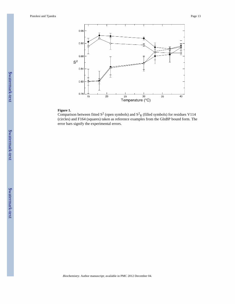
Figure 1.Comparison between fitted S2 (open symbols) and S2
R (filled symbols) for residues V114(circles) and F164 (squares) taken as reference examples from the GlnBP bound form. Theerror bars signify the experimental errors.
Pistolesi and Tjandra Page 13
Biochemistry. Author manuscript; available in PMC 2012 December 04.
$waterm
ark-text$w
atermark-text
$waterm
ark-text
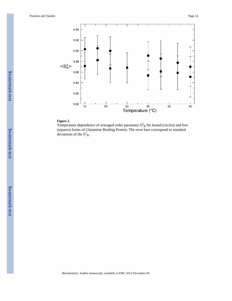
Figure 2.Temperature dependence of averaged order parameter S2
R for bound (circles) and free(squares) forms of Glutamine Binding Protein. The error bars correspond to standarddeviations of the S2
R.
Pistolesi and Tjandra Page 14
Biochemistry. Author manuscript; available in PMC 2012 December 04.
$waterm
ark-text$w
atermark-text
$waterm
ark-text
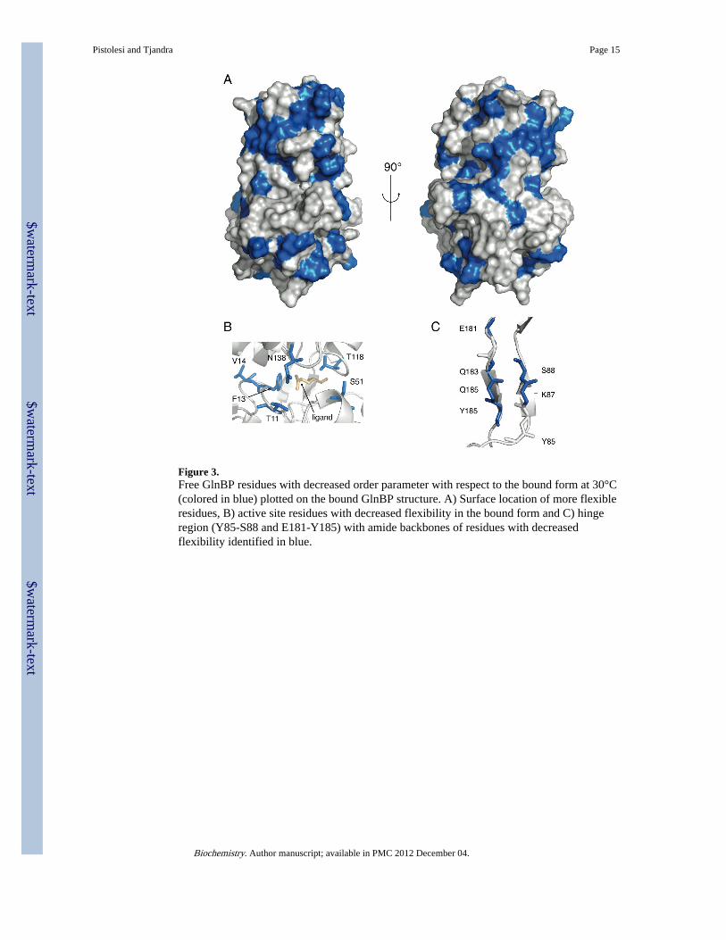
Figure 3.Free GlnBP residues with decreased order parameter with respect to the bound form at 30°C(colored in blue) plotted on the bound GlnBP structure. A) Surface location of more flexibleresidues, B) active site residues with decreased flexibility in the bound form and C) hingeregion (Y85-S88 and E181-Y185) with amide backbones of residues with decreasedflexibility identified in blue.
Pistolesi and Tjandra Page 15
Biochemistry. Author manuscript; available in PMC 2012 December 04.
$waterm
ark-text$w
atermark-text
$waterm
ark-text
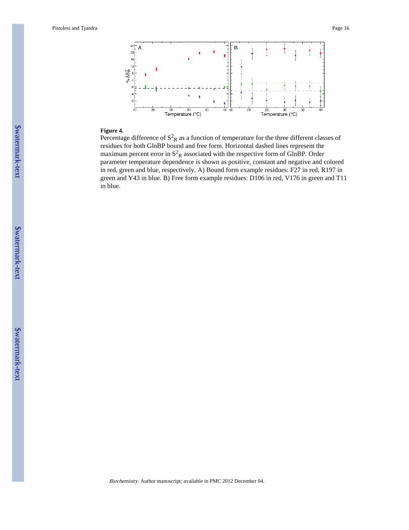
Figure 4.Percentage difference of S2
R as a function of temperature for the three different classes ofresidues for both GlnBP bound and free form. Horizontal dashed lines represent themaximum percent error in S2
R associated with the respective form of GlnBP. Orderparameter temperature dependence is shown as positive, constant and negative and coloredin red, green and blue, respectively. A) Bound form example residues: F27 in red, R197 ingreen and Y43 in blue. B) Free form example residues: D106 in red, V176 in green and T11in blue.
Pistolesi and Tjandra Page 16
Biochemistry. Author manuscript; available in PMC 2012 December 04.
$waterm
ark-text$w
atermark-text
$waterm
ark-text
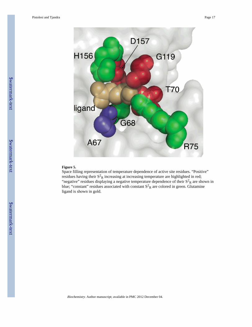
Figure 5.Space filling representation of temperature dependence of active site residues. “Positive”residues having their S2
R increasing at increasing temperature are highlighted in red;“negative” residues displaying a negative temperature dependence of their S2
R are shown inblue; “constant” residues associated with constant S2
R are colored in green. Glutamineligand is shown in gold.
Pistolesi and Tjandra Page 17
Biochemistry. Author manuscript; available in PMC 2012 December 04.
$waterm
ark-text$w
atermark-text
$waterm
ark-text
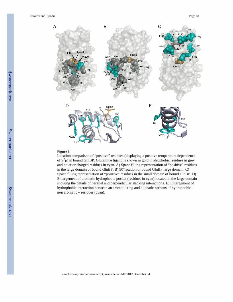
Figure 6.Location comparison of “positive” residues (displaying a positive temperature dependenceof S2
R) in bound GlnBP. Glutamine ligand is shown in gold, hydrophobic residues in greyand polar or charged residues in cyan. A) Space filling representation of “positive” residuesin the large domain of bound GlnBP. B) 90°rotation of bound GlnBP large domain. C)Space filling representation of “positive” residues in the small domain of bound GlnBP. D)Enlargement of aromatic hydrophobic pocket (residues in cyan) located in the large domainshowing the details of parallel and perpendicular stacking interactions. E) Enlargement ofhydrophobic interaction between an aromatic ring and aliphatic carbons of hydrophobic –non aromatic – residues (cyan).
Pistolesi and Tjandra Page 18
Biochemistry. Author manuscript; available in PMC 2012 December 04.
$waterm
ark-text$w
atermark-text
$waterm
ark-text
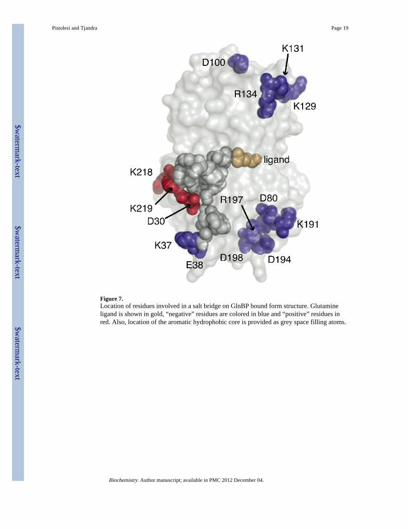
Figure 7.Location of residues involved in a salt bridge on GlnBP bound form structure. Glutamineligand is shown in gold, “negative” residues are colored in blue and “positive” residues inred. Also, location of the aromatic hydrophobic core is provided as grey space filling atoms.
Pistolesi and Tjandra Page 19
Biochemistry. Author manuscript; available in PMC 2012 December 04.
$waterm
ark-text$w
atermark-text
$waterm
ark-text
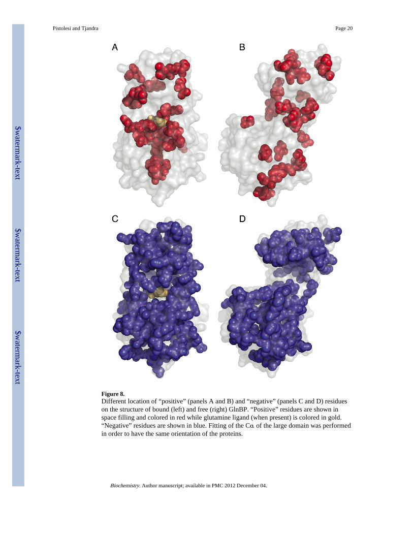
Figure 8.Different location of “positive” (panels A and B) and “negative” (panels C and D) residueson the structure of bound (left) and free (right) GlnBP. “Positive” residues are shown inspace filling and colored in red while glutamine ligand (when present) is colored in gold.“Negative” residues are shown in blue. Fitting of the Cα of the large domain was performedin order to have the same orientation of the proteins.
Pistolesi and Tjandra Page 20
Biochemistry. Author manuscript; available in PMC 2012 December 04.
$waterm
ark-text$w
atermark-text
$waterm
ark-text