telomere/telomerase interplay in virus-driven and virus-independent lymphomagenesis: pathogenic and...
TRANSCRIPT

Telomere/Telomerase Interplay inVirus-Driven andVirus-IndependentLymphomagenesis: Pathogenic and
Clinical Implications
Riccardo Dolcetti1 and Anita De Rossi2
1Cancer Bio-Immunotherapy Unit, Department of Medical Oncology, CRO - IRCCS, National Cancer Institute,
Aviano, Italy2Unit of Viral Oncology, Department of Oncology and Surgical Sciences, University of Padova and IOV-IRCCS,
Padova, Italy
in Wiley InterScience (www.interscience.wiley.com).DOI 10.1002/med.20211
.
Abstract: Telomerase is a ribonucleoprotein complex critically involved in extending and maintaining
telomeres. Unlike the majority of somatic cells, in which hTERT and telomerase activity are generally
silent, normal lymphocytes show transient physiological hTERT expression and telomerase activity
according to their differentiation/activation status. During lymphomagenesis, induction of persistent
telomerase expression and activity may occur before or after telomere shortening, as a consequence of
the different mechanisms through which transforming factors/agents may activate telomerase. Avail-
able data indicate that the timing of telomerase activation may allow the distinction of two different
lymphomagenetic models: (i) an early activation of telomerase via exogenous regulators of hTERT,
along with an increased lymphocyte growth and a subsequent selection of cells with increased trans-
forming potential may characterize several virus-related lymphoid malignancies; (ii) a progressive
shortening of telomeres, leading to genetic instability which favors a subsequent activation of telo-
merase via endogenous regulators may occur in most virus-unrelated lymphoid tumors. These models
may have clinically relevant implications, particularly for the tailoring of therapeutic strategies targeting
telomerase.
Key words: telomerase; telomere length; telomerase inhibitors; virus-driven lymphomagenesis
Contract grant sponsor:European Community FP6 VITAL;Contract grant number: 037874; Contract grant sponsors: Italian Min-
istry of Health Program ‘‘Alleanza Contro il Cancro (ACC-4)’’; Program Integrato Oncologia; Contract grant number: RO 4/2007;
Contract grant sponsors: Italian Ministry of University and Research, Prin 2007; Associazione Italiana per la Ricerca sul Cancro.
Correspondence to: Riccardo Dolcetti, Cancer Bioimmunotherapy Unit, Centro di Riferimento Oncologico - IRCCS, National
Cancer Institute,Via Franco Gallini 2, 33081, Aviano (PN), Italy, E-mail: [email protected]
& 2010 Wiley Periodicals, Inc.
Published online 14 June 2 010
& 2010 Wiley Periodicals, Inc. Med Res Rev, 32, No. 2, 233–253, 2012
Medicinal Research Reviews, 32,No. 2, 233--253, 2012

1. INTRODUCTION
Telomerase is a ribonucleoprotein complex critically involved in extending and maintainingthe telomeres. Activation of this enzyme is required for cells to overcome replicative senes-cence and acquire the ability to unlimitedly divide, a prerequisite for neoplastic transfor-mation. Telomerase activity is constitutively expressed in germ-line cells and in the majorityof malignant tumor cells, whereas it is repressed in most human somatic cells. A notableexception is constituted by normal lymphocytes in which telomerase activity is expressed in ahighly regulated manner; therefore, in these cells, also the length of the telomeres variesaccording to their functional and differentiation status. Considering the relevance of theseaspects with regard to lymphomagenesis, we herein review available data on telomere andtelomerase in normal and transformed B and T lymphocytes. Moreover, because virusesinvolved in lymphomagenesis may directly up-regulate telomerase, it seems of interest tocompare the mechanisms underlying telomerase activation in virus-driven and virus-unrelated lymphoid malignancies. These pathogenic aspects are of relevance also from aclinical point of view, considering the increasing number of therapeutic strategies targetingtelomerase.
2. TELOMERASE AND CELL TRANSFORMATION
A. Telomerase Plays a Key Role in a Cell’s Decision Between Deathand Immortalization
Telomerase is a ribonucleoprotein enzyme that de novo extends telomeric sequences at theends of eukaryotic chromosomes, thus preventing replicative senescence, a state of irrever-sible cell growth arrest, and death. Telomeres are specialized DNA structures located at theend of chromosomes; they are composed of (TTAGGG)n tandem repeats and are essential tostabilizing chromosomes by protecting them from end-to-end fusion and DNA degradation.Telomeres are progressively shortened during each cell division by replication-dependent lossof sequences at DNA termini, due to the failure of DNA polymerase to completely replicatethe 30 end of chromosomes.1 When telomeres become critically short (the Hayflick limit), cellsundergo replicative senescence and apoptosis; further erosion of telomeres may impair theirfunction in protecting chromosome ends, resulting in genetic instability. Thus, telomereerosion has been proposed to have two conflicting roles: tumor suppression by inducing celldeath and tumor promotion by inducing genetic instability, a key event in the initiation ofcarcinogenesis.2 Nonetheless, cell division-associated telomere shortening prevents unlimitedcell proliferation and, thus, tumor development/progression. To escape this proliferationbarrier, cells must stabilize their telomeres.3 Most tumors maintain their ability to growindefinitely through inappropriate expression of telomerase,4 a ribonucleoprotein complexcontaining an internal RNA template (hTR), used as a template for elongation of telomeres,and a catalytic protein with telomere-specific reverse transcriptase activity (hTERT). hTRbelongs to the family of small nuclear RNAs and contains an H/ACA domain essential fortelomerase activity.5 While hTR has broad tissue distribution and is constitutively present innormal and tumor cells, hTERT is the rate-limiting component of the telomerase complex,6
and its expression generally correlates with telomerase activity. Overexpression of hTR alongwith hTERT may increase telomerase activity,7 while specific hTR variants may reduce itsassociation with hTERT, thus diminishing the telomerase activity in telomere lengthening.8
Expression of hTERT is generally restricted to stem cells and is usually repressed in normalsomatic cells. However, it may be expressed at low levels in normal hematopoietic cells
K DOLCETTI ANDDEROSSI
Medicinal Research Reviews DOI 10.1002/med
232 K DOLCETTI ANDDEROSSI234

according to their state of differentiation/activation. In contrast, hTERT is expressed in thevast majority of immortalized and fully transformed cells. Overexpression of hTERT innormal cellular systems allows cells to grow for longer periods of time and confers increasedsusceptibility to accumulating additional genetic alterations, thus contributing to themalignant phenotype.9–11 Conversely, hTERT inhibition limits the growth of human cancercells.12 Furthermore, several pieces of evidence suggest that hTERT is also involved in othercellular functions, including activation of pro-proliferative signaling pathways and anti-apoptotic functions, by mechanisms independent of its ability to prevent telomere ero-sion.13–16 Notably, high levels of telomerase confer resistance to several antineoplasticdrugs.17,18
B. Regulation of Telomerase Expression
Regulation of telomerase operates at several biological levels: transcription, mRNA splicing,subcellular localization of each component, assembly of hTR and hTERT in an activeribonucleoprotein, and activity on telomere extension. Transcription of the hTERT gene isprobably the key determinant in the regulation of telomerase activity; notably, hTERTtranscriptional activity is specifically up-regulated in cancer cells, while it is silent in mostnormal cells.19 The hTERT gene comprises about 35Kb DNA and codes for an mRNAcomposed of 16 exons and 15 introns.20 At the transcriptional level, more than 20 tran-scription factor-binding sites acting as activators or repressors have been identified within thehTERT promoter. Cooperation of c-Myc and Sp1 is required for full activation of hTERTpromoter,21 while p53, through its interaction with Sp1, down-regulates hTERT.22 hTERT isalso directly activated by nuclear factor (NF)-kB,23 hypoxia-inducible factor (HIF)-1,24 andEts/c-Myc complex.25 The histone methyltransferase Smyd3 also directly contributes to in-ducible and constitutive hTERT expression in normal and malignant human cells.26 hTERTexpression is suppressed by the oncosuppressor genes WT127 and Menin,28 and through theMad1/c-Myc29 and the TGF-beta/Smad330 pathways. The cell cycle inhibitors p16INK4a 31
and p27KIP1 32 were also shown to down-regulate hTERT expression in cancer cells.Regulation of hTERT transcription may also involve DNA methylation, as the hTERT
promoter contains a cluster of CpG sites33 and histone acetylation/deacetylation.34 At thepost-transcriptional level, modulation of telomerase may occur by alternative splicing me-chanisms: mRNA variants lacking a and/or b regions translate truncated and dysfunctionalprotein products.35 It has been proposed that some of these splicing products, such as theisoform a, may exert a dominant negative function by competitive interaction with com-ponents of the telomerase complex.36 Moreover, telomerase activity is also controlledthrough post-translational modifications of the hTERT protein. Phosphorylation of theprotein at critical sites by the PI3K/Akt kinase pathway seems to be crucial for telomeraseactivity.37,38 A peculiar mechanism of post-translational regulation of telomerase activity wasdescribed in natural killer cells, in which IL-2 promotes the association between hTERT andthe complex of Akt, Hsp90, mTOR, and S6K.39 Moreover, in breast cancer cells, proteinkinase C a-dependent hTERT phosphorylation was shown to constitute an essential step fortelomerase activation.40
Telomerase activity at the chromosome ends is controlled by telomere-associated pro-teins. Mammalian telomere has a special t-loop conformation in which the telomere terminusis not accessible to telomerase. This structure is associated with a 6-protein complex (TRF1,TRF2, Rap1, TIN2, TPP1, and POT1), the shelterin, which functions to protect chromosomeends from all aspects of DNA damage and regulates telomere maintenance by telomerase.TPP1 directly interacts with telomerase and may facilitate recruitment of the enzyme to
TELOMERE/TELOMERASE INTERPLAY K 3
Medicinal Research Reviews DOI 10.1002/med
TELOMERE/TELOMERASE INTERPLAY K 235

telomeres, while POT1 is a negative regulator due to its ability to compete with the 30
overhang of telomere, the substrate of telomerase.41
3. TELOMERASE EXPRESSION AND ACTIVITY IN NORMAL LYMPHOCYTES
Telomerase expression and activity may be up-regulated in a tightly controlled fashionduring the differentiation of normal B and T lymphocytes. In particular, telomerase acti-vation occurs in concomitance with cell activation following the encounter with a cognateantigen. Nevertheless, B and T cells show differences in terms of the kinetics of telomeraseactivation and telomere length along with their respective differentiation pathways.
A. T Cells
Effective immune responses against pathogens or transformed cells usually require the re-peated expansion of a limited number of antigen-specific T lymphocytes. Limitations in theproliferative potential of these effectors may severely compromise immune function. Uponantigen stimulation, naıve CD41 and CD81 T lymphocytes are activated to proliferate andgenerate effector as well as memory T cells that can be distinguished from naıve T cells byimmunophenotypic and functional properties.42 Notably, the homeostatic cytokine IL-15was shown to activate telomerase in memory CD81 T cells via Jak3 and PI3-K signalingpathways.43 The induction of telomerase was stable over time and minimized telomere loss,thus preserving the replicative lifespan of proliferating memory CD81 T cells.43 Moreover,IL-7 was also shown to activate telomerase,44 with higher levels in naıve rather than inmemory CD41 cells.45 Studies carried out in total peripheral blood lymphocytes demon-strated that the pro-inflammatory cytokine TNF-a promotes a rapid induction and nucleartranslocation of telomerase activity through PI3-K/Akt/(NF)-kB activation, resulting inprevention of cell apoptosis.46
Earlier studies have shown that the length of telomeres in circulating T lymphocytesdecreases with age.47 The observation that the average telomere length decreases with eachcell division in vitro suggested that telomere length could be used as an indicator of thenumber of cell divisions that have occurred in vivo. Available data indicate that cord blood Tcells have the longest telomeres, followed by naıve CD45RA1 T cells and CD45RO1
memory T lymphocytes, which show the shortest telomere length.48,49 Stimulation of T cellswith antigens, mitogens, cytokines, or activating antibodies was shown to readily up-regulatetelomerase activity, although this activity decreased progressively and was almostundetectable after 3 weeks.50 While secondary antigenic stimulation also results inreactivation of telomerase, CD81 T cells lose this ability by the third antigenic encounter,showing progressive telomere erosion concomitantly with further antigen-driven prolifera-tion. In contrast, CD41 T lymphocytes maintain telomerase activity even after the seventhantigenic stimulation.50
Among the complex signaling pathways activated by T cell receptor triggering, proteinkinase C-y was shown to promote hTERT expression by transcriptional regulation, mediatedby NF-kB.51 Furthermore, growth-promoting stimuli were shown to induce hTERTexpression and telomerase activity in normal T lymphocytes through Ser10 phosphorylationof histone H3 at the hTERT promoter mediated by MAPK signaling.52 This effect waspotentiated by the synergistic activity of Lys14 histone H3 acetylation, which resulted inconstitutive telomerase activation in both normal and malignant T lymphocytes.52 Effectivetelomerase up-regulation in T cells also requires CD28 costimulation, as shown by thefinding that blocking the interaction between CD28 and its B7 ligands expressed by antigen
4 K DOLCETTI ANDDEROSSI
Medicinal Research Reviews DOI 10.1002/med
K DOLCETTI ANDDEROSSI236

presenting cells markedly inhibited telomerase activation.52 More recently, it has been shownthat telomerase activity can also be induced by other costimulatory signals, including CD137,CD134, ICOS, and OX40.53 Notably, T cell cultures with phenotypic and functional featuresof replicative senescence show enrichment in cells lacking CD28 expression. Moreover, cross-sectional analysis of T cells from different age groups clearly showed that the number ofCD28� T cells progressively increased with age.54 Consistently, ex vivo analyses demon-strated that telomeres of CD28� T cells were shorter than those of other T lymphocytes fromthe same individuals, suggesting a more extensive replicative history.55–57 These cells alsoshow decreased proliferative responses to both antigenic and nonspecific stimulations,55
lower levels of telomerase activity,57 and relative resistance to apoptosis.58 High numbers ofsenescent CD28� T cells have been correlated to several pathological conditions includingpoor control of infectious agents, reduced responses to vaccines, bone loss, and early mor-tality.59–61 These effects may be related to as yet poorly defined suppressor functions ofsenescent CD28� T cells or to alterations in the size and/or composition of the T cell pool,consequent to the increased number of these exhausted and poorly functional cells. Furtherdifferentiation of CD81CD28�CD271 T cells into CD81CD28�CD27� populations leads toa loss of telomerase activity that cannot be reversed by alternative costimulation. Down-regulation of telomerase activity in CD81CD28�CD27� is not due to reduced expression ofhTERT, but is the consequence of defective Akt phosphorylation on Ser473, an essentialregulator of telomerase activity.62 While transduction with telomerase is necessary and suf-ficient to stabilize telomeres and to achieve full immortalization in vitro,63 the transientphysiological activation of endogenous telomerase after T cell activation is insufficient toimmortalize T cells.64
B. B Cells
Similarly to T cells, quiescent circulating B lymphocytes express low to undetectable levels oftelomerase.65 Nevertheless, peripheral blood B cells show peculiar telomere dynamics, dis-tinctly different from other blood cell populations. In fact, when compared to T cells, naturalkiller cells, and monocytes, B lymphocytes have notably longer telomeres and higher telo-merase activity.66,67 Telomere length and telomerase activity are highly regulated also duringactivation and differentiation of B lymphocytes. In particular, several lines of evidenceindicate that germinal center B cells, a subpopulation characterized by extensive clonalexpansion and antigen-driven selection, have significantly longer telomeres than those ofprecursor naıve or descendant memory B lymphocytes.67 Telomerase activity is increased ingerminal center B cells and correlates with increased telomere length.67 Moreover, CD271
memory B cells have significantly longer telomeres than their CD27� counterparts. Thisstrongly contrasts with the observed telomere shortening that occurs during conversion ofnaıve to memory CD41 T cells,66 and suggests that telomerase may actually elongate telo-meres in germinal center B cells to preserve the replicative lifespan of progeny memory Bcells. Signals mediated by several cell surface receptors have been shown to regulate telo-merase activity in B lymphocytes. In particular, telomerase can be induced in human tonsilnaıve and memory B cells by the mitogen Staphylococcus aureus Cowan strain or by en-gagement of the B cell receptor in combination with costimulatory signals provided by CD40triggering or interleukin (IL)-4.67,68 Notably, these stimulatory conditions also promote Bcell proliferation, whereas apoptotic signals are unable to induce telomerase.67 Therefore,telomerase activity and proliferation seem to be closely linked in B cells, consistently with anadaptive role of telomerase in preserving the proliferative potential of clonally expanding Blymphocytes.
TELOMERE/TELOMERASE INTERPLAY K 5
Medicinal Research Reviews DOI 10.1002/med
TELOMERE/TELOMERASE INTERPLAY K 237

Long-term in vitro culture of normal B lymphocytes can be obtained after EBV infection(see below) or by triggering CD40 in the presence of IL-4.69 Optimization of culture con-ditions with CD40/IL-4 allows the conditional immortalization of normal human B cells,without requiring genetic manipulation.70 These B lymphocytes have a constant phenotype,are free from EBV, remain dependent on CD40 ligation, and have constitutive telomeraseactivity and stabilized telomere length.70
Studies aimed at investigating the regulation of telomerase expression and activity inlymphocytes demonstrated that transcriptional regulation of hTERT alone is not sufficient toup-regulate telomerase activity.71 In these cells, hTERT is phosphorylated and translocatedfrom cytoplasm to the nucleus, indicating the need for additional post-transcriptional me-chanisms to fully activate telomerase in human lymphocytes.71
4. TELOMERASE EXPRESSION AND ACTIVITY IN VIRUS-UNRELATEDLYMPHOID MALIGNANCIES
While hemopoietic stem cells have an extraordinary capacity of self-renewal and high levelsof telomerase, their differentiation and related acquisition of specialized functions result indecreased telomere length and telomerase activity. Conversely, hematological cancer cellsgenerally express higher levels of telomerase than normal counterparts. Besides the tran-scriptional activation of the hTERT gene, post-transcriptional mechanisms may also increasetelomerase activity in cancer cells. Indeed, while alternatively spliced variants are present innormal and neoplastic T cells, the full-length hTERT RNA, which encodes the functionalhTERT protein, is constitutively present in malignant cells, but only after mitogenic sti-mulation in normal lymphocytes.72 While maintenance of telomerase activity and telomerelength preservation are essential for neoplastic cell transformation, the kinetics of telomereerosion/telomere stabilization mediated by hTERT activation is currently a matter of debate.
A. T Cell Malignancies
Although only limited data are available, evidence accumulated so far suggests that a pro-gressive shortening of telomeres and telomerase activation may be involved even in thepathogenesis of virus-unrelated T cell malignancies. Significantly higher levels of telomeraseactivity and shortened telomere lengths were observed in tumor cells of patients with cuta-neous T cell lymphomas as compared to normal counterparts.73 It has been reported that Tcell prolymphocytic leukemia, a rare aggressive disease characterized by expansion of a CD21
CD71CD41 T cell clone with genetic rearrangement of chromosome 14q32, has very shorttelomeres and express high levels of hTERT; as this leukemia shows a high proliferativeindex, high levels of telomerase might compensate for the telomere loss due to cell pro-liferation.74 Moreover, it has been demonstrated that T cell acute leukemias are also positivefor hTERT, but at levels lower than those observed in B-cell acute leukemias.75 Additionalsupport for an involvement of telomere shortening in the pathogenesis of T cell malignancieswas provided by studies carried out in patients with ataxia teleangectasia, a rare diseaseassociated with an increased risk of developing T cell leukemias and lymphomas. In fact, theaccelerated telomere shortening demonstrated in these patients could at least partly explaintheir enhanced susceptibility to developing T cell malignancies.76 Most of the available dataon the role of telomerase in T cell malignancies, however, deal with Adult T-cell leukemia/lymphoma (ATLL), a tumor associated with Human T-cell Leukemia Virus (HTLV)-1infection (see below).
6 K DOLCETTI ANDDEROSSI
Medicinal Research Reviews DOI 10.1002/med
K DOLCETTI ANDDEROSSI238

B. B Cell Malignancies
Studies of mature B cell lymphoproliferative disorders demonstrated that, overall, telomerelength was greater in diffuse large cell lymphoma, Burkitt’s lymphoma, and follicular lym-phoma than in mantle cell lymphoma, marginal zone lymphoma, and chronic lymphocyticleukemia.77 Telomerase activity has been reported in all the above malignancies, except forlow-grade marginal zone lymphomas, and is positively correlated with the proliferation indexof tumor cells.78 A relationship between telomere length and histopathogenesis according togerminal center (GC) has been advanced; GC-derived tumors have longer telomeres thanthose originated from GC-inexperienced cells.77–80 During a classical T cell-mediated GCexperience, normal B cells have high levels of telomerase and exhibit telomere lengthen-ing.65,67 Within this context, studies on B cell chronic lymphocytic leukemia (B-CLL) are ofparticular interest. B-CLL is the most common leukemia in adults and is clinically highlyheterogeneous. Based on the presence of somatic mutations within the immunoglobulin (Ig)heavy chain gene variable regions (IgVH), two subgroups of B-CLL have been identified, withthe unmutated IgVH profile being associated with a more aggressive disease than the mutatedone.81,82 Somatic hypermutation of Ig genes is a physiological process, as part of the antigen-driven affinity maturation of antibodies occurring in the GC; thus, B-CLL cases with un-mutated IgVH genes are considered to originate from pre-GC B lymphocytes and those withmutated IgVH genes from GC-experienced B lymphocytes. Overall, B-CLL had shorter tel-omeres than normal B cells,79 but mutated IgVH GC-experienced B-CLL had longer telo-meres than the unmutated IgVH B-CLL.79,80,83 Of interest, the unmutated IgVH B-CLL hadhigher levels of hTERT and telomerase activity than the mutated IgVH cases.79,84 The findingthat B-CLL with shorter telomeres have higher levels of telomerase than those with longertelomere emphasizes the concept that telomere length in tumors, rather than being associatedwith hTERT levels, reflects the initial kinetics of telomere length erosion by cell proliferationand telomere length stabilization due to hTERT activation.
Notably, high levels of hTERT and/or telomerase activity are correlated with poorclinical outcome.79,85–87 Furthermore, the discordant unmutated cases with low levels ofhTERT had an overall survival similar to mutated cases with high hTERT levels, thussupporting the notion that hTERT mediates effects that contribute to lymphomagenesisbeyond preservation of telomere length.87
5. TELOMERASE EXPRESSION AND ACTIVITY IN VIRUS-DRIVENLYMPHOID MALIGNANCIES
Evidence from animal models supports the hypothesis that viruses interplay with telomere/telomerase complex to promote lymphomagenesis. In fact, a virus-encoded telomerase RNAhas been shown to promote malignant T cell transformation in animals.88 Marek’s diseaseherpesvirus (MDV), which induces a malignant T cell lymphoma in chickens, harbors in itsgenome two identical copies of a viral telomerase RNA (vR), with 88% sequence identity toendogenous chicken TR. MDV mutants, lacking both copies of vTR, were significantlyimpaired in their ability to induce T cell lymphomas; tumor incidence was reduced by >60%in chickens infected with vTR-negative viruses, and these lymphomas were also significantlysmaller in size and less disseminated than those induced by unmutated MDV.88 Additionalevidence links telomerase activation and the development of rapid-onset chicken B celllymphomas induced by avian leukosis virus (ALV).89 In this model, the hTERT promoter/enhancer region has been shown to constitute a common integration site for the ALV pro-virus.89 This integration leads to hTERT overexpression and up-regulation of telomerase
TELOMERE/TELOMERASE INTERPLAY K 7
Medicinal Research Reviews DOI 10.1002/med
TELOMERE/TELOMERASE INTERPLAY K 239

activity, without effects on telomere length.89 These findings are consistent with the possi-bility that telomerase contributes to B cell lymphomagenesis independently of its activity ontelomere length.
A. HTLV-1-Associated Lymphomas
The HTLV-1 is the etiologic agent of ATLL, a lymphoproliferative disorder of CD41 T cells.Peripheral blood cells from HTLV-1 carriers have longer telomeres than age-matched healthysubjects, whereas ATLL cells are characterized by short telomeres, despite strong telomeraseactivity.90,91 Of note, HTLV-1-infected CD251 ATLL cells retain shorter telomeres thanthose of uninfected CD25� cells isolated from the same patients.92 It has been demonstratedthat the telomere-binding proteins, TRF1, TRF2, and TIN2, are overexpressed in ATLLcells compared to those from asymptomatic carriers; the up-regulation of TIN2 along withTRF1 may stabilize TRF2 on telomeres, thus preventing apoptosis in cancer cells with shorttelomeres. Of note, the expression of POT1, a negative regulator of telomerase activity,41
does not significantly increase in ATLL.92 Reactivation of telomerase seems to be a criticalevent in the development and progression of ATLL. In fact, telomerase activity is high inlymphocytes from patients with acute smoldering and chronic disease, and disease progres-sion is associated with a further increase in telomerase activity.90–92 Infection of primary Tlymphocytes with HTLV-1 results in up-regulation of hTERT, thus confirming the direct roleof the virus in the telomerase activation.93 Several lines of evidence have established that thetransactivator viral protein Tax plays a central role in the conversion of normal T lym-phocytes to neoplastic cells. The effects of the Tax protein on telomerase expression are stillill-defined. A study performed by transduction of lymphocytes with Tax-expressing vectorand hTERT promoter–luciferase reporter gene suggests that Tax plays a negative role inhTERT activation; this inhibitory effect may occur by competitive binding with the positiveregulator c-Myc to its canonical-binding site, E-box, located within the hTERT promoter.94
Nonetheless, other studies indicate that Tax is a strong, positive regulator of the endogenoushTERT promoter through the NF-kB signaling pathway.93,95 This discrepancy may be duenot only to the different assays employed, but it may also be linked to the proliferative statusof the cells. Indeed, it has been demonstrated that Tax repressed the hTERT promoter inproliferating cells, while it activated the hTERT promoter in quiescent cells, and this acti-vation was associated with cell cycle progression.96 Notably, in ATLL cells, Tax expression isvery low-to-undetectable, yet these cells retain strong telomerase activity. This suggests thatalternative/additional mechanisms, independent of Tax protein, may induce hTERTexpression and telomerase activity. The HTLV-1 basic leucine zipper protein HBZ encodedby the minus strand of HTLV-1 genome and expressed in ATLL cells, increases thetranscriptional activity of JunD, an AP-1 protein. The hTERT promoter has severalAP-1-binding sites, and it has been demonstrated that HBZ in association with JunD acti-vates the hTERT promoter.97 Furthermore, it has also been shown that IL-2 signaling wasassociated with a PI3K-dependent transcriptional up-regulation of the hTERT promoter inHTLV-1 transformed cells. Activation of the PI3K pathway mediates cytoplasmic retentionof the WT1 protein, which strongly suppresses the hTERT promoter; this may explain thehigh levels of hTERT expression in IL-2-dependent HTLV-1 infected cells and in ATLLsamples, even in the absence of Tax protein.98
B. EBV-Associated Lymphoproliferative Disorders
Epstein-Barr virus (EBV) is involved in the pathogenesis of several lymphoproliferativedisorders, including Burkitt’s and Hodgkin’s lymphomas, post-transplant lymphoprolifera-tions, and a subset of T/NK cell lymphomas.99 This spectrum closely reflects the ability of
8 K DOLCETTI ANDDEROSSI
Medicinal Research Reviews DOI 10.1002/med
K DOLCETTI ANDDEROSSI240

EBV to preferentially infect B lymphocytes, although a latent infection can be establishedalso in T or natural killer cells. EBV infection of resting B cells in vitro results in thegeneration of continuously proliferating lymphoblastoid cell lines (LCLs), which closelyresemble most of the EBV-related lymphoproliferations associated with immune suppression.These cells, in fact, harbor EBV mainly in its latent state and express a broad array of latentproteins, including six nuclear antigens (EBNAs) and three latent membrane proteins(LMP-1, LMP-2A, and LMP-2B). Among EBV latency gene products, LMP-1 is consideredthe strongest oncoprotein, being essential for immortalization of B cells. Expression of latentEBV proteins, however, does not suffice to fully immortalize EBV-infected B cells. In fact,only EBV-carrying B cells with sustained telomerase activity are truly immortalized, whereastelomerase-negative cells, although exhibiting a prolonged lifespan, eventually undergocellular senescence and terminate their lifespan by shortening of telomeres.100,101 This notionhas been further supported by recent findings indicating that sustained EBV viralfunctions and continuous telomerase activity are both required to achieve a complete LCLimmortalization.102
In early-passage EBV-infected B lymphocytes, activation of hTERT occurs con-comitantly with the induction of latent EBV genes and the down-regulation of lytic EBV geneexpression.84 In addition, while ectopic hTERT expression on telomerase-negative cells in-hibits EBV replication, hTERT silencing results in the induction of the EBV lytic cycle.84
These results are consistent with a crucial role of hTERT activation in inhibiting virusreplication, thereby favoring the induction and maintenance of EBV latency in B lympho-cytes, a crucial prerequisite for EBV-driven transformation. Moreover, overexpression ofhTERT has been shown to induce resistance to EBV lytic cycle induction in B lymphocytes,indicating that a stable latent EBV infection in these cells requires continuous hTERTexpression.84 hTERT activation also promotes the proliferation of primary B lymphocytes,84
further supporting the hypothesis that the functional relevance of hTERT goes beyond themere regulation of telomere length.
Studies aimed at defining the mechanism underlying EBV-induced telomerase activationhave demonstrated that the LMP-1 oncoprotein up-regulates telomerase activity in bothepithelial cells103 and B lymphocytes.104,105 LMP-1 transactivates the hTERT promoter inboth carcinoma (HeLa) cells and B lymphocytes (BJAB and DG75 cells).105 LMP-1-driventransformation mainly relies on the ability of this protein to hijack cellular signaling path-ways that are critical for B cell growth and survival, including some cascades that are alsoknown to regulate hTERT expression and telomerase activity. While CD40 signaling isprobably not involved in LMP1-mediated hTERT activation, both the ERK1/2 and NF-kBpathways are strongly and independently activated in B cells following ectopic LMP-1expression.105 Considering that pathways involving ERK1/2 activation are known to reg-ulate telomerase activity in response to exogenous growth stimuli, even independently ofproliferation,106,107 LMP-1 expression in B lymphocytes may mimic the effects of growthfactors by directly activating telomerase via ERK1/2, thus contributing to cell im-mortalization. LMP-1 induces telomerase activity also in nasopharyngeal carcinoma cells, anepithelial tumor closely associated with EBV infection. In these cells, LMP-1 up-regulatestelomerase through NF-kB activation,108 an effect that is c-Myc-dependent, since muta-genesis of c-Myc-responsive E-box elements in the hTERT promoter inhibits hTERTtransactivation induced by LMP-1.103 In B cells, however, c-Myc is not involved in mediatingthe hTERT expression and telomerase activation induced by LMP-1, as shown by severallines of evidence: (i) lack of c-Myc up-regulation following ectopic LMP-1 expression;(ii) c-Myc silencing does not inhibit LMP1-induced telomerase activation; (iii) mutagenesis inthe NF-kB-binding site, but not in the c-Myc-binding sites, inhibits LMP-1-induced acti-vation of the hTERT promoter.105 In nasopharyngeal carcinoma cells, LMP-1 enhances
TELOMERE/TELOMERASE INTERPLAY K 9
Medicinal Research Reviews DOI 10.1002/med
TELOMERE/TELOMERASE INTERPLAY K 241

telomerase activity also at the post-transcriptional level, by promoting NF-kB RelA/p65-mediated binding to and nuclear localization of hTERT.108 Whether a similar mechanismalso occurs in B cells remains to be elucidated.
The observation that LMP-2A is able to interfere with B cell differentiation and toprovide survival signals suggests that this viral protein could be involved in cell transfor-mation.109 In exploring its possible role in modulating telomerase activity, LMP-2A has beenshown to inhibit hTERT promoter activity in both epithelial cells and B lymphocytes.110
Although this apparently paradoxical effect deserves further investigation, particularly in Blymphocytes, it can be advanced so that the inhibitory activity of LMP-2A on telomerasecould be related to its role in suppressing B cell activation.
Unlike other oncogenic viruses known to enhance telomerase activity, EBV is currentlythe only virus for which virus-infected tumor cells have been associated with increased tel-omere length. In fact, EBV-positive Burkitt’s lymphoma cell lines show increases in telomerelength as compared to those of EBV-unrelated cells.111,112 These observations are limited toin vitro grown cell lines and confirmation of these findings will, therefore, require mea-surement of telomere length in freshly obtained lymphoma cells.
C. KSHV/HHV-8-Related Lymphoproliferative Disorders
Kaposi’s sarcoma associated herpesvirus (KSHV) or Human Herpesvirus-8 (HHV-8) is anoncogenic g- herpesvirus etiopathogenically associated with Kaposi’s sarcoma113 and withtwo B cell lymphoproliferative disorders, multicentric Castleman’s disease (MCD) and pri-mary effusion lymphomas (PEL).114 In vitro KSHV/HHV-8 infection of primary humanendothelial cells115 or keratinocytes116 was shown to up-regulate telomerase activity. Thesearch for virus-encoded proteins able to mediate telomerase activation was focused on thelatency-associated nuclear antigen (LANA), a multifunctional protein that is consistentlyexpressed in all KSHV/HHV-8-associated malignancies. LANA is required for long-termmaintenance of viral episomal DNA in dividing cells,117 and it has been shown to modulatethe cellular transcription program in KSHV/HHV-8-infected cells. In particular, LANAaffects the p53, Rb, and b-catenin pathways and promotes B cell lymphoma development.118
In vitro transcriptional reporter assays revealed that LANA transactivates the hTERTpromoter,119 an effect mediated by direct interaction of LANA with the Sp1 transcriptionfactor.120 Notably, LANA-induced hTERT transcriptional activation was also observed in Bcell lines derived from Burkitt’s lymphomas and PEL.119,120 Although these findings areconsistent with a pathogenic role of KSHV/HHV-8-induced hTERT up-regulation, noinformation is currently available on the role exerted in vivo by KSHV/HHV-8 in activatingtelomerase in MCD and PEL, and no data on telomere length in these disorders have beenreported thus far.
6. CLINICAL IMPLICATIONS
As discussed above, hematological malignancies are generally characterized by deregulatedtelomere length and telomerase activity compared to normal cellular counterparts, suggestingthat both these markers may be of diagnostic and/or prognostic value for early detection ofcancer, monitoring response to therapy, or predicting clinical outcome. The expression ofhTERT in tumor cells and the requirement of a sustained telomerase activity for the un-limited proliferation capability of tumor cells make telomerase a particularly attractive targetfor cancer therapy. Several strategies targeting telomerase are being explored at the pre-clinical level, and two major approaches are currently under clinical investigation.121,122
10 K DOLCETTI ANDDEROSSI
Medicinal Research Reviews DOI 10.1002/med
K DOLCETTI ANDDEROSSI242
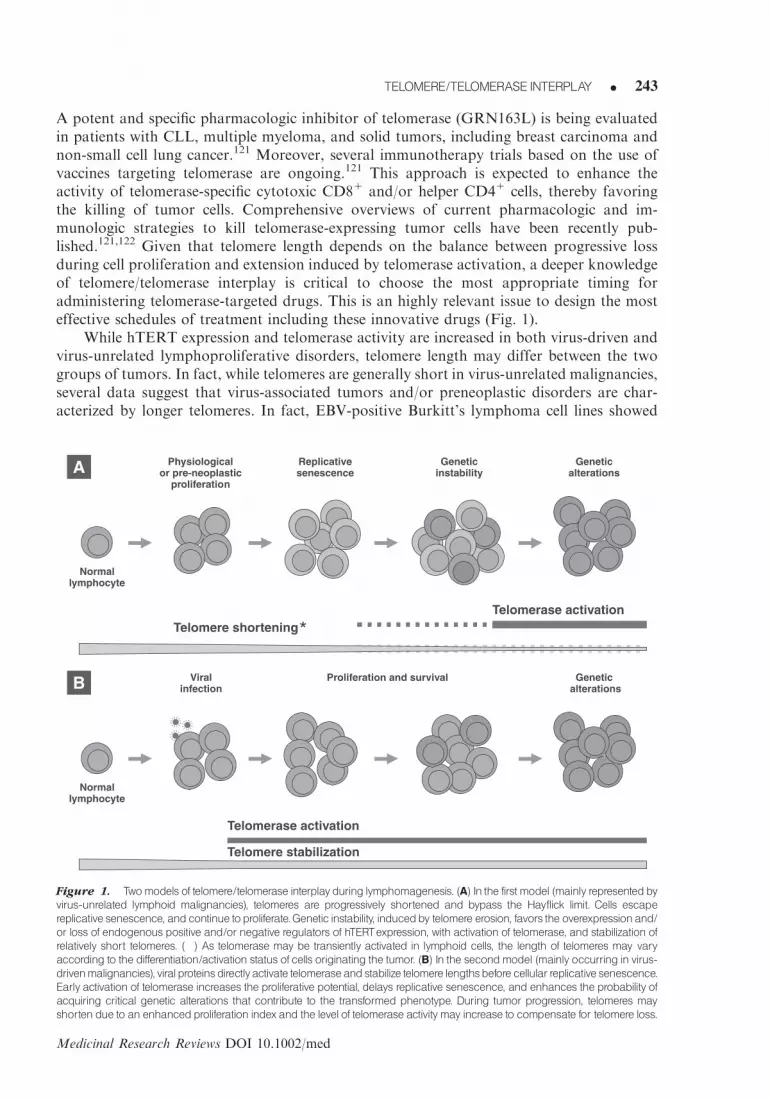
A potent and specific pharmacologic inhibitor of telomerase (GRN163L) is being evaluatedin patients with CLL, multiple myeloma, and solid tumors, including breast carcinoma andnon-small cell lung cancer.121 Moreover, several immunotherapy trials based on the use ofvaccines targeting telomerase are ongoing.121 This approach is expected to enhance theactivity of telomerase-specific cytotoxic CD81 and/or helper CD41 cells, thereby favoringthe killing of tumor cells. Comprehensive overviews of current pharmacologic and im-munologic strategies to kill telomerase-expressing tumor cells have been recently pub-lished.121,122 Given that telomere length depends on the balance between progressive lossduring cell proliferation and extension induced by telomerase activation, a deeper knowledgeof telomere/telomerase interplay is critical to choose the most appropriate timing foradministering telomerase-targeted drugs. This is an highly relevant issue to design the mosteffective schedules of treatment including these innovative drugs (Fig. 1).
While hTERT expression and telomerase activity are increased in both virus-driven andvirus-unrelated lymphoproliferative disorders, telomere length may differ between the twogroups of tumors. In fact, while telomeres are generally short in virus-unrelated malignancies,several data suggest that virus-associated tumors and/or preneoplastic disorders are char-acterized by longer telomeres. In fact, EBV-positive Burkitt’s lymphoma cell lines showed
Physiologicalor pre-neoplastic
proliferation
Telomere shortening
A Replicativesenescence
Geneticinstability
Geneticalterations
Telomerase activation
Normallymphocyte
Viralinfection
Telomere stabilization
B Geneticalterations
Telomerase activation
Normallymphocyte
Proliferation and survival
*
Figure 1. Twomodels of telomere/telomerase interplay during lymphomagenesis. (A) In the first model (mainly representedbyvirus-unrelated lymphoid malignancies), telomeres are progressively shortened and bypass the Hayflick limit. Cells escape
replicative senescence, and continue toproliferate.Genetic instability, inducedby telomere erosion, favors the overexpressionand/
or loss of endogenous positive and/or negative regulators of hTERTexpression, with activation of telomerase, and stabilization of
relatively short telomeres. (�) As telomerase may be transiently activated in lymphoid cells, the length of telomeres may vary
according to the differentiation/activation status of cells originating the tumor. (B) In the secondmodel (mainly occurring in virus-drivenmalignancies), viral proteinsdirectlyactivate telomeraseandstabilize telomere lengthsbefore cellular replicative senescence.
Early activation of telomerase increases the proliferative potential, delays replicative senescence, and enhances the probability of
acquiring critical genetic alterations that contribute to the transformed phenotype. During tumor progression, telomeres may
shorten due to an enhancedproliferation index and the level of telomerase activity may increase to compensate for telomere loss.
TELOMERE/TELOMERASE INTERPLAY K 11
Medicinal Research Reviews DOI 10.1002/med
TELOMERE/TELOMERASE INTERPLAY K 243

longer telomeres than EBV-unrelated cells.111,112 Moreover, peripheral blood cells fromHTLV-1 carriers had longer telomeres than those of uninfected and ATLL cells.91,92 Thismay reflect differences in the timing of hTERT activation and, therefore, telomere lengthstabilization. From a theoretical perspective, shorter or longer telomeres could each con-tribute to oncogenesis.2,123 Data from human cancers, including hematological malig-nancies,124 support the hypothesis that shortening of telomeres is a key promoter of geneticinstability; activation of hTERT may thus occur as a subsequent step, necessary for theimmortalization of cells with acquired oncogenic potential. In contrast, long telomeres intumors and/or preneoplastic lesions suggest an early activation of hTERT that may con-tribute to increase cell proliferation; increased telomere length delays replicative senescenceand enhances the likelihood of acquiring genetic alterations critical for the induction of afully transformed phenotype. Of interest, long telomeres in peripheral blood cells has beenrecently suggested as potential marker of increased risk for Non-Hodgkin lymphoma (NHL);unfortunately, no data are available about viral infections and their possible association withNHLs in this study.125
Unlike the large majority of somatic cells in which hTERT and telomerase activity aregenerally silent, normal lymphocytes show transient physiological hTERT expression andtelomerase activity according to their differentiation/activation status. Whether activation ofpersistent telomerase expression and activity occurs before or after telomere shortening insuch cells may depend on the various mechanisms through which transforming factors/agentsmay activate telomerase. In virus-unrelated cancers, shortening of telomeres and subsequentgenetic instability may determine the activation and/or loss of endogenous positive(i.e., oncogenes) and/or negative (i.e., tumor suppressor genes) regulators of the hTERTpromoter. In EBV-negative Burkitt’s lymphomas, the translocated and overexpressed c-Mycplays a key role in hTERT activation. Cytogenetic changes involving negative regulators ofp53 may contribute to hTERT activation in B-CLL.126 Furthermore, genetic alterations mayaffect microRNAs involved in regulating hTERT mRNA expression.127,128 In virus-drivenlymphoid tumors, the preexisting viral infection and the ability of distinct viral proteins todirectly activate hTERT may lead to telomere lengthening/stabilization before tumor onsetand even during malignant progression. Viral proteins, such as LMP-1 and Tax, may act asexogenous inducers of hTERT expression by directly activating the hTERT promoter. Theseviral products may also modulate telomerase activity by acting at the post-transcriptionallevel.
On these grounds, the timing of telomerase activation allows the distinction of twodifferent lymphomagenetic models: (i) the shortening of telomeres leading to genetic in-stability and subsequent increase of replicative potential induced by endogenous regulatorsof hTERT in most virus-unrelated tumors, and (ii) an early activation of telomerase viaexogenous regulators of hTERT along with an increased lymphocyte growth and survivaland then selection of cells with increased transforming potential in several virus-relatedtumors. These different models may have clinically relevant implications, particularly in theevaluation of the possible diagnostic/prognostic value of telomere length in lymphoidmalignancies. From a therapeutic viewpoint, the clinical response to telomerase inhibitorsmay be delayed in tumors with long telomeres, owing to the time needed to achieve criticaltelomere shortening. This aspect would be more evident in lymphoid tumors with a cancerstem cell subpopulation characterized by slow proliferative kinetics. Furthermore, the lengthof telomeres could be related to different levels of telomerase activation, which may beresponsible for differences in the sensitivity to drugs targeting telomerase. It should beemphasized that, during tumor progression, telomerase activity should maintain adequatetelomere length in order to preserve the unlimited proliferative capacity of transformed cells.Notably, the variable levels of telomerase activity observed in transformed lymphocytes may
12 K DOLCETTI ANDDEROSSI
Medicinal Research Reviews DOI 10.1002/med
K DOLCETTI ANDDEROSSI244

correlate with the length of telomeres at the time of their stabilization. Indeed, telomeraseactivity is higher in unmutated B-CLL that are characterized by shorter telomeres as com-pared to mutated B-CLL that usually carry longer telomeres.79,84 Moreover, telomeraseactivity is also higher in EBV-negative Burkitt’s lymphoma cell lines with shorter telomeresthan in EBV-positive LCL cell lines with longer telomeres.105,112 Thus, the timing of hTERTactivation during lymphomagenesis may influence the level of hTERT expression and telo-mere length. The persistently increased levels of telomerase activity observed in lymphoidmalignancies may constitute a target of therapeutic relevance not only because the enzymeelongates telomeres, but also for the additional pleiotropic properties of telomerase known tocontribute to cell transformation, particularly the promotion of cell proliferation andresistance to apoptosis.84,129 These functions make telomerase a particularly attractivetherapeutic target for all hematological malignancies. Virus-driven lymphomas/leukemiasoffer additional therapeutic opportunities based on the possibility to target viral proteinsknown to up-regulate telomerase activity with the goal to reduce hTERT expression and thusprevent tumor progression. Available data allow the design and activation of clinical studiesbased on combination therapies including telomerase inhibitors or immunotherapyapproaches targeting telomerase. The best schedule for combination therapies is still beingexplored in animal models, but promising results have been recently obtained in ATLLpatients with the combination of interferon and zidovudine, a reverse transcriptase inhibitor,that acts against reverse transcriptase activity of both hTERT and HTLV-1.130
Deregulation of telomere/telomerase interplay is, therefore, a key mechanism in lym-phomagenesis. A number of endogenous and exogenous activators and repressors of hTERTexpression and telomerase activity have been identified. Furthermore, multiple factors mayactivate hTERT at different time points during lymphomagenesis, leading to variations intelomere length and in levels of telomerase activity in different lymphoid malignancies.Further studies in this area will shed light on critical pathogenic events/factors involved inlymphomagenesis and will provide the rational background for a tailored therapeuticapproach targeting telomere/telomerase.
REFERENCES
1. Harley CB, Futcher AB, Greider CW. Telomeres shorten during ageing of human fibroblasts.
Nature 1990;345:458–460.
2. Hackett JA, Greider CW. Balancing instability: Dual roles for telomerase and telomere
dysfunction in tumorigenesis. Oncogene 2002;21:619–626.
3. Greider CW, Blackburn EH. A telomeric sequence in the RNA of tetrahymena telomerase
required for telomere repeat synthesis. Nature 1989;337:331–337.
4. Kim NW, Piatyszek MA, Prowse KR, Harley CB, West MD, Ho PL, Coviello GM, Wright WE,
Weinrich SL, Shay JW. Specific association of human telomerase activity with immortal cells and
cancer. Science 1994;266:2011–2015.
5. Bachand F, Triki I, Autexier C. Human telomerase RNA-protein interactions. Nucleic Acids Res
2001;29:3385–3393.
6. Nakamura TM, Morin GB, Chapman KB, Weinrich SL, Andrews WH, Lingner J, Harley CB,
Cech TR. Telomerase catalytic subunit homologs from fission yeast and human. Science
1997;277:955–959.
7. Cristofari G, Lingner J. Telomere length homeostasis requires that telomerase levels are limiting.
EMBO J 2006;25:565–574.
8. Robart AR, Collins K. Investigation of human telomerase holoenzyme assembly, activity, and
processivity using disease-linked subunit variants. J Biol Chem 2010;285:4375–4386.
TELOMERE/TELOMERASE INTERPLAY K 13
Medicinal Research Reviews DOI 10.1002/med
TELOMERE/TELOMERASE INTERPLAY K 245
9. Bodnar AG, Ouellette M, Frolkis M, Holt SE, Chiu CP, Morin GB, Harley CB, Shay JW,
Lichtsteiner S, Wright WE. Extension of life-span by introduction of telomerase into normal
human cells. Science 1998;279:349–352.
10. Milyavsky M, Shats I, Erez N, Tang X, Senderovich S, Meerson A, Tabach Y, Goldfinger N,
Ginsberg D, Harris CC, Rotter V. Prolonged culture of telomerase-immortalized human
fibroblasts leads to a premalignant phenotype. Cancer Res 2003;63:7147–7157.
11. Serakinci N, Guldberg P, Burns JS, Abdallah B, Schrødder H, Jensen T, Kassem M. Adult
human mesenchymal stem cell as a target for neoplastic transformation. Oncogene 2004;23:
5095–5098.
12. Hahn WC, Stewart SA, Brooks MW, York SG, Eaton E, Kurachi A, Beijersbergen RL, Knoll JH,
Meyerson M, Weinberg RA. Inhibition of telomerase limits the growth of human cancer cells. Nat
Med 1999;5:1164–1170.
13. Cao Y, Li H, Deb S, Liu JP. TERT regulates cell survival independent of telomerase enzymatic
activity. Oncogene 2002;21:3130–3138.
14. Stewart SA, Hahn WC, O’Connor BF, Banner EN, Lundberg AS, Modha P, Mizuno H,
Brooks MW, Fleming M, Zimonjic DB, Popescu NC, Weinberg RA. Telomerase contributes to
tumorigenesis by a telomere length-independent mechanism. Proc Natl Acad Sci USA
2002;99:12606–12611.
15. Rahman R, Latonen L, Wiman KG. hTERT antagonizes p53-induced apoptosis independently of
telomerase activity. Oncogene 2005;24:1320–1327.
16. Massard C, Zermati Y, Pauleau AL, Larochette N, Metivier D, Sabatier L, Kroemer G, Soria JC.
hTERT: A novel endogenous inhibitor of the mitochondrial cell death pathway. Oncogene
2006;25:4505–4514.
17. Yamada O, Kawauchi K, Akiyama M, Ozaki K, Motoji T, Adachi T, Aikawa E. Leukemic
cells with increased telomerase activity exhibit resistance to imatinib. Leuk Lymphoma 2008;
49:1168–1177.
18. Guo XL, Ma NN, Zhou FG, Zhang L, Bu XX, Sun K, Song JR, Li R, Zhang BH, Wu MC,
Wei LX. Up-regulation of hTERT expression by low-dose cisplatin contributes to chemotherapy
resistance in human hepatocellular cancer cells. Oncol Rep 2009;22:549–556.
19. Takakura M, Kyo S, Kanaya T, Hirano H, Takeda J, Yutsudo M, Inoue M. Cloning of human
telomerase catalytic subunit (hTERT) gene promoter and identification of proximal core promoter
sequences essential for transcriptional activation in immortalized and cancer cells. Cancer Res
1999;59:551–557.
20. Cong YS, Wen J, Bacchetti S. The human telomerase catalytic subunit hTERT: Organization of
the gene and characterization of the promoter. Hum Mol Genet 1999;8:137–142.
21. Kyo S, Takakura M, Taira T, Kanaya T, Itoh H, Yutsudo M, Ariga H, Inoue M. Sp1 cooperates
with c-Myc to activate transcription of the human telomerase reverse transcriptase gene (hTERT).
Nucleic Acids Res 2000;28:669–677.
22. Xu D, Wang Q, Gruber A, Bjorkholm M, Chen Z, Zaid A, Selivanova G, Peterson C,
Wiman KG, Pisa P. Downregulation of telomerase reverse transcriptase mRNA expression by
wild type p53 in human tumor cells. Oncogene 2000;19:5123–5133.
23. Akiyama M, Hideshima T, Hayashi T, Tai YT, Mitsiades CS, Mitsiades N, Chauhan D,
Richardson P, Munshi NC, Anderson KC. Cytokines modulate telomerase activity in a human
multiple myeloma cell line. Cancer Res 2002;62:3876–3882.
24. Koshiji M, Kageyama Y, Pete EA, Horikawa I, Barrett JC, Huang LE. HIF-1alpha induces cell
cycle arrest by functionally counteracting Myc. EMBO J 2004;23:1949–1956.
25. Xu D, Dwyer J, Li H, Duan W, Liu JP. Ets2 maintains hTERT gene expression and breast cancer
cell proliferation by interacting with c-Myc. J Biol Chem 2008;283:23567–23580.
26. Liu C, Fang X, Ge Z, Jalink M, Kyo S, BjorkholmM, Gruber A, Sjoberg J, Xu D. The telomerase
reverse transcriptase (hTERT) gene is a direct target of the histone methyltransferase SMYD3.
Cancer Res 2007;67:2626–2631.
14 K DOLCETTI ANDDEROSSI
Medicinal Research Reviews DOI 10.1002/med
K DOLCETTI ANDDEROSSI246

27. Oh S, Song Y, Yim J, Kim TK. The Wilms’ tumor 1 tumor suppressor gene represses transcription
of the human telomerase reverse transcriptase gene. J Biol Chem 1999;274:37473–37478.
28. Hashimoto M, Kyo S, Hua X, Tahara H, Nakajima M, Takakura M, Sakaguchi J, Maida Y,
Nakamura M, Ikoma T, Mizumoto Y, Inoue M. Role of menin in the regulation of telomerase
activity in normal and cancer cells. Int J Oncol 2008;33:333–340.
29. Lin SY, Elledge SJ. Multiple tumor suppressor pathways negatively regulate telomerase. Cell
2003;113:881–889.
30. Li H, Liu JP. Mechanisms of action of TGF-beta in cancer: Evidence for Smad3 as a repressor of
the hTERT gene. Ann N Y Acad Sci 2007;1114:56–68.
31. Saito M, Nakagawa K, Hamada K, Hirose S, Harada H, Kohno S, Nagato S, Ohnishi T.
Introduction of p16INK4a inhibits telomerase activity through transcriptional suppression of human
telomerase reverse transcriptase expression in human gliomas. Int J Oncol 2004;24:1213–1220.
32. Kanzawa T, Komata T, Kyo S, Germano IM, Kondo Y, Kondo S. Down-regulation of
telomerase activity in malignant glioma cells by p27KIP1. Int J Oncol 2003;23:1703–1708.
33. Dessain SK, Yu H, Reddel RR, Beijersbergen RL, Weinberg RA. Methylation of the human
telomerase gene CpG island. Cancer Res 2000;60:537–541.
34. Cong YS, Bacchetti S. Histone deacetylation is involved in the transcriptional repression of
hTERT in normal human cells. J Biol Chem 2000;275:35665–35668.
35. Ulaner GA, Hu JF, Vu TH, Giudice LC, Hoffman AR. Telomerase activity in human
development is regulated by human telomerase reverse transcriptase (hTERT) transcription and
by alternate splicing of hTERT transcripts. Cancer Res 1998;58:4168–4172.
36. Colgin LM, Wilkinson C, Englezou A, Kilian A, Robinson MO, Reddel RR. The
hTERTalpha splice variant is a dominant negative inhibitor of telomerase activity. Neoplasia
2000;2:426–432.
37. Haendeler J, Hoffmann J, Rahman S, Zeiher AM, Dimmeler S. Regulation of telomerase activity
and anti-apoptotic function by protein-protein interaction and phosphorylation. FEBS Lett
2003;536:180–186.
38. Okawa T, Michaylira CZ, Kalabis J, Stairs DB, Nakagawa H, Andl CD, Johnstone CN, Klein-
Szanto AJ, El-Deiry WS, Cukierman E, Herlyn M, Rustgi AK. The functional interplay between
EGFR overexpression, hTERT activation, and p53 mutation in esophageal epithelial cells with
activation of stromal fibroblasts induces tumor development, invasion, and differentiation. Gene
Dev 2007;21:2788–2803.
39. Kawauchi K, Ihjima K, Yamada O. IL-2 increases human telomerase reverse transcriptase activity
transcriptionally and posttranslationally through phosphatidylinositol 30-kinase/Akt, heat shock
protein 90, and mammalian target of rapamycin in transformed NK cells. J Immunol
2005;174:5261–5269.
40. Li H, Zhao L, Yang Z, Funder JW, Liu JP. Telomerase is controlled by protein kinase C alpha in
human breast cancer cells. J Biol Chem 1998;273:33436–33442.
41. Palm W, de Lange T. How shelterin protects mammalian telomeres. Annu Rev Genet
2008;42:301–334.
42. Hamann D, Baars PA, Rep MH, Hooibrink B, Kerkhof-Garde SR, Klein MR, van Lier RA.
Phenotypic and functional separation of memory and effector human CD81T cells. J Exp Med
1997;186:1407–1418.
43. Li Y, Zhi W, Wareski P, Weng NP. IL-15 activates telomerase and minimizes telomere loss and
may preserve the replicative life span of memory CD81T cells in vitro. J Immunol
2005;174:4019–4024.
44. Soares MV, Borthwick NJ, Maini MK, Janossy G, Salmon M, Akbar AN. IL-7-dependent
extrathymic expansion of CD45RA1T cells enables preservation of a naive repertoire. J Immunol
1998;161:5909–5917.
45. Yang Y, An J, Weng NP. Telomerase is involved in IL-7-mediated differential survival of naive
and memory CD41T cells. J Immunol 2008;180:3775–3781.
TELOMERE/TELOMERASE INTERPLAY K 15
Medicinal Research Reviews DOI 10.1002/med
TELOMERE/TELOMERASE INTERPLAY K 247

46. Akiyama M, Yamada O, Hideshima T, Yanagisawa T, Yokoi K, Fujisawa K, Eto Y, Yamada H,
Anderson KC. TNFalpha induces rapid activation and nuclear translocation of telomerase in
human lymphocytes. Biochem Biophys Res Commun 2004;316:528–532.
47. Vaziri H, Schachter F, Uchida I, Wei L, Zhu X, Effros R, Cohen D, Harley CB. Loss of telomeric
DNA during aging of normal and trisomy 21 human lymphocytes. Am J Hum Genet 1993;
52:661–667.
48. Rufer N, Dragowska W, Thornbury G, Roosnek E, Lansdorp PM. Telomere length dynamics in
human lymphocyte subpopulations measured by flow cytometry. Nat Biotechnol 1998;16:743–747.
49. Weng NP. Telomere and adaptive immunity. Mech Ageing Dev 2008;129:60–66.
50. Valenzuela HF, Effros RB. Divergent telomerase and CD28 expression patterns in human CD4
and CD8 T cells following repeated encounters with the same antigenic stimulus. Clin Immunol
2002;105:117–125.
51. Sheng WY, Chen YR, Wang TC. A major role of PKC theta and NFkappaB in the regulation of
hTERT in human T lymphocytes. FEBS Lett 2006;580:6819–6824.
52. Ge Z, Liu C, Bjorkholm M, Gruber A, Xu D. Mitogen-activated protein kinase cascade-mediated
histone H3 phosphorylation is critical for telomerase reverse transcriptase expression/telomerase
activation induced by proliferation. Mol Cell Biol 2006;26:230–237.
53. Plunkett FJ, Franzese O, Finney HM, Fletcher JM, Belaramani LL, Salmon M, Dokal I,
Webster D, Lawson AD, Akbar AN. The loss of telomerase activity in highly differentiated CD81
CD28-CD27- T cells is associated with decreased Akt (Ser473) phosphorylation. J Immunol
2007;178:7710–7719.
54. Boucher N, Dufeu-Duchesne T, Vicaut E, Farge D, Effros RB, Schachter F. CD28 expression in T
cell aging and human longevity. Exp Gerontol 1998;33:267–282.
55. Effros RB, Allsopp R, Chiu CP, Hausner MA, Hirji K, Wang L, Harley CB, Villeponteau B,
West MD, Giorgi JV. Shortened telomeres in the expanded CD28-CD81cell subset in HIV disease
implicate replicative senescence in HIV pathogenesis. AIDS 1996;10:F17–F22.
56. Monteiro J, Batliwalla F, Ostrer H, Gregersen PK. Shortened telomeres in clonally expanded
CD28-CD81T cells imply a replicative history that is distinct from their CD281CD81counterparts. J Immunol 1996;156:3587–3590.
57. Lin J, Epel E, Cheon J, Kroenke C, Sinclair E, Bigos M, Wolkowitz O, Mellon S, Blackburn E.
Analyses and comparisons of telomerase activity and telomere length in human T and B cells: Insights
for epidemiology of telomere maintenance. J Immunol Methods 2010;352:71–80.
58. Posnett DN, Edinger JW, Manavalan JS, Irwin C, Marodon G. Differentiation of human CD8 T
cells: Implications for in vivo persistence of CD81CD28�cytotoxic effector clones. Int Immunol
1999;11:229–241.
59. Goronzy JJ, Fulbright JW, Crowson CS, Poland GA, O’Fallon WM, Weyand CM. Value of
immunological markers in predicting responsiveness to influenza vaccination in elderly
individuals. J Virol 2001;75:12182–12187.
60. Saurwein-Teissl M, Lung TL, Marx F, Gschosser C, Asch E, Blasko I, Parson W, Bock G,
Schonitzer D, Trannoy E, Grubeck-Loebenstein B. Lack of antibody production following
immunization in old age: Association with CD8(1)CD28(�) T cell clonal expansions and an
imbalance in the production of Th1 and Th2 cytokines. J Immunol 2002;168:5893–5899.
61. Ouyang Q, Wagner WM, Wikby A, Remarque E, Pawelec G. Compromised interferon gamma
(IFN-gamma) production in the elderly to both acute and latent viral antigen stimulation:
Contribution to the immune risk phenotype? Eur Cytokine Netw 2002;13:392–394.
62. Kang SS, Kwon T, Kwon DY, Do SI. Akt protein kinase enhances human telomerase activity
through phosphorylation of telomerase reverse transcriptase subunit. J Biol Chem 1999;274:
13085–13090.
63. Hooijberg E, Ruizendaal JJ, Snijders PJ, Kueter EW, Walboomers JM, Spits H. Immortalization
of human CD81T cell clones by ectopic expression of telomerase reverse transcriptase. J Immunol
2000;165:4239–4245.
16 K DOLCETTI ANDDEROSSI
Medicinal Research Reviews DOI 10.1002/med
K DOLCETTI ANDDEROSSI248

64. Son NH, Murray S, Yanovski J, Hodes RJ, Weng N. Lineage-specific telomere shortening and
unaltered capacity for telomerase expression in human T and B lymphocytes with age. J Immunol
2000;165:1191–1196.
65. Norrback KF, Dahlenborg K, Carlsson R, Roos G. Telomerase activation in normal B
lymphocytes and non-Hodgkin’s lymphomas. Blood 1996;88:222–229.
66. Martens UM, Brass V, Sedlacek L, Pantic M, Exner C, Guo Y, Engelhardt M, Lansdorp PM,
Waller CF, Lange W. Telomere maintenance in human B lymphocytes. Br J Haematol
2002;119:810–818.
67. Weng NP, Granger L, Hodes RJ. Telomere lengthening and telomerase activation during human
B cell differentiation. Proc Natl Acad Sci USA 1997;94:10827–10832.
68. Igarashi H, Sakaguchi N. Telomerase activity is induced in human peripheral B lymphocytes by
the stimulation to antigen receptor. Blood 1997;89:1299–1307.
69. Banchereau J, de Paoli P, Valle A, Garcia E, Rousset F. Long-term human B cell lines dependent
on interleukin-4 and antibody to CD40. Science 1991;251:70–72.
70. Wiesner M, Zentz C, Mayr C, Wimmer R, Hammerschmidt W, Zeidler R, Moosmann A.
Conditional immortalization of human B cells by CD40 ligation. PLoS One 2008;3:e1464.
71. Liu K, Hodes RJ, Weng Np. Cutting edge: Telomerase activation in human T lymphocytes does
not require increase in telomerase reverse transcriptase (hTERT) protein but is associated with
hTERT phosphorylation and nuclear translocation. J Immunol 2001;166:4826–4830.
72. Jalink M, Ge Z, Liu C, Bjorkholm M, Gruber A, Xu D. Human normal T lymphocytes and
lymphoid cell lines do express alternative splicing variants of human telomerase reverse
transcriptase (hTERT) mRNA. Biochem Biophys Res Commun 2007;353:999–1003.
73. Wu KD, Hansen ER. Shortened telomere length is demonstrated in T-cell subsets together with a
pronounced increased telomerase activity in CD4 positive T cells from blood of patients with
mycosis fungoides and parapsoriasis. Exp Dermatol 2001;10:329–336.
74. Roth A, Durig J, Himmelreich H, Bug S, Siebert R, Duhrsen U, Lansdorp PM, Baerlocher GM.
Short telomeres and high telomerase activity in T-cell prolymphocytic leukemia. Leukemia
2007;21:2456–2462.
75. Cogulu O, Kosova B, Gunduz C, Karaca E, Aksoylar S, Erbay A, Karapinar D, Vergin C,
Vural F, Tombuloglu M, Cetingul N, Ozkinay F. The evaluation of hTERT mRNA expression in
acute leukemia children and 2 years follow-up of 40 cases. Int J Hematol 2008;87:276–283.
76. Metcalfe JA, Parkhill J, Campbell L, Stacey M, Biggs P, Byrd PJ, Taylor AM. Accelerated
telomere shortening in ataxia telangiectasia. Nat Genet 1996;13:350–353.
77. Ladetto M, Compagno M, Ricca I, Pagano M, Rocci A, Astolfi M, Drandi D, di Celle PF,
Dell’Aquila M, Mantoan B, Vallet S, Pagliano G, De Marco F, Francese R, Santo L, Cuttica A,
Marinone C, Boccadoro M, Tarella C. Telomere length correlates with histopathogenesis
according to the germinal center in mature B-cell lymphoproliferative disorders. Blood
2004;103:4644–4649.
78. Ely SA, Chadburn A, Dayton CM, Cesarman E, Knowles DM. Telomerase activity in B-cell non-
Hodgkin lymphoma. Cancer 2000;89:445–452.
79. Damle RN, Batliwalla FM, Ghiotto F, Valetto A, Albesiano E, Sison C, Allen SL, Kolitz J,
Vinciguerra VP, Kudalkar P, Wasil T, Rai KR, Ferrarini M, Gregersen PK, Chiorazzi N.
Telomere length and telomerase activity delineate distinctive replicative features of the B-CLL
subgroups defined by immunoglobulin V gene mutations. Blood 2004;103:375–382.
80. Grabowski P, Hultdin M, Karlsson K, Tobin G, Aleskog A, Thunberg U, Laurell A,
Sundstrom C, Rosenquist R, Roos G. Telomere length as a prognostic parameter in chronic
lymphocytic leukemia with special reference to VH gene mutation status. Blood 2005;105:4807–4812.
81. Fais F, Ghiotto F, Hashimoto S, Sellars B, Valetto A, Allen SL, Schulman P, Vinciguerra VP,
Rai K, Rassenti LZ, Kipps TJ, Dighiero G, Schroeder HW, Jr, Ferrarini M, Chiorazzi N. Chronic
lymphocytic leukemia B cells express restricted sets of mutated and unmutated antigen receptors.
J Clin Invest 1998;102:1515–1525.
TELOMERE/TELOMERASE INTERPLAY K 17
Medicinal Research Reviews DOI 10.1002/med
TELOMERE/TELOMERASE INTERPLAY K 249

82. Hamblin TJ, Davis Z, Gardiner A, Oscier DG, Stevenson FK. Unmutated Ig V(H) genes are associated
with a more aggressive form of chronic lymphocytic leukemia. Blood 1999;94:1848–1854.
83. Ricca I, Rocci A, Drandi D, Francese R, Compagno M, Lobetti Bodoni C, De Marco F,
Astolfi M, Monitillo L, Vallet S, Calvi R, Ficara F, Omede P, Rosato R, Gallamini A, Marinone C,
Bergui L, Boccadoro M, Tarella C, Ladetto M. Telomere length identifies two different prognostic
subgroups among VH-unmutated B-cell chronic lymphocytic leukemia patients. Leukemia
2007;21:697–705.
84. Terrin L, Dolcetti R, Corradini I, Indraccolo S, Col JD, Bertorelle R, Bonaldi L, Esposito G, De
Rossi A. hTERT inhibits the Epstein-Barr virus lytic cycle and promotes the proliferation of
primary B lymphocytes: Implications for EBV-driven lymphomagenesis. Int J Cancer
2007;121:576–587.
85. Trentin L, Ballon G, Ometto L, Perin A, Basso U, Chieco-Bianchi L, Semenzato G, De Rossi A.
Telomerase activity in chronic lymphoproliferative disorders of B-cell lineage. Br J Haematol
1999;106:662–668.
86. Ballon G, Trentin L, De Rossi A, Semenzato G. Telomerase activity and clinical progression in
chronic lymphoproliferative disorders of B-cell lineage. Leuk Lymphoma 2001;41:35–45.
87. Terrin L, Trentin L, Degan M, Corradini I, Bertorelle R, Carli P, Maschio N, Bo MD, Noventa F,
Gattei V, Semenzato G, De Rossi A. Telomerase expression in B-cell chronic lymphocytic
leukemia predicts survival and delineates subgroups of patients with the same IgVH mutation
status and different outcome. Leukemia 2007;21:965–972.
88. Trapp S, Parcells MS, Kamil JP, Schumacher D, Tischer BK, Kumar PM, Nair VK, Osterrieder N.
A virus-encoded telomerase RNA promotes malignant T cell lymphomagenesis. J Exp Med
2006;203:1307–1317.
89. Yang F, Xian RR, Li Y, Polony TS, Beemon KL. Telomerase reverse transcriptase expression
elevated by avian leukosis virus integration in B cell lymphomas. Proc Natl Acad Sci USA
2007;104:18952–18957.
90. Uchida N, Otsuka T, Arima F, Shigematsu H, Fukuyama T, Maeda M, Sugio Y, Itoh Y, Niho Y.
Correlation of telomerase activity with development and progression of adult T-cell leukemia.
Leuk Res 1999;23:311–316.
91. Kubuki Y, Suzuki M, Sasaki H, Toyama T, Yamashita K, Maeda K, Ido A, Matsuoka H,
Okayama A, Nakanishi T, Tsubouchi H. Telomerase activity and telomere length as prognostic
factors of adult T-cell leukemia. Leuk Lymphoma 2005;46:393–399.
92. Bellon M, Datta A, Brown M, Pouliquen JF, Couppie P, Kazanji M, Nicot C. Increased
expression of telomere length regulating factors TRF1, TRF2 and TIN2 in patients with adult
T-cell leukemia. Int J Cancer 2006;119:2090–2097.
93. Sinha-Datta U, Horikawa I, Michishita E, Datta A, Sigler-Nicot JC, Brown M, Kazanji M,
Barrett JC, Nicot C. Transcriptional activation of hTERT through the NF-kappaB pathway in
HTLV-I-transformed cells. Blood 2004;104:2523–2531.
94. Gabet AS, Mortreux F, Charneau P, Riou P, Duc-Dodon M, Wu Y, Jeang KT, Wattel E.
Inactivation of hTERT transcription by Tax. Oncogene 2003;22:3734–3741.
95. Datta A, Bellon M, Sinha-Datta U, Bazarbachi A, Lepelletier Y, Canioni D, Waldmann TA,
Hermine O, Nicot C. Persistent inhibition of telomerase reprograms adult T-cell leukemia to p53-
dependent senescence. Blood 2006;108:1021–1029.
96. Hara T, Matsumura-Arioka Y, Ohtani K, Nakamura M. Role of human T-cell leukemia
virus type I Tax in expression of the human telomerase reverse transcriptase (hTERT) gene in
human T-cells. Cancer Sci 2008;99:1155–1163.
97. Kuhlmann AS, Villaudy J, Gazzolo L, Castellazzi M, Mesnard JM, Duc Dodon M.HTLV-1 HBZ
cooperates with JunD to enhance transcription of the human telomerase reverse transcriptase gene
(hTERT). Retrovirology 2007;4:92.
98. Bellon M, Nicot C. Central role of PI3K in transcriptional activation of hTERT in HTLV-I-
infected cells. Blood 2008;112:2946–2955.
18 K DOLCETTI ANDDEROSSI
Medicinal Research Reviews DOI 10.1002/med
K DOLCETTI ANDDEROSSI250

99. Dolcetti R, Masucci MG. Epstein-Barr virus: Induction and control of cell transformation. J Cell
Physiol 2003;196:207–218.
100. Kataoka H, Tahara H, Watanabe T, Sugawara M, Ide T, Goto M, Furuichi Y, Sugimoto M.
Immortalization of immunologically committed Epstein-Barr virus-transformed human
B-lymphoblastoid cell lines accompanied by a strong telomerase activity. Differentiation
1997;62:203–211.
101. Sugimoto M, Tahara H, Ide T, Furuichi Y. Steps involved in immortalization and tumorigenesis
in human B-lymphoblastoid cell lines transformed by Epstein-Barr virus. Cancer Res
2004;64:3361–3364.
102. Jeon JP, Nam HY, Shim SM, Han BG. Sustained viral activity of Epstein-Barr virus contributes
to cellular immortalization of lymphoblastoid cell lines. Mol Cells 2009;27:143–148.
103. Yang J, Deng X, Deng L, Gu H, Fan W, Cao Y. Telomerase activation by Epstein-Barr virus
latent membrane protein 1 is associated with c-Myc expression in human nasopharyngeal
epithelial cells. J Exp Clin Cancer Res 2004;23:495–506.
104. Mei YP, Zhu XF, Zhou JM, Huang H, Deng R, Zeng YX. siRNA targeting LMP1-induced
apoptosis in EBV-positive lymphoma cells is associated with inhibition of telomerase activity and
expression. Cancer Lett 2006;232:189–198.
105. Terrin L, Dal Col J, Rampazzo E, Zancai P, Pedrotti M, Ammirabile G, Bergamin S,
Rizzo S, Dolcetti R, De Rossi A. Latent membrane protein 1 of Epstein-Barr virus activates the
hTERT promoter and enhances telomerase activity in B lymphocytes. J Virol 2008;82:
10175–10187.
106. Maida Y, Kyo S, Kanaya T, Wang Z, Yatabe N, Tanaka M, Nakamura M, Ohmichi M,
Gotoh N, Murakami S, Inoue M. Direct activation of telomerase by EGF through Ets-mediated
transactivation of TERT via MAP kinase signaling pathway. Oncogene 2002;21:4071–4079.
107. Goueli BS, Janknecht R. Upregulation of the catalytic telomerase subunit by the transcription
factor ER81 and oncogenic HER2/Neu, Ras, or Raf. Mol Cell Biol 2004;24:25–35.
108. Ding L, Li LL, Yang J, Tao YG, Ye M, Shi Y, Tang M, Yi W, Li XL, Gong JP, Cao Y. Epstein-
Barr virus encoded latent membrane protein 1 modulates nuclear translocation of telomerase
reverse transcriptase protein by activating nuclear factor-kappaB p65 in human nasopharyngeal
carcinoma cells. Int J Biochem Cell Biol 2005;37:1881–1889.
109. Longnecker R. Epstein-Barr virus latency: LMP2, a regulator or means for Epstein-Barr virus
persistence? Adv Cancer Res 2000;79:175–200.
110. Chen F, Liu C, Lindvall C, Xu D, Ernberg I. Epstein-Barr virus latent membrane 2A (LMP2A)
down-regulates telomerase reverse transcriptase (hTERT) in epithelial cell lines. Int J Cancer
2005;113:284–289.
111. Mochida A, Gotoh E, Senpuku H, Harada S, Kitamura R, Takahashi T, Yanagi K. Telomere size
and telomerase activity in Epstein-Barr virus (EBV)-positive and EBV-negative Burkitt’s
lymphoma cell lines. Arch Virol 2005;150:2139–2150.
112. Kamranvar SA, Gruhne B, Szeles A, Masucci MG. Epstein-Barr virus promotes genomic
instability in Burkitt’s lymphoma. Oncogene 2007;26:5115–5123.
113. Chang Y, Cesarman E, Pessin MS, Lee F, Culpepper J, Knowles DM, Moore PS. Identification
of herpesvirus-like DNA sequences in AIDS-associated Kaposi’s sarcoma. Science 1994;266:
1865–1869.
114. Cesarman E, Chang Y, Moore PS, Said JW, Knowles DM. Kaposi’s sarcoma-associated
herpesvirus-like DNA sequences in AIDS-related body-cavity-based lymphomas. N Engl J Med
1995;332:1186–1191.
115. Flore O, Rafii S, Ely S, O’Leary JJ, Hyjek EM, Cesarman E. Transformation of primary human
endothelial cells by Kaposi’s sarcoma-associated herpesvirus. Nature 1998;394:588–592.
116. Cerimele F, Curreli F, Ely S, Friedman-Kien AE, Cesarman E, Flore O. Kaposi’s sarcoma-
associated herpesvirus can productively infect primary human keratinocytes and alter their growth
properties. J Virol 2001;75:2435–2443.
TELOMERE/TELOMERASE INTERPLAY K 19
Medicinal Research Reviews DOI 10.1002/med
TELOMERE/TELOMERASE INTERPLAY K 251

117. Ballestas M, Kaye KM. Kaposi’s sarcoma associated herpesvirus latency associated nuclear
antigen 1 mediates episome persistence through cis-acting terminal repeat (TR) sequence and
specifically binds DNA. J Virol 2001;75:3250–3258.
118. Hu J, Garber AC, Renne R. The latency-associated nuclear antigen of Kaposi’s sarcoma-
associated herpesvirus supports latent DNA replication in dividing cells. J Virol 2002;76:
11677–11687.
119. Knight JS, Cotter MA, 2nd, Robertson ES. The latency-associated nuclear antigen of Kaposi’s
sarcoma-associated herpesvirus transactivates the telomerase reverse transcriptase promoter.
J Biol Chem 2001;276:22971–22978.
120. Verma SC, Borah S, Robertson ES. Latency-associated nuclear antigen of Kaposi’s sarcoma-
associated herpesvirus up-regulates transcription of human telomerase reverse transcriptase
promoter through interaction with transcription factor Sp1. J Virol 2004;78:10348–10359.
121. Harley CB. Telomerase and cancer therapeutics. Nat Rev Cancer 2008;8:167–179.
122. Liu JP, Chen W, Schwarer AP, Li H. Telomerase in cancer immunotherapy. Biochim Biophys
Acta 2010;1805:35–42.
123. Mooi WJ, Peeper DS. Oncogene-induced cell senescence—Halting on the road to cancer. N Engl J
Med 2006;355:1037–1046.
124. Widmann TA, Herrmann M, Taha N, Konig J, Pfreundschuh M. Short telomeres in
aggressive non-Hodgkin’s lymphoma as a risk factor in lymphomagenesis. Exp Hematol
2007;35:939–946.
125. Lan Q, Cawthon R, Shen M, Weinstein SJ, Virtamo J, Lim U, Hosgood HD, 3rd, Albanes D,
Rothman N. A prospective study of telomere length measured by monochrome
multiplex quantitative PCR and risk of non-Hodgkin lymphoma. Clin Cancer Res 2009;15:
7429–7433.
126. Salin H, Ricoul M, Morat L, Sabatier L. Increased genomic alteration complexity and telomere
shortening in B-CLL cells resistant to radiation-induced apoptosis. Cytogenet Genome Res
2008;122:343–349.
127. Miura N, Sato R, Tsukamoto T, Shimizu M, Kabashima H, Takeda M, Takahashi S, Harada T,
West JE, Drabkin H, Mejia JE, Shiota G, Murawaki Y, Virmani A, Gazdar AF, Oshimura M,
Hasegawa J. Noncoding RNA gene on chromosome 10p15.3 may function upstream of hTERT.
BMC Mol Biol 2009;10:5.
128. Trahan C, Martel C, Dragon F. Effects of dyskeratosis congenita mutations in dyskerin, NHP2
and NOP10 on assembly of H/ACA pre-RNPs. Hum Mol Genet 2010;19:825–836.
129. Bollmann FM. The many faces of telomerase: Emerging extratelomeric effects. Bioessays
2008;30:728–732.
130. Taylor GP, Matsuoka M. Natural history of adult T-cell leukemia/lymphoma and approaches to
therapy. Oncogene 2005;24:6047–6057.
Dr. Riccardo Dolcetti is a senior Oncologist and Immunologist at the Department of MedicalOncology, CRO, National Cancer institute, Aviano, and a senior lecturer at the University ofFerrara. Currently, Dr. Dolcetti is heading the Cancer Bio-Immunotherapy Unit at the CRO,National Cancer institute, Aviano, and is the President Elect of the Italian Society for CancerResearch. His interests at present include clinical and laboratory research into infection-driventumors, with a special interest in experimental therapeutics (biotherapy and immunotherapy)for the treatment of these malignancies. He is a coauthor of 140 articles published in qualifiedinternational journals, one patent, and about 100 communications at national and internationalmeetings.
20 K DOLCETTI ANDDEROSSI
Medicinal Research Reviews DOI 10.1002/med
K DOLCETTI ANDDEROSSI252

Dr. Anita De Rossi is full Professor of Pathology, Medical School, University of Padova andHead of Viral Oncology Unit, Istituto Oncologico Veneto-IRCCS, Padova. She has beenworking in virus-driven immunodeficiency and lymphomagenesis since 1984, and extended theresearch to telomerase field in the last 12 years. Her studies on this field address the telomeraseactivity and expression in hematological malignancies and virus-driven diseases. Her studies aresome of the first to indicate that telomerase activity and expression are predictor markers ofdisease outcome in chronic lymphocytic leukemia. She is coauthor of 180 articles published inpeer-reviewed journals.
TELOMERE/TELOMERASE INTERPLAY K 21
Medicinal Research Reviews DOI 10.1002/med
TELOMERE/TELOMERASE INTERPLAY K 253