the effects of telomerase inhibition on prostate tumor-initiating cells
TRANSCRIPT
The effects of telomerase inhibition on prostatetumor-initiating cells
Calin O. Marian, Woodring E. Wright and Jerry W. Shay
Department of Cell Biology, University of Texas Southwestern Medical Center, Dallas, TX
Prostate cancer is the most common malignancy in men, and patients with metastatic disease have poor outcome even with
the most advanced therapeutic approaches. Most cancer therapies target the bulk tumor cells, but may leave intact a small
population of tumor-initiating cells (TICs), which are believed to be responsible for the subsequent relapse and metastasis.
Using specific surface markers (CD44, integrin a2b1 and CD133), Hoechst 33342 dye exclusion, and holoclone formation, we
isolated TICs from a panel of prostate cancer cell lines (DU145, C4-2 and LNCaP). We have found that prostate TICs have
significant telomerase activity which is inhibited by imetelstat sodium (GRN163L), a new telomerase antagonist that is
currently in Phase I/II clinical trials for several hematological and solid tumor malignancies. Prostate TICs telomeres were of
similar average length to the telomeres of the main population of cells and significant telomere shortening was detected in
prostate TICs as a result of imetelstat treatment. These findings suggest that telomerase inhibition therapy may be able to
efficiently target the prostate TICs in addition to the bulk tumor cells, providing new opportunities for combination therapies.
Early detection combined with androgen depletion therapysignificantly reduces morbidity in patients with localizedprostate cancer, but for the patients with metastatic diseasethe therapeutic options are limited.1 The development of cas-trate- and drug-resistant tumors poses further challenges inthe treatment of prostate cancer.2 Therefore, understandingthe etiology of prostate cancer may lead to the developmentof new chemotherapeutic agents, which can circumvent thelimitations of current therapies.
The initial tumor formation and subsequent tumor relapseare believed to be caused by small populations of cells,known as tumor-initiating cells (TICs) or cancer stem cells.3,4
The existence of TICs was suggested by the observation thatcancers are composed of heterogeneous cell populations, withdifferent capacities of tumor initiation.5,6 According to thishypothesis, targeting the TICs may be the only viable methodto eliminate the tumor and achieve a significant therapeuticresponse. Several experimental strategies have been used toidentify prostate TICs. One of the most popular strategy usesspecific surface markers such as CD44,7–13 integrin a2b1,
14,15
CD13316 or a combination of the above.17–19 A differentapproach is to isolate side population (SP) cells based on theexclusion of Hoechst 33342 dye.20,21 Finally, an innovativestrategy is based on the hypothesis that only the holoclones(tightly packed round colonies of cells with distinct morphol-ogy) are able to re-initiate tumor growth.22,23
Telomeres are specialized nucleoprotein complexes thatprotect the ends of linear chromosomes24 and in the vast ma-jority of human tumors telomere lengths are maintained bytelomerase.25 Previous studies have shown that almost allprostate carcinomas have detectable telomerase activity,26–30
and there is a direct correlation between the total amount oftelomerase and the Gleason score.31,32 By contrast, in normalprostate tissues, telomerase activity is absent.33 The increasedlevel of telomerase activity almost universally present in car-cinomas and the lack of telomerase in most normal tissuesmake it an attractive target for anticancer therapy.34–40 Oneof the most efficient telomerase inhibitors is a N30-P50 thio-phosphoamidate oligonucleotide antagonist (GRN163, GeronCorporation, Menlo Park, CA), which causes telomerase inhi-bition and progressive telomere shortening in numerouscancer cell types.40–43 The second generation of GRN163,designated imetelstat sodium (GRN163L), shows increasedintracellular uptake, increased telomerase inhibition and telo-mere shortening in several cancer cell lines.44–47 Imetelstathas now entered early stage clinical trials as single agent forchronic lymphocytic leukemia and multiple myeloma and incombination with standard chemotherapeutics for non-smallcell lung cancer and breast cancer.48
We previously hypothesized that telomerase inhibition canefficiently target the TICs,49 but there are few rigorous scien-tific investigations that study the telomere biology of TICs. Itis generally believed, but not well documented, that TICs are
Key words: prostate, telomerase, cancer stem cells, imetelstat
Additional Supporting Information may be found in the online
version of this article.
Grant sponsor: Department of Defense; Grant number: PC074128;
Grant sponsor: Southland Financial Corporation
DOI: 10.1002/ijc.25043
History: Received 19 Aug 2009; Accepted 3 Nov 2009; Online 11
Nov 2009
Correspondence to: Jerry W. Shay, Department of Cell Biology,
University of Texas Southwestern Medical Center, 5323 Harry Hines
Boulevard, Dallas, TX 75390-9039, USA, Fax: þ1-214-648-8694,
E-mail: [email protected]
Can
cerCellBiology
Int. J. Cancer: 127, 321–331 (2010) VC 2009 UICC
International Journal of Cancer
IJC
telomerase-positive, but little is known about the telomerelength of these cells.50 In this study, we set out to investigateif prostate TICs have telomerase activity and if these cellscould be efficiently targeted by telomerase inhibitor drugssuch as imetelstat. This report demonstrates that prostateTICs have high levels of telomerase activity and that treat-ment with imetelstat leads to telomerase inhibition and sub-sequent telomere shortening in the prostate TICs. Theseresults have important therapeutic implications for telomer-ase inhibitor drugs in prostate cancer therapy.
Material and MethodsCell lines
The prostate cancer cell line DU145 was maintained in a 4:1mixture of Dulbecco’s modified Eagle’s medium and medium199 supplemented with 10% cosmic calf serum (HyClone,Logan, UT). The PC3, C4-2 and LNCaP prostate cancer celllines were grown in T-medium (Invitrogen, Carlsbad, CA)supplemented with 5% fetal calf serum (HyClone, Logan,UT). All the cell lines were kept in a humidified incubatorwith 5% CO2, at 37�C.
Imetelstat treatment
Imetelstat (50-Palm-TAGGGTTAGACAA-NH2-30) is an oli-gonucleotide containing a sequence complementary to thehTR template region of telomerase. For short-term telomer-ase inhibition, the cells were treated with 1 lM drug 72 hprior to TIC isolation. For long-term treatment, leading totelomere shortening, the cells were passaged weekly andtreated with 2 lM drug every 3 days.
Isolation of TICs using surface markers
Cells were grown on 15-cm tissue culture dishes (BD Falcon,Bedford, MA) until they became subconfluent, then gentlydetached using 0.05% Trypsin EDTA (Invitrogen, Carlsbad,CA). After detachment, the total number of cells was deter-mined using a Z1 Coulter Counter (Beckman Coulter, Fuller-ton, CA) and the cells were resuspended in cold 1� PBS at adensity of 1 � 107 cells/100 ll. The following antibodies anddilutions were used: 1:10 integrin alpha 2 (AK7) mousemonoclonal FITC-conjugated antibody (Abcam, Cambridge,MA), 1:10 CD44 (G44-26) mouse monoclonal PE-conjugatedantibody (BD Biosciences, San Jose, CA) and 1:10 CD133(AC133) mouse monoclonal PE-conjugated antibody (Milte-nyi Biotec, Auburn, CA). The cells were incubated with theantibodies on ice for 20 min, then washed twice with cold1� PBS. After washes, the cells were strained through a ny-lon mesh (70-lm cell strainer, BD Falcon, Bedford, MA) andmaintained on ice until FACS analysis. Cell sorting was per-formed on a Becton-Dickinson FACSAria (BD Biosciences,San Jose, CA). IgG samples were used as negative controlsand the positive cells gated out of the living cell population.Tumor-initiating fractions were sorted for both controls andimetelstat treatment groups.
Isolation of SP (Side Population)
The SP protocol was based on Goodell et al.51 The cells (1 �106/ml) were incubated in warm T-medium with 5% fetal bo-vine serum containing 5 lg/ml Hoechst 33342 for 1 h at37�C with occasional mixing. A control sample was incu-bated with 50 lM verapamil to confirm the nature of the SP.After incubation, the cells were resuspended in cold 1� PBSand propidium iodide was added to a final concentration of2 lg/ml before FACS analysis. The samples were analyzed ona MoFlo flow cytometer (Beckman Coulter, Fullerton, CA)with UV excitation at 360 nm. The fluorescence was meas-ured with a 670-nm filter and a 405-nm filter.
Isolation of holoclones and spheroid formation assays
Cells were plated low density (500 cells/10-cm dishes) and af-ter 10 days the colonies were counted and holoclones wereisolated based on their morphology using small diametercloning rings. Holoclones are tightly packed colonies of smallcells with round morphology; meroclones possess an interme-diate phenotype and paraclones have irregular shape and arecomposed of large, loosely packed cells. The clones werebriefly expanded, then harvested for subsequent analysis. Forthe assessment of clonogenicity in long-term imetelstat-treated cells, we used both serial dilutions in 96-well platesand 10-cm dishes. For the clonogenic spheroid formationassays, the cells were plated on ultra-low attachment dishes(Corning Life Sciences, Lowell, MA) and the spheroids werecounted after 10 days of culture.
Telomerase activity
The telomerase activity was measured using a TelomericRepeat Amplification Protocol (TRAP) with the TRAPeze kit(Chemicon, Temecula, CA) according to the manufacturer’sinstructions. The cells were pelleted, lysed in CHAPS buffer(on ice) and after preparing the PCR reactions with celllysates equivalent to equal number of cells, the telomeraseextension products were amplified using a PTC-200 PeltierThermal Cycler (MJ Research, Waltham, MA). The sampleswere resolved on a 10% polyacrylamide gel and visualizedusing a Typhoon Trio Variable Mode Imager (AmershamBiosciences, Piscataway, NJ). The telomerase products (6-bpladder) and the 36-bp internal control (ITAS) bands werequantified using the AlphaImager 2000 software (AlphaInnotech, San Leandro, CA). The relative telomerase activity(RTA) was calculated as the intensity ratio of the TRAP lad-der to that of the ITAS band, and the relative intensity ofeach sample was normalized to that of the positive control.
Telomere length
Total DNA was extracted from the cancer cells using theDNeasy Blood and Tissue Kit (Qiagen Sciences, MD). Telo-mere restriction fragment (TRF) analysis was performed asdescribed previously.46 Briefly, 1 lg of total DNA wasdigested with a mixture of 6 enzymes and separated on an
Can
cerCellBiology
322 Telomerase inhibition and tumor-initiating cells
Int. J. Cancer: 127, 321–331 (2010) VC 2009 UICC
agarose gel. The gel was denatured, dried and neutralized in1.5 M NaCl and 0.5 M Tris–HCl at pH 8.0. The gel was thenhybridized with a 32P-labeled telomeric probe overnight at42�C. After several washes, the gel was exposed to a Phos-phor screen overnight, which was analyzed using a TyphoonTrio Variable Mode Imager (Amersham Biosciences, Piscat-away, NJ).
ResultsInhibition of telomerase activity in prostate cancer cell
lines by imetelstat leads to telomere shortening
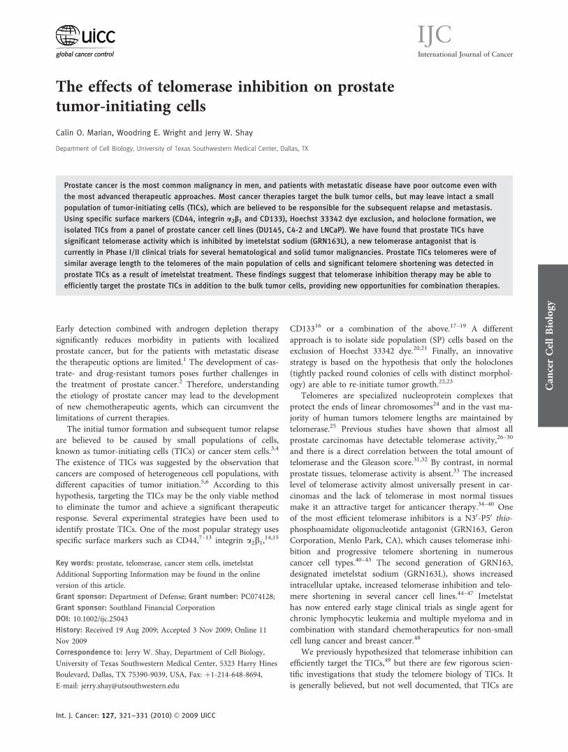
First, we set out to evaluate the effects of imetelstat on thewhole population of prostate cancer cells. Figure 1a shows theexpected 6-bp TRAP ladder for 4 different prostate cancer celllines (DU145, PC3, C4-2 and LNCaP), quantified in Figure 1b.All the cell lines used in this study have significant levels oftelomerase activity, 3 of them (DU145, C4-2 and LNCaP) havemore RTA (relative telomerase activity) when compared toHeLa cells. Treatment with imetelstat leads to efficient telomer-
ase inhibition (Fig. 1c) in a dose-dependent fashion (gel datanot shown). Prolonged telomerase inhibition due to imetelstattreatment leads to telomere shortening in all the prostate cancercell lines analyzed (Fig. 1d). The telomere lengths of the cellsused in our experiments vary in average size, from short(LNCaP) to relatively long (DU145), and there is no correlationbetween telomere length and telomerase activity. If telomeraseinhibition (2 lM every 3 days) was maintained until the telo-meres became critically short, the cells ceased to proliferate andultimately died (data not shown). More importantly, there is acorrelation between the interval of time required for the onsetof apoptosis as a result of telomere shortening (due to imetel-stat treatment) and the initial telomere length.
Prostate TICs isolated using established surface markers
are telomerase-positive and sensitive to telomerase
inhibition by imetelstat
It was previously shown that DU145 CD44þ/integrin a2b1hi
cancer cells possess traits of tumor stem/progenitor cells and
Figure 1. Prostate cell lines have high levels of telomerase activity which can be inhibited by imetelstat. (a) Telomeric repeat amplification
protocol (TRAP) assay of 4 prostate cancer cell lines compared with the HeLa cells. (b) Quantification of the TRAP signal presented as a
ratio between the intensity of the telomerase ladder signal versus the intensity of internal amplification standard (ITAS) band. (c) Imetelstat
(1 lM) inhibits telomerase activity efficiently in all the cell lines analyzed. Relative telomerase activity (RTA) was normalized to the
untreated control cells. (d) TRF (telomere analysis) shows that sustained telomerase inhibition with 2 lM imetelstat leads to telomere
shortening. Lysate equivalent to the same number of cells was used for TRAP with all the cell lines.
Can
cerCellBiology
Marian et al. 323
Int. J. Cancer: 127, 321–331 (2010) VC 2009 UICC
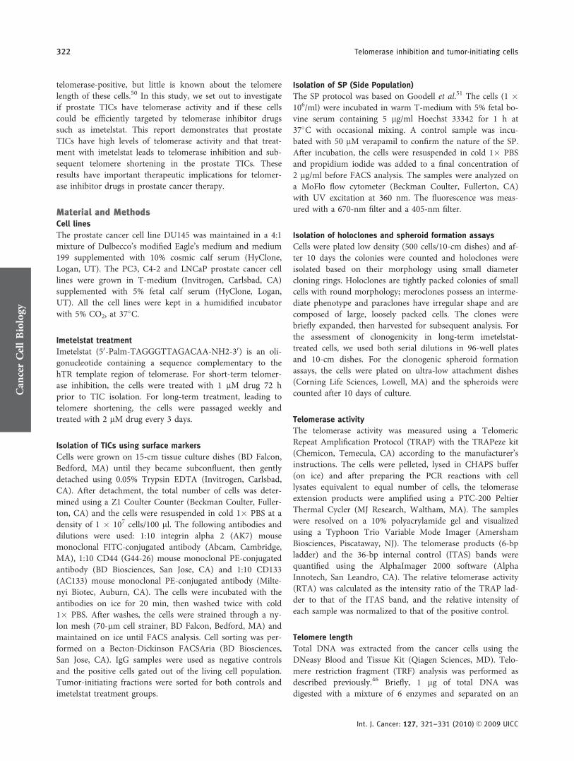
are more proliferative, clonogenic, tumorigenic and metastaticthan the CD44þ/integrin a2b1
low cells.15 We sorted the top10% cells stained with each antibody, which translated to�2% of the total population (Fig. 2a). Equal numbers ofCD44hi/integrin a2b1
hi cells were collected for the untreatedand imetelstat-treated samples and subsequently used for theTRAP assay. The DU145 CD44hi/integrin a2b1
hi cells havetelomerase levels similar to that of total population, and ime-telstat inhibition of telomerase activity is equally efficient inthis putative stem/progenitor cell fraction (Fig. 2b).
In the LNCaP cell line, the isolated CD44þ/CD24� popu-lation is highly tumorigenic and expresses specific genesknown to be important in stem cell maintenance.12 Consist-ent with this previous study, the percentage of LNCaP cellsthat stained positive for CD44 was relatively small, less than1% (Fig. 2c). The LNCaP CD44þ/CD24� cells had high lev-els of telomerase activity, which was similar to the main pop-ulation of LNCaP cells. Again, imetelstat was able to robustlyinhibit telomerase activity in both fractions (Fig. 2d).
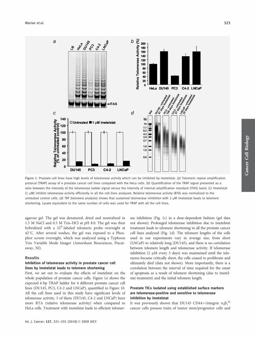
CD133þ cells isolated from the DU145 line have thecapacity of self-renewal and differentiation, as well as high
proliferative and tumorigenic potential.19 An antibody againstCD133 (prominin-1) was used to sort a small population ofCD133þ cells from the DU145 line (Fig. 3a). DU145CD133þ cells have high levels of telomerase activity, similarto the levels found in the total population of cells, and ime-telstat is effective at inhibiting telomerase in these putativestem-like cells (Fig. 3b).
These results show that populations of prostate TICs iso-lated using most of the surface markers cited in the literatureare telomerase-positive and are sensitive to the telomeraseinhibitor imetelstat.
The SP (side population) cells have high levels of
telomerase activity which is inhibited by imetelstat
The SP, believed to harbor the TICs, can be isolated by sort-ing cells which exclude the Hoechst 33342 dye.21,52 Out ofthe 4 cell lines analyzed, we detected a small SP only in theC4-2 prostate cell line (an LNCaP derivative), whichaccounted for �0.1% of the total population (Fig. 3c). TRAPassays show that the SP cells isolated from the C4-2 cellshave high levels of telomerase activity, slightly higher than
Figure 2. Imetelstat acts efficiently on prostate TICs isolated using the CD44 surface marker. (a) DU145 cells with the CD44hi/integrin a2b1hi
phenotype were isolated using fluorescence-activated cell sorting (FACS). (b) RTA in the untreated and imetelstat-treated cells for total and
CD44hi/integrin a2b1hi cells. (c) LNCaP cells possess a small population of CD44/CD24� cells as illustrated by FACS. (d) RTA in the
untreated and imetelstat-treated cells for total and CD44þ/CD24� cells. The cells were treated with 1 lM imetelstat before sorting and
analysis.
Can
cerCellBiology
324 Telomerase inhibition and tumor-initiating cells
Int. J. Cancer: 127, 321–331 (2010) VC 2009 UICC
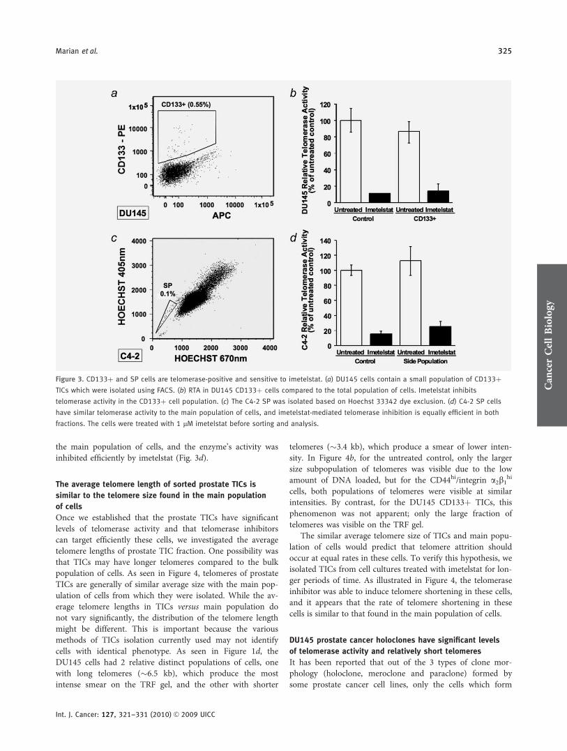
the main population of cells, and the enzyme’s activity wasinhibited efficiently by imetelstat (Fig. 3d).
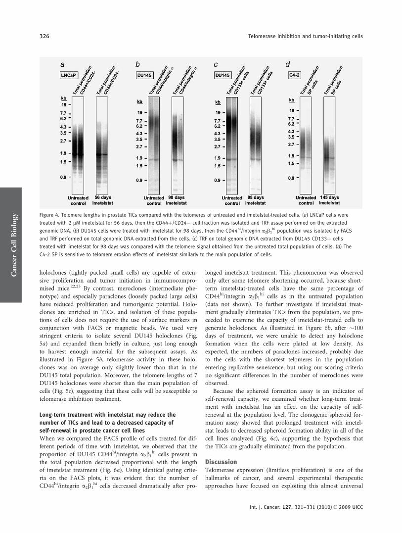
The average telomere length of sorted prostate TICs is
similar to the telomere size found in the main population
of cells
Once we established that the prostate TICs have significantlevels of telomerase activity and that telomerase inhibitorscan target efficiently these cells, we investigated the averagetelomere lengths of prostate TIC fraction. One possibility wasthat TICs may have longer telomeres compared to the bulkpopulation of cells. As seen in Figure 4, telomeres of prostateTICs are generally of similar average size with the main pop-ulation of cells from which they were isolated. While the av-erage telomere lengths in TICs versus main population donot vary significantly, the distribution of the telomere lengthmight be different. This is important because the variousmethods of TICs isolation currently used may not identifycells with identical phenotype. As seen in Figure 1d, theDU145 cells had 2 relative distinct populations of cells, onewith long telomeres (�6.5 kb), which produce the mostintense smear on the TRF gel, and the other with shorter
telomeres (�3.4 kb), which produce a smear of lower inten-sity. In Figure 4b, for the untreated control, only the largersize subpopulation of telomeres was visible due to the lowamount of DNA loaded, but for the CD44hi/integrin a2b1
hi
cells, both populations of telomeres were visible at similarintensities. By contrast, for the DU145 CD133þ TICs, thisphenomenon was not apparent; only the large fraction oftelomeres was visible on the TRF gel.
The similar average telomere size of TICs and main popu-lation of cells would predict that telomere attrition shouldoccur at equal rates in these cells. To verify this hypothesis, weisolated TICs from cell cultures treated with imetelstat for lon-ger periods of time. As illustrated in Figure 4, the telomeraseinhibitor was able to induce telomere shortening in these cells,and it appears that the rate of telomere shortening in thesecells is similar to that found in the main population of cells.
DU145 prostate cancer holoclones have significant levels
of telomerase activity and relatively short telomeres
It has been reported that out of the 3 types of clone mor-phology (holoclone, meroclone and paraclone) formed bysome prostate cancer cell lines, only the cells which form
Figure 3. CD133þ and SP cells are telomerase-positive and sensitive to imetelstat. (a) DU145 cells contain a small population of CD133þTICs which were isolated using FACS. (b) RTA in DU145 CD133þ cells compared to the total population of cells. Imetelstat inhibits
telomerase activity in the CD133þ cell population. (c) The C4-2 SP was isolated based on Hoechst 33342 dye exclusion. (d) C4-2 SP cells
have similar telomerase activity to the main population of cells, and imetelstat-mediated telomerase inhibition is equally efficient in both
fractions. The cells were treated with 1 lM imetelstat before sorting and analysis.
Can
cerCellBiology
Marian et al. 325
Int. J. Cancer: 127, 321–331 (2010) VC 2009 UICC
holoclones (tightly packed small cells) are capable of exten-sive proliferation and tumor initiation in immunocompro-mised mice.22,23 By contrast, meroclones (intermediate phe-notype) and especially paraclones (loosely packed large cells)have reduced proliferation and tumorigenic potential. Holo-clones are enriched in TICs, and isolation of these popula-tions of cells does not require the use of surface markers inconjunction with FACS or magnetic beads. We used verystringent criteria to isolate several DU145 holoclones (Fig.5a) and expanded them briefly in culture, just long enoughto harvest enough material for the subsequent assays. Asillustrated in Figure 5b, telomerase activity in these holo-clones was on average only slightly lower than that in theDU145 total population. Moreover, the telomere lengths of 7DU145 holoclones were shorter than the main population ofcells (Fig. 5c), suggesting that these cells will be susceptible totelomerase inhibition treatment.
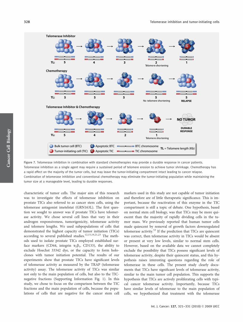
Long-term treatment with imetelstat may reduce the
number of TICs and lead to a decreased capacity of
self-renewal in prostate cancer cell lines
When we compared the FACS profile of cells treated for dif-ferent periods of time with imetelstat, we observed that theproportion of DU145 CD44hi/integrin a2b1
hi cells present inthe total population decreased proportional with the lengthof imetelstat treatment (Fig. 6a). Using identical gating crite-ria on the FACS plots, it was evident that the number ofCD44hi/integrin a2b1
hi cells decreased dramatically after pro-
longed imetelstat treatment. This phenomenon was observedonly after some telomere shortening occurred, because short-term imetelstat-treated cells have the same percentage ofCD44hi/integrin a2b1
hi cells as in the untreated population(data not shown). To further investigate if imetelstat treat-ment gradually eliminates TICs from the population, we pro-ceeded to examine the capacity of imetelstat-treated cells togenerate holoclones. As illustrated in Figure 6b, after �100days of treatment, we were unable to detect any holocloneformation when the cells were plated at low density. Asexpected, the numbers of paraclones increased, probably dueto the cells with the shortest telomeres in the populationentering replicative senescence, but using our scoring criteriano significant differences in the number of meroclones wereobserved.
Because the spheroid formation assay is an indicator ofself-renewal capacity, we examined whether long-term treat-ment with imetelstat has an effect on the capacity of self-renewal at the population level. The clonogenic spheroid for-mation assay showed that prolonged treatment with imetel-stat leads to decreased spheroid formation ability in all of thecell lines analyzed (Fig. 6c), supporting the hypothesis thatthe TICs are gradually eliminated from the population.
DiscussionTelomerase expression (limitless proliferation) is one of thehallmarks of cancer, and several experimental therapeuticapproaches have focused on exploiting this almost universal
Figure 4. Telomere lengths in prostate TICs compared with the telomeres of untreated and imetelstat-treated cells. (a) LNCaP cells were
treated with 2 lM imetelstat for 56 days, then the CD44þ/CD24� cell fraction was isolated and TRF assay performed on the extracted
genomic DNA. (b) DU145 cells were treated with imetelstat for 98 days, then the CD44hi/integrin a2b1hi population was isolated by FACS
and TRF performed on total genomic DNA extracted from the cells. (c) TRF on total genomic DNA extracted from DU145 CD133þ cells
treated with imetelstat for 98 days was compared with the telomere signal obtained from the untreated total population of cells. (d) The
C4-2 SP is sensitive to telomere erosion effects of imetelstat similarly to the main population of cells.
Can
cerCellBiology
326 Telomerase inhibition and tumor-initiating cells
Int. J. Cancer: 127, 321–331 (2010) VC 2009 UICC
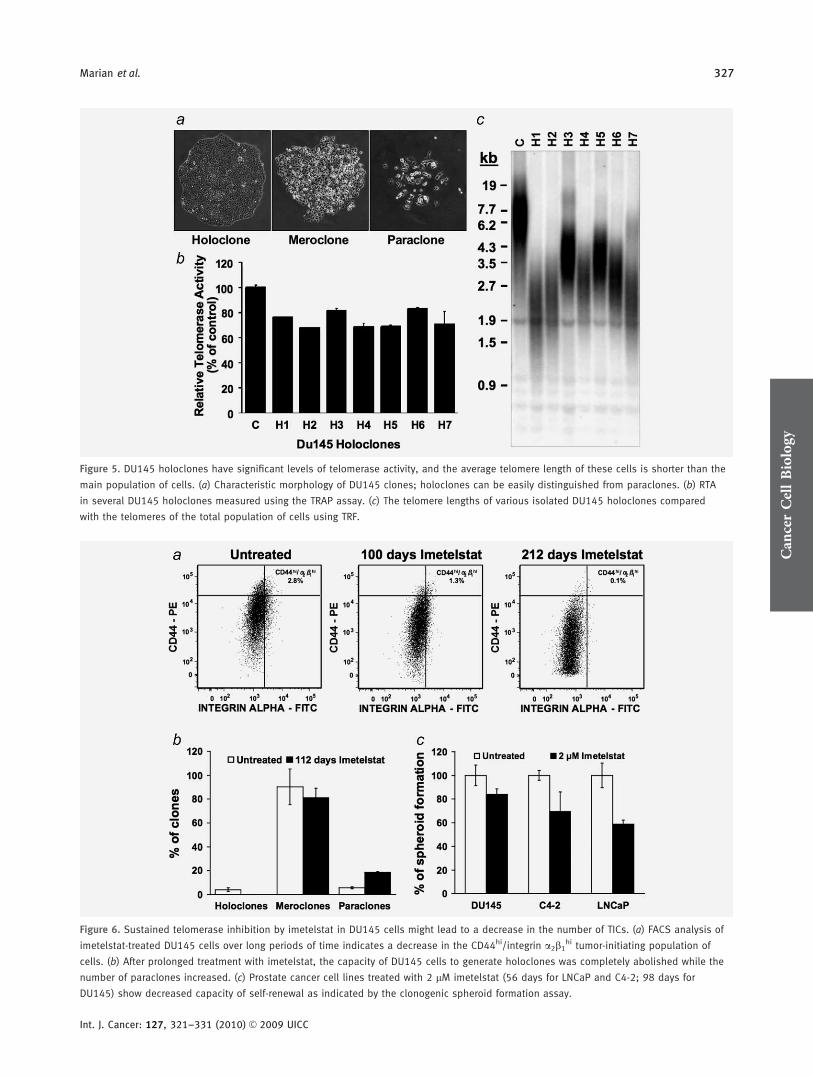
Figure 5. DU145 holoclones have significant levels of telomerase activity, and the average telomere length of these cells is shorter than the
main population of cells. (a) Characteristic morphology of DU145 clones; holoclones can be easily distinguished from paraclones. (b) RTA
in several DU145 holoclones measured using the TRAP assay. (c) The telomere lengths of various isolated DU145 holoclones compared
with the telomeres of the total population of cells using TRF.
Figure 6. Sustained telomerase inhibition by imetelstat in DU145 cells might lead to a decrease in the number of TICs. (a) FACS analysis of
imetelstat-treated DU145 cells over long periods of time indicates a decrease in the CD44hi/integrin a2b1hi tumor-initiating population of
cells. (b) After prolonged treatment with imetelstat, the capacity of DU145 cells to generate holoclones was completely abolished while the
number of paraclones increased. (c) Prostate cancer cell lines treated with 2 lM imetelstat (56 days for LNCaP and C4-2; 98 days for
DU145) show decreased capacity of self-renewal as indicated by the clonogenic spheroid formation assay.
Can
cerCellBiology
Marian et al. 327
Int. J. Cancer: 127, 321–331 (2010) VC 2009 UICC
characteristic of tumor cells. The major aim of this researchwas to investigate the effects of telomerase inhibition onprostate TICs also referred to as cancer stem cells, using thetelomerase antagonist imetelstat (GRN163L). The first ques-tion we sought to answer was if prostate TICs have telomer-ase activity. We chose several cell lines that vary in theirandrogen responsiveness, tumorigenicity, telomerase activityand telomere lengths. We used subpopulations of cells thatdemonstrated the highest capacity of tumor initiation (TICs)according to several published studies.12,15,19,21,23 The meth-ods used to isolate prostate TICs employed established sur-face markers (CD44, integrin a2b1, CD133), the ability toexclude Hoechst 33342 dye, or the capacity to form holo-clones with tumor initiation potential. The results of ourexperiments show that prostate TICs have significant levelsof telomerase activity as measured by the TRAP (telomeraseactivity) assay. The telomerase activity of TICs was similarnot only to the main population of cells, but also to the TIC-negative fractions (Supporting Information Fig. 1). In thisstudy, we chose to focus on the comparison between the TICfractions and the main population of cells, because the popu-lations of cells that are negative for the cancer stem cell
markers used in this study are not capable of tumor initiationand therefore are of little therapeutic significance. This is im-portant, because the reactivation of this enzyme in the TICcompartment is still a topic of debate. One hypothesis, basedon normal stem cell biology, was that TICs may be more qui-escent than the majority of rapidly dividing cells in the tu-mor mass. We previously reported that human tumor cellsmade quiescent by removal of growth factors downregulatedtelomerase activity.53 If the prediction that TICs are quiescentwas correct, then telomerase activity in TICs would be absentor present at very low levels, similar to normal stem cells.However, based on the available data we cannot completelyexclude the possibility that TICs possess significant levels oftelomerase activity, despite their quiescent status, and this hy-pothesis raises interesting questions regarding the role oftelomerase in these cells. The present study clearly docu-ments that TICs have significant levels of telomerase activity,similar to the main tumor cell population. This supports thehypothesis that TICs are actively proliferating cells with typi-cal cancer telomerase activity. Importantly, because TICshave similar levels of telomerase to the main population ofcells, we hypothesized that treatment with the telomerase
Figure 7. Telomerase inhibition in combination with standard chemotherapies may provide a durable response in cancer patients.
Telomerase inhibition as a single agent may require a sustained period of telomere erosion to achieve tumor shrinkage. Chemotherapy has
a rapid effect on the majority of the tumor cells, but may leave the tumor-initiating compartment intact leading to cancer relapse.
Combination of telomerase inhibition and conventional chemotherapy may eliminate the tumor-initiating population while maintaining the
tumor size at a manageable level, leading to durable responses.
Can
cerCellBiology
328 Telomerase inhibition and tumor-initiating cells
Int. J. Cancer: 127, 321–331 (2010) VC 2009 UICC
inhibitor imetelstat would have similar effects in this com-partment. The experimental data in the present study showthat imetelstat treatment efficiently inhibits telomerase activ-ity in all the prostate TICs populations analyzed.
Because there is not always a direct correlation betweentelomerase activity and average telomere lengths in variouscultured prostate cancer cells, it was important to determinethe average telomere length of TICs. We found that the telo-meres of these cells were approximately the same length orshorter than the average telomere size found in the mainpopulation; therefore, we assumed that telomerase inhibitorscurrently being tested in clinical trials will induce telomereattrition in the rare populations of TICs with equal efficiencyas the bulk tumor cells. We were able to show that the telo-merase inhibition by imetelstat induced telomere shorteningin the TIC compartment and postulate that prolonged telo-merase inhibition will lead to apoptosis and cell death ofTICs, similar to the main population of cells.
Investigating the telomere length of prostate TICs was im-portant, since it was theoretically possible for these cells tohave longer telomeres, similar to normal stem cells. Thereduced telomere length in the tumor-initiating compartmentmay also shed some light on the process of malignant trans-formation in prostate. Telomere shortening can be detected asearly as prostatic intraepithelial neoplasia (PIN) and is re-stricted to the luminal compartment.54,55 This suggests thatthe TICs originate from a subset of transient amplifying cellswhich under chronic inflammation pressure and genomicinstability caused by short telomeres reactivate telomerase andafter additional mutations lead to prostate cancer.
Another important observation we made relates to theeffects of prolonged telomerase inhibition on the fraction ofTICs present in the population. The experimental data showsthat long-term treatment with imetelstat leads to a decreasein the number of DU145 CD44hi/integrin a2b1
hi cells presentin the total population of cells due to a reduction in the fluo-rescence of the whole population. Interesting, the CD133þcells did not show the same trend (data not shown). Pro-longed treatment with imetelstat also correlated with adecreased capacity of DU145 cells to form holoclones.Because one of the main characteristics of TICs is theircapacity of self-renewal, it was important to investigate theimpact of long-term treatment with imetelstat on the capacityof cells to form spheroids when plated at clonal density inattachment-independent conditions, which was used as ameasure of self-renewal capacity for prostate cancer cells.56
When we performed clonogenic spheroid formation assayson long-term imetelstat-treated LNCaP, C4-2 and DU145cells, telomere shortening positively correlated with adecrease in the sphere-forming ability of these cells, indicat-ing a decreased capacity of self-renewal. This is important,because it was well documented by our group and others57
that telomere shortening is associated with a decreased tumorformation ability in immunocompromised mice.
These experiments support the hypothesis that long-termtelomerase inhibition by imetelstat coupled with telomereshortening may lead to the elimination of certain populationsof prostate TICs. Whether this is a direct result of the elimi-nation of TICs from the population remains unclear andfuture experiments will be aimed at answering this importantquestion.
One of the ongoing concerns about telomerase inhibitiontherapy is related to the effects of long-term telomerase inhi-bition on normal cells. However, normal prostate cells lacktelomerase activity and have longer telomere lengths com-pared to cancer cells.31,58 Moreover, we have shown thattreatment with imetelstat has no effect on the proliferation ofnormal cells.46 Normal stem cells are known to be relativelyquiescent and have low or no telomerase activity exceptwhen dividing. Most importantly, normal stem cells possessrelatively long telomeres,59 and we predict that the effect ofimetelstat on these normal stem cells would be less toxic incomparison to the shorter telomere length of TICs. Thus,there may be an optimal therapeutic window that would leadto cancer cell death without irreversibly affecting the normalcells.
Telomerase inhibition as single agent therapy is believed tobe most effective only after critical telomere erosion occurred,and this may require relatively long periods of treatment(depending on the initial average telomere length of the tu-mor) to achieve a reduction in tumor mass. In contrast, con-ventional therapies (such as surgery, radiation and chemother-apy) lead to a dramatic reduction in tumor burden relativelyquickly, but do not lead to durable responses in advancedstage cancers. This may be due to the inherent resistance ofTICs to conventional therapeutic agents, behavior stronglydocumented in glioblastoma.60–62 We believe that an idealprostate cancer therapy should combine conventional thera-peutic approaches with telomerase inhibitors, such as imetel-stat. While conventional approaches will initially target thebulk tumor, after a certain interval of time imetelstat-mediatedtelomerase inhibition will shorten the telomeres in the tumor-initiating compartment to a critical level, inducing cell apopto-sis and death in this small fraction of cells. This therapyapproach could potentially lead to durable responses (Fig. 6).
In summary, this preclinical study shows that telomeraseinhibition has a great potential for the treatment of prostatecancer and may be able to target the TICs that contribute torelapse and metastasis.
AcknowledgementsWe would like to thank Geron Corporation (Menlo Park, CA) for providingthe imetelstat telomerase inhibitor, Ms. Erin Kitten for technical supportand Ms. Angela Diehl for the graphic design. This work was supported by aDepartment of Defense Prostate Cancer Training Award (Grant no.PC074128 to C.O.M.) and by the Southland Financial Corporation toW.E.W. and J.W.S.
Can
cerCellBiology
Marian et al. 329
Int. J. Cancer: 127, 321–331 (2010) VC 2009 UICC
References
1. Mike S, Harrison C, Coles B, Staffurth J,Wilt TJ, Mason MD. Chemotherapy forhormone-refractory prostatecancer.Cochrane Database Syst Rev(Online) 2006:CD005247.
2. Uzzo RG, Haas NB, Crispen PL, KolenkoVM. Mechanisms of apoptosis resistanceand treatment strategies to overcome themin hormone-refractory prostate cancer.Cancer 2008;112:1660–71.
3. Wicha MS, Liu S, Dontu G. Cancer stemcells: an old idea—a paradigm shift. CancerRes 2006;66:1883–90, discussion 95–6.
4. Ward RJ, Dirks PB. Cancer stem cells: atthe headwaters of tumor development.Annu Rev Pathol 2007;2:175–89.
5. Sabbath KD, Ball ED, Larcom P, Davis RB,Griffin JD. Heterogeneity of clonogeniccells in acute myeloblastic leukemia. J ClinInvest 1985;75:746–53.
6. Hamburger AW, Salmon SE. Primarybioassay of human tumor stem cells.Science 1977;197:461–63.
7. Liu AY, True LD, LaTray L, Nelson PS,Ellis WJ, Vessella RL, Lange PH, Hood L,van den Engh G. Cell–cell interaction inprostate gene regulation andcytodifferentiation. Proc Natl Acad Sci USA1997;94:10705–10.
8. Avigdor A, Goichberg P, Shivtiel S, Dar A,Peled A, Samira S, Kollet O, Hershkoviz R,Alon R, Hardan I, Ben-Hur H, Naor D,et al. CD44 and hyaluronic acid cooperatewith SDF-1 in the trafficking of humanCD34þ stem/progenitor cells to bonemarrow. Blood 2004;103:2981–9.
9. Oswald J, Boxberger S, Jorgensen B,Feldmann S, Ehninger G, Bornhauser M,Werner C. Mesenchymal stem cells can bedifferentiated into endothelial cells in vitro.Stem Cells 2004;22:377–84.
10. Schwartz PH, Bryant PJ, Fuja TJ, Su H,O’Dowd DK, Klassen H. Isolation andcharacterization of neural progenitor cellsfrom post-mortem human cortex.J Neurosci Res 2003;74:838–51.
11. Gudjonsson T, Villadsen R, Nielsen HL,Ronnov-Jessen L, Bissell MJ, Petersen OW.Isolation, immortalization, andcharacterization of a human breastepithelial cell line with stem cell properties.Genes Dev 2002;16:693–706.
12. Hurt EM, Kawasaki BT, Klarmann GJ,Thomas SB, Farrar WL. CD44þ CD24(�)prostate cells are early cancer progenitor/stem cells that provide a model for patientswith poor prognosis. Br J Cancer 2008;98:756–65.
13. Patrawala L, Calhoun T, Schneider-Broussard R, Li H, Bhatia B, Tang S, ReillyJG, Chandra D, Zhou J, Claypool K,Coghlan L, Tang DG. Highly purifiedCD44þ prostate cancer cells from
xenograft human tumors are enriched intumorigenic and metastatic progenitorcells. Oncogene 2006;25:1696–708.
14. Collins AT, Habib FK, Maitland NJ, NealDE. Identification and isolation of humanprostate epithelial stem cells based onalpha(2)beta(1)-integrin expression. J CellSci 2001;114:3865–72.
15. Patrawala L, Calhoun-Davis T, Schneider-Broussard R, Tang DG. Hierarchicalorganization of prostate cancer cells inxenograft tumors: the CD44þalpha2beta1þcell population is enriched in tumor-initiating cells. Cancer Res 2007;67:6796–805.
16. Vander Griend DJ, Karthaus WL,Dalrymple S, Meeker A, DeMarzo AM,Isaacs JT. The role of CD133 in normalhuman prostate stem cells and malignantcancer-initiating cells. Cancer Res 2008;68:9703–11.
17. Richardson GD, Robson CN, Lang SH,Neal DE, Maitland NJ, Collins AT. CD133,a novel marker for human prostaticepithelial stem cells. J Cell Sci 2004;117:3539–45.
18. Collins AT, Berry PA, Hyde C, Stower MJ,Maitland NJ. Prospective identification oftumorigenic prostate cancer stem cells.Cancer Res 2005;65:10946–51.
19. Wei C, Guomin W, Yujun L, Ruizhe Q.Cancer stem-like cells in human prostatecarcinoma cells DU145: the seedsof the cell line? Cancer Biol Ther 2007;6:763–8.
20. Brown MD, Gilmore PE, Hart CA, SamuelJD, Ramani VA, George NJ, Clarke NW.Characterization of benign and malignantprostate epithelial Hoechst 33342 sidepopulations. Prostate 2007;67:1384–96.
21. Patrawala L, Calhoun T, Schneider-Broussard R, Zhou J, Claypool K, TangDG. Side population is enriched intumorigenic, stem-like cancer cells, whereasABCG2þ and ABCG2� cancer cells aresimilarly tumorigenic. Cancer Res 2005;65:6207–19.
22. Locke M, Heywood M, Fawell S,Mackenzie IC. Retention of intrinsic stemcell hierarchies in carcinoma-derived celllines. Cancer Res 2005;65:8944–50.
23. Li H, Chen X, Calhoun-Davis T, ClaypoolK, Tang DG. PC3 human prostatecarcinoma cell holoclones contain self-renewing tumor-initiating cells. Cancer Res2008;68:1820–5.
24. Moyzis RK, Buckingham JM, Cram LS,Dani M, Deaven LL, Jones MD, Meyne J,Ratliff RL, Wu JR. A highly conservedrepetitive DNA-sequence, (TTAGGG)n,present at the telomeres of human-chromosomes. Proc Natl Acad Sci USA1988;85:6622–26.
25. Greider CW, Blackburn EH. Identificationof a specific telomere terminal transferaseactivity in Tetrahymena extracts. Cell 1985;43:405–13.
26. Zhang W, Kapusta LR, Slingerland JM,Klotz LH. Telomerase activity in prostatecancer, prostatic intraepithelial neoplasia,and benign prostatic epithelium. CancerRes 1998;58:619–21.
27. Takahashi C, Miyagawa I, Kumano S,Oshimura M. Detection oftelomerase activity in prostate cancerby needle biopsy. Eur Urol 1997;32:494–8.
28. Lin Y, Uemura H, Fujinami K, Hosaka M,Iwasaki Y, Kitamura H, Harada M, KubotaY. Detection of telomerase activity inprostate needle-biopsy samples. Prostate1998;36:121–8.
29. Lin Y, Uemura H, Fujinami K, Hosaka M,Harada M, Kubota Y. Telomerase activityin primary prostate cancer. J Urol 1997;157:1161–5.
30. Kallakury BV, Brien TP, Lowry CV,Muraca PJ, Fisher HA, Kaufman RP Jr,Ross JS. Telomerase activity in humanbenign prostate tissue and prostaticadenocarcinomas. Diagn Mol Pathol 1997;6:192–8.
31. Sommerfeld HJ, Meeker AK, PiatyszekMA, Bova GS, Shay JW, Coffey DS.Telomerase activity: a prevalent marker ofmalignant human prostate tissue. CancerRes 1996;56:218–22.
32. Kamradt J, Drosse C, Kalkbrenner S,Rohde V, Lensch R, Lehmann J, FixemerT, Bonkhoff H, Stoeckle M, Wullich B.Telomerase activity and telomerase subunitgene expression levels are not related inprostate cancer: a real-time quantificationand in situ hybridization study. Lab Invest2003;83:623–33.
33. Liu BC, LaRose I, Weinstein LJ, Ahn M,Weinstein MH, Richie JP. Expression oftelomerase subunits in normal andneoplastic prostate epithelial cells isolatedby laser capture microdissection. Cancer2001;92:1943–8.
34. Koga S, Hirohata S, Kondo Y, Komata T,Takakura M, Inoue M, Kyo S, Kondo S.FADD gene therapy using the humantelomerase catalytic subunit (hTERT) genepromoter to restrict induction of apoptosisto tumors in vitro and in vivo. AnticancerRes 2001;21:1937–43.
35. Vonderheide RH. Telomerase as auniversal tumor-associated antigen forcancer immunotherapy. Oncogene 2002;21:674–9.
36. Naasani I, Oh-Hashi F, Oh-Hara T, FengWY, Johnston J, Chan K, Tsuruo T.Blocking telomerase by dietary polyphenolsis a major mechanism for limiting the
Can
cerCellBiology
330 Telomerase inhibition and tumor-initiating cells
Int. J. Cancer: 127, 321–331 (2010) VC 2009 UICC
growth of human cancer cells in vitro andin vivo. Cancer Res 2003;63:824–30.
37. Sun D, Thompson B, Cathers BE, SalazarM, Kerwin SM, Trent JO, Jenkins TC,Neidle S, Hurley LH. Inhibition ofhuman telomerase by a G-quadruplex-interactive compound. J Med Chem 1997;40:2113–6.
38. Strahl C, Blackburn EH. The effects ofnucleoside analogs on telomerase andtelomeres in Tetrahymena. Nucleic AcidsRes 1994;22:893–900.
39. Yokoyama Y, Takahashi Y, Shinohara A,Lian Z, Wan X, Niwa K, Tamaya T.Attenuation of telomerase activity by ahammerhead ribozyme targeting thetemplate region of telomerase RNA inendometrial carcinoma cells. Cancer Res1998;58:5406–10.
40. Herbert BS, Pongracz K, Shay JW,Gryaznov SM. Oligonucleotide N30!P50
phosphoramidates as efficient telomeraseinhibitors. Oncogene 2002;21:638–42.
41. Canales BK, Li Y, Thompson MG, GleasonJM, Chen Z, Malaeb B, Corey DR, HerbertBS, Shay JW, Koeneman KS. Smallmolecule, oligonucleotide-based telomerasetemplate inhibition in combination withcytolytic therapy in an in vitro androgen-independent prostate cancer model. UrolOncol 2006;24:141–51.
42. Asai A, Oshima Y, Yamamoto Y, UochiTA, Kusaka H, Akinaga S, Yamashita Y,Pongracz K, Pruzan R, Wunder E,Piatyszek M, Li S, et al. A novel telomerasetemplate antagonist (GRN163) as apotential anticancer agent. Cancer Res2003;63:3931–9.
43. Ozawa T, Gryaznov SM, Hu LJ, PongraczK, Santos RA, Bollen AW, Lamborn KR,Deen DF. Antitumor effects of specifictelomerase inhibitor GRN163 in humanglioblastoma xenografts. Neuro Oncol 2004;6:218–26.
44. Herbert BS, Gellert GC, Hochreiter A,Pongracz K, Wright WE, Zielinska D, ChinAC, Harley CB, Shay JW, Gryaznov SM.
Lipid modification of GRN163, anN30!P50 thio-phosphoramidateoligonucleotide, enhances the potency oftelomerase inhibition. Oncogene 2005;24:5262–8.
45. Djojosubroto MW, Chin AC, Go N,Schaetzlein S, Manns MP, Gryaznov S,Harley CB, Rudolph KL. Telomeraseantagonists GRN163 and GRN163L inhibittumor growth and increasechemosensitivity of human hepatoma.Hepatology 2005;42:1127–36.
46. Gellert GC, Dikmen ZG, Wright WE,Gryaznov S, Shay JW. Effects of a noveltelomerase inhibitor. GRN163L, in humanbreast cancer. Breast Cancer Res Treat2006;96:73–81.
47. Dikmen ZG, Gellert GC, Jackson S,Gryaznov S, Tressler R, Dogan P, WrightWE, Shay JW. In vivo inhibition of lungcancer by GRN163L: a novel humantelomerase inhibitor. Cancer Res 2005;65:7866–73.
48. Harley CB. Telomerase and cancertherapeutics. Nat Rev 2008;8:167–79.
49. Shay JW, Keith WN. Targeting telomerasefor cancer therapeutics. Br J Cancer 2008;98:677–83.
50. Ponti D, Costa A, Zaffaroni N, Pratesi G,Petrangolini G, Coradini D, Pilotti S,Pierotti MA, Daidone MG. Isolation and invitro propagation of tumorigenic breastcancer cells with stem/progenitor cellproperties. Cancer Res 2005;65:5506–11.
51. Goodell MA, Brose K, Paradis G, ConnerAS, Mulligan RC. Isolation and functionalproperties of murine hematopoietic stemcells that are replicating in vivo. J Exp Med1996;183:1797–806.
52. Pascal LE, Oudes AJ, Petersen TW, GooYA, Walashek LS, True LD, Liu AY.Molecular and cellular characterization ofABCG2 in the prostate. BMC Urol 2007;7:6.
53. Holt SE, Shay JW. Role of telomerase incellular proliferation and cancer. J CellPhysiol 1999;180:10–8.
54. Meeker AK, Gage WR, Hicks JL, Simon I,Coffman JR, Platz EA, March GE, DeMarzo AM. Telomere length assessment inhuman archival tissues: combined telomerefluorescence in situ hybridization andimmunostaining. Am J Pathol 2002;160:1259–68.
55. Meeker AK, Hicks JL, Platz EA, MarchGE, Bennett CJ, Delannoy MJ, De MarzoAM. Telomere shortening is an earlysomatic DNA alteration in humanprostate tumorigenesis. Cancer Res 2002;62:6405–9.
56. Tang DG, Patrawala L, Calhoun T, BhatiaB, Choy G, Schneider-Broussard R, Jeter C.Prostate cancer stem/progenitor cells:identification, characterization, andimplications. Mol Carcinog 2007;46:1–14.
57. Guo C, Geverd D, Liao R, Hamad N,Counter CM, Price DT. Inhibition oftelomerase is related to the life span andtumorigenicity of human prostate cancercells. J Urol 2001;166:694–8.
58. Koeneman KS, Pan CX, Jin JK, Pyle JM,3rd, Flanigan RC, Shankey TV, Diaz MO.Telomerase activity, telomere length, andDNA ploidy in prostatic intraepithelialneoplasia (PIN). J Urol 1998;160:1533–9.
59. Allen ND, Baird DM. Telomere lengthmaintenance in stem cell populations.Biochim Biophys Acta 2009;1792:324–8.
60. Bao S, Wu Q, McLendon RE, Hao Y, ShiQ, Hjelmeland AB, Dewhirst MW, BignerDD, Rich JN. Glioma stem cells promoteradioresistance by preferential activation ofthe DNA damage response. Nature 2006;444:756–60.
61. Liu G, Yuan X, Zeng Z, Tunici P, Ng H,Abdulkadir IR, Lu L, Irvin D, Black KL,Yu JS. Analysis of gene expression andchemoresistance of CD133þ cancer stemcells in glioblastoma. Mol Cancer 2006;5:67.
62. Kang MK, Kang SK. Tumorigenesis ofchemotherapeutic drug-resistant cancerstem-like cells in brain glioma. Stem CellsDev 2007;16:837–47.
Can
cerCellBiology
Marian et al. 331
Int. J. Cancer: 127, 321–331 (2010) VC 2009 UICC