human telomerase rna and telomerase activity in immortal cell lines and tumor tissues
TRANSCRIPT
[CANCER RESEARCH56. 645-650. February 1. 1996[
levels of telomerase activity to the levels of hTR.3 Whereas many celllines and tumors had both increased hTR and telomerase activity, wefound that hTR was present in cell lines and tissues that lackedtelomerase activity, indicating that the RNA is not limiting for telomerase activity, and that the RNA component is not a good predictor ofthe presence of enzyme activity.
MATERIALS AND METhODS
Cell Extracts and Telomerase Assays. HEK cells were cultured ina-MEM and 10% FBS, grown on 15-cm plates, and split 1:4. Human B cellswere cultured in suspension in RPMI containing 10% }@BS.All culturescontained 0.03% penicillin and streptomycin. Tumor tissue was obtained fromThe University of Texas Southwestern Medical Center at Dallas and fromNorth Shore University Hospital (Manhasset, New York). Cell and tissueextracts were prepared and assayed using a PCR based telomerase assay(TRAP) as described previously (4, 21) using a modified (CCCTAA)n “CX―primer.4 The PCR products were run on a 12—15% acrylamide gels. In
experiments with cell lines, enzyme activity was quantified by scanning gels
on a Molecular Dynamics Phospholmager, and the radioactivity in each bandof the repeat ladder was determined using ImageQuant software (MolecularDynamics). Total activity was expressed as a percentage of that in 293 cellextracts (positive control) after normalizing for protein content. Extracts were
considered negative if no products were detected after 7 days of exposure.RNA Preparation and Northern Blots. RNA was isolated from 10@to 108
cultured cells or 50—500mg of tissue according to the acid guanidiniumthiocyanate/phenol/chloroform extraction procedure as described by Ausubel
el a!. (22). Briefly, cells were lysed in a denaturing solution of 4 M guanidine
thiocyanate-25 mM sodium citrate (pH 7.0)-0.l M (3-mercaptoethanol-0.5%Sarkosyl. The lysate was extracted with saturated phenol, chloroform, isoamyl
alcohol, and sodium acetate (pH 4.0), and the RNA was precipitated from thewater phase with isopropyl alcohol or ethanol. The final pellet was resuspended in diethylpyrocarbonate-treated water. The absorbance at 260 nm wasmeasured to determine the RNA concentration, assuming that 1 absorbance
unit per ml contains 40 ,xg of RNA. For purifying RNA from tissues, sampleswere lysed by Dounce homogenization in the denaturing solution using al.5-ml Eppendorf tube with a fitted pestle (Kimble).
RNA was electrophoresed on 1.5% agarose-2.2 Mformaldehyde gels in 20mM 4-morpholinepropanesulfonic acid (pH 7.0)-8 mM sodium acetate-l nmi
EDTA for4—8h at 5 V/cm (110V using a 21-cm gel). The RNA was passivelytransferred onto a Nytran maximum strength membrane (Schleicher andSchuell), and the RNA was covalently attached to the membrane by baking for1—2h at 80°C.Blots were probed overnight in Church hybridization solution[500 mM sodium phosphate (pH 7.2)-l misiEDTA-l% BSA-7% SDS) contaming 15% formamide. Blots were rinsed 5 times in 2X SSC and 0.1% SDS,at room temperature, for 5 mm each. One final wash was performed at thehybridization temperature for 30 rain in SSC containing 0.1% SDS (for
specific details regarding each probe and the concentration of SSC in the last
wash, see below). Blots hybridized with the hTR component gene or ribosomal55 RNAwereprobedat 65°C;blotsprobedwithanoligonucleotidecomplimentary to the RNase P RNA were probed at 50°C.All blots were exposed toKodak XAR film at —70°Cwith an enhancing screen and to a Phospholmagerscreen, and were scanned with a Fuji BAS200 Phospholmager. Typical autoradiographic exposures for all the experiments were as follows: 3—10days for
3 The abbreviations used are: hTR, human telomerase RNA; HEK, human embryonic
kidney; PD, population doubling; RNP, ribonucleoprotein.4 N. Kim, personal communication.
ABSTRACT
Telomerase activity has been detected in many human immortal cell
lines and in tumor tissues, whereas it is generally absent from primary cell
strains and from many tumor adjacent tissue samples. With the recently
cloned human telomerase RNA (hTR), we used Northern analysis tofollow the levels of hTR in primary, precrisis, and immortalized cells. Itwas surprising that the amount of hTR was high in cell strains that lackedtelomerase activity, and the levels did not parallel the increases in telomerase activity, which accompanies immortalization. In addition, although the hTR levels were somewhat higher in tumor samples compared
to nontumor tissues, the level of hTR in a variety of different humantumors did not predict the level of telomerase activity in the tumor. Thus,
whereas hTR was detected in all samples that have telomerase activity, thepresence of the RNA was not a good a predictor of the presence or amount
of telomerase activity.
INTRODUCTION
Chromosomal rearrangements and end associations are ubiquitousin cancer cells. The loss of telomeres, which normally provide stability to chromosome ends, may initiate or drive the genomic instabilitythat results in abnormal chromosomes and unchecked cell growth(1—5).Telomeres are often shorter in tumor tissue than in normaladjacent tissue (reviewed in Refs. 5, 6). In many immortal organismslike yeast and Tetrahymena, telomere length is maintained by theunusual DNA polymerase, called telomerase. Telomerase is a ribonucleoprotein polymerase that adds telomeric sequences onto chromosome ends (7, 8). In humans, telomerase activity is present in thegermline but is not detected in many normal adult tissues (9). Theabsence of telomerase may lead to the telomere shortening seen insomatic tissues in vivo (reviewed in Ref. 10). In contrast to normaltissues, telomerase activity is found in many human tumors (for
review, see Refs. 6, 11—14).Tissue culture models of cell immortalization suggest that telomerase-positive cells are selected for at crisis,and that telomerase is required for the growth of most immortal cellswith short telomeres (14—17).Thus, telomerase has been proposed asa target for cancer chemotherapy (15). In yeast, telomerase RNA isessential; in cells that are deleted for the RNA component, telomeresshorten and cells die after 50—70divisions (18, 19). In addition,antisense experiments with the RNA component of human telomeraseindicate that telomerase inhibition may lead to telomere shorteningand cell death in human tumor cell lines (20). To gain insight into theregulation of telomerase in cell lines and tumors, we compared the
Received I0/25/95: accepted I2/I 2/95.The costs of publication of this article were defrayed in part by the payment of page
charges. This article must therefore be hereby marked advertisement in accordance with18 U.S.C. Section 1734 solely to indicate this fact.
I This work was supported by NIH Grants AG09383 (C. W. G., A. A. A.) and
AG07992 (J. W. S.), USAMR Grant DAMD-94-J-4077 (J. W. S.), the Susan KomenFoundation (J. W. S.), Medical Research Council-Canada (S. B.), and Geron Corp. (MenloPark, CA).
2 To whom requests for reprints should be addressed, at Cold Spring Harbor Labora
tory. Box 100, Cold Spring Harbor, NY I 1724. Phone: (516) 367-8449; Fax: (516)367-88 15: E-mail: [email protected].
645
Human Telomerase RNA and Telomerase Activity in Immortal Cell Lines andTumor Tissues'
Ariel A. Avilion, Mieczyslaw A. Piatyszek, Jyothi Gupta, Jerry W. Shay, Silvia Bacchetti, and Carol W. Greider@
Cold Spring Harbor Laborazoiy. Cold Spring Harbor, New York 11724 (A. A. A., C. W. G.J: Department of Pathology, McMaster University, Hamilton, OntarioLSN 3Z5. Canada Ii. G.. S. 8.1; and Department of Cell Biology and Neuroscience, University of Texas Southwestern Medical Center at Dallas, Dallas, Texas 75235-9039IM. A. P.. J. W. S.)
Research. on January 31, 2016. © 1996 American Association for Cancercancerres.aacrjournals.org Downloaded from
hTR LEVELS IN CELL LINES AND TUMORS
blots probed with telomerase RNA, 5 h for blots probed with RNase P RNA,and less than 1 h for blots probed with ribosomal 55 RNA.
Quantitative Analysis of Telomerase RNA Abundance. To control forloading errors, telomerase RNA was normalized either to the levels of RNaseP or to 55 RNA signals in the same lane. The tumorstudy was performedblind, with the identity and telomerase activity status of each sample unknownuntil after Northern analysis. Quantification of all RNAs was performed usinga Fuji Phospholmager. The RNA signal for each lane was determined bydrawing a rectangle around each RNA band and integrating the signal over the
area. Some of the samples initially analyzed for telomerase activity were foundto have degraded RNA; these samples were omitted from the analysis presented in Fig. 3.
RESULTS
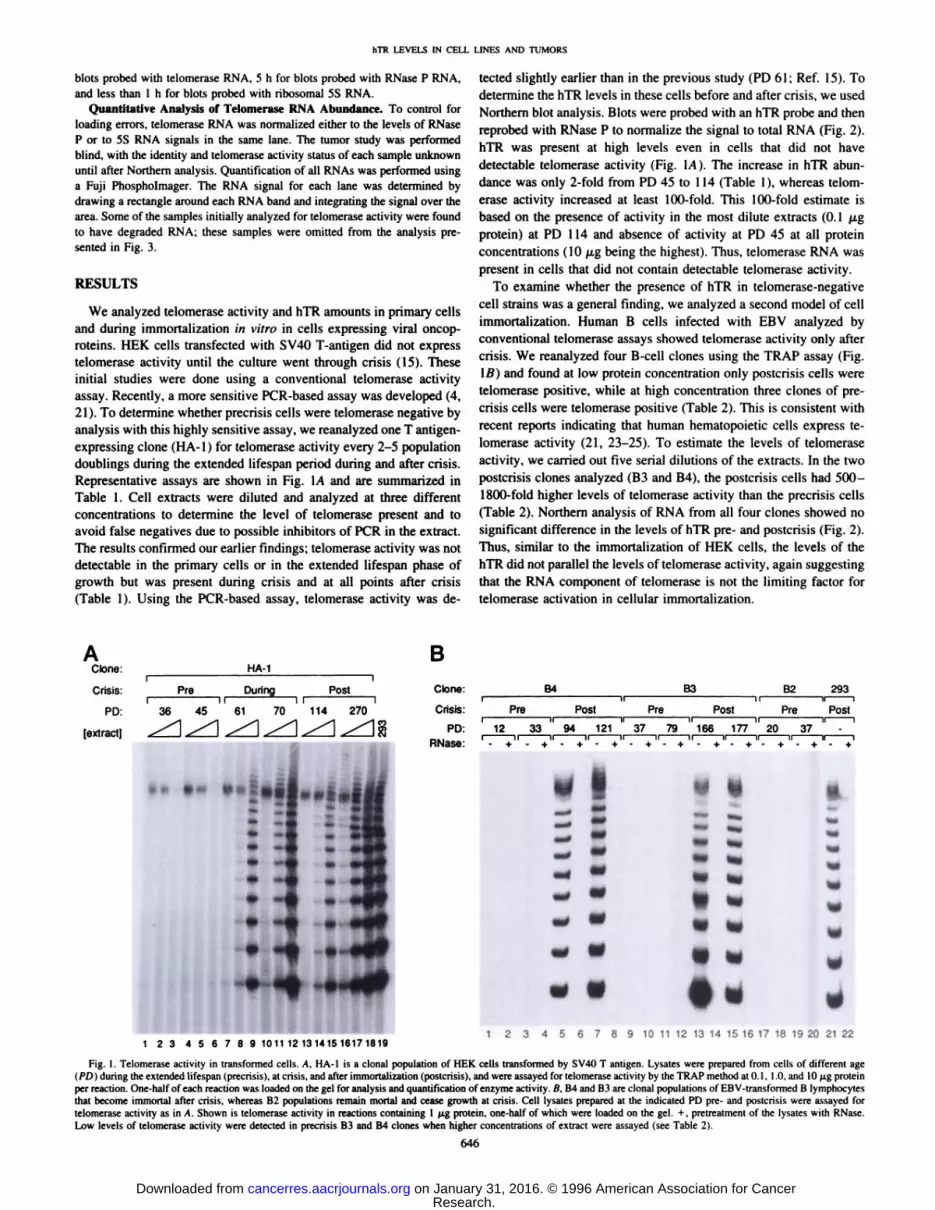
We analyzed telomerase activity and hTR amounts in primary cellsand during immortalization in vitro in cells expressing viral oncoproteins. HEK cells transfected with 5V40 T-antigen did not expresstelomerase activity until the culture went through crisis (15). Theseinitial studies were done using a conventional telomerase activityassay. Recently, a more sensitive PCR-based assay was developed (4,21). To determine whether precrisis cells were telomerase negative byanalysis with this highly sensitive assay, we reanalyzed one T antigenexpressing clone (HA-l) for telomerase activity every 2—Spopulationdoublings during the extended lifespan period during and after crisis.Representative assays are shown in Fig. lA and are summarized inTable 1. Cell extracts were diluted and analyzed at three differentconcentrations to determine the level of telomerase present and toavoid false negatives due to possible inhibitors of PCR in the extract.The results confirmed our earlier findings; telomerase activity was notdetectable in the primary cells or in the extended lifespan phase ofgrowth but was present during crisis and at all points after crisis(Table 1). Using the PCR-based assay, telomerase activity was de
tected slightly earlier than in the previous study (PD 61 ; Ref. 15). Todetermine the hTR levels in these cells before and after crisis, we usedNorthern blot analysis. Blots were probed with an hTR probe and thenreprobed with RNase P to normalize the signal to total RNA (Fig. 2).hTR was present at high levels even in cells that did not havedetectable telomerase activity (Fig. 1A). The increase in hTR abundance was only 2-fold from PD 45 to 114 (Table 1), whereas telomerase activity increased at least 100-fold. This 100-fold estimate isbased on the presence of activity in the most dilute extracts (0.1 @.tgprotein) at PD 114 and absence of activity at PD 45 at all proteinconcentrations (10 p.g being the highest). Thus, telomerase RNA waspresent in cells that did not contain detectable telomerase activity.
To examine whether the presence of hTR in telomerase-negativecell strains was a general finding, we analyzed a second model of cellimmortalization. Human B cells infected with EBV analyzed byconventional telomerase assays showed telomerase activity only aftercrisis. We reanalyzed four B-cell clones using the TRAP assay (Fig.1B) and found at low protein concentration only postcrisis cells weretelomerase positive, while at high concentration three clones of precrisis cells were telomerase positive (Table 2). This is consistent withrecent reports indicating that human hematopoietic cells express telomerase activity (21, 23—25).To estimate the levels of telomeraseactivity, we carried out five serial dilutions of the extracts. In the twopostcrisis clones analyzed (B3 and B4), the postcrisis cells had 500—1800-fold higher levels of telomerase activity than the precrisis cells(Table 2). Northern analysis of RNA from all four clones showed nosignificant difference in the levels of hTR pre- and postcrisis (Fig. 2).Thus, similar to the immortalization of HEK cells, the levels of thehTR did not parallel the levels of telomerase activity, again suggestingthat the RNA component of telomerase is not the limiting factor fortelomerase activation in cellular immortalization.
AClone:
Crisis:
PD:
(extracti
HA-iB
Clone:
Crisis:
PD:RNase:
B4 B3 B2 293
. Pre Post @‘ Pre Post Pre Posti II II It ii
12 33 94 121 37 79 166 177 20 37 -‘I. + . + . + . + - + - + - +- +- + - + - +
w
ww
I 2 3 4 5 6 7 8 9 10 11 12 13 14 15 16 17 18 19 20 21 22
Fig. I. Telomerase activity in transformed cells. A, HA-l is a clonal population of HEK cells transformed by SV4O I antigen. Lysates were prepared from cells of different age(PD) during the extended lifespan (precrisis), at crisis, and after immortalization (postcrisis), and were assayed for telomerase activity by the TRAP method at 0.1, 1.0, and 10 @.sgproteinper reaction. One-half of each reaction was loaded on the gel for analysis and quantification of enzyme activity. B, B4 and B3 are clonal populations of EBV-transformed B lymphocytesthatbecomeimmortalaftercrisis,whereasB2populationsremainmortalandceasegrowthat crisis.Cell lysatespreparedat the indicatedPD pre-andpostcrisiswereassayedfortelomerase activity as in A. Shown is telomerase activity in reactions containing 1 @.tgprotein, one-half of which were loaded on the gel. + , pretreatment of the lysates with RNase.Low levels of telomerase activity were detected in precrisis B3 and B4 clones when higher concentrations of extract were assayed (see Table 2).
646
1 2 3 4 5 6 7 8 9 10111213141516171819
Pre During PostI II 1 t I
36 45 61 70 114 270
@1 L@1@1@1@
.. .—
II
Research. on January 31, 2016. © 1996 American Association for Cancercancerres.aacrjournals.org Downloaded from
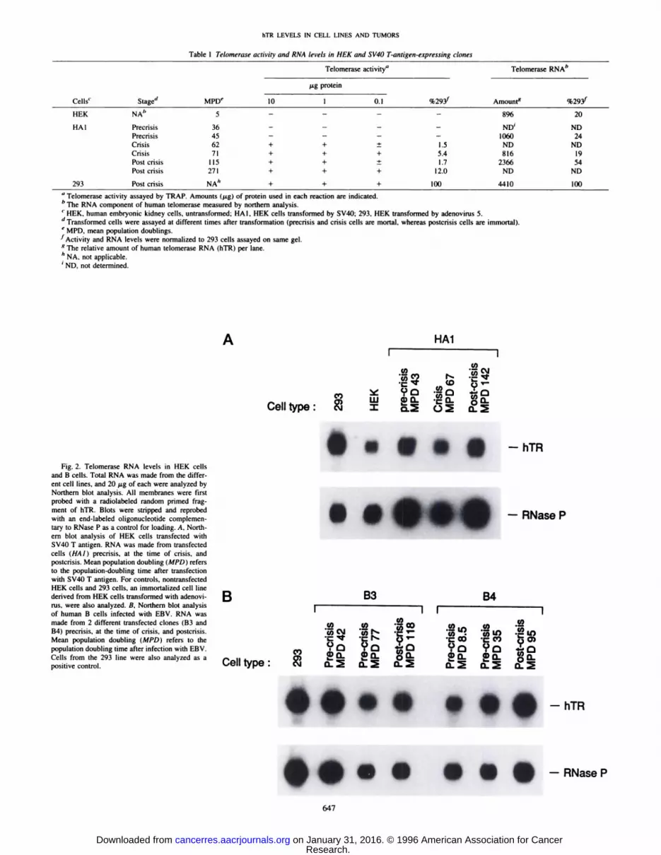
Table 1 Telomerase activity andRNA levels in HEK and SV4O T-anrigen-expressingclonesCellscStagedMPDeTelomerase
activity―TelomeraseRNA'@'1.Lg
protein%293@Amounts%2931101
0.1HEKNAb89620HAlPrecrisis
PrecrisisCrisisCrisisPost crisisPostcrisis36
456271
115271—
—
++++—
—
— —
+ ±+ ++ ±+ +—
—
1.55.4I .7
12.0NJY
1060ND816
2366NDND
24ND1954
ND293Post
crisisNA―++ +1004410100
hTR LEVELS IN CELL LINES AND TUMORS
a Telomerase activity assayed by TRAP. Amounts (p@g) of protein used in each reaction are indicated.
b The RNA component of human telomerase measured by northern analysis.
C HEK, human embryonic kidney cells, untransformed; HAl, HEK cells transformed by SV4O; 293, HEK transformed by adenovirus 5.
d Transformed cells were assayed at different times after transformation (precrisis and crisis cells are mortal, whereas postcrisis cells are immortal).
e MPD, mean population doublings.
I Activity and RNA levels were normalized to 293 cells assayed on same gel.g The relative amount of human telomerase RNA (hTR) per lane.
h NA, not applicable.â€ND, not determined.
A HAlI :@@ :@
(V)@ 9cl -@ 1@5c@n@ W a@o@ -CQ-@
Celitype:@ =@ Q@ o_@
I....Fig. 2. Telomerase RNA levels in HEK cells
and B cells. Total RNA was made from the different cell lines, and 20 j.sg of each were analyzed byNorthern blot analysis. All membranes were firstprobed with a radiolabeled random primed fragment of hTR. Blots were stripped and reprobedwith an end-labeled oligonucleotide complementao' to RNase P as a control for loading. A, Northem blot analysis of HEK cells transfected withSV4OT antigen. RNA was made from transfectedcells (HAl) precrisis, at the time of crisis, andpostcrisis. Mean population doubling (MPD) refersto the population-doubling time after transfectionwith SV4OI antigen. For controls, nontransfectedHEK cells and 293 cells, an immortalized cell linederived from HEK cells transformed with adenovi- Bnis, were also analyzed. B, Northern blot analysis . __________________________of human B cells infected with EBV. RNA wasmade from 2 different transfected clones (B3 andB4) precrisis, at the time of crisis, and postcrisis.Mean population doubling (MPD) refers to thepopulation doubling time after infection with EBV.Cells from the 293 line were also analyzed as a II 4@positive control. e@. type
•I.. •SI-hTR
••.. ••S-RNaseP
647
S.
B3 B4
ANase P
I 11
C,, .@? -@a:@ @, (I) SQ-@;c'J (I) N. .@ 1-@ tq .@@_c'I@. -@t@-@ U'- -C@ .CCV) @5O)
@ c@o ?c3 41'c@ ?c@ 9cl 4-'•Q_ WQ@@ @Q_ !Q
@@ o_@@ o_@
Research. on January 31, 2016. © 1996 American Association for Cancercancerres.aacrjournals.org Downloaded from
TabIc 2 Telomerase activityand RNA levels in B-cell clonesexpressingEBVCells'StagedMPDeTelomerase
activity―TelomeraseRNAb@Lg
protein%293@amount5%293@1051
0.10.01B3Precrisis42++—
——0.16503132B3Near
crisis77++++—13.07056143B3Postcrisis118+++++186.06666135B4Precrisis8.5++———0.15818118B4Near
crisis35++———0.17638155B4Postcrisis95+++
++50.08179166B2Precrisis12±———ND―5162105B2Precrisis26±—ND427887B5Precrisis9——425186B5Precrisis71——454492293PostcrisisNA'+++
++1004929100
hTR LEVELS IN CELL LINES AND TUMORS
a Telomerase activity assayed by TRAP. Amounts (@xg) of protein used in each reaction are indicated.
I) The RNA component of human telomerase measured by northem analysis.
C B2—B5, human B lymphocytes transformed by EBV.
d Transformed cells were assayed at different times after transformation (precrisis and crisis cells are mortal, whereas postcrisis cells are immortal).
e MPD, mean population doublings.
1Activity and RNA levels were normalized to 293 cells assayed on same gel.gThe relativeamountof humantelomeraseRNA (hTR) per lane.S ND, not determined.
â€NA, not applicable.
To further examine the correlation of telomerase RNA abundanceand activity, we analyzed 2 1 human tumor samples and 5 normaltissues (Fig. 3). Extracts were made from tumors, and telomeraseactivity was measured. Assays were done using 6.0 p.g of protein, andthose extracts that were negative were tested for possible inhibitorsusing serial dilutions of the extracts. Telomerase activity was notdetected in the normal tissues analyzed but was detected in most of thetumor samples [activity levels are designated as 0 for no activity and1—4(1 is the lowest level) in Fig. 3]. The levels of telomerase activityvaried greatly between the different tumors. Northern analysis wascarried out, and the hTR signal quantitated in a blind study on RNAisolated from all of the tumor samples (Fig. 3). The hTR was detectedin all of the samples analyzed, including the normal tissues that lackeddetectable telomerase activity. The level of hTR was higher in thetumor samples (median signal 14,017) than in the normal tissue(median signal 1, 112), although there was a high degree ofvariation. In some cases, the level of hTR in a telomerase-positivetumor was similar to that of telomerase-negative normal tissue (e.g.,compare samples 10 and 11). Among the tumor samples, the level ofhTR did not reflect the level of telomerase activity detected. Althoughsome variability may be introduced during the surgical handling oftumor tissue before extract preparation, we have not found variabilityin the time of tissue freezing after removal to significantly affecttelomerase activity levels (2 1). In addition, a recent study on mousetumors in which all tissues were handled identically has also foundtumor to tumor variations in both telomerase activity and telomeraseRNA (34). Finally, the data on hTR levels in tumors and normal tissueare consistent with the study in cell lines that suggest that telomeraseRNA levels do not reflect the level of telomerase activity. Thus,telomerase RNA is not a good marker for the presence of telomeraseactivity in a given tumor.
To compare hTR levels in tumor and normal tissues from matchedsamples from the same individual, we analyzed colorectal carcinomasamples. RNA was extracted from three tumors and adjacent tissuesamples. hTR was present in both the tumors and the adjacent tissue,although in all cases there was a higher amount in the tumor samples(Fig. 4). Telomerase activity assays were also done at three differentprotein concentrations (6, 0.6, and 0.06 p.g) on each sample. All of thetumor samples had telomerase activity. One of the three adjacentsamples (sample 55) showed low levels of telomerase activity, and theother two were telomerase negative (data not shown). Telomerase
activity has been reported previously in tumor adjacent tissues (21)and may be due to a low percentage of contaminating tumor cells inthe adjacent sample (occult micrometastasis). None of the six extractscaused inhibition of PCR in control reactions, indicating that the lackof signal was not due to inhibition of the PCR-based telomerase assay.Thus, as for the cell lines and the blind tumor study, telomerase RNAlevels did not parallel telomerase activity in human colorectal carcinoma samples.
DISCUSSION
Telomerase activity is up-regulated in a variety of immortal celllines and in tumors in both human and mouse (reviewed in Refs. 6, 11,13, 26). To understand the regulation of telomerase during tumorigenesis, we analyzed the levels of the recently cloned hTR componentduring telomerase activation. In both cell lines and in tumors, wefound that hTR levels did not always parallel the level of telomeraseactivity. Although there was an increase in the hTR level when tumorand normal tissues were compared, the amount of telomerase activitydid not always parallel the amount of hTR. The activity and RNAlevels in tumors may vary at the cellular level. Some large tumors thathave little RNA may only express telomerase RNA (and perhapsactivity) in a portion of the cells. This intratumor heterogeneity mightcontribute to the variable level of RNA seen in tumors. An in situassay will be required to test this hypothesis.
Similar results showing the presence of telomerase RNA in normalhuman tissues that are telomerase negative were recently reported(20). In addition, a lack of correlation between telomerase RNA andactivity was recently found during progression of multistage tumorigenesis in transgenic mice (34). Taken together, these results indicatethat the telomerase RNA component is not a good predictor oftelomerase activity.
The presence of telomerase RNA in cell cultures that lack telomerase activity suggests that telomerase is regulated at several different levels. Telomerase RNA was present in EBV- and SV4O Tantigen-expressing cell clones that did not have detectable telomeraseactivity. Recent evidence from transgenic mice suggests that viraloncogenes or their cellular effects may directly up-regulate telomeraseRNA but not telomerase activity (34). Thus, the increase in hTR in theHAl clones may represent a direct or indirect effect of the viraloncogenes.
648
Research. on January 31, 2016. © 1996 American Association for Cancercancerres.aacrjournals.org Downloaded from

:@•!/@ @.-.-—i
—J@
flH@1@@ n i -@ I flFlr@@ I@@@
0@@@ L1@@@ —@ —I I I@ @r@
@@ @°Uil U@t@ @°r'@@tr2@
@ @O@N SooOC@@@ In‘@0@@ cn m —:@@ C@lcfl cn In Cfl C@ICfl —\O 00@ @fl
@d@ — — Cs@l,- .@
C,)
hTR LEVELS IN CELL LINES AND TUMORS
5
25000
20000
Fig. 3. hTR levels in tumor (T) versus normal(N) tissues. Total RNA was made from differenttissues, and 25 @gof each were analyzed by Northem blot analysis. *, normal tissues tested; othertissues are from tumors. The graph shows the relative amounts of telomerase RNA (Relative hTR)in different tumors normalized to the amount of 5 SRNA. Telomerase activity levels are plotted on theright as 0 (for no activity) or 1—4to indicate therelative level of telomerase activity detected. Extracts that showed activity at 0.06 @xgwere designated 4 for telomerase activity. Extracts that hadweak activity at higher concentrations (6 @zg)weredesignated 1 for weak activity, 2 for strong activityat 6 @g.or 3 for strong activity at 0.6 @g.
The lack of activity in cells that contain high levels of hTR may bedue to the absence of a protein component of telomerase or to specificdown-regulators of telomerase present in primary cells and tissues(27). In Tetrahymena, overexpression of telomerase RNA does notlead to an increase in the steady-state amount of telomerase RNA,suggesting that the RNA component must be bound by a limitingfactor, possibly the protein components to be stabilized against degradation in vivo. (28) Thus, it will be of interest to determine whethertelomerase proteins are present in cells that lack telomerase activity orwhether telomerase RNA is packaged as a RNP in these cells.Telomerase proteins components have been isolated from the ciliate
ColonTissueI@ —@ I
:@ #52 #55
@) I IE@ IC)CsJ
15000
10000
5@yJt'J@** * * *
@@0
m
#56
T N T
0 S...,
S — 5S
Fig. 4. Northern blot analysis of human colon tumor (T) samples and normal (N)adjacent tissue from three patients. The Northem blots were probed with hTR and thenwith 5 S RNA gene. and the levels of hTR were normalized to the 5 S RNA levels in thesame lanes.
649
c@i::@i@i@=4@y I
@ ‘@
@@:1@ :Ei@ H3@
L@, @.
Tetrahymena (29) but not yet from human cells; thus, it is not yetpossible to determine whether they are limiting for telomerase activityin vivo.
Cell fusion experiments between telomerase-positive immortalcells and telomerase-negative immortal or normal cells have shownthat telomerase activity is down-regulated in at least some cases,suggesting a trans-repression activity may be present in telomerasenegative cells (27, 30, 31). This trans-acting repression could actdirectly on the availability of one or more telomerase polypeptides orit may represent a direct repressor of the telomerase enzyme activity.Mixing extracts from telomerase-positive cells with those from telomerase-negative cells did not inhibit telomerase activity in thepositive extracts (15, 30, 32). Thus, lack of activity is not due to asimple, diffusible telomerase inhibitor. However, the presence of atightly associated specific telomerase inhibitor has not been excluded.
Telomerase activity is commonly found in a variety of cancers (4,33). Telomere shortening and eventual cell death were seen in yeastcells deleted for telomerase RNA component and in human cellsexpressing antisense RNA (18—20).Thus, telomerase appears to berequired for the growth of at least some immortal cell types. Thisrequirement has raised the possibility that telomerase inhibitors may
— hTR be useful in cancer chemotherapy (reviewed in Ref. 13). Understand
ing the details of telomerase activity and component regulation isimportant to evaluate the potential of new therapeutic and diagnosticapproaches to cancer.
ACKNOWLEDGMENTS
We thank Maria Blasco and Calvin Harley for critical reading of themanuscript; M. Blasco, M. Rizen, and D. Hanahan for sharing data beforepublication; and I. Wang for pointing out the potential hTR increase inresponse to T-ag. We thank Dr. Margaret Kemeny (North Shore UniversityHospital) for providing colon tumor samples to Cold Spring Harbor Laboratoryand Dr. Adi Gazdar for providing the University of Texas Southwestern tumorsamples.
Research. on January 31, 2016. © 1996 American Association for Cancercancerres.aacrjournals.org Downloaded from
hTR LEVELS IN CELL LINES AND TUMORS
epithelial cells but not in human fibroblasts. Oncogene, 8: 1407—1413,1993.18. Singer, M. S., and Gottschling, D. E. TLC1: template RNA component of Saccha
romyces cerevisiae telomerase. Science (Washington DC), 266: 404—409, 1994.19. McEachern, M. J., and Blackburn, E. H. Runaway telomere elongation caused by
telomerase RNA gene mutations. Nature (Land.), 376: 403—409,1995.20. Feng, J., Funk, W., Wang, S., Weinrich, S., Avilion, A., Chiu, C-P., Adams, R.,
Chang. E., Yu, J., La, S., West, M., Harley, C. B., Andrews, W., Greider, C. W.,Villeponteau, B. The human telomerase RNA component. Science (Washington DC),269: 1236—1241,1995.
21. Piatyszek, M. A., Kim, N. W., Weinrich, S. L., Hiyama, K., Hiyama, E., Wright,w. E.,andShay,J. W.Detectionof telomeraseactivityinhumancellsandtumorsbya telomeric repeat amplification protocol (TRAP). Methods Cell Sci., 17: 1—15,1995.
22. Ausubel, F. M., Brent, R., Kingston, R., Moore, D., Seidman, J. G., Smith, J. A., andStruM, K. Current Protocols in Molecular Biology. New York: Wiley and Sons, Inc.,1993.
23. Counter, C. M., Gupta, J., Harley, C. B., Leber, B., and Bacchetti, S. Telomeraseactivity in normal leukocytes and in hematologic malignancies. Blood, 85: 2315—2320, 1995.
24. Broccoli, D., Young, J. W., and de Lange, T. Telomerase activity in normal andmalignant heamatopoietic cells. Proc. Nail. Acad. Sci. USA, 92: 9082—9086,1995.
25. Hiyama, K., Hirai, Y., Kyoizumi, S., Akiyama, M., Hiyama, E., Piatyszek, M. A.,Shay, J., Ishioka, S., and Yamakido, M. Activation of telomerase in human lymphocytes and hematopoietic progenitor cells. J. Immunol., 155: 3711—3715,1995.
26. Harley, C. B., and Villeponteau, B. Telomeres and telomerase in aging and cancer.Curr. Opin. Genet. Dcv., 5: 249—255,1995.
27. Ohmura, H., Tahara, H., Suzuki, M., Ide, T., Shimizu, M., Yoshida, M. A., Tahara,E., Shay, J. W., Barrett, J. C., and Oshimura, M. Restoration of the cellular senescenceprogram and repression of telomerase by chromosome 3. Jpn. J. Cancer Res., 86:899—904, 1995.
28. Yu, G-L., Bradley, J. D., Attardi, L. D., and Blackburn, E. H. In vivo alteration oftelomere sequences and senescence caused by mutated Tetrahymena telomeraseRNAs. Nature (Lond.), 344: 126—132,1990.
29. Collins, K., Koybayashi, R., and Greider, C. W. Purification of Tetrahymena telomerase and cloning of the genes for the two protein components of the enzyme. Cell,81: 677—686,1995.
30. Bryan, T. R., Englezou, A., Gupta, J., Bacchetti, S., and Reddel, R. Telomereelongation in immortal human cells without detectable telomerase activity. EMBO J.,14: 4240—4248, 1995.
31. Wright, W. e., Brasiskyte, D., Piatyszek, M. A., and Shay, J. W. Experimentalelongation of telomeres in immortal human cells extends the lifespan of immortal xnormal cell hybrids. EMBO J., in press, 1996.
32. Allsopp, R. C., Chang, E., Kashefi-Aazam, M., Rogaev, E. I., Piatyszek, M. A., Shay,J., and Harley, C. B. Telomere shortening is associated with cell division in vitro andin vivo. Exp. Cell Res., 220: 194—200,1995.
33. Counter, G. M., Hirte, H. W., Bacchetti, S., and Harley, C. B. Telomerase activity inhuman ovarian carcinoma. Proc. Nail. Acad. Sci. USA, 91: 2900—2904, 1994.
34. Blasco, M., Risen, M., Grieder, C. W., and Hanahan, D. Differential regulation oftelomerase activity and telomerase RNA during multistage tumorigenesis. Nat. Genet., in press, 1996.
650
REFERENCES
1. Harley, C. B., Futcher, A. B., and Greider, C. W. Telomeres shorten during ageing ofhuman fibroblasts. Nature (Lond.), 345: 458—460, 1990.
2. Hastie, N. D., Dempster, M., Dunlop, M. G.. Thompson, A. M., Green, D. K., andAllshire, R. C. Telomere reduction in human colorectal carcinoma and with ageing.Nature (Land.), 346: 866—868, 1990.
3. de Lange, T., Shiue, L., Myers, R., Cox, D. R., Naylor, S. L., Killery, A. M., andVarmus, H. E. Structure and variability of human chromosome ends. Mol. Cell. Biol.,10: 518—527,1990.
4. Kim, N. W., Piatyszek, M. A., Prowse, K. R., Harley, C. B., West, M. D., Ho, P. L.,Coviello, G. M., Wright, W. E., Weinrich, S. L., and Shay, J. W. Specific associationof human telomerase activity with immortal cells and cancer. Science (WashingtonDC), 266: 2011—2014, 1994.
5. de Lange, T. Telomere dynamics and genomic instability in human cancer. In: E. H.Blackbum and C. W. Greider (eds.), Telomeres, pp. 265—293.Cold Spring Harbor,NY: Cold Spring Harbor Laboratory, 1995.
6. Bacchetti, S., and Counter, C. M. Telomeres and telomerase in human cancer(Review). Int. J. Oncol., 7: 423—432,1995.
7. Greider, C. W., and Blackburn, E. H. Identification of a specific telomere terminaltransferase activity in Tetrahymena extracts. Cell, 43: 405—413, 1985.
8. Greider, C. W., and Blackbum, E. H. The telomere terminal transferase of Tetrahymena is a ribonucleoprotein enzyme with two kinds of primer specificity. Cell, 51:887—898, 1987.
9. Wright, W. E., Piatyszek, M. A., Rainey, W. E., Bryd, W., and Shay, J. W.Telomerase activity in human germline and embryonic tissues and cells. Dev. Genet.,in press, 1996.
10. Harley, C. B. Telomeres and aging. In: E. H. Blackburn and C. W. Greider (eds.),Telomeres, pp. 247—263.Cold Spring Harbor, NY: Cold Spring Harbor Laboratory,1995.
11. Shay, J. W., and Wright, W. E. Telomerase activity in human cancer. Curr. Opin.Oncol., in press, 1996.
12. Wright, W. E., and Shay, J. W. Time telomeres and tumors: is cellular senescencemore than an anti-cancer mechanism? Trends Cell. Biol., 5: 293—297,1995.
13. Harley, C. B., Kim, N. W., Prowse, K. R., Weinrich, S. L., Hirsh, K. S., West, M. D.,Bacchetti, S., Hirte, H. W., Counter, C. M., Greider, C. W., Wright, W. E., Shay, J.W. Telomerase, cell immortality and cancer. Cold Spring Harbor Symp. Quant. Biol.,59: 307—315,1994.
14. Shay, J. W. Aging and cancer: are telomeres and telomerase the connection? Mol.Med. Today, 1: 376—382, 1995.
15. Counter, C. M., Avilion, A. A., LeFeuvre, C. E., Stewart, N. G., Greider, C. W.,Harley, C. B., and Bacchetti, S. Telomere shortening associated with chromosomeinstability is arrested in immortal cells which express telomerase activity. EMBO J.,11:1921—1929,1992.
16. Counter, C. M., Botelho, F. M., Wang, P., Harley, C. B., and Baccheui, S. Stabilization of short telomeres and telomerase activity accompany immortalization ofEpstein-Barr virus transfromed human B lymphocytes. J. Virol., 68: 3410—3414,1994.
17. Shay, J. W., Wright, W. E., Brasiskyte, D., and Van der Hagen, B. A. £6of humanpapiloma type 16 can overcome the Ml stage of immortalization in human mammary
Research. on January 31, 2016. © 1996 American Association for Cancercancerres.aacrjournals.org Downloaded from

1996;56:645-650. Cancer Res Ariel A. Avilion, Mieczyslaw A. Piatyszek, Jyothi Gupta, et al. Cell Lines and Tumor TissuesHuman Telomerase RNA and Telomerase Activity in Immortal
Updated version
http://cancerres.aacrjournals.org/content/56/3/645
Access the most recent version of this article at:
E-mail alerts related to this article or journal.Sign up to receive free email-alerts
Subscriptions
Reprints and
To order reprints of this article or to subscribe to the journal, contact the AACR Publications
Permissions
To request permission to re-use all or part of this article, contact the AACR Publications
Research. on January 31, 2016. © 1996 American Association for Cancercancerres.aacrjournals.org Downloaded from